SPORTS MEDICINE SPAIN PARK HIGH SCHOOL Anatomy & Injuries to the Abdomen & Thorax.
108
SPORTS MEDICINE SPAIN PARK HIGH SCHOOL Anatomy & Injuries to the Abdomen & Thorax
-
Upload
margery-bryant -
Category
Documents
-
view
219 -
download
0
Transcript of SPORTS MEDICINE SPAIN PARK HIGH SCHOOL Anatomy & Injuries to the Abdomen & Thorax.

SPORTS MEDICINE SPAIN PARK HIGH SCHOOL
Anatomy & Injuries to the Abdomen & Thorax
Anatomy
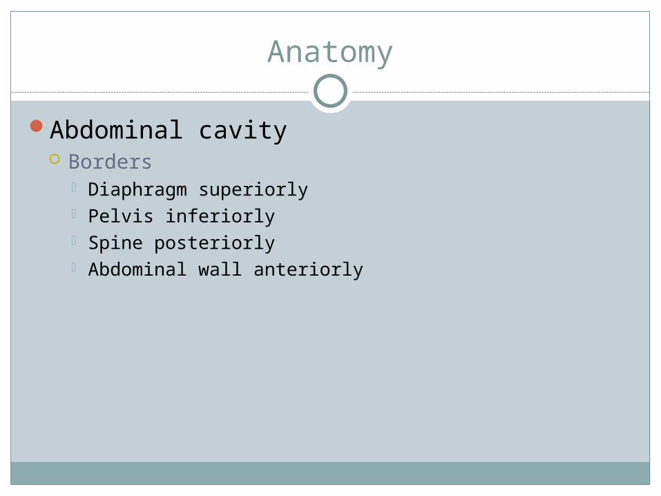
Abdominal cavity Borders
Diaphragm superiorly Pelvis inferiorly Spine posteriorly Abdominal wall anteriorly

Anatomy
Divided into 4 quadrants Line runs through navel at midline of body UL UR LL LR
Anatomy--Quadrants
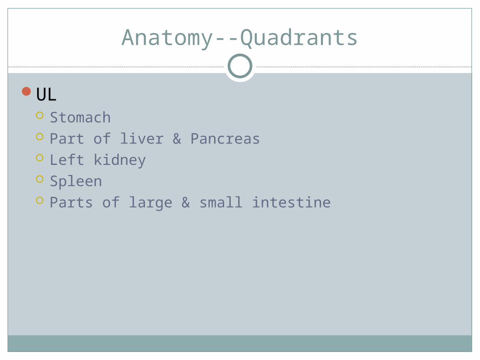
UL Stomach Part of liver & Pancreas Left kidney Spleen Parts of large & small intestine
Anatomy--Quadrants
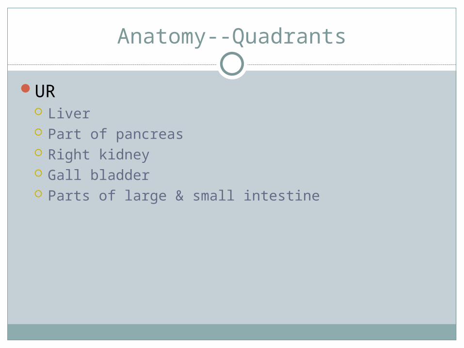
UR Liver Part of pancreas Right kidney Gall bladder Parts of large & small intestine

Anatomy--Quadrants
LL Parts of Lg & Sm intestines Part of bladder Uterus-females Left ovary-females Prostate- males Ureter-male

Anatomy--Quadrants
LR Parts of Lg & Sm intestines Appendix Part of bladder Uterus-female Right ovary-female Prostate-male Ureter-male


Anatomy
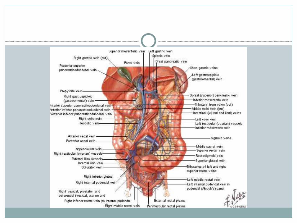
Organs are part of: Urinary system
Kidneys, bladder Digestive system
Stomach, liver, pancreas, gall bladder, large & small intestine, spleen
Reproductive system Uterus, ovaries, prostate, seminal vesicles
Anatomy
Solid organs More often & easily injured Can cause rapid death due to large blood supply—
internal bleeding Spleen, liver, kidney, pancreas
Hollow organs Injuries are rare because tubes are hollow—assist in
transporting substances from one organ to another
Digestive organs
Digestion begins in mouthStomach
Secretes gastric juices that assist in breaking down food before entering small intestine
Liver Detoxifies chemicals that the body perceives as
poisonous, stores vitamins, produces bile, assists with food metabolism
Gall bladder Storage tank for bile, which passes into sm intestine
where it assists in metabolism of fat

Digestive Organs
Pancreas Produces insulin and enzymes for digestion
Small intestine Completes digestion, absorbed the products into
circulatory system. Peristalsis (sequential contraction/relaxation of intestinal muscle) pushes the food through the intestines to the large intestine. At this point all material that has not been absorbed is considered waste.
Large intestine All materials not absorbed into the system in sm
intestine is passed into large intestine as waste. Water is absorbed leaving solid waste for excretion

Digestive organs
Appendix Part of large intestine No known function
Spleen Produces & destroys red blood cells Storage site for blood Aids in destruction of harmful microorganisms

Urinary system
Kidneys Responsible for maintaining acid-base in body, which
if changed causes body system to shut down eventually resulting in death
Filter blood and remove waste products of metabolism to maintain stable acid-base relationship
If blood supply is inadequate, can cause hypertension from chemical constriction of body’s blood vessels
Bladder Holding tank for liquid waste in body

Reproductive system
Females Ovaries
Produces eggs and estrogen (stimulates development of & maintains feminine characteristics)
Uterus Fertilized eggs develop here
Males seminal vesicles Prostate gland
Responsible for adding fluid & nutrients to seminal fluid

Anatomy
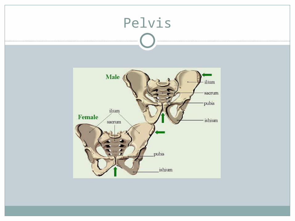
Pelvis Provides bony base and protection for internal organs Wider in females to accommodate childbirth
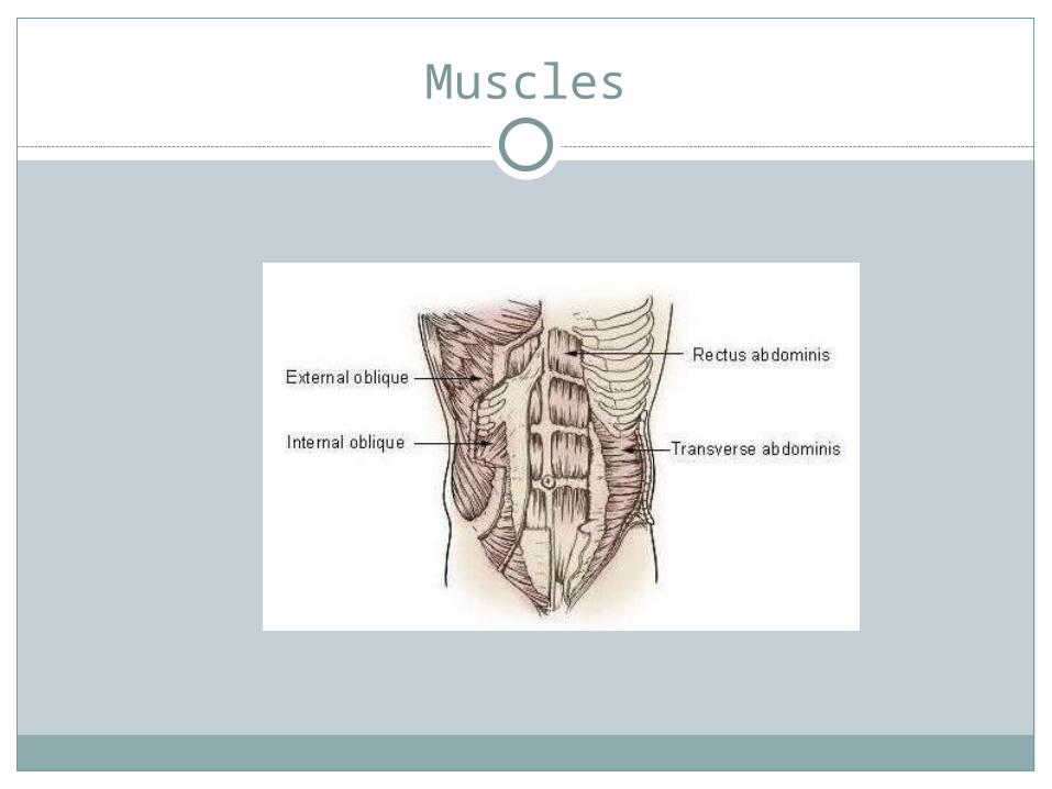
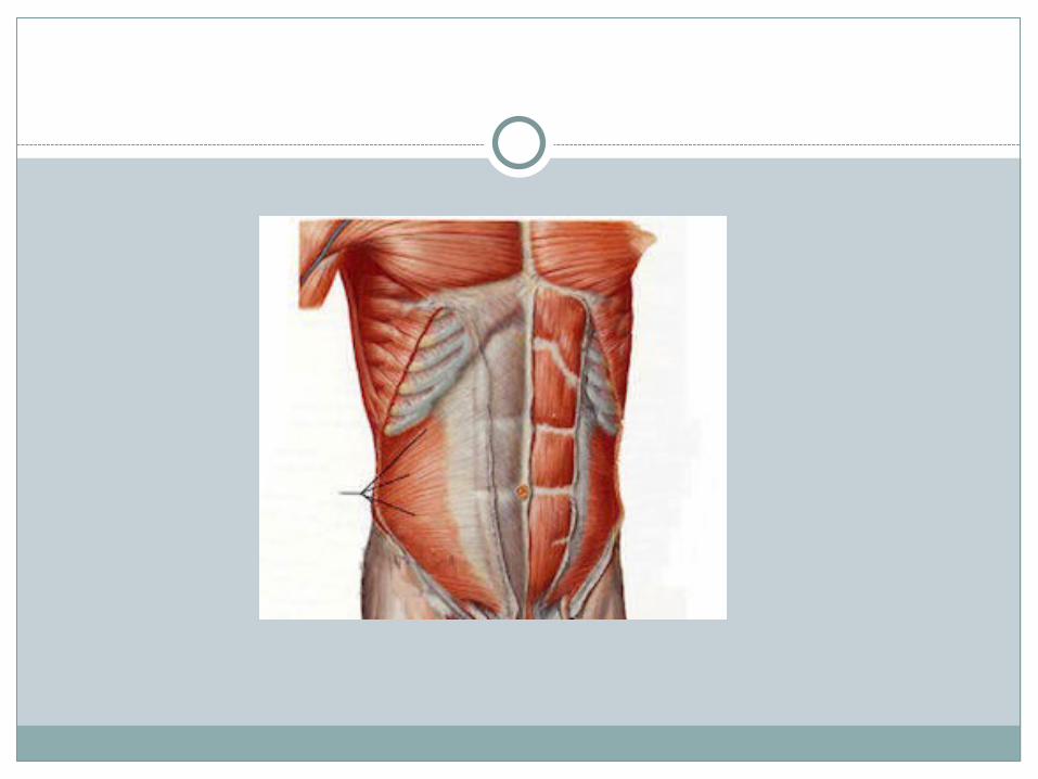
Abdominal muscles Provides protection for organs Rectus abdominus gives “washboard” affect; attaches
to pelvis & lower ribs & sternum—trunk flexion Obliques (external & internal) attaches to lateral
aspect of lower ribs & runs diagonally to pelvis—flexion and rotation
Transverse abdominus—holds internal organs in cavity
Pelvis
Muscles
Anatomy of thorax
Part of body between neck & abdomenContains heart & lungs

Anatomy
Throat Carotid arteries
One on each side of trachea Carry oxygenated blood to brain
Jugular veins One on each side of trachea Carry unoxygenated blood away from brain
Anatomy
Larynx Modified upper part of trachea Contains vocal chords
Trachea Made up of circular rings of cartilage Main trunk of system of tubes by which air passes to &
from lungs for exchange of CO2 and O2 Esophagus
Passageway for food going from the mouth to the stomach
Sits in front of the cervical vertebrae and behind the trachea & larynx
Anatomy of thorax
Bony structures Thoracic vertebrae posterior 12 ribs on each side Sternum anterior
Protects organs of thorax
Anatomy
Heart Size of fist Pumps blood to all parts of body
Divided into 4 chambers Right & left atrium
Upper chambers Right & left ventricle
Lower chambers Larger with thicker walls

Heart Pumps blood to lungs and throughout body Right atrium fills w/ blood from vein
Carries waste products and CO2 Right ventricle receives blood from atrium (through
tricuspid valve) Pulmonary arteries carry UNOXYGENATED blood to
lungs

Heart Blood is mixed with O2 in the lungs OXYGENATED blood is carried back to heart by the
pulmonary veins Goes into the left atrium Flows to the left ventricle through the bicuspid valve Is pumped to the rest of the body through the aorta

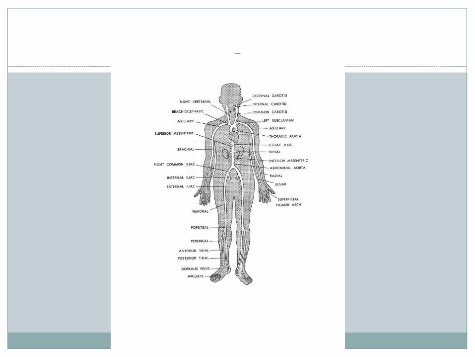
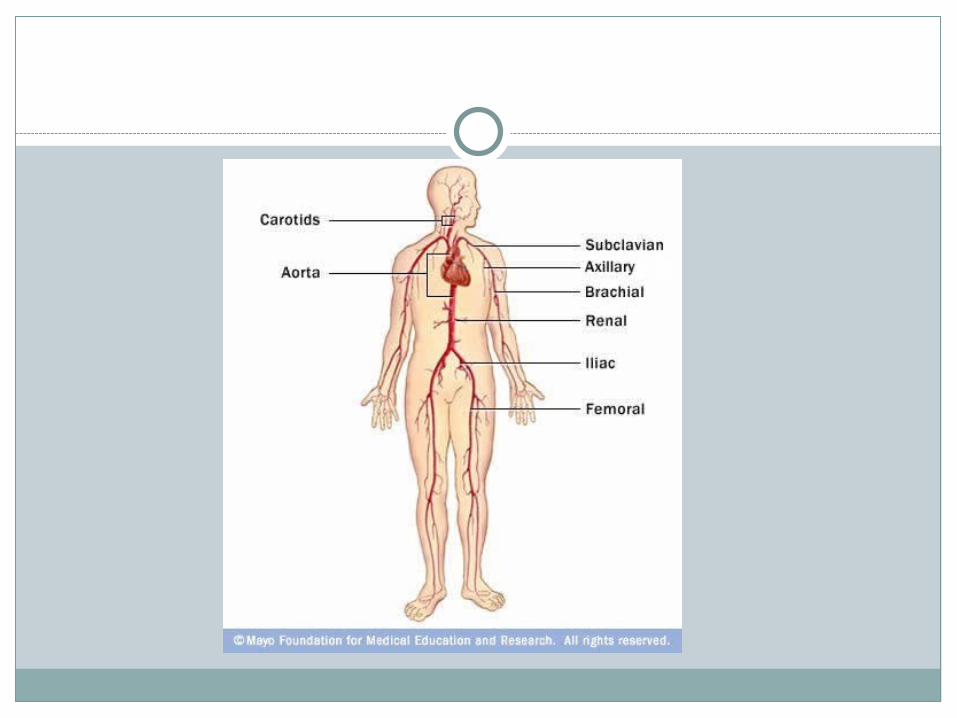
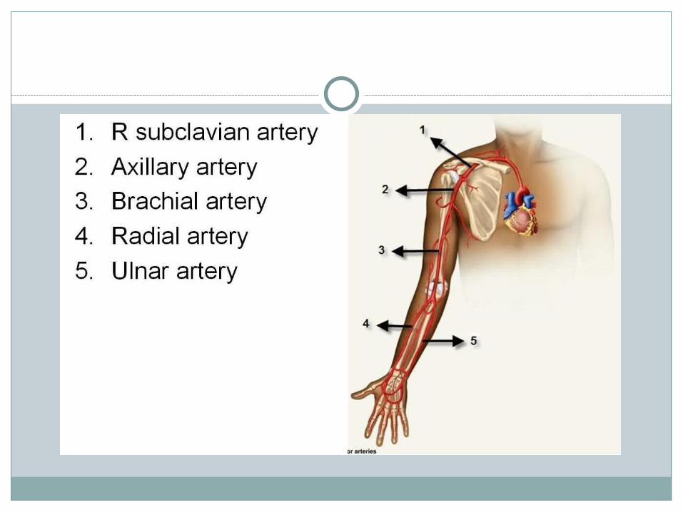
Heart Main branches (arteries) off the aorta Ascending and descending aortas
Carotid Subclavian
Axillary Brachial
• Radial & ulnar Common iliac
Femoral• Anterior & Posterior Tibial


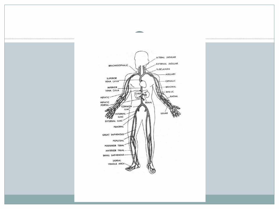
VeinsRun parallel with the arteries
Superior vena cava Inferior vena cava
Two extra in arm Cephalic basilic
Two extra in leg Greater saphenous Lesser saphenous

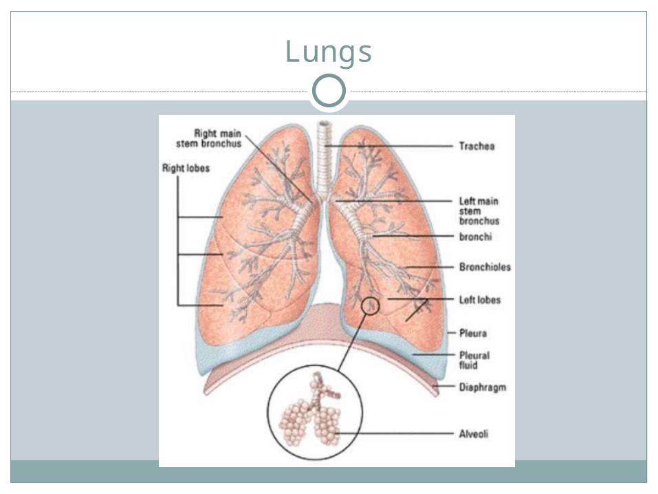
Lungs Right has 3 lobes Left has 2 lobes
Function: to exchange O2 and CO2 To dissipate heat from the body
Trachea divides into two bronchi Bronchi are filled with cilia
Hair like projections that help remove foreign substances like dust & pollen
Bronchi divide into bronchiolesBronchioles end in alveoli
Alveoli are air containing cells of the lungs O2 and CO2 are exchanged here
Coughing & sneezing help keep trachea and bronchi clear & remove phlegm and allergy-causing agents from lungs
Respiration rate
Lung function & breathing rate controlled by CO2 receptors
If there is too much CO2, inhalation occurs to bring in more O2
Exercise increases cell metabolism Causes cells to need more O2 and eliminate more CO2
Respiration Rate
With exercise lungs’ ability to exchange air more efficiently increases
Breaths become deeper & more forceful
Return to normal breathing quicker
Pleura Thin lubricated tissue Lines each half of thorax Folded back over the surface of the lung on same side Allow for smooth movement of lungs as they
encounter the wall of ribs during inhalation & exhalation

Lungs
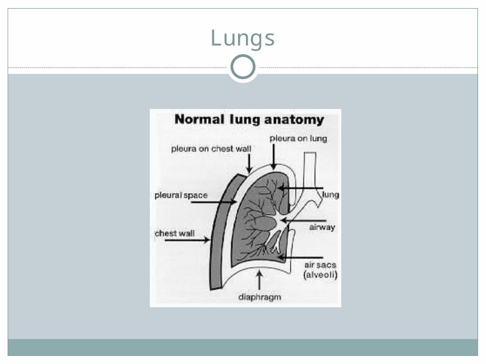
Lungs
Muscular Anatomy
Intercostal muscles sit between ribs Internal and external intercostals
Aid in inhalation and exhalation

Intercostal muscles

Intercostals

Abdominal & Thoracic Injuries
Injuries are rareSolid organs most often injuredLife threatening

Abdominal Strains
Rectus abdominus most often injuredPotentially can be incapacitatingMxn:
sudden twisting of trunk or reaching overhead
S/S: pain with movements of the trunk, POT over affected
muscle, tightness of muscles
TX: ice, compression, gentle stretching, no exercise until
ROM is pain free
Abdominal contusions
Not common but most likely to occur in collision sports
Mxn: direct blow to abdomen, compressive force to
abdominal wall
S/S: pain, tightness, hematoma formation under the fascial
tissue surrounding muscle
Tx: ice, compression, look for signs of internal injury, no
activity until pain free
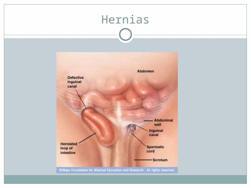
Hernia
Protrusion of abdominal viscera through a portion of abdominal wall
Those resulting from sports occur in groin area
Inguinal hernias occur most often in men ( more than 75%)
Femoral hernias occur most often in women
Hernias
Inguinal Results from abnormal enlargement of opening in
inguinal canal through which vessels and nerves of male reproductive system pass
Femoral Arises in canal that transports vessels & nerves that
go the thigh and lower limb
Hernias
When intra-abdominal tension is produced in these areas, muscles produce a contraction around canal openings.
If muscles fail to react abdominal contents may be pushed through opening
Hernias
Mxn: improperly lifting a heavy object, increased abdominal
pressure, blow to groin area, weakness of abdominal wall
S/S: pain and prolonged discomfort in groin area,
protrusion in the groin area that is present when standing (or when coughing) but goes away when lying down, feeling of weakness or pulling sensation in groin area
Tx: surgical repair
hernias
Complications: Strangulated hernia:
If hernia is not treated, the bulge can get stuck in abdominal wall or inguinal canal.
The blood supply to the tissue will be cut off and eventually die.
If the intestine is involved, a bowel obstruction will result and prevents the passage of waste material from the body causing pain and illness

Hernias
Hernias

Hernias

Hernias

Blow to Solar Plexus
Commonly known as “getting the wind knocked out”
Mxn: blow to the middle of the abdomen or solar plexus
S/S: transitory paralysis of diaphragm, inability to breath (inhale) or trouble breathing for a brief period of time, cyanosis, short term panic
Tx: calm athlete, loosen belt/clothing around abdomen, bend knees, , control breathing-short inspirations, long expirations
Blow to solar plexus
Complications: fear of not being able to breath may cause athlete to hyperventilate Increased rate of ventilations that results in increases
levels of O2 which can cause dizziness, lump in throat, pounding of heart, fainting, tingling/numbness in hands, face, feet
Care for hyperventilation: Have athlete breath into a paper bag to increase levels of
CO2 to restore normal breathing If athlete does pass out, normal breathing should be
restored
Stitch in Side
Name given to an idiopathic condition that occurs in some athletes
Causes: Constipation Intestinal gas Overeating Diaphragmatic spasm as result of poor conditioning Lace of visceral support because of weak abdominal
muscles Distended spleen Breathing techniques that lead to lack of O2 in
diaphragm Ischemia of either the diaphragm or intercostal muscles
Stitch in Side
S/S: Cramp like pain in side at either right or left costal
border during hard physical activity
Tx: Relax the spasm
Stretch arm on affected side above the head and side bend to the same side
Flex the trunk over the thighs Ice If pain/spasm persists seek medical evaluation
Spleen injury
Mxn: Direct blow to upper left quadrant Falling on UL quadrant Infectious mononucleosis causes enlarged spleen
putting athlete at risk If spleen is enlarged due to mono, may resume activity
after 3 weeks if the spleen is no longer enlarged or painful and there is no fever

Spleen injury
S/S: History of injury Signs of shock-dizziness, thirst, pale, sweating, rapid
pulse/respirations Abdominal rigidity Nausea Vomiting Kehr’s sign
Reflex (referred) pain that comes on about 30 minutes after injury where pain radiates to the left shoulder and 1/3 the way down the left arm Referred pain—pain felt in one part of the body other
than its actual origin

Spleen injury
Can hemorrhage profusely into abdominal cavity causing athlete to die of internal bleeding days or weeks after injury
Tx: Call 911, monitor athlete, conservative, non-operative
treatment with about 1 week of hospitalization At 3 weeks can engage in light conditioning Return to full activity at 4 weeks If surgical repair is needed athlete will return to activity
at 3 months If surgical removal is necessary, return to activity at 6
months

Spleen injury
Kidney Contusion
Mxn: blow to the backS/S:
signs of shock nausea vomiting rigidity of muscles of back hematuria (blood in urine) referred pain radiates forward around the trunk into
the lower abdominal region

Kidney Contusion
Tx: Have athlete urinate 2-3 times to determine if there is
blood in urine Call 911 if necessary Treat for shock Immediate physician referral there is hematuria 24 hour hospitalization for observation Gradual increase in fluid intake If hemorrhage fails to stop, surgical intervention Usually takes 2 weeks bed rest prior to return to
activity
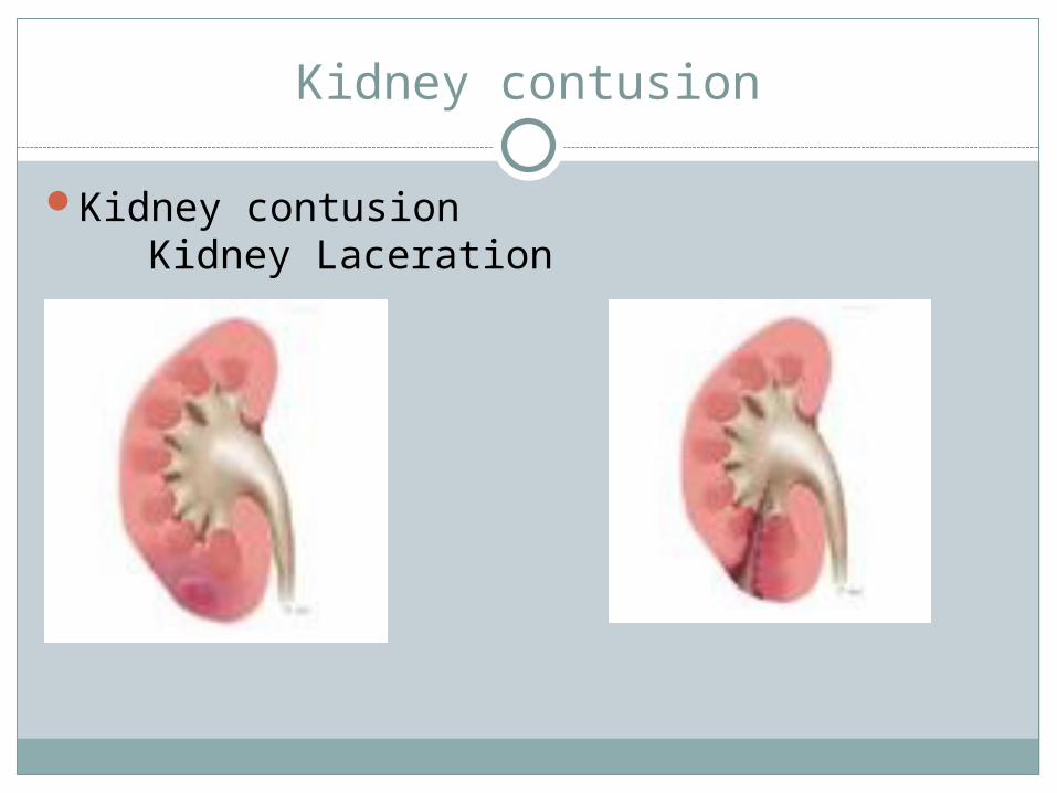
Kidney contusion
Kidney contusion Kidney Laceration

Liver Contusion/laceration
Mxn: hard blow to right side of abdomen
S/S: hemorrhage signs of shock referred pain
just below the right scapula right shoulder substernal area anterior left side of chest (occasionally)
Liver contusion/laceration
Tx: Call 911 Treat for shock Monitor athlete Immediate surgical intervention

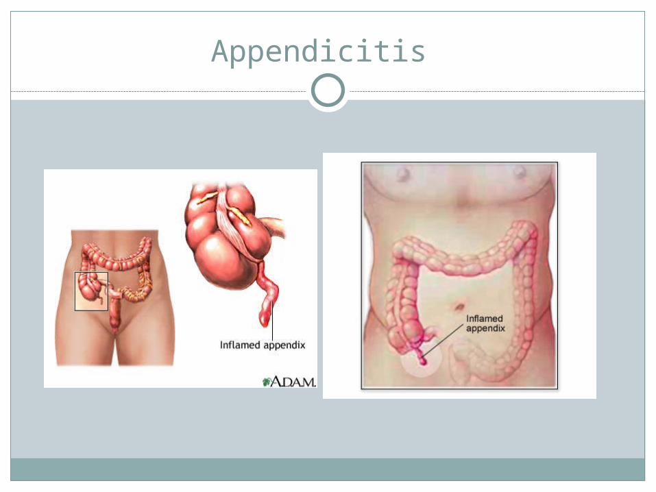
Appendicitis
Mxn: inflammation of appendix Chronic or acute Caused by fecal obstruction Initially appendix is red & swollen Can become gangrenous, rupture into bowels &
cause peritonitis

Appendicitis
S/S: Mild to sever pain in lower abdomen Nausea Vomiting Low grade fever Cramps in lower right side Abdominal rigidity Referred pain is at McBurney’s point (between the
ASIS and the umbilicus)

Appendicitis
Tx: Monitor athlete Refer when necessary Surgical removal of inflamed appendix Not an emergency unless there is a bowel obstruction An obstructed bowel, with an acute rupture is life-
threatening
Highest incidence in males between ages of 15 & 25.
Appendicitis
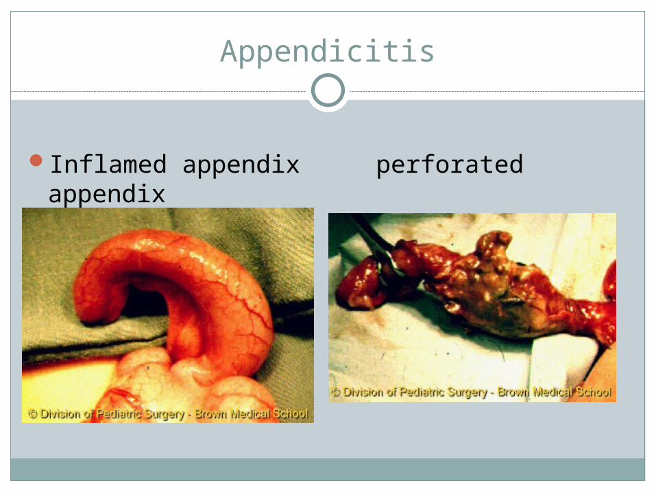
Appendicitis
Inflamed appendix perforated appendix
Injuries to Bladder
Mxn: blunt force to lower abdominal region if the bladder
is distended by urine Hematuria associated with contusion of bladder
during running Known as runner’s bladder
S/S: blood in urine Referred pain to lower trunk, upper thigh anteriorly With rupture, athlete will be unable to urinate
Bladder injuries
Tx: Monitor athlete Physician referral if necessary
Testicular/Scrotal contusion
Due to considerable sensitivity & vulnerability, contusions to the scrotum & testicles cause extreme pain, nausea and disability
Important for males to wear proper protection to prevent incidence of contusions
Mxn: direct blow to the genitalia
Testicular/Scrotal contusion
S/S: hemorrhage fluid effusion muscle spasm Vomiting is severe
Tx: place athlete on his side flex thighs to chest ice to scrotum as pain diminishes Immediate medical referral for increasing or
unresolved pain after 15-20 minutes
Rib Contusion
Mxn: blow to rib cage
S/S: sharp pain with breathing POT over contused area pain with compression of rib cage
Tx: RICE NSAIDS cessation of activity until pain subsides self limiting
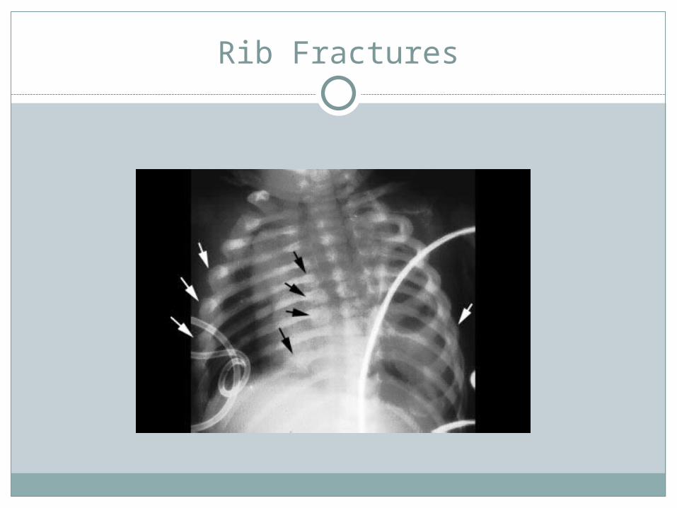
Rib Fractures
Most common in collision sportsRibs 5-9 most commonly fracturedPossibility of cause damage to or puncturing
a lung
Mxn: direct impact compression of rib cage

Rib Fractures
S/S: severe pain during inspiration POT over fracture site Crepitus Pain with movement of trunk

Rib Fractures
Tx: Refer for x-ray ice Support Rest Heal within 3-4 weeks Rib brace may offer some stabilization and comfort
Rib Fractures

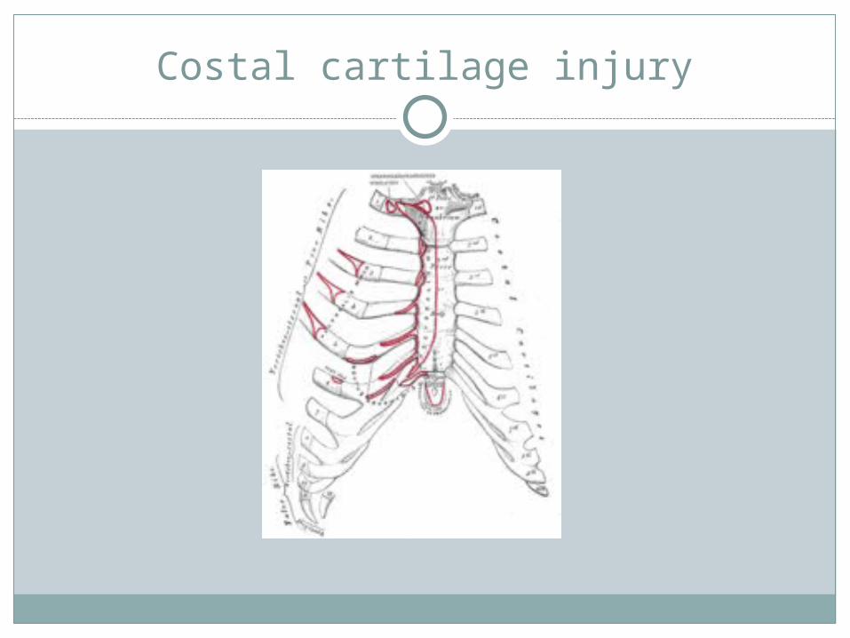
Costal Cartilage Injury
More common than rib fracturesMxn:
direct blow to thorax Indirectly from sudden twist of fall on a ball
compressing the rib cage

Costal cartilage injury
S/S: similar to rib fracture except pain is localized in the
junction of the rib cartilage and the rib Sharp pain during sudden movements Difficulty in breathing deeply POT with swelling Rib deformity Ribs make crackling noise (crepitus) as it moves in
and out of place
Costal cartilage injury
Tx: Ice Rest Immobilization with rib brace 1-2 months healing time
Costal cartilage injury

Intercostal Muscle Strain
Mxn: Direct blow Sudden torsion of trunk
S/S: Pain w/ active motion Pain w/ inspiration/expiration, laughing, coughing,
sneezing
Tx: Ice Compression Immobilization for comfort

Lung Injuries
Injures to lungs are rare but can be life threatening
PneumothoraxTension PneumothoraxHemothoraxTraumatic asphyxia
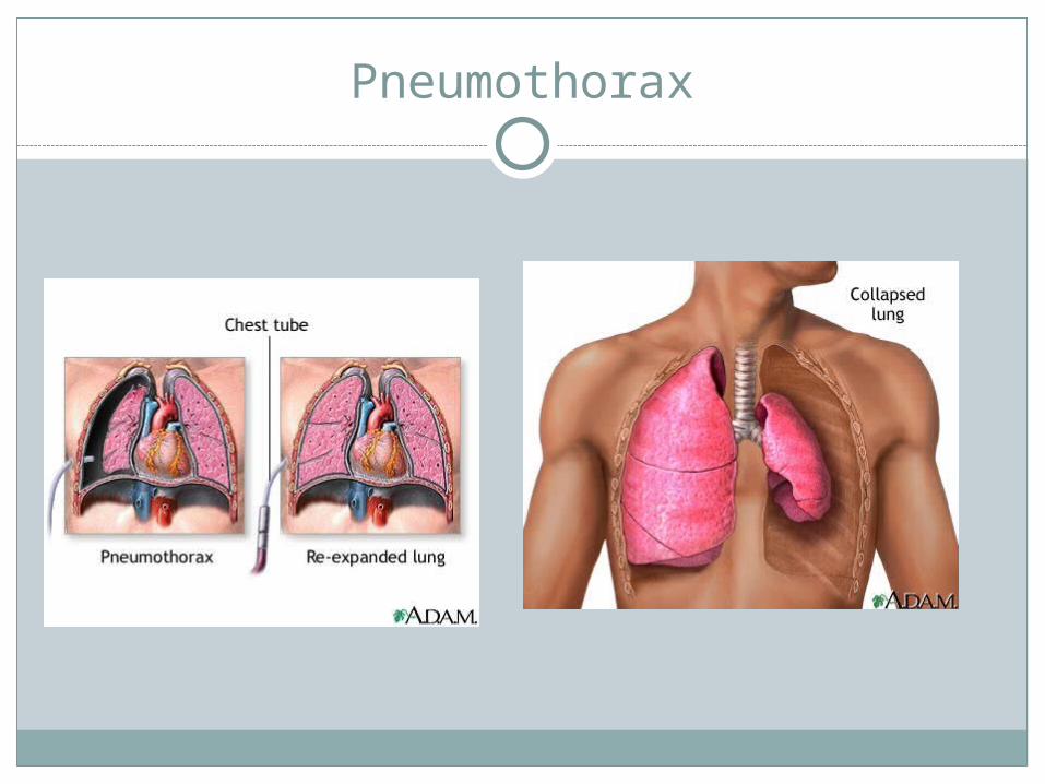
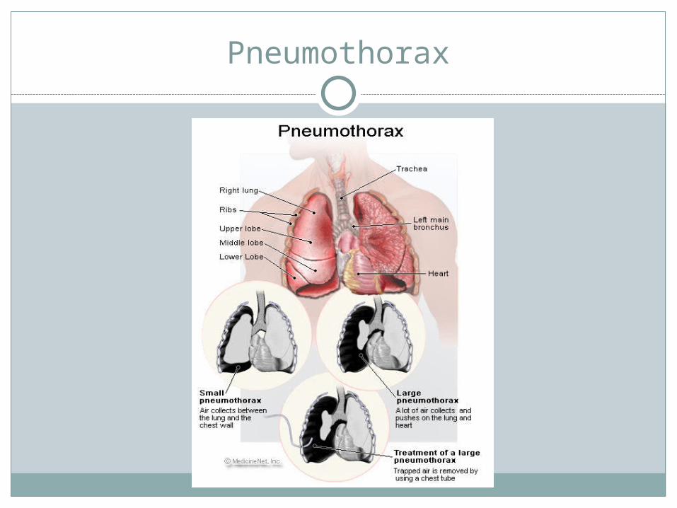
Pneumothorax
Condition in which pleural cavity surround lung becomes filled w/ air that has entered through an opening in the chest
As pleural cavity fills with air, lung on that side collapses

Tension pneumothorax
Occurs when the pleural cavity on one side fills with air & displaces the lung and the heart toward the opposite side, this compressing the opposite lung

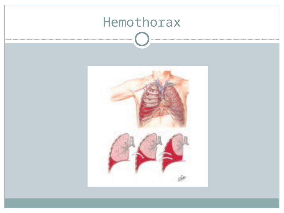
Hemothorax
Presence of blood within the pleural cavity or pleural tissue involving the blood vessels in the area

Traumatic asphyxia
Occurs as the result of a violent blow to or compression of the rib cage causing a cessation of breathing.
Demands immediate mouth-to-mouth resuscitation & immediate medical attention
Lung injuries
S/S: Difficulty breathing Shortness of breath Chest pain on side of injury Coughing up blood Cyanosis shock
Tx: Call 911 Monitor athlete Treat for shock
Pneumothorax
Pneumothorax

Tension pneumothorax
Hemothorax
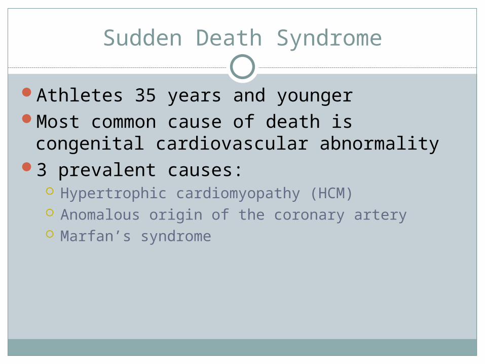
Sudden Death Syndrome
Athletes 35 years and youngerMost common cause of death is congenital
cardiovascular abnormality3 prevalent causes:
Hypertrophic cardiomyopathy (HCM) Anomalous origin of the coronary artery Marfan’s syndrome

Hypertrophic Cardiomyopathy
Condition in which there is thickened cardiac muscle with no evidence or chamber enlargement or extensive myocardial scarring
Anomalous origin of coronary artery
One of the two coronary vessels originates at a different site than normal
This compromises or obstructs the artery because of its unusual course
Marfan’s syndrome
Abnormality of connective tissue resulting weakening of the aorta and cardiac valves which can lead to a rupture of either a valve or of the aorta itself
Coronary artery disease (CAD)
One other potential cause of sudden cardiac death
Results from atherosclerosis which causes a narrowing of the coronary arteries due to hypercholesterolemia in the young athlete

Sudden death
S/S: chest pain or discomfort during exertion Heart palpitations or flutters Syncope Nausea Profuse sweating Heart murmurs Shortness of breath General malaise Fever

Sudden death
Tx: Life-threateningCall 911 Be prepared to perform CPRHave AED ready to useEarly detection/screening/identification could
prevent sudden death from occuring