Spine Joints and Joint Surfaces - Squarespace · PDF fileSpine Joints and Joint Surfaces ATLAS...
34
1 Spine Joints and Joint Surfaces ATLAS (5 letters) • Superior articular facet joint – 2 2 superior articular facets o (Atlanto occipital joint) • Inferior articular facet joint – 2 2 inferior articular facets o (Atlanto axial joint) • Anterior atlanto odontoid joint – 1 1 anterior articular facet AXIS (9 strokes to write axis) • Superior articular facet joint – 2 2 superior articular facets o (Atlanto axial joint) • Inferior articular facet joint – 2 2 inferior articular facets • Lateral inter body articulation – 2 2 uncinate processes • Anterior odontoid joint – 1 2 anterior articular facet o (Axial odontioid joint) • Posterior odontoid joint – 1 1 posterior articular facet • Intervertebral disc joint – 1 1 intervertebral disc surface CERVICAL (The neck is a “perfect 10”) • Intervertebral disc joint – 2 2 intervertebral disc surfaces • Lateral inter body articulation – 4 4 uncinate processes • Superior articular facet joints – 2 2 superior articular processes • Inferior articular facet joints – 2 2 inferior articular processes THORACIC (12 thoracic vertebrae) • Intervertebral disc joint – 2 2 intervertebral disc surfaces • Costovertebral joint – 4 2 sup. costal facets & 2 inf. costal facets • Costotransverse joint – 2 2 transverse costal facets • Superior articular facet joint – 2 2 superior articular processes • Inferior articular facet joint – 2 2 inferior articular processes LUMBAR (6 letters) • Intervertebral disc joint – 2 2 intervertebral disc surfaces • Superior articular facet joint – 2 2 superior articular processes • Inferior articular facet joint – 2 2 inferior articular processes SACRUM (6 letters) • Sacroiliac joint – 2 2 auricular surfaces • Sacrococcygeal joint – 1 1 sacrococcygeal jt surface • Lumbosacral joint – 1 1 lumbosacral jt surface • Superior articular facet joint – 2 2 superior articular processes
Transcript of Spine Joints and Joint Surfaces - Squarespace · PDF fileSpine Joints and Joint Surfaces ATLAS...

1
Spine Joints and Joint Surfaces
ATLAS (5 letters)
• Superior articular facet joint – 2 2 superior articular facets o (Atlanto occipital joint)
• Inferior articular facet joint – 2 2 inferior articular facets o (Atlanto axial joint)
• Anterior atlanto odontoid joint – 1 1 anterior articular facet AXIS (9 strokes to write axis)
• Superior articular facet joint – 2 2 superior articular facets o (Atlanto axial joint)
• Inferior articular facet joint – 2 2 inferior articular facets • Lateral inter body articulation – 2 2 uncinate processes • Anterior odontoid joint – 1 2 anterior articular facet
o (Axial odontioid joint) • Posterior odontoid joint – 1 1 posterior articular facet • Intervertebral disc joint – 1 1 intervertebral disc surface
CERVICAL (The neck is a “perfect 10”)
• Intervertebral disc joint – 2 2 intervertebral disc surfaces • Lateral inter body articulation – 4 4 uncinate processes • Superior articular facet joints – 2 2 superior articular processes • Inferior articular facet joints – 2 2 inferior articular processes
THORACIC (12 thoracic vertebrae)
• Intervertebral disc joint – 2 2 intervertebral disc surfaces • Costovertebral joint – 4 2 sup. costal facets & 2 inf. costal facets • Costotransverse joint – 2 2 transverse costal facets • Superior articular facet joint – 2 2 superior articular processes • Inferior articular facet joint – 2 2 inferior articular processes
LUMBAR (6 letters)
• Intervertebral disc joint – 2 2 intervertebral disc surfaces • Superior articular facet joint – 2 2 superior articular processes • Inferior articular facet joint – 2 2 inferior articular processes
SACRUM (6 letters)
• Sacroiliac joint – 2 2 auricular surfaces • Sacrococcygeal joint – 1 1 sacrococcygeal jt surface • Lumbosacral joint – 1 1 lumbosacral jt surface • Superior articular facet joint – 2 2 superior articular processes
2
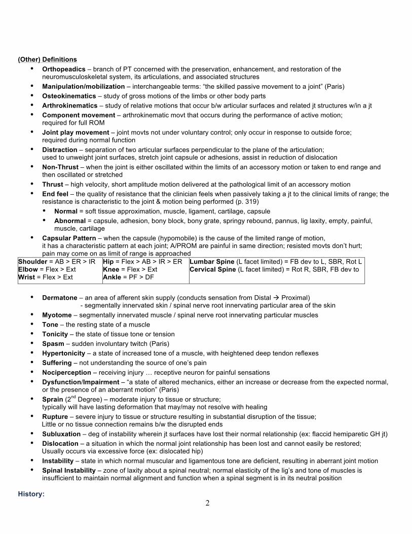
(Other) Definitions
• Orthopeadics – branch of PT concerned with the preservation, enhancement, and restoration of the neuromusculoskeletal system, its articulations, and associated structures
• Manipulation/mobilization – interchangeable terms: “the skilled passive movement to a joint” (Paris) • Osteokinematics – study of gross motions of the limbs or other body parts • Arthrokinematics – study of relative motions that occur b/w articular surfaces and related jt structures w/in a jt • Component movement – arthrokinematic movt that occurs during the performance of active motion;
required for full ROM • Joint play movement – joint movts not under voluntary control; only occur in response to outside force;
required during normal function • Distraction – separation of two articular surfaces perpendicular to the plane of the articulation;
used to unweight joint surfaces, stretch joint capsule or adhesions, assist in reduction of dislocation • Non-Thrust – when the joint is either oscillated within the limits of an accessory motion or taken to end range and
then oscillated or stretched • Thrust – high velocity, short amplitude motion delivered at the pathological limit of an accessory motion • End feel – the quality of resistance that the clinician feels when passively taking a jt to the clinical limits of range; the
resistance is characteristic to the joint & motion being performed (p. 319) • Normal = soft tissue approximation, muscle, ligament, cartilage, capsule • Abnormal = capsule, adhesion, bony block, bony grate, springy rebound, pannus, lig laxity, empty, painful,
muscle, cartilage • Capsular Pattern – when the capsule (hypomobile) is the cause of the limited range of motion,
it has a characteristic pattern at each joint; A/PROM are painful in same direction; resisted movts don’t hurt; pain may come on as limit of range is approached
Shoulder = AB > ER > IR Elbow = Flex > Ext Wrist = Flex > Ext
Hip = Flex > AB > IR > ER Knee = Flex > Ext Ankle = PF > DF
Lumbar Spine (L facet limited) = FB dev to L, SBR, Rot L Cervical Spine (L facet limited) = Rot R, SBR, FB dev to
• Dermatone – an area of afferent skin supply (conducts sensation from Distal à Proximal)
- segmentally innervated skin / spinal nerve root innervating particular area of the skin • Myotome – segmentally innervated muscle / spinal nerve root innervating particular muscles • Tone – the resting state of a muscle • Tonicity – the state of tissue tone or tension • Spasm – sudden involuntary twitch (Paris) • Hypertonicity – a state of increased tone of a muscle, with heightened deep tendon reflexes • Suffering – not understanding the source of one’s pain • Nociperception – receiving injury … receptive neuron for painful sensations • Dysfunction/Impairment – “a state of altered mechanics, either an increase or decrease from the expected normal,
or the presence of an aberrant motion” (Paris) • Sprain (2nd Degree) – moderate injury to tissue or structure;
typically will have lasting deformation that may/may not resolve with healing • Rupture – severe injury to tissue or structure resulting in substantial disruption of the tissue;
Little or no tissue connection remains b/w the disrupted ends • Subluxation – deg of instability wherein jt surfaces have lost their normal relationship (ex: flaccid hemiparetic GH jt) • Dislocation – a situation in which the normal joint relationship has been lost and cannot easily be restored;
Usually occurs via excessive force (ex: dislocated hip) • Instability – state in which normal muscular and ligamentous tone are deficient, resulting in aberrant joint motion • Spinal Instability – zone of laxity about a spinal neutral; normal elasticity of the lig’s and tone of muscles is
insufficient to maintain normal alignment and function when a spinal segment is in its neutral position History:

3
l Cyriax/McKenzie – Disc l Maitland – Reproducible signs l Kaltenborn – arthrokinematics l Chiropractors – Vertebral subluxation, now segmental mechanics l Kirkaidy Willis – Degenerative cascade – a dysfunction in one segment will affect all segmental levels l Paris – Eclectic, functional approach (arthrokinematic)
History
• Hippocrates – first to describe/illustrate jt manipulation and traction techniques, “father of Physical Therapy” • Andrew Taylor Still – founded osteopathic medicine & sx in America in 1874, “The Law of the Artery” • Daniel David Palmer – founded Chiropractic in 1895, but the foundation was from medicine • Physiotherapy was founded in the United Kingdom in 1899 • Physical Therapy was founded in the U.S. in 1921 • E. A. Codman – 1934 published the book
“The Shoulder” advocating repetitive motions at the joints to centralize pain (Codman’s exercises) • James Cyriax – popularized the term “end feel”, advocated manipulation, emphasized evaluation then tx • John Mennell – 1960 published book “Joint Pain” in which he ascribed primary cause of joint pain to be arising from synovial
joints and not the disc; coined term “joint play” and defined it as the “wiggle” or “slack” present in all mechanical & biomech structures
• Stanley Paris – published the “Spinal Lesion” in 1965, First section president of Orthopedic Section of APTA in 1974, founded IFOMT
• Geoffrey Maitland – 1964 published “Vertebral Manipulation” in which he refined the art of oscillatory manipulation and used it to treat “reproducible signs”
• Melzack & Wall – 1966 “Gate control” theory of pain – large nerve fiber stimulation from joints & muscles blocks the transmission & perception of pain
• Robin McKenzie – 1970s popularized concept of spinal extension for the treatment of low back pain; believes centralization of pain is due to reduction of disc protrusion
• American medicine today refers manual methods of tx to PT’s and increasingly to chiropractors & other alternative health care prof Present Day Schools of Thought
1. Manipulation philosophies based on relieving nerve root pressure: § Bonesetters – purpose is to click bones back into place, specific techniques, few practioners remain today § Chiropractic (traditional)– purpose is to move vertebra to relieve nerve root pressure, non-specific techniquesàmultiple cracks § Cyriax – spinal techniques designed to move the disc & thus relieve nerve root pressure, nonspecific techniques affecting many structures including muscle & facets
2. Manipulation philosophies based on relieving pain: § Maitland – oscillations used to eliminate reproducible signs, specific techniques § Maigne – manipulations performed must produce “no pain”, specific techniques, direction of force is contrary to direction of greatest hypomobility § McKenzie* – repetitive motion used to centralize pain; many interpret this as centralizing the disc
3. Manipulation philosophies based on normalizing joint mobility: § Osteopathy – joints & body structures mobilized to increase movement; specific techniques are used § Mennell–emphasis on “joint play”, techniques specific to extremities, less so to the spine § Kaltenborn – emphasis on arthrokinematics, esp. convex-concave relationships, techniques very specific and eclectic § Paris – emphasis on restoring normal arthrokinematics, especially component & jt play. Pain is de-emphasized. Techniques are specific and eclectic. § Mulligan – promotes natural facet glides by assisting motions being performed by the pt (usually in wt bearing position) – MWM
*McKenzie Extension is not manipulation by our definition or that adopted by the APTA. Chiropractic: Theoretical Basis of Chiropractic = The Law of the Nerve
4
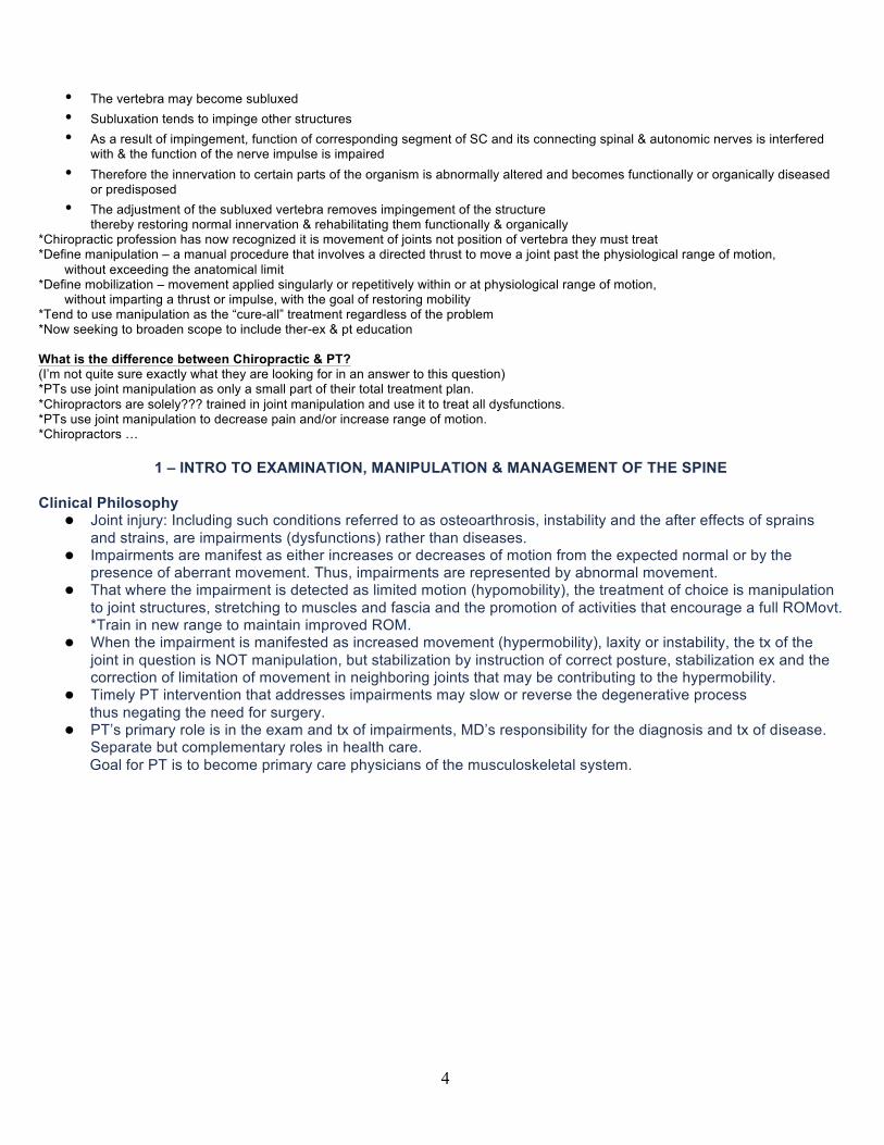
• The vertebra may become subluxed • Subluxation tends to impinge other structures • As a result of impingement, function of corresponding segment of SC and its connecting spinal & autonomic nerves is interfered
with & the function of the nerve impulse is impaired • Therefore the innervation to certain parts of the organism is abnormally altered and becomes functionally or organically diseased
or predisposed • The adjustment of the subluxed vertebra removes impingement of the structure
thereby restoring normal innervation & rehabilitating them functionally & organically *Chiropractic profession has now recognized it is movement of joints not position of vertebra they must treat *Define manipulation – a manual procedure that involves a directed thrust to move a joint past the physiological range of motion,
without exceeding the anatomical limit *Define mobilization – movement applied singularly or repetitively within or at physiological range of motion,
without imparting a thrust or impulse, with the goal of restoring mobility *Tend to use manipulation as the “cure-all” treatment regardless of the problem *Now seeking to broaden scope to include ther-ex & pt education What is the difference between Chiropractic & PT? (I’m not quite sure exactly what they are looking for in an answer to this question) *PTs use joint manipulation as only a small part of their total treatment plan. *Chiropractors are solely??? trained in joint manipulation and use it to treat all dysfunctions. *PTs use joint manipulation to decrease pain and/or increase range of motion. *Chiropractors …
1 – INTRO TO EXAMINATION, MANIPULATION & MANAGEMENT OF THE SPINE Clinical Philosophy
l Joint injury: Including such conditions referred to as osteoarthrosis, instability and the after effects of sprains and strains, are impairments (dysfunctions) rather than diseases.
l Impairments are manifest as either increases or decreases of motion from the expected normal or by the presence of aberrant movement. Thus, impairments are represented by abnormal movement.
l That where the impairment is detected as limited motion (hypomobility), the treatment of choice is manipulation to joint structures, stretching to muscles and fascia and the promotion of activities that encourage a full ROMovt. *Train in new range to maintain improved ROM.
l When the impairment is manifested as increased movement (hypermobility), laxity or instability, the tx of the joint in question is NOT manipulation, but stabilization by instruction of correct posture, stabilization ex and the correction of limitation of movement in neighboring joints that may be contributing to the hypermobility.
l Timely PT intervention that addresses impairments may slow or reverse the degenerative process thus negating the need for surgery.
l PT’s primary role is in the exam and tx of impairments, MD’s responsibility for the diagnosis and tx of disease. Separate but complementary roles in health care. Goal for PT is to become primary care physicians of the musculoskeletal system.
5
l Since impairment is the cause of pain, the primary goal of PT should be to correct the impairment rather than the pain. However when the nature of the pain interferes with correcting the impairment, the pain will need to be addressed as part of the tx program. *Find out what movements are causing the pain
l The key to understanding impairment and thus being able to examine and treat it, is understanding anatomy & biomechanics to safely assume leadership in the non operative mgmt of neruomusculoskeletal disorders.
l It is the patient’s responsibility to restore, maintain and enhance their health. PT role is to serve as an educator and to be an example to the pt, reinforcing a healthy and productive life style.
Classification of Movement
l Classical (osteokinematic) movement: active or passive and is the traditional description of movement � Active: those motions that take place within the joint as a result of voluntary muscle action � Passive: those motions in which a joint is passively moved through a range of motion � Ex: FB, BB, SB, Rot
l Accessory (arthrokinematic) movements: motions that accompany the classical movts and are essential to normal full range and painless function. Accessory movements consist of component motions and joint plays: � Component motion: motions that take place in a joint complex or related joint to facilitate a particular active
motion, typically under voluntary control, typically a glide movement, and tied to a specific classical ROM � Joint play: movements not under voluntary control which occur only in response to an outside force, typically a distraction, and tied to all classical ROM; the “give” in the capsule � Ex: US, DS, Gapping
Dr. Paris' Favorite Vertebrae
l C3 Dr. Rot's Favorite Vertebrae
• C1 (Atlas) • 5 joint surfaces (*know all*): 2 Superior Articular Facets, 2 Inferior Articular Facets, 1 Anterior Articular
Facet • Attachment for transverse ligament
(most important ligament in the human body b/c it keeps the dens off the spinal cord) is the medial tubercle • Lateral interbody articulation is a synovial joint • Very mobile, not many attachments, always moves in same direction as head b/c driven by occipital
condyles
Pain: l “An unpleasant sensory and emotional experience associated with actual or potential tissue damage or
described in terms of such damage.” (Nolan) l Pain is NOT a warning sign, NOT related to morbidity, NOT related to progress, NOT referred into
dermatomes Suffering:
l Suffering is when you don't know why you are in pain Three Aspects of Pain:
l Physical – physiology behind the pain (tissue injury, nociceptive firing) l Rational – understanding (low when first come in, increases after initial eval) l Emotional – pts concern about the intensity, duration, and effects of the condition on their lifestyle
(high when first come in, decreases after initial eval) How can we influence these aspects?
l We listen to the patient l Our hands are placed on the patient l We seek to find and reproduce pts pain l We are not afraid to explain our findings l We alleviate their fears; they have come to someone who understands their c/o and has found their
problem How do we measure pain?
l Intensity: Visual/Verbal analog scale l Location: Pain drawing (body chart) l Type: McGill pain questionnaire l Behavior: Functional index
6
STRUCTURE SECTION (p 32 notes)
• Differential Dx: • Spine (Scoliosis) • SI Joint (Torsion / Rotation)
(Asymmetrical Femoral Neck Angles Slipped Femoral Epiphysis
Legg-Calve Perthes Disease (AVN) DJD
• Hip Joint (Anomoly – osteitis pubis, rugby players) • LE (LLD) • Foot (Pronated)
3 – ACTIVE MOTION ASSESSMENT RULES OF COMBINED MOTION (p14)
7
Paris vs. McKenzie Backward Bending:
l McKenzie states that BB pushes nucleus forward which reduces bulge and takes pressure off nerve root l Paris agrees that this does happen but only in a normal healthy disc. Therefore:
� How might BB Relieve Pain? u Gate control theory: the firing of large fiber proprioceptors blocks onward transmission of nociception
and that will reduce and therefore centralize pain (Codman 1934) u Elevates the water content of the disc – repetitive motion has been shown on T2 weight MRI to ↑ water
content and thus lessen the irritability of the disc to its peripheral and perhaps invading nociceptors. u Mobilizes the facet joint – backwards bending stretches the facets more so than forward bending u Promotes circulation generally relieving irritability u Help relieve the fear of movement thus promotes functional recovery u Neural tension is ↓’ed due to slackening of the cauda equina when a lordosis is achieved & maintained u Over time the disc protrusion loses its proteoglycans and thus its ability to attract water & shrinks
CHARACTERISTICS OF NORMAL VS ABNORMAL MOTION Normal Motion:
l Takes place smoothly regardless of speed l Adequate relaxation of antagonists l Range is full l Pain free l Muscles are of normal length l PROM > AROM
Abnormal Motion: l Limited Range l Unwillingness to move l Painful arc l Compensatory movement l Presence of crepitus l Signs of instability (shaking, juddering) l Pain at end range l PROM no greater than AROM
FACET CAPSULAR PATTERNS OF RESTRICTION IN THE SPINE LUMBAR SPINE Capsular Pattern of L- Spine (if L facet were limited):
l FB – deviation to the L l SBR – limited l RL – limited l SBL – free l RR – free
Myofascial Pattern of the Lumbar Spine: l Limited FB l Free SB
CERVICAL SPINE Capsular Pattern of C- Spine (if L facet were limited):
l FB – some deviation to L l SBR – restricted l RR – most restricted l SBL – relatively free l RL – relatively free
8
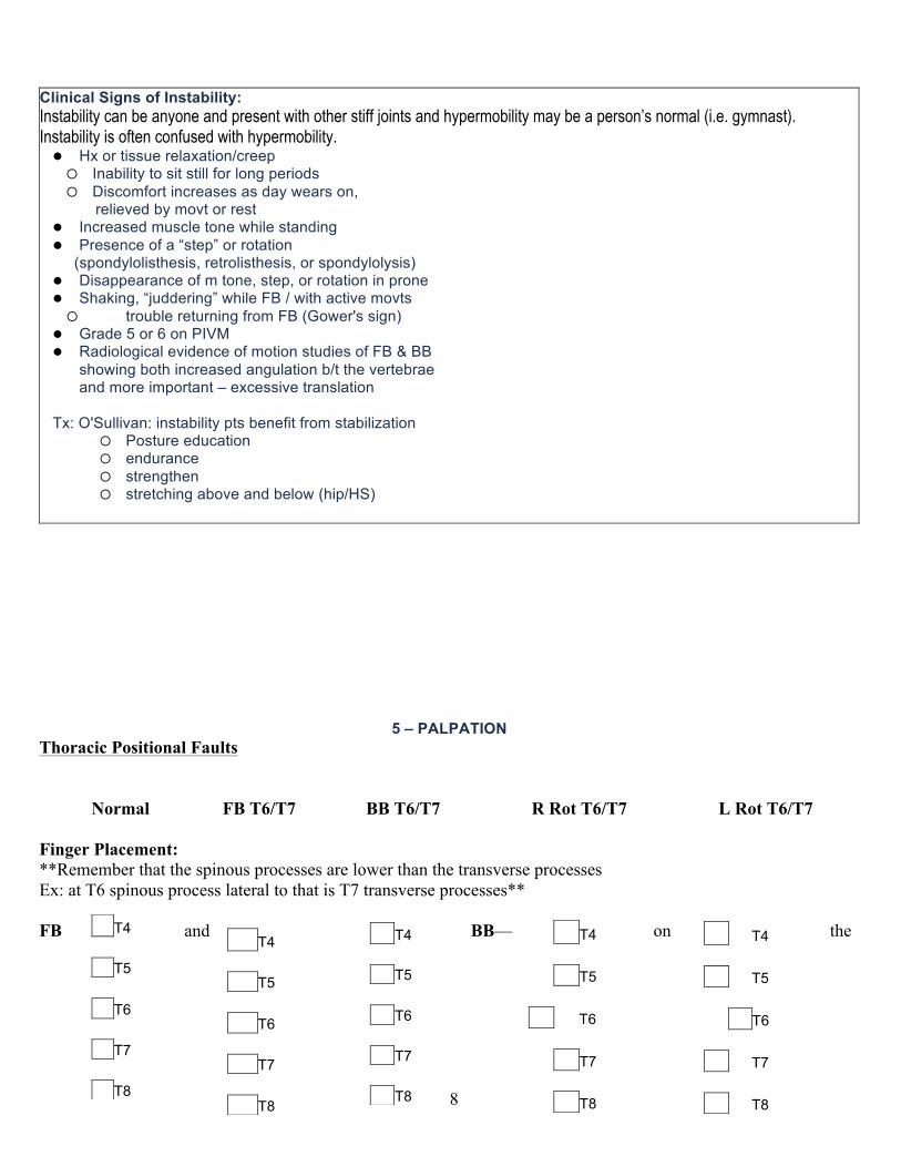
Clinical Signs of Instability: Instability can be anyone and present with other stiff joints and hypermobility may be a person’s normal (i.e. gymnast). Instability is often confused with hypermobility. l Hx or tissue relaxation/creep
� Inability to sit still for long periods � Discomfort increases as day wears on,
relieved by movt or rest l Increased muscle tone while standing l Presence of a “step” or rotation (spondylolisthesis, retrolisthesis, or spondylolysis) l Disappearance of m tone, step, or rotation in prone l Shaking, “juddering” while FB / with active movts
� trouble returning from FB (Gower's sign) l Grade 5 or 6 on PIVM l Radiological evidence of motion studies of FB & BB showing both increased angulation b/t the vertebrae and more important – excessive translation Tx: O'Sullivan: instability pts benefit from stabilization
� Posture education � endurance � strengthen � stretching above and below (hip/HS)
5 – PALPATION Thoracic Positional Faults
Normal FB T6/T7 BB T6/T7 R Rot T6/T7 L Rot T6/T7
Finger Placement: **Remember that the spinous processes are lower than the transverse processes Ex: at T6 spinous process lateral to that is T7 transverse processes** FB and BB— on the
T4 T5 T6 T7 T8
T4 T5 T6 T7 T8
T4 T5 T6 T7 T8
T4 T5 T6 T7 T8
T4 T5 T6 T7 T8

9
transverse processes of the lower level of the fault (ie: T7) R Rot—on the L lower level and R upper level (ie: L T7 and R T6 creating L Rot due to the Rule of Lower Finger) L Rot—on the R lower level and L upper level (ie: R T7 and L T6) Positional Fault
l FB: name for the larger space � Tx @ the level below, ex: T6/T7 will tx @ T7 � Postero-Antero glide of a mid-thoracic vertebra with progressive stretch
l BB: name for smaller space � Tx @ the level below � P/A glide of mid-T spine with progressive stretch
l RR: spinous process off to the left, in right rot and needs to be rot to the left � Tx @ the level moved � Mid-Thoracic Rotation via the Transverse Process � ”Rule of the lower finger” – place left finger lower
l LR: spinous process off to the right, in left rot and needs to be rot to the right � Tx @ the level moved � Mid-Thoracic Rotation via the Transverse Process � ”Rule of the lower finger” – place right finger lower
6 – PIVM
PIVM Scale: (Passive Inter-Vertebral Motion) l 0 – Ankylosis – ignore l 1 – Considerable Hypomobility – Non-thrust l 2 – Slight Hypomobility – Non-thrust + thrust l 3 – Normal l 4 – Slight hypermobility – Stabilize l 5 – Considerable hypermobility – Stabilize l 6 – Unstable – Stabilize / Fusion
Lumbar Spine FB/BB 1)Sidelying – Single Leg Flexion 2)Sidelying – Double Leg Flexion
SB 1)Prone – Leg ABD 2)Sidelying – Raising Legs 3)Sidelying – Rocking the Pelvis
ROT 1)Prone – Raising Pelvis (Rule Gluts) 2)Prone – Rolling Legs (Rule of Legs) 3)Prone – Impulse over Trans Proc
7 – SPINAL MANIPULATION Manipulation:
l “The skilled passive movement to a joint with a therapeutic intent.” (Paris) l “A manual therapy technique comprised of a continuum of skilled passive movements to the joints and/or
related soft tissues that are applied at varying speeds and amplitudes, including a small amplitude high velocity therapeutic movement.” (Guide to PT Practice)
l oscillations w/in range are not only the most tolerated, but seem to be the most effective l usually best achieved by techniques done at end range
Effects of Manipulation:
l Psychological • Hands-on provides treatment confidence & assurance to patient • Providing a thorough exam • Having manually “found” the source of pain by palpation • Reproducing pt’s pain on examination
10
• Dramatic motion or maybe a “pop” l Neurophysiological (possible effects)
• Fire articular mechanoreceptors (Wyke 1972-1985) • Fire cutaneous & muscular receptors • Activates gate control mechanism (Melzack & Wall 1966) • Abates nociception – reduces pain • Centralizes pain • Reduces hypertonicity and muscle holding
l Mechanical (Stress-Strain Curve) •Stretching tight capsules •Stretch out adhesions •Snap adhesions •Alter positional relationships
l Chemical •Little is known, but suspected that increased endorphin release eases pain
Indications for Manipulation:
l ↓ Pain l ↑ ROM l ↑ Function and performance l ↑ Nutrition and repair of joint surfaces
Ex: Steady stretch, progressive oscillation, or Grade IV oscillation. Thrust is effective when end range restriction is minor Manipulation is not ethical nor reasonable practice for psychological benefits only! Precautions for Manipulation:
l Few, if any, absolute CONTRAINDICATIONS (ex: Grade I oscillation) l Instabilities l Fractures l Tumors l All diseases, likelihood of causing osseous and ligamentous damage l Rotational techniques in the presence of ligamentous (annular disc) weakness
Joint Mechanoreceptors:
• Tiny nerves located in the ligament, capsule of the joints. • Sensory receptors that give us proprioceptive sense • Function: To communicate with the brain to tell it where the body part/body is in space (proprioception) • To help with responses to prevent injury
TYPE (PDIN) LOCATION FIRED BY WHEN FIRED?
HOW DO THEY ADAPT? Type I Postural Capsule
(Prox Jts: Core, Head) Oscillations Stretch (graded or progressive)
- Slowly/Constantly Adapt
Type II Dynamic Capsule (Dist Jts: wrist, elbow)
Oscillations Stretch (graded or progressive)
- Rapidly Adapting - Change in Direction & Velocity

11
Type III
Inhibitive (GTO-like)
Capsule Ligament (outside of joint)
Stretch Sustained Pressure Thrust
- Very Slow Adapting - Joint Pop - Stretched Capsule/
Type IV Nociperceptive (Not painful until reaches brain)
Most Tissues; EXCEPT: Brain and nerves, Articular cartilage, Nucleus pulposus
Injury Inflammation
- Non-Adapting - Damage
Golgi Tendon Organs:
• Nerve receptors located in the tendon • Function: Upon stimulation with response to tension,
they cause a reflex relaxation / inhibition to stretch and relax the muscle

12
STRESS / STRAIN (Mechanical Effects) Stress and Strain
l Solid mechanics – the study of the mechanical properties of tissues with the fundamental concern on stress and strain relationships.
l Stress – the resistance of a material to deformation that occurs in the presence of an applied load —the force developed inside the material. Ex: compression, tension, shear and combinations
l Strain – the deformation that occurs in response to an external load l Load – the application of the force or moment (torque). Ex: compression, tension, shearing, torsion, bending.
Stress/Strain Curve: l Toe region – takes up the slack in the tissue l Elastic region – deformation at this stage is completely reversible.
“Hooke’s Law” states the stress and strain are directly proportional to each other within the elastic range. l Yield point – critical point where elastic limit has been reached and damage begins l Plastic region- tissue deforms or lengthens at a rate disproportionate to the stress; usually permanent
deformation. l Ultimate stress – peak of the stress-strain curve and represents max load tissue can tolerate before failure. l Necking – tissues under failure begin to narrow…the narrowing = necking. l Failure – sudden decrease in stress while strain continues. l Biological memory – biological tissues have the ability to slowly contract back to their original length, even
after plastic deformation. Why it is vital that patients continue to stretch at home?
THE “POP” OR “CRACK” OF A JOINT
l Gas, primarily nitrogen, is released from the synovial fluid and remains intra-capsular l Gas distends the joint and stretches it l Distended capsule fires Type III (Inhibitory Type III), which relaxes/inhibits muscle relieving tension l Gas can be seen on Radiograph
POSITIVE EFFECTS OF THE “POP”
l A continuing stretch on the capsule l Firing of Type III articular mechanoreceptors & GTO’s of attaching musculature (multifidus) l Thus, reflex relaxation/inhibition of muscle tone occurs in neighboring muscles
NEGATIVE EFFECT OF THE “POP”
l If already a hypermobile segment, then it may become more hypermobile l Increased stress on a disc l A “Dependency” brought on by the feeling of immediate relieve & perhaps by release of endogenous hormones l Muscle inhibition

13
14
Disc: Everything! l LARGEST LIGAMENT OF THE SPINE! l The disc carries the greatest responsibility for the preservation of function of the vertebral column. l Discs make up 25% of total length of spine; at birth they make up 50% of the total length l 3 functions of the disc:
�bind together the vertebral bodies �permit movement within the segment �transmit loads
l Composed of 1) annulus fibrosus and 2) nucleus pulposus l 23 vertebral discs, 23 motion segments l Paris’ 5 zones of the disc:
� Neurovascular capsule- simply the most outer annulus with nerve and blood supply � Fibrous outer annulus � Inner less fibrous annulus � Nucleus pulposus � Cartilagenous end plate
l Innervation: recurrent nerve (off anterior ramus?), sinuvertebral nerve, branches from gray communicans and sometimes branches from the mixed spinal nerve.
l Annulus Fibrosus �10-12 layers of the disc �outer layers organized with type 1 collagen �inner layers less organized with type II collagen � Function of the annulus fibrosus
u Containment of the nucleus u Stabilization- due to alternating layers and binding to cartilaginous end plates via Sharpey’s
fibers u Permission of Movement u Minimal shock absorption- much less than the vertebral body itself u Herringbone orientation of fibers vulnerable to twisting b/c only ½ fibers resist at a time
l Nucleus Pulposus �remnant of the embryological notochord �80% water at birth �decreasing to 70% by 60 years old � Function of the nucleus pulposus
u Imbibition u Nutrition u Transmission of force u Equalization of stress- will transmit force in equal directions to the water property of disc u Movement- acts like a “ball-bearing” and provides a “rocking” action
l Imbibition- due to the pressure changes in the disc; works when we unweight the spine � Nucleus is lattice of collagen fibrils in protein polysaccharide gel complex � Cartilaginous end plates are semi-permeable � Fluid is moved across cartilagenous end-plates via osmotic forces and release of weight bearing � Nutrition gets to disc this way � Fluid is gradually expressed throughout day through the annulus to the vertebral bodies � Cycle repeats when spine unweighted again
l To obtain good nutrition � Supine or sidelying w/ knees/hips 90/90 � most neutral position of spine � unweights the disc by taking lordosis out of spine � 80% on nutrition in 1-2 hours
l Osteokinematic Movements � Flexion- annulus bulges anteriorly, flattens (becomes taut) posteriorly, nucleus deforms posteriorly � Extension- annulus bulges posteriorly, flattens anteriorly, nucleus deforms anteriorly � Side-bending- annulus bulges on same side, flattens on opposite side � Rotation- disc is compressed as a result of the torque and only ½ of the annulus layers resist the
rotation Fibrocartilage:
l Dense network of Type I collagen fibers with chondrocytes to resist tension and strethinc

15
SPONDYLOLISTHESIS
l Type I- Fatigue fracture of pars- spondylosis
� Cause: repeated over load (prevalent in weightlifters, gymnasts, and springboard divers)— combination torque motions of extension/rotation � S&S: step on standing, disappears if unstable, hypertonicity at level, ligamentous dull ache, rotational component if only one side � Tx: Stabilization, flexion biased tx, possibly manipulate joint above
l Type II- Degenerative- facet arthrosis and tropism
� Cause: congenital tropism (alteration of facet planes from mostly coronal to sagittal), degeneration process- facet arthrosis, overload of above � S & S: step will appear at level of slip, x-ray must be in standing loaded at end of day, step in standing that doesn’t show on oblique x-ray � Tx: Stabilization, manipulate joint above-, myofascia (same as in type I)
l Type III- Isthmic Spondylolisthesis � Cause: Pars interarticularis (isthmus) lengthens allowing vertebrae to slip fwd, childhood obesity = precursor � S&S: X-ray may show elongated pars/isthmus, may also show fracture of pars and thus miss initial cause � Tx: Same as Type I & II, weight control
l Unstable � S&S: dull ache- ligamentous- towards the end of the day, STEP DISAPPEARS ON LYING, muscle guarding
on standing that disappears on lying, rotational fault if just one side (i.e. Spondylolisis) � TX: Stabilization, manipulation if may reduce stress, edu on decreasing loading and extension activities,
Fusion, Pedicle screws MUSCLES OF THE SPINE:
l Function to activate and restrain motion and support (tone of muscles) l Desirable = moderate development with good postural tone. l NOT desirable = overly strong spine musculature b/c à severe compression forces in the spine when activated.
Multifidus:
l Innervation: branches from the Posterior Primary Rami (Dorsal Rami) of spinal nerves

16
l Attachments: C2 – Sacrum, transverse process à spinous processes C2/C3-S3/S4. Bipennate at origin and insertion; deepest layer of the back
l Segmental stabilizer of the spine: attaches deep and on close approximation to facet jts and is intersegmental l Action: fires at all times for stabilization, Bilateral: BB, Unilateral: SB same side, Rot opposite side l Multifidus muscle recovery is not spontaneous on remission of painful symptoms (Hides et al, 1996) in LBP it
atrophies on same side as back pain, leaving pt. more susceptible to re-injury. Need to retrain l Attaches to the posterior facet capsule to prevent pinching; each capsule has 2 multifidi inserting on to it.
According to Winkelstein’s 2001 article. Theory: since it attaches to the capsule, contraction of it may pull capsule out of facet and prevent nipping.
l Train: quad/prone – opposite leg and arm. Why is the multifidus a stabilizing muscle? • It is deep and segmental so the patient would need to exercise to prevent pain and dysfunction • Superficial muscle activity without deep muscle activity, causes spinal buckling and unstable segments. • Deep muscle activity “stiffins” spinal segments
Quadratus Lumborum:
l Innervation: Subcostal N, Ventral Rami L1-L4 l Origin: posterior iliac crest to erector spinae (and iliolumbar ligament-myoplasia of QL after 30) l Insertion: medial aspect of 12th rib; costal transverse process of L1-L4 l Thick rectangular muscle sheet forming part of posterior abdominal wall.
Lies ventral to the deep muscles of back and lateral to psoas major. l Stabilizer of the spine: attaches deep and on close approximation to facet joints and is intersegmental l Action: Bilateral: BB, Unilateral: SB same side, Rot opposite side,
helps elongate thorax during inspiration by anchoring 12th rib; lateral tilt of pelvis l Train: sidelying on table with elbow and lift pelvis
Transverse Abdominus:
l Innervation via Intercostal T5-T12, Ilioinguinal, Iliohypogastric, Genitofemoral, Subcostal ? l Origin: thoracolumbar fascia, anterior 2/3 of iliac crest/ASIS, lateral ½ of inguinal ligament, lower 6 ribs l Insertion: Linea alba, pubic crest, forms conjoint tendon with internal oblique l Weakness of the abdominals allows the abdomen to protrude, increasing the abdominal wt arm on the L- spine. l Action: Unilateral: Rot same side, Bilateral: active in expiration, maintains/increases intra-abdominal pressure,
stabilization of pelvis and trunk, supports abdominal viscera l In normal pts it is the first to fire prior to loading of spine. In pts with low back pain it is no longer the 1st to fire.
Longus Capitus / Longus Colli:
l Innervation: Capitus (Ventral Rami C1-C4) Colli (Ventral Rami C1-C7) l Attachments: Capitus (anterior tubercle of TP’s C3-6 – Basilar part of occipal bone) Colli (ant vert bodies--TPs) l Action: Deep muscles that “stiffen” the spinal segment, control intervertebral motion & control cervical lordosis l Test using Cranio-Cervical Flexion Test (C-CFT) (See handout) l Impairment leads to:
�↓’ed holding capacity of inner range positions and ↑’ed activity of superficial muscles (SCOM, Ant Scalene) �Superficial muscles w/o deep activity leads to spinal buckling and unstable segments; �over-activity of superficial M seen in people with chronic neck pain
l Cervical Stabilization: 80% provided by muscle; 20% ligaments l Does not recover spontaneously
Dermatomes:
l Segmentally innervated skin l Since a dermatome is an area of afferent skin supply, it can only conduct sensation from distal à proximal.
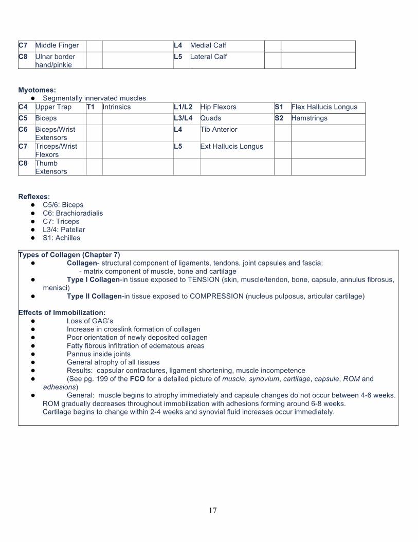
C4 Upper Trap T1 Medial Forearm L1 S1 Lateral border of foot C5 Lateral Delt T2 Medial Arm L2 Lateral Thigh? C6 Thumb/Index L3 Medial Knee
17
C7 Middle Finger L4 Medial Calf C8 Ulnar border
hand/pinkie L5 Lateral Calf
Myotomes:
l Segmentally innervated muscles C4 Upper Trap T1 Intrinsics L1/L2 Hip Flexors S1 Flex Hallucis Longus C5 Biceps L3/L4 Quads S2 Hamstrings C6 Biceps/Wrist
Extensors L4 Tib Anterior
C7 Triceps/Wrist Flexors
L5 Ext Hallucis Longus
C8 Thumb Extensors
Reflexes:
l C5/6: Biceps l C6: Brachioradialis l C7: Triceps l L3/4: Patellar l S1: Achilles
Types of Collagen (Chapter 7)
l Collagen- structural component of ligaments, tendons, joint capsules and fascia; - matrix component of muscle, bone and cartilage
l Type I Collagen-in tissue exposed to TENSION (skin, muscle/tendon, bone, capsule, annulus fibrosus, menisci)
l Type II Collagen-in tissue exposed to COMPRESSION (nucleus pulposus, articular cartilage) Effects of Immobilization:
l Loss of GAG’s l Increase in crosslink formation of collagen l Poor orientation of newly deposited collagen l Fatty fibrous infiltration of edematous areas l Pannus inside joints l General atrophy of all tissues l Results: capsular contractures, ligament shortening, muscle incompetence l (See pg. 199 of the FCO for a detailed picture of muscle, synovium, cartilage, capsule, ROM and
adhesions) l General: muscle begins to atrophy immediately and capsule changes do not occur between 4-6 weeks.
ROM gradually decreases throughout immobilization with adhesions forming around 6-8 weeks. Cartilage begins to change within 2-4 weeks and synovial fluid increases occur immediately.
18
Healing of Articular Cartilage (Salter's Work) l Injury to articular cartilage can be caused by degradation of macromolecules or direct trauma.
Specifically: � Injury � Immobilization � Repeated stress � Weak and poorly coordinated muscles � Incongruous surfaces � Trauma- burst type (not sure what that means??)
l Partial thickness cuts and tears DO NOT heal l Full thickness defects DO heal with continuous passive motion l Lesions from diseases causing breakdown DO NOT heal
Facts on Cartilage:
l Cartilage needs to contact cartilage or synovium will destroy the cartilage l Prolonged compression should be avoided l 3 hydrocortisone injections can kill cartilage l 6 weeks immobilization kills cartilage l 4 weeks of constant mobilization helps restore cartilage with holes drilled in it l Note: In Vail, CO Dr. Steadman does a surgery called a “microfracture” that is indicated for a full
thickness lesion of articular cartilage. It is an arthroscopic surgery to encourage the cartilage to regenerate. A pick is used to cause mini-fractures on the cartilage surface and encourage bleeding. Stem cells of type II collagen are found in the area. The entire degenerative surface is treated. The patients are NWB, but with full PROM on day 1. Partial thickness lesions are contraindicated.
Levers:
l There are 5 parts to every lever: 1) Fulcrum, 2) Force arm, 3) Resistance arm, 4) Force and 5) Restistance.
l Lever- motion of a solid object restricted by a fulcrum l Fulcrum- point of attachment to rotating object, serving as a fixed point of rotation l Forces applied to levers create moments, AKA torque. l Torque= Force x distance
Types of Levers
l Class I- pulley system with fulcrum in center (therapeutic exercise) l Class II- resistance or body weight is closest to fulcrum with a very long moment arm (toe raises) l Class III- moment arm is very short with resistance or body having mechanical advantage (most common lever in the body = Quads).
19
Codman's Paradox: l Flex elbow to 90* and IR next to body. Elevate the arm over your head in the sagittal plane. Descend
the arm in coronal plane. Notice the difference in hand position. You never changed your hand position, so the altered position must be due to rotation at the shoulder joint.
l Elevate the arm in the coronal plane and ER occurs l Descend the arm in the sagittal plane and IR occurs
Instant Center of Rotation (ICR): l Surgeons call it the “isometric point.” The point where the graft can be attached and remain at a constant length throughout the entire ROM. l All synovial joints, except planar joints, have this. l The motion is angular around an axis of rotation called the “ICR.” l The location of the ICR is critical for clinical application of manual therapy techniques, orthotics and
machines. l In many joints, the ICR continually changes with movement.
2 methods of calculation of ICR l Circular estimation- via the curve of the convexity; it is the geometric center of the convex surface.
This is a “guess” of the ICR. l Method of Reuleaux- via 2 points on an X-ray. Example: picture taken of ankle and medial malleolus and neck of the talus are the reference points. The ankle is moved. Another picture is taken and the same reference points are noted. Overlay the X-rays & connect the medial malleolus on one with the medial malleolus of the other…repeat for talus. Draw a perpendicular line to these 2 lines and watch where they intersect. This is the ICR and is accurate.
Neurogenic vs. Vascular Claudication Test (bike and treadmill test)
l Bike: Have pt ride upright on bike until symptoms come on, time how long this takes. Have pt rest until symptoms alleviate. Have pt ride bike again, this time in fwd bent position (opening foramen). Time how long before symptoms come on. If neurogenic claudication, symptoms should take longer to come on in FB position. If vascular, symptoms will be same time.
l Treadmill: Have pt walk on level treadmill until symptoms come on, time how long this takes. Have pt rest until symptoms alleviate. Have pt walk again, this time with treadmill at an increased grade (in FB position which opens the foramen). Time how long before symptoms come on. If neurogenic claudication, symptoms should take longer to come on in forward bent position. If vascular, symptoms will actually come on sooner b/c increasing intensity and O2 demand.
Musculoskeletal vs Non-Musculoskeletal Problems:
Musculoskeletal Non-Musculoskeletal S/S Can reproduce with AROM and provocation tests Cannot reproduce with AROM or provocation tests
Systemic, Autonomic Movements Certain actions cause / relieve pain Not related to activity
Movements / positions don’t change symptoms Pattern Pattern makes sense from musculoskel standpoint When only have pain at night, unrelenting,
not changed by positions, RED FLAG: Cyclical, same time every night
History MOI Insidious onset Release Phenomenon:
l Pain from revascularization due to sympathetic nerve fibers in blood vessels LBP and Posterior Buttock/Thigh Pain:
20
3 Potential Causes and how to differentiate: l Disc Herniation:
�(+) neurological signs, �limited SLR (35-40 deg) reproduces S/S
l SI Joint: �(+) provocation tests, �ipsilateral, �typically female (3:1), �usually below L5/S1 over sacral sulcus, �tender medial to sacral sulcus
l Facet: �AROM limited capsular pattern/entrapment, �(-) neurologic & provocation �pain higher in l-spine, �interspaces tender.
Pisoelectic vs. Streaming Potentials:
l Pisoelectic Effect – Related to bones � Wolff's Law: stressing bone makes it stronger � Stress bone: convex and concave side � More (-) ions on concave side, thus, more cellular metabolism occurs on that side and that side strengthens
l Streaming Potentials – Related to ligaments � Ligament mostly water � When stressed, water and (-) ions stream out � Wherever there are more (-) ions = more strengthening occurs (fibroblasts laying down more ligament)
SYNDROMES: 1. Abnormal Muscle (Myofascial) States 2. Facet Dysfunctions 3. Sacroiliac Dysfunction 4. Ligamentous Weakness 5. Instability 6. Disc Dysfunction 7. Spondylolisthesis 8. Lumbar Spine Stenosis
a. Central Spine Stenosis b. Lateral Foramenal
9. Cervical Spine Stenosis a. Central Spine Stenosis & Myelopathy b. Lateral Foramenal
10. Whiplash – Acceleration & Deceleration 11. Thoracic Outlet Syndrome (TOS) 12. Headaches (HA) 13. Lesion Complex 14. Lumbar Spine:
a. Kissing Lamina (Bastraps Disease) 15. Cervical Spine:
a. Kissing Lamina 16. Thoracolumbar Syndrome (Maignes Syndrome) 17. Lateral Shift 18. Scoliosis
1) ABNORMAL MUSCLE STATES: HYPERTONIC
l *Involuntary Guarding � Cause: injury, dysfunction (ex: spondylo) � S/S: Hypertonicity, protective muscle guarding, elevated resting tone, abnormal elastic response to pressure � Tx: treat cause of impairment (Pinched capsule, instability, sprain/strain, etc)
l *Chemical Muscle Guarding � Cause: sustained involuntary guarding, may lead to compartment syndrome
21
� S/S: doughy to touch, non-elastic response to pressure, limited ROM � Tx: Remove irritants, restore elasticity of tissue:
■Heat & massage (deep finger kneading, trying to bring new circulation in and get waste products out) ■Think compartmental syndrome-multifidus
l *Voluntary Muscle Guarding � Cause: anxiety, pain or fear of pain often follows involuntary and chemical states � S/S: slow and guarded movements, trunk moves as a whole � Tx: *once sure no fracture*,
■ignore, give reassurance, ■Movt with repetitive motion (painfree, low load/resistance)
l Spasm � Cause: may be provoked on examination (i.e. spring) � S/S: momentary, involuntary twitch � Tx: ?
l Hypertrophy l Psychosomatic Tension/Stress
HYPOTONIC
1. Disuse Atrophy 2. Wasting and Fibrosus 3. Denervated
NORMAL TONE
1. Adaptive Shortening (Normal Tone/Shortened) � Cause: follows chemical muscle holding, slouching posture (ex: sub cranial extensors) � S/S: normal tone, shortened length, loss of ROM, altered posture (↑’ed lordosis 2° to psoas shortening) � Tx: myofascial stretching
OTHER
1. Fibromyalgia – management rather than treatment
2) FACET DYSFUNCTIONS: 1. Synovitis / Hemarthrosis
� Cause: awkward movement of catch, gross trauma � S/S: good but guarded movt, involuntary and voluntary muscle holding � Tx:
■Cervical: rest, soft collar, careful movt- circular, consider interferential for swelling * ROM painfree*, anti-inflammatories, massage, ice, estim- don’t flare up ■Lumbar: rest, soft corset, and careful mvt,
2. Chronic Facet Arthrosis � Cause: poor posture, trauma, overuse � S/S: dull ache, local pain, stiffness � Tx- Posture, mobilize adjacent areas
3. Stiffness / Restriction � Cause: Resolved synovitis hemarthrosis not symptomatic � S/S: none- stiffness does not hurt, lowered tolerance to insult leading to strain & associated pain in neighboring joints � Tx: manipulation
4. Mechanical Block � Cause: idiopathic (not painful), loose body, impaction � S/S: sudden onset, block to motion, relatively pain-free � Tx:
■Cervical- traction and SB away, rotation to blocked side ■Lumbar- rotational manipulation over a bolster to open affected side
5. Painful Entrapment � Cause: awkward movement in eccentric range � S/S: unable to slide inferior articular process down, head held away from painful side-torticollis � Tx:
■Cervical & Lumbar- multifidus isometric

22
■Lumbar- also manip over a bolster Mechanical Block vs Painful Entrapment:
l Painful entrapment is painful Painful Entrapment S/S:
l Cervical: AROM assessment painful and limited: Rot (SS), SB (SS), BB l Lumbar: AROM assessment painful and limited: Rot (OS), SB (SS), BB
Facet Capsular Entrapment in Lumber spine ®: l Hx: Limited SBR, Rot L, BB (pain local on R side) l MOI: return from FB/BB and have “catch”, can't get straight l Shifted away from painful side l (-) Neurologic signs l Tender/guarded in surrounding musculature l Traction does not relieve l Tx: Multifidus isometric: Prone w/ 2 pillow under abdomen, resist opp LE (5 sec hold, 4 sets, 4 reps), re-eval! Facet Capsular Entrapment in Cervical spine ®: l Hx: Limited SBR, Rot R, BB (pain local on R side) – pinching capsule w/ DS on right l MOI: sudden onset, catch, pain, woke up with l Shifted away from painful side (SB or Rot to L) l (-) Neurologic signs l Tender/guarded neck movements, increased tone, swollen facet l Tx: Multifidus isometric: Stand on pts L side on diagonal, resist (5 sec hold, 4 sets, 4 reps) then re-eval! l Tx: ice, NSAIDs as directed on box l Tx: Address pillow, 20-30 degrees of flexion in supine, should fill space in sidelying l Tx: Address posture, strengthen, body mechanics
Facet Joints: l Function to permit, guide and restrict the motion segment l Facets have two main actions: slide and gapping l 3 things that prevent the facet capsule from pinching: (multiple men are flavorful)
�1) Multifidus �2) Meniscus �3) Ligamentum Flavum
l Facet joints formed by superior and inferior articular processes of vertebrae l Facets in cervical spine at 45* l Facets in lumbar spine in sagittal plane l Blood supply from periarticular plexus l Innervation from 3 levels – ascending, local, and descending branches of dorsal ramus
Cervical Nerve Roots (Why are there 8?) l Cervical spine NR’s exit above the vertebrae and the nerve roots of the thoracic spine exit below the vertebrae, therefore, the C8
nerve root is in a transitional area between C7 and T1 b/c: C7 nerve root exits above C7 and T1 nerve root exits below T1.
3) SACROILIAC DYSFUNCTIONS (L5/S1*)
1. Sprain / Strain 2. Hypermobility 3. Displacement
Schwarzer – “The SI jt in chronic LBP” – Spine. 1995.
• SI jt source of pain in 30% subjects w/ chronic LBP utilizing anesthetic joint blocks + fluoroscopy Cibulka – “Changes in Innominate Tilt after Manipulation of SI joint in patients with LBP: experimental study” – Physical Therapy. 1998.
• Poor intertester reliability • Intertester acceptable when 3-4 / 6 tests positive to indicate SI jt dysfxn • Equal but opposite change was noted in innominate rotation s/p manipulation
o Measures innominate - displacment Sturesson – “Movements of SI jts: A Roentgen Stereophotogrammetric Analysis” – Spine. 1989.
• SI jt does indeed move but motions are small. 2.5 deg of motion and 0.7mm (mean avg) Laslett – “Dx Painful SI jt…” JMMT 2008, Australian J PT 2003, Spine 1994.
• 3+ provocation tests and no centralization or peripheralization w/ repitive extension = 3-20x more likely to have SI jt dysfunction
Walker – “SI jt; critical review” PT. 1992
23
• SI jt is part synovial and part syndesmosis • Female > male
Richardson – “Relationship b/w TrA and SI jt and LBP” Spine. 2002. • During both specific contraction of TrA and general abdominal bracing, measurements of SI jt laxity were
decreased, but laxity was reduced to a significantly greater extent w/ TrA –used ultrasound. Alan Stoddard – osetopath – specific joint Cyriax – general manip, disk, disagreed w/ SI source of LBP Mckensive = disc Maitland and kaltenbord did not discuss syndromes Paris accepts broad range
• SI jt source of LBP! • Ovarian cysts mimic SI jt pain • Ligaments stabilize primarily • No SI muscles (indirect = multifidus, glut max, abdominals) • Ilio-lumbar ligaments – defined well in females • Sacroiliac ligaments:
o Deep posterior – very strong and bind pelvic together on wt bearing o Long posterior – not sig; extension of biceps femoris tendon over the bursa on the tuberosity o Sacrotuberous and sacrospinous – principal stabilizers against rotary forces in females that occur in
normal standing and inc on SLS • Symphysis pubis – key stone to pelvic for stability (if ruptured = unstable girdle)
Signs and Symptoms of SI joint problem
l Pain unilateral and below L5/S1 junction l Pain reference (no pattern) l Pain accentuated by springing and provocation testing l Pain altered by torsion (stresses to ligs) l No central pain l Ipsiliateral tension multifidus, and erector spine, QL & muscle guarding (protect jt) l Swelling over dorsal sacrum l Ligaments tender to touch over sacral sulcus l Symptom centralization does NOT occur w/ BB (it will cause px)
Who is more prone to SI joint problems and why?
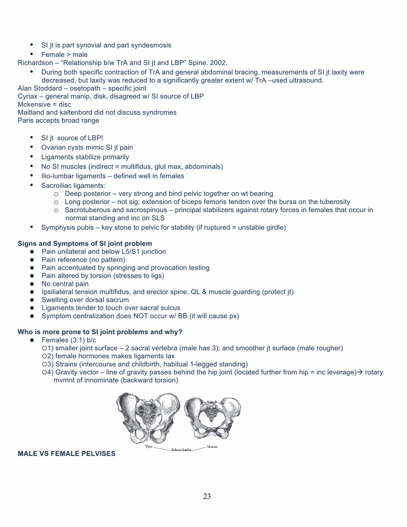
l Females (3:1) b/c �1) smaller joint surface – 2 sacral vertebra (male has 3); and smoother jt surface (male rougher) �2) female hormones makes ligaments lax �3) Strains (intercourse and childbirth, habitual 1-legged standing) �4) Gravity vector – line of gravity passes behind the hip joint (located further from hip = inc leverage)à rotary
mvmnt of innominate (backward torsion)
MALE VS FEMALE PELVISES
24
4) LIGAMENTOUS WEAKNESS l Causes:
�repeated minor strains, �obesity, �poor posture, �vibration
l S/S: �pain w/ fixed position, �relieved by changing positions, �relieved by “cracking” back, �lig’s sensitive to touch
l Tx: �Early stages: exercises, stabilization, posture, back school �Later Stages: pre-discal, rest/controlled activity, corset, bracing, taping, Instruction in First Aid- BB if injury
5) INSTABILITY (see Clinical Signs of Instability above) • Where the osseo-ligamentous structures and the neuromuscular control systems are unable to hold a spine in
neutral and during motion against buckling and slippage/shear. • Causes:
o Ligamentous stress & strain from sports & poor posture, o Lack of meuromuscular training and exercise, o Surgical (ex: wide laminectomy or secondary to fusion – level above), o Medical/surgical as with chymopapain
• Characterized by: o Ligamentous weakness/laxity, o Muscle weakness/atrophy, o Fatigue, o Poor posture, o Pain on assuming fixed position, o Chronic pain
• Physical Exam: o History of catches and twinges, o sudden pain, o pain on prolonged sitting/standing
• Structural Exam: o Obesity and poor posture, o Spondylolisthesis, o Involuntary muscle guarding,
25
o Step on standing that disappears on lying • Active Movements:
o Uneven, Slippage, Juddering, Pain at end range, o Poor balance and neuromuscular control as in “hound dog”
• Palpation: o Tenderness to palpation of ligaments, o Grade 5 or 6 on PIVM
• Treatement Principles: o Ensure slow, o continuous improvement, o Train for health & not performance
• Exercises: o Transverse Abdominis (Abdominal setting), o Multifidus Isometric, o Quadratus Lumborum (esp in Fem), o Quads, Ab’s, Gluts, Manual Therapy-according to what is found
6) DISC DYSFUNCTION Paris vs. McNabs’ Disc Classifications McNab’s Classification (Pg 160 S1 manual)
l Disc Protrusion—Bulging Annulus � Type I—localized annular bulge, one side � Type II—diffuse annular bulge, both sides
l Disc Herniation—Torn Annulus � Type I—prolapsed nucleus, to edge of annulus border � Type II—extruded nucleus, past edge of annulus border � Type III—sequestrated nucleus (free body)
Clinical info needed to detect/determine disc herniation:
l Neurologic signs: �Dermatomes �Myotomes (***most reliable***) �Reflexes �Neural Tension Test (SLR)
Disc Herniation vs Lateral Foraminal Stenosis:
l Age: � Disc: younger, 30-50 � Stenosis: older, 60-70
l History of onset: � Disc: MOI bending and twisting � Stenosis: Gradual
l Sitting: � Disc:
■doesn't like to sit, ■increases intradiscal pressure, ■stretches posterior annulus which is already torn.
� Stenosis: ■likes to sit, ■opens foramen, ■unbuckles ligamentum flavum; can't stand or walk

26
Disc vs. Stenosis (Nerve Roots)
l Stenosis/Disc l Example: L4 nerve root: l If disc, L3/L4 l If stenosis, L4/L5
Positional Distraction:
l Opens foramen which: �relieves compression on nerve root, �increases circulation, bringing nutrition, flushing chemical irritants, ultimately DECREASIN INFLAMMATION
l SB also flattens disc bulge further relieving compression.
Paris Treatment Classifications Four Disc Dysfunctions:
1. Pre-prolapse � Hx:
■Dull muscular ache on sitting (need to get up and move), ■Hx of self-cracking, ■LBP occasionally radiating into buttock, ■No neuro signs
� Physical: ■Demonstrates many signs of instability, ■Grade 5-6 to PIVM
� Tx: ■Stabilization, ■Back School, ■Manipulation (Hypo above or below), ■Instruction in 1st aid to maintain lordosis (approximate the tissue), ■Promote healing and have scar tissue form
2. Tear or Strain
� Hx: ■Sudden unguarded motion resulting in acute but deep pain, ■Usually with flex and occasionally rotation, ■Pt may say it “tore,” “ripped,” or “gave out”
� Signs and Sx: ■Sudden and deep pain, ■May refer to buttocks, ■Very guarded motions
� Physical: ■Don’t do a physical (no FB and Rot), ■Neuro may show exaggerated responses
� Tx: ■Maintain lordosis for 4 wks, ■Stabilization, ■Manipulation (no rot until after 3 wks), ■Myofascial technique
l Prolapse
� Acute (day 1-4):
27
■Minimal bed rest as disc swells with rest (max 3 days), ■Try backward bending, ■Medical palliative measures, ■Education (move, don’t rest too long)
� Sub acute (day 4 and improving): ■Initiate movt, ■Myofascial manipulation, ■Corset (decrease pressure, increase support), Stabilization
� Settled (3-4 wks slow improvement): ■Commence positional distraction (opens lat foramen, centralizes pain), ■Stabilization, ■Possibly neural manipulation, ■Lifestyle changes
l Chronic Disc
� Hx: ■Serious debilitating back pain with hx of neuro signs and possibly surgery
� Physical: ■Sad and depressed pt on meds; ■often obese, smoker, unfit, diabetic; ■ROM deficit from pain; ■PIVM restrictions and instability; ■Myofascia restrictions and poor tone/condition
� Tx: ■Lifestyle Education, ■Stabilization, ■Positional distraction, ■Possibly neural manipulation, ■Manipulation, ■Fitness training and work hardening, counseling
7) SPONDYLOLISTHESIS (see above)
8) LUMBAR SPINE STENOSIS Central Spine Stenosis:
l Cause: �degeneration, �wear and tear, �poor posture, �abdominal protrusion/lordosis, �tight iliopsoas, �tight lumbar spine myofascia, �DISC protrusion, �prolapse (33% asymptomatic)
l S&S: �Chronic dull low back pain, �leg pain on walking any distance
(neurogenic claudication vs vascular claudication --- bike vs. treadmill test) l Tx:
�Myofascial manipulation and stretching (psoas and low back muscles), �increase physical fitness (pool, unweighted treadmill), �lifestyle changes, �surgery
28
Lateral Foraminal Stenosis l Cause:
� lateral disc protrusion, � loss of disc heights, � degenerative changes to ligamentum flavum and facets
l S&S: llateral symptoms- (pain, subjective numbness, hyper neurological response), ltrue neurological signs, paresis>skin sensation> reflexes> neural tension (SLR) ldiscogenic * 28-50 y.o * male>female
l Tx: lposture education, lstabilization, lstretch myofascia (back and psoas); lmanipulate stiff joints, lpositional distraction, and lpossible heel lift on unaffected side to open affected foramen;
9) CERVICAL SPINE STENOSIS Central Spine Stenosis& Myelopathy:
l *May mimic neurological problems such as ALS, MS, Parkinson’s l Cause:
�Congenital narrowing of cervical spinal canal, �Hypermobility, �Enfolding of ligamentum Flavum
l Contributing Factors: �poor posture, �cervical stress, �strain, �sports, �MVA, �compensatory Hyper mobility to UT kyphosis/stiffness, �C5/6- most common degenerative level in spine
l S&S: �B UE symptoms, �UMN signs + babinski and clonus, �vague transient neuro signs may be in arms and legs
l Tx: �POSTURE (AXIAL EXT), �STABILIZE (DEEP ANT MUSCLES),

29
�avoid bckwd bending, �Manipulate upper thoracic to help reduce MC stress, �Surgery to remove impingements and then fusion
Myelopathy:

30
• Disease to the spinal cord secondary to
compression
Causes of Myelopathy:
• Stenosis 2° to osteophytes (bosses and bars)
• bone spurs • posterior disc bulge • ligamentum flavum infolding or hypertrophy • spinal cord canal tumor • trauma
Lateral Foraminal Stenosis
l * >50 y.o l Cause:
�degenerative changes, �osteophytes from lateral interbody articulations (uncinate processes, Von Luschka joints), �thickening of ligamentum flava, �arthrosis of facet joints
l S&S: �neck/arm pain and paresthesia, �Neurological S&S- muscle, skin, reflex
l Tx: �Joint & Myofascial release, �Posture, �Positional distraction
l Sx: �Foraminectomy- surgeon will most often tell pt. they need disc surgery
10) WHIPLASH – ACCELERATION & DECELERATION INJURY
l Cause: �MVA, �fall down stairs, �skiing, �struck by yacht
l S&S: �VERY UNRELIABLE , �often initially minimal, �minor to bizarre, �sympathetics
l Tx: �CONSERVATIVE!!
31
Whiplash Protocol: l Based on:
� Healing time � Imaging (or lack of) –
■not reliable, doesn't always detect fracture, ■need open mouth x-ray to detect fx to dens, ■may need more than one over several days
� 60% healing: 2-4 weeks, � 80% healing: 6 weeks, � 100% healing: 12 weeks � Once clear with imaging:
u Rigid immobilization for 2 weeks u Plastic collar at one week if symptom free u Soft collar at 4 weeks if symptoms free u ****No traction EVER until after 8 weeks**** u Miminal movement first 2 weeks u No exercise for 4 weeks u No resisted exercise for 8 weeks--Open mouth X- Ray, but cannot rely!
11) THORACIC OUTLET SYNDROME
l Definition: Compromise of the neurovascular structures of the upper extremity l Causes (Functional):
�Elevation or Hypermobile 1st Rib, �Bony Exostosis of 1st Rib, �Hypertrophy of Anterior Scalene, �Hypertrophy of Subclavius, �Adaptive Shortening of Pectoralis Minor
l Causes (Congenital): �Broad insertion or two-banded insertion of Anterior Scalene muscle, �Fibrous slip running from Ant-Mid Scalene, �Presence of cervical rib or fibrous band from C7
l S&S: �Pain & Paresthesia in upper extremity, �deep aching ill-defined, �Intermittent claudication, �Raynaud’s phenomena, �Intermittent edema, �venous engorgement, �cyanosis, �Dorsal Scapula pain
l Tx/findings: �Manipulation of 1st Rib, �Myofascial manipulation for tight muscles, �Postural re-education (attention to head and scapular position), �Instruction in diaphragmatic breathing, �Home program of self-stretching & mobilizing, �Special treatment for RELEASE PHENOMENON
12) HEADACHES l Indicators we may be able to help:
� Pain begins in cervical or thoracic spine � Headache can be affected by change in posture or movement � History of trauma preceded HA � Physical or emotional stress brings on HA
32
l RED FLAGS: � Very short Hx � New HA haven’t had before � Worse than ever before � Behavioral/mood changes
13) LESION COMPLEX
l Lesion complex (SVP 1965) – multiple , “degenerative cascade” l Treatment is of each syndrome
� Soft tissue restrictions * myofascial techniques � Limited segmental motion * manipulation � Restricted hip function * manipulation � Instability * stabilization routines � Poor general condition * general conditioning exercises
KISSING SPINES
14) Lumbar Spine: Kissing Spines (Bastraps disease) l Artificial joint b/w SP’s in lumbar spine. Not uncommon source of back pain in short stocky men at middle life l “Arthritic Joint” forms between spinus processes l Cause:
�spinous processes rubbing in post mid line, �inflammatory reaction is source of pain
l Contributing factors: �poor posture, �pot belly w/ excessive lordosis, �stocky males
l Treatment: �pelvic tilt, �stretch psoas and myofascia, �weight loss, �healthy back living
l Surgery: �removal of portion of spinous process, �dennervation of medial branch of posterior primary rami, �sclerosants – phenol to deaden nerve?
15) Cervical Spines: Kissing Lamina
l (SVP- 1985) l Cause:
�friction between lamina or vertebrae l S&S:
�central neck pain l Contributing Factors:
�excessive M/C lordosis, �U/T stiffness, �M/C hypermobility/instability, �Loss of disc height, �collapse of lateral interbody joints
l Treatment: �Posture and stabilization- avoid BB and Circumduction, �Manipulate U/T
16) Thoracolumbar Syndrome (Maignes Syndrome)
l Pain over lateral thigh and occasional giving away of leg. Often confused for hip impairment.

33
l Effects Lateral Femoral Cutaneous Nerve l Cause:
�Instability at the T/L junction involving the lateral cutaneous nerve to thigh l S&S:
�Pain over lateral thigh, �spontaneous giving away of leg, �tenderness over iliac crest
l Treatment: �stabilization of T/L junction- mulifidus exercises
17) Lateral Shift
l *3 Causes*:
1.Disc – (slow onset), neurological signs 2.Facet – (quick onset), NO neuro signs usually due to facet capsular entrapment so do multifidus isometric 3.Mechanical Block – do lumbar roll
l S&S: 1.Loss of normal lordosis (shifted laterally, obvious kyphosis), 2.pain or painless (see cause), 3.Inability to freely straighten up, most likely male 20:1 ratio and at L4/L5
l Tx: see technique pg 177 manual
18) Scoliosis
l Causes: � Surgeons most are idiopathic, hemi vertebral, polio � PT think of biomechanical causes: ■ Unleveled sacral base, short leg, flat foot, slipped femoral capitus, faulty femoral neck angle. ■ Atlas displacement resulting in SB occiput on spine--this forms a compensatory scoliosis to keep eyes level.
l Treatment: �Postural correction, �pressure pads- (on lower half of convexity), �look always for pelvic alignment and position of atlas, �in Idiopathic Scoliosis, treat the rotary component using pressure pad technique. �(SB and Rot opp. Are true only at lower half of curve)
l Medical/Surgical: �Traction, plaster jacket, Milwaukee or Boston Brace, Cast correction and fusion, �Harrington rods, Dwyer instrumentation
Oral Questions from Class
34
Define “degenerative cascade” and who said it?
• Pathology will affect all structures of the segment, not just one (Kirkaldy Willis). How many total vertebrae are there? How many C, T, L spine? How many discs?
• C – 7 • T – 12 • L – 5 • S – 5 • Total Vertebrae = 33 (Scottie Pippen) • Total Discs = 23 (Michael Jordan)
Why are there 8 nerves and only 7 vertebrae? Why the offset in nerve and vertebrae?
• Nerve roots exit above at C-spine, when t-spine begins • Nerve root is below for the remainder of the spine to extra nerve root called C8
Why aren’t there as many disc herniations in the C-Spine?
1. Uncinate process prevents the motion in c-spine 2. Weight bearing structures minimize the stress in c-spine 3. Discs dry out quicker, nucleus pulposes becomes solidified, and disc can’t be herniated as easily without
hydration in c-spine 4. Nucleus pulposes is more anterior in the c-spine 5. Posterior Longitudinal Ligament narrows towards lumbar spine
What is significant about the supraspinous ligament?
• Ends at L3-L4 • Replaced by erector spinae tendons forming an angle going down into the sacrum and will allow further FB
What are the attachments of the iliolumbar ligament?
• It is attached above to the lower and front part of the transverse process of the fifth lumbar vertebra. • It radiates as it passes laterally and is attached by two main bands to the pelvis. • The lower bands run to the base of the sacrum, blending with the anterior sacroiliac ligament; • The upper is attached to the crest of the ilium immediately in front of the sacroiliac articulation, and is
continuous above with the lumbodorsal fascia.
IMPAIRMENTS: TREATMENTS: 1. Poor postural awareness -pt ed, scapular taping 2. Decreased AROM -manipulations 3. Decreased pec minor length -stretching 4. Decreased muscle strength -strengthening 5. Hypertonicity, tenderness -heat, soft tissue mobs 6. Decreased mobility -manipulations 7. Decreased function -treadmill, pt ed, functional activities