Articulations - drcarman.info · Synovial Joints, aka Diarthrotic Joints . 12 Movements ......
69
1 Articulations With Movements
-
Upload
duongduong -
Category
Documents
-
view
234 -
download
0
Transcript of Articulations - drcarman.info · Synovial Joints, aka Diarthrotic Joints . 12 Movements ......
1
Articulations
With Movements
2
Fibrous Joints
• Have no synovial cavity
• Have little or no movement
3
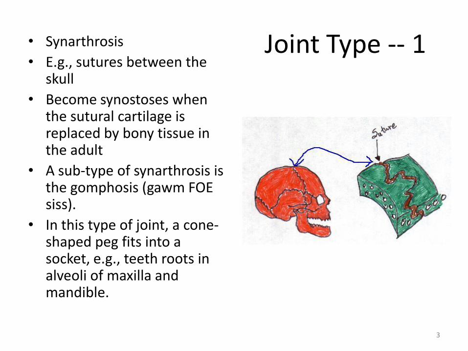
Joint Type -- 1 • Synarthrosis
• E.g., sutures between the skull
• Become synostoses when the sutural cartilage is replaced by bony tissue in the adult
• A sub-type of synarthrosis is the gomphosis (gawm FOE siss).
• In this type of joint, a cone-shaped peg fits into a socket, e.g., teeth roots in alveoli of maxilla and mandible.
4
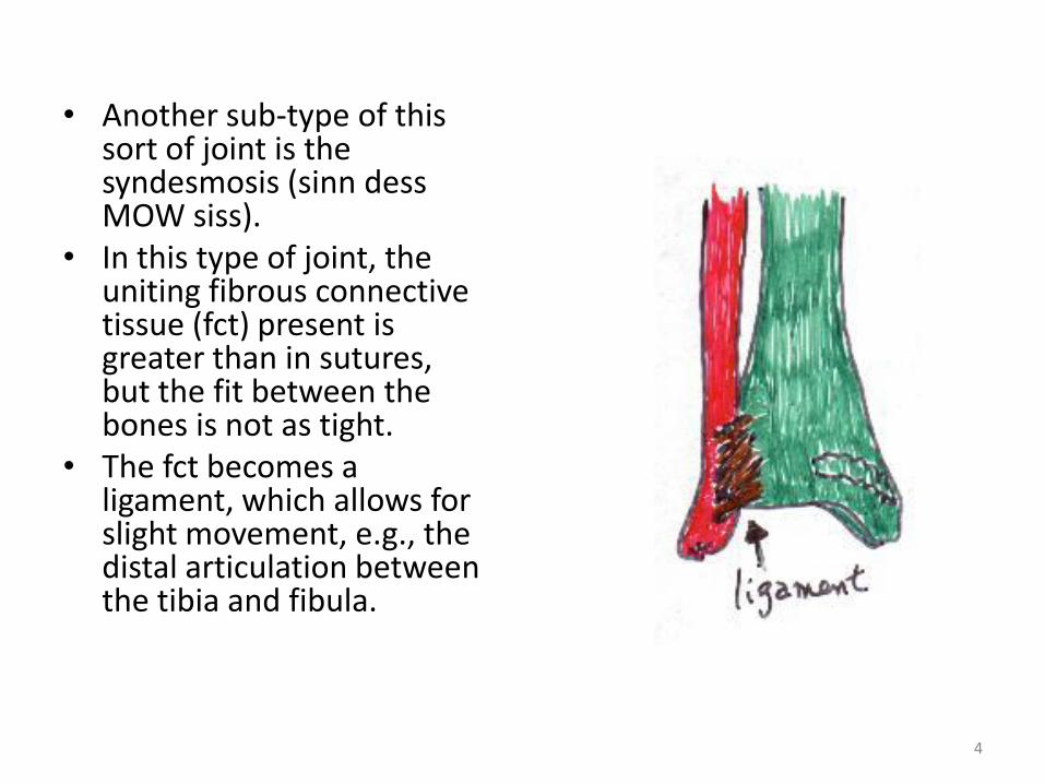
• Another sub-type of this sort of joint is the syndesmosis (sinn dess MOW siss).
• In this type of joint, the uniting fibrous connective tissue (fct) present is greater than in sutures, but the fit between the bones is not as tight.
• The fct becomes a ligament, which allows for slight movement, e.g., the distal articulation between the tibia and fibula.
5
Amphiarthrosis • In these joints, the articulating bones are tightly connected by cartilage and
have little to no movement.
• There are two sub-types that we're interested in: the synchondrosis (sinn konn DROW siss) and the symphysis (SIMM fuh siss).
• The former contains hyalin cartilage as the connective tissue.
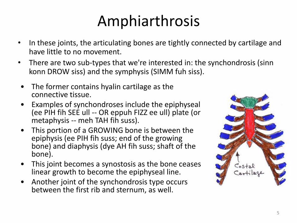
• Examples of synchondroses include the epiphyseal (ee PIH fih SEE ull -- OR eppuh FIZZ ee ull) plate (or metaphysis -- meh TAH fih suss).
• This portion of a GROWING bone is between the epiphysis (ee PIH fih suss; end of the growing bone) and diaphysis (dye AH fih suss; shaft of the bone).
• This joint becomes a synostosis as the bone ceases linear growth to become the epiphyseal line.
• Another joint of the synchondrosis type occurs between the first rib and sternum, as well.
6
Amphiarthrosis • In these joints, the articulating bones are tightly connected by cartilage and
have little to no movement.
• There are two sub-types that we're interested in: the synchondrosis (sinn konn DROW siss) and the symphysis (SIMM fuh siss).
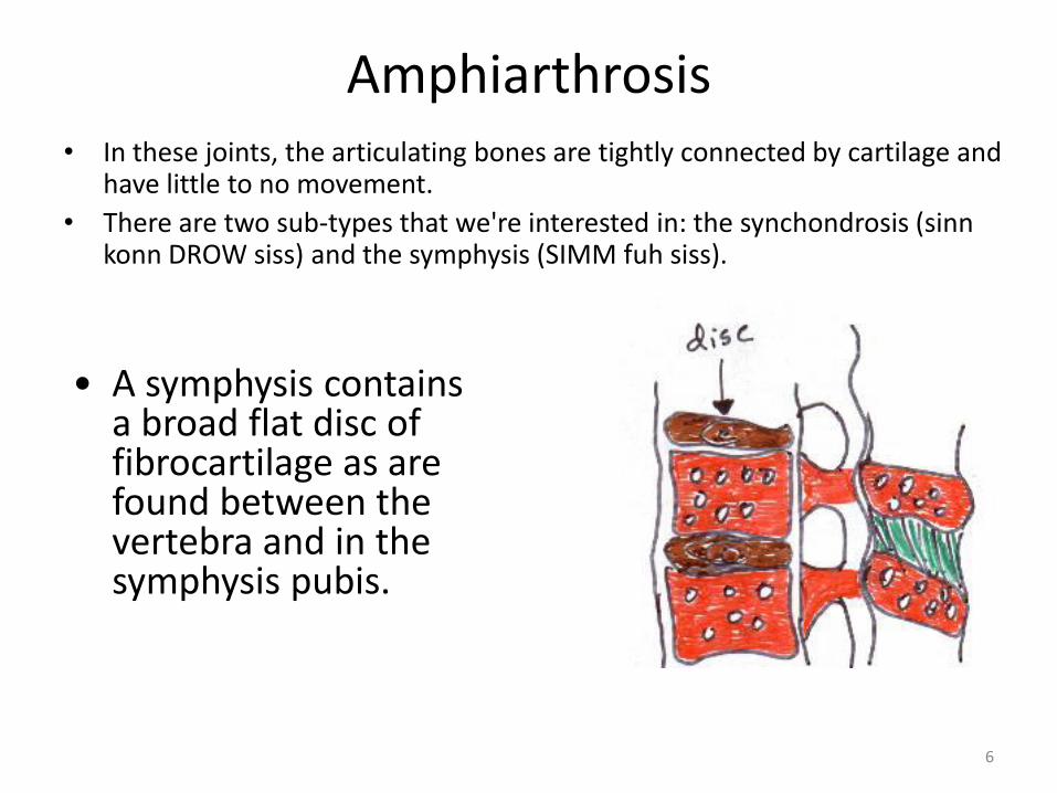
• A symphysis contains a broad flat disc of fibrocartilage as are found between the vertebra and in the symphysis pubis.
7
Synovial Joints, aka Diarthrotic Joints
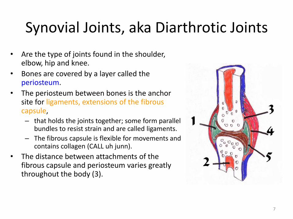
• Are the type of joints found in the shoulder, elbow, hip and knee.
• Bones are covered by a layer called the periosteum.
• The periosteum between bones is the anchor site for ligaments, extensions of the fibrous capsule, – that holds the joints together; some form parallel
bundles to resist strain and are called ligaments.
– The fibrous capsule is flexible for movements and contains collagen (CALL uh junn).
• The distance between attachments of the fibrous capsule and periosteum varies greatly throughout the body (3).
8
Synovial Joints, aka Diarthrotic Joints
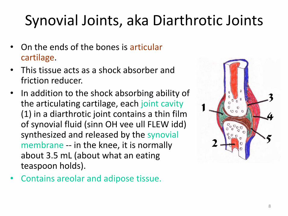
• On the ends of the bones is articular cartilage.
• This tissue acts as a shock absorber and friction reducer.
• In addition to the shock absorbing ability of the articulating cartilage, each joint cavity (1) in a diarthrotic joint contains a thin film of synovial fluid (sinn OH vee ull FLEW idd) synthesized and released by the synovial membrane -- in the knee, it is normally about 3.5 mL (about what an eating teaspoon holds).
• Contains areolar and adipose tissue.
9
Synovial Joints, aka Diarthrotic Joints
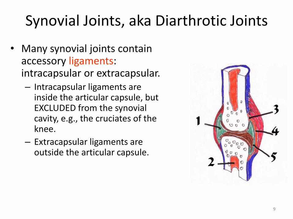
• Many synovial joints contain accessory ligaments: intracapsular or extracapsular. – Intracapsular ligaments are
inside the articular capsule, but EXCLUDED from the synovial cavity, e.g., the cruciates of the knee.
– Extracapsular ligaments are outside the articular capsule.
10
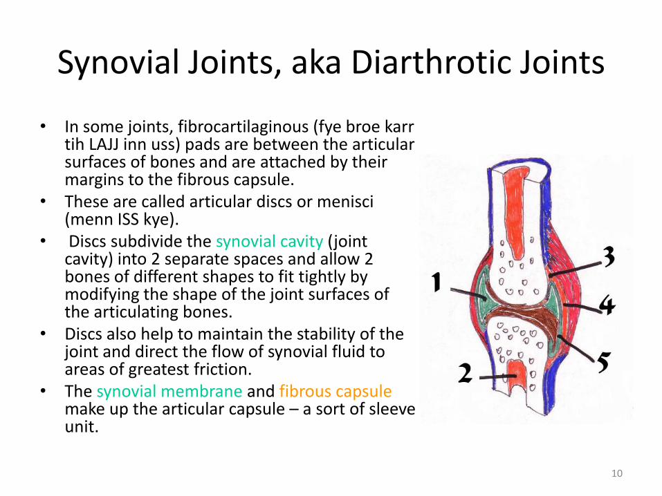
• In some joints, fibrocartilaginous (fye broe karr tih LAJJ inn uss) pads are between the articular surfaces of bones and are attached by their margins to the fibrous capsule.
• These are called articular discs or menisci (menn ISS kye).
• Discs subdivide the synovial cavity (joint cavity) into 2 separate spaces and allow 2 bones of different shapes to fit tightly by modifying the shape of the joint surfaces of the articulating bones.
• Discs also help to maintain the stability of the joint and direct the flow of synovial fluid to areas of greatest friction.
• The synovial membrane and fibrous capsule make up the articular capsule – a sort of sleeve unit.
Synovial Joints, aka Diarthrotic Joints
11
• The different movements create friction between the moving parts. • To decrease friction, sac-like structures called BURSAE (BRRRR suh) are situated in
body tissues. • They resemble joint capsules due to the fact that their walls consist of connective
tissue lined by a synovial membrane. • They are also filled with a fluid similar to synovial fluid. • The bursa are located between the skin and bone where skin rubs over bone. • They are also found between tendons and bones, muscle and bones and ligaments
and bones. • The bursa cushion the movement of one part of the body over the other.
Inflammation of the bursa causes bursitis. • In synovial joints, the articular surfaces are kept in contact with each other by
several factors: – 1) the fit of the articulating bones, e.g., hip (the femoral head and the acetabulum [ass
uh TABB you lumm]), – 2) the strength and tension (tautness) of the joint ligaments and – 3) arrangements and tension of the muscles around the joint.
Synovial Joints, aka Diarthrotic Joints
12
Movements
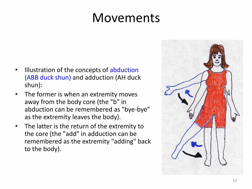
• Illustration of the concepts of abduction (ABB duck shun) and adduction (AH duck shun):
• The former is when an extremity moves away from the body core (the "b" in abduction can be remembered as "bye-bye" as the extremity leaves the body).
• The latter is the return of the extremity to the core (the "add" in adduction can be remembered as the extremity "adding" back to the body).
13
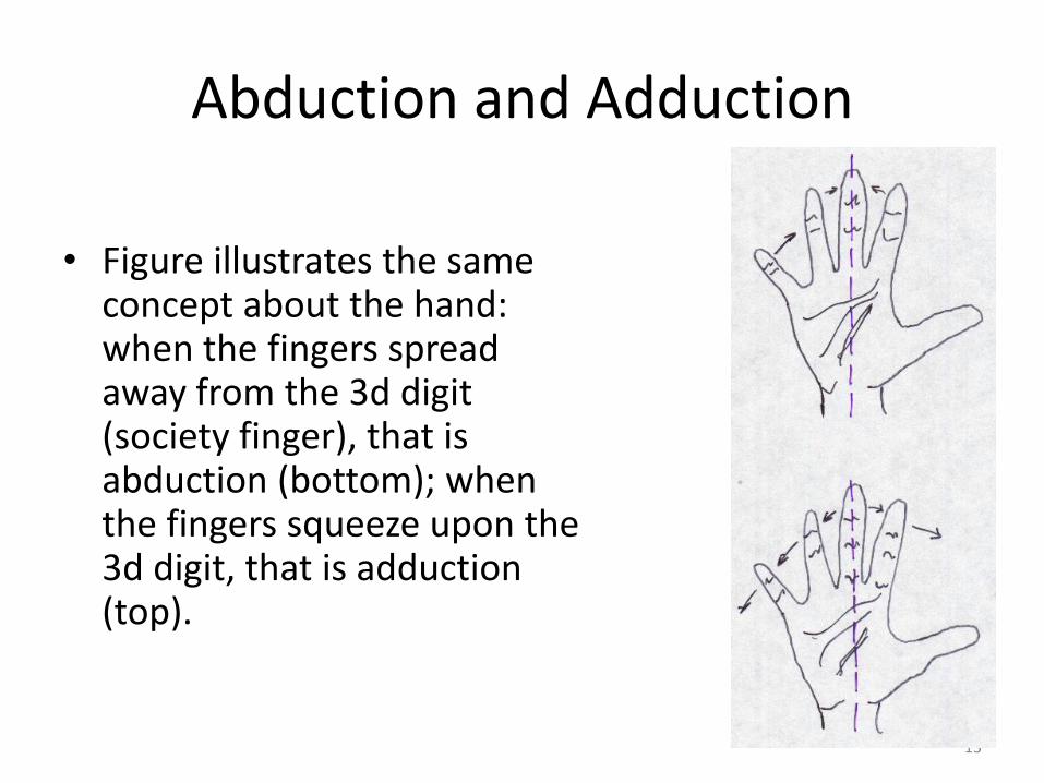
Abduction and Adduction
• Figure illustrates the same concept about the hand: when the fingers spread away from the 3d digit (society finger), that is abduction (bottom); when the fingers squeeze upon the 3d digit, that is adduction (top).
14
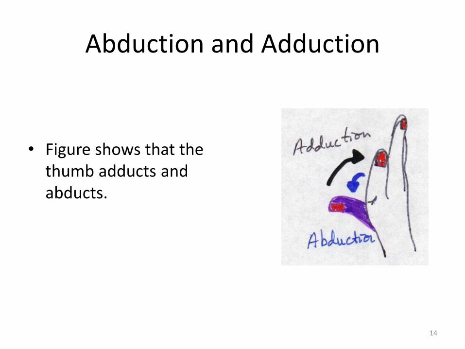
• Figure shows that the thumb adducts and abducts.
Abduction and Adduction
15
Flexion and Extension
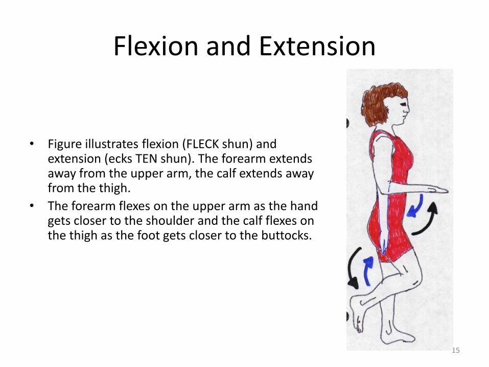
• Figure illustrates flexion (FLECK shun) and extension (ecks TEN shun). The forearm extends away from the upper arm, the calf extends away from the thigh.
• The forearm flexes on the upper arm as the hand gets closer to the shoulder and the calf flexes on the thigh as the foot gets closer to the buttocks.
16
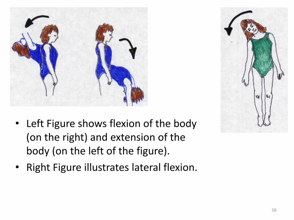
• Left Figure shows flexion of the body (on the right) and extension of the body (on the left of the figure).
• Right Figure illustrates lateral flexion.
17
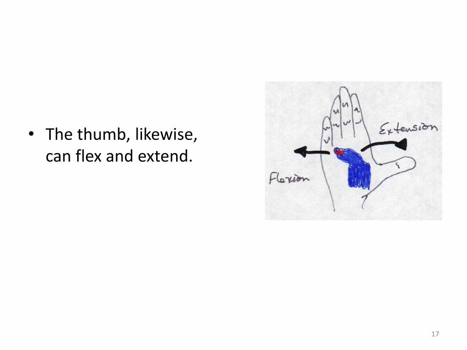
• The thumb, likewise, can flex and extend.
18
Supination and Pronation
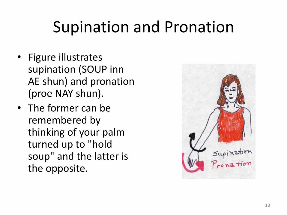
• Figure illustrates supination (SOUP inn AE shun) and pronation (proe NAY shun).
• The former can be remembered by thinking of your palm turned up to "hold soup" and the latter is the opposite.
19
Medial and Lateral Rotation
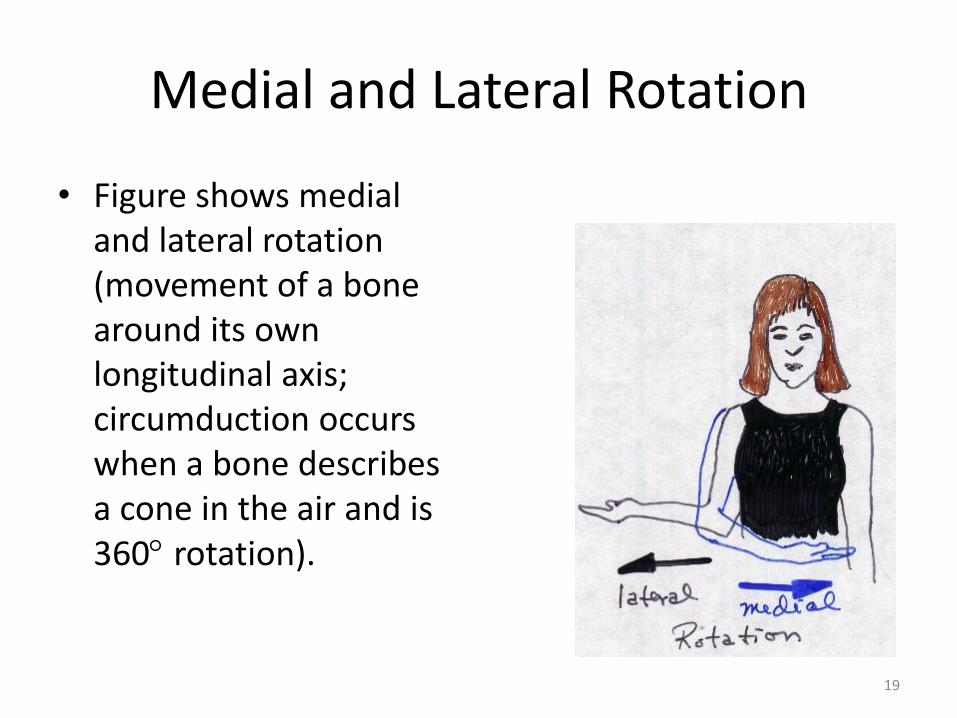
• Figure shows medial and lateral rotation (movement of a bone around its own longitudinal axis; circumduction occurs when a bone describes a cone in the air and is 360 rotation).
20
Feet Movements
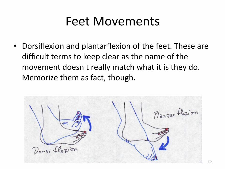
• Dorsiflexion and plantarflexion of the feet. These are difficult terms to keep clear as the name of the movement doesn't really match what it is they do. Memorize them as fact, though.
21
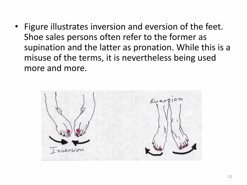
• Figure illustrates inversion and eversion of the feet. Shoe sales persons often refer to the former as supination and the latter as pronation. While this is a misuse of the terms, it is nevertheless being used more and more.
22
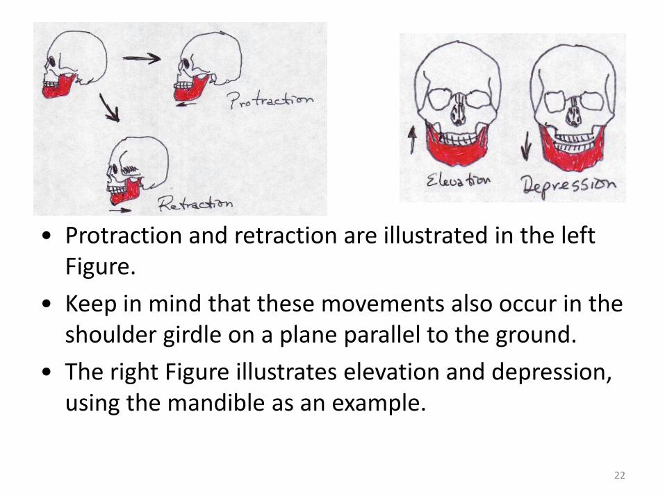
• Protraction and retraction are illustrated in the left Figure.
• Keep in mind that these movements also occur in the shoulder girdle on a plane parallel to the ground.
• The right Figure illustrates elevation and depression, using the mandible as an example.
23
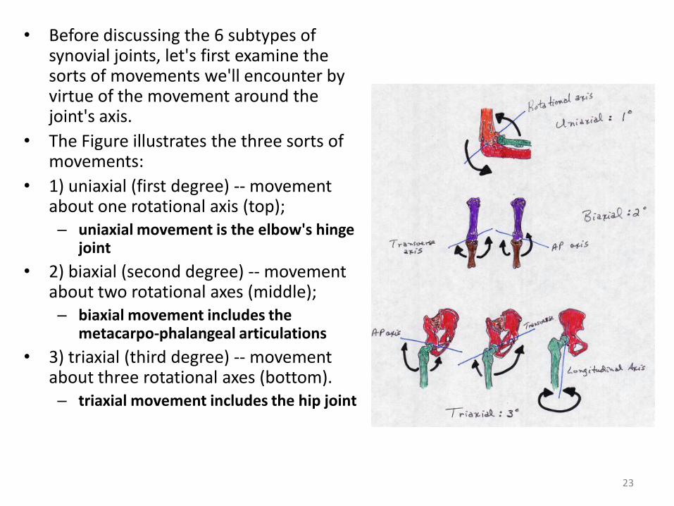
• Before discussing the 6 subtypes of synovial joints, let's first examine the sorts of movements we'll encounter by virtue of the movement around the joint's axis.
• The Figure illustrates the three sorts of movements:
• 1) uniaxial (first degree) -- movement about one rotational axis (top); – uniaxial movement is the elbow's hinge
joint
• 2) biaxial (second degree) -- movement about two rotational axes (middle); – biaxial movement includes the
metacarpo-phalangeal articulations
• 3) triaxial (third degree) -- movement about three rotational axes (bottom). – triaxial movement includes the hip joint
24
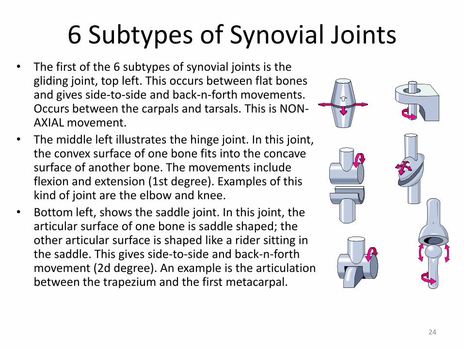
6 Subtypes of Synovial Joints • The first of the 6 subtypes of synovial joints is the
gliding joint, top left. This occurs between flat bones and gives side-to-side and back-n-forth movements. Occurs between the carpals and tarsals. This is NON-AXIAL movement.
• The middle left illustrates the hinge joint. In this joint, the convex surface of one bone fits into the concave surface of another bone. The movements include flexion and extension (1st degree). Examples of this kind of joint are the elbow and knee.
• Bottom left, shows the saddle joint. In this joint, the articular surface of one bone is saddle shaped; the other articular surface is shaped like a rider sitting in the saddle. This gives side-to-side and back-n-forth movement (2d degree). An example is the articulation between the trapezium and the first metacarpal.
25
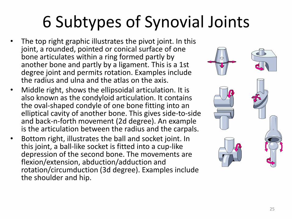
6 Subtypes of Synovial Joints • The top right graphic illustrates the pivot joint. In this
joint, a rounded, pointed or conical surface of one bone articulates within a ring formed partly by another bone and partly by a ligament. This is a 1st degree joint and permits rotation. Examples include the radius and ulna and the atlas on the axis.
• Middle right, shows the ellipsoidal articulation. It is also known as the condyloid articulation. It contains the oval-shaped condyle of one bone fitting into an elliptical cavity of another bone. This gives side-to-side and back-n-forth movement (2d degree). An example is the articulation between the radius and the carpals.
• Bottom right, illustrates the ball and socket joint. In this joint, a ball-like socket is fitted into a cup-like depression of the second bone. The movements are flexion/extension, abduction/adduction and rotation/circumduction (3d degree). Examples include the shoulder and hip.
26
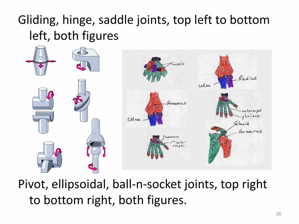
Gliding, hinge, saddle joints, top left to bottom left, both figures
Pivot, ellipsoidal, ball-n-socket joints, top right to bottom right, both figures.
27
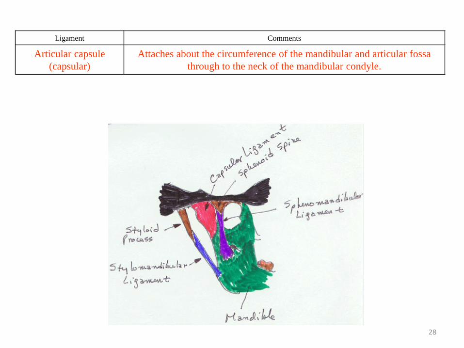
Tempero-Mandibular Joint (TMJ)
• Consists of the temporal bone and mandible bilaterally
28
Ligament Comments
Articular capsule
(capsular)
Attaches about the circumference of the mandibular and articular fossa
through to the neck of the mandibular condyle.
29
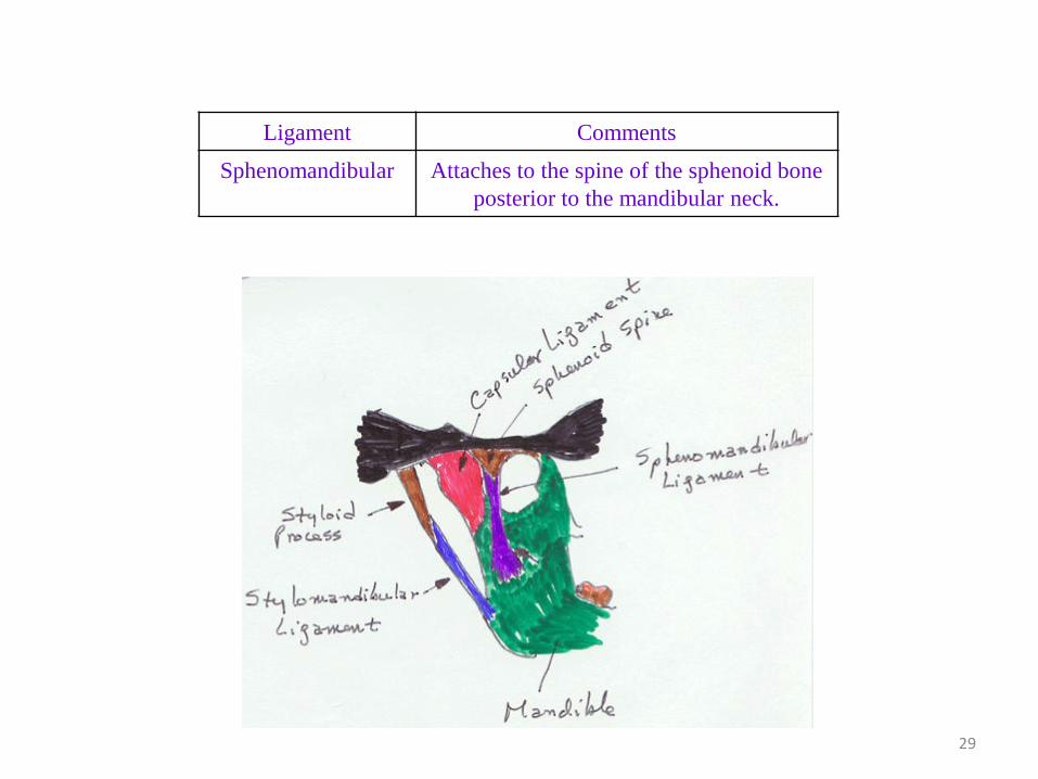
Ligament Comments
Sphenomandibular Attaches to the spine of the sphenoid bone
posterior to the mandibular neck.
30
Ligament Comments
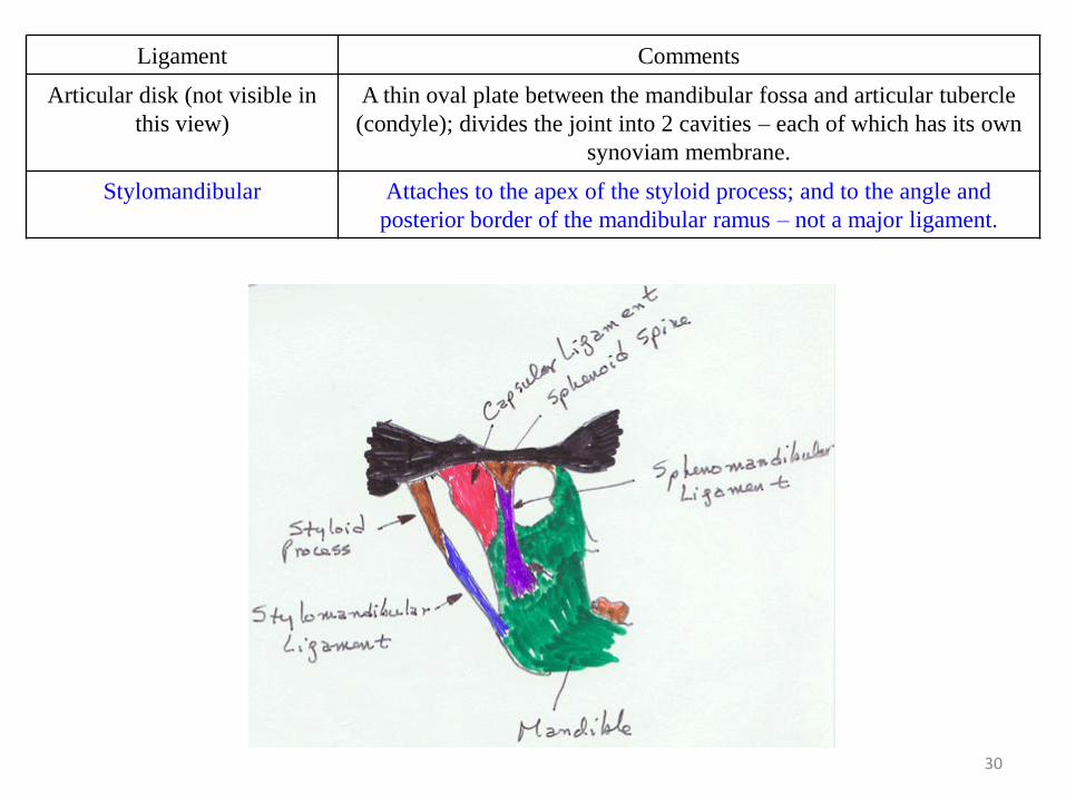
Articular disk (not visible in
this view)
A thin oval plate between the mandibular fossa and articular tubercle
(condyle); divides the joint into 2 cavities – each of which has its own
synoviam membrane.
Stylomandibular Attaches to the apex of the styloid process; and to the angle and
posterior border of the mandibular ramus – not a major ligament.
31
Ligament Comments
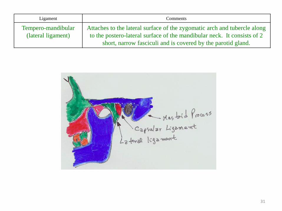
Tempero-mandibular
(lateral ligament)
Attaches to the lateral surface of the zygomatic arch and tubercle along
to the postero-lateral surface of the mandibular neck. It consists of 2
short, narrow fasciculi and is covered by the parotid gland.
32
Sternocostal Articulation
• Consists of articulations between the sternum and ribs
33
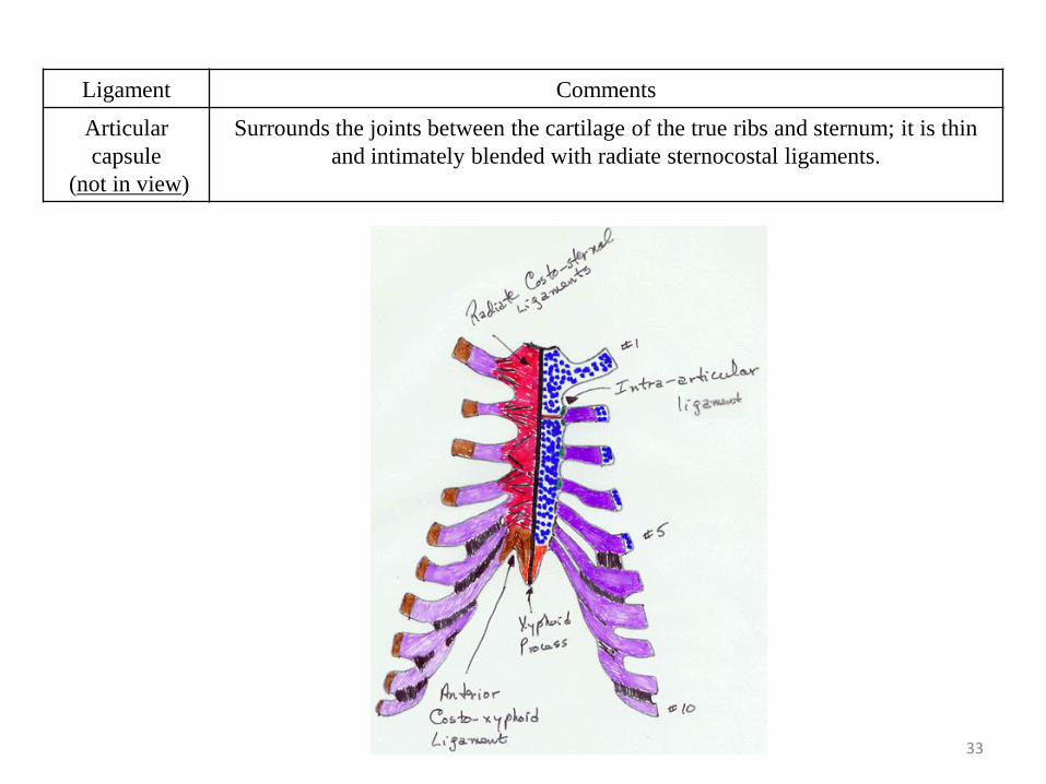
Ligament Comments
Articular
capsule
(not in view)
Surrounds the joints between the cartilage of the true ribs and sternum; it is thin
and intimately blended with radiate sternocostal ligaments.

34
Ligament Comments
Radiate sternocostal Attaches to the ventral and dorsal surfaces of the sternal ends of the
cartilages of the true ribs across to the anterior/posterior surface of the
sternum; has three fasciculi: superior (fibers ascend abliquely), middle
(fibers run horizontally) and inferior (fibers descend obliquely).
35
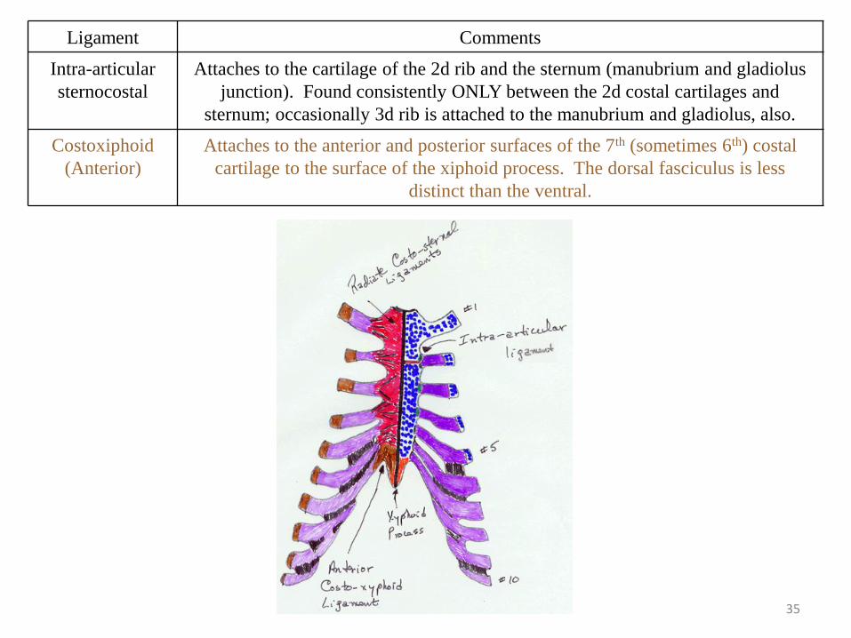
Ligament Comments
Intra-articular
sternocostal
Attaches to the cartilage of the 2d rib and the sternum (manubrium and gladiolus
junction). Found consistently ONLY between the 2d costal cartilages and
sternum; occasionally 3d rib is attached to the manubrium and gladiolus, also.
Costoxiphoid
(Anterior)
Attaches to the anterior and posterior surfaces of the 7th (sometimes 6th) costal
cartilage to the surface of the xiphoid process. The dorsal fasciculus is less
distinct than the ventral.
36
The Shoulder Joint
• The shoulder joint consists of three bones: the clavicle, the scapula and the humerus.
• The shoulder joint consists of three regions: – the acromioclavicular,
– the non-acromioclavicular and
– the shoulder, proper.
• These regions contain the following ligaments:
37
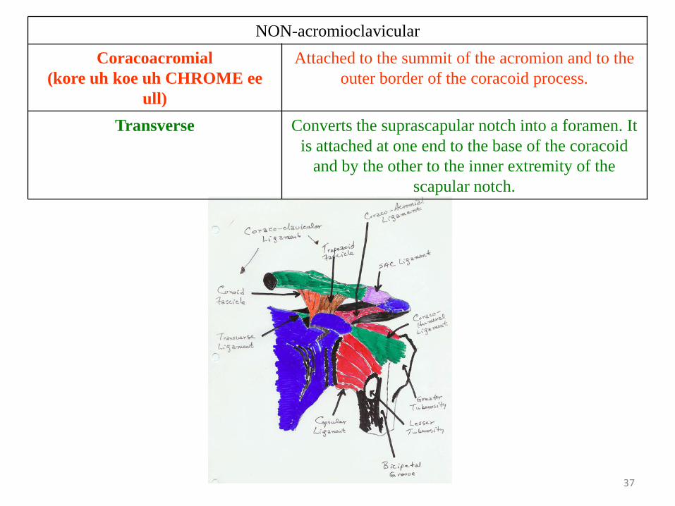
NON-acromioclavicular
Coracoacromial
(kore uh koe uh CHROME ee
ull)
Attached to the summit of the acromion and to the
outer border of the coracoid process.
Transverse Converts the suprascapular notch into a foramen. It
is attached at one end to the base of the coracoid
and by the other to the inner extremity of the
scapular notch.
38
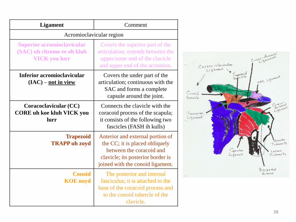
Ligament Comment
Acromioclavicular region
Superior acromioclavicular
(SAC) uh chrome ee oh kluh
VICK you lurr
Covers the superior part of the
articulation; extends between the
upper/outer end of the clavicle
and upper end of the acromion.
Inferior acromioclavicular
(IAC) – not in view
Covers the under part of the
articulation; continuous with the
SAC and forms a complete
capsule around the joint.
Coracoclavicular (CC)
CORE uh koe kluh VICK you
lurr
Connects the clavicle with the
coracoid process of the scapula;
it consists of the following two
fascicles (FASH ih kulls)
Trapezoid
TRAPP uh zoyd
Anterior and external portion of
the CC; it is placed obliquely
between the coracoid and
clavicle; its posterior border is
joined with the conoid ligament.
Conoid
KOE noyd
The posterior and internal
fasciculus; it is attached to the
base of the coracoid process and
to the conoid tubercle of the
clavicle.
39
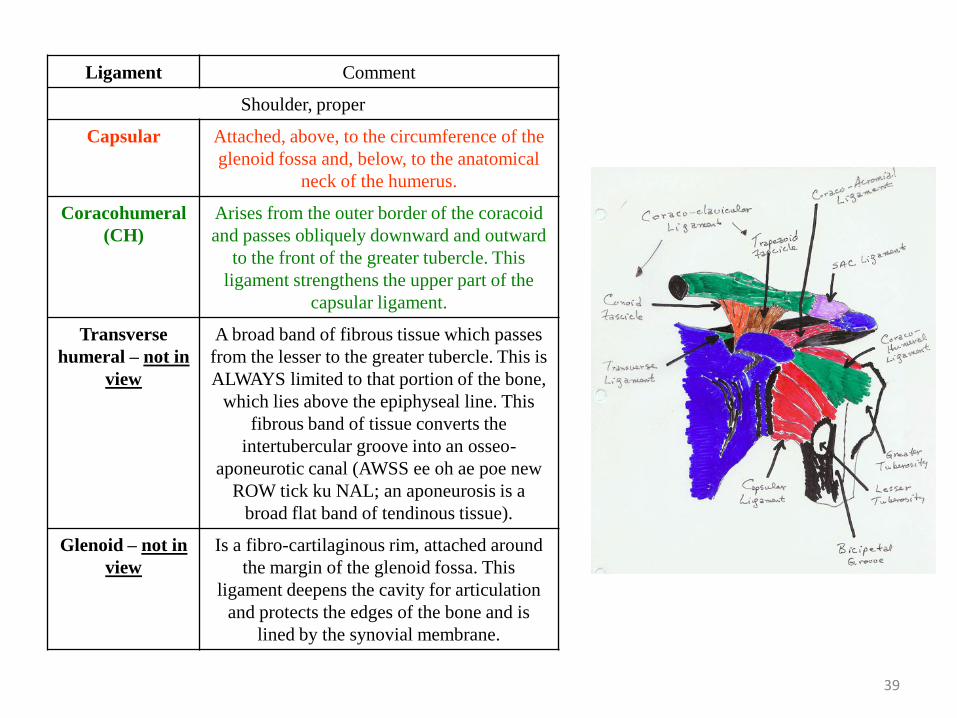
Ligament Comment
Shoulder, proper
Capsular Attached, above, to the circumference of the
glenoid fossa and, below, to the anatomical
neck of the humerus.
Coracohumeral
(CH)
Arises from the outer border of the coracoid
and passes obliquely downward and outward
to the front of the greater tubercle. This
ligament strengthens the upper part of the
capsular ligament.
Transverse
humeral – not in
view
A broad band of fibrous tissue which passes
from the lesser to the greater tubercle. This is
ALWAYS limited to that portion of the bone,
which lies above the epiphyseal line. This
fibrous band of tissue converts the
intertubercular groove into an osseo-
aponeurotic canal (AWSS ee oh ae poe new
ROW tick ku NAL; an aponeurosis is a
broad flat band of tendinous tissue).
Glenoid – not in
view
Is a fibro-cartilaginous rim, attached around
the margin of the glenoid fossa. This
ligament deepens the cavity for articulation
and protects the edges of the bone and is
lined by the synovial membrane.
40
The Elbow
• The elbow consists of three bones:
– the humerus,
– the radius and
– the ulna.
• Four ligaments are of significance, here:
41
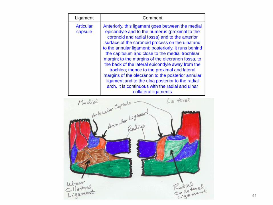
Ligament Comment
Articular
capsule
Anteriorly, this ligament goes between the medial
epicondyle and to the humerus (proximal to the
coronoid and radial fossa) and to the anterior
surface of the coronoid process on the ulna and
to the annular ligament; posteriorly, it runs behind
the capitulum and close to the medial trochlear
margin; to the margins of the olecranon fossa, to
the back of the lateral epicondyle away from the
trochlea; thence to the proximal and lateral
margins of the olecranon to the posterior annular
ligament and to the ulna posterior to the radial
arch. It is continuous with the radial and ulnar
collateral ligaments
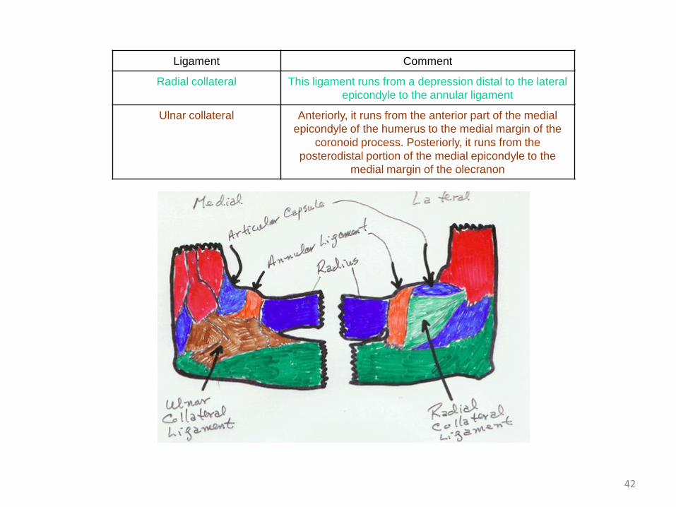
42
Ligament Comment
Radial collateral This ligament runs from a depression distal to the lateral
epicondyle to the annular ligament
Ulnar collateral Anteriorly, it runs from the anterior part of the medial
epicondyle of the humerus to the medial margin of the
coronoid process. Posteriorly, it runs from the
posterodistal portion of the medial epicondyle to the
medial margin of the olecranon
43
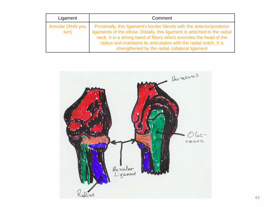
Ligament Comment
Annular (ANN you
lurr)
Proximally, this ligament's border blends with the anterior/posterior
ligaments of the elbow. Distally, this ligament is attached to the radial
neck. It is a strong band of fibers which encircles the head of the
radius and maintains its articulation with the radial notch. It is
strengthened by the radial collateral ligament
44
The Wrist
• Consists of the
– radius,
– Ulna and
– carpal bones
45
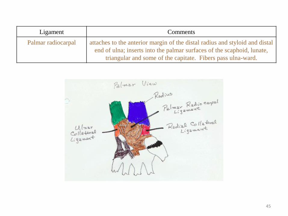
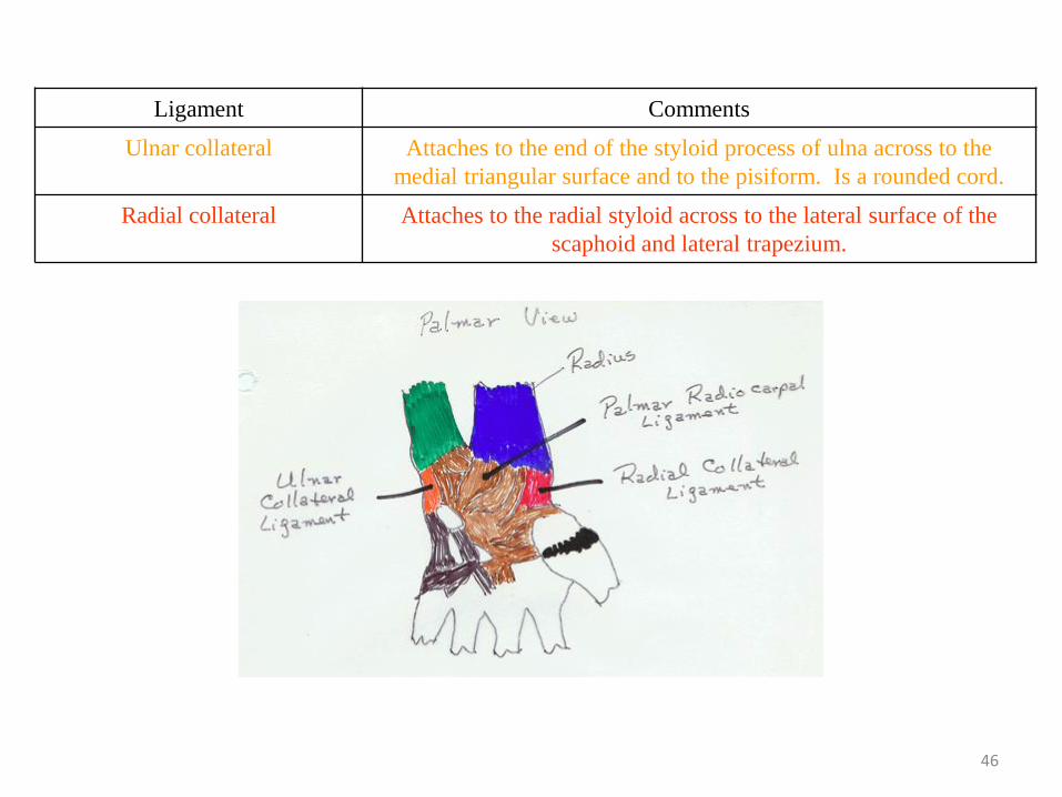
Ligament Comments
Palmar radiocarpal attaches to the anterior margin of the distal radius and styloid and distal
end of ulna; inserts into the palmar surfaces of the scaphoid, lunate,
triangular and some of the capitate. Fibers pass ulna-ward.
46
Ligament Comments
Ulnar collateral Attaches to the end of the styloid process of ulna across to the
medial triangular surface and to the pisiform. Is a rounded cord.
Radial collateral Attaches to the radial styloid across to the lateral surface of the
scaphoid and lateral trapezium.
47
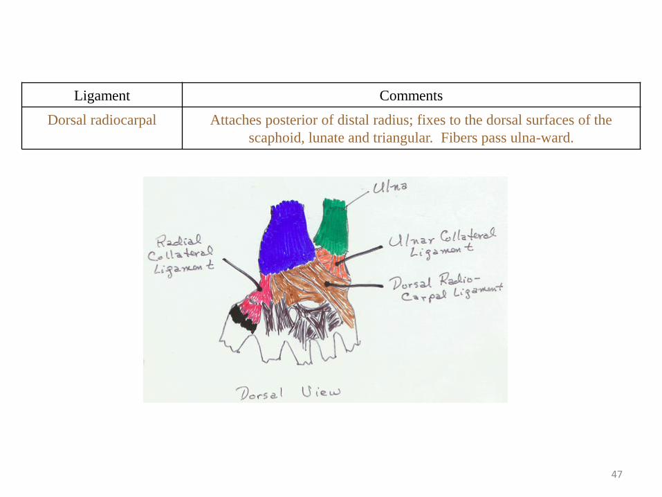
Ligament Comments
Dorsal radiocarpal Attaches posterior of distal radius; fixes to the dorsal surfaces of the
scaphoid, lunate and triangular. Fibers pass ulna-ward.
48
The Hip
• The hip joint consists of 2-4 bones, depending on how you look at it.
• For two bones, – it consists of the femur and an os coxae;
• for 4 bones (during bone development and growth), – it consists of the femur and three bones that make up an
immature os coxae: the ilium (ILL ee umm; upper flared part), the ischium (ISH ee um; lower part upon which you are sitting right now) and the os pubis (PYEW biss; the bone that makes up the front of the pelvis).
49
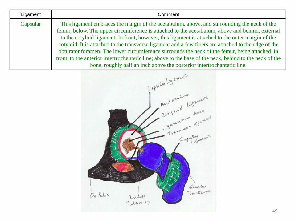
Ligament Comment
Capsular This ligament embraces the margin of the acetabulum, above, and surrounding the neck of the
femur, below. The upper circumference is attached to the acetabulum, above and behind, external
to the cotyloid ligament. In front, however, this ligament is attached to the outer margin of the
cotyloid. It is attached to the transverse ligament and a few fibers are attached to the edge of the
obturator foramen. The lower circumference surrounds the neck of the femur, being attached, in
front, to the anterior intertrochanteric line; above to the base of the neck, behind to the neck of the
bone, roughly half an inch above the posterior intertrochanteric line.
50
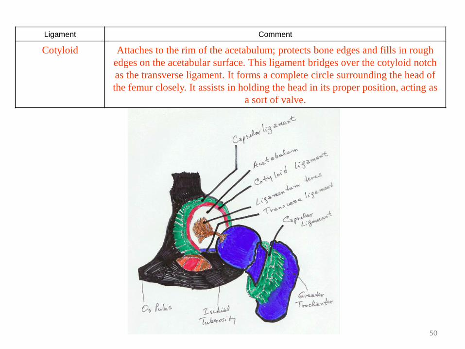
Ligament Comment
Cotyloid Attaches to the rim of the acetabulum; protects bone edges and fills in rough
edges on the acetabular surface. This ligament bridges over the cotyloid notch
as the transverse ligament. It forms a complete circle surrounding the head of
the femur closely. It assists in holding the head in its proper position, acting as
a sort of valve.
51
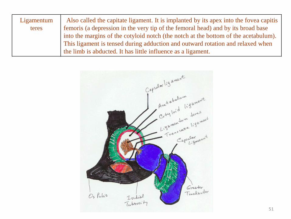
Ligamentum
teres
Also called the capitate ligament. It is implanted by its apex into the fovea capitis
femoris (a depression in the very tip of the femoral head) and by its broad base
into the margins of the cotyloid notch (the notch at the bottom of the acetabulum).
This ligament is tensed during adduction and outward rotation and relaxed when
the limb is abducted. It has little influence as a ligament.
52
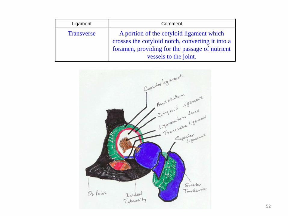
Ligament Comment
Transverse A portion of the cotyloid ligament which
crosses the cotyloid notch, converting it into a
foramen, providing for the passage of nutrient
vessels to the joint.
53
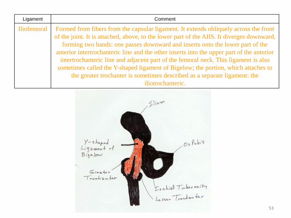
Ligament Comment
Iliofemoral Formed from fibers from the capsular ligament. It extends obliquely across the front
of the joint. It is attached, above, to the lower part of the AIIS. It diverges downward,
forming two bands: one passes downward and inserts onto the lower part of the
anterior intertrochanteric line and the other inserts into the upper part of the anterior
intertrochanteric line and adjacent part of the femoral neck. This ligament is also
sometimes called the Y-shaped ligament of Bigelow; the portion, which attaches to
the greater trochanter is sometimes described as a separate ligament: the
iliotrochanteric.
54
The Knee
• The knee is the most unstable joint in the body.
• It consists of 4 bones: – the femur,
– the patella (kneecap),
– the tibia and
– the fibula.
• The ligaments of significance are tabulated, below:
55
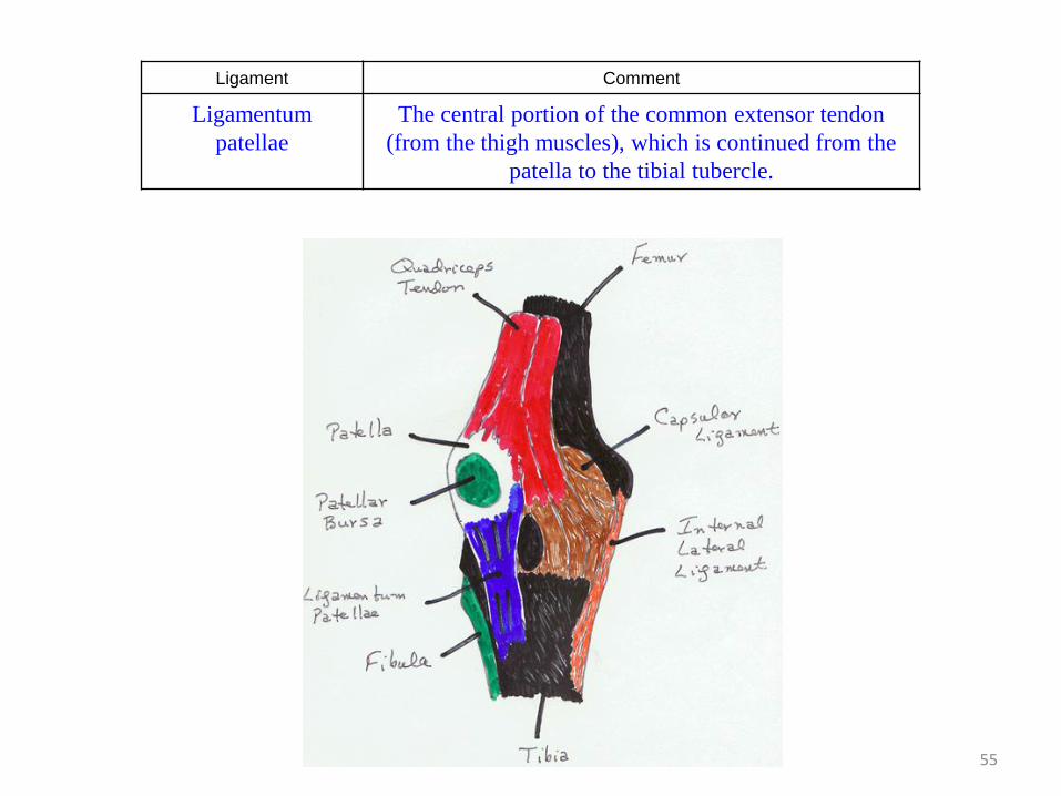
Ligament Comment
Ligamentum
patellae
The central portion of the common extensor tendon
(from the thigh muscles), which is continued from the
patella to the tibial tubercle.
56
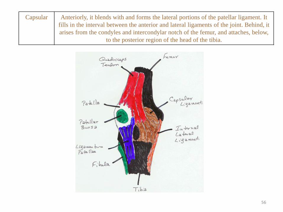
Capsular Anteriorly, it blends with and forms the lateral portions of the patellar ligament. It
fills in the interval between the anterior and lateral ligaments of the joint. Behind, it
arises from the condyles and intercondylar notch of the femur, and attaches, below,
to the posterior region of the head of the tibia.
57
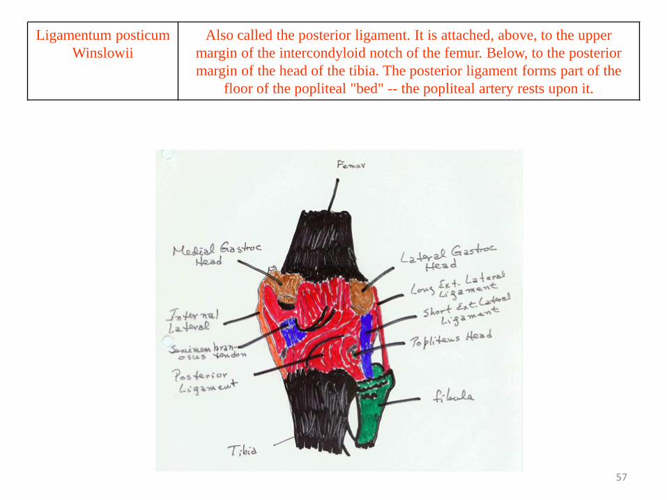
Ligamentum posticum
Winslowii
Also called the posterior ligament. It is attached, above, to the upper
margin of the intercondyloid notch of the femur. Below, to the posterior
margin of the head of the tibia. The posterior ligament forms part of the
floor of the popliteal "bed" -- the popliteal artery rests upon it.
58
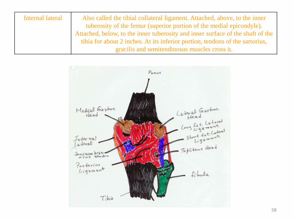
Internal lateral Also called the tibial collateral ligament. Attached, above, to the inner
tuberosity of the femur (superior portion of the medial epicondyle).
Attached, below, to the inner tuberosity and inner surface of the shaft of the
tibia for about 2 inches. At its inferior portion, tendons of the sartorius,
gracilis and semitendinosus muscles cross it.
59
Ligament Comment
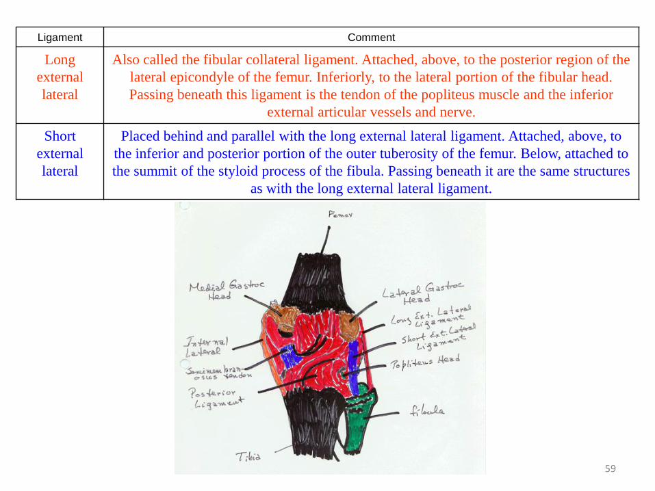
Long
external
lateral
Also called the fibular collateral ligament. Attached, above, to the posterior region of the
lateral epicondyle of the femur. Inferiorly, to the lateral portion of the fibular head.
Passing beneath this ligament is the tendon of the popliteus muscle and the inferior
external articular vessels and nerve.
Short
external
lateral
Placed behind and parallel with the long external lateral ligament. Attached, above, to
the inferior and posterior portion of the outer tuberosity of the femur. Below, attached to
the summit of the styloid process of the fibula. Passing beneath it are the same structures
as with the long external lateral ligament.
60
CRUCIATES
• (KROO shee utts): there are two
– Anterior
– Posterior
61
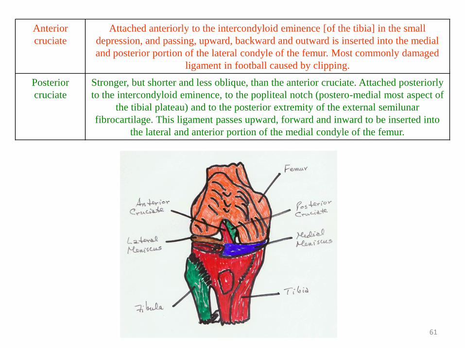
Anterior
cruciate
Attached anteriorly to the intercondyloid eminence [of the tibia] in the small
depression, and passing, upward, backward and outward is inserted into the medial
and posterior portion of the lateral condyle of the femur. Most commonly damaged
ligament in football caused by clipping.
Posterior
cruciate
Stronger, but shorter and less oblique, than the anterior cruciate. Attached posteriorly
to the intercondyloid eminence, to the popliteal notch (postero-medial most aspect of
the tibial plateau) and to the posterior extremity of the external semilunar
fibrocartilage. This ligament passes upward, forward and inward to be inserted into
the lateral and anterior portion of the medial condyle of the femur.
62
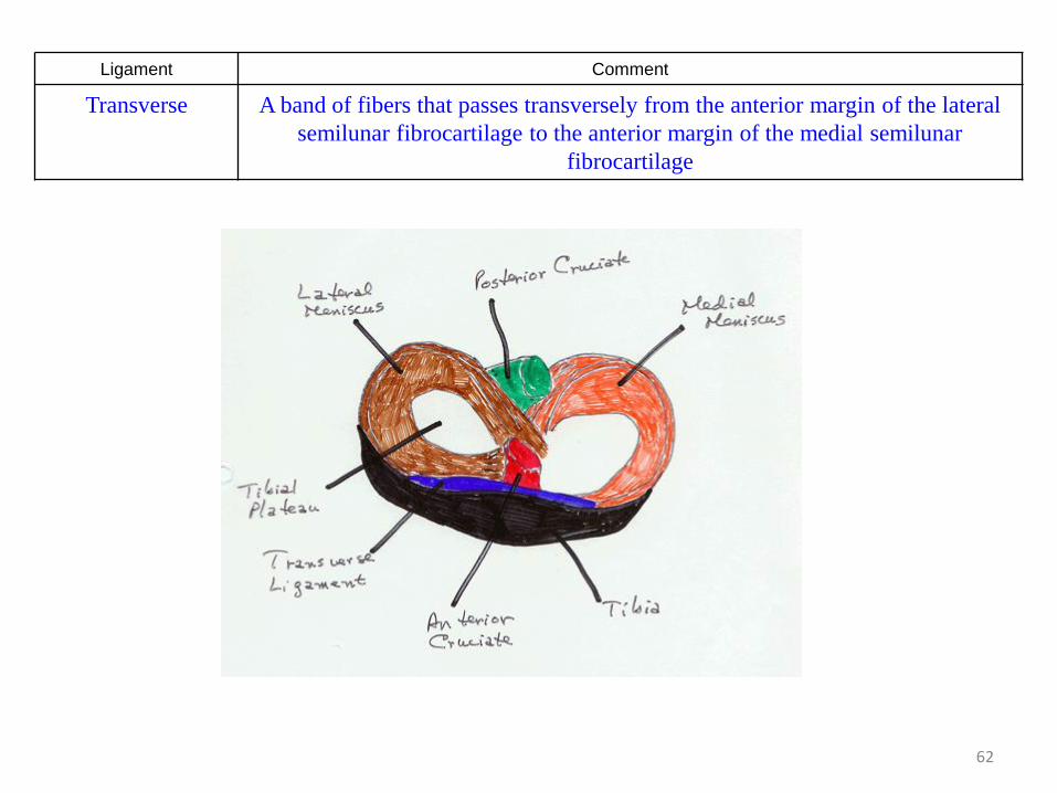
Ligament Comment
Transverse A band of fibers that passes transversely from the anterior margin of the lateral
semilunar fibrocartilage to the anterior margin of the medial semilunar
fibrocartilage
63
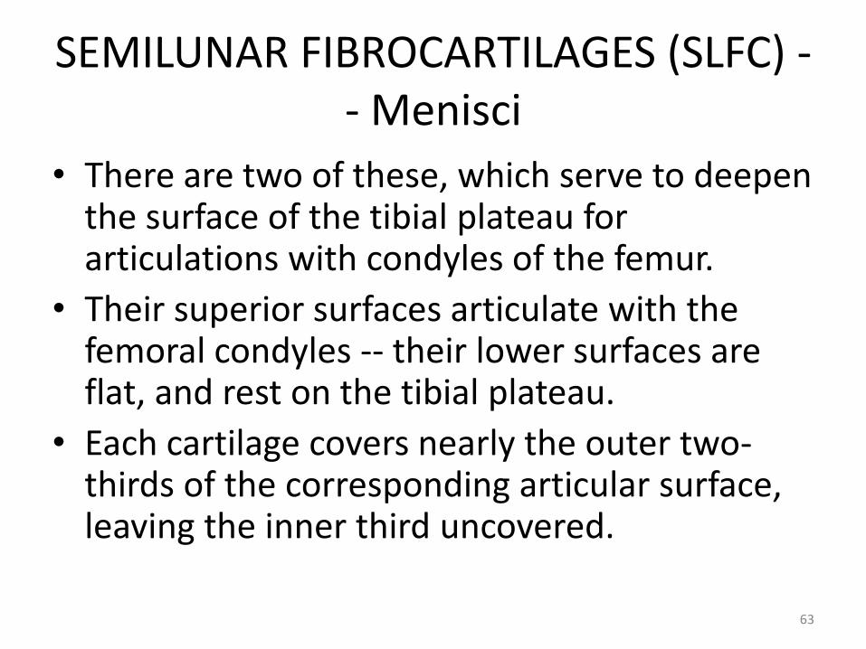
SEMILUNAR FIBROCARTILAGES (SLFC) -- Menisci
• There are two of these, which serve to deepen the surface of the tibial plateau for articulations with condyles of the femur.
• Their superior surfaces articulate with the femoral condyles -- their lower surfaces are flat, and rest on the tibial plateau.
• Each cartilage covers nearly the outer two-thirds of the corresponding articular surface, leaving the inner third uncovered.
64
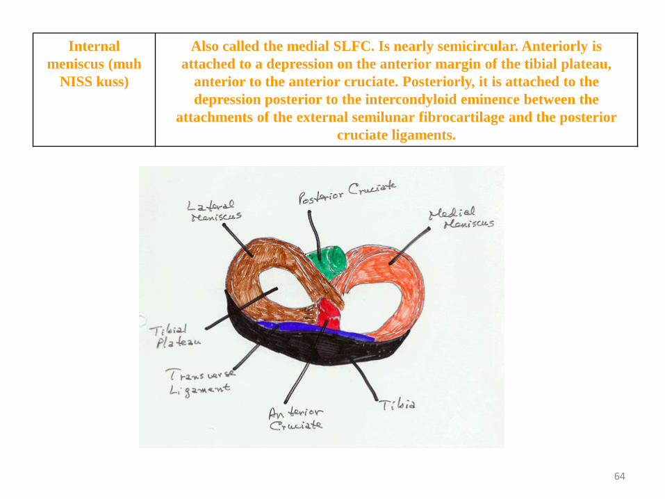
Internal
meniscus (muh
NISS kuss)
Also called the medial SLFC. Is nearly semicircular. Anteriorly is
attached to a depression on the anterior margin of the tibial plateau,
anterior to the anterior cruciate. Posteriorly, it is attached to the
depression posterior to the intercondyloid eminence between the
attachments of the external semilunar fibrocartilage and the posterior
cruciate ligaments.
65
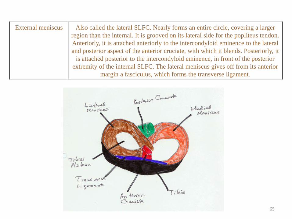
External meniscus Also called the lateral SLFC. Nearly forms an entire circle, covering a larger
region than the internal. It is grooved on its lateral side for the popliteus tendon.
Anteriorly, it is attached anteriorly to the intercondyloid eminence to the lateral
and posterior aspect of the anterior cruciate, with which it blends. Posteriorly, it
is attached posterior to the intercondyloid eminence, in front of the posterior
extremity of the internal SLFC. The lateral meniscus gives off from its anterior
margin a fasciculus, which forms the transverse ligament.
66
The Ankle
• Consists of the
– tibia,
– fibula and
– tarsal bones
67
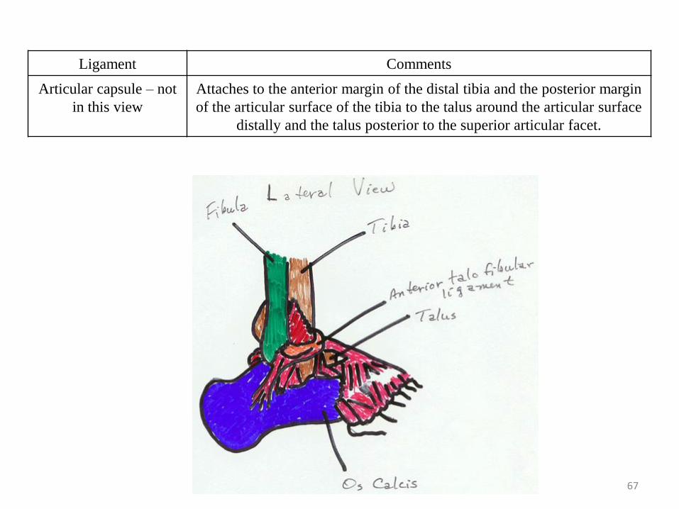
Ligament Comments
Articular capsule – not
in this view
Attaches to the anterior margin of the distal tibia and the posterior margin
of the articular surface of the tibia to the talus around the articular surface
distally and the talus posterior to the superior articular facet.
68
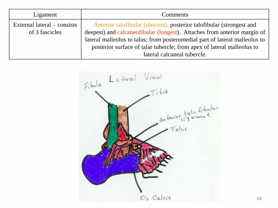
Ligament Comments
External lateral – consists
of 3 fascicles
Anterior talofibular (shortest), posterior talofibular (strongest and
deepest) and calcaneofibular (longest). Attaches from anterior margin of
lateral malleolus to talus; from posteromedial part of lateral malleolus to
posterior surface of talar tubercle; from apex of lateral malleolus to
lateral calcaneal tubercle.
69
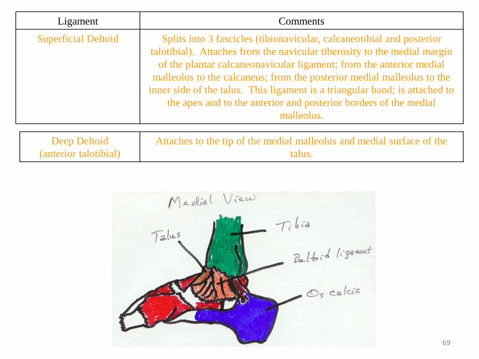
Ligament Comments
Superficial Deltoid Splits into 3 fascicles (tibionavicular, calcaneotibial and posterior
talotibial). Attaches from the navicular tiberosity to the medial margin
of the plantar calcaneonavicular ligament; from the anterior medial
malleolus to the calcaneus; from the posterior medial malleolus to the
inner side of the talus. This ligament is a triangular band; is attached to
the apex and to the anterior and posterior borders of the medial
malleolus.
Deep Deltoid
(anterior talotibial)
Attaches to the tip of the medial malleolus and medial surface of the
talus.