Sorting Out the p63 Signaling Network -...
13
COMMENTARY See related article on pg 1676 Sorting Out the p63 Signaling Network Maranke I. Koster 1 and Dennis R. Roop 1 p63 is a transcription factor required for normal epidermal development and differentiation. Because of the complexity of these processes, p63 is expected to regulate a myriad of target genes, providing impetus to many laboratories to identify these genes. p63 target genes have been shown to encode a diverse group of proteins, including structural proteins, proteins that control cell cycle withdrawal, and proteins that regulate the epidermal differentiation program. In this issue, Antonini et al. describe a novel p63 target gene whose evolutionary conservation suggests a critical role for this gene in the epidermis. Journal of Investigative Dermatology (2008), 128, 1617–1619. doi:10.1038/jid.2008.149 Epidermal morphogenesis begins dur- ing embryonic development when cells of the single-layered surface ectoderm, which initially covers the developing embryo, adopt an epider- mal fate and differentiate into active- ly proliferating basal keratinocytes (reviewed in Koster and Roop, 2007). The first morphological sign of stratifi- cation occurs when these basal kera- tinocytes give rise to a suprabasal cell layer. Cells in this so-called interme- diate layer undergo a few rounds of cell division before they permanently withdraw from the cell cycle and become spinous keratinocytes. During subsequent stages of epidermal mor- phogenesis, spinous keratinocytes differentiate into cells of the granular and cornified cell layers, the latter of which are primarily responsible for the barrier function of the epidermis. The end point of the terminal kerati- nocyte differentiation program is the shedding of the dead and enucleated keratinocytes of the cornified cell layers into the environment. The embryonic differentiation program described above is maintained, with few modifications, during postnatal development. This program is essential to the continuous replacement of cells that are lost and to the maintenance of epidermal tissue architecture. One striking difference between the terminal differentiation programs exe- cuted during embryogenesis and those executed postnatally is the mechanism by which the spinous layer is formed. Whereas this layer develops from intermediate keratinocytes during embryonic skin development, basal keratinocytes directly differentiate into spinous keratinocytes in postnatal epi- dermis. In addition to generating the epidermis, basal keratinocytes also produce and secrete crucial compo- nents of the basement membrane, the dermal–epidermal interface, which anchors epidermal keratinocytes to the dermis. Finally, basal keratinocytes participate in epithelial–mesenchymal interactions that are required for the formation of epithelial appendages, including teeth, hair follicles, mam- mary glands, and limbs. A key role for the transcription factor p63 in the above-described processes was first inferred from the phenotype of p63-deficient mice. These mice do not develop stratified epithelia or epithelial appendages (Yang et al., 1999; Mills et al., 1999). Instead of an epidermis, these mice are covered with a single-layered epithelium that resembles the sur- face ectoderm that covers the early embryo, suggesting that loss of p63 leads to obstruction of keratinocyte commitment and differentiation and thus to aborted skin development. As a consequence, p63-deficient mice die shortly after birth owing to uncon- trolled water loss caused by a lack of skin barrier function. The p63 gene encodes six differ- ent proteins, each of which can func- tion as a transcriptional activator or transcriptional repressor. Because p63-deficient mice lack expression of all six proteins, it is not possible to determine the isoform(s) required for normal skin and appendage devel- opment using this animal model. In late embryonic and postnatal epider- mis, ∆Np63α is the predominantly expressed p63 isoform, whereas expression of the other isoforms is barely detectable (Yang et al., 1998). To determine the contribution of ∆Np63α to epidermal development and differentiation, two different groups downregulated ∆Np63 expres- sion in the epidermis using different approaches. We used an epidermal- specific promoter to generate trans- genic mice that inducibly express a hairpin sequence consisting of sequences unique to the ∆Np63 iso- forms. The hairpin was designed to be processed into short interfering RNA (siRNA) sequences that subsequently degrade endogenous ∆Np63 tran- scripts (Koster et al., 2007). Truong and colleagues (2006) took an in vitro approach by downregulating ∆Np63 expression in human primary kerati- nocytes using an siRNA specific for ∆Np63. These keratinocytes were then placed into organotypic cultures to allow them to regenerate epidermis. In both the transgenic mouse epider- mis and the human skin equivalents, the spinous layer failed to develop properly, as demonstrated by a delay in the expression of a marker of the spinous layer, keratin 1 (K1). As a con- sequence, subsequent stages of termi- nal differentiation failed to proceed normally. The data summarized above clearly highlight the importance of ∆Np63α in epidermal differentiation. 1 Department of Dermatology and Charles C. Gates Regenerative Medicine and Stem Cell Biology Program, University of Colorado Denver, Aurora, Colorado, USA Correspondence: Dr Maranke I. Koster, Department of Dermatology, Charles C. Gates Regenerative Medicine and Stem Cell Biology Program, University of Colorado Denver, 12800 E. 19th Avenue, Room P18-8131, P.O. Box 6511, Mail Stop 8320, Aurora, Colorado 80045, USA. E-mail: Maranke. [email protected] © 2008 The Society for Investigative Dermatology www.jidonline.org 1617
Transcript of Sorting Out the p63 Signaling Network -...
COMMENTARY
See related article on pg 1676
Sorting Out the p63 Signaling NetworkMaranke I. Koster1 and Dennis R. Roop1
p63 is a transcription factor required for normal epidermal development and differentiation. Because of the complexity of these processes, p63 is expected to regulate a myriad of target genes, providing impetus to many laboratories to identify these genes. p63 target genes have been shown to encode a diverse group of proteins, including structural proteins, proteins that control cell cycle withdrawal, and proteins that regulate the epidermal differentiation program. In this issue, Antonini et al. describe a novel p63 target gene whose evolutionary conservation suggests a critical role for this gene in the epidermis.
Journal of Investigative Dermatology (2008), 128, 1617–1619. doi:10.1038/jid.2008.149
Epidermal morphogenesis begins dur-ing embryonic development when cells of the single-layered surface ectoderm, which initially covers the developing embryo, adopt an epider-mal fate and differentiate into active-ly proliferating basal keratinocytes (reviewed in Koster and Roop, 2007). The first morphological sign of stratifi-cation occurs when these basal kera-tinocytes give rise to a suprabasal cell layer. Cells in this so-called interme-diate layer undergo a few rounds of cell division before they permanently withdraw from the cell cycle and become spinous keratinocytes. During subsequent stages of epidermal mor-phogenesis, spinous keratinocytes differentiate into cells of the granular and cornified cell layers, the latter of which are primarily responsible for the barrier function of the epidermis. The end point of the terminal kerati-nocyte differentiation program is the shedding of the dead and enucleated keratinocytes of the cornified cell layers into the environment. The embryonic differentiation program described above is maintained, with few modifications, during postnatal development. This program is essential to the continuous replacement of cells
that are lost and to the maintenance of epidermal tissue architecture.
One striking difference between the terminal differentiation programs exe-cuted during embryogenesis and those executed postnatally is the mechanism by which the spinous layer is formed. Whereas this layer develops from intermediate keratinocytes during embryonic skin development, basal keratinocytes directly differentiate into spinous keratinocytes in postnatal epi-dermis. In addition to generating the epidermis, basal keratinocytes also produce and secrete crucial compo-nents of the basement membrane, the dermal–epidermal interface, which anchors epidermal keratinocytes to the dermis. Finally, basal keratinocytes participate in epithelial–mesenchymal interactions that are required for the formation of epithelial appendages, including teeth, hair follicles, mam-mary glands, and limbs.
A key role for the transcription factor p63 in the above-described processes was first inferred from the phenotype of p63-deficient mice. These mice do not develop stratified epithelia or epithelial appendages (Yang et al., 1999; Mills et al., 1999). Instead of an epidermis, these mice
are covered with a single-layered epithelium that resembles the sur-face ectoderm that covers the early embryo, suggesting that loss of p63 leads to obstruction of keratinocyte commitment and differentiation and thus to aborted skin development. As a consequence, p63-deficient mice die shortly after birth owing to uncon-trolled water loss caused by a lack of skin barrier function.
The p63 gene encodes six differ-ent proteins, each of which can func-tion as a transcriptional activator or transcriptional repressor. Because p63-deficient mice lack expression of all six proteins, it is not possible to determine the isoform(s) required for normal skin and appendage devel-opment using this animal model. In late embryonic and postnatal epider-mis, ∆Np63α is the predominantly expressed p63 isoform, whereas expression of the other isoforms is barely detectable (Yang et al., 1998). To determine the contribution of ∆Np63α to epidermal development and differentiation, two different groups downregulated ∆Np63 expres-sion in the epidermis using different approaches. We used an epidermal-specific promoter to generate trans-genic mice that inducibly express a hairpin sequence consisting of sequences unique to the ∆Np63 iso-forms. The hairpin was designed to be processed into short interfering RNA (siRNA) sequences that subsequently degrade endogenous ∆Np63 tran-scripts (Koster et al., 2007). Truong and colleagues (2006) took an in vitro approach by downregulating ∆Np63 expression in human primary kerati-nocytes using an siRNA specific for ∆Np63. These keratinocytes were then placed into organotypic cultures to allow them to regenerate epidermis. In both the transgenic mouse epider-mis and the human skin equivalents, the spinous layer failed to develop properly, as demonstrated by a delay in the expression of a marker of the spinous layer, keratin 1 (K1). As a con-sequence, subsequent stages of termi-nal differentiation failed to proceed normally. The data summarized above clearly highlight the importance of ∆Np63α in epidermal differentiation.
1Department of Dermatology and Charles C. Gates Regenerative Medicine and Stem Cell Biology Program, University of Colorado Denver, Aurora, Colorado, USA
Correspondence: Dr Maranke I. Koster, Department of Dermatology, Charles C. Gates Regenerative Medicine and Stem Cell Biology Program, University of Colorado Denver, 12800 E. 19th Avenue, Room P18-8131, P.O. Box 6511, Mail Stop 8320, Aurora, Colorado 80045, USA. E-mail: [email protected]
© 2008 The Society for Investigative Dermatology www.jidonline.org 1617
COMMENTARY
1618 Journal of Investigative Dermatology (2008), Volume 128
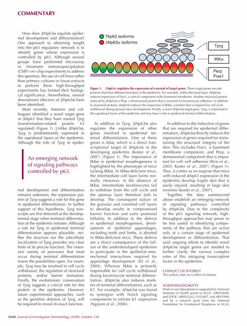
How does ∆Np63α regulate epider-mal development and differentiation? One approach to obtaining insight into the p63 regulatory network is to identify genes whose expression is controlled by p63. Although several groups have performed microarray or chromatin immunoprecipitation (ChIP)–on–chip experiments to address this question, the use of cell lines rather than primary cultures or tissue extracts to perform these high-throughput experiments has limited their biologi-cal significance. Nevertheless, several downstream effectors of ∆Np63α have been identified.
Most recently, Antonini and col-leagues identified a novel target gene of ∆Np63 that they have named Tprg (transformation-related protein 63 regulated) (Figure 1). Unlike ∆Np63α, Tprg is predominantly expressed in the suprabasal layers of the epidermis. Although the role of Tprg in epider-
mal development and differentiation remains unknown, the expression pat-tern of Tprg suggests a role for this gene in epidermal differentiation. In further support of this hypothesis, Tprg tran-scripts are first detected at the develop-mental stage when terminal differentia-tion of the epidermis initiates. Although a role for Tprg in epidermal terminal differentiation appears plausible, nei-ther the structure nor the subcellular localization of Tprg provides any clear hints of its precise function. The exten-sive variety of processes that must occur during terminal differentiation leaves the possibilities open. For exam-ple, Tprg may be involved in cell cycle withdrawal, the regulation of structural proteins, and/or barrier formation. Finally, the evolutionary conservation of Tprg suggests a critical role for this protein in the epidermis. However, future experimental approaches, such as the germline deletion of Tprg, will be required to reveal its exact function.
In addition to Tprg, ∆Np63α also regulates the expression of other genes involved in epidermal ter-minal differentiation. One of these genes is Ikkα, which is a direct tran-scriptional target of ∆Np63α in the developing epidermis (Koster et al., 2007) (Figure 1). The importance of IKKα in epidermal morphogenesis is highlighted by the phenotype of mice lacking IKKα. In IKKα-deficient mice, the intermediate cell layer forms nor-mally. However, in the absence of IKKα, intermediate keratinocytes fail to withdraw from the cell cycle and the spinous layer therefore fails to develop. The consequent failure of the granular and cornified cell layers to develop results in compromised barrier function and early postnatal lethality. In addition to the defects in epidermal morphogenesis, devel-opment of epithelial appendages, including teeth and limbs, is aborted in IKKα-deficient mice. These defects are a direct consequence of the fail-ure of the underdeveloped epidermis to participate in the epithelial–mes-enchymal interactions required for appendage development (Sil et al., 2004). Whereas IKKα is primarily responsible for cell cycle withdrawal during keratinocyte terminal differen-tiation, ∆Np63α also induces mark-ers of terminal differentiation, such as K1. For example, ∆Np63α was found to synergize with Notch signaling components to initiate K1 expression (Nguyen et al., 2006).
In addition to the induction of genes that are required for epidermal differ-entiation, ∆Np63α directly induces the expression of genes required for main-taining the structural integrity of the skin. This includes Fras1, a basement membrane component, and Perp, a desmosomal component that is impor-tant for cell–cell adhesion (Ihrie et al., 2005; Koster et al., 2007) (Figure 1). Thus, it comes as no surprise that mice with reduced ∆Np63 expression in the epidermis develop fragile skin that is easily injured, resulting in large skin erosions (Koster et al., 2007).
Together, the data summarized above establish an emerging network of signaling pathways controlled by ∆Np63α. Due to the complexity of the p63 signaling network, high-throughput approaches may prove to be less useful in identifying compo-nents of the pathway that are active only at a certain stage of epidermal development or differentiation. That said, ongoing efforts to identify novel ∆Np63α target genes are needed to further clarify the various complex roles of this intriguing transcription factor in the epidermis.
CONFLICT OF INTERESTThe authors state no conflict of interest.
ACKNOWLEDGMENTSWork in our laboratories is supported by National Institutes of Health grants to M.I.K. (AR054696) and D.R.R. (AR052263, CA52607, and AR47898) and by a research grant from the National Foundation for Ectodermal Dysplasias to M.I.K.
Figure 1. ∆Np63α regulates the expression of a myriad of target genes. These target genes encode proteins that have different functions in the epidermis. For example, within the basal layer, ∆Np63α induces expression of Fras1, a critical component of the basement membrane. Another structural protein induced by ∆Np63α is Perp, a desmosomal protein that is essential for keratinocyte adhesion. In addition to structural proteins, ∆Np63α induces the expression of IKKα, a protein that is required for cell cycle withdrawal during spinous layer development. Finally, a novel ∆Np63α target gene, Tprg, is expressed in the suprabasal layers of the epidermis and may have a role in epidermal terminal differentiation.
TAp63 isoforms∆Np63α isoforms
Tprg
IKKα
PerpFras1
|An emerging network of signaling pathways controlled by p63.
COMMENTARY
www.jidonline.org 1619
and D.R.R. We thank Dr Peter J. Koch for his constructive comments on the manuscript.
REFERENCESAntonini D, Dentice M, Mahtani P, De Rosa L,
Gatta GD, Mandinova A et al. (2008) Tprg, a gene predominantly expressed in skin, is a direct target of the transcription factor p63. J Invest Dermatol 128:1676–85
Ihrie RA, Marques MR, Nguyen BT, Horner JS, Papazoglu C, Bronson RT et al. (2005) Perp is a p63-regulated gene essential for epithelial integrity. Cell 120:843–56
Koster MI, Roop DR (2007) Mechanisms regulating epithelial stratification. Annu Rev Cell Dev Biol 23:93–113
Koster MI, Dai D, Marinari B, Sano Y, Costanzo A, Karin M et al. (2007) p63 induces key target genes required for epidermal morphogenesis. Proc Natl Acad Sci USA 104:3255–60
Mills AA, Zheng B, Wang XJ, Vogel H, Roop DR, Bradley A (1999) p63 is a p53 homologue required for limb and epidermal morphogenesis. Nature 398:708–13
Nguyen BC, Lefort K, Mandinova A, Antonini D, Devgan V, Della GG et al. (2006) Cross-regulation between Notch and p63 in keratinocyte commitment to differentiation. Genes Dev 20:1028–42
Sil AK, Maeda S, Sano Y, Roop DR, Karin M (2004) IkappaB kinase-alpha acts in the epidermis to control skeletal and craniofacial
morphogenesis. Nature 428:660–4
Truong AB, Kretz M, Ridky TW, Kimmel R, Khavari PA (2006) p63 regulates proliferation and differentiation of developmentally mature keratinocytes. Genes Dev 20:3185–97
Yang A, Kaghad M, Wang Y, Gillett E, Fleming MD, Dotsch V et al. (1998) p63, a p53 homolog at 3q27-29, encodes multiple products with transactivating, death-inducing, and dominant-negative activities. Mol Cell 2:305–16
Yang A, Schweitzer R, Sun D, Kaghad M, Walker N, Bronson RT et al. (1999) p63 is essential for regenerative proliferation in limb, craniofacial and epithelial development. Nature 398:714–8
Tprg, a Gene Predominantly Expressed in Skin, Is aDirect Target of the Transcription Factor p63Dario Antonini1, Monica Dentice2, Parvesh Mahtani3, Laura De Rosa1, Giusy Della Gatta4,Anna Mandinova5, Domenico Salvatore2, Elia Stupka6 and Caterina Missero1
p63 and p73 are highly homologous members of the p53 family that originated by gene duplication at theinvertebrate-to-vertebrate transition. We characterize here a previously unreported gene, Transformation-related protein 63 regulated (Tprg), located upstream of the p63 gene in the vertebrate genome, with strikingsimilarity to Transformation related protein 63 regulated like (Tprgl), an uncharacterized gene located upstreamof p73, suggesting that p63/Tprg and p73/Tprgl are embedded in a paralogue region originated from a singleduplication event. Tprg is predominantly expressed in the epithelial compartment of the skin, more abundantlyin differentiated cells. Consistent with its relative higher expression in differentiated keratinocytes, finely tunedp63 expression levels are required for optimal Tprg expression in primary keratinocytes. p63 is essential for Tprgexpression as shown in p63-knockdown keratinocytes; however, high levels of p63 result in Tprg down-regulation. p63 directly binds in vivo to a canonical p63-binding site in an evolutionary conserved genomicregion located in Tprg intron 4. This genomic region is sufficient to function as a p63-inducible enhancer inpromoter studies. Thus, we demonstrate that the Tprg gene is predominantly expressed in skin, is physicallyassociated with the p63 gene during evolution, and directly regulated by p63 through a long-distance enhancerlocated within the Tprg locus.
Journal of Investigative Dermatology (2008) 128, 1676–1685; doi:10.1038/jid.2008.12; published online 7 February 2008
INTRODUCTIONA number of transcription factors are known to regulate skindevelopment and differentiation. Among these, p63 is a keymodulator of these processes, as is clearly demonstratedin vivo by knockout studies. p63�/� mice fail to form astratified epidermis, resulting in lack of barrier formation,consequent dehydration, and death within hours after birth(Mills et al., 1999; Yang et al., 1999).
p63 belongs to the p53 gene family consisting of threegenes, p53, p63, and p73, that share a significant sequencehomology (reviewed by Yang et al., 2002). Each p53 familymember contains a transactivation domain at the amino
terminus, a DNA-binding domain, and an oligomerizationdomain. In addition, all family members share some commonfunctions, and bind to a canonical p53-binding site, thuscontrolling the expression of a subset of p53 target genes(Yang et al., 2002, 2006). The use of alternative promotersand transcription start sites gives rise to two classes of p63transcripts, those encoding proteins with an amino-terminaltransactivation domain (TA isoforms) and those encodingproteins lacking this domain (DN isoforms) (Yang et al.,1998). Three different carboxyl-termini, designated a, b, andg, are generated by alternative splicing. The carboxyl-terminus of p63-a is the longest and contains a sterilea-motif domain and a transactivation-inhibitory domain (Chiet al., 1999; Thanos and Bowie, 1999; Serber et al., 2002).Accordingly, DNp63-a has been shown to act as a repressorand to display dominant-negative function against bothTAp63 isoforms and p53 (Yang et al., 1998; Ghioni et al.,2002; Westfall et al., 2003; Chan et al., 2004). However,DNp63-a also positively regulates the expression of sometarget genes, such as integrins and other adhesion-associatedgenes (Kurata et al., 2004; Carroll et al., 2006; Truong et al.,2006), as well as keratin 14 (K14) (Romano et al., 2006).
p63 is predominantly expressed in the basal and spinouslayers of the epidermis, and is downregulated uponkeratinocyte differentiation both in vitro and in vivo (Parsaet al., 1999; Yang et al., 1999; Liefer et al., 2000; Bambergerand Schmale, 2001; Pellegrini et al., 2001; Westfall et al.,2003; Nguyen et al., 2006). In the basal layer, p63 is mainly
See related commentary on pg 1617ORIGINAL ARTICLE
1676 Journal of Investigative Dermatology (2008), Volume 128 & 2008 The Society for Investigative Dermatology
Received 29 June 2007; revised 23 November 2007; accepted 4 December2007; published online 7 February 2008
1CEINGE Biotecnologie Avanzate, Napoli, Italy; 2Department of Molecularand Clinical Endocrinology and Oncology, University ‘‘Federico II’’, Napoli,Italy; 3School of Biological Sciences, National University of Singapore,Singapore, Republic of Singapore; 4Telethon Institute of Genetics andMedicine, Napoli, Italy; 5Cutaneous Biology Research Center, MassachusettsGeneral Hospital and Harvard Medical School, Charlestown, Massachusetts,USA and 6CBM S.c.r.l., Trieste, Italy
Correspondence: Dr Elia Stupka, CBM S.c.r.l., c/o AREA Science Park,Basovizza, SS 14 km, 163,5, Trieste, Italy. E-mail: [email protected] orDr Caterina Missero, CEINGE Biotecnologie Avanzate, Via ComunaleMargherita 482, Napoli 80145, Italy. E-mail: [email protected]
Abbreviations: aa, amino acids; ChIP, chromatin immunoprecipitation; GFP,green-fluorescent protein; hrs, hours; PBS, phosphate-buffered saline;RT–PCR, reverse transcriptase–polymerase chain reaction; siRNA, shortinterfering RNA; TK, thymidine kinase; Tprg, Transformation-related protein63 regulated; Tprgl, Transformation-related protein 63 regulated like
involved in maintaining cell proliferation and cell adhesion(Koster et al., 2004; Deyoung et al., 2006; Sbisa et al., 2006;Truong et al., 2006).
As keratinocytes detach from the basement membrane,they begin a program of terminal differentiation, characteri-zed by a change in keratin expression from K5/K14 to asuprabasal pair in the spinous layer (K1/K10). Furtherkeratinocyte differentiation leads to the expression offilaggrin, loricrin, and other cornified envelope componentsin the granular layer, contributing to the formation of thecutaneous barrier (reviewed by Fuchs, 1998). It has beenproposed that p63 plays a dual role in keratinocytedifferentiation, as it is required for initiating epithelialstratification (Koster et al., 2004; Nguyen et al., 2006; Truonget al., 2006), whereas concurrently it inhibits the expressionof some differentiation markers, at least in part throughtranscriptional repression of the Notch effector Hes1 (Kinget al., 2003; Nguyen et al., 2006). Interestingly, some p63direct targets, such as Perp, whose expression in skin requiresp63, are predominantly localized in the suprabasal layers(Ihrie et al., 2005).
Here, we identify Transformation-related protein 63regulated (Tprg), a previously uncharacterized gene locatedupstream of the genomic locus of p63, whose expression isregulated by p63. Tprg encodes for a cytoplasmic proteinwith a high degree of homology with the gene product ofTransformation related protein 63 regulated like (Tprgl),located upstream of the p73 gene. Both loci are conservedthroughout vertebrate evolution upstream of p63 and p73,respectively. Tprg is significantly expressed from embryonicday 15.5 in a cell type-specific manner in the epidermis andin the hair follicle in contrast to the ubiquitous expression ofTprgl. We show that Tprg expression is suppressed by p63knockdown in mouse primary keratinocytes, and is specifi-cally affected by knockdown of the DNp63-a isoform.Interestingly, DNp63-a overexpression also negatively reg-ulates Tprg, suggesting that a finely tuned p63 activity isrequired for optimal Tprg expression. p63, moreover, directlybinds and activates a long-distance enhancer located in aTprg intronic region. Taken together, our data indicate thatTprg is a previously uncharacterized gene, conservedthroughout evolution in proximity of p63, and whoseexpression is under direct control of p63.
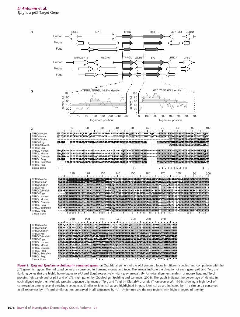
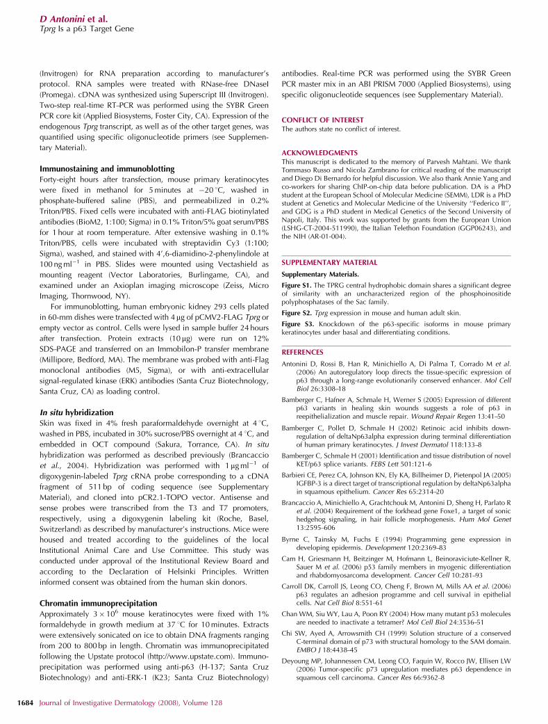
RESULTSIn vertebrates, p63 and p73 proteins are more closely relatedto one another than to p53, owing to a higher percentage ofsimilarity in the DNA-binding domain as well as the presenceof the sterile a-motif domain, absent in p53 (Saccone et al.,2002; Yang et al., 2002). As p63 and p73 are known to havederived from a gene duplication event, we investigatedwhether they are embedded in paralogous regions, whichwould indicate duplication of nearby genes. Interestingly, anuncharacterized gene located 260 kb upstream of the p63gene in mouse, indicated by the full-length cDNA5430420C16Rik, is highly homologous to another unchar-acterized gene located 101 kb upstream of the p73 gene,indicated by the full-length cDNA 1200015A19Rik (Kawai
et al., 2001; Figure 1a). On the basis of the results describedbelow, we have named the above two transcripts Tprg andTprgl, respectively. They are both transcribed in the samedirection as the corresponding p63 and p73 genes, and share44.1% of identity at the protein level (Figure 1b). p63 and p73share a comparable percentage of identity (56.6%), suggest-ing that they have evolved in a parallel manner, and may beembedded in a paralogue region originated from a singleduplication event.
Tprg is transcribed in a 1,087-bp transcript originallyisolated as a full-length cDNA from 6 days neonate head andadult female vagina cDNA library (Kawai et al., 2001). Tprglis transcribed in a 1,756-bp transcript originally isolated froma cDNA library of adult male lung (Kawai et al., 2001). Tocharacterize the expression pattern of the two genes, wemeasured their expression in mouse adult tissues by real timereverse transcriptase–polymerase chain reaction (RT–PCR).Tprg was specifically expressed in skin, and to a much lesserextent in tongue and esophagus (Figure 2a). In contrast, Tprglwas abundantly expressed in all tested tissues (Figure 2b). Aspreviously reported (Yang et al., 1998; Nakamuta andKobayashi, 2003; Cam et al., 2006), p63 was expressed athigh levels in skin, tongue, muscles, and testis, and to a lesserextent in esophagus and heart (Figure 2c). DNp63 was thepredominant isoform in skin, tongue, and esophagus,whereas TAp63 was highly expressed in muscle and testis(Figure 2d). Thus, Tprg expression correlates with DNp63expression in adult mouse tissues. The Tprg gene is predictedto encode for a putative protein of 279 amino acids (aa),whereas the Tprgl gene is predicted to encode for a putativeprotein of 266 aa. Both proteins have clear orthologues in 26annotated vertebrate genomes, including other mammals,xenopus, chicken, zebrafish, and fugu (Figure 1c), but nohomology in non vertebrate genomes, indicating them asclearly vertebrate-specific proteins. The putative Tprg andTprgl proteins share two highly conserved domains, one inthe central region (78% of identity in 32 aa) and the other atthe carboxyl-terminus of the protein (76% of identity in29 aa). No known protein domains were found in theirsequences. The central portion of the sequence shares asignificant degree of similarity with members of the Sacfamily of phosphoinositide phosphatases in Drosophilamelanogaster (Figure S1); however, the similarity occurswithin uncharacterized protein domains. Moreover, mouseorthologues of these Drosophila genes exist and they do notdisplay any similarity to Tprg and Tprgl. In the absence ofspecific antibodies, Tprg protein expression was evaluated bytransient transfection of a FLAG-tagged construct. Immuno-blotting analysis revealed that FLAG-Tprg protein run at anapparent molecular weight of approximately 36 kDa, con-sistent with the theoretical molecular weight of the wild-typeprotein (31 kDa) (Figure 3a). In mouse primary keratinocytes,immunofluorescence with anti-FLAG antibodies revealed thatexogenous Tprg protein was localized in the cytoplasm,whereas being absent from the nucleus (Figure 3b). A similarlocalization was observed in both undifferentiated (0 hour)keratinocytes and in keratinocytes induced to differentiate byCa2þ addition at 8 and 24 hours. Thus, Tprg is unlikely to be
www.jidonline.org 1677
D Antonini et al.Tprg Is a p63 Target Gene
BCL6 LPP TPRG p63 LEPREL1 CLDN1
DFFBLRRC47p73WDR8TPRGL
TPRG/TPRGL 44.1% identity
MEGF6ARHGEF16
Human
Mouse
Fugu
Human
Mouse
Fugu
100
80604020
00
10
110 120 130 140 150 160 170 180 190 200
210 220 230 240 250 260 270
20 30 40 50 60 70 80 90 100
40 80 120 160 200 240 280 0 100 200 300 400 500 600 700
Alignment position
p63/p73 56.6% identity
Alignment position
Iden
tity
(%)
100
80604020
0
Iden
tity
(%)
TPRG Mouse
TPRGL Mouse
TPRG Human
TPRGL Human
TPRG Chicken
TPRGL Chicken
TPRG Frog
TPRGL Frog
TPRG Fugu
TPRGL FuguClustal Cons.
TPRG Zebrafish
TPRG MouseTPRG HumanTPRG ChickenTPRG Frog
TPRG FuguTPRG Zebrafish
TPRG MouseTPRG HumanTPRG ChickenTPRG Frog
TPRG FuguTPRG Zebrafish
TPRGL Zebrafish
TPRGL MouseTPRGL Human
TPRGL ChickenTPRGL Frog
TPRGL FuguClustal Cons.
TPRGL Zebrafish
TPRGL MouseTPRGL Human
TPRGL ChickenTPRGL Frog
TPRGL FuguClustal Cons.
TPRGL Zebrafish
Figure 1. Tprg and Tprgl are evolutionarily conserved genes. (a) Graphic alignment of the p63 genomic locus in different species, and comparison with the
p73 genomic region. The indicated genes are conserved in humans, mouse, and fugu. The arrows indicate the direction of each gene. p63 and Tprg are
flanking genes that are highly homologous to p73 and Tprgl, respectively, (dark gray arrows). (b) Pairwise alignment analysis of mouse Tprg and Tprgl
proteins (left panel) and of p63 and of p73 (right panel) by GraphAlign (Spalding and Lammers, 2004). The graph indicates the percentage of identity in
each aligned region. (c) Multiple protein sequence alignment of Tprg and Tprgl by ClustalW analysis (Thompson et al., 1994), showing a high level of
conservation among several vertebrate sequences. Similar or identical aa are highlighted in gray. Identical aa are indicated by ‘‘*’’; similar aa conserved
in all sequences by ‘‘:’’; and similar aa not conserved in all sequences by ‘‘.’’. Underlined are the two regions with highest degree of identity.
1678 Journal of Investigative Dermatology (2008), Volume 128
D Antonini et al.Tprg Is a p63 Target Gene
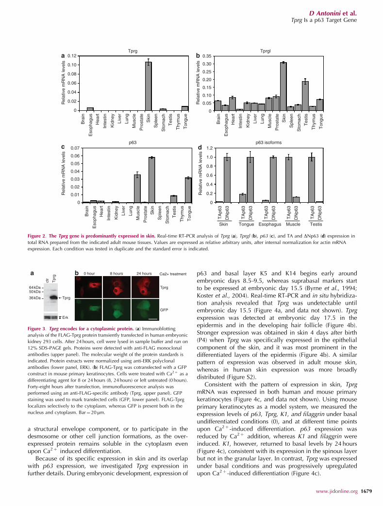
a structural envelope component, or to participate in thedesmosome or other cell junction formations, as the over-expressed protein remains soluble in the cytoplasm evenupon Ca2þ induced differentiation.
Because of its specific expression in skin and its overlapwith p63 expression, we investigated Tprg expression infurther details. During embryonic development, expression of
p63 and basal layer K5 and K14 begins early aroundembryonic days 8.5–9.5, whereas suprabasal markers startto be expressed at embryonic day 15.5 (Byrne et al., 1994;Koster et al., 2004). Real-time RT–PCR and in situ hybridiza-tion analysis revealed that Tprg was undetectable untilembryonic day 15.5 (Figure 4a, and data not shown). Tprgexpression was detected at embryonic day 17.5 in theepidermis and in the developing hair follicle (Figure 4b).Stronger expression was obtained in skin 4 days after birth(P4) when Tprg was specifically expressed in the epithelialcomponent of the skin, and it was most prominent in thedifferentiated layers of the epidermis (Figure 4b). A similarpattern of expression was observed in adult mouse skin,whereas in human skin expression was more broadlydistributed (Figure S2).
Consistent with the pattern of expression in skin, TprgmRNA was expressed in both human and mouse primarykeratinocytes (Figure 4c, and data not shown). Using mouseprimary keratinocytes as a model system, we measured theexpression levels of p63, Tprg, K1, and filaggrin under basalundifferentiated conditions (0), and at different time pointsupon Ca2þ -induced differentiation. p63 expression wasreduced by Ca2þ addition, whereas K1 and filaggrin wereinduced. K1, however, returned to basal levels by 24 hours(Figure 4c), consistent with its expression in the spinous layerbut not in the granular layer. In contrast, Tprg was expressedunder basal conditions and was progressively upregulatedupon Ca2þ -induced differentiation (Figure 4c).
0.12 0.35Tprg
p63 p63 isoforms
Tprgl
0.30
0.25
0.20
0.15
0.10
0.05
0
0.10R
elat
ive
mR
NA
leve
lsR
elat
ive
mR
NA
leve
ls
Rel
ativ
e m
RN
A le
vels
Rel
ativ
e m
RN
A le
vels
0.08
0.06
0.04
0.02
0
Bra
in
Eso
phag
us
Hea
rt
Inte
stin
Kid
ney
Live
r
Lung
Mus
cle
Pro
stat
e
Ski
n
Spl
een
Sto
mac
h
Tes
tis
Thy
mus
Ton
gue
Bra
in
Eso
phag
us
Hea
rt
Inte
stin
Kid
ney
Live
r
Lung
Mus
cle
Pro
stat
e
Ski
n
Spl
een
Sto
mac
h
Tes
tis
Thy
mus
Ton
gue
Bra
in
Eso
phag
us
Hea
rt
Inte
stin
Kid
ney
Live
r
Lung
Mus
cle
Pro
stat
e
Ski
n
Spl
een
Sto
mac
h
Tes
tis
Thy
mus
Ton
gue
TA
p63
DN
p63
TA
p63
DN
p63
TA
p63
DN
p63
TA
p63
DN
p63
TA
p63
DN
p63
0.07 1.2
1.0
0.8
0.6
0.4
0.2
0
0.06
0.05
0.04
0.03
0.02
0.01
0
Skin Tongue Esophagus Muscle Testis
a b
c d
Figure 2. The Tprg gene is predominantly expressed in skin. Real-time RT–PCR analysis of Tprg (a), Tprgl (b), p63 (c), and TA and DNp63 (d) expression in
total RNA prepared from the indicated adult mouse tissues. Values are expressed as relative arbitrary units, after internal normalization for actin mRNA
expression. Each condition was tested in duplicate and the standard error is indicated.
64kDa50kDa
36kDa Tprg
0 hour 8 hours 24 hours Ca2+ treatment
Tprg
GFP
Tpr
g
ctr
Erk
Figure 3. Tprg encodes for a cytoplasmic protein. (a) Immunoblotting
analysis of the FLAG-Tprg protein transiently transfected in human embryonic
kidney 293 cells. After 24 hours, cell were lysed in sample buffer and run on
12% SDS–PAGE gels. Proteins were detected with anti-FLAG monoclonal
antibodies (upper panel). The molecular weight of the protein standards is
indicated. Protein extracts were normalized using anti-ERK polyclonal
antibodies (lower panel, ERK). (b) FLAG-Tprg was cotransfected with a GFP
construct in mouse primary keratinocytes. Cells were treated with Ca2þ as a
differentiating agent for 8 or 24 hours (8, 24 hours) or left untreated (0 hours).
Forty-eight hours after transfection, immunofluorescence analysis was
performed using an anti-FLAG-specific antibody (Tprg, upper panel). GFP
staining was used to mark transfected cells (GFP, lower panel). FLAG-Tprg
localizes selectively to the cytoplasm, whereas GFP is present both in the
nucleus and cytoplasm. Bar¼ 20mm.
www.jidonline.org 1679
D Antonini et al.Tprg Is a p63 Target Gene
Given that Tprg and p63 are predominantly expressed inskin, and their temporal patterns of expression in skin, weasked whether p63 might control Tprg transcription. To testthis possibility, we measured the expression of Tprg in p63-knockdown keratinocytes, using previously characterizedtotal p63-specific short interfering RNA (siRNA) (Antoniniet al., 2006). Forty-eight hours after transfection of p63 siRNAin mouse primary keratinocytes, a strong reduction of Tprgexpression was observed (Figure 5a). In contrast, all the othertested genes spanning a genomic region �1.7 Mbþ 687 kbfrom the p63 gene (Figure 1a) were not affected by p63knockdown (Figure 5a). Similar results were obtained with apreviously characterized independent p63 siRNA oligonu-cleotide (data not shown) (Antonini et al., 2006). We thenasked which specific p63 isoform controls Tprg expression bytransfection of isoform-specific siRNA oligonucleotides.
Knockdown of the DNp63 or the a-isoforms strongly inhibitedTprg expression, whereas knockdown of the TA andg-isoforms was unable to affect Tprg, both under basalconditions and upon Ca2þ induced differentiation (Figure 5b;Figure S3). Thus, DNp63-a is required for proper expressionof the Tprg gene in mouse primary keratinocytes.
To further investigate the regulation of Tprg expression byDNp63-a, we infected primary keratinocytes with a retrovirusexpressing DNp63-a protein fused to an estrogen-receptordomain (ERp63) and maintained under basal conditions in aninactive form (Nguyen et al., 2006). Total RNA was preparedat early time points after ERp63 activation by tamoxifentreatment, and Tprg expression was measured by real-timeRT–PCR. Upon DNp63-a activation, Tprg expression wassignificantly reduced by ERp63 between 40 minutes and1 hour, suggesting that Tprg is likely to be directly regulated
0.08E17.5
P4
P4
0.06
0.04
0.02
E10
.5
E12
.5
E14
.5
E15
.5
Ski
n
0
0.08
0.06
0.04
0.02
0
0.008 Tprg p63
K1Filaggrin
0.006
0.004
0.002
0
0.016
0.012
0.12
0.09
0.06
0.03
0
0.008
0.004
0
0 hour
1 hour
2 hours
4 hours
6 hours
8 hours
16 hours
24 hours
0 hour
1 hour
2 hours
4 hours
6 hours
8 hours
16 hours
24 hours
0 hour
1 hour
2 hours
4 hours
6 hours
8 hours
16 hours
24 hours
0 hour
1 hour
2 hours
4 hours
6 hours
8 hours
16 hours
24 hours
Ca2+ treatment
Ca2+ treatment
Rel
ativ
e T
prg
mR
NA
leve
ls
Rel
ativ
e m
RN
A le
vels
Rel
ativ
e m
RN
A le
vels
Rel
ativ
e m
RN
A le
vels
Rel
ativ
e m
RN
A le
vels
Figure 4. Tprg is expressed in the epidermis and in the hair follicle and is more abundant in differentiated keratinocytes. (a) Real-time RT–PCR analysis
of whole-embryo RNA at different time points during embryonic development or in adult skin as indicated. Values are expressed and normalized as in
Figure 3. (b) RNA in situ hybridization of mouse skin sections at embryonic day 17.5 (upper left panel) and at postnatal day 4 (P4, lower left panel, and
right panel) using a digoxygenin-labeled antisense probe for mouse Tprg. The dashed line indicates the dermal–epidermal junction. Similar results were
observed using an independent probe, whereas a Tprg sense probe gave no detectable signal under the same conditions (data not shown). Bars¼ 50 mm
for the left panels, and 10mm for the right panel. (c) Real-time RT–PCR analysis of total RNA extracted from primary mouse keratinocytes at different time
points upon Ca2þ treatment, as indicated (hours), reveals an induction of the Tprg expression upon differentiation, whereas p63 is modestly upregulated at
early time points, and then strongly downregulated. Expression of filaggrin and K1 are shown for comparison. Values are expressed as described in Figure 3.
1680 Journal of Investigative Dermatology (2008), Volume 128
D Antonini et al.Tprg Is a p63 Target Gene
by p63 (Figure 5c). Interestingly, DNp63-a activation resultedin Tprg downregulation rather than induction. DNp63-aactivation is unlikely to repress Tprg due to a genericsquelching effect, in agreement with a global gene expressionanalysis, which revealed that most genes that are repressedby p63 activation are induced by p63 knockdown (GiusyDella Gatta and Caterina Missero, in preparation). A similarinhibition of Tprg expression was observed in primarykeratinocytes infected with an adenovirus expressingDNp63-a (Figure 5d), suggesting that Tprg expression isfinely regulated by DNp63-a in keratinocytes.
Regulation of Tprg expression by p63 could either occurdirectly through p63 binding to a Tprg-regulatory sequence orcould be mediated by other mechanisms. To identifypotential p63-binding sites in the Tprg gene, we examinedthe entire genomic region containing the Tprg locus for p63-binding sites, using a recently performed genome-widechromatin immunoprecipitation (ChIP)-on-chip analysis inhuman carcinoma cells (Yang et al., 2006). The Tprg genespans 151 kb in the human genome and has five introns.Interestingly, two genomic regions located in human Tprgintron 4 displayed p63-binding activity with a significantbinding score, whereas no p63 binding could be detected in
the Tprg putative proximal promoter or in upstream regions(up to 50 kb from the transcription start site). Both genomicregions identified are conserved throughout evolution (Figure6a, and data not shown); however, only one displays a p53/p63 consensus sequence that is conserved between humanand chicken (Figure 6b). We proceeded to test whether p63could bind in vivo to the latter. ChIP was performed usinganti-p63-specific antibodies in human primary keratinocytes,and amplifying three sequences approximately 500 bp apart(Figure 6a). Interestingly, p63 specifically bound to thesequence corresponding to the most highly conserved regionand containing the conserved p63-binding site (Figure 6c).A strong binding was obtained with the corresponding mousesequence in mouse primary keratinocytes, where p63 boundthe Tprg-binding site to a similar extent of as that of a high-affinity long-distance enhancer (C40 enhancer; Antoniniet al., 2006; Yang et al., 2006; Figure 6d). This genomicregion is likely to contain a functional p63-binding site andalso to function as an enhancer. We tested this possibility bycloning the conserved genomic region containing the bindingsite upstream of a thymidine kinase (TK) minimal promoterdriving the expression of a luciferase gene. Transienttransfection assays in HeLa cells revealed that the Tprg
1.5
1.2
0.9
0.6
0.3
Rel
ativ
e m
RN
A le
vels
0– +
Bcl6 Lpp Tprg p63
1.6 14
12
10
8
6
4
2
0
1.41.21.00.80.6
Rel
ativ
e T
prg
mR
NA
leve
ls
0.40.2
0– TA siRNATAΔN ΔNα αγ γ–
Leprel Cldn1
– + – + – + – + – + p63 siRNA
Undifferentiated ker. Differentiated ker.
1.6 1.2
0.8
0.4
0– Adp63
1.2
0.8
0.4
00 20
minutes40
minutes1
hour2
hours3
hours4
hoursTamoxifen treatment
Rel
ativ
e T
prg
mR
NA
leve
ls
Rel
ativ
e T
prg
mR
NA
leve
lsFigure 5. Tprg gene expression is controlled by p63. (a) Real-time RT–PCR analysis of total RNA prepared from primary mouse keratinocytes transfected
with siRNA specific for p63 (þ ) or with an siRNA-negative control (�) reveals a downregulation of Tprg that parallels p63 expression, whereas the other
indicated genes located in the p63 locus are unaffected. Values for each gene are expressed as fold changes versus the siRNA-negative control set to 1.
(b) Analysis of Tprg expression by real-time RT–PCR of total RNA prepared from primary mouse keratinocytes transfected with siRNA specific for p63a,
p63g, TAp63, and DNp63 isoforms, or negative control (c), and either grown under basal conditions (undifferentiated ker.) or induced 0.2 mM Ca2þ to
differentiate by for 24 hours (differentiated ker.). Values are expressed as fold changes versus the siRNA-negative control in basal conditions set to 1.
Knockdown efficiency and specificity for all p63 isoforms are shown in Figure S3. (c) Expression profile of Tprg at early time points upon induction of p63
activity. Primary mouse keratinocytes were infected with a retrovirus carrying an ER-DNp63-a fusion protein or empty vector control and subsequently
treated with 20 nM tamoxifen for the indicated times. Total RNA was used for cDNA preparation followed by real-time RT–PCR. Values are expressed as
changes in relative mRNA levels in the ER-p63-expressing versus control keratinocytes. (d) Down-modulation of Tprg mRNA expression by DNp63-a.
Primary mouse keratinocytes were infected with a recombinant adenovirus expressing DNp63-a or a control GFP-expressing adenovirus (c) for 24 hours.
Tprg mRNA levels were quantified by real-time RT–PCR. Values are expressed as relative arbitrary units, after internal normalization for GAPDH. GAPDH,
glycerladehyde-3-phosphate dehydrogenase.
www.jidonline.org 1681
D Antonini et al.Tprg Is a p63 Target Gene
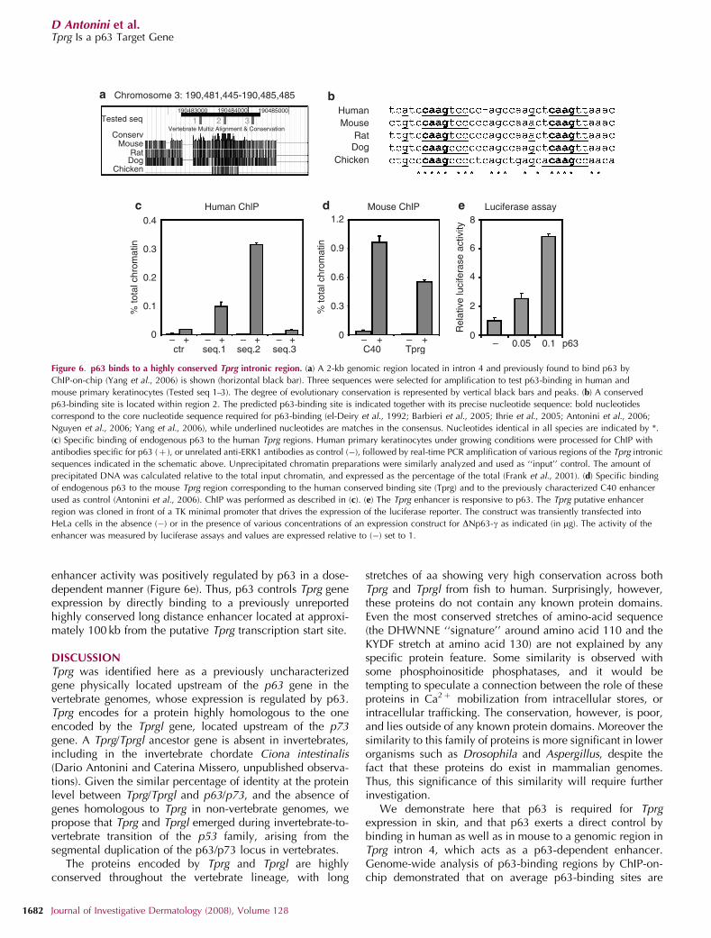
enhancer activity was positively regulated by p63 in a dose-dependent manner (Figure 6e). Thus, p63 controls Tprg geneexpression by directly binding to a previously unreportedhighly conserved long distance enhancer located at approxi-mately 100 kb from the putative Tprg transcription start site.
DISCUSSIONTprg was identified here as a previously uncharacterizedgene physically located upstream of the p63 gene in thevertebrate genomes, whose expression is regulated by p63.Tprg encodes for a protein highly homologous to the oneencoded by the Tprgl gene, located upstream of the p73gene. A Tprg/Tprgl ancestor gene is absent in invertebrates,including in the invertebrate chordate Ciona intestinalis(Dario Antonini and Caterina Missero, unpublished observa-tions). Given the similar percentage of identity at the proteinlevel between Tprg/Tprgl and p63/p73, and the absence ofgenes homologous to Tprg in non-vertebrate genomes, wepropose that Tprg and Tprgl emerged during invertebrate-to-vertebrate transition of the p53 family, arising from thesegmental duplication of the p63/p73 locus in vertebrates.
The proteins encoded by Tprg and Tprgl are highlyconserved throughout the vertebrate lineage, with long
stretches of aa showing very high conservation across bothTprg and Tprgl from fish to human. Surprisingly, however,these proteins do not contain any known protein domains.Even the most conserved stretches of amino-acid sequence(the DHWNNE ‘‘signature’’ around amino acid 110 and theKYDF stretch at amino acid 130) are not explained by anyspecific protein feature. Some similarity is observed withsome phosphoinositide phosphatases, and it would betempting to speculate a connection between the role of theseproteins in Ca2þ mobilization from intracellular stores, orintracellular trafficking. The conservation, however, is poor,and lies outside of any known protein domains. Moreover thesimilarity to this family of proteins is more significant in lowerorganisms such as Drosophila and Aspergillus, despite thefact that these proteins do exist in mammalian genomes.Thus, this significance of this similarity will require furtherinvestigation.
We demonstrate here that p63 is required for Tprgexpression in skin, and that p63 exerts a direct control bybinding in human as well as in mouse to a genomic region inTprg intron 4, which acts as a p63-dependent enhancer.Genome-wide analysis of p63-binding regions by ChIP-on-chip demonstrated that on average p63-binding sites are
Human ChlP0.4
– – – – + – +C40
1.2 8
6
4
2
0
Rel
ativ
e lu
cife
rase
act
ivity
0.9
0.6
0.3
0
Tprg– – 0.05 0.1 p63++++
ctr seq.1 seq.2 seq.3
0.3
0.2
0.1% to
tal c
hrom
atin
% to
tal c
hrom
atin
0
Mouse ChlP Luciferase assay
HumanMouse
RatDog
Chicken
Chromosome 3: 190,481,445-190,485,485
Tested seq190483000
1Vertebrate Multiz Alignment & Conservation
2 3190484000 190485000
ConservMouse
RatDog
Chicken
Figure 6. p63 binds to a highly conserved Tprg intronic region. (a) A 2-kb genomic region located in intron 4 and previously found to bind p63 by
ChIP-on-chip (Yang et al., 2006) is shown (horizontal black bar). Three sequences were selected for amplification to test p63-binding in human and
mouse primary keratinocytes (Tested seq 1–3). The degree of evolutionary conservation is represented by vertical black bars and peaks. (b) A conserved
p63-binding site is located within region 2. The predicted p63-binding site is indicated together with its precise nucleotide sequence: bold nucleotides
correspond to the core nucleotide sequence required for p63-binding (el-Deiry et al., 1992; Barbieri et al., 2005; Ihrie et al., 2005; Antonini et al., 2006;
Nguyen et al., 2006; Yang et al., 2006), while underlined nucleotides are matches in the consensus. Nucleotides identical in all species are indicated by *.
(c) Specific binding of endogenous p63 to the human Tprg regions. Human primary keratinocytes under growing conditions were processed for ChIP with
antibodies specific for p63 (þ ), or unrelated anti-ERK1 antibodies as control (�), followed by real-time PCR amplification of various regions of the Tprg intronic
sequences indicated in the schematic above. Unprecipitated chromatin preparations were similarly analyzed and used as ‘‘input’’ control. The amount of
precipitated DNA was calculated relative to the total input chromatin, and expressed as the percentage of the total (Frank et al., 2001). (d) Specific binding
of endogenous p63 to the mouse Tprg region corresponding to the human conserved binding site (Tprg) and to the previously characterized C40 enhancer
used as control (Antonini et al., 2006). ChIP was performed as described in (c). (e) The Tprg enhancer is responsive to p63. The Tprg putative enhancer
region was cloned in front of a TK minimal promoter that drives the expression of the luciferase reporter. The construct was transiently transfected into
HeLa cells in the absence (�) or in the presence of various concentrations of an expression construct for DNp63-g as indicated (in mg). The activity of the
enhancer was measured by luciferase assays and values are expressed relative to (�) set to 1.
1682 Journal of Investigative Dermatology (2008), Volume 128
D Antonini et al.Tprg Is a p63 Target Gene
38.6% nucleotide-identical between human and mouse(Yang et al., 2006). The p63-binding site in the Tprg intron4 is conserved in mammals and in chicken, and is more than90% identical between human and mouse, thus being amongthe most conserved binding regions identified to date.Interestingly, we identify that p63 binds to several otherhighly conserved binding sites in the genomic regionencompassing Tprg and p63 (Antonini et al., 2006; Yanget al., 2006, and Dario Antonini and Caterina Missero,unpublished data), and their functional relevance will requirefurther investigation.
Although p63 is required for Tprg expression, Tprg starts tobe expressed in embryonic skin much later than p63, and unlikep63, it is more abundant in differentiated keratinocytes, at leastin the developing and newborn skin, suggesting either that othertranscription factors may be involved in Tprg expression in thesuprabasal compartment, or that a balance between differentp63 isoforms may trigger Tprg expression. The expression andputative function of the various p63 isoforms in skin iscontroversial. It has been proposed that during embryogenesisDNp63-a is required to counterbalance the inhibitory effect ofTAp63-a on terminal differentiation (Koster et al., 2004).However, DNp63 isoforms are highly expressed even beforeepidermal stratification, whereas the TAp63 isoforms areexpressed at very low levels (Laurikkala et al., 2006). Similarly,in normal human and mouse epidermis, DNp63-a is the mostabundant p63 splice variant, whereas very weak expression ofTAp63-a and DNp63-g is detected at the RNA but not at theprotein level (Bamberger et al., 2002, 2005). In primarykeratinocytes, DNp63-a is readily detectable at the proteinlevel under proliferating conditions, and declines uponCa2þ -induced differentiation. The onset of differentiation inhuman keratinocytes does not change the ratio of two othervery weakly expressed isoforms (Bamberger et al., 2002). Inmouse keratinocytes, it has been reported that the TAp63-gisoform is induced upon Ca2þ addition (King et al., 2006),although under our culture conditions, we could not detectany significant change in TAp63 expression during differ-entiation (data not shown). Our knockdown studies clearlydemonstrate that Tprg expression is dependent on DNp63-a,whereas the TAp63 and p63-g isoforms do not alter Tprgexpression under proliferating or differentiating conditions.Consistent with these data, Tprg expression in the adultmouse follows the tissue distribution of DNp63, whereas it isnot expressed in tissues where TAp63 is abundant.
In contrast to p63, Tprg expression in skin and in isolatedkeratinocytes is higher in differentiated keratinocytes than inbasal keratinocytes. Accordingly, p63 is required for Tprgexpression in keratinocytes (Figure 5a and b), but high levelsof p63 results in Tprg downregulation (Figure 5c and d),suggesting that optimal Tprg expression may require levels ofp63 expression lower than those present in the basal layer.Thus, DNp63-a finely regulates Tprg expression possibly inconjunction with other transcription factors.
In conclusion, we have identified Tprg as a gene,predominantly expressed in skin, likely to be co-regulatedwith its adjacent gene p63. We have shown that p63participates directly in the transcriptional control of Tprg
expression in skin. Tprg gene and its paralogue Tprgl encodefor proteins, which are specific to the vertebrate lineage andhighly conserved in sequence; however, their functions willrequire further investigation.
MATERIALS AND METHODSCell cultures, transfections, and reporter assays
Mouse primary keratinocytes were isolated from 2-day-old Swiss
CD1 mice and cultured under low-Ca2þ conditions (0.05 mM) in the
presence of 4% Ca2þ -chelated fetal bovine serum (Invitrogen,
Carlsbad, CA), and epidermal growth factor (Invitrogen), as
previously described (Antonini et al., 2006). Terminal differentiation
was induced by addition of 0.2 mM calcium chloride to the medium.
Human primary keratinocytes were kindly provided by Dr GP Dotto,
and cultured in keratinocyte-serum-free medium medium supple-
mented with bovine pituitary extracts and epidermal growth factor
(Invitrogen). Human embryonic kidney 293 and HeLa cells were
cultured in Dulbecco’s modified Eagle’s medium with 10% fetal
bovine serum. All cell types were transfected using Lipofectamine
2000 (Invitrogen) following the manufacturer’s protocol. Confluent
mouse primary keratinocytes were transfected 5 days after plating.
Keratinocytes in 60-mm dishes were transfected with 2 mg of
pCMV2-FLAG-Tprg and pCMV2-FLAG (control) together with
0.2 mg of pCMV–GFP (green-fluorescent protein; Clontech, Palo
Alto, CA), or with 200 mM of stealth siRNA for mouse p63-a, -g,
DNp63, Tap63, or an siRNA recognizing all isoforms (Antonini et al.,
2006; Supplementary Material), or a control medium GC-rich stealth
siRNA (Invitrogen).
HeLa cells in 12-well dishes were transfected with a construct
carrying the Tprg enhancer and the TK minimal promoter driving the
expression of the luciferase gene (see below) (0.25 mg); the DNp63-g(0.05 and 0.1 mg); and CMV–Renilla (0.02 mg; Promega, Madison,
WI). Luciferase activity was determined 48 hours after transfection
with the dual-luciferase reporter assay kit (Promega). Renilla
luciferase activity was used to normalize transfection efficiency.
Plasmids and constructs
For the retrovirus expressing inducible p63, a modified ER ligand-
binding domain (Littlewood et al., 1995) was cloned in frame
between the FLAG epitope and the DNp63-a cDNA lacking the first
ATG, and inserted into the HindIII–NotI sites under the control of the
CMV promoter in the PINCO retroviral vector (Nocentini et al.,
1997). The pCMV2-FLAG Tprg expression vector was obtained by
amplifying the putative coding sequence of Tprg lacking the ATG
from mouse primary keratinocyte cDNA using the PfuI polymerase
(Stratagene, La Jolla, CA) with specific oligonucleotide primers (see
Supplementary Material), and by cloning it in frame in pCMVFLAG2
(Sigma, St. Louis, MO) in NotI–XbaI. The construct was sequence
verified and tested by immunoblotting for its ability to encode for a
protein. The Tprg enhancer sequence (597 bp) was amplified by PCR
from mouse genomic DNA using specific oligonucleotide primers
(see Supplementary Material), and cloned in the pGL3-TK-Luc
construct (Ohno et al., 1999) at the KpnI site. The enhancer
sequence was verified by sequencing.
Real-time RT–PCR and microarray
Mouse embryos and adult tissues derived from adult CD1 female
mice were snap frozen, pulverized, and dissolved in TRIzol reagent
www.jidonline.org 1683
D Antonini et al.Tprg Is a p63 Target Gene
(Invitrogen) for RNA preparation according to manufacturer’s
protocol. RNA samples were treated with RNase-free DNaseI
(Promega). cDNA was synthesized using Superscript III (Invitrogen).
Two-step real-time RT–PCR was performed using the SYBR Green
PCR core kit (Applied Biosystems, Foster City, CA). Expression of the
endogenous Tprg transcript, as well as of the other target genes, was
quantified using specific oligonucleotide primers (see Supplemen-
tary Material).
Immunostaining and immunoblotting
Forty-eight hours after transfection, mouse primary keratinocytes
were fixed in methanol for 5 minutes at �20 1C, washed in
phosphate-buffered saline (PBS), and permeabilized in 0.2%
Triton/PBS. Fixed cells were incubated with anti-FLAG biotinylated
antibodies (BioM2, 1:100; Sigma) in 0.1% Triton/5% goat serum/PBS
for 1 hour at room temperature. After extensive washing in 0.1%
Triton/PBS, cells were incubated with streptavidin Cy3 (1:100;
Sigma), washed, and stained with 4’,6-diamidino-2-phenylindole at
100 ng ml�1 in PBS. Slides were mounted using Vectashield as
mounting reagent (Vector Laboratories, Burlingame, CA), and
examined under an Axioplan imaging microscope (Zeiss, Micro
Imaging, Thornwood, NY).
For immunoblotting, human embryonic kidney 293 cells plated
in 60-mm dishes were transfected with 4mg of pCMV2-FLAG Tprg or
empty vector as control. Cells were lysed in sample buffer 24 hours
after transfection. Protein extracts (10 mg) were run on 12%
SDS–PAGE and transferred on an Immobilon-P transfer membrane
(Millipore, Bedford, MA). The membrane was probed with anti-Flag
monoclonal antibodies (M5, Sigma), or with anti-extracellular
signal-regulated kinase (ERK) antibodies (Santa Cruz Biotechnology,
Santa Cruz, CA) as loading control.
In situ hybridization
Skin was fixed in 4% fresh paraformaldehyde overnight at 4 1C,
washed in PBS, incubated in 30% sucrose/PBS overnight at 4 1C, and
embedded in OCT compound (Sakura, Torrance, CA). In situ
hybridization was performed as described previously (Brancaccio
et al., 2004). Hybridization was performed with 1mg ml�1 of
digoxygenin-labeled Tprg cRNA probe corresponding to a cDNA
fragment of 511 bp of coding sequence (see Supplementary
Material), and cloned into pCR2.1-TOPO vector. Antisense and
sense probes were transcribed from the T3 and T7 promoters,
respectively, using a digoxygenin labeling kit (Roche, Basel,
Switzerland) as described by manufacturer’s instructions. Mice were
housed and treated according to the guidelines of the local
Institutional Animal Care and Use Committee. This study was
conducted under approval of the Institutional Review Board and
according to the Declaration of Helsinki Principles. Written
informed consent was obtained from the human skin donors.
Chromatin immunoprecipitation
Approximately 3� 106 mouse keratinocytes were fixed with 1%
formaldehyde in growth medium at 37 1C for 10 minutes. Extracts
were extensively sonicated on ice to obtain DNA fragments ranging
from 200 to 800 bp in length. Chromatin was immunoprecipitated
following the Upstate protocol (http://www.upstate.com). Immuno-
precipitation was performed using anti-p63 (H-137; Santa Cruz
Biotechnology) and anti-ERK-1 (K23; Santa Cruz Biotechnology)
antibodies. Real-time PCR was performed using the SYBR Green
PCR master mix in an ABI PRISM 7000 (Applied Biosystems), using
specific oligonucleotide sequences (see Supplementary Material).
CONFLICT OF INTERESTThe authors state no conflict of interest.
ACKNOWLEDGMENTSThis manuscript is dedicated to the memory of Parvesh Mahtani. We thankTommaso Russo and Nicola Zambrano for critical reading of the manuscriptand Diego Di Bernardo for helpful discussion. We also thank Annie Yang andco-workers for sharing ChIP-on-chip data before publication. DA is a PhDstudent at the European School of Molecular Medicine (SEMM), LDR is a PhDstudent at Genetics and Molecular Medicine of the University ‘‘Federico II’’,and GDG is a PhD student in Medical Genetics of the Second University ofNapoli, Italy. This work was supported by grants from the European Union(LSHG-CT-2004-511990), the Italian Telethon Foundation (GGP06243), andthe NIH (AR-01-004).
SUPPLEMENTARY MATERIAL
Supplementary Materials.
Figure S1. The TPRG central hydrophobic domain shares a significant degreeof similarity with an uncharacterized region of the phosphoinositidepolyphosphatases of the Sac family.
Figure S2. Tprg expression in mouse and human adult skin.
Figure S3. Knockdown of the p63-specific isoforms in mouse primarykeratinocytes under basal and differentiating conditions.
REFERENCES
Antonini D, Rossi B, Han R, Minichiello A, Di Palma T, Corrado M et al.(2006) An autoregulatory loop directs the tissue-specific expression ofp63 through a long-range evolutionarily conserved enhancer. Mol CellBiol 26:3308–18
Bamberger C, Hafner A, Schmale H, Werner S (2005) Expression of differentp63 variants in healing skin wounds suggests a role of p63 inreepithelialization and muscle repair. Wound Repair Regen 13:41–50
Bamberger C, Pollet D, Schmale H (2002) Retinoic acid inhibits down-regulation of deltaNp63alpha expression during terminal differentiationof human primary keratinocytes. J Invest Dermatol 118:133–8
Bamberger C, Schmale H (2001) Identification and tissue distribution of novelKET/p63 splice variants. FEBS Lett 501:121–6
Barbieri CE, Perez CA, Johnson KN, Ely KA, Billheimer D, Pietenpol JA (2005)IGFBP-3 is a direct target of transcriptional regulation by deltaNp63alphain squamous epithelium. Cancer Res 65:2314–20
Brancaccio A, Minichiello A, Grachtchouk M, Antonini D, Sheng H, Parlato Ret al. (2004) Requirement of the forkhead gene Foxe1, a target of sonichedgehog signaling, in hair follicle morphogenesis. Hum Mol Genet13:2595–606
Byrne C, Tainsky M, Fuchs E (1994) Programming gene expression indeveloping epidermis. Development 120:2369–83
Cam H, Griesmann H, Beitzinger M, Hofmann L, Beinoraviciute-Kellner R,Sauer M et al. (2006) p53 family members in myogenic differentiationand rhabdomyosarcoma development. Cancer Cell 10:281–93
Carroll DK, Carroll JS, Leong CO, Cheng F, Brown M, Mills AA et al. (2006)p63 regulates an adhesion programme and cell survival in epithelialcells. Nat Cell Biol 8:551–61
Chan WM, Siu WY, Lau A, Poon RY (2004) How many mutant p53 moleculesare needed to inactivate a tetramer? Mol Cell Biol 24:3536–51
Chi SW, Ayed A, Arrowsmith CH (1999) Solution structure of a conservedC-terminal domain of p73 with structural homology to the SAM domain.EMBO J 18:4438–45
Deyoung MP, Johannessen CM, Leong CO, Faquin W, Rocco JW, Ellisen LW(2006) Tumor-specific p73 upregulation mediates p63 dependence insquamous cell carcinoma. Cancer Res 66:9362–8
1684 Journal of Investigative Dermatology (2008), Volume 128
D Antonini et al.Tprg Is a p63 Target Gene
el-Deiry WS, Kern SE, Pietenpol JA, Kinzler KW, Vogelstein B (1992)Definition of a consensus binding site for p53. Nat Genet 1:45–9
Frank SR, Schroeder M, Fernandez P, Taubert S, Amati B (2001) Binding ofc-Myc to chromatin mediates mitogen-induced acetylation of histoneH4 and gene activation. Genes Dev 15:2069–82
Fuchs E (1998) Beauty is skin deep: the fascinating biology of the epidermisand its appendages. Harvey Lect 94:47–77
Ghioni P, Bolognese F, Duijf PH, Van Bokhoven H, Mantovani R, Guerrini L(2002) Complex transcriptional effects of p63 isoforms: identification ofnovel activation and repression domains. Mol Cell Biol 22:8659–68
Ihrie RA, Marques MR, Nguyen BT, Horner JS, Papazoglu C, Bronson RT et al.(2005) Perp is a p63-regulated gene essential for epithelial integrity. Cell120:843–56
Kawai J, Shinagawa A, Shibata K, Yoshino M, Itoh M, Ishii Y et al. (2001)Functional annotation of a full-length mouse cDNA collection. Nature409:685–90
King KE, Ponnamperuma RM, Gerdes MJ, Tokino T, Yamashita T, Baker CCet al. (2006) Unique domain functions of p63 isotypes that differentiallyregulate distinct aspects of epidermal homeostasis. Carcinogenesis27:53–63
King KE, Ponnamperuma RM, Yamashita T, Tokino T, Lee LA, Young MF et al.(2003) DeltaNp63alpha functions as both a positive and a negativetranscriptional regulator and blocks in vitro differentiation of murinekeratinocytes. Oncogene 22:3635–44
Koster MI, Kim S, Mills AA, DeMayo FJ, Roop DR (2004) p63 is the molecularswitch for initiation of an epithelial stratification program. Genes Dev18:126–31
Kurata S, Okuyama T, Osada M, Watanabe T, Tomimori Y, Sato S et al. (2004)p51/p63 Controls subunit alpha3 of the major epidermis integrinanchoring the stem cells to the niche. J Biol Chem 279:50069–77
Laurikkala J, Mikkola ML, James M, Tummers M, Mills AA, Thesleff I (2006)p63 regulates multiple signalling pathways required for ectodermalorganogenesis and differentiation. Development 133:1553–63
Liefer KM, Koster MI, Wang XJ, Yang A, McKeon F, Roop DR (2000)Downregulation of p63 is required for epidermal UV-B-inducedapoptosis. Cancer Res 60:4016–20
Littlewood TD, Hancock DC, Danielian PS, Parker MG, Evan GI (1995)A modified oestrogen receptor ligand-binding domain as an improvedswitch for the regulation of heterologous proteins. Nucleic Acids Res23:1686–90
Mills AA, Zheng B, Wang XJ, Vogel H, Roop DR, Bradley A (1999) p63 is ap53 homologue required for limb and epidermal morphogenesis. Nature398:708–13
Nakamuta N, Kobayashi S (2003) Expression of p63 in the testis of mouseembryos. J Vet Med Sci 65:853–6
Nguyen BC, Lefort K, Mandinova A, Antonini D, Devgan V, Della Gatta Get al. (2006) Cross-regulation between Notch and p63 in keratinocytecommitment to differentiation. Genes Dev 20:1028–42
Nocentini G, Giunchi L, Ronchetti S, Krausz LT, Bartoli A, Moraca R et al.(1997) A new member of the tumor necrosis factor/nerve growth factorreceptor family inhibits T cell receptor-induced apoptosis. Proc NatlAcad Sci USA 94:6216–21
Ohno M, Zannini M, Levy O, Carrasco N, di Lauro R (1999) The paired-domain transcription factor Pax8 binds to the upstream enhancer of therat sodium/iodide symporter gene and participates in both thyroid-specific and cyclic-AMP-dependent transcription. Mol Cell Biol19:2051–60
Parsa R, Yang A, McKeon F, Green H (1999) Association of p63 withproliferative potential in normal and neoplastic human keratinocytes.J Invest Dermatol 113:1099–105
Pellegrini G, Dellambra E, Golisano O, Martinelli E, Fantozzi I, Bondanza Set al. (2001) p63 identifies keratinocyte stem cells. Proc Natl Acad SciUSA 98:3156–61
Romano RA, Birkaya B, Sinha S (2006) A functional enhancer ofkeratin14 is a direct transcriptional target of deltaNp63. J InvestDermatol 127:1175–86
Saccone C, Barome PO, D’Erchia AM, D’Errico I, Pesole G, Sbisa E et al.(2002) Molecular strategies in metazoan genomic evolution. Gene300:195–201
Sbisa E, Mastropasqua G, Lefkimmiatis K, Caratozzolo MF, D’Erchia AM,Tullo A (2006) Connecting p63 to cellular proliferation: the example ofthe adenosine deaminase target gene. Cell Cycle 5:205–12
Serber Z, Lai HC, Yang A, Ou HD, Sigal MS, Kelly AE et al. (2002) AC-terminal inhibitory domain controls the activity of p63 byan intramolecular mechanism. Mol Cell Biol 22:8601–11
Spalding JB, Lammers PJ (2004) BLAST Filter and GraphAlign: rule-basedformation and analysis of sets of related DNA and protein sequences.Nucleic Acids Res 32:W26–32
Thanos CD, Bowie JU (1999) p53 Family members p63 and p73 are SAMdomain-containing proteins. Protein Sci 8:1708–10
Thompson JD, Higgins DG, Gibson TJ (1994) CLUSTAL W: improving thesensitivity of progressive multiple sequence alignment through sequenceweighting, position-specific gap penalties and weight matrix choice.Nucleic Acids Res 22:4673–80
Truong AB, Kretz M, Ridky TW, Kimmel R, Khavari PA (2006) p63 regulatesproliferation and differentiation of developmentally mature keratino-cytes. Genes Dev 20:3185–97
Westfall MD, Mays DJ, Sniezek JC, Pietenpol JA (2003) The delta Np63 alphaphosphoprotein binds the p21 and 14-3-3 sigma promoters in vivo andhas transcriptional repressor activity that is reduced by Hay–Wellssyndrome-derived mutations. Mol Cell Biol 23:2264–76
Yang A, Kaghad M, Caput D, McKeon F (2002) On the shoulders of giants:p63, p73 and the rise of p53. Trends Genet 18:90–5
Yang A, Kaghad M, Wang Y, Gillett E, Fleming MD, Dotsch V et al. (1998)p63, a p53 homolog at 3q27-29, encodes multiple products withtransactivating, death-inducing, and dominant-negative activities. MolCell 2:305–16
Yang A, Schweitzer R, Sun D, Kaghad M, Walker N, Bronson RT et al. (1999)p63 is essential for regenerative proliferation in limb, craniofacial andepithelial development. Nature 398:714–8
Yang A, Zhu Z, Kapranov P, McKeon F, Church GM, Gingeras TR et al.(2006) Relationships between p63 binding, DNA sequence,transcription activity, and biological function in human cells. Mol Cell24:593–602
www.jidonline.org 1685
D Antonini et al.Tprg Is a p63 Target Gene