p63 and potential p63 targets in squamous cell carcinoma of the head and neck
p63 induces key target genes required forepidermal morphogenesisMaranke I. Koster*, Daisy Dai†, Barbara Marinari‡, Yuji Sano§, Antonio Costanzo‡, Michael Karin§¶,and Dennis R. Roop*†�**
Departments of *Molecular and Cellular Biology and �Dermatology and †Program in Developmental Biology, Baylor College of Medicine,Houston, TX 77030; ‡Department of Dermatology, University of Rome ‘‘Tor Vergata’’, 000173 Rome, Italy; and §Departmentof Pharmacology, University of California at San Diego, La Jolla, CA 92093
Contributed by Michael Karin, January 3, 2007 (sent for review December 6, 2006)
Mice lacking p63, a single gene that encodes a group of transcrip-tion factors that either contain (TA) or lack (�N) a transactivationdomain, fail to develop stratified epithelia as well as epithelialappendages and limbs. �Np63 isoforms are predominantly ex-pressed during late embryonic and postnatal epidermal develop-ment, however, the function of these proteins remains elusive.Using an epidermal-specific inducible knockdown mouse model,we demonstrate that �Np63 proteins are essential for maintainingbasement membrane integrity and terminal differentiation ofkeratinocytes. Furthermore, we have identified two �Np63� targetgenes that mediate these processes. We propose that �Np63�initially induces expression of the extracellular matrix componentFras1, which is required for maintaining the integrity of theepidermal–dermal interface at the basement membrane. Subse-quently, induction of I�B kinase-� by �Np63� initiates epidermalterminal differentiation resulting in the formation of the spinouslayer. Our data provide insights into the role of �Np63� inepidermal morphogenesis and homeostasis, and may contribute toour understanding of the pathogenic mechanisms underlying dis-orders caused by p63 mutations.
I�B kinase-�
During epidermal morphogenesis, the sequential and coor-dinated action of transcription factors ultimately results in
the formation of a mature epidermis that protects the organismfrom dehydration and environmental insults (reviewed in ref. 1).Two critical transitions that occur during normal epidermalmorphogenesis are the commitment to stratification, when cellsof the single-layered surface ectoderm are induced to becomekeratinocytes, and the commitment to terminal differentiation.The execution of the terminal differentiation program results inthe migration of basal cells to the suprabasal cell layer and isassociated with an increase in cellular adhesion, growth arrest,and the induction of biochemical markers of differentiation, suchas keratin K1 (reviewed in ref. 2).
One gene that is critical for controlling epidermal morphogenesisis p63, a transcription factor that can be expressed as isoforms thatcontain (TA) or lack (�N) a transactivation domain (3). The criticalrole for p63 in regulating epidermal morphogenesis is illustrated bythe phenotype of p63�/� mice, which fail to develop an epidermis,other stratified epithelia, and epithelial appendages (4, 5). Thesingle layer of epithelial cells covering p63�/� mice at birth fails toprovide barrier function, resulting in early postnatal lethality due tosevere dehydration. However, despite the striking phenotype ofp63�/� mice, the precise role of p63 in epidermal morphogenesis iscontroversial. Initial reports suggested a role for p63 in epithelialstem cell maintenance (5, 6). However, the finding that p63 is notenriched in epidermal stem cells challenges this notion (7–10). Incontrast, we previously demonstrated that TAp63 is required for thecommitment to stratification during epidermal morphogenesis, aprocess that is mediated, in part, by induction of AP-2� (11, 12).
Whereas TAp63 isoforms function during the initial stages ofepidermal morphogenesis, the role of �Np63 isoforms, the
predominantly expressed p63 isoforms during later stages ofepidermal morphogenesis (11, 13) as well as in mature epidermis(3, 14) has remained largely elusive. �Np63� is mainly expressedin keratinocytes of the proliferative basal layer, whereas itsexpression is down-regulated in postmitotic keratinocytes of thesuprabasal differentiated layers of mature epidermis (3, 15).Based on this expression pattern and subsequent in vitro studies,it was proposed that �Np63� functions by maintaining theproliferative potential of basal keratinocytes while preventingtheir premature entry into terminal differentiation (16–18). Thisnotion is further supported by recent findings demonstrating thatdown-regulating �Np63 in human primary keratinocytes causeshypoproliferation (19). The ability of �Np63 to maintain pro-liferation is accomplished in part by preventing expression ofp21, a cyclin-dependent kinase inhibitor that is required for cellcycle exit during keratinocyte terminal differentiation (19–22).�Np63� inhibits p21 expression both through direct repressionby binding to the p21 promoter (21) and by preventing Notchsignaling, an upstream activator of p21 in the epidermis (22, 23).
In contrast to a role for �Np63� in maintaining basal kera-tinocytes, �Np63� also regulates the expression of genes re-quired for keratinocyte terminal differentiation. For example,�Np63� directly induces p57Kip2 (24), a cyclin-dependent ki-nase inhibitor that is induced when keratinocytes undergoterminal differentiation (25). In addition, �Np63� directly re-presses the expression of genes required for cell cycle progres-sion, including cyclin B2 and cdc2 (26). Finally, �Np63� syner-gizes with Notch to induce K1 expression in primarykeratinocytes (22).
The contradictory in vitro findings summarized above war-ranted an in vivo analysis of the role of �Np63� in epidermaldevelopment and differentiation. To this end, we have generatedmice in which �Np63 expression can be down-regulated specif-ically in the epidermis. Surprisingly, we found that down-regulating �Np63 isoforms caused severe epidermal defects,including impaired terminal differentiation and impaired base-ment membrane formation, culminating in the development ofsevere skin erosions. Furthermore, we found that these defectswere caused in part by a failure of �Np63� to induce expressionof two previously uncharacterized �Np63� target genes, Fras1
Author contributions: M.I.K., M.K., and D.R.R. designed research; M.I.K., D.D., B.M., and Y.S.performed research; M.I.K., A.C., and M.K. analyzed data; and M.I.K. and D.R.R. wrote thepaper.
The authors declare no conflict of interest.
Abbreviations: Ad, adenovirus; E3�, exon 3�; IKK�, I�B kinase-alpha; i-kd, inducible knock-down; K, keratin; pD, pDECAP; p53RE, p53 response element; TA, transactivation domain.
¶To whom correspondence may be addressed. E-mail: [email protected].
**To whom correspondence may be addressed at: Department of Molecular and CellularBiology, Baylor College of Medicine, One Baylor Plaza, Room T721, Houston, TX 77030.E-mail: [email protected].
This article contains supporting information online at www.pnas.org/cgi/content/full/0611376104/DC1.
© 2007 by The National Academy of Sciences of the USA
www.pnas.org�cgi�doi�10.1073�pnas.0611376104 PNAS � February 27, 2007 � vol. 104 � no. 9 � 3255–3260
DEV
ELO
PMEN
TAL
BIO
LOG
Y
and I�B kinase-� (Ikk�), which are required for maintainingbasement membrane integrity and terminal differentiation,respectively.
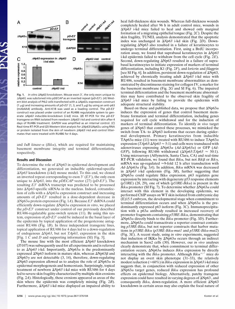
Results and DiscussionTo determine the role of �Np63 in epidermal development anddifferentiation, we generated an inducible epidermal-specific�Np63 knockdown (i-kd) mouse model. To this end, we clonedan inverted repeat corresponding to exon 3� (E3�), the only exonunique to �Np63, into the pDECAP vector (27) (pD-E3�). Theresulting E3� dsRNA transcript was predicted to be processedinto �Np63-specific siRNAs in the nucleus. Indeed, cotransfec-tion of cells with a �Np63� expression construct and increasingamounts of pD-E3� resulted in a dose-dependent decrease in�Np63� protein expression (Fig. 1A). Because E3� dsRNA couldefficiently down-regulate �Np63� expression in vitro, we placedthe pD-E3� construct under control of our previously describedRU486-regulatable gene-switch system (11). By using this sys-tem, expression of pD-E3� could be induced in the basal layer ofthe epidermis by topical application of the progesterone antag-onist RU486 (Fig. 1B). In three independent transgenic lines,topical application of RU486 for 4 days led to a down-regulationof endogenous �Np63, but not TAp63, expression in the skin[Fig. 1 C and D and supporting information (SI) Fig. 5].
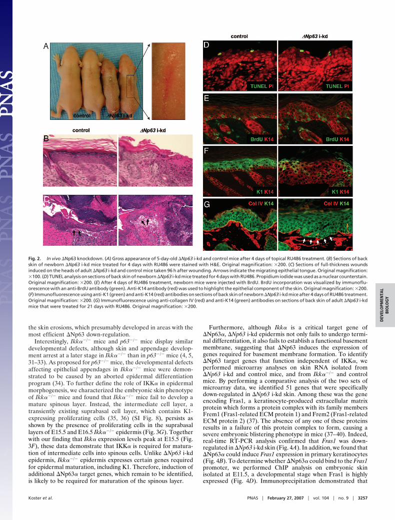
The mouse line with the most efficient �Np63 knockdown(J5107) was subsequently used for all experiments and is referredto as �Np63 i-kd. Importantly, �Np63� is the predominantlyexpressed �Np63 isoform in mature skin, whereas �Np63� and�Np63� are not detectable (3, 14), therefore, down-regulating�Np63 expression allowed us to analyze the role of �Np63� inepidermal morphogenesis and homeostasis. Surprisingly, topicaltreatment of newborn �Np63 i-kd mice with RU486 for 4 daysled to severe skin fragility characterized by multiple skin erosions(Fig. 2A). Histologically, these erosions appeared as areas of theskin where the epidermis was completely missing (Fig. 2B).Furthermore, �Np63 i-kd mice displayed an impaired ability to
heal full-thickness skin wounds. Whereas full-thickness woundscompletely healed after 96 h in adult control mice, wounds in�Np63 i-kd mice failed to heal and showed only marginalformation of a migrating epithelial tongue (Fig. 2C). Despite theskin fragility, TUNEL analysis demonstrated that the apoptoticindex was unchanged in �Np63 i-kd skin (Fig. 2D). Down-regulating �Np63 also resulted in a failure of keratinocytes toundergo terminal differentiation. First, using a BrdU incorpo-ration assay, we found that suprabasal keratinocytes in �Np63i-kd epidermis failed to withdraw from the cell cycle (Fig. 2E).Second, down-regulating �Np63 resulted in a failure of supra-basal keratinocytes to initiate expression of markers of terminaldifferentiation, including K1 (Fig. 2F), and loricrin and filaggrin[see SI Fig. 6]. In addition, persistent down-regulation of �Np63,achieved by chronically treating adult �Np63 i-kd mice withRU486, resulted in basement membrane abnormalities as dem-onstrated by discontinuous staining for collagen IV, a marker forthe basement membrane (Fig. 2G and SI Fig. 6). The impairedterminal differentiation and the basement membrane abnormal-ities may have contributed to the observed skin fragility of�Np63 i-kd mice by failing to provide the epidermis withadequate structural stability.
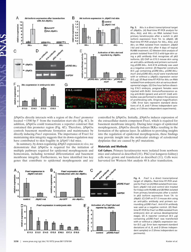
Based on these and published data, we propose that �Np63�induces the expression of genes that control basement mem-brane formation and terminal differentiation, including genesrequired for cell cycle withdrawal and for the induction ofmarkers of terminal differentiation. To identify in vivo targetgenes of �Np63 that control these processes, we mimicked theswitch from TA- to �Np63 isoforms that occurs during epider-mal development. Primary keratinocytes from inducibleTAp63� mice (11) were treated with RU486 to induce TAp63�expression (TAp63:�Np63 � 5:1) and cells were transduced withadenoviruses expressing �Np63� (Ad-�Np63�) or GFP (Ad-GFP), following RU486 withdrawal (�Np63:TAp63 � 50:1).Using microarrays (Affymetrix, Santa Clara, CA) and real-timeRT-PCR validation, we found that Ikk�, but not Ikk� or Ikk�,mRNA was up-regulated �9-fold 12 h after transduction withAd-�Np63� (Fig. 3A). In addition, Ikk� mRNA was decreasedin �Np63 i-kd epidermis (Fig. 3B), further suggesting that�Np63� could regulate Ikk� expression. p63 regulates geneexpression by interacting with degenerate p53 response elements(p53RE) (28, 29), one of which (p53RE-Ikk�) is present in theIkk� promoter (SI Fig. 7). To determine whether �Np63� couldinteract with this element in the developing epidermis, weperformed ChIP assays on WT skin isolated from embryonic day(E)15.5 embryos, the developmental stage when commitment toterminal differentiation occurs and when �Np63� is the pre-dominantly expressed p63 isoform (Fig. 3C). Immunoprecipita-tion with a p63� antibody resulted in increased recovery ofpromoter fragments containing p53RE-Ikk�, demonstrating that�Np63� directly binds to the Ikk� promoter (Fig. 3D). Further-more, �Np63� could transactivate a reporter construct contain-ing p53RE-Ikk�, but not reporter constructs that harbor muta-tions in p53RE-Ikk� (p53RE-Ikk�-mut1 and p53RE-Ikk�-mut2)(Fig. 3E). A recent study, using in vitro experiments, suggestedthat induction of IKK� by �Np63� occurs through an indirectmechanism in Saos2 cells (30). However, our in vivo analysesclearly demonstrate that, when commitment to terminal differ-entiation occurs, �Np63� induces Ikk� expression by directlyinteracting with the Ikk� promoter. Although Ikk��/� mice donot display an overt skin phenotype (31–33), the relativelymodest reduction (�60%) in Ikk� expression in �Np63 i-kd micesuggests that, in conjunction with reduced expression of other�Np63� target genes, reduced Ikk� expression has profoundeffects on epidermal biology. Alternatively, patchy transgeneexpression could have resulted in varying degrees of �Np63 , andconsequently Ikk�, down-regulation. A more efficient �Np63knockdown in certain areas may also explain the focal nature of
Fig. 1. In vitro �Np63 knockdown. Mouse exon 3�, the only exon unique to�Np63, was subcloned into pDECAP as an inverted repeat (pD-E3�). (A) West-ern blot analysis of Ptk2 cells transfected with a �Np63� expression construct(1 �g) and increasing amounts of pD-E3� (1, 3, and 5 �g) by using an anti-p63(mAb4A4) antibody. Anti-K18 was used as a loading control. The pD-E3�construct was placed under control of an RU486 regulatable system to gen-erate �Np63 inducible-knockdown (i-kd) mice. (B) RT-PCR for the pD-E3�transgene on RNA isolated from newborn �Np63 i-kd and control skin after 4days of RU486 treatment. GAPDH was amplified as an internal control. (C)Real-time RT-PCR and (D) Western blot analysis for �Np63/�Np63� using RNAor protein isolated from the skin of newborn �Np63 i-kd and control litter-mates that were treated with RU486 for 4 days.
3256 � www.pnas.org�cgi�doi�10.1073�pnas.0611376104 Koster et al.
the skin erosions, which presumably developed in areas with themost efficient �Np63 down-regulation.
Interestingly, Ikk��/� mice and p63�/� mice display similardevelopmental defects, although skin and appendage develop-ment arrest at a later stage in Ikk��/� than in p63�/� mice (4, 5,31–33). As proposed for p63�/� mice, the developmental defectsaffecting epithelial appendages in Ikk��/� mice were demon-strated to be caused by an aborted epidermal differentiationprogram (34). To further define the role of IKK� in epidermalmorphogenesis, we characterized the embryonic skin phenotypeof Ikk��/� mice and found that Ikk��/� mice fail to develop amature spinous layer. Instead, the intermediate cell layer, atransiently existing suprabasal cell layer, which contains K1-expressing proliferating cells (35, 36) (SI Fig. 8), persists asshown by the presence of proliferating cells in the suprabasallayers of E15.5 and E16.5 Ikk��/� epidermis (Fig. 3G). Togetherwith our finding that Ikk� expression levels peak at E15.5 (Fig.3F), these data demonstrate that IKK� is required for matura-tion of intermediate cells into spinous cells. Unlike �Np63 i-kdepidermis, Ikk��/� epidermis expresses certain genes requiredfor epidermal maturation, including K1. Therefore, induction ofadditional �Np63� target genes, which remain to be identified,is likely to be required for maturation of the spinous layer.
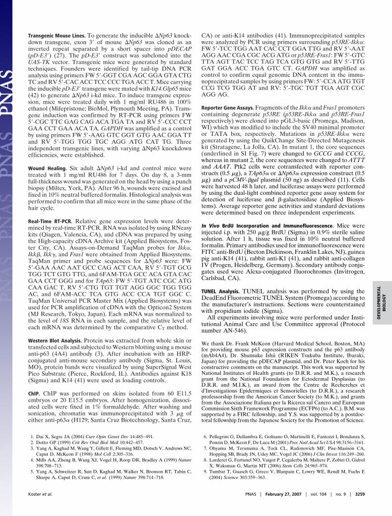
Furthermore, although Ikk� is a critical target gene of�Np63�, �Np63 i-kd epidermis not only fails to undergo termi-nal differentiation, it also fails to establish a functional basementmembrane, suggesting that �Np63 induces the expression ofgenes required for basement membrane formation. To identify�Np63 target genes that function independent of IKK�, weperformed microarray analyses on skin RNA isolated from�Np63 i-kd and control mice, and from Ikk��/� and controlmice. By performing a comparative analysis of the two sets ofmicroarray data, we identified 51 genes that were specificallydown-regulated in �Np63 i-kd skin. Among these was the geneencoding Fras1, a keratinocyte-produced extracellular matrixprotein which forms a protein complex with its family membersFrem1 (Fras1-related ECM protein 1) and Frem2 (Fras1-relatedECM protein 2) (37). The absence of any one of these proteinsresults in a failure of this protein complex to form, causing asevere embryonic blistering phenotype in mice (37–40). Indeed,real-time RT-PCR analysis confirmed that Fras1 was down-regulated in �Np63 i-kd skin (Fig. 4A). In addition, we found that�Np63� could induce Fras1 expression in primary keratinocytes(Fig. 4B). To determine whether �Np63� could bind to the Fras1promoter, we performed ChIP analysis on embryonic skinisolated at E11.5, a developmental stage when Fras1 is highlyexpressed (Fig. 4D). Immunoprecipitation demonstrated that
Fig. 2. In vivo �Np63 knockdown. (A) Gross appearance of 5-day-old �Np63 i-kd and control mice after 4 days of topical RU486 treatment. (B) Sections of backskin of newborn �Np63 i-kd mice treated for 4 days with RU486 were stained with H&E. Original magnification: �200. (C) Sections of full-thickness woundsinduced on the heads of adult �Np63 i-kd and control mice taken 96 h after wounding. Arrows indicate the migrating epithelial tongue. Original magnification:�100. (D) TUNEL analysis on sections of back skin of newborn �Np63 i-kd mice treated for 4 days with RU486. Propidium iodide was used as a nuclear counterstain.Original magnification: �200. (E) After 4 days of RU486 treatment, newborn mice were injected with BrdU. BrdU incorporation was visualized by immunoflu-orescence with an anti-BrdU antibody (green). Anti-K14 antibody (red) was used to highlight the epithelial component of the skin. Original magnification: �200.(F) Immunofluorescence using anti-K1 (green) and anti-K14 (red) antibodies on sections of back skin of newborn �Np63 i-kd mice after 4 days of RU486 treatment.Original magnification: �200. (G) Immunofluorescence using anti-collagen IV (red) and anti-K14 (green) antibodies on sections of back skin of adult �Np63 i-kdmice that were treated for 21 days with RU486. Original magnification: �200.
Koster et al. PNAS � February 27, 2007 � vol. 104 � no. 9 � 3257
DEV
ELO
PMEN
TAL
BIO
LOG
Y
�Np63� directly interacts with a region of the Fras1 promoterlocated �1500 bp 5� from the translation start site (Fig. 4C). Inaddition, �Np63� could transactivate a reporter construct thatcontained this promoter region (Fig. 4E). Therefore, �Np63�controls basement membrane formation and maintenance bydirectly inducing Fras1 expression. The importance of Fras1 formaintaining skin integrity suggests that its down-regulation mayhave contributed to skin fragility in �Np63 i-kd mice.
In summary, by down-regulating �Np63 expression in vivo, wedemonstrate that �Np63� is required for the initiation ofmultiple pathways required for epidermal morphogenesis andhomeostasis, including terminal differentiation and basementmembrane integrity. Furthermore, we have identified two keygenes that contribute to epidermal morphogenesis and are
controlled by �Np63�. Initially, �Np63� induces expression ofthe extracellular matrix component Fras1, which is required forbasement membrane integrity. During later stages of epidermalmorphogenesis, �Np63� directly induces IKK� resulting in theformation of the spinous layer. In addition to providing insightsinto the regulation of epidermal morphogenesis, these findingsmay provide insight into the molecular etiology of ectodermaldysplasias that are caused by p63 mutations.
Materials and MethodsCell Culture. Primary keratinocytes were isolated from newbornmice and cultured as described (41). Ptk2 (rat kangaroo kidney)cells were grown and transfected as described (11). Cells wereharvested for Western blot analysis 48 h after transfection.
Fig. 4. Fras1 is a direct transcriptionaltarget of �Np63�. Real-time RT-PCR anal-ysis for Fras1 on (A) RNA isolated from new-born �Np63 i-kd and control skin treatedfor 4 days with RU486 and (B) RNA isolatedfrom primary keratinocytes after a switchin p63 isoform expression from TA- to�Np63. (C) ChIP on E11.5 mouse skin usingan anti-p63� antibody and primers sur-rounding p53RE-Fras1. Anti-K14 antibodywas used as a negative control. (D) Real-time RT-PCR for Fras1 on RNA isolated fromembryonic skin at various developmentalstages. (E) A reporter construct (0.5 �g)containing p53RE-Fras1 was transfectedwith or without a �Np63� expression vec-tor (0.5 �g). Error bars represent standarddeviations of A, B, and D (three indepen-dent samples) or E (three independent ex-periments).
Fig. 3. Ikk� is a direct transcriptional targetof �Np63�. (A) Real-time RT-PCR analysis forIkk�, Ikk�, and Ikk� on RNA isolated fromprimary keratinocytes after a switch in p63isoform expression from TA- to �Np63. (B)Real-time RT-PCR analysis for Ikk�, Ikk�, andIkk� on RNA isolated from newborn �Np63i-kd and control skin after 4 days of topicalRU486 treatment. (C) Western blot analysis ofprotein isolated from E15.5 wild-type skin us-ing a p63 antibody that recognizes all p63isoforms. (D) ChIP on E15.5 mouse skin usingan anti-p63� antibody and primers surround-ing p53RE-Ikk�. Anti-K14 antibody was usedas a negative control. (E) Reporter constructs(0.5 �g) containing p53RE-Ikk�, p53RE-Ikk�-mut1 and p53RE-Ikk�-mut2 were transfectedwith or without a �Np63� expression vector(0.5 �g). (F) Real-time RT-PCR for Ikk� on RNAisolated from embryonic skin at various devel-opmental stages. (G) One hour before dissect-ing E16.5 embryos, pregnant females wereinjected with BrdU. Immunofluorescence us-ing anti-BrdU (green) and anti-K1 (red) anti-bodies was performed to detect the presenceof proliferating cells. Original magnification:�200. Error bars represent standard devia-tions of A, B, and F (three independent sam-ples), or E (three independent experiments).
3258 � www.pnas.org�cgi�doi�10.1073�pnas.0611376104 Koster et al.
Transgenic Mouse Lines. To generate the inducible �Np63 knock-down transgene, exon 3� of mouse �Np63 was cloned as aninverted repeat separated by a short spacer into pDECAP(pD-E3�) (27). The pD-E3� construct was subcloned into theUAS-TK vector. Transgenic mice were generated by standardtechniques. Founders were identified by tail-tip DNA PCRanalysis using primers FW 5�-GGT CGA AGC GGA GTA CTGTC and RV 5�-CAC ACC TCC CCC TGA ACC T. Mice carryingthe inducible pD-E3� transgene were mated with K14.Glp65 mice(42) to generate �Np63 i-kd mice. To induce transgene expres-sion, mice were treated daily with 1 mg/ml RU486 in 100%ethanol (Mifepristone; BioMol, Plymouth Meeting, PA). Trans-gene induction was confirmed by RT-PCR using primers FW5�-CGC TTC GAG CAG ACA TGA TA and RV 5�-CCC CCTGAA CCT GAA ACA TA. GAPDH was amplified as a controlby using primers FW 5�-AAG GTC GGT GTG AAC GGA TTand RV 5�-TGG TGG TGC AGG ATG CAT TG. Threeindependent transgenic lines, with varying �Np63 knockdownefficiencies, were established.
Wound Healing. Six adult �Np63 i-kd and control mice weretreated with 1 mg/ml RU486 for 7 days. On day 8, a 3-mmfull-thickness wound was generated on the head by using a punchbiopsy (Miltex, York, PA). After 96 h, wounds were excised andfixed in 10% neutral buffered formalin. Histological analysis wasperformed to confirm that all mice were in the same phase of thehair cycle.
Real-Time RT-PCR. Relative gene expression levels were deter-mined by real-time RT-PCR. RNA was isolated by using RNeasykits (Qiagen, Valencia, CA), and cDNA was prepared by usingthe High-capacity cDNA Archive kit (Applied Biosystems, Fos-ter City, CA). Assays-on-Demand TaqMan probes for Ikk�,Ikk�, Ikk�, and Fras1 were obtained from Applied Biosystems.TaqMan primer and probe sequences for �Np63 were: FW5�-GAA AAC AAT GCC CAG ACT CAA, RV 5�-TGT GCGTGG TCT GTG TTG, and 6FAM-TGA GCC ACA GTA CACGAA CCT GGG and for TAp63: FW 5�-TGT ATC CGC ATGCAA GAC T, RV 5�-CTG TGT TGT AGG GGC TGG TGGAC, and 6FAM-ACC TCA GTG ACC CCA TGT GGC C.TaqMan Universal PCR Master Mix (Applied Biosystems) wasused for PCR amplification of cDNA with the Opticon2 System(MJ Research, Tokyo, Japan). Each mRNA was normalized tothe level of 18S RNA in each sample, and the relative level ofeach mRNA was determined by the comparative CT method.
Western Blot Analysis. Protein was extracted from whole skin ortransfected cells and subjected to Western blotting using a mouseanti-p63 (4A4) antibody (3). After incubation with an HRP-conjugated anti-mouse secondary antibody (Sigma, St. Louis,MO), protein bands were visualized by using SuperSignal WestPico Substrate (Pierce, Rockford, IL). Antibodies against K18(Sigma) and K14 (41) were used as loading controls.
ChIP. ChIP was performed on skins isolated from 60 E11.5embryos or 20 E15.5 embryos. After homogenization, dissoci-ated cells were fixed in 1% formaldehyde. After washing andsonication, chromatin was immunoprecipitated with 3 �g ofeither anti-p63� (H129; Santa Cruz Biotechnology, Santa Cruz,
CA) or anti-K14 antibodies (41). Immunoprecipitated sampleswere analyzed by PCR using primers surrounding p53RE-Ikk�:FW 5�-TCC TGG AAT CAC CCT GGA TTG and RV 5�-AATAGG AAC CGA CGC ACG ATG or p53RE-Fras1: FW 5�-GTCTTA AGT TAC TCC TAG TCA GTG GTG and RV 5�-TTGGAT GGA ACC TGA GTC CT. GAPDH was amplified ascontrol to confirm equal genomic DNA content in the immu-noprecipitated samples by using primers FW: 5�-CCA ATG TGTCCG TCG TGG AT and RV: 5�-TGC TGT TGA AGT CGCAGG AG.
Reporter Gene Assays. Fragments of the Ikk� and Fras1 promoterscontaining degenerate p53RE (p53RE-Ikk� and p53RE-Fras1respectively) were cloned into pGL3-basic (Promega, Madison,WI) which was modified to include the SV40 minimal promoteror TATA box, respectively. Mutations in p53RE-Ikk� weregenerated by using the QuikChange Site-Directed Mutagenesiskit (Stratagene, La Jolla, CA). In mutant 1, the core sequences(underlined in SI Fig. 7) were changed to GCCG and CCCG,whereas in mutant 2, the core sequences were changed to ATTTand AAAT. Ptk2 cells were cotransfected with reporter con-structs (0.5 �g), a TAp63� or �Np63� expression construct (0.5�g) and a pCMV-�gal plasmid (50 ng) as described (11). Cellswere harvested 48 h later, and luciferase assays were performedby using the dual-light combined reporter gene assay system fordetection of luciferase and �-galactosidase (Applied Biosys-tems). Average reporter gene activities and standard deviationswere determined based on three independent experiments.
In Vivo BrdU Incorporation and Immunofluorescence. Mice wereinjected i.p. with 250 �g/g BrdU (Sigma) in 0.9% sterile salinesolution. After 1 h, tissue was fixed in 10% neutral bufferedformalin. Primary antibodies used for immunofluorescence wereFITC anti-BrdU (Becton Dickinson, Franklin Lakes, NJ), guineapig anti-K14 (41), rabbit anti-K1 (41), and rabbit anti-collagenIV (Progen, Heidelberg, Germany). Secondary antibody conju-gates used were Alexa-conjugated fluorochromes (Invitrogen,Carlsbad, CA).
TUNEL Analysis. TUNEL analysis was performed by using theDeadEnd Fluorometric TUNEL System (Promega) according tothe manufacturer’s instructions. Sections were counterstainedwith propidium iodide (Sigma).
All experiments involving mice were performed under Insti-tutional Animal Care and Use Committee approval (Protocolnumber AN-546).
We thank Dr. Frank McKeon (Harvard Medical School, Boston, MA)for providing mouse p63 expression constructs and the p63 antibody(mAb4A4), Dr. Shunsuke Ishii (RIKEN Tsukuba Institute, Ibaraki,Japan) for providing the pDECAP plasmid, and Dr. Peter Koch for hisconstructive comments on the manuscript. This work was supported byNational Institutes of Health grants (to D.R.R. and M.K.), a researchgrant from the National Foundation for Ectodermal Dysplasias (toD.R.R. and M.I.K.), an award from the Centre de Recherches etd’Investigations Epidermiques et Sensorielles (to D.R.R.), a researchprofessorship from the American Cancer Society (to M.K.), and grantsfrom the Associazione Italiana per la Ricerca sul Cancro and EuropeanCommission Sixth Framework Programme (ECFP6) (to A.C.). B.M. wassupported by a FIRC fellowship, and Y.S. was supported by a postdoc-toral fellowship from the Japanese Society for the Promotion of Science.
1. Dai X, Segre JA (2004) Curr Opin Genet Dev 14:485–491.2. Dotto GP (1999) Crit Rev Oral Biol Med 10:442–457.3. Yang A, Kaghad M, Wang Y, Gillett E, Fleming MD, Dotsch V, Andrews NC,
Caput D, McKeon F (1998) Mol Cell 2:305–316.4. Mills AA, Zheng B, Wang XJ, Vogel H, Roop DR, Bradley A (1999) Nature
398:708–713.5. Yang A, Schweitzer R, Sun D, Kaghad M, Walker N, Bronson RT, Tabin C,
Sharpe A, Caput D, Crum C, et al. (1999) Nature 398:714–718.
6. Pellegrini G, Dellambra E, Golisano O, Martinelli E, Fantozzi I, Bondanza S,Ponzin D, McKeon F, De Luca M (2001) Proc Natl Acad Sci USA 98:3156–3161.
7. Ohyama M, Terunuma A, Tock CL, Radonovich MF, Pise-Masison CA,Hopping SB, Brady JN, Udey MC, Vogel JC (2006) J Clin Invest 116:249–260.
8. Larderet G, Fortunel NO, Vaigot P, Cegalerba M, Maltere P, Zobiri O, GidrolX, Waksman G, Martin MT (2006) Stem Cells 24:965–974.
9. Tumbar T, Guasch G, Greco V, Blanpain C, Lowry WE, Rendl M, Fuchs E(2004) Science 303:359–363.
Koster et al. PNAS � February 27, 2007 � vol. 104 � no. 9 � 3259
DEV
ELO
PMEN
TAL
BIO
LOG
Y
10. Morris RJ, Liu Y, Marles L, Yang Z, Trempus C, Li S, Lin JS, Sawicki JA,Cotsarelis G (2004) Nat Biotechnol 22:411–417.
11. Koster MI, Kim S, Mills AA, DeMayo FJ, Roop DR (2004) Genes Dev18:126–131.
12. Koster MI, Kim S, Huang J, Williams T, Roop DR (2006) Dev Biol 289:253–261.13. Laurikkala J, Mikkola ML, James M, Tummers M, Mills AA, Thesleff I (2006)
Development (Cambridge, UK) 133:1553–1563.14. Liefer KM, Koster MI, Wang XJ, Yang A, McKeon F, Roop DR (2000) Cancer
Res 60:4016–4020.15. Parsa R, Yang A, McKeon F, Green H (1999) J Invest Dermatol 113:1099–1105.16. King KE, Ponnamperuma RM, Yamashita T, Tokino T, Lee LA, Young MF,
Weinberg WC (2003) Oncogene 22:3635–3644.17. King KE, Ponnamperuma RM, Gerdes MJ, Tokino T, Yamashita T, Baker CC,
Weinberg WC (2006) Carcinogenesis 27:53–63.18. Ellisen LW, Ramsayer KD, Johannessen CM, Yang A, Beppu H, Minda K,
Oliner JD, McKeon F, Haber DA (2002) Mol Cell 10:995–1005.19. Truong AB, Kretz M, Ridky TW, Kimmel R, Khavari PA (2006) Genes Dev
20:3185–3197.20. Missero C, Calautti E, Eckner R, Chin J, Tsai LH, Livingston DM, Dotto GP
(1995) Proc Natl Acad Sci USA 92:5451–5455.21. Westfall MD, Mays DJ, Sniezek JC, Pietenpol JA (2003) Mol Cell Biol
23:2264–2276.22. Nguyen BC, Lefort K, Mandinova A, Antonini D, Devgan V, Della GG, Koster
MI, Zhang Z, Wang J, Tommasi DV, et al. (2006) Genes Dev 20:1028–1042.23. Rangarajan A, Talora C, Okuyama R, Nicolas M, Mammucari C, Oh H, Aster
JC, Krishna S, Metzger D, Chambon P, et al. (2001) EMBO J 20:3427–3436.24. Beretta C, Chiarelli A, Testoni B, Mantovani R, Guerrini L (2005) Cell Cycle
4:1625–1631.
25. Martinez LA, Chen Y, Fischer SM, Conti CJ (1999) Oncogene 18:397–406.26. Testoni B, Mantovani R (2006) Nucleic Acids Res 34:928–938.27. Shinagawa T, Ishii S (2003) Genes Dev 17:1340–1345.28. Zeng X, Levine AJ, Lu H (1998) Proc Natl Acad Sci USA 95:6681–6686.29. Bian J, Sun Y (1997) Proc Natl Acad Sci USA 94:14753–14758.30. Candi E, Terrinoni A, Rufini A, Chikh A, Lena AM, Suzuki Y, Sayan BS,
Knight RA, Melino G (2006) J Cell Sci 119:4617–4622.31. Hu Y, Baud V, Delhase M, Zhang P, Deerinck T, Ellisman M, Johnson R,
Karin M (1999) Science 284:316–320.32. Takeda K, Takeuchi O, Tsujimura T, Itami S, Adachi O, Kawai T, Sanjo H,
Yoshikawa K, Terada N, Akira S (1999) Science 284:313–316.33. Li Q, Lu Q, Hwang JY, Buscher D, Lee KF, Izpisua-Belmonte JC, Verma IM
(1999) Genes Dev 13:1322–1328.34. Sil AK, Maeda S, Sano Y, Roop DR, Karin M (2004) Nature 428:660–664.35. Smart IH (1970) Br J Dermatol 82:276–282.36. Lechler T, Fuchs E (2005) Nature 437:275–280.37. Kiyozumi D, Sugimoto N, Sekiguchi K (2006) Proc Natl Acad Sci USA
103:11981–11986.38. Vrontou S, Petrou P, Meyer BI, Galanopoulos VK, Imai K, Yanagi M,
Chowdhury K, Scambler PJ, Chalepakis G (2003) Nat Genet 34:209–214.39. Jadeja S, Smyth I, Pitera JE, Taylor MS, van Haelst M, Bentley E, McGregor
L, Hopkins J, Chalepakis G, Philip N, et al. (2005) Nat Genet 37:520–525.40. Smyth I, Du X, Taylor MS, Justice MJ, Beutler B, Jackson IJ (2004) Proc Natl
Acad Sci USA 101:13560–13565.41. Yuspa SH, Kilkenny AE, Steinert PM, Roop DR (1989) J Cell Biol 109:1207–
1217.42. Cao T, He W, Roop DR, Wang XJ (2002) Genesis 32:189–190.
3260 � www.pnas.org�cgi�doi�10.1073�pnas.0611376104 Koster et al.