RESEARCH Open Access Identification of p63 keratinocyte ...
13
RESEARCH Open Access Identification of p63 + keratinocyte progenitor cells in circulation and their matrix-directed differentiation to epithelial cells Renjith P Nair and Lissy K Krishnan * Abstract Introduction: In the event of chronic diabetes or burn wounds, accomplishing skin regeneration is a major concern. Autologous skin grafting is the most effective remedy, but the tissue harvest may create more nonhealing wounds. Currently available skin substitutes have a limited clinical outcome because of immune reactions arising from the xenobiotic scaffold or allogenous cells. Autologous stem cells that can be collected without an additional injury may be a viable option for skin-tissue engineering. Presence of a low number of keratinocyte progenitor cells (KPCs) within the peripheral blood mononuclear cell (PBMNC) population has been indicated. Identification, isolation, expansion, and differentiation of KPCs is necessary before they are considered for skin regeneration, which is the focus of this study. Methods: Culture of isolated human PBMNCs on a cell-specific matrix was carried out to induce differentiation of KPCs. Flow cytometry and reverse transcriptase polymerase chain reaction were done for epithelial stem cell marker p63 and lineage markers cytokeratin 5 and cytokeratin 14, to track differentiation. Proliferation was confirmed by quantifying the proliferating cell nuclear antigen-expressing cells. Immunostaining with epithelial cell markers, involucrin and filaggrin, was carried out to establish terminal differentiation. Microscopic analysis confirmed growth and survival of KPCs on the dermal fibroblast monolayer and on a transplantable fibrin sheet. Results: We demonstrated that KPCs are p63 + and CD34 - . The specifically designed composition of the extracellular matrix was found to support selective adhesion, proliferation, and differentiation of p63 + KPCs. The PBMNC culture for 12 days under controlled conditions resulted in a homogenous population that expressed cytokeratins, and >90% of the cells were found to proliferate. Subculture for 5 days resulted in expression of filaggrin and involucrin, suggesting terminal differentiation. Transfer of matrix-selected KPCs to a dermal fibroblast monolayer or fibrin supported cell proliferation and showed typical hexagonal morphology of keratinocytes within 15 days. Conclusions: Circulating KPCs were identified with p63, which differentiated into keratinocytes with expression of the cytokeratins, involucrin and filaggrin. Components of the specifically designed matrix favored KPC attachment, directed differentiation, and may turn out to be a potential vehicle for cell transplantation. Keywords: Keratinocyte progenitor cells, p63, Cytokeratins, Peripheral blood mononuclear cells, Epithelial cells, Fibrin, Matrix-directed differentiation, Skin-tissue engineering * Correspondence: [email protected] Thrombosis Research Unit, Biomedical Technology Wing, Sree Chitra Tirunal Institute for Medical Sciences and Technology, Trivandrum 695012, India © 2013 Nair and Krishnan; licensee BioMed Central Ltd. This is an Open Access article distributed under the terms of the Creative Commons Attribution License (http://creativecommons.org/licenses/by/2.0), which permits unrestricted use, distribution, and reproduction in any medium, provided the original work is properly cited. Nair and Krishnan Stem Cell Research & Therapy 2013, 4:38 http://stemcellres.com/content/4/2/38
Transcript of RESEARCH Open Access Identification of p63 keratinocyte ...

Nair and Krishnan Stem Cell Research & Therapy 2013, 4:38http://stemcellres.com/content/4/2/38
RESEARCH Open Access
Identification of p63+ keratinocyte progenitorcells in circulation and their matrix-directeddifferentiation to epithelial cellsRenjith P Nair and Lissy K Krishnan*
Abstract
Introduction: In the event of chronic diabetes or burn wounds, accomplishing skin regeneration is a majorconcern. Autologous skin grafting is the most effective remedy, but the tissue harvest may create more nonhealingwounds. Currently available skin substitutes have a limited clinical outcome because of immune reactions arisingfrom the xenobiotic scaffold or allogenous cells. Autologous stem cells that can be collected without an additionalinjury may be a viable option for skin-tissue engineering. Presence of a low number of keratinocyte progenitor cells(KPCs) within the peripheral blood mononuclear cell (PBMNC) population has been indicated. Identification,isolation, expansion, and differentiation of KPCs is necessary before they are considered for skin regeneration, whichis the focus of this study.
Methods: Culture of isolated human PBMNCs on a cell-specific matrix was carried out to induce differentiation ofKPCs. Flow cytometry and reverse transcriptase polymerase chain reaction were done for epithelial stem cell markerp63 and lineage markers cytokeratin 5 and cytokeratin 14, to track differentiation. Proliferation was confirmed byquantifying the proliferating cell nuclear antigen-expressing cells. Immunostaining with epithelial cell markers,involucrin and filaggrin, was carried out to establish terminal differentiation. Microscopic analysis confirmed growthand survival of KPCs on the dermal fibroblast monolayer and on a transplantable fibrin sheet.
Results: We demonstrated that KPCs are p63+ and CD34-. The specifically designed composition of the extracellularmatrix was found to support selective adhesion, proliferation, and differentiation of p63+ KPCs. The PBMNC culturefor 12 days under controlled conditions resulted in a homogenous population that expressed cytokeratins, and>90% of the cells were found to proliferate. Subculture for 5 days resulted in expression of filaggrin and involucrin,suggesting terminal differentiation. Transfer of matrix-selected KPCs to a dermal fibroblast monolayer or fibrinsupported cell proliferation and showed typical hexagonal morphology of keratinocytes within 15 days.
Conclusions: Circulating KPCs were identified with p63, which differentiated into keratinocytes with expression ofthe cytokeratins, involucrin and filaggrin. Components of the specifically designed matrix favored KPC attachment,directed differentiation, and may turn out to be a potential vehicle for cell transplantation.
Keywords: Keratinocyte progenitor cells, p63, Cytokeratins, Peripheral blood mononuclear cells, Epithelial cells,Fibrin, Matrix-directed differentiation, Skin-tissue engineering
* Correspondence: [email protected] Research Unit, Biomedical Technology Wing, Sree Chitra TirunalInstitute for Medical Sciences and Technology, Trivandrum 695012, India
© 2013 Nair and Krishnan; licensee BioMed Central Ltd. This is an Open Access article distributed under the terms of theCreative Commons Attribution License (http://creativecommons.org/licenses/by/2.0), which permits unrestricted use,distribution, and reproduction in any medium, provided the original work is properly cited.

Nair and Krishnan Stem Cell Research & Therapy 2013, 4:38 Page 2 of 13http://stemcellres.com/content/4/2/38
IntroductionFor the cure of chronic ulcers, tissue-engineered skin sub-stitute has been proven to produce a positive outcome.However, because the commercially available skin substi-tutes use xenobiotic scaffold material or allogenic cells [1],the outcome is limited. Therefore, identification of a suit-able cell source and a biocompatible culture matrix is crit-ical for successful skin-tissue engineering.The major requirements of the cells to be used for skin-
tissue engineering are that they should not be terminallydifferentiated, they must possess proliferation potential,and they must be lineage committed. It has been shownthat the bone marrow-derived stem cells (BMDSCs) coulddifferentiate into a number of different cell types, includ-ing epidermis [2]. In many instances, BMDSCs leave bonemarrow, circulate in the blood, and are found in peripheralblood mononuclear cell (PBMNC) fractions [3]. A subsetof PBMNCs is considered to be multipotent and to ac-quire macrophagic, epithelial, endothelial, neuronal, andhepatocytic phenotypes [4]. The bone marrow-derivedmesenchymal stem cells (MSCs) have been shown to dif-ferentiate into keratinocyte-like cells in vitro [5]. Sasakiet al. [6] showed that MSCs are recruited into woundedskin and contribute to wound repair by differentiation intoskin cells. Medina et al. [7] reported that circulating bonemarrow-derived CD34+ cells may have the capacity totransdifferentiate into epithelium-like cells. They alsoshowed recently that CD14+ monocytes could differentiateinto keratinocyte-like epithelial cells [8]. All these resultssuggests that, other than the basal stem cells that are re-sponsible for normal epithelial homeostasis, circulatingprogenitors may also contribute to epithelial layer regener-ation. However, none of these studies used a specificmarker for demonstrating the presence of keratinocytelineage-committed stem cells in peripheral blood, whichcould be directed to skin epithelium.A homologue of p53, p63, is an essential transcription
factor for epithelial development and proliferation. Thismolecule was shown to distinguish keratinocyte stem cellsfrom their transiently amplifying (TA) progenitors involvedin skin generation [9]. It was also shown that TA cell cul-tures express a cell proliferation-associated nuclear antigen(PCNA). Therefore, the presence of p63 indicates that thecells can proliferate, form clones, and maintain stemness.The transcription factor p63 is also required for epidermallineage commitment, epidermal differentiation, cell adhe-sion, and basement membrane formation [10]. Once thebasal stem cells reach the suprabasal region, p63 may bedownregulated; subsequently proliferation may be arrested.Therefore, it is a probable candidate to be present onlineage-committed keratinocyte progenitor cells (KPCs) aswell. Because KPCs are present in low numbers in circula-tion, it is essential for them to have good proliferation po-tential if they are to be involved in epithelial regeneration.
In addition to p63, the stem cells in the basal layer alsocontain keratin bundles such as cytokeratin 5 (CK5) andcytokeratin 14 (CK14), and once they differentiate, add-itional molecules such as filaggrin and involucrin areexpressed.Attachment and multiplication of the identified stem or
progenitor cell in vitro and in vivo require an appropriatesubstrate that has adhesion sites and signaling molecules.In physiology, p63+ stem cell proliferation takes place onthe basement membrane, which consists of fibronectin,laminin, collagen, and growth factors. Fibrin clot is the nat-ural scaffold that promotes wound healing in almost all re-gions of the human body and more specifically in skinrepair. A naturally formed fibrin clot at the injury site con-tains important adhesive proteins, such as fibronectin, andgrowth factors released from platelets and from other cellssuch as fibroblasts. Exogenous fibrin matrix, carefully com-posed of cell-specific growth factors, is proven to be suit-able for differentiation of endothelial progenitor cell [11]and neural progenitors [12]. Therefore, a fibrin-based, cell-specific matrix composition may be designed to supportattachment and differentiation of KPCs. The extracellularmatrix (ECM) molecules, which are important for growthof skin cells, are epidermal growth factor (EGF), angiogenicgrowth factors (AGFs), hyaluronic acid (HA), gelatin, lam-inin, and fibronectin.In this study, p63, cytokeratins (CK5 and CK14), in-
volucrin, and filaggrin antigens were used as markers toestablish the lineage of KPCs and their differentiation afterculture on the specifically designed matrix. The resultsmay lead to a novel approach for treating chronic woundswith a tissue-engineered skin substitute derived from au-tologous keratinocyte progenitors isolated from blood andgrown on a human plasma-derived fibrin matrix.
MethodsGelatin, formaldehyde, Histopaque 1077, ascorbic acid,heparin, insulin, 40,6-diamidino-2-phenylindole (DAPI),hydrocortisone, agarose, and TritonX-100 were from SigmaAldrich (St. Louis, MO, USA). DMEM: F12 medium,trypsin-EDTA, and antibiotic-antimycotic were fromGIBCO BRL (Now part of Invitrogen Corporation, GrandIsland NY, USA). Epidermal growth factor (EGF) was fromR &D Systems (Minneapolis, MN, USA), and polystyreneculture dishes were from Nunc (Roskilder, Denmark). In-house prepared materials used were bovine hypothalamusextract (BHE) prepared from young calf tissue, according tothe method of Maciag et al. [13], hyaluronic acid (HA) pre-pared from human umbilical cord [14] and dialyzed new-born calf serum, cryoprecipitated fibrinogen concentratefrom human plasma, and purified human thrombin [15].The complete medium contained 10% newborn calf serum,5 μg/ml insulin, 0.3 mM ascorbic acid, 25 μg/ml BHE, 0.5μg/ml hydrocortisone, and 20 ng/ml EGF, and 10 μl/ml

Nair and Krishnan Stem Cell Research & Therapy 2013, 4:38 Page 3 of 13http://stemcellres.com/content/4/2/38
antibiotic-antimycotic. Anti-human p63 antibody (NovaCastra, Newcastle upon Tyne, UK), phycoerythrin (PE) con-jugated anti-human cytokeratin5 (Santa Cruz Biotechnology,Santa Cruz, CA, USA), anti-human cytokeratin14 (NovaCastra), FITC-conjugated antihuman proliferating cell nu-clear antigen (PCNA) (Caltag, now part of Invitrogen Cor-poration, Grand Island NY, USA), antihuman involucrin(Nova Castra), and antihuman filaggrin (Santa Cruz Biotech-nology) were used as markers for various experiments.Secondary antibodies used were FITC- or PE-conjugatedanti-mouse IgG from Santa Cruz Biotechnology or Abcam(Cambridge Science Park, Cambridge, UK).
Matrix preparationCulture plates were coated with fibrin composite, accor-ding to the established procedure of Chennazhy andKrishnan [15], with a modified fibrinogen cocktail (FC).The modified FC consists of fibrinogen (2 mg/ml), fi-bronectin (200 μg/ml), gelatin (0.2%), BHE (25 μg/ml),EGF (20 ng/ml), and HA (0.1 mg/ml). The fibrin net-work in the culture dish were lyophilized and stored at4°C until use. A fibrin sheet was prepared, as describedearlier [16]. In brief, 1.0 ml of fibrin composite, as de-scribed earlier, was mixed with 1.0 ml (5 IU) thrombinto make a clot in a culture well (about 3.3 cm diameter)and was lyophilized to form a stable sheet. The fibrinsheet was suspended in the medium for 5 to 10 minutesbefore using for cell culture.
Isolation of peripheral blood mononuclear cellsIsolation of PBMNCs from the buffy coat (discards fromBlood Bank collected with informed consent) was donewith Histopaque-1077 density gradient centrifugation. Asper the standard operating procedure of Institutional Eth-ics Committee, Sree Chitra Tirunal Institute for MedicalSciences and Technology (IEC-SCTIMST), the collectionof discarded buffy coat is exempted from review if thedonor identity is not publicized. In brief, red blood cells(RBCs) were settled by centrifugation of the buffy coat at1,200 g for 15 minutes in 15-ml centrifuge tubes (HareusStratos, Hanau, Germany). Superficial plasma wasdiscarded; PBMNCs at the interface were collected and di-luted with equal volumes of Hank Balanced Salt Solution(HBSS), layered over Histopaque-1077 in centrifuge tubes(Nunc, Roskilder, Denmark), and centrifuged at 400 g for30 minutes at 25°C. The layer containing PBMNCs wascarefully separated from the plasma-Histopaque interfaceand was washed with serum-free DMEM: F12 by centrifu-gation at 150 g at 4°C for 10 minutes. The washedPBMNCs were suspended in complete medium.
Cell cultureThe PBMNCs suspended in complete medium were ini-tially seeded in bare 20-cm2 culture dishes and were
incubated for 1 hour in a humidified incubator under 5%CO2 at 37°C. After 1 hour, the medium was removed gen-tly, and fresh medium was added. After 48 hours, themedium was removed, and nonadherent but settled cellswere flushed with complete keratinocyte culture medium,and the cells attached to the bare polystyrene surface werediscarded. The plastic-nonadherent cells were seeded ontomatrix-coated dishes and were incubated in a humidifiedincubator under 5% CO2 at 37°C. The medium was re-placed with fresh medium each day until 72 hours; after-ward, the medium was changed on alternate days. Themorphology of cultured cells was observed with a phase-contrast microscope (Leica Microsystems, DMIRB Wetzlar,Germany).Fibroblasts were isolated from human foreskin from a
local hospital (collected with informed consent) by using astandard protocol [17]. Cells were subcultured, and eitherthe third- or the fourth-passage monolayer was used forseeding KPCs. The protocol for co-culture was to seedPBMNCs in an uncoated dish for 48 hours, transfer to acoated dish, and after 4 days on biomimetic matrix, KPCswere flushed out and seeded onto either a fibroblastmonolayer or a fibrin sheet. Culture was continued in thesame medium composition as described earlier.
Immunostaining for fluorescence microscopyCells cultured on FC-coated 1.75-cm2 wells for a specificperiod of time were rinsed with phosphate-buffered saline(PBS), fixed with 3.7% formaldehyde in PBS, washed withPBS, and permeated with Triton X-100 (0.1%) in PBS. Cellswere then incubated with primary antibodies against p63(1:50), FITC-conjugated cytokeratin 14 antibody (1:50), orwith phycoerythrin (PE)-conjugated cytokeratin 5 (1:50),antiinvolucrin (1:100), and antifilaggrin (1:100). Conjugatedsecondary antibody was used at 1:200 dilution, and cul-tures were viewed by using a fluorescence microscope(Leica Microsystems, DMIRB, Wetzlar, Germany). Actinstaining was done by using phalloidin conjugated withTexas Red (Molecular Probes, now part of Invitrogen Cor-poration, Grand Island NY, USA), and for nuclear stainingof cells, DAPI (Sigma Aldrich) was used.
Flow cytometryCells in culture for 1 hour, 4 days, 8 days, and 12 dayswere harvested by flushing out or by using the standardtrypsinization protocol (0.25% Trypsin-EDTA for 3 mi-nutes), washed with PBS, fixed with 3.7% formaldehyde inPBS for 20 minutes, washed with PBS, and permeatedwith Triton X-100 (0.1%) in PBS for 5 minutes. The cellswere treated with 1% bovine serum albumin (Sigma-Al-drich, St. Louis, MO, USA) in PBS for 30 minutes and in-cubated with FITC-conjugated antibodies against PCNA(1:100), CK14 (1:200), phycoerythrin-conjugated CK5(1:100), and nonconjugated p63 (1:100) antibody for 2

Nair and Krishnan Stem Cell Research & Therapy 2013, 4:38 Page 4 of 13http://stemcellres.com/content/4/2/38
hours. The cells incubated with primary p63 antibodieswere washed and treated with FITC-conjugated secondaryantibody (1:200) for 1 hour. After the staining procedure,the cells were washed and analyzed by using a flowcytometer (FACS ARIA, BD Biosciences, San Jose, CA,USA). The percentage of marker-expressed cells was cal-culated by using BD FACS Diva software (BD Biosciences,San Jose, CA, USA).Sorting of CD34+ cells from the PBMNCs was done
from the enriched Histopaque gradient centrifugationstep, seeded on bare polystyrene for at least 1 hour, andthe cells harvested from bare polystyrene were stainedwith CD34 antibodies conjugated with APC (BD Biosci-ences, Franklin Lakes, NJ, USA) and sorted by usingFACS Aria (BD Biosciences). A portion of the sortedcells was used for analysis of purity, and the remainingcells were fixed, permeated, and stained with p63, as de-scribed earlier, and analyzed.
Reverse transcriptase-polymerase chain reactionFor detecting expression of markers before and after cul-ture, RT-PCR analysis was performed. Total RNA wasextracted from the cells in culture on day 1 and day 12by using Trizol reagent (Invitrogen Invitrogen Corpor-ation, Grand Island NY, USA) according to manufac-turer’s protocol. RNA, 1 μg, was converted to cDNA byusing superscript III reverse transcriptase enzyme(Invitrogen Corporation). The expression of keratinocytelineage markers, such as TAp63, ΔNp63, CK5, and CK14,was analyzed with RT-PCR. For amplification reactions,Master cycler from Eppendorff (Hamburg, Germany) wasused for 40 cycles; the annealing temperature for all reac-tions was 54°C, and the product was analyzed for molecu-lar size by using agarose gel electrophoresis. GAPDH wasused as a positive control. The primer sequence used foreach gene is given in Table 1. For each gene, an assess-ment of quality was performed by examining PCR meltcurves after real-time PCR by using a Chromo4 system(MJ Research, now part of Bio-Rad, Hercules, CA, USA)to ensure specificity.
ResultsIn the preliminary experiments, different compositions ofmatrices were found to support adhesion of KPCs fromPBMNCs with scanty cell survival. For promoting their
Table 1 Primers used for the polymerase chain reaction analy
Gene Forward primer
GAPDH 50-gcttgtcatcaatggaaatccc-30
TAp63 50-aagatggtgcgacaaacaag-30
ΔNp63 50-ggaaaacaatgcccagactc-30
CK5 50-cttgtggagtgggtggctat-30
CK14 50-gaccattgaggacctgagga-30
survival, multiplication, lineage commitment, and differen-tiation, several modifications were necessary. The finalcomposition of fibrinogen composite (FC) described earlierwas found to be suitable for attaining cell survival andmultiplication. The cells were initially seeded on bare poly-styrene culture wells, and the floating cells were removed atleast 3 times within a period of 48 hours of starting the cul-ture. The process involved light swirling of the cultureplates and aspiration of medium, which was replaced withfresh medium to eliminate many of the nonspecific cellsfrom the culture. The unattached cells that had settled atthe bottom were likely to have many multipotent adult pro-genitor cells (MAPCs), so they were flushed out and seededonto the FC-coated dishes. Within 1 hour of seeding onFC, the floating cells were removed. The attached cells werefound to form small groups of four to six cells and wereuniformly distributed in the culture well (Figure 1A). Whenthe medium was changed on alternate days, more floatingcells were removed. Cells were analyzed periodically, andon days 4, 8, and 12, many fields were viewed to check thefrequency of cell colonies and morphologic variations, ifany, and were micrographed. The size of the colony wasfound to increase with time (Figure 1B through D). In mostof the experiments, the cells grew around the initial colonyand filled the culture well in 12 days (Figure 1D). A con-tinuous bed of cells was seen in 15 days (Figure 1E, F).Until the fourth day, cells were not tightly adhered to theculture substrate and could be harvested by flushing withmedium, for transfer to other substrates or for analysis. Atlater periods, cells were attached tightly to the matrix andcould be harvested only by the standard trypsinizationmethod.For identification of KPCs among the cells seeded onto
the FC, p63, CK5, and CK14 were analyzed by usingflow cytometry. Compiled data of flow-cytometric ana-lysis of cells harvested on days 1, 4, 8, and 12 is shownin Figure 2A. The sample that was analyzed on day 1was obtained after 1 hour of PBMNC culture on an un-coated dish. On all days, excluding the debris, all cellsfrom the FSC-versus-SSC plot were gated; and in theSSC-versus-FL1/FL2 plot, the gate having <0.5% was se-lected for detection of the positively stained population.Among the cells obtained after 1 hour, few were p63+ inthe gated region. The p63+ cells were prominent in themonocyte region of the FSC-versus-SSC plot. No CK14+
sis
Reverse primer Amplicon size
50-tccacacccatgacgaacatg- 30 210 bp
50-agagagcatcgaaggtggag-30 234 bp
50-gtggaatacgtccaggtggc-30 294 bp
50-ccacttggtgtccagaacct-30 439 bp
50-attgatgtcggcttccacac-30 157 bp

Figure 1 Representative phase-contrast micrographs of cells on fibrinogen composite (FC) matrix. (A) Cells on the day of seeding; (B) onday 4 of seeding; (C) on day 8; (D) on day 12; (E, F), on day 15. A through E are at low magnification (scale bar, 200 μm), and F is at highermagnification (scale bar, 50 μm).
Nair and Krishnan Stem Cell Research & Therapy 2013, 4:38 Page 5 of 13http://stemcellres.com/content/4/2/38
or CK5+cells were detectable on day 1 of the culture.Tracking of progenitor /differentiation markers fromdays 1 to 12 was made possible by using culture ofsingle-donor PBMNCs in at least four culture wells of10 cm2 area, and cells from each well were aspirated formeasurement on each specified day (one dish perperiod). From day 8 onward, almost all cells in the FSC-versus-SSC plots were in a single cluster, which indi-cated that the cells were homogeneous as comparedwith previous periods in terms of size and granularity.By day 12, about 90% of cells were positive for both p63and CK14, but the percentage of CK5 was lowest amongthe three antigens. Of the cells that survived on day 4,69% ± 8% were p63+, 4% ± 2.5% were CK5+, and 4.9% ±3% were CK14+. This finding suggests that all p63+ cellsmay have survived, and some of them seemed to havedifferentiated into keratinocyte lineage with CK5 andCK14 expression by day 4. Obviously, specific KPC ad-hesion to the FC occurred, and floating cells were elimi-nated from the culture over a period of 4 days. Thehigher percentage of p63+ cells on day 4 as comparedwith day 1 may be due to the elimination of large
number of nonspecific cells from the culture and prolifera-tion of p63+ KPCs. A remarkable difference was seen inCK5 and CK14 expression on cells cultured for 8 days.Whereas p63+ cells were about 80% of the total viablepopulation, >95% were CK5+, and >80% were CK14+.Average ± standard deviation (SD) of the values from threedonors (Figure 2A) show similar progression of expressionfor p63, CK5, and CK14, with the lowest SD by day 12 ofculture. A differentiation experiment was done by usingPBMNCs from many other donors for confirming differen-tiation; however, tracking and quantification of marker ex-pression by using three donors only is presented here.Overall, it was observed that if the PBMNC yield was good,the cell survival and expression of markers was also asgood, as the data show in Figure 2. But if PBMNC yieldand seeding density were poor, cell survival and marker ex-pression were poor, too. It appeared that seeding density iscrucial for KPC survival, with the best results when theseeding density of leukocytes was about 1 × 10⁶/cm2 of thebiomimetic matrix.In Figure 2B, results of the gene-expression analysis are
shown. In cells on day 1, one isoform of p63 (TAp63) was

Figure 2 Evidence for differentiation. (A) Compiled data offlow-cytometric analysis during cell culture. The percentage of cellspositive for p63, CK5, and CK14 on each day of analysis during theperiods of culture is presented. Periods of analysis against thepercentage of positive cells for each parameter are marked in therespective axis. Average ± SD of replicate experiments (n = 3),carried out by using different donor PBMNCs are presented. Thevalues are plotted in the bar diagram for comparison of percentageof positive cells on each day. CK14- and CK5-positive cells wereabsent on day 1. (B) Expressions of p63, CK5, and CK14 genes on theday of starting the culture and after 12 days of culture. GAPDH wasthe housekeeping gene, and TAp63 was the only KPC-specific genethat amplified on day 1. ΔNp63, CK5, and CK14 were expressed onday 12, and the products showed specific molecular size.
Nair and Krishnan Stem Cell Research & Therapy 2013, 4:38 Page 6 of 13http://stemcellres.com/content/4/2/38
expressed, but none of the other tested markers was ampli-fied in 40 cycles of reaction. In cells on day 12, TAp63,ΔNp63, CK5, and CK14 genes were clearly expressed.These data confirm that p63, CK5, and CK14 are expressedwhen PBMNCs were cultured on the KPC-specific niche inthis study. How cells were stimulated to the keratinocytelineage during the later period of cell culture is beyond thescope of this study. It is likely that the molecules present onthe culture matrix and in the medium together would haveinfluenced the keratinocyte lineage commitment and ex-pression of more keratinocyte markers.The KPCs were found to proliferate during the culture
period, and big clusters were found frequently throughout
the culture surface. The proliferation marker used wasPCNA, and an average of 60% of the gated cells werePCNA positive on day 1; gradually the average reached>95% by day 8, but by day 12, the percentage of proliferat-ing cells decreased to 80% (Figure 3). The reason for thelower percentage of proliferating cells on the initial daymay be that the cells were more heterogeneous at that time,and other nonspecific and nondividing cells contaminatedthe gated region; large donor-to-donor variability was seenduring this period. Once they became more homogeneouson day 8 with >95% of cells expressing CK5 (Figure 2A),they were all expressing PCNA as well (Figure 3). The pro-liferation potential was reduced by day 12, may be becauseterminal differentiation progressed.The surface marker CD34 has been reported to be posi-
tive on many MAPCs in circulation. To test for its pres-ence on p63+ KPCs, CD34+ cells were sorted by usingFACS Aria. Purity of the sorted cell population was >70%.When the sorted cells were permeated and stained withp63 antibodies, none of the CD34+ cells was found to bep63+ (Figure 4A through F). Even though KPCs are likelyto be of bone-marrow origin, once committed to thelineage and expressed p63, they do not seem to be CD34+.The data were consistent in three sorting experiments,which were done by using three different donor PBMNCs.In this study, we found that p63 is a useful marker to
identify keratinocyte progenitors in the circulating blood,which could survive in the culture. With a nuclear anti-gen, permeation of cells is necessary to stain p63, so itcannot be used as a marker to sort KPCs from PBMNCfractions. Because the cells are CD34-, it too cannot beused to sort KPCs from PBMNCs; therefore, matrix-directed cell attachment, proliferation, and differenti-ation are the most suitable strategies to culture-amplifynumbers of KPCs for use in regenerative medicine.To confirm keratinocyte lineage commitment and differ-
entiation, cultures were fixed and stained with fivemarkers, which included p63, CK5, CK14, filaggrin, andinvolucrin. Fibroblasts from human foreskin were used asnegative control for all antigens (not shown). Most of thecells in the clusters formed by day 4 of culture (Figure 5A,B) stained positive for p63 (Figure 5C); from the magnifiedmicrograph (Figure 5D, D1), antigen seems to be located inthe nucleus. These data correlated well with the flow-cytometry data obtained on day 4 (Figure 2A), which sug-gests that about 70% of the cells in culture dishes are p63+.Some of the cells in the periphery were also p63+, butcould not be focused together with the cluster when im-ages were taken.For CK5, CK14, filaggrin, and involucrin immunostain-
ing, the cells in first-passage culture were used, andstained cells are shown in Figure 6. After subculture, theKPCs were able to survive when they were seeded onto afresh matrix-coated dish. Both antigens CK14 (Figure 6A)
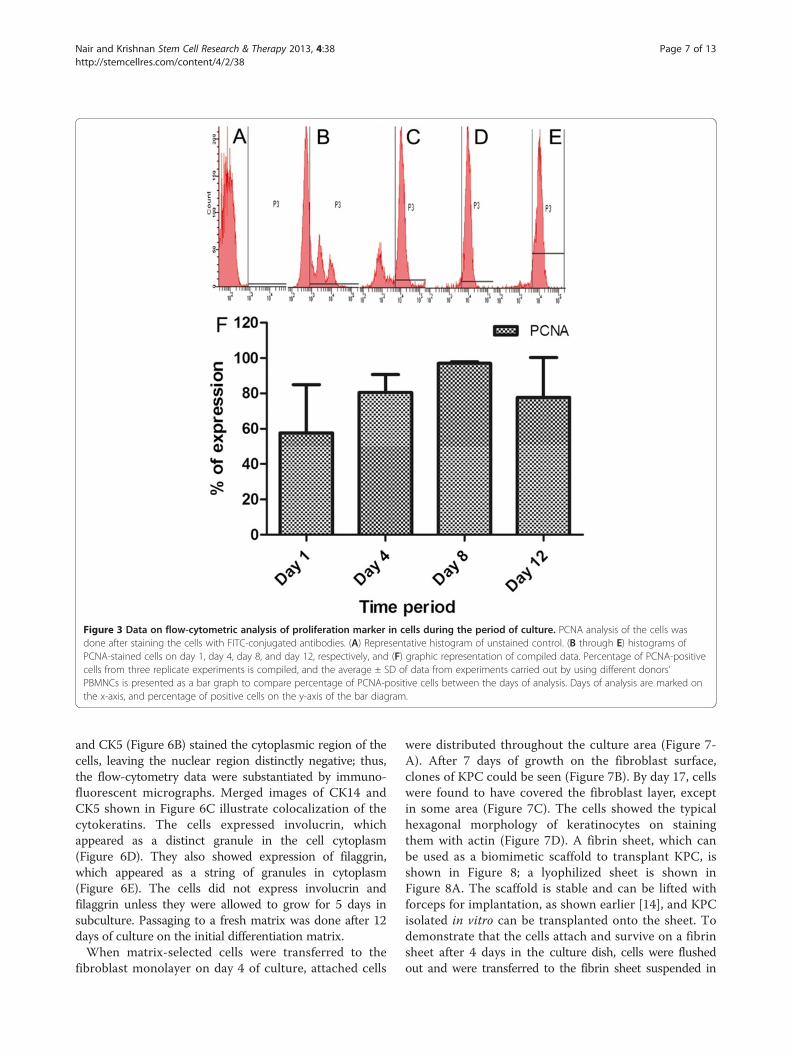
Figure 3 Data on flow-cytometric analysis of proliferation marker in cells during the period of culture. PCNA analysis of the cells wasdone after staining the cells with FITC-conjugated antibodies. (A) Representative histogram of unstained control. (B through E) histograms ofPCNA-stained cells on day 1, day 4, day 8, and day 12, respectively, and (F) graphic representation of compiled data. Percentage of PCNA-positivecells from three replicate experiments is compiled, and the average ± SD of data from experiments carried out by using different donors’PBMNCs is presented as a bar graph to compare percentage of PCNA-positive cells between the days of analysis. Days of analysis are marked onthe x-axis, and percentage of positive cells on the y-axis of the bar diagram.
Nair and Krishnan Stem Cell Research & Therapy 2013, 4:38 Page 7 of 13http://stemcellres.com/content/4/2/38
and CK5 (Figure 6B) stained the cytoplasmic region of thecells, leaving the nuclear region distinctly negative; thus,the flow-cytometry data were substantiated by immuno-fluorescent micrographs. Merged images of CK14 andCK5 shown in Figure 6C illustrate colocalization of thecytokeratins. The cells expressed involucrin, whichappeared as a distinct granule in the cell cytoplasm(Figure 6D). They also showed expression of filaggrin,which appeared as a string of granules in cytoplasm(Figure 6E). The cells did not express involucrin andfilaggrin unless they were allowed to grow for 5 days insubculture. Passaging to a fresh matrix was done after 12days of culture on the initial differentiation matrix.When matrix-selected cells were transferred to the
fibroblast monolayer on day 4 of culture, attached cells
were distributed throughout the culture area (Figure 7-A). After 7 days of growth on the fibroblast surface,clones of KPC could be seen (Figure 7B). By day 17, cellswere found to have covered the fibroblast layer, exceptin some area (Figure 7C). The cells showed the typicalhexagonal morphology of keratinocytes on stainingthem with actin (Figure 7D). A fibrin sheet, which canbe used as a biomimetic scaffold to transplant KPC, isshown in Figure 8; a lyophilized sheet is shown inFigure 8A. The scaffold is stable and can be lifted withforceps for implantation, as shown earlier [14], and KPCisolated in vitro can be transplanted onto the sheet. Todemonstrate that the cells attach and survive on a fibrinsheet after 4 days in the culture dish, cells were flushedout and were transferred to the fibrin sheet suspended in

Figure 4 Data on flow-cytometric analysis of sorted CD34+ cells for the presence of p63. For analysis of CD34, primary antibodiesconjugated with APC (FL1), and for bound p63 antibody, secondary antibodies conjugated with FITC (FL2) were used. (A) RepresentativeFSC-versus-SSC plot of unstained PBMNCs flushed from uncoated PS plates. (B) Plot of FL1 versus SSC of unstained PBMNCs. (C) Representativeplot of FL1-CD34-stained PBMNCs before sorting. (D) Representative plot of FL1-CD34 sorted cells. (E) Representative plot of FL2-versus-SSCsorted cells. (F) Representative plot of SSC-versus-FL2-p63 stained cells.
Nair and Krishnan Stem Cell Research & Therapy 2013, 4:38 Page 8 of 13http://stemcellres.com/content/4/2/38
the medium (Figure 8B). Within 2 days of culture, the fibrinscaffold appeared as a thin membrane-like structure thatcould be lifted (Figure 8C). Handling of the sheet after cul-ture was difficult for imunostaining; so simple DAPI stain-ing was done to demonstrate that the cells transferredfrom the biomimetric matrix-coated culture dish survivedon the fibrin sheet as well. Because of the three-dimensional and porous structure of the sheet, the cellspenetrated into the matrix and DAPI-stained nucleus(Figure 8D) and were seen in different planes under thefluorescence microscope. The flow-cytometric analysis had
proven that by day 4, >70% of the cells in the fibrin-coatedculture dish were p63+ (Figure 2A), so the DAPI-stainedcells are expected to be p63+ KPCs.Thus when PBMNCs were cultured under the speci-
fied conditions on the fibrin matrix, which comprisedgrowth factors, adhesive proteins, and hyaluronic acid, ahomogeneous population of KPCs was obtained. The re-sults are promising because KPCs differentiate on thematrix and can be transplanted by using a dermal equiva-lent such as a fibrin sheet or a fibroblast sheet to supportKPC survival and differentiation.

Figure 5 Identification of p63-specific cells in clusters. (A) Light micrograph of a representative cluster on day 4. (B) DAPI-stained cell cluster.(C) Immunostained p63-positive cells in the cluster. (D) Magnified micrograph of p63+ cells. (A through C) The same cluster is imaged withdifferent filter settings (scale bar, 100 μm). (D) Scale bar, 50 μm; D1 40×.
Nair and Krishnan Stem Cell Research & Therapy 2013, 4:38 Page 9 of 13http://stemcellres.com/content/4/2/38
DiscussionApplication of stem cells in skin grafting and tissue engin-eering is an important area of research in regenerativemedicine. An important step in the use of stem cells for tis-sue engineering is to obtain a relatively pure populationthat retains multiplication potential. The primary objectiveof this study was to find an easy-to-access autologous adultstem cell or progenitor cells for skin-tissue engineering.Further, a well-defined and efficient protocol must be devel-oped for directing the commitment and differentiation ofstem cells into the keratinocyte lineage for epidermal regen-eration. The development of such protocols would reducethe likelihood of spontaneous differentiation of multipotentstem cells into undesired lineages on transplantation.In this study, we identified p63+ KPCs in the circulation
by using flow cytometry, but the origin is not clear. Theymay have reached circulation either from the bone mar-row or from the epidermal compartment. Once commit-ted to the specific lineage that expresses p63, they do notseem to be CD34+. Use of p63 as a marker for identifica-tion of circulating KPCs is a novel approach that we usedin this study. To the best of our understanding, this is thefirst report on the presence of p63+ cells in blood. Weused a matrix-directed signaling strategy to amplify thenumber of progenitors and thus generated a homogeneouslineage committed culture that co-expressed p63, CK5,and CK14.The presence of transit-amplifying cells at the injured tis-
sue has been reported, and they probably originated frombone marrow and differentiated into keratinocytes [18].
They showed that the extent of skin damage influences theengraftment of bone marrow-derived progenitors, and theprocess continues until 3 weeks after the injury. Korblingand colleagues [19] found that on transplanting peripheralblood stem cells, they differentiated into mature epithelialcells in skin, lungs, gastrointestinal tract, and liver. Theresults of other studies suggest that adult stem/precursorcells may cross the lineage and germ-layer boundaries andgive rise to nonhematopoietic cells. Such transdiffe-rentiation is dependent on the niche to which they homeand also on the signals that are received from the local mi-lieu [20,21]. It is likely that circulating progenitors may mi-grate to damaged areas and participate in the local tissueregeneration [22].The results of the current study suggest that PBMNCs
could be a probable source for isolation and proliferationof KPCs in vitro for their potential use in regenerativemedicine. It may be relevant in the event that the localmilieu is unfavorable for engraftment, mainly due topoor blood circulation at the injured area. Under suchcircumstances, transplantation of KPCs along with a bio-mimetic scaffold as cell carrier is likely to enhanceepithelialization.Various studies have identified the roles of p63 in epi-
dermal regeneration, which include lineage commitment,differentiation, cell adhesion, and basement membraneformation. This transcription factor p63 was identified onthe basis of its sequence homology with the tumor-suppressor gene p53 [23]. However, unlike p53, p63 isexpressed in a tissue-specific manner and is expressed in

Figure 6 Immunofluorescence of cells for differentiation markers. (A) Representative image of cell stained with FITC-conjugated CK14antibody. (B) Representative image of PE-conjugated CK5 antibody. (C) Merged images of A and B. Both CK14 and CK5 antigens are distinctlylocated in the cytoplasmic region. (D) Representative image of cells stained with anti-involucrin antibody developed by PE-conjugated secondaryantibody on day 17. (E) Representative image of cells stained with anti-filaggrin antibody developed by using FITC-conjugated secondaryantibody on day 17. For negative control, fibroblasts were stained with each antibody (not shown). Scale bar is 100 μm for cytokeratin-stainedimages and 50 μm for filaggrin- and involucrin-stained images.
Nair and Krishnan Stem Cell Research & Therapy 2013, 4:38 Page 10 of 13http://stemcellres.com/content/4/2/38
stratified epithelia, such as the epidermis. During normalepidermal development, basal stem cells are induced toexpress epidermal keratins CK5 and CK14 [24,25], and theinduction process is dependent on p63. Expression of p63in cultured cells, single-layered epithelia, or embryonicstem cells also resulted in the expression of CK5 andCK14 [26-29]. Therefore, coexpression of p63, CK5, andCK14 in our experiment is an excellent indication of theepidermal lineage commitment of KPCs.Our hypothesis was proven true as we found that circu-
lating KPCs express p63. Here, analysis of sorted CD34+
cells confirmed that they are p63-. We found that afterenrichment by Histopaque gradient centrifugation and re-moval of plastic adherent cells, about 4% of cells are p63+;gene amplification was clearly evident. Considering themultipotency of progenitors in PBMNCs, predifferentiatedand lineage-committed KPCs may be better suited forregenerative medicine to heal chronic wounds. With pro-gress in the culture period on the niche that was designed
during this study, the cells became CK5+ and CK14+ inflow cytometry, immunofluorescent staining, and RT-PCR.It is interesting to note that the proliferation potential ofthe cells in culture tends to be reduced by day 12, probablybecause the lineage commitment progressed. When thecells were subcultured and stained for terminal differenti-ation markers involucrin and filaggrin, they turned positivefor both these antigens. Filaggrin is an intermediate fila-ment (IF)-associated protein that aggregates keratin IFsin vitro and is thought to perform a similar function duringthe terminal differentiation of epidermal keratinocytes [30].Filaggrin expression resulted in reduced keratinocyte prolif-eration and caused an alteration in cell-cycle distributionconsistent with a post-G1-phase arrest. We also found thatPCNA expression was reduced by 15 days of KPC culture.In keratinocytes, expression of involucrin may be regulatedby cell-substratum contact, and fibronectin at high concen-trations can inhibit the expression [31]. In the matrix thatwe composed, fibronectin content is <2 μg/cm2 of the

Figure 7 Representative images of keratinocyte progenitor cells (KPCs) on fibroblast monolayer. (A) Representative image of KPCs onfibroblast monolayer on the day of seeding. (B) KPCs on fibroblast on day 7. (C) KPCs on fibroblast on day 17. (D) Actin-stained KPCs onfibroblast. Magnification is the same for A and B (scale bar, 200 μm); and for C and D, the scale bar is 50 μm.
Nair and Krishnan Stem Cell Research & Therapy 2013, 4:38 Page 11 of 13http://stemcellres.com/content/4/2/38
culture surface, and it has not affected involucrin expres-sion on KPCs.Previously, we reported that the fibrin matrix can be
composed with growth factors and glycosaminoglycans forspecific requirements such as growth and differentiation
Figure 8 Fibrin sheet developed for transplantation of keratinocyte pculture dish (3.3-cm diameter). (B) Fibrin sheet suspended in medium for s(D) DAPI-stained KPCs grown on fibrin sheet for 2 days after transferring fro
of vascular cells and neural cells. Fibrin is abundant, wellcharacterized for its soft elasticity, and is useful as ahemostatic agent and as a scaffold for tissue engineering.Specific binding of fibrin with fibronectin, hyaluronic acid,and many growth factors, as well as clot components, is
rogenitor cells (KPCs). (A) Lyophilized fibrin sheet that was cast ineeding KPCs. (C) Skin-like fibrin sheet after 2 days of KPC culture.m FC matrix.

Nair and Krishnan Stem Cell Research & Therapy 2013, 4:38 Page 12 of 13http://stemcellres.com/content/4/2/38
well established [32]. The bioactivity of RGD sites on fi-brin and fibronectin, through which it can interact withintegrin, makes it an attractive matrix for stem cell differ-entiation and tissue engineering. Modulation of its pro-perties has even permitted differentiation of MSCs intoosteoblasts and mouse embryonic stem cells to neural andastroglial lineages [33,34].In this study, we demonstrated that a fibrin substrate
composed with specific growth factors and hyaluronicacid permits attachment and growth of circulating kera-tinocyte progenitors.Survival of keratinocytes in vitro and in vivo requires a
dermal-equivalent matrix as the niche. When p63+ cellswere enriched by using a biomimetic matrix-coated dishand transferred to the fibroblast monolayer, KPCs formedcolonies and multiplied to cover the fibroblast layer. Inskin, stem cells reside on the underlying basement mem-brane, where they self-renew and form transit-amplifyingcells, but they do not anchor tightly to the underlyingniche. In the initial period, as no trypsinization is neededto transfer the KPC from the selection niche to the fibro-blast sheet or the fibrin sheet, the protocols standardizedin this study have potential applications in KPC transplant-ation and for in vitro construction of a skin substitute forepidermal regeneration.
ConclusionIt is a novel finding that p63 can be used as marker toidentify/enumerate KPCs in PBMNCs, which can be col-lected by using a noninvasive procedure. In 4 days ofPBMNC culture on the designed biomimetic niche, KPCwas enriched to about 70% homogeneity. Within 12 daysof culture, the cells coexpressed p63, CK5, and CK14. Thedifferentiation progressed during subculture and expressedinvolucrin and filaggrin and showed morphologic featuresof keratinocytes. After 4 days of PBMNC culture on thebiomimetic niche, cells can be easily transferred to a fibrin/fibroblast sheet for continued growth and differentiation.The niche that was standardized for KPC isolation, expan-sion, differentiation, cell survival, and the protocol fortransfer of cells to dermal equivalent may be translated intoclinical use for cell/skin-substitute transplantation.
AbbreviationsAGF: angiogenic growth factor; BHE: bovine hypothalamus extract;BMDSC: bone marrow-derived stem cell; cDNA: complimentarydeoxyribonucleic acid; CK14: cytokeratin 14; CK5: cytokeratin 5; CO2: carbondioxide; DAPI: 40,6-diamidino-2-phenylindole; DMEM: Dulbecco ModifiedEagle Medium; ECM: extracellular matrix; EGF: epidermal growth factor;FACS: fluorescence-activated cell sorting; FC: fibrinogen composite;FITC: fluorescein isothiocyanate; HA: hyaluronic acid; HBSS: Hank balancedsalt solution; IgG: immunoglobulin G; IU: international unit; KPC: keratinocyteprogenitor cell; MAPCs: multipotent adult progenitor cells;MSC: mesenchymal stem cell; PBMNC: peripheral blood mononuclear cell;PBS: phosphate buffered saline; PCNA: proliferating cell nuclear antigen;PE: phycoerythrin; RNA: ribonucleic acid; RT-PCR: reverse transcriptase-polymerase chain reaction; SD: standard deviation; TA: transiently amplifying.
Competing interestsAuthors have no competing interests with regard to publication of thismanuscript.
Authors’ contributionsThe first author carried out the experiments and standardized several newprotocols, compiled the data, and prepared the draft manuscript. Thesecond author conceived the idea, analyzed the data, and fine-tuned themanuscript. Both authors read and approved the final manuscript.
Authors’ informationRPN is a graduate student nearly completing PhD research. This work is partof his PhD dissertation. Currently he is evaluating in vivo models to test thehypothesis that the niche standardized in this study may be used for celltransplantation.LKK has been a scientist and leader for many tissue-engineering programs,conceptualized that a fibrin-based cell-specific niche may be used fordifferentiation of stem cells (MSC) and circulating progenitors to becomedifferentiated endothelial cells, smooth muscle cells, neurons, dermalfibroblasts, keratinocytes, and cardiomyocytes for future applications inregenerative medicine.
AcknowledgementsThe authors acknowledge the director, SCTIMST, and the head, BMT Wing,SCTIMST, for the facilities provided. We acknowledge the support from allcolleagues of the Thrombosis Research Unit, especially Ms. Priyanka S andMr. Ranjith S for providing human fibrinogen and thrombin. Weacknowledge both the research grant to Dr. LKK and the individualfellowship support to Mr. RPN from the CSIR, Government of India.
Received: 18 December 2012 Revised: 12 March 2013Accepted: 20 March 2013 Published: 11 April 2013
References1. Boyce ST: Cultured skin substitutes: a review. Tissue Eng 1996, 2:255–266.2. Kawada H, Fujita J, Kinjo K, Matsuzaki Y, Tsuma M, Miyatake H, Muguruma Y,
Tsuboi K, Itabashi Y, Ikeda Y, Ogawa S, Okano H, Hotta T, Ando K, Fukuda K:Nonhematopoietic mesenchymal stem cells can be mobilized anddifferentiate into cardiomyocytes after myocardial infarction. Blood 2004,104:3581–3587.
3. Krause DS, Theise ND, Collector MI, Henegariu O, Hwang S, Gardner R,Neutzel S, Sharkis SJ: Multi-organ, multi-lineage engraftment by a singlebone marrow-derived stem cell. Cell 2001, 105:369–377.
4. Zhao Y, Glesne D, Huberman E: A human peripheral blood monocyte-derived subset acts as pluripotent stem cells. Proc Natl Acad Sci U S A2003, 100:2426–2431.
5. Chun-mao H, Su-yi W, Ping-ping L, Hang-hui C: Human bone marrow-derived mesenchymal stem cells differentiate into epidermal-like cellsin vitro. Differentiation 2007, 75:292–298.
6. Sasaki M, Abe R, Fujita Y, Ando S, Inokuma D, Shimizu H: Mesenchymalstem cells are recruited into wounded skin and contribute to woundrepair by transdifferentiation into multiple skin cell type. J Immunol 2008,180:2581–2587.
7. Medina A, Kilani RT, Carr N, Brown E, Ghahary A: Transdifferentiation ofperipheral blood mononuclear cells into epithelial-like cells. Am J Pathol2007, 171:1140–1152.
8. Medina A, Brown E, Carr N, Ghahary A: Circulating monocytes have thecapacity to be transdifferentiated into keratinocyte-like cells. WoundRepair Regen 2009, 17:268–277.
9. Pellegrini G, Dellambra E, Golisano O, Martinelli E, Fantozzi I, Bondanza S,Ponzin D, McKeon F, De Luca M: p63 identifies keratinocyte stem cells.Proc Natl Acad Sci U S A 2001, 98:3156–3161.
10. Koster MI: p63 in skin development and ectodermal dysplasias. J InvestDermatol 2010, 130:2352–2358.
11. Sreerekha PR, Divya P, Krishnan LK: Adult stem cell homing anddifferentiation in vitro on composite fibrin matrix. Cell Prolif 2006, 39:301.
12. Jose A, Krishnan LK: Effect of matrix composition on differentiation ofnestin-positive neural progenitors from circulation into neurons. J NeuralEng 2010, 7. doi:10.1088/1741-2560/7/3/036009.

Nair and Krishnan Stem Cell Research & Therapy 2013, 4:38 Page 13 of 13http://stemcellres.com/content/4/2/38
13. Maciag TCJ, Ilsley S, Kelley PR, Forand R: An endothelial cell growth factorfrom bovine hypothalamus: identification and partial characterization.Proc Natl Acad Sci U S A 1979, 76:5674.
14. Anilkumar TV, Muhamed J, Jose A, Jyothi A, Mohanan PV, Krishnan LK:Advantages of hyaluronic acid as a component of fibrin sheet for care ofacute wound. Biologicals 2011, 39:81–88.
15. Prasad Chennazhy K, Krishnan LK: Effect of passage number and matrixcharacteristics on differentiation of endothelial cells cultured for tissueengineering. Biomaterials 2005, 26:5658–5667.
16. Krishnan LK, Mohanty M, Umashankar PR, Vijayan Lal A: Comparativeevaluation of absorbable hemostats: advantages of fibrin-based sheets.Biomaterials 2004, 25:5557–5563.
17. Ma L, Gao C, Mao Z, Zhou J, Shen J, Hu X, Han C: Collagen/chitosanporous scaffolds with improved biostability for skin tissue engineering.Biomaterials 2003, 24:4833–4841.
18. Borue X, Lee S, Grove J, Herzog EL, Harris R, Diflo T, Glusac E, Hyman K,Theise ND, Krause DS: Bone marrow-derived cells contribute to epithelialengraftment during wound healing. Am J Pathol 2004, 165:1767–1772.
19. Korbling M, Katz RL, Khanna A, Ruifrok AC, Rondon G, Albitar M, ChamplinRE, Estrov Z: Hepatocytes and epithelial cells of donor origin in recipientsof peripheral-blood stem cells. N Engl J Med 2002, 346:738–746.
20. Korbling M, Estrov Z: Adult stem cells for tissue repair: a new therapeuticconcept? N Engl J Med 2003, 349:570–582.
21. Eisenberg LM, Eisenberg CA: Adult stem cells and their cardiac potential.Anat Rec A Discov Mol Cell Evol Biol 2004, 276A:103–112.
22. Quesenberry PJ, Abedi M, Aliotta J, Colvin G, Demers D, Dooner M, Greer D,Hebert H, Menon MK, Pimentel J, Paggioli D: Stem cell plasticity:an overview. Blood Cells Mol Dis 2004, 32:1–4.
23. Yang A, Kaghad M, Wang Y, Gillett E, Fleming MD, Dotsch V, Andrews NC,Caput D, McKeon F: p63, a p53 homolog at 3q27–29, encodes multipleproducts with transactivating, death-inducing, and dominant-negativeactivities. Mol Cell 1998, 2:305–316.
24. Jackson BW, Grund C, Winter S, Franke WW, Illmensee K: Formation ofcytoskeletal elements during mouse embryogenesis: ii, epithelialdifferentiation and intermediate-sized filaments in early postimplantation embryos. Differentiation 1981, 20:203–216.
25. Byrne C, Tainsky M, Fuchs E: Programming gene expression in developingepidermis. Development 1994, 120:2369–2383.
26. Koster MI, Kim S, Mills AA, DeMayo FJ, Roop DR: p63 is the molecularswitch for initiation of an epithelial stratification program. Genes Dev2004, 18:126–131.
27. Aberdam E, Barak E, Rouleau M, de LaForest S, Berrih-Aknin S, Suter DM,Krause K-H, Amit M, Itskovitz-Eldor J, Aberdam D: A pure population ofectodermal cells derived from human embryonic stem cells. Stem Cells2008, 26:440–444.
28. Medawar A, Virolle T, Rostagno P, de la Forest-Divonne S, Gambaro K,Rouleau M, Aberdam D: ΔNp63 is essential for epidermal commitment ofembryonic stem cells. PLoS One 2008, 3:e3441.
29. Romano R-A, Ortt K, Birkaya B, Smalley K, Sinha S: An active role of the ΔNisoform of p63 in regulating basal keratin genes K5 and K14 anddirecting epidermal cell fate. PLoS One 2009, 4:e5623.
30. Presland RB, Kuechle MK, Lewis SP, Fleckman P, Dale BA: Regulatedexpression of human filaggrin in keratinocytes results in cytoskeletaldisruption, loss of cell–cell adhesion, and cell cycle arrest. Exp Cell Res2001, 270:199–213.
31. Adams JC, Watt FM: Fibronectin inhibits the terminal differentiation ofhuman keratinocytes. Nature 1989, 340:307–309.
32. Weisel JW: Fibrinogen and fibrin. In Advances in Protein Chemistry. Volume70. Edited by David ADP, John MS. San Diego: Academic Press;2005:247–299.
33. Willerth SM, Arendas KJ, Gottlieb DI, Sakiyama-Elbert SE: Optimization offibrin scaffolds for differentiation of murine embryonic stem cells intoneural lineage cells. Biomaterials 2006, 27:5990–6003.
34. Catelas I, Sese N, Wu BM, Dunn JCY, Helgerson S, Tawil B: Humanmesenchymal stem cell proliferation and osteogenic differentiation infibrin gels in vitro. Tissue Eng 2006, 12:2385–2396.
doi:10.1186/scrt186Cite this article as: Nair and Krishnan: Identification of p63+ keratinocyteprogenitor cells in circulation and their matrix-directed differentiationto epithelial cells. Stem Cell Research & Therapy 2013 4:38.
Submit your next manuscript to BioMed Centraland take full advantage of:
• Convenient online submission
• Thorough peer review
• No space constraints or color figure charges
• Immediate publication on acceptance
• Inclusion in PubMed, CAS, Scopus and Google Scholar
• Research which is freely available for redistribution
Submit your manuscript at www.biomedcentral.com/submit