Sonographic Assessment of the Umbilical Cord The umbilical cord (UC) is the essential...
11
Introduction ! The umbilical cord (UC) is the essential life-sus- taining connection between fetus and placenta. It constitutes a stable connection to the fetomater- nal interface, while allowing fetal mobility that is essential for fetal development in general and neuromotor development in particular. This com- bination of mechanical stability and flexibility is due to the architecture of the UC. There is how- ever a range of umbilical cord complications that may be life threatening to the fetus, and these too can be explained to a large extent by the cordʼs structural characteristics. Examination of cord vessels using Doppler ultrasound enables investi- gators to deduce the state of the fetoplacental vas- cular bed, providing essential information on the condition of the fetus. Development and Architecture of the Umbilical Cord ! In its embryonic stage the UC develops in the re- gion of the body stalk to become the embryoʼs connection to the fetal portion of the placenta (fetal placenta). The amniotic cavity expands from dorsal to ventral while the chorionic cavity shrinks in volume. During cephalo-caudal and lateral folding the early UC arises as it is “envel- oped” by the expanding amnion (l " Fig. 1) (see also textbooks of embryology, www.embryol- ogy.ch). In this early stage at around 7–8 weeks postmenstrual age the UC contains the body stalk with umbilical vessels as well as other structures that will later regress and disappear entirely: the allantoic diverticulum (an out- pouching from the endoderm connected to the (future) urinary bladder, later the urachus), as well as the extra-embryonic coelom that at this early stage still forms a connection to the cho- Abstract ! The umbilical cord (UC) is a vital connection be- tween fetus and placenta. It constitutes a stable connection to the fetomaternal interface, while allowing the fetal mobility that is of great impor- tance for fetal development in general and fetal neuromotor development in particular. This com- bination of mechanical stability and flexibility is due to the architecture of the UC. There is howev- er a range of umbilical cord complications that may be life threatening to the fetus and these too can be explained to a large extent by the cordʼs structural characteristics. This review article dis- cusses clinically relevant aspects of UC ultra- sound. Zusammenfassung ! Die Nabelschnur ist die lebenswichtige Verbin- dung zwischen Fetus und Plazenta. Sie bildet ei- nerseits eine stabile Verbindung zur zentralen Einheit des fetomaternalen Stoffwechsels und er- möglicht dem Feten andererseits eine Beweglich- keit, die für die körperliche – insbesondere die neuromotorische – Entwicklung von großer Be- deutung ist. Diese Kombination aus mechanischer Stabilität und Flexibilität begründet sich in der Architektur der Nabelschnur. Es gibt jedoch eine Reihe an Nabelschnurkomplikationen, die den Feten z.T. lebensbedrohlich gefährden können. Auch diese lassen sich vor allem auf die struktu- rellen Eigenschaften der Nabelschnur zurückfüh- ren. Im Rahmen einer Übersichtsarbeit werden klinisch relevante Aspekte der sonografischen Diagnostik der Nabelschnur besprochen. Sonographic Assessment of the Umbilical Cord Sonografie der Nabelschnur Authors S. Bosselmann 1,2 , G. Mielke 2 Affiliations 1 Frauenklinik, Universitätsklinikum Heidelberg, Heidelberg 2 Pränatalzentrum Stuttgart, Stuttgart Key words l " ultrasound l " fetal period l " umbilical cord Schlüsselwörter l " Ultraschall l " Fetalzeit l " Nabelschnur received 13. 4. 2015 revised 30. 5. 2015 accepted 3. 6. 2015 Bibliography DOI http://dx.doi.org/ 10.1055/s-0035-1557819 Geburtsh Frauenheilk 2015; 75: 808–818 © Georg Thieme Verlag KG Stuttgart · New York · ISSN 0016‑5751 Correspondence Dr. Stephan Bosselmann Universitätsklinikum Heidelberg Frauenklinik Im Neuenheimer Feld 440 69120 Heidelberg Stephan.Bosselmann@ med.uni-heidelberg.de 808 Bosselmann S and Mielke G. Sonographic Assessment of … Geburtsh Frauenheilk 2015; 75: 808–818 GebFra Science Deutschsprachige Zusatzinformation unter: www.thieme-connect.de/ ejournals/gebfra
Transcript of Sonographic Assessment of the Umbilical Cord The umbilical cord (UC) is the essential...

Abstract!
The umbilical cord (UC) is a vital connection be-tween fetus and placenta. It constitutes a stableconnection to the fetomaternal interface, whileallowing the fetal mobility that is of great impor-tance for fetal development in general and fetalneuromotor development in particular. This com-bination of mechanical stability and flexibility isdue to the architecture of the UC. There is howev-er a range of umbilical cord complications thatmay be life threatening to the fetus and these toocan be explained to a large extent by the cordʼsstructural characteristics. This review article dis-cusses clinically relevant aspects of UC ultra-sound.
Zusammenfassung!
Die Nabelschnur ist die lebenswichtige Verbin-dung zwischen Fetus und Plazenta. Sie bildet ei-nerseits eine stabile Verbindung zur zentralenEinheit des fetomaternalen Stoffwechsels und er-möglicht dem Feten andererseits eine Beweglich-keit, die für die körperliche – insbesondere dieneuromotorische – Entwicklung von großer Be-deutung ist. Diese Kombination ausmechanischerStabilität und Flexibilität begründet sich in derArchitektur der Nabelschnur. Es gibt jedoch eineReihe an Nabelschnurkomplikationen, die denFeten z.T. lebensbedrohlich gefährden können.Auch diese lassen sich vor allem auf die struktu-rellen Eigenschaften der Nabelschnur zurückfüh-ren. Im Rahmen einer Übersichtsarbeit werdenklinisch relevante Aspekte der sonografischenDiagnostik der Nabelschnur besprochen.
Sonographic Assessment of the Umbilical CordSonografie der Nabelschnur
Authors S. Bosselmann1,2, G. Mielke2
Affiliations 1 Frauenklinik, Universitätsklinikum Heidelberg, Heidelberg2 Pränatalzentrum Stuttgart, Stuttgart
Key wordsl" ultrasoundl" fetal periodl" umbilical cord
Schlüsselwörterl" Ultraschalll" Fetalzeitl" Nabelschnur
received 13.4.2015revised 30.5.2015accepted 3.6.2015
BibliographyDOI http://dx.doi.org/10.1055/s-0035-1557819Geburtsh Frauenheilk 2015; 75:808–818 © Georg ThiemeVerlag KG Stuttgart · New York ·ISSN 0016‑5751
CorrespondenceDr. Stephan BosselmannUniversitätsklinikum HeidelbergFrauenklinikIm Neuenheimer Feld 44069120 [email protected]
808
Bosselmann S and Mielke G. Sonogra
GebFra Science
Deutschsprachige
Zusatzinformation unter:
www.thieme-connect.de/
ejournals/gebfra
Introduction!
The umbilical cord (UC) is the essential life-sus-taining connection between fetus and placenta. Itconstitutes a stable connection to the fetomater-nal interface, while allowing fetal mobility that isessential for fetal development in general andneuromotor development in particular. This com-bination of mechanical stability and flexibility isdue to the architecture of the UC. There is how-ever a range of umbilical cord complications thatmay be life threatening to the fetus, and these toocan be explained to a large extent by the cordʼsstructural characteristics. Examination of cordvessels using Doppler ultrasound enables investi-gators to deduce the state of the fetoplacental vas-cular bed, providing essential information on thecondition of the fetus.
phic Assessment of… Geburtsh Frauenheilk 2015; 75: 808–818
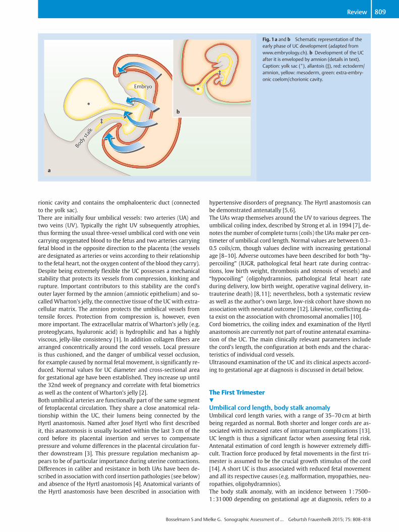
Development and Architectureof the Umbilical Cord!
In its embryonic stage the UC develops in the re-gion of the body stalk to become the embryoʼsconnection to the fetal portion of the placenta(fetal placenta). The amniotic cavity expandsfrom dorsal to ventral while the chorionic cavityshrinks in volume. During cephalo-caudal andlateral folding the early UC arises as it is “envel-oped” by the expanding amnion (l" Fig. 1) (seealso textbooks of embryology, www.embryol-ogy.ch). In this early stage at around 7–8 weekspostmenstrual age the UC contains the bodystalk with umbilical vessels as well as otherstructures that will later regress and disappearentirely: the allantoic diverticulum (an out-pouching from the endoderm connected to the(future) urinary bladder, later the urachus), aswell as the extra-embryonic coelom that at thisearly stage still forms a connection to the cho-
**
*
‡‡
‡‡
Body
stal
k
Body
stal
kEmbryoEmbryo
a
b
Fig. 1a and b Schematic representation of theearly phase of UC development (adapted fromwww.embryology.ch). b Development of the UCafter it is enveloped by amnion (details in text).Caption: yolk sac (*), allantois (‡), red: ectoderm/amnion, yellow: mesoderm, green: extra-embry-onic coelom/chorionic cavity.
809Review
rionic cavity and contains the omphaloenteric duct (connectedto the yolk sac).There are initially four umbilical vessels: two arteries (UA) andtwo veins (UV). Typically the right UV subsequently atrophies,thus forming the usual three-vessel umbilical cord with one veincarrying oxygenated blood to the fetus and two arteries carryingfetal blood in the opposite direction to the placenta (the vesselsare designated as arteries or veins according to their relationshipto the fetal heart, not the oxygen content of the blood they carry).Despite being extremely flexible the UC possesses a mechanicalstability that protects its vessels from compression, kinking andrupture. Important contributors to this stability are the cordʼsouter layer formed by the amnion (amniotic epithelium) and so-calledWhartonʼs jelly, the connective tissue of the UCwith extra-cellular matrix. The amnion protects the umbilical vessels fromtensile forces. Protection from compression is, however, evenmore important. The extracellular matrix of Whartonʼs jelly (e.g.proteoglycans, hyaluronic acid) is hydrophilic and has a highlyviscous, jelly-like consistency [1]. In addition collagen fibers arearranged concentrically around the cord vessels. Local pressureis thus cushioned, and the danger of umbilical vessel occlusion,for example caused by normal fetal movement, is significantly re-duced. Normal values for UC diameter and cross-sectional areafor gestational age have been established. They increase up untilthe 32nd week of pregnancy and correlate with fetal biometricsas well as the content of Whartonʼs jelly [2].Both umbilical arteries are functionally part of the same segmentof fetoplacental circulation. They share a close anatomical rela-tionship within the UC, their lumens being connected by theHyrtl anastomosis. Named after Josef Hyrtl who first describedit, this anastomosis is usually located within the last 3 cm of thecord before its placental insertion and serves to compensatepressure and volume differences in the placental circulation fur-ther downstream [3]. This pressure regulation mechanism ap-pears to be of particular importance during uterine contractions.Differences in caliber and resistance in both UAs have been de-scribed in associationwith cord insertion pathologies (see below)and absence of the Hyrtl anastomosis [4]. Anatomical variants ofthe Hyrtl anastomosis have been described in association with
Bosselmann S and Mi
hypertensive disorders of pregnancy. The Hyrtl anastomosis canbe demonstrated antenatally [5,6].The UAs wrap themselves around the UV to various degrees. Theumbilical coiling index, described by Strong et al. in 1994 [7], de-notes the number of complete turns (coils) the UAsmake per cen-timeter of umbilical cord length. Normal values are between 0.3–0.5 coils/cm, though values decline with increasing gestationalage [8–10]. Adverse outcomes have been described for both “hy-percoiling” (IUGR, pathological fetal heart rate during contrac-tions, low birth weight, thrombosis and stenosis of vessels) and“hypocoiling” (oligohydramnios, pathological fetal heart rateduring delivery, low birth weight, operative vaginal delivery, in-trauterine death) [8,11]; nevertheless, both a systematic reviewas well as the authorʼs own large, low-risk cohort have shown noassociation with neonatal outcome [12]. Likewise, conflicting da-ta exist on the association with chromosomal anomalies [10].Cord biometrics, the coiling index and examination of the Hyrtlanastomosis are currently not part of routine antenatal examina-tion of the UC. The main clinically relevant parameters includethe cordʼs length, the configuration at both ends and the charac-teristics of individual cord vessels.Ultrasound examination of the UC and its clinical aspects accord-ing to gestational age at diagnosis is discussed in detail below.
The First Trimester!
Umbilical cord length, body stalk anomalyUmbilical cord length varies, with a range of 35–70 cm at birthbeing regarded as normal. Both shorter and longer cords are as-sociated with increased rates of intrapartum complications [13].UC length is thus a significant factor when assessing fetal risk.Antenatal estimation of cord length is however extremely diffi-cult. Traction force produced by fetal movements in the first tri-mester is assumed to be the crucial growth stimulus of the cord[14]. A short UC is thus associated with reduced fetal movementand all its respective causes (e.g. malformation, myopathies, neu-ropathies, oligohydramnios).The body stalk anomaly, with an incidence between 1:7500–1:31000 depending on gestational age at diagnosis, refers to a
elke G. Sonographic Assessment of… Geburtsh Frauenheilk 2015; 75: 808–818
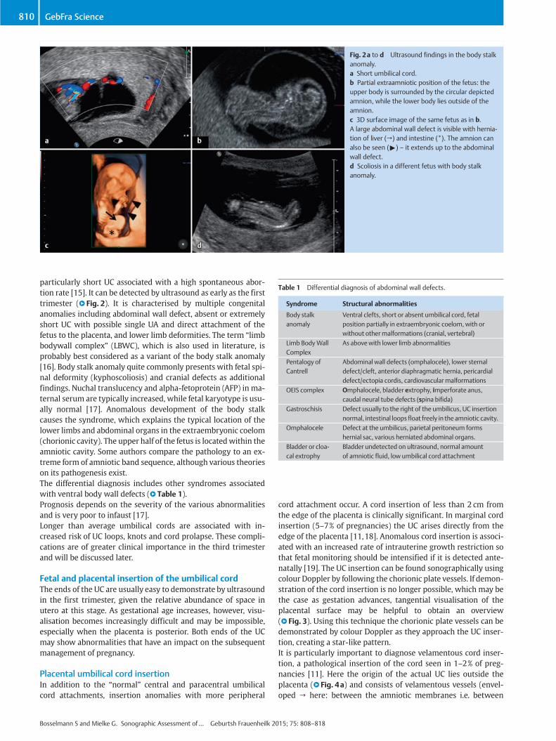
Table 1 Differential diagnosis of abdominal wall defects.
Syndrome Structural abnormalities
Body stalkanomaly
Ventral clefts, short or absent umbilical cord, fetalposition partially in extraembryonic coelom, with orwithout othermalformations (cranial, vertebral)
Limb BodyWallComplex
As abovewith lower limb abnormalities
Pentalogy ofCantrell
Abdominal wall defects (omphalocele), lower sternaldefect/cleft, anterior diaphragmatic hernia, pericardialdefect/ectopia cordis, cardiovascular malformations
OEIS complex Omphalocele, bladder extrophy, imperforate anus,caudal neural tube defects (spina bifida)
Gastroschisis Defect usually to the right of the umbilicus, UC insertionnormal, intestinal loops float freely in the amniotic cavity.
Omphalocele Defect at the umbilicus, parietal peritoneum formshernial sac, various herniated abdominal organs.
Bladder or cloa-cal extrophy
Bladder undetected on ultrasound, normal amountof amniotic fluid, low umbilical cord attachment
Fig. 2a to d Ultrasound findings in the body stalkanomaly.a Short umbilical cord.b Partial extraamniotic position of the fetus: theupper body is surrounded by the circular depictedamnion, while the lower body lies outside of theamnion.c 3D surface image of the same fetus as in b.A large abdominal wall defect is visible with hernia-tion of liver (→) and intestine (*). The amnion canalso be seen (") – it extends up to the abdominalwall defect.d Scoliosis in a different fetus with body stalkanomaly.
810 GebFra Science
particularly short UC associated with a high spontaneous abor-tion rate [15]. It can be detected by ultrasound as early as the firsttrimester (l" Fig. 2). It is characterised by multiple congenitalanomalies including abdominal wall defect, absent or extremelyshort UC with possible single UA and direct attachment of thefetus to the placenta, and lower limb deformities. The term “limbbodywall complex” (LBWC), which is also used in literature, isprobably best considered as a variant of the body stalk anomaly[16]. Body stalk anomaly quite commonly presents with fetal spi-nal deformity (kyphoscoliosis) and cranial defects as additionalfindings. Nuchal translucency and alpha-fetoprotein (AFP) in ma-ternal serum are typically increased, while fetal karyotype is usu-ally normal [17]. Anomalous development of the body stalkcauses the syndrome, which explains the typical location of thelower limbs and abdominal organs in the extraembryonic coelom(chorionic cavity). The upper half of the fetus is locatedwithin theamniotic cavity. Some authors compare the pathology to an ex-treme form of amniotic band sequence, although various theorieson its pathogenesis exist.The differential diagnosis includes other syndromes associatedwith ventral body wall defects (l" Table 1).Prognosis depends on the severity of the various abnormalitiesand is very poor to infaust [17].Longer than average umbilical cords are associated with in-creased risk of UC loops, knots and cord prolapse. These compli-cations are of greater clinical importance in the third trimesterand will be discussed later.
Fetal and placental insertion of the umbilical cordThe ends of the UC are usually easy to demonstrate by ultrasoundin the first trimester, given the relative abundance of space inutero at this stage. As gestational age increases, however, visu-alisation becomes increasingly difficult and may be impossible,especially when the placenta is posterior. Both ends of the UCmay show abnormalities that have an impact on the subsequentmanagement of pregnancy.
Placental umbilical cord insertionIn addition to the “normal” central and paracentral umbilicalcord attachments, insertion anomalies with more peripheral
Bosselmann S and Mielke G. Sonographic Assessment of… Geburtsh Frauenheilk 20
cord attachment occur. A cord insertion of less than 2 cm fromthe edge of the placenta is clinically significant. In marginal cordinsertion (5–7% of pregnancies) the UC arises directly from theedge of the placenta [11,18]. Anomalous cord insertion is associ-ated with an increased rate of intrauterine growth restriction sothat fetal monitoring should be intensified if it is detected ante-natally [19]. The UC insertion can be found sonographically usingcolour Doppler by following the chorionic plate vessels. If demon-stration of the cord insertion is no longer possible, which may bethe case as gestation advances, tangential visualisation of theplacental surface may be helpful to obtain an overview(l" Fig. 3). Using this technique the chorionic plate vessels can bedemonstrated by colour Doppler as they approach the UC inser-tion, creating a star-like pattern.It is particularly important to diagnose velamentous cord inser-tion, a pathological insertion of the cord seen in 1–2% of preg-nancies [11]. Here the origin of the actual UC lies outside theplacenta (l" Fig. 4a) and consists of velamentous vessels (envel-oped → here: between the amniotic membranes i.e. between
15; 75: 808–818

Fig. 3a and b Normal umbilical cord insertion.a Demonstration of the cord insertion and a num-ber of chorionic plate vessels. Depending on fetal lievisualisation may be difficult even if the placenta isanterior. For further differentiation it may be helpfulto examine the placenta tangentially as in b. Thestar-like pattern of the chorionic plate vessels asthey approach the cord insertion is seen. Placentaltissue surrounds them. The yellow line in a repre-sents the level and orientation of the image in b.
Fig. 4a to d Pathological cord insertions.a Velamentous insertion at the end of the first tri-mester. The UC insertion is demonstrable oppositethe chorion frondosum.b Transvaginal view of the same pregnancy as in a.A velamentous vessel runs directly across the cervi-cal os (≙ vasa praevia).c and d Demonstration of velamentous vessels inthe second trimester: c Longitudinal section.d Transverse section. These findings must alwaysbe followed by further assessment of placental UCinsertion (to exclude velamentous insertion) andexamination of the lower uterine segment (to ex-clude vasa praevia). Caption: chorion frondosum/placenta (*); cord insertion (→); cervical canal (").
811Review
amnion and chorion). In contrast to normal UC vessels, velamen-tous vessels are not protected by the stabilising effect of Whar-tonʼs jelly. There is an increased risk of cord compression, fetalgrowth restriction, premature labour, placental abruption, CTGabnormalities and low Apgar scores [11]. It is important to beaware that velamentous cord insertion is associated with an in-creased rate of vasa praevia.The term vasa praevia describes velamentous vessels that lie indirect proximity to the cervix and below the fetal “presentingpart” (l" Figs. 4b and d); the prevalence of this anomaly is 0.04%[20]. Risk factors for vasa praevia, apart from velamentous cordinsertion (vasa praevia type I), include accessory placenta (vasapraevia type II) [21], and multiple pregnancy and pregnancy inthe setting of assisted reproduction [22]. The closer the vesselsare to the internal os (which exposes them to traction and shear-ing forces during birthing efforts, especially in the context of pre-mature rupture of membranes) the greater the danger of vesselrupture and massive fetal haemorrhage. Prenatal diagnosis of va-
Bosselmann S and Mi
sa praevia followed by delivery by primary caesarean section pri-or to the onset of labour can increase survival from 44 to 97%[20]. Depending on gestational age RDS prophylaxis should beconsidered since increased rates of preterm delivery are probable[21,23]. A current systematic review describes antenatal detec-tion rates of vasa praevia of between 53 and 100% [24]. Transvag-inal colour Doppler ultrasound is best for diagnosis. Detectionrates may be much lower in the abdominal plane. According to ascreening algorithm proposed by Rebarber et al. transvaginal ul-trasound should be performed in every pregnant woman whopresents with any of the following criteria [25]:" Placenta praevia in the current pregnancy that is subsequently
no longer demonstrable" Vasa praevia in a previous pregnancy" Velamentous cord insertion in the lower uterine segment" Accessory placenta in the lower uterine segment" Multiple pregnancy
elke G. Sonographic Assessment of… Geburtsh Frauenheilk 2015; 75: 808–818
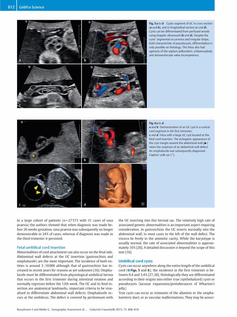
Fig. 5a to d Cystic segment of UC in cross section(a und b), and in longitudinal section (c und d).Cysts can be differentiated from perfused vesselsusing Doppler ultrasound (b und d). Despite thecystsʼ segmental occurrence and irregular shape,both characteristic of pseudocysts, differentiation isonly possible on histology. This fetus also hadagenesis of the septum pellucidum, schizencephalyand atrioventricular valve incompetence.
Fig. 6a to da and b Demonstration of an UC cyst in a centralcord segment in the first trimester;c and d Fetus with a large UC cyst located at thefetal cord insertion. The echogenic appearance ofthe cyst margin nearest the abdominal wall (")raises the suspicion of an abdominal wall defect.An omphalocele was subsequently diagnosed.Caption: yolk sac (*).
812 GebFra Science
In a large cohort of patients (n = 27573 with 31 cases of vasapraevia) the authors showed that when diagnosis was made be-fore 26 weeks gestation, vasa praevia was subsequently no longerdemonstrable in 24% of cases, whereas if diagnosis was made inthe third trimester it persisted.
Fetal umbilical cord insertionAbnormalities of cord attachment can also occur on the fetal side.Abdominal wall defects at the UC insertion (gastroschisis andomphalocele) are the most important. The incidence of both en-tities is around 3:10000 although that of gastroschisis has in-creased in recent years for reasons as yet unknown [16]. Ompha-locele must be differentiated from physiological umbilical herniathat occurs in the first trimester during intestinal rotation andnormally regresses before the 12th week. The UC and its fetal in-sertion are anatomical landmarks, important criteria to be visu-alised to differentiate abdominal wall defects. Omphalocele oc-curs at the umbilicus. The defect is covered by peritoneum with
Bosselmann S and Mielke G. Sonographic Assessment of… Geburtsh Frauenheilk 20
the UC inserting into this hernial sac. The relatively high rate ofassociated genetic abnormalities is an important aspect requiringconsideration. In gastroschisis the UC inserts normally into theabdominal wall, in most cases to the left of the wall defect. Theviscera lie freely in the amniotic cavity. While the karyotype isusually normal, the rate of associated abnormalities is approxi-mately 10% [26]. A detailed discussion is beyond the scope of thistext [16].
Umbilical cord cystsCysts can occur anywhere along the entire length of the umbilicalcord (l" Figs. 5 and 6); the incidence in the first trimester is be-tween 0.4 and 3.4% [27,28]. Histologically they are differentiatedaccording to their origins into either true (epithelialized) cysts orpseudocysts (lacunar expansions/protuberances of Whartonʼsjelly).True cysts can occur as remnants of the allantois or the ompha-loenteric duct, or as vascular malformations. They may be associ-
15; 75: 808–818

Fig. 7a to da Perivesical course of the UAs right up to the fetalcord insertion.b Cross section of a normal umbilical cord. Thethree vessel lumens appear as a stylized MickeyMouse.c Demonstration of two UAs in their perivesicalcourse in the first trimester up to the fetal cord in-sertion.d Due to their close proximity in the first trimester,the UAs may be confused with the femoral arteries("). Caption: urinary bladder (*).
813Review
ated with malformations (eg. omphalocele, urachus fistula) andchromosomal abnormalities (especially trisomy 18). As most ofthe available literature on the subject is in the form of case re-ports and small case series, data on complication rates and clini-cal course are limited.Cases of UC cysts should be followed up by detailed investigationfor associated malformations. Many first trimester cysts disap-pear by the second trimester (ca. 80% [11]), a development asso-ciated with a good prognosis [29]. UC cysts in the second andthird trimester have a poorer prognosis due to the above-men-tioned complications [28].
Number of vessels/single umbilical arteryEarly in its development the umbilical cord is formed from fourvessels. At this stage there are two umbilical arteries (UA) andtwo umbilical veins (UV) of which the right UV typically atro-phies by the 6th week of gestation. Thus the normal umbilicalcord consists of two UAs and one UV.While a persistent four-ves-sel umbilical cord rarely occurs [30], the most common cord var-iant, with an incidence of approx. 0.5% in the second trimester[31], is a single UA (two vessels: one vein and only one artery;SUA = single umbilical artery). Visualisation of the umbilical ar-teries is done either in the free umbilical cord or preferably atthe level of the fetal urinary bladder by demonstrating the cordʼsperivesical course using colour Doppler ultrasound (l" Fig. 7).Using colour Doppler and high definition ultrasound, SUA can bediagnosed at this level as early as the first trimester. A low pulserepetition frequency (PRF) and a high colour gain are recom-mended, as comparatively small vessels (compared to the oppo-site side) may otherwise be overlooked (l" Fig. 7c and d). Theperivesical course of the UAs should be visualised all the way tothe umbilicus as there is a danger of confusion with fetal femoralarteries in early gestation (l" Fig. 8c and d).If single umbilical artery is suspected in the first trimester the di-agnosis should be confirmed at a later stage (l" Fig. 8a and b). Theexisting (single) UA compensates and its lumen is usually biggerthan that of a three-vessel cord, with a diameter ratio of UV/UA≤ 2 (in three-vessel cord UV/UA the ratio is > 2) [32]. The mainclinical relevance of SUA is the association with structural abnor-malities of the fetal cardiovascular and genitourinary systems, as
Bosselmann S and Mi
well as more rare gastrointestinal and CNS abnormalities [11].Approximately one third of fetuses with SUA have structural ab-normalities, and chromosomal anomalies are found in 10% [31],although according to current data isolated occurrence of SUAdoes not significantly increase the risk of chromosomal anoma-lies [31,33]. SUA is regarded as a marker for the development offetal growth restriction and premature birth [34].The demonstration of an SUA should always prompt a detailedanatomical ultrasound examination with particular focus on thecardiovascular and genitourinary systems. Increased pregnancymonitoring because of the higher risk of IUGR is currently contro-versial [11,31].
The Second Trimester!
Assessment of the UC in the second trimester can be difficult dueto fetal interference when the placenta is posterior, or a convo-luted cord. However, because of fetal growth, the now larger indi-vidual vessels are easier to assess. Thus SUA is often first diag-nosed in the second trimester. Structural changes of the umbilicalvein are also mostly diagnosed in the second trimester.
Structural changes of the umbilical veinThere are two structural UV variants which are of clinical impor-tance, both involving its intraabdominal course.
Persistent right umbilical vein (PRUV)By the 7th week of gestation one of the initially paired UVs (usu-ally the right one) has typically atrophied. The left UV initiallyruns dorsally in the abdomen and joins the portal system. In ab-dominal transverse section (at a level slightly caudal of the bio-metry level) the umbilicoportal vessels arch to the right appear-ing to “encircle” the gallbladder while passing the stomach tan-gentially (l" Fig. 9a).Portal vessels branch off along its course, as does the ductus ve-nosus approximately at the peak of the dorsal arch (l" Fig. 9b).Chaoui et al. recently published a detailed description of the he-patic veins as a CME article in German and English [35].
elke G. Sonographic Assessment of… Geburtsh Frauenheilk 2015; 75: 808–818
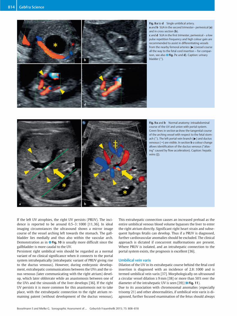
Fig. 8a to d Single umbilical artery.a and b SUA in the second trimester– perivesical (a)and in cross section (b).c and d SUA in the first trimester, perivesical – a lowpulse repetition frequency and high colour gain arerecommended to assist in differentiating vesselsfrom the nearby femoral arteries (") (vessel courseall the way to the fetal cord insertion – for compar-ison, see also l" Fig. 7c und d). Caption: urinarybladder (*).
Fig. 9a and b Normal anatomy: intraabdominalcourse of the UV and union with portal system.Green lines in section a show the tangential courseof the arching vessel with respect to the fetal stom-ach (*). The left portal vein branch (") and ductusvenosus (→) are visible. In section b a colour changeallows identification of the ductus venosus (“alias-ing” caused by flow acceleration). Caption: hepaticveins (‡).
814 GebFra Science
If the left UV atrophies, the right UV persists (PRUV). The inci-dence is reported to be around 0.5–3:1000 [11,36]. In idealimaging circumstances the ultrasound shows a mirror imagecourse of the vessel arching left towards the stomach. The gall-bladder lies medially and thus also within the vascular arch.Demonstration as in l" Fig. 10 is usually more difficult since thegallbladder is more caudal to the UV.Persistent right umbilical vein should be regarded as a normalvariant of no clinical significance when it connects to the portalsystem intrahepatically (intrahepatic variant of PRUV giving riseto the ductus venosus). However, during embryonic develop-ment, extrahepatic communications between the UVs and the si-nus venosus (later communicating with the right atrium) devel-op, which later obliterate while an anastomosis between one ofthe UVs and the sinusoids of the liver develops [36]. If the rightUV persists it is more common for this anastomosis not to takeplace, with the extrahepatic connection to the right atrium re-maining patent (without development of the ductus venosus).
Bosselmann S and Mielke G. Sonographic Assessment of… Geburtsh Frauenheilk 20
This extrahepatic connection causes an increased preload as theentire umbilical venous blood volume bypasses the liver to enterthe right atrium directly. Significant right heart strain and subse-quent hydrops fetalis can develop. Thus if a PRUV is diagnosed,further cardiovascular anomalies should be excluded. The clinicalapproach is dictated if concurrent malformations are present.Where PRUV is isolated, and an intrahepatic connection to theportal system exists, the prognosis is excellent [36].
Umbilical vein varixDilation of the UV in its extrahepatic course behind the fetal cordinsertion is diagnosed with an incidence of 2.8 :1000 and istermed umbilical vein varix [37]. Morphologically on ultrasounda circular vessel dilation ≥ 9mm [38] or more than 50% over thediameter of the intrahepatic UV is seen [39] (l" Fig. 11).Due to its association with chromosomal anomalies (especiallytrisomy 21) and other abnormalities, if umbilical vein varix is di-agnosed, further focused examination of the fetus should always
15; 75: 808–818
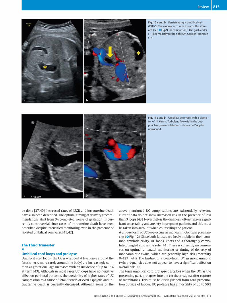
Fig. 10a and b Persistent right umbilical vein(PRUV). The vascular arch runs towards the stom-ach (see l" Fig. 9 for comparison). The gallbladder(→) lies medially to the right UV. Caption: stomach(*).
Fig. 11a and b Umbilical vein varix with a diame-ter of 11.6mm. Turbulent flow within the out-pouching/vessel dilatation is shown on Dopplerultrasound.
815Review
be done [37,40]. Increased rates of IUGR and intrauterine deathhave also been described. The optimal timing of delivery (recom-mendations start from 34 completed weeks of gestation) is cur-rently controversial since cases of intrauterine death have beendescribed despite intensified monitoring even in the presence ofisolated umbilical vein varix [41,42].
The Third Trimester!
Umbilical cord loops and prolapseUmbilical cord loops (the UC is wrapped at least once around thefetusʼs neck, more rarely around the body) are increasingly com-mon as gestational age increases with an incidence of up to 33%at term [43]. Although in most cases UC loops have no negativeeffect on perinatal outcome, the possibility of higher rates of UCcompression as a cause of fetal distress or even asphyxia and in-trauterine death is currently discussed. Although some of the
Bosselmann S and Mi
above-mentioned UC complications are existentially relevant,current data do not show increased risk in the presence of lessthan 3 loops [43]. Nevertheless the diagnosis often triggers signif-icant uncertainty and anxiety in pregnant patients and this mustbe taken into account when counselling the patient.A unique form of UC loop occurs in monoamniotic twin pregnan-cies (l" Fig. 12). Since both fetuses are freely mobile in their com-mon amniotic cavity, UC loops, knots and a thoroughly convo-luted/tangled cord is the rule [44]. There is currently no consen-sus on optimal antenatal monitoring or timing of delivery ofmonoamniotic twins, which are generally high risk (mortality8–42% [44]). The finding of a convoluted UC in monoamniotictwin pregnancies does not appear to have a significant effect onoverall risk [45].The term umbilical cord prolapse describes when the UC, as thepresenting part, prolapses into the cervix or vagina after ruptureof membranes. This must be distinguished from cord presenta-tion outside of labour. UC prolapse has a mortality of up to 50%
elke G. Sonographic Assessment of… Geburtsh Frauenheilk 2015; 75: 808–818

Fig. 12a to d Convoluted umbilical cord in amonoamniotic twin pregnancy.a Demonstration on ultrasound.b Macroscopic view of tangled umbilical cords.c and d Doppler ultrasound assessment of tangledumbilical cords. Two independent curves are seen inparallel (fetus 1 and fetus 2).
816 GebFra Science
[11], although current data show a reduction in mortality tounder 10% in the context of rising caesarean section rates [46].The main risk factor for cord prolapse necessitating emergent de-livery, is abnormal fetal lie. Numerous other factors are also asso-ciated with increased risk including prematurity and low birthweight, abnormalities of the maternal pelvis, polyhydramnios,UC length > 80 cm and others (male sex, multiple pregnancy,placenta praevia and obstetric interventions) [46].Risk factors such as multiple cord loops, very low placental cordinsertion or polyhydramnios should be known to the obstetricteam as this knowledge is essential for the correct interpretationof the progress of labour and particularly the CTG performedduring labour, with labour management adjusted to take accountof this information.
True and false knotsUmbilical cord compression can also be caused by tightening of atrue UC knot. So-called false knots, in contrast, are varicose dila-tations of umbilical vessels with an appearance sometimes imi-tating true knots. Their course is snaked but the UC does not forman actual knot that can tighten. UC knots are usually diagnosedpostnatally. Antenatal diagnosis of true knots is possible, alsocausing major maternal anxiety. Well-defined diagnostic criteriaandmanagement standards are currently lacking andmisdiagno-sis is relatively common. Nevertheless, diagnosis of a UC knotshould be followed by detailed Doppler ultrasound of the UCand stringent pregnancy monitoring [47,48].
Umbilical cord tumoursUmbilical cord tumours are very rare entities. In most cases theyare haemangiomas or teratomas. As in the case of UC haemato-mas (see below), tumours can cause umbilical vessel compres-sion. Antenatally haemangiomas appear as echogenic structureswith demonstrable perfusion on Doppler ultrasound. Multicysticvariants make the differentiation from teratomas and haemato-mas particularly difficult [11]. Mortality is estimated to be 40%.Associated fetal malformations are described without any specif-ic preponderance [49].Teratomas of the UC are usually benign tumours. Their appear-ance on ultrasound is heterogeneous often with both cystic and
Bosselmann S and Mielke G. Sonographic Assessment of… Geburtsh Frauenheilk 20
solid components. Concurrent malformations are described inabout half of published cases. Teratomas can reach considerablesize and are a cause of cardiac failure in the fetus (“high-outputheart failure”). Rupture of cord vessels is a particular danger asso-ciated with UC teratomas [50].
Special Considerations!
Doppler ultrasoundDoppler ultrasound of the UC is relatively easy to carry out evenin the later weeks of pregnancy thanks to the length and variablecourse of the UC. Due to the flexibility of the cord, vessels canusually be examined at the appropriate angle for good qualityspectral curves. Nevertheless the cordʼs length and windingcourse make standardisation and the exact definition of the ex-amined segment and precise Doppler measurement at a specificangle difficult.The umbilical arteries are the most commonly examined vesselsin the assessment of fetal circulation. They provide informationon the fetoplacental capillary bed; where resistance is increased,investigation of the UAs allows early detection of risk to the fetus.Assessment of UC blood flow is done particularly in the setting offetal growth restriction to differentiate between compensatedand decompensated placental insufficiency. Various (semi-)quantitative and qualitative measures can be assessed. The um-bilical vein also provides information on the fetal status, allowingassessment of the fetal central venous vasculature, e.g. increasedpreload in the setting of right heart failure. The examination ofother fetal and/or maternal vessels is often necessary for a com-plete assessment. We refer the reader to textbooks on the appli-cation of Doppler ultrasound in obstetrics for a complete descrip-tion of Doppler ultrasound of the UC.
Invasive procedures and umbilical cord haematomaFetal blood sampling for the investigation of fetal anaemia, con-genital infections and specialised genetic investigations is possi-ble through UC puncture. Ultrasound-guided puncture of the UVallows both blood sampling and, via the same route, blood trans-fusion for the correction of fetal anaemia as indicated. The proce-
15; 75: 808–818

817Review
dural risk of abortion is 1% (fetus without malformations) [51]. Avery rare complication is the development of a UC haematomathat, if large enough, can compress UC vessels and impair fetalcirculation [52]. Spontaneous formation of a haematoma is alsopossible and the following risk factors have been reported: shortUC, cord prolapse, velamentous cord insertion [53] and fetal clot-ting disorders [54]. UC haematomas are associated with a highrate of asphyxia and fetal mortality.
Conclusion!
Detailed examination of the umbilical cord provides informationon different aspects of fetal development. Targeted examinationshould be undertaken in early pregnancy as many details of corddevelopment become increasingly difficult to demonstrate on ul-trasound with increasing gestational age. The detection of abnor-malities in the number, structure or course of cord vessels shouldprompt further extended investigation since associations withother structural (especially cardiovascular) and chromosomalanomalies exist. Isolated abnormalities of the cord mostly carrya favourable prognosis. Particular attention should be paid to fe-tal and placental umbilical cord insertions.
Conflict of Interest!
None.
References1 Ferguson VL, Dodson RB. Bioengineering aspects of the umbilical cord.Eur J Obstet Gynecol Reprod Biol 2009; 144 (Suppl. 1): S108–S113
2 Raio L, Ghezzi F, Di Naro E et al. Sonographic measurement of the um-bilical cord and fetal anthropometric parameters. Eur J Obstet GynecolReprod Biol 1999; 83: 131–135
3 Gordon Z, Eytan O, Jaffa AJ et al.Hemodynamic analysis of Hyrtl anasto-mosis in human placenta. Am J Physiol Regul Integr Comp Physiol2007; 292: R977–R982
4 Bhutia KL, Sengupta R, Upreti B et al. Pregnancy-induced hypertensionis associated with altered anatomical patterns of Hyrtlʼs anastomosis.Anat Rec (Hoboken) 2014; 297: 819–825
5 Raio L, Ghezzi F, di Naro E et al. In-utero characterization of the bloodflow in the Hyrtl anastomosis. Placenta 2001; 22: 597–601
6 Raio L, Ghezzi F, Di Naro E et al. Prenatal assessment of the Hyrtl anas-tomosis and evaluation of its function: case report. Hum Reprod 1999;14: 1890–1893
7 Strong TH jr., Jarles DL, Vega JS et al. The umbilical coiling index. AmJ Obstet Gynecol 1994; 170: 29–32
8 Mittal A, Nanda S, Sen J. Antenatal umbilical coiling index as a predictorof perinatal outcome. Arch Gynecol Obstet 2015; 291: 763–768
9 Narayan R, Saaid R, Pedersen L et al. Ultrasound assessment of umbili-cal cord morphology in the first trimester: a feasibility study. Fetal Di-agn Ther 2015; [Epub ahead of print]
10 Verkleij CP, van Oppen AC, Mulder EJ et al. Evaluation of antenatal um-bilical coiling index at 16–21 weeks of gestation as a predictor of tris-omy 21 and other chromosomal defects. Ultrasound Obstet Gynecol2013; 42: 545–552
11 Moshiri M, Zaidi SF, Robinson TJ et al. Comprehensive imaging review ofabnormalities of the umbilical cord. Radiographics 2014; 34: 179–196
12 Jessop FA, Lees CC, Pathak S et al. Umbilical cord coiling: clinical out-comes in an unselected population and systematic review. VirchowsArch 2014; 464: 105–112
13 Rayburn WF, Beynen A, Brinkman DL. Umbilical cord length and intra-partum complications. Obstet Gynecol 1981; 57: 450–452
14 Hill LM, DiNofrio DM, Guzick D. Sonographic determination of first tri-mester umbilical cord length. J Clin Ultrasound 1994; 22: 435–438
15 Murphy A, Platt LD. First-trimester diagnosis of body stalk anomaly us-ing 2- and 3-dimensional sonography. J Ultrasound Med 2011; 30:1739–1743
Bosselmann S and Mi
16 Prefumo F, Izzi C. Fetal abdominal wall defects. Best Pract Res Clin Ob-stet Gynaecol 2014; 28: 391–402
17 Smrcek JM, Germer U, Krokowski M et al. Prenatal ultrasound diagnosisandmanagement of body stalk anomaly: analysis of nine singleton andtwo multiple pregnancies. Ultrasound Obstet Gynecol 2003; 21: 322–328
18 Ugurlucan FG, Yuksel A. Is complete umbilical cord scanning possible atthe second-trimester ultrasound scan? J Clin Ultrasound 2014; DOI:10.1002/jcu.22242
19 Hasegawa J, Matsuoka R, Ichizuka K et al. Ultrasound diagnosis andmanagement of umbilical cord abnormalities. Taiwan J Obstet Gynecol2009; 48: 23–27
20 Oyelese Y, Catanzarite V, Prefumo F et al. Vasa previa: the impact of pre-natal diagnosis on outcomes. Obstet Gynecol 2004; 103: 937–942
21 Rao KP, Belogolovkin V, Yankowitz J et al. Abnormal placentation: evi-dence-based diagnosis and management of placenta previa, placentaaccreta, and vasa previa. Obstet Gynecol Surv 2012; 67: 503–519
22 Oyelese Y, Smulian JC. Placenta previa, placenta accreta, and vasa pre-via. Obstet Gynecol 2006; 107: 927–941
23 Gagnon R, Morin L, Bly S et al. SOGC CLINICAL PRACTICE GUIDELINE:guidelines for the management of vasa previa. Int J Gynaecol Obstet2010; 108: 85–89
24 Ruiter L, Kok N, Limpens J et al. Systematic review of accuracy of ultra-sound in the diagnosis of vasa previa. Ultrasound Obstet Gynecol 2015;45: 516–522
25 Rebarber A, Dolin C, Fox NS et al. Natural history of vasa previa acrossgestation using a screening protocol. J Ultrasound Med 2014; 33:141–147
26 Mastroiacovo P, Lisi A, Castilla EE et al. Gastroschisis and associated de-fects: an international study. Am J Med Genet A 2007; 143A: 660–671
27 Bonilla F jr., Raga F, Villalaiz E et al. Umbilical cord cysts: evaluationwith different 3-dimensional sonographic modes. J Ultrasound Med2010; 29: 281–285
28 Zangen R, Boldes R, Yaffe H et al. Umbilical cord cysts in the second andthird trimesters: significance and prenatal approach. Ultrasound Ob-stet Gynecol 2010; 36: 296–301
29 Hannaford K, Reeves S, Wegner E. Umbilical cord cysts in the first tri-mester: are they associated with pregnancy complications? J Ultra-sound Med 2013; 32: 801–806
30 Avnet H, Shen O, Mazaki E et al. Four-vessel umbilical cord. UltrasoundObstet Gynecol 2011; 38: 604–606
31 Voskamp BJ, Fleurke-Rozema H, Oude-Rengerink K et al. Relationship ofisolated single umbilical artery to fetal growth, aneuploidy and perina-tal mortality: systematic review and meta-analysis. Ultrasound ObstetGynecol 2013; 42: 622–628
32 Sepulveda W, Peek MJ, Hassan J et al. Umbilical vein to artery ratio infetuses with single umbilical artery. Ultrasound Obstet Gynecol 1996;8: 23–26
33 Dagklis T, Defigueiredo D, Staboulidou I et al. Isolated single umbilicalartery and fetal karyotype. Ultrasound Obstet Gynecol 2010; 36: 291–295
34 Mailath-Pokorny M, Worda K, Schmid M et al. Isolated single umbilicalartery: evaluating the risk of adverse pregnancy outcome. Eur J ObstetGynecol Reprod Biol 2015; 184: 80–83
35 Chaoui R, Heling KS, Karl K. Ultrasound of the fetal veins part 1: the in-trahepatic venous system. Ultraschall Med 2014; 35: 208–228
36 Weichert J, Hartge D, Germer U et al. Persistent right umbilical vein: aprenatal condition worth mentioning? Ultrasound Obstet Gynecol2011; 37: 543–548
37 Lee SW, Kim MY, Kim JE et al. Clinical characteristics and outcomes ofantenatal fetal intra-abdominal umbilical vein varix detection. ObstetGynecol Sci 2014; 57: 181–186
38 Allen SL, Bagnall C, Roberts AB et al. Thrombosing umbilical vein varix.J Ultrasound Med 1998; 17: 189–192
39 SepulvedaW, Mackenna A, Sanchez J et al. Fetal prognosis in varix of theintrafetal umbilical vein. J Ultrasound Med 1998; 17: 171–175
40 Byers BD, Goharkhay N, Mateus J et al. Pregnancy outcome after ultra-sound diagnosis of fetal intra-abdominal umbilical vein varix. Ultra-sound Obstet Gynecol 2009; 33: 282–286
41 Zalel Y, Lehavi O, Heifetz S et al. Varix of the fetal intra-abdominal um-bilical vein: prenatal sonographic diagnosis and suggested in uteromanagement. Ultrasound Obstet Gynecol 2000; 16: 476–478
42 Valsky DV, Rosenak D, Hochner-Celnikier D et al. Adverse outcome ofisolated fetal intra-abdominal umbilical vein varix despite close moni-toring. Prenat Diagn 2004; 24: 451–454
elke G. Sonographic Assessment of… Geburtsh Frauenheilk 2015; 75: 808–818

818 GebFra Science
43 Kong CW, Chan LW, To WW. Neonatal outcome and mode of delivery inthe presence of nuchal cord loops: implications on patient counsellingand the mode of delivery. Arch Gynecol Obstet 2015; 292: 283–289
44 Dias T, Mahsud-Dornan S, Bhide A et al. Cord entanglement and peri-natal outcome in monoamniotic twin pregnancies. Ultrasound ObstetGynecol 2010; 35: 201–204
45 Rossi AC, Prefumo F. Impact of cord entanglement on perinatal outcomeof monoamniotic twins: a systematic review of the literature. Ultra-sound Obstet Gynecol 2013; 41: 131–135
46 Lin MG. Umbilical cord prolapse. Obstet Gynecol Surv 2006; 61: 269–277
47 Hasbun J, Alcalde JL, Sepulveda W. Three-dimensional power Dopplersonography in the prenatal diagnosis of a true knot of the umbilicalcord: value and limitations. J Ultrasound Med 2007; 26: 1215–1220
48 Abuhamad A. Three-dimensional ultrasound with color Doppler imag-ing of an umbilical cord true knot. Ultrasound Obstet Gynecol 2014;43: 360
Bosselmann S and Mielke G. Sonographic Assessment of… Geburtsh Frauenheilk 20
49 Sathiyathasan S, Jeyanthan K, Hamid R. Umbilical hemangioma: a casereport. Arch Gynecol Obstet 2011; 283 (Suppl. 1): 15–17
50 Satge DC, Laumond MA, Desfarges F et al. An umbilical cord teratoma ina 17-week-old fetus. Prenat Diagn 2001; 21: 284–288
51 Berry SM, Stone J, Norton ME et al. Fetal blood sampling. Am J ObstetGynecol 2013; 209: 170–180
52 Tongsong T, Wanapirak C, Kunavikatikul C et al. Cordocentesis at 16–24weeks of gestation: experience of 1,320 cases. Prenat Diagn 2000; 20:224–228
53 Seoud M, Aboul-Hosn L, Nassar A et al. Spontaneous umbilical cord he-matoma: a rare cause of acute fetal distress. Am J Perinatol 2001; 18:99–102
54 Jouannelle C, Giansily-Blaizot M, Monpoux F et al. Spontaneous umbili-cal cord haematoma and congenital factor VII deficiency. Haemophilia2012; 18: e24–e25
15; 75: 808–818