![1 CHAPTER 1 The fetal circulation - John Wiley & Sons...in fetal sheep [5]. Umbilical venous blood passing through the ductus venosus into the inferior vena cava is preferentially](https://static.fdocuments.in/doc/165x107/5e54dd7197391d1eec3463a2/1-chapter-1-the-fetal-circulation-john-wiley-sons-in-fetal-sheep-5.jpg)
Umbilical Coiling Index: An Important Predictor of Fetal ... Coiling... · Umbilical cord is an...
4
44 AIM Publications www.pajog.com Umbilical Coiling Index: An Important Predictor of Fetal Outcome 1 Aruna Nigam, 2 Anshu Sharma, 3 Arifa A Elahi, 4 Swaraj Batra 1,4 Professor, 2 Senior Resident, 3 Associate Professor 1-4 Department of Obstetrics and Gynecology, Hamdard Institute of Medical Sciences and Research, New Delhi, India Case Report To cite: Nigam A, Sharma A, Elahi AA, Batra S. Umbilical Coiling Index: An Important Predictor of Fetal Outcome. Pan Asian J Obs Gyn 2018;1(1):44-47 Received on: 06/07/2018 Accepted on: 27/07/2018 Source of Support: Nil Conflict of Interest: None ABSTRACT Umbilical cord is an important structure for fetal survival. Any abnormality of the umbilical cord can lead to fetal distress, growth restriction and intrauterine death. A rare case of long cord with high umbilical coiling index leading to growth retardation and fetal distress is reported and importance of umbilical coiling index is discussed. Keywords: Umbilical cord, Fetal distress, nonstress test. IntroduCtIon Umbilical cord represents the only connection between the mother and the fetus that determines not only fetal welfare but also its existence. Umbilical cord lies freely in the amniotic fluid making it vulnerable to kinking, torsion, entanglement and compression. The fetal distress which is a common entity during labor monitoring usually occurs due to umbilical cord compression and cord around neck. A very rare case of excessive long umbilical cord with high umbilical coiling index causing fetal distress is reported. Case report A 26 year second gravida with parity one and no live issue at 34 weeks gestation from last menstrual period presented with decreased movements since night. There was no history headache, pain in abdomen, blurring of vision, vaginal leaking or bleeding. She was on regular antenatal checkup. She had preterm vaginal delivery of dead fetus at 35 weeks of gestation one year back. No details were available regarding that pregnancy. Her general physical and systemic examination was unremarkable. Her pulse rate was 80/minute and blood pressure was 112/70 mm of Hg. Abdominal examination revealed 30–32 weeks size relaxed uterus, adequate liquor and with fetus in breech presentation. On auscultation, fetal heart rate was 90/minute initially but picked to 120/min immediately. Her hemoglobin was 11 gm%. Her ultrasound showed single live fetus of approximately 31 weeks gestation and intrauterine growth retardation of 3 weeks with central placenta praevia. Biophysical profile was 8/8 and fetal weight was 1.6 kg. Doppler studies revealed S/D ratio of 3.7 in umbilical artery and 4.4 in middle cerebral artery. Her nonstress test on cardiotocograph showed decreased beat to beat fetal heart rate variability (Fig. 1) with prolonged variable deceleration (Fig. 2). Immediate cesarean section was performed in view of previous history of preterm intrauterine death at 35 weeks gestation, decreased fetal movements, and repeated prolonged variable deceleration with decreased fetal heart rate variability. Baby was distressed with apgar score 2, 4 and 5 at 1, 5 and 10 minutes and needed artificial ventilation. Baby’s weight was 1800 grams and placental weight was 450 gm. The umbilical cord was excessive long (170 cm) with left sided hypercoiling (Fig. 3). There were 74 coils with umbilical cord coiling index of 0.44 coils/cm. Histopathological examination of cord and placenta showed increased syncytial knots, intervillous hemorrhage, intraluminal thrombus formation with thickening of trophoblastic layer.
Transcript of Umbilical Coiling Index: An Important Predictor of Fetal ... Coiling... · Umbilical cord is an...

44 AIM Publications www.pajog.com
Umbilical Coiling Index: An Important Predictor of Fetal Outcome1Aruna Nigam, 2Anshu Sharma, 3Arifa A Elahi, 4Swaraj Batra1,4Professor, 2Senior Resident, 3Associate Professor1-4Department of Obstetrics and Gynecology, Hamdard Institute of Medical Sciences and Research, New Delhi, India
Case Report
To cite: Nigam A, Sharma A, Elahi AA, Batra S. Umbilical Coiling Index: An Important Predictor of Fetal Outcome. Pan Asian J Obs Gyn 2018;1(1):44-47
Received on: 06/07/2018
Accepted on: 27/07/2018
Source of Support: Nil
Conflict of Interest: None
ABSTRACTUmbilical cord is an important structure for fetal survival. Any abnormality of the umbilical cord can lead to fetal distress, growth restriction and intrauterine death. A rare case of long cord with high umbilical coiling index leading to growth retardation and fetal distress is reported and importance of umbilical coiling index is discussed.Keywords: Umbilical cord, Fetal distress, nonstress test.
IntroduCtIonUmbilical cord represents the only connection between the mother and the fetus that determines not only fetal welfare but also its existence. Umbilical cord lies freely in the amniotic fluid making it vulnerable to kinking, torsion, entanglement and compression. The fetal distress which is a common entity during labor monitoring usually occurs due to umbilical cord compression and cord around neck. A very rare case of excessive long umbilical cord with high umbilical coiling index causing fetal distress is reported.
Case reportA 26 year second gravida with parity one and no live issue at 34 weeks gestation from last menstrual period presented with decreased movements since night. There was no history headache, pain in abdomen, blurring of vision, vaginal leaking or bleeding. She was on regular antenatal checkup. She had preterm vaginal delivery of dead fetus at 35 weeks of gestation one year back. No details were available regarding that pregnancy. Her general physical and systemic examination was unremarkable. Her pulse rate was 80/minute and blood pressure was 112/70 mm of Hg. Abdominal examination revealed 30–32 weeks size relaxed uterus,
adequate liquor and with fetus in breech presentation. On auscultation, fetal heart rate was 90/minute initially but picked to 120/min immediately. Her hemoglobin was 11 gm%. Her ultrasound showed single live fetus of approximately 31 weeks gestation and intrauterine growth retardation of 3 weeks with central placenta praevia. Biophysical profile was 8/8 and fetal weight was 1.6 kg. Doppler studies revealed S/D ratio of 3.7 in umbilical artery and 4.4 in middle cerebral artery. Her nonstress test on cardiotocograph showed decreased beat to beat fetal heart rate variability (Fig. 1) with prolonged variable deceleration (Fig. 2). Immediate cesarean section was performed in view of previous history of preterm intrauterine death at 35 weeks gestation, decreased fetal movements, and repeated prolonged variable deceleration with decreased fetal heart rate variability. Baby was distressed with apgar score 2, 4 and 5 at 1, 5 and 10 minutes and needed artificial ventilation. Baby’s weight was 1800 grams and placental weight was 450 gm. The umbilical cord was excessive long (170 cm) with left sided hypercoiling (Fig. 3). There were 74 coils with umbilical cord coiling index of 0.44 coils/cm. Histopathological examination of cord and placenta showed increased syncytial knots, intervillous hemorrhage, intraluminal thrombus formation with thickening of trophoblastic layer.
Umbilical Coiling Index
45Pan Asian Journal of Obstetrics & Gynecology, May-August 2018;1(1):44-47
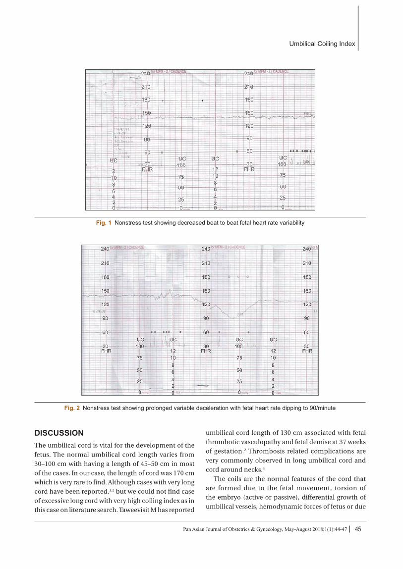
Fig. 1 Nonstress test showing decreased beat to beat fetal heart rate variability
Fig. 2 Nonstress test showing prolonged variable deceleration with fetal heart rate dipping to 90/minute
dIsCussIonThe umbilical cord is vital for the development of the fetus. The normal umbilical cord length varies from 30–100 cm with having a length of 45–50 cm in most of the cases. In our case, the length of cord was 170 cm which is very rare to find. Although cases with very long cord have been reported.1,2 but we could not find case of excessive long cord with very high coiling index as in this case on literature search. Taweevisit M has reported
umbilical cord length of 130 cm associated with fetal thrombotic vasculopathy and fetal demise at 37 weeks of gestation.2 Thrombosis related complications are very commonly observed in long umbilical cord and cord around necks.3
The coils are the normal features of the cord that are formed due to the fetal movement, torsion of the embryo (active or passive), differential growth of umbilical vessels, hemodynamic forces of fetus or due

Aruna Nigam et al.
46 AIM Publications www.pajog.com
Fig. 3 Peroperative picture of hypercoiled long cord and inset showing normal cord
to presence of Roach muscle.4 It has been observed that only pressure gradient between the umbilical vein and the fetal inferior vena cava is not sufficient to transport the necessary amount of blood to the fetus. Therefore, the other theory has been postulated for the blood flow towards the foetus which was supported by the presence of coils in the umbilical cord. The arteries in the umbilical cord stretch in length during pulsations, leading to relative negative pressure in the umbilical vein causing forward venous blood flow. Umbilical cord also becomes more flexible and strong due to the presence of coils. Coils also provide resistance to external forces which could compromise blood flow. The normal range of coils varies from 0 to 40. The umbilical cord coiling index is the ratio of coils to the length of the cord. The normal UCI is 0.17 ± .009 coils/cm.5 The 10th and 90th centiles are 0.07 and 0.30 coils/cm but in this case it was much higher (0.44 coils/cm). The coiling orientation in the present case is of left side which is the most common type.6
Hypercoiling as well as hypocoiling, both are associated with poor fetal outcome. In presence of hypercoiling, the blood flow will be less straight and
more blood will come in contact with the vessel-wall leading to more turbulence and further stasis down the blood flow. Thus hypercoiling is associated with fetal growth retardation, intrauterine death, preterm delivery, intrapartum fetal heart rate decelerations, operative delivery for fetal distress, meconium staining which occurred in our case. In a study of fetal growth restriction with hypercoiling, it has been found that these fetuses are at high risk of hypoxia due to fetal venous system alteration.7 Few authors have given the hypothesis that the hypo as well as hypercoiled cords are less flexible, making them more prone for kinking and torsion during labor.8,9 Prenatal sonographic assessment of umbilical cord coiling index during 2nd trimester scan of congenital anomaly can predict small for gestational age babies and nonreassuring pattern on cardiotocogram during labor.10
ConClusIonThe length and the umbilical cord coiling index must be assessed in the second trimester ultrasonographic examination more so in cases of intrauterine growth restriction and previous history of unexplained intrauterine death. Prolonged variable deceleration and the decreased beat to beat heart rate variability are a sign of fetal distress requiring intervention.
address for CorrespondenceAruna Nigam Flat No. 6, Type 4 Quarters, Bangla Sahib Road, LHMC Campus, New Delhi 110001 [email protected]
referenCes 1. Schmid A, Jacquemyn Y, Loor JD. Intrauterine growth
restriction associated with excessively long umbilical cord. Clin Pract. 2013;3:e23.
2. Taweevisit M, Thorner PS. Massive fetal thrombotic vasculopathy associated with excessively long umbilical cord and fetal demise: case report and literature review. Paediatr Dev Pathol. 2010;13:112-5.
3. Tantbirojn P, Saleemuddin A, Sirois K, Crum CP, Boyd TK, Tworoger S, Parast MM. Gross abnormalities of the umbilical cord: related placental histology and clinical significance. Placenta. 2009;30:1083-8.
4. de Laat MW, Nikkels PG, Franx A, Visser GH. The Roach muscle bundle and umbilical cord coiling. Early Hum Dev. 2007;83:571-4.
5. De Laat MW, franx A, Van aldermen ED, Nikkels PG, Viser GH. The umbilical coiling index, a review of literature. J Matern Fetal Neonatal Med. 2005;17:93-100.

Umbilical Coiling Index
47Pan Asian Journal of Obstetrics & Gynecology, May-August 2018;1(1):44-47
6. Ernst LM, Minturn L, Huang MH, Curry E, Su EJ. Gross patterns of umbilical cord coiling: correlations with placental histology and stillbirth. Placenta. 2013;34(7):583-8.
7. Clerici G, antonelli C, Rizzo G, Kanninen TT, DDi Renzo GC. Atypical hemodynamic pattern in fetuses with hypercoiled umbilical cord and growth restriction. J Matern Fetal Neonatal Med. 2013;26:558-62.
8. Strong THJr., Elliott JP, Radin TG. Non-coiled umbilical blood vessels: a new marker for the fetus at risk. Obstetrics and Gynecology. 1993;81:409-11.
9. De Laat MWM, Franx A, VanAlderen ED, Nikkels PGJ, Visser GHA. The umbilical coiling index, a review of the literature. Journal of Maternal-Fetal and Neonatal Medicine. 2005;17:93-100.
10. Predanic M, Perni SC, Chasen ST, Baergen RN, Chervenak FA. Assessment of umbilical cord coiling during routine fetal sonographic anatomic survey in the second trimester. J Ultrasound Med. 2005;24:185-91.