Severe maxillofacial renal osteodystrophy in two patients with … maxillofacial... · CASE REPORT...
7
CASE REPORT Severe maxillofacial renal osteodystrophy in two patients with chronic kidney disease Maria Luiza Diniz de Sousa Lopes 1 & Assis Filipe Medeiros Albuquerque 2 & Adriano Rocha Germano 2 & Lélia Maria Guedes Queiroz 1 & Márcia Cristina da Costa Miguel 1 & Éricka Janine Dantas da Silveira 1 Received: 7 December 2014 /Accepted: 9 March 2015 /Published online: 19 March 2015 # Springer-Verlag Berlin Heidelberg 2015 Abstract Renal osteodystrophy (ROD) is the bone pathology that occurs as an uncommon complication related to the several alterations in mineral metabolism present in patients with chronic kidney disease (CKD). This paper describes two cases of severe ROD affecting the maxilla and mandible and causing facial dis- figurement of a young and a middle-aged female patient with CKD. Both patients had a history of secondary hyperparathy- roidism, previously treated by surgery. The pathogenesis of the disease, as well as its clinical, imaging, and histopathological features, and management of the patient are discussed. Keywords Renal osteodystrophy . Chronic kidney disease . Secondary hyperparathyroidism . Chronic kidney disease-mineral bone disorder Introduction Chronic kidney disease (CKD) is the progressive and irrevers- ible loss of renal function, usually leading to end-stage renal disease, a potentially fatal condition, requiring chronic hemo- dialysis or kidney transplantation for survival [1, 2]. One of the complications that can arise in patients with CKD is the clinical syndrome chronic kidney disease-mineral and bone disorder (CKD-MBD), which involves changes in laboratory tests such as serum calcium, organic phosphate, parathyroid hormone (PTH), and concentrations of vitamin D derivatives; changes in bone turnover; and extraskeletal calcifications [3, 4]. Renal osteodystrophy (ROD) has recently been redefined to refer to the bone pathology from this syndrome [5]. Advancements in the knowledge about CKD and its com- plications, such as secondary hyperparathyroidism (HPT-II) and ROD, resulted in a decreased frequency of significant macroscopic bone alterations in kidney patients. Nonetheless, radiographic changes of the facial skeleton still eventually occur, occasionally involving the jaws, and may represent an early sign of ROD [6–8]. This manuscript reports the details of two unusually severe maxillofacial manifesta- tions of ROD in patients with CKD, emphasizing the laboratorial, clinical, imaging, and histological features as well as patient management. Case reports Patient 1 A 23-year-old female was referred to the Oral and Maxillofacial Surgery Service of our department with a 4- year history of an asymptomatic, expansive mass in the right side of the face causing airway compromise and facial disfig- urement (Fig. 1a). On intraoral examination, there were mac- roscopic enlargement of the right maxilla and a less severe increase in volume of the right mandible (Fig. 1b). In addition, the patient presented gingivitis, carious teeth, and malocclu- sion. She had a history of idiopathic CKD in advanced stage and had been receiving hemodialysis three times a week for 10 years, while on a kidney transplant waiting list. During this period, she developed HPT-II reaching 1452 pg/mL as a * Maria Luiza Diniz de Sousa Lopes [email protected] 1 Postgraduate Program in Oral Pathology, Dentistry Department, Federal University of Rio Grande do Norte, Avenida Salgado Filho, 1787-Lagoa Nova, Natal, RN CEP 59.056-000, Brazil 2 Oral and Maxillofacial Surgery and Traumatology Residency, Dentistry Department, Federal University of Rio Grande do Norte, Natal, RN, Brazil Oral Maxillofac Surg (2015) 19:321–327 DOI 10.1007/s10006-015-0490-9
Transcript of Severe maxillofacial renal osteodystrophy in two patients with … maxillofacial... · CASE REPORT...

CASE REPORT
Severe maxillofacial renal osteodystrophy in two patientswith chronic kidney disease
Maria Luiza Diniz de Sousa Lopes1 & Assis Filipe Medeiros Albuquerque2 &
Adriano Rocha Germano2 & Lélia Maria Guedes Queiroz1 &
Márcia Cristina da Costa Miguel1 & Éricka Janine Dantas da Silveira1
Received: 7 December 2014 /Accepted: 9 March 2015 /Published online: 19 March 2015# Springer-Verlag Berlin Heidelberg 2015
Abstract Renal osteodystrophy (ROD) is the bone pathologythat occurs as an uncommon complication related to the severalalterations inmineralmetabolism present in patientswith chronickidney disease (CKD). This paper describes two cases of severeROD affecting the maxilla and mandible and causing facial dis-figurement of a young and a middle-aged female patient withCKD. Both patients had a history of secondary hyperparathy-roidism, previously treated by surgery. The pathogenesis of thedisease, as well as its clinical, imaging, and histopathologicalfeatures, and management of the patient are discussed.
Keywords Renal osteodystrophy . Chronic kidney disease .
Secondary hyperparathyroidism . Chronic kidneydisease-mineral bone disorder
Introduction
Chronic kidney disease (CKD) is the progressive and irrevers-ible loss of renal function, usually leading to end-stage renaldisease, a potentially fatal condition, requiring chronic hemo-dialysis or kidney transplantation for survival [1, 2]. One ofthe complications that can arise in patients with CKD is theclinical syndrome chronic kidney disease-mineral and bone
disorder (CKD-MBD), which involves changes in laboratorytests such as serum calcium, organic phosphate, parathyroidhormone (PTH), and concentrations of vitamin D derivatives;changes in bone turnover; and extraskeletal calcifications [3,4]. Renal osteodystrophy (ROD) has recently been redefinedto refer to the bone pathology from this syndrome [5].
Advancements in the knowledge about CKD and its com-plications, such as secondary hyperparathyroidism (HPT-II)and ROD, resulted in a decreased frequency of significantmacroscopic bone alterations in kidney patients.Nonetheless, radiographic changes of the facial skeleton stilleventually occur, occasionally involving the jaws, and mayrepresent an early sign of ROD [6–8]. This manuscript reportsthe details of two unusually severe maxillofacial manifesta-tions of ROD in patients with CKD, emphasizing thelaboratorial, clinical, imaging, and histological features aswell as patient management.
Case reports
Patient 1
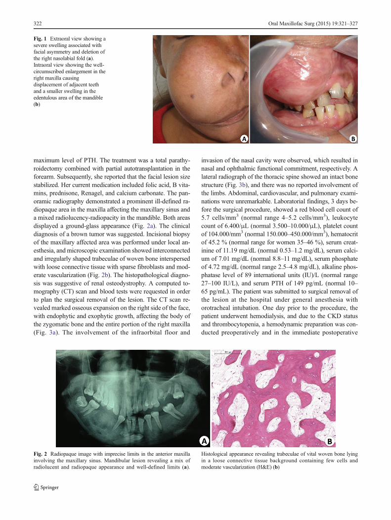
A 23-year-old female was referred to the Oral andMaxillofacial Surgery Service of our department with a 4-year history of an asymptomatic, expansive mass in the rightside of the face causing airway compromise and facial disfig-urement (Fig. 1a). On intraoral examination, there were mac-roscopic enlargement of the right maxilla and a less severeincrease in volume of the right mandible (Fig. 1b). In addition,the patient presented gingivitis, carious teeth, and malocclu-sion. She had a history of idiopathic CKD in advanced stageand had been receiving hemodialysis three times a week for10 years, while on a kidney transplant waiting list. During thisperiod, she developed HPT-II reaching 1452 pg/mL as a
* Maria Luiza Diniz de Sousa [email protected]
1 Postgraduate Program in Oral Pathology, Dentistry Department,Federal University of Rio Grande do Norte, Avenida Salgado Filho,1787-Lagoa Nova, Natal, RN CEP 59.056-000, Brazil
2 Oral and Maxillofacial Surgery and Traumatology Residency,Dentistry Department, Federal University of Rio Grande do Norte,Natal, RN, Brazil
Oral Maxillofac Surg (2015) 19:321–327DOI 10.1007/s10006-015-0490-9
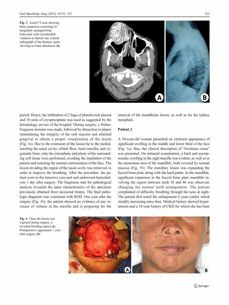
maximum level of PTH. The treatment was a total parathy-roidectomy combined with partial autotransplantation in theforearm. Subsequently, she reported that the facial lesion sizestabilized. Her current medication included folic acid, B vita-mins, prednisone, Renagel, and calcium carbonate. The pan-oramic radiography demonstrated a prominent ill-defined ra-diopaque area in the maxilla affecting the maxillary sinus anda mixed radiolucency-radiopacity in the mandible. Both areasdisplayed a ground-glass appearance (Fig. 2a). The clinicaldiagnosis of a brown tumor was suggested. Incisional biopsyof the maxillary affected area was performed under local an-esthesia, and microscopic examination showed interconnectedand irregularly shaped trabeculae of woven bone interspersedwith loose connective tissue with sparse fibroblasts and mod-erate vascularization (Fig. 2b). The histopathological diagno-sis was suggestive of renal osteodystrophy. A computed to-mography (CT) scan and blood tests were requested in orderto plan the surgical removal of the lesion. The CT scan re-vealed marked osseous expansion on the right side of the face,with endophytic and exophytic growth, affecting the body ofthe zygomatic bone and the entire portion of the right maxilla(Fig. 3a). The involvement of the infraorbital floor and
invasion of the nasal cavity were observed, which resulted innasal and ophthalmic functional commitment, respectively. Alateral radiograph of the thoracic spine showed an intact bonestructure (Fig. 3b), and there was no reported involvement ofthe limbs. Abdominal, cardiovascular, and pulmonary exami-nations were unremarkable. Laboratorial findings, 3 days be-fore the surgical procedure, showed a red blood cell count of5.7 cells/mm3 (normal range 4–5.2 cells/mm3), leukocytecount of 6.400/μL (normal 3.500–10.000/μL), platelet countof 104.000/mm3 (normal 150.000–450.000/mm3), hematocritof 45.2 % (normal range for women 35–46 %), serum creat-inine of 11.19 mg/dL (normal 0.53–1.2 mg/dL), serum calci-um of 7.01 mg/dL (normal 8.8–11 mg/dL), serum phosphateof 4.72 mg/dL (normal range 2.5–4.8 mg/dL), alkaline phos-phatase level of 89 international units (IU)/L (normal range27–100 IU/L), and serum PTH of 149 pg/mL (normal 10–65 pg/mL). The patient was submitted to surgical removal ofthe lesion at the hospital under general anesthesia withorotracheal intubation. One day prior to the procedure, thepatient underwent hemodialysis, and due to the CKD statusand thrombocytopenia, a hemodynamic preparation was con-ducted preoperatively and in the immediate postoperative
Fig. 1 Extraoral view showing asevere swelling associated withfacial asymmetry and deletion ofthe right nasolabial fold (a).Intraoral view showing the well-circumscribed enlargement in theright maxilla causingdisplacement of adjacent teethand a smaller swelling in theedentulous area of the mandible(b)
Fig. 2 Radiopaque image with imprecise limits in the anterior maxillainvolving the maxillary sinus. Mandibular lesion revealing a mix ofradiolucent and radiopaque appearance and well-defined limits (a).
Histological appearance revealing trabeculae of vital woven bone lyingin a loose connective tissue background containing few cells andmoderate vascularization (H&E) (b)
322 Oral Maxillofac Surg (2015) 19:321–327
period. Hence, the infiltration of 2 bags of platelet-rich plasmaand 10 units of cryoprecipitate was used as suggested by thehematology service of the hospital. During surgery, a Weber-Ferguson incision was made, followed by dissection in planes(maintaining the integrity of the oral mucosa and attachedgingiva) to obtain a proper visualization of the lesion(Fig. 4a). Due to the extension of the lesion far to the medial,reaching the nasal cavity, orbital floor, hemi-maxilla, and zy-gomatic bone, only the osteoplasty and plasty of the surround-ing soft tissue were performed, avoiding the mutilation of thepatient and restoring the normal conformation of the face. Thelesion invading the region of the nasal cavity was removed, inorder to improve the breathing. After the procedure, the pa-tient went to the intensive care unit and underwent hemodial-ysis 1 day after surgery. The fragments sent for pathologicalanalysis revealed the same characteristics of the specimenpreviously obtained from incisional biopsy. The final patho-logic diagnosis was consistent with ROD. One year after thesurgery (Fig. 4b), the patient showed no evidence of any in-crease of volume in the maxilla and is preparing for the
removal of the mandibular lesion, as well as for the kidneytransplant.
Patient 2
A 44-year-old woman presented an extraoral appearance ofsignificant swelling in the middle and lower third of the face(Fig. 5a); thus, the clinical description of “leontiasis ossea”was presented. On intraoral examination, a hard and asymp-tomatic swelling in the right maxilla was evident, as well as inthe mentonian area of the mandible, both covered by normalmucosa (Fig. 5b). The maxillary lesion was expanding thebuccal bone plate along with the hard palate. In the mandible,significant expansion in the buccal bone plate mandible in-volving the region between teeth 36 and 46 was observed,changing the normal teeth arrangement. The patientcomplained of difficulty breathing through the nose at night.The patient first noted the enlargement 6 years earlier whichsteadily increasing since then. Medical history showed hyper-tension and a 10-year history of CKD for which she has been
Fig. 3 Axial CT scan showingbone expansion consisting ofirregularly arranged bonetrabeculae with considerablevariation in density (a). Lateralradiograph of the thoracic spineshowing no bone alterations (b)
Fig. 4 Once the lesion wasexposed during surgery, itrevealed bleeding aspect (a).Postoperative appearance 1 yearafter surgery (b)
Oral Maxillofac Surg (2015) 19:321–327 323

receiving hemodialysis three times a week. During this period,the patient developed HPT-II, achieving a maximum value ofserum parathyroid hormone (PTH) of 1056 pg/mL (normal10–65 pg/mL). The patient underwent total parathyroidecto-my associated with partial autotransplantation in the forearm1 year before being referred to our department. Her medica-tion included losartan potassium and clonidine hydrochloride.Panoramic radiography showed a marked expansion of themaxilla and mandible, with a ground-glass appearance andteeth hypercementosis (Fig. 6). There were no bone manifes-tations at other areas of the body. Laboratory examinationshowed current red blood cell count of 4.2 cells/mm3 (normalrange 4–5.2 cells/mm3), leukocyte count of 6.200/μL (normal3.500–10.000/μL), platelet count of 325.000/mm3 (normal150.000–450.000/mm3), hematocrit of 37.8 % (normal rangefor women 35–46 %), serum creatinine of 11.17 mg/dL (nor-mal 0.53–1.2 mg/dL), serum calcium of 6.9 mg/dL (normal8.8–11 mg/dL), serum phosphate of 4.72 mg/dL (normalrange 2.5–4.8 mg/dL), alkaline phosphatase of 244 IU/L)(normal range 27–100 IU/L), and serum PTH of 208 pg/mL(normal 10–65 pg/mL). The clinical diagnostic hypothesiswas ROD and brown tumor. The patient underwent incisionalbiopsies of both maxillary and mandibular lesions under localanesthesia. Microscopic examination revealed vital and
irregular trabeculae of woven bone in different maturationstages, occasionally showing osteoblastic rimming as well asmultinucleated osteoclast-type giant cells into a vascularizedfibrous connective tissue (Fig. 7). The clinical, biochemical,imaging, and microscopic studies lead to a diagnosis of oste-itis fibrosa consistent with ROD. The patient was submitted toosteoplasty of both lesions by an intraoral approach in thehospital under general anesthesia. Sections of the collectedspecimen confirmed the histopathological diagnosis osteitisfibrosa consistent with ROD. Ten months after the surgery,the patient shows no noticeable bone changes on her facialcontour and is under nephrologic care (Fig. 8).
Discussion
Patients with CKD commonly have alterations in several min-eral metabolisms including hyperphosphatemia, hypocalce-mia, and 1,25-dihydroxyvitamin D3 or calcitrol (active metab-olite of vitamin D), which eventually lead to a compensatoryoverproduction of PTH by parathyroid glands in a conditionknown as HPT-II [9–11]. As renal function declines, 1,25-dihydroxyvitamin D3 synthesis becomes impaired, which
Fig. 5 Extraoral view (patient 2) showing a swelling in the middle and lower third of the face resembling a leonine face (a). Intraoral view of severemaxillary and mandibular enlargement causing teeth separation (b)
Fig. 6 Panoramic radiography (patient 2) displaying marked expansionof bone structures with ground-glass appearance and several teeth withhypercementosis
Fig. 7 Multinucleated giant cells throughout the stroma and plumposteoblasts adjacent to the osteoid matrix (patient 2; H&E)
324 Oral Maxillofac Surg (2015) 19:321–327

leads to deficiency in renal calcium absorption, resulting inhypocalcemia. The calcium sensing receptor (CaSR), which ishighly expressed in the parathyroid glands, senses the lowserum calcium concentrations, leading to a compensatory in-crease in PTH (HPT-II) in the attempt to maintain the homeo-stasis of calcium [10]. In the meantime, the reduction of glo-merular filtration also leads to impaired phosphate excretion,elevating the serum concentrat ion of phosphate.Hyperphosphatemia can act by directly stimulating PTH syn-thesis and parathyroid hyperplasia and indirectly promotingHPT-II by decreasing the serum calcium level [1, 2, 10].Excess of PTH acts in the transformation of the inactive formof vitamin D to 1,25-dihydroxyvitamin D3 in the kidney andinfluences the development and activity of osteoclasts, osteo-blasts, and osteocytes, thereby affecting bone turnover [1, 4].
Due to the mineral alterations, patients with ROD shouldrun basic blood tests, including electrolytes, full blood cellcount, PTH, and alkaline phosphatase levels [2]. The labora-tory findings of both patients revealed a serum PTH levelslightly elevated, but when it is compared to the levels beforethe removal of the parathyroid glands, the considerable im-provement in the HPT-II status became evidently unmistak-able. Especially in patient 1, this fact was supported by thedemonstrated normal levels of calcium, phosphorus, and alka-line phosphatase, the latter being a biochemical marker and,together with the PTH, commonly used in the diagnosis ofdifferent forms of bone disease associated with CKD [12].
Usually, the alterations in bone metabolism are diagnosedby laboratory tests before the development of macroscopicskeletal changes [7, 10]. The majority of macroscopic mani-festations of ROD present as diffuse jaw enlargement [13].However, recently, the pattern of localized expansion was re-ported to be more common than originally recognized [5].Patient 1 exhibited localized enlargements of the maxilla andmandible, causing facial disfigurement, while the bone alter-ations of patient 2 looked more diffuse. In addition, the alter-ations were exclusively in the jaw bones, any manifestation inlong bones and aorta being discarded.
The extreme clinical manifestation of ROD is denominatedleontiasis ossea represented by hyperostotic changes in thefacial bones and may cause an expansion of the malar process-es, a reduced nasomaxillary angle, widening of the nares,
flattening of the nasal bridge, and an increase of interdentalspace [9]. This ultimate condition results in lion-like facialfeatures observed in patient 2, which clinically and histologi-cally resembles the big head disease observed in animals withhyperparathyroidism secondary to kidney problems or to dietsrich in phosphorus and calcium deficiency [2].
Such facial disfigurement as in leontiasis ossea is an un-common feature of ROD and is not specific because it can bepresent in other lesions such as fibrous dysplasia, or Paget’sdisease of bone [9]. Craniofacial fibrous dysplasia is asym-metric and can cause cranial neuropathies, headaches, andocclusal discrepancies [2]. In contrast to ROD, fibrous dyspla-sia typically occurs in childhood and accompanies a biochem-ical profile within normal limits [5, 13]. Concerning Paget’sdisease, the rates of osteoclastic and osteoblastic activities areelevated, resulting in skeletal remodeling and weakened de-formed bones. However, when it affects jawbones, it is typi-cally bilateral [14]. Moreover, even showing increased levelsof alkaline phosphatase, the serum level of calcium in Paget’sdisease is normal [2].
Some radiographic findings of ROD of the jaws also occurin fibrous dysplasia, or Paget’s disease of bone, such asground-glass appearance, observed in both current patients;hypercementosis noticed on patient 2; and bone resorptionwith loss of lamina dura [13]. Some of the radiographic alter-ations of ROD include demineralization of bone, delayedtooth eruption, mixed osteolysis and sclerosis (salt-and-pep-per) trabecular appearance, absence of lamina dura, thickeningof the diploic space of the calvarium, dental spacing and mo-bility, jaw fracture, and poor definition of anatomical land-marks [9]. The CT scan can show changes in the trabecularpattern and bone expansion and, along with magnetic reso-nance imaging, is helpful for diagnosis and treatment planning[15].
Microscopic and pathologic features of ROD are dynamicin their progression and can be subdivided into low- and high-turnover bone diseases or with a combined pattern of the twocategories [16]. Osteitis fibrosa represents increased boneturnover as it is associated with hyperparathyroidism, whiledecreased turnover mainly characterizes osteomalacia andadynamic (aplastic) bone disease [7, 13]. Osteitis fibrosa usedto be the most common type of ROD; however, in the lastdecades, adynamic bone disease has become the most com-mon form [17]. Microscopic findings of osteitis fibrosa revealprominent peritrabecular fibrosis associated with an increasednumber and activity of osteoclasts associated with increasednumbers of osteoblasts and osteoid [2, 13, 18]. Those featureswere present in patient 2, justifying the diagnosis of osteitisfibrosa. On the other hand, osteomalacia is characterized bydefective mineralization and increased osteoid productionmainly caused by the decrease of 1,25-dihydroxyvitamin D3
concentrations, usually causing osseous pain and musculo-skeletal disability and making the patient at risk of osteopenia,
Fig. 8 Facial appearance 10 months after operation (patient 2)
Oral Maxillofac Surg (2015) 19:321–327 325

bone deformity, and fracture [1, 4, 5]. Adynamic bone diseaseis most frequent in CKD patients without HPT-II and is usu-ally related to increased risk of hypercalcemia, cardiovascularcalcification, and mortality. No osteoid accumulation ispresent, and there are only few or absent osteoblastsand little or no fibrosis in either the bone marrow orperitrabecular area [17]. The absence of osteoid deposi-tion or osteoclastic activity in patient 1 specimens reflectsthe lack of active bone metabolism, or even normal bonemetabolism, probably because the patient’s bone diseasewas under control.
Differential diagnosis of ROD, besides fibrous dysplasiaand Paget’s disease of the bone, should include brown tumorsof HPT-II, which generally can be discarded by radiographicappearance or/and histopathological examination. Brown tu-mors are more likely to occur in primary hyperparathyroidism,showing a radiographic appearance of a well-circumscribedradiolucent lesion and typically revealingmultinucleated giantcells and prominent hemosiderin deposition under light mi-croscopy [5, 6, 8]. As observed in patient 2, the presence ofmultinucleated giant cells can also occur in ROD, but accord-ing to Lerman et al. [5], it is less prominent than brown tu-mors. The Sagliker syndrome, recently described by Saglikeret al. [19], also must be considered in the differential diagnosisof ROD, since it is seen in 0.5 % of patients with CKD [20].However, in addition to HPT-II and CKD, the diagnosticcriteria for this syndrome include short stature, extremely se-vere maxillary and mandibular changes (uglifying the face),soft and histopathologically verified benign epithelial hyper-plasia, severe teeth/dental abnormalities, fingertip changes,knee and scapula deformities, abnormalities in audition, andsevere neurological and psychological disturbances [20, 21].
Management of ROD is based on control of factors respon-sible for the development of altered bone metabolism [22].ROD associated with HPT-II can be improved with the cor-rection of calcium and vitamin D levels and a decrease inphosphate levels. This can be accomplished by conservativetreatment, such as the use of phosphate-binding agents, treat-ment with calcitriol and other active vitamin D sterols, andfollowing a phosphate-restricted diet [1, 2, 9]. Despite medicaltherapy, in several patients, the altered PTH levels persist,requiring surgical intervention with total parathyroidectomywith or without autotransplantation or subtotal parathyroidec-tomy. The ultimate treatment for persistent HPT-II in the twocu r r e n t p a t i e n t s wa s p a r a t hy r o i d e c t omy w i t hautotransplantation, which involves the transplantation of asmall amount of parathyroid tissue into the forearm, aimingto maintain normal parathyroid function. If recurrent hyper-parathyroidism occurs, this technique allows a simple access,with locoregional anesthesia, for removal of the fragmentsfrom the forearm [23].
Even with the adjustment of the PTH levels, the total re-gression of bone alterations is unusual; therefore, most cases
require surgical recontouring. There are few reports of RODmanifestations in jaws resolved after the correction of theHPT-II condition, without surgical intervention [24, 25]. Thejaw enlargement in the present patients ceased after the treat-ment of HPT-II, but surgery was necessary mainly for im-provement of breathing and for esthetics consideration.
This manuscript emphasizes the importance of a detailedanamnesis and clinical and microscopic investigation for thediagnosis and appropriate treatment of ROD affecting themaxillofacial region. Even though the altered bone metabo-lism usually is biochemically detected before patients haveany significant symptomatology, the cases reported here high-light that in unusual circumstances it may develop into severebone alterations.
Conflict of interest The authors declare no conflict of interest.
References
1. Antonelli JR, Hottel TL (2003) Oral manifestations of renalosteodystrophy: case report and review of the literature. SpecCare Dentist 23(1):28–34
2. Collum J, Jones RHB, Liynham A, Hirst J (2013) Leontiasis ossea:a presentation of hyperparathyroidism in an indigenous Australianman secondary to chronic renal failure. J Oral Maxillofac Surg71(1):56–61. doi:10.1016/j.joms.2012.04.008
3. Chauhan V, Kelepouris E, Chauhan N, Vaid M (2012) Currentconcepts and management strategies in chronic kidney disease-mineral and bone disorder. South Med J 105(9):479–485. doi:10.1097/SMJ.0b013e318261f7fe
4. LimCY, OngKO (2013) Variousmusculoskeletal manifestations ofchronic renal insufficiency. Clin Radiol 68(7):e397–e411. doi:10.1016/j.crad.2013.01.025
5. LermanMA, Do C, Gunaratnam L, Kulkarni C, Tucker K,Woo SB(2012) Localized mandibular enlargement in end-stage renal dis-ease: two case reports and a review of the literature. Oral Surg OralMed Oral Pathol Oral Radiol 113(3):384–390. doi:10.1016/j.tripleo.2011.04.039
6. Damm DD, Neville BW, McKenna S, Jones AC, Freedman PD,AndersonWR et al (1997)Macrognathia of renal osteodystrophy indialysis patients. Oral Surg Oral Med Oral Pathol Oral RadiolEndod 83(4):489–495
7. Chang JI, Som PM, Lawson W (2007) Unique imaging findings inthe facial bones of renal osteodystrophy. Am J Neuroradiol 28(4):608–609
8. Karsburg RM, Campos KR, Peres MPSM, Bologna SB, LourençoSV, Franco JB (2012) Facial lesions caused by renal osteodystrophyin a patient with chronic renal insufficiency: a case report. RevOdontol Cienc 27(2):161–165
9. Fatahzadeh M (2011) The spectrum of orofacial manifestations inrenal osteodystrophy: diagnostic challenges of an uncommon pre-sentation. Quintessence Int 42(7):e78–e88
10. Heymann EP, Jenkins M, Goldsmith D (2011) Clinical features andmanifestations of CKD-MBD. Clin Rev Bone Miner Metab 10(3):142–148
11. Way FM, Lessard M, Lafage-Proust MH (2012) Pathophysiologyof chronic kidney disease-mineral and bone disorder. Joint BoneSpine 79(6):544–549
326 Oral Maxillofac Surg (2015) 19:321–327

12. Martin KJ, Olgaard K, Coburn JW, Coen GM, Fukagawa M,Langman C et al (2004) Bone Turnover Work Group: diagnosis,assessment, and treatment of bone turnover abnormalities in renalosteodystrophy. Am J Kidney Dis 43(3):558–565
13. Kalyvas D, Tosios KI, LeventisMD, Tsiklakis K, Angelopoulos AP(2004) Localized jaw enlargement in renal osteodystrophy: reportof a case and review of the literature. Oral Surg Oral Med OralPathol Oral Radiol Endod 97(1):68–74
14. Brooks JK, Rivera-Ramirez LE, Errington LW, Scheper MA (2011)Synchronous Paget disease of bone and hyperparathyroidism: re-port of a case with extensive craniofacial involvement. Oral SurgOral Med Oral Pathol Oral Radiol Endod 111(4):e19–e24. doi:10.1016/j.tripleo.2010.11.004
15. Abdel Razek AA (2014) Computed tomography and magnetic res-onance imaging of maxillofacial lesions in renal osteodystrophy. JCraniofac Surg 25(4) :1354–1357. doi :10.1097/SCS.0000000000000819
16. Thomas R, Kanso A, Sedor JR (2008) Chronic kidney disease andits complications. Prim Care 35(2):329–344
17. MorrowB, QunibiW (2012) Specific bone andmineral disorders inpatients with chronic kidney disease. Clin Rev Bone Miner Metab10(3):184–208
18. Adachi PLG, Santos PSS, Magalhães MHCG, Martins MT (2007)Renal osteodystrophy manifesting as localized enlargement of thejaw. Nephrol Dial Transplant 22:2398–2399. doi:10.1093/ndt/gfm232
19. Sagliker Y, Balal M, Ozkaynak PS, Paydas S, Sagliker C, SaglikerHS et al (2004) Sagliker syndrome: uglifying human face appear-ance in late and severe secondary hyperparathyroidism in chronicrenal failure. Semin Nephrol 24(5):449–455
20. Yavascan O, Kose E, Alparslan C, Kose SS, Bal A, Kanik A et al(2013) Severe renal osteodystrophy in a pediatric patient with end-stage renal disease: Sagliker syndrome? J Ren Nutr 23(4):326–330.doi:10.1053/j.jrn.2012.07.002
21. Giray S, Sagliker Y, Yildiz I, Halvaci I, Paylar N, Ocal F et al (2006)Neurologic manifestations in Sagliker syndrome: uglifying humanface appearance in severe and late secondary hyperparathyroidismin chronic renal failure patients. J Ren Nutr 16(3):233–236
22. Malluche HH, Mawad HW, Monier-Faugere MC (2011) Renalosteodystrophy in the first decade of the new millennium: analysisof 630 bone biopsies in black and white patients. J BoneMiner Res26(6):1368–1376. doi:10.1002/jbmr.309
23. Conti-Freitas LC, Foss-Freitas MC, Lucca LJ, da Costa JA,Mamede RC, Foss MC (2009) Dynamics of parathyroid hormonesecretion after total parathyroidectomy and autotransplantation.World J Surg 33(7):1403–1407. doi:10.1007/s00268-009-0057-8
24. Adornato MC, Mayne RW (2000) Macrognathia of renalosteodystrophy in a dialysis patient. Report of a case. N Y StateDent J 66(10):30–34
25. Hata T, Irei I, Tanaka K, Nagatsuka H, Hosoda M (2006)Macrognathia secondary to dialysis-related renal osteodystrophytreated successfully by parathyroidectomy. Int J Oral MaxillofacSurg 35(4):378–382
Oral Maxillofac Surg (2015) 19:321–327 327