MAXILLOFACIAL TRAUMATOLOGY Department of Maxillofacial...
68
MAXILLOFACIAL TRAUMATOLOGY Department of Maxillofacial Surgery Semmelweis University, Budapest
Transcript of MAXILLOFACIAL TRAUMATOLOGY Department of Maxillofacial...
MAXILLOFACIAL TRAUMATOLOGY
Department of Maxillofacial Surgery
Semmelweis University, Budapest
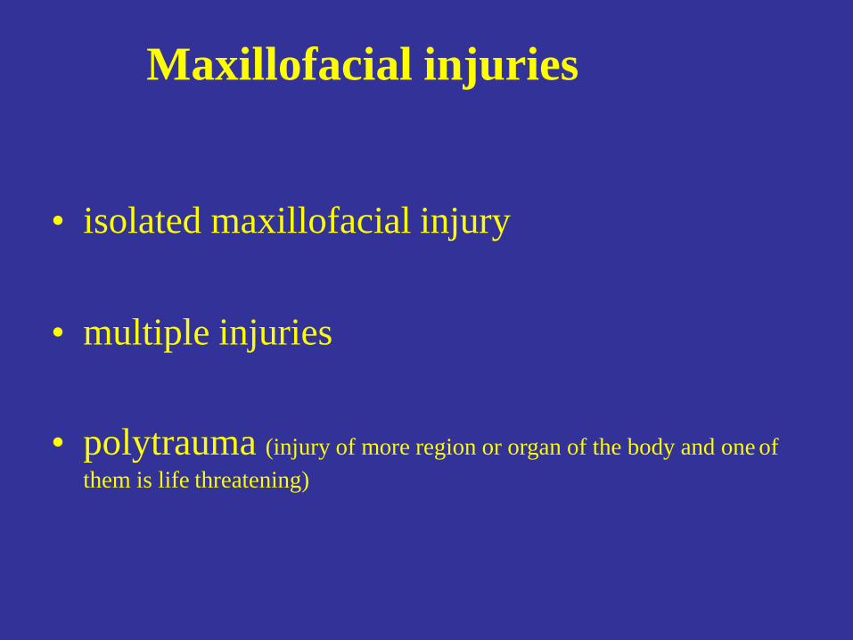
• isolated maxillofacial injury
• multiple injuries
• polytrauma (injury of more region or organ of the body and one of
them is life threatening)
Maxillofacial injuries
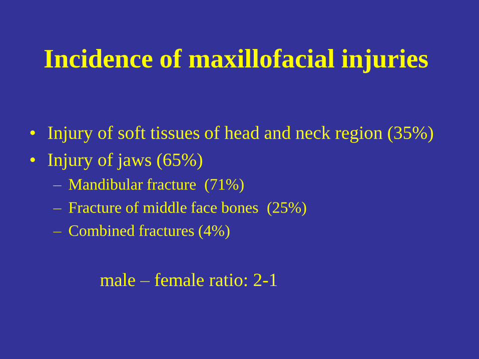
Incidence of maxillofacial injuries
• Injury of soft tissues of head and neck region (35%)
• Injury of jaws (65%)
– Mandibular fracture (71%)
– Fracture of middle face bones (25%)
– Combined fractures (4%)
male – female ratio: 2-1
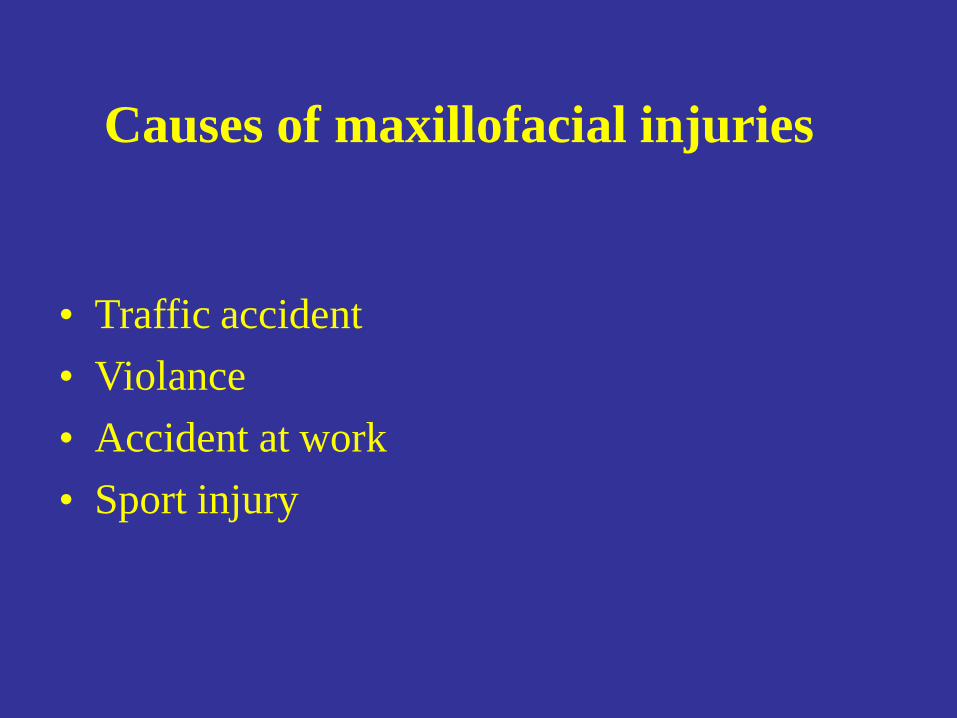
• Traffic accident
• Violance
• Accident at work
• Sport injury
Causes of maxillofacial injuries
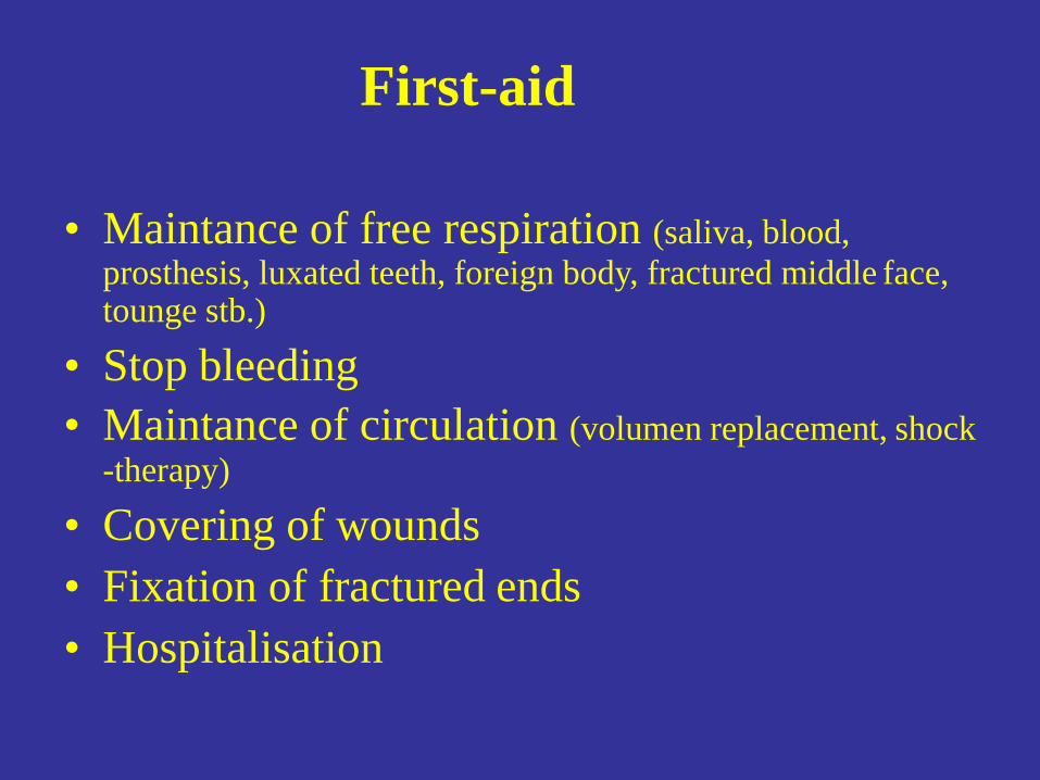
First-aid
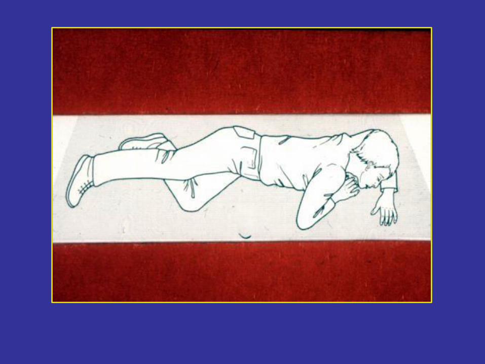
• Maintance of free respiration (saliva, blood,
prosthesis, luxated teeth, foreign body, fractured middle face, tounge stb.)
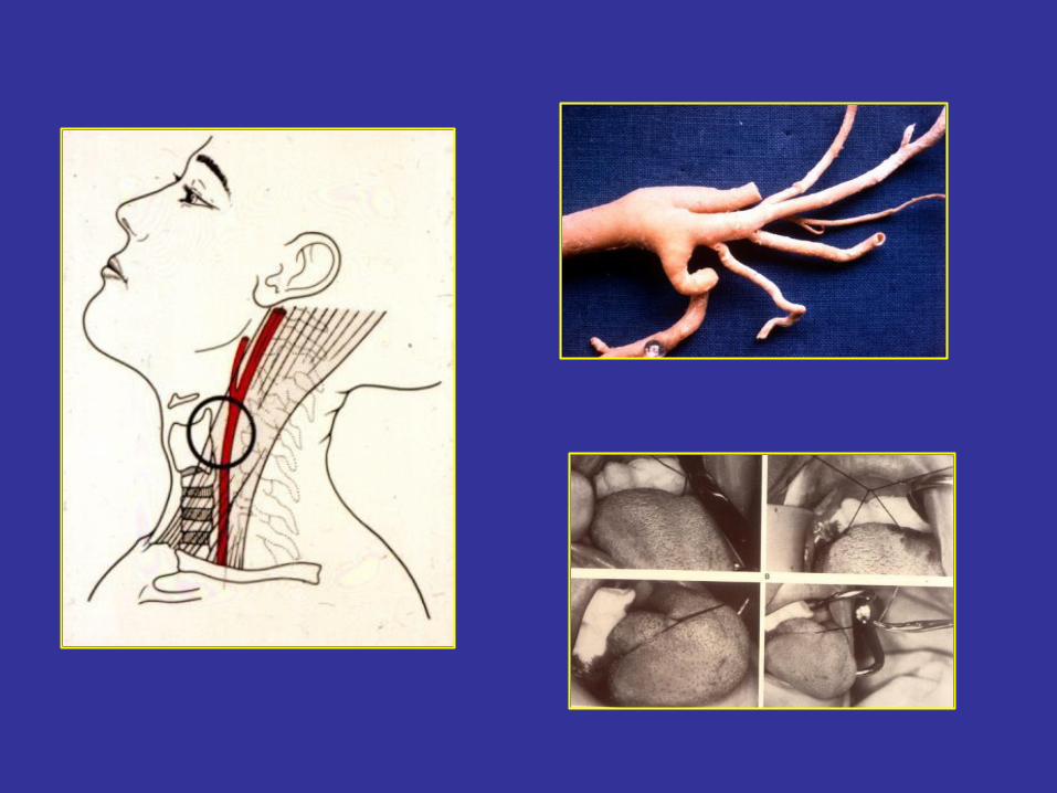
• Stop bleeding
• Maintance of circulation (volumen replacement, shock
-therapy)
• Covering of wounds
• Fixation of fractured ends
• Hospitalisation
Treatment in hospital
if it is possible immediate and definitive!!!
• diagnosis (clinical symptomes, rtg.)
• treatment of soft tissue injuries
• reposition of fractured bone ends, immobilisation
• antibiotic administration
• nutrition, rehabilitation
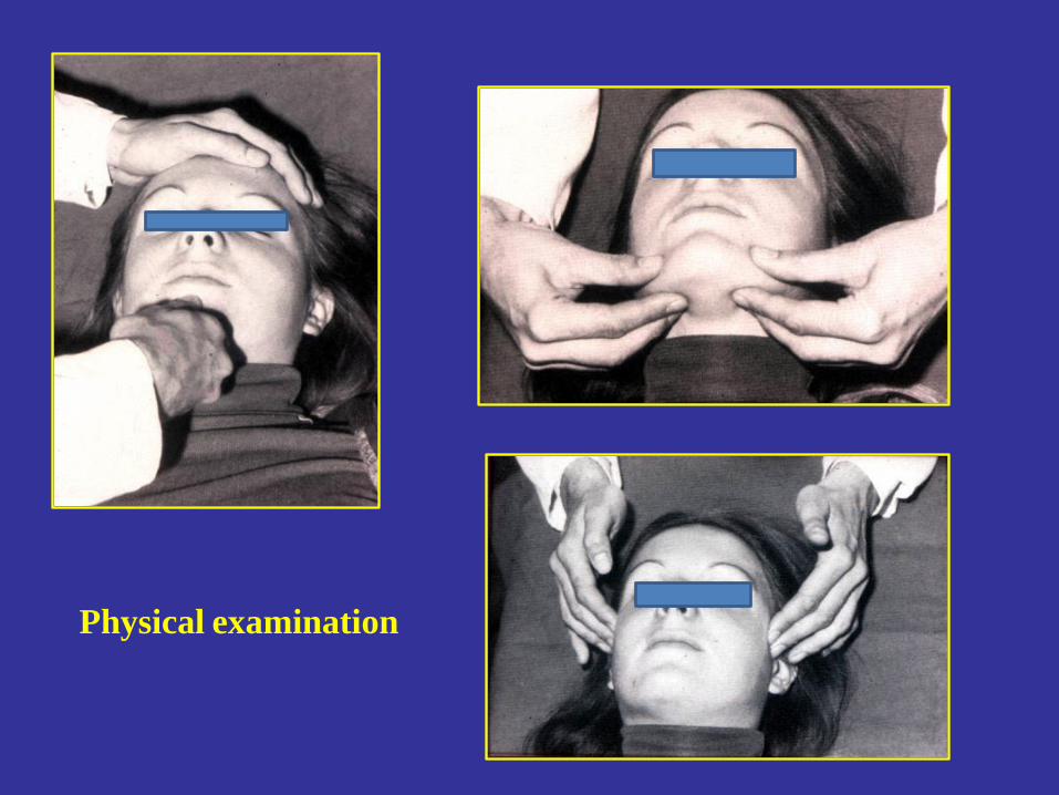
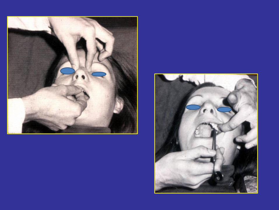
Physical examination
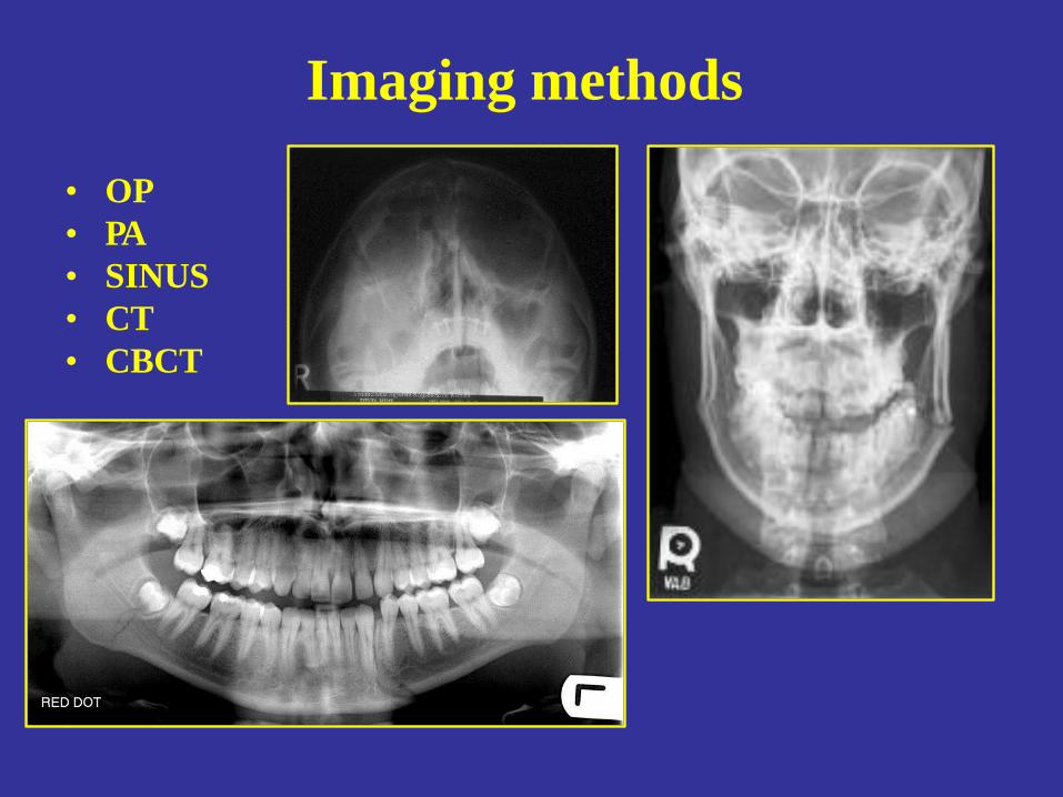
Imaging methods
• OP
• PA
• SINUS
• CT
• CBCT
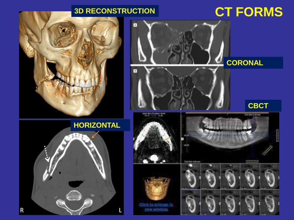
CT FORMS
CORONAL
CBCT
3D RECONSTRUCTION
HORIZONTAL
Mandibular fractures
• 75 % of jaw fractures
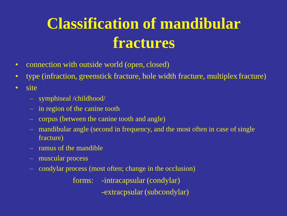
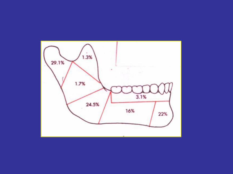
Classification of mandibular
fractures
• connection with outside world (open, closed)
• type (infraction, greenstick fracture, hole width fracture, multiplex fracture)
• site
– symphiseal /childhood/
– in region of the canine tooth
– corpus (between the canine tooth and angle)
– mandibular angle (second in frequency, and the most often in case of single
fracture)
– ramus of the mandible
– muscular process
– condylar process (most often; change in the occlusion)
forms: -intracapsular (condylar)
-extracpsular (subcondylar)
Diagnosis
• anamnesis
• inspection
• physical examination
• imaging methods ( x-ray, CT, CBCT)
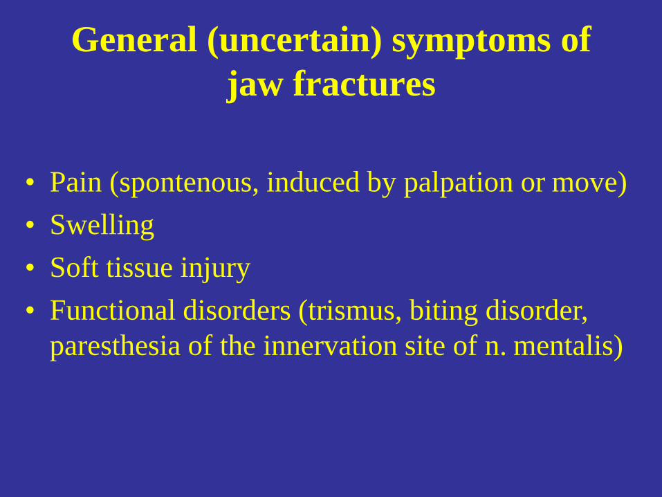
General (uncertain) symptoms of
jaw fractures
• Pain (spontenous, induced by palpation or move)
• Swelling
• Soft tissue injury
• Functional disorders (trismus, biting disorder,
paresthesia of the innervation site of n. mentalis)
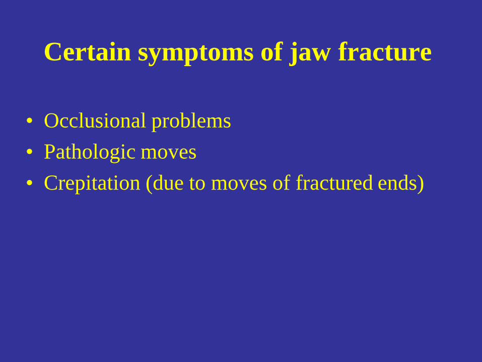
Certain symptoms of jaw fracture
• Occlusional problems
• Pathologic moves
• Crepitation (due to moves of fractured ends)
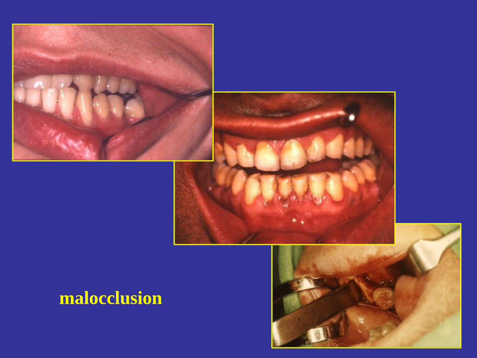
malocclusion
Therapy of mandibular fractures
-Conservative
-Surgical
-Sugical-conservative
• Aim: to reach the orginial function and anatomic situation
• Type of the treatment:
•
•
• Conservative:
– intermaxillary fixation (IMF) with dental splints for 4-6 weeks (Schuchardt-, Stout-, Sauer splints, Gunning splint in case of total toothless)
– Circumferencial fixation – Problems: nutrition, oral higiene, morbus sacer,
unedentoulness, mental retardation)
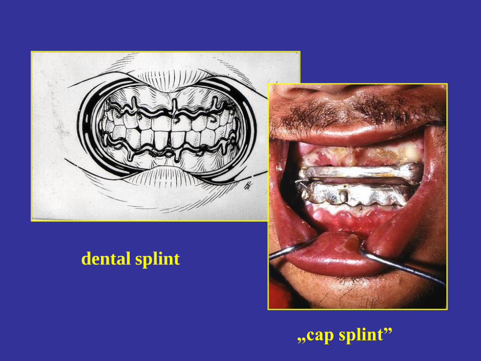
dental splint
„cap splint”
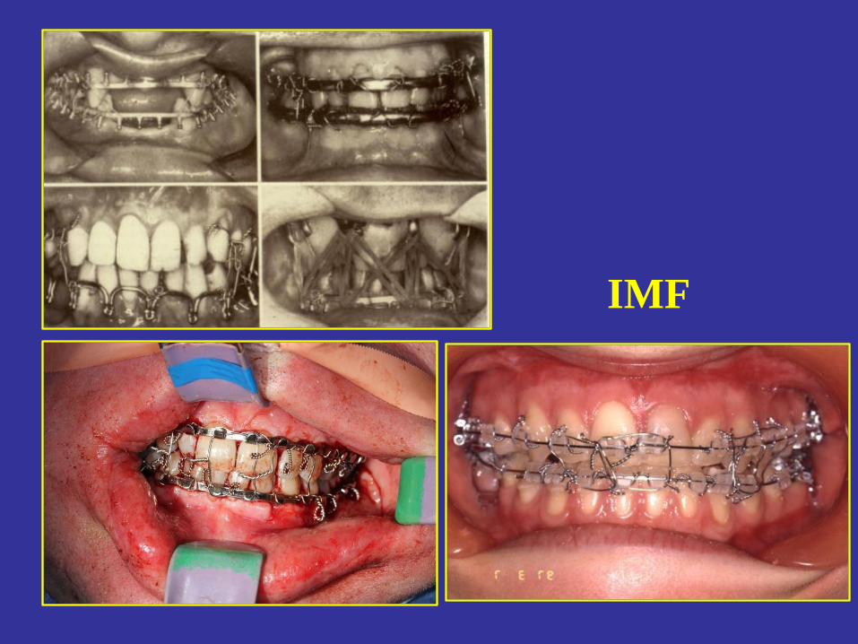
IMF
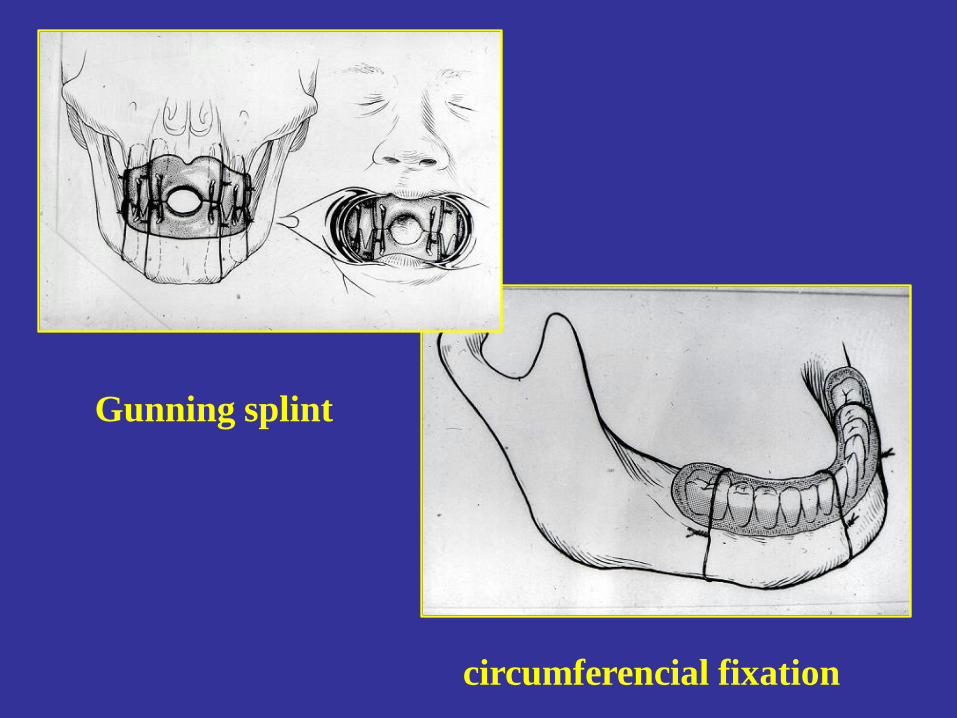
circumferencial fixation
Gunning splint
Surgical therapy of mandibular
fractures
•
• Osteosynthesis (extra and/or intraoral)
Types:
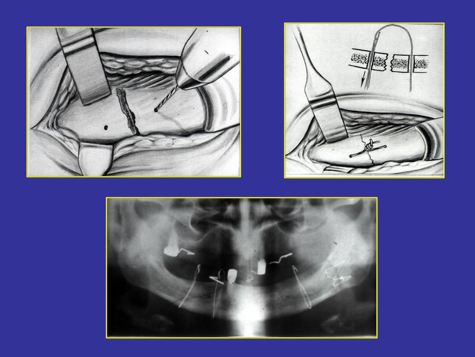
-with wire (Wassmund, Neuner) + IMF
-with pin fixation
-with compression plates (first: Luhr in 1968; most modern)
-systems:
• -Luhr • -ASIF (Association for the Study of Internal fixation) – DCP plate (Dynamic Compression
Plate)
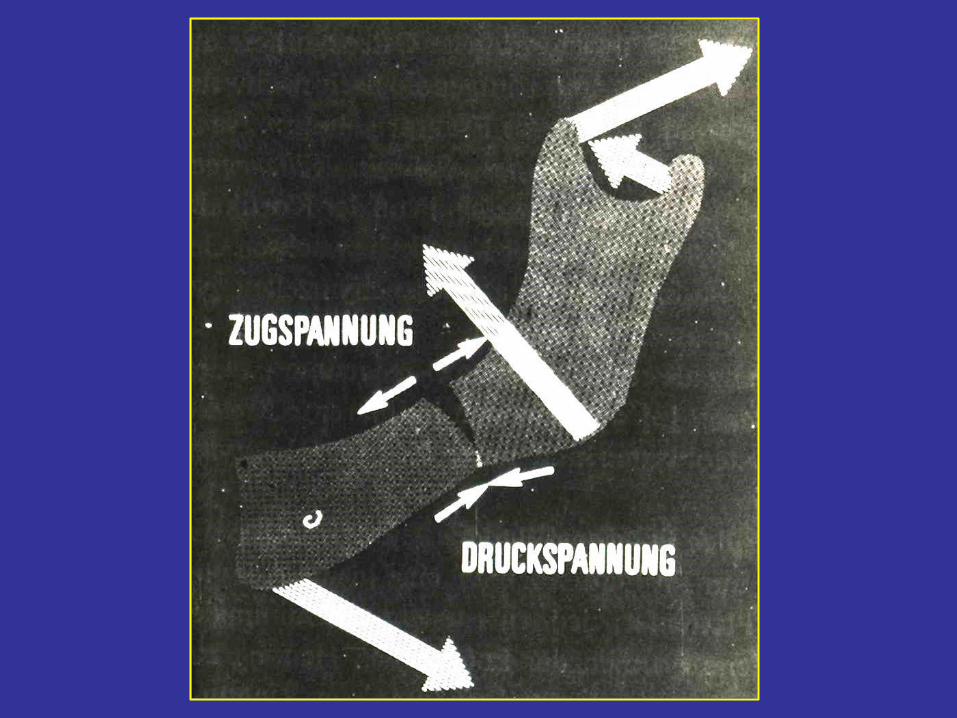
• -Miniplate (by Champy)– non-compression plate selfcompression by muscles
• Microplate
• Absorbable plates
• -AO plates
• Indications of compression osteosynthesis
– total toothless
– corpus fracture together with high (intercapsular) condylar fracture
– big dislocation
– open fracture
– when IMF is contraindicated (epilepsia, hyperemesis, respiratory disorders, etc.)
• Contraindications of compression osteosynthesis
– childhood (dental bulb injury)
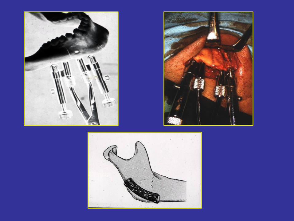
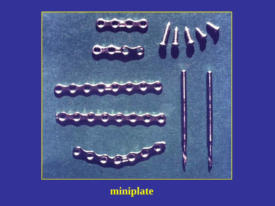
miniplate
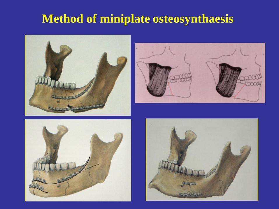
Method of miniplate osteosynthaesis
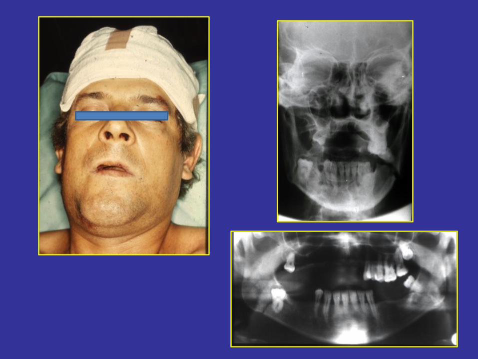
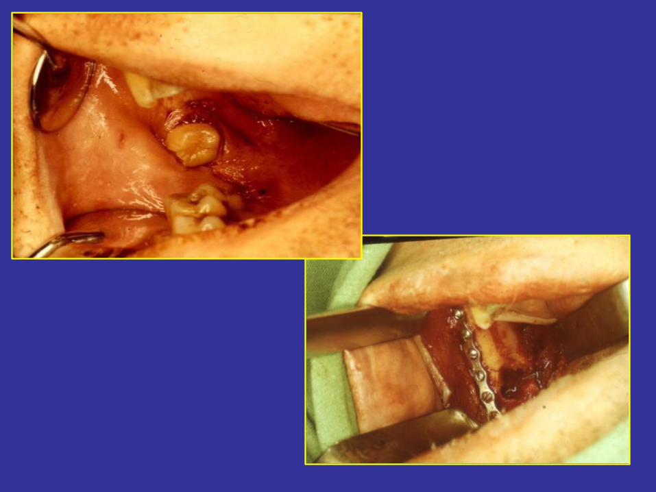
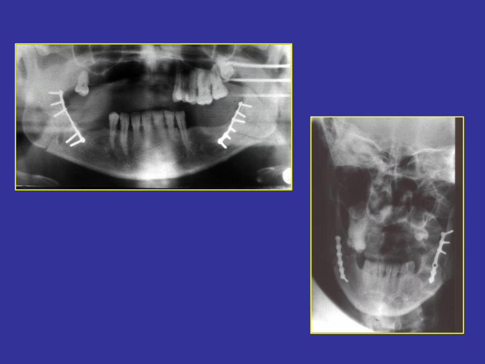
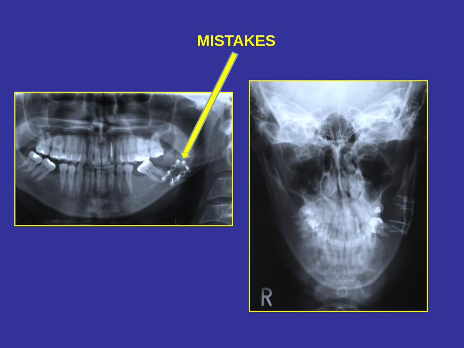
MISTAKES
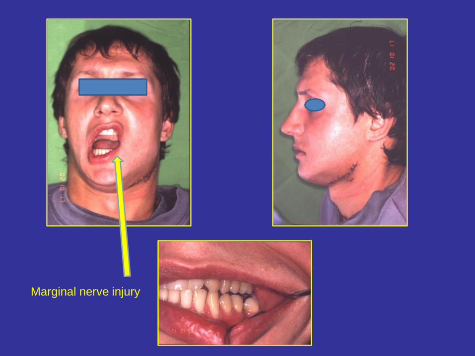
Marginal nerve injury
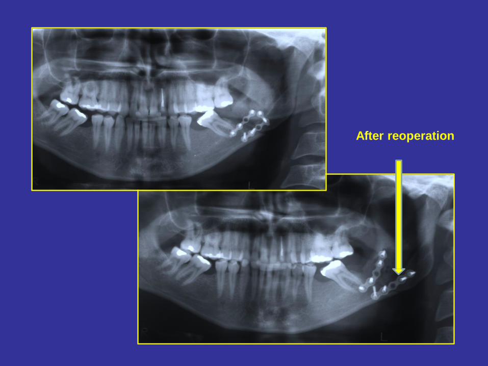
After reoperation
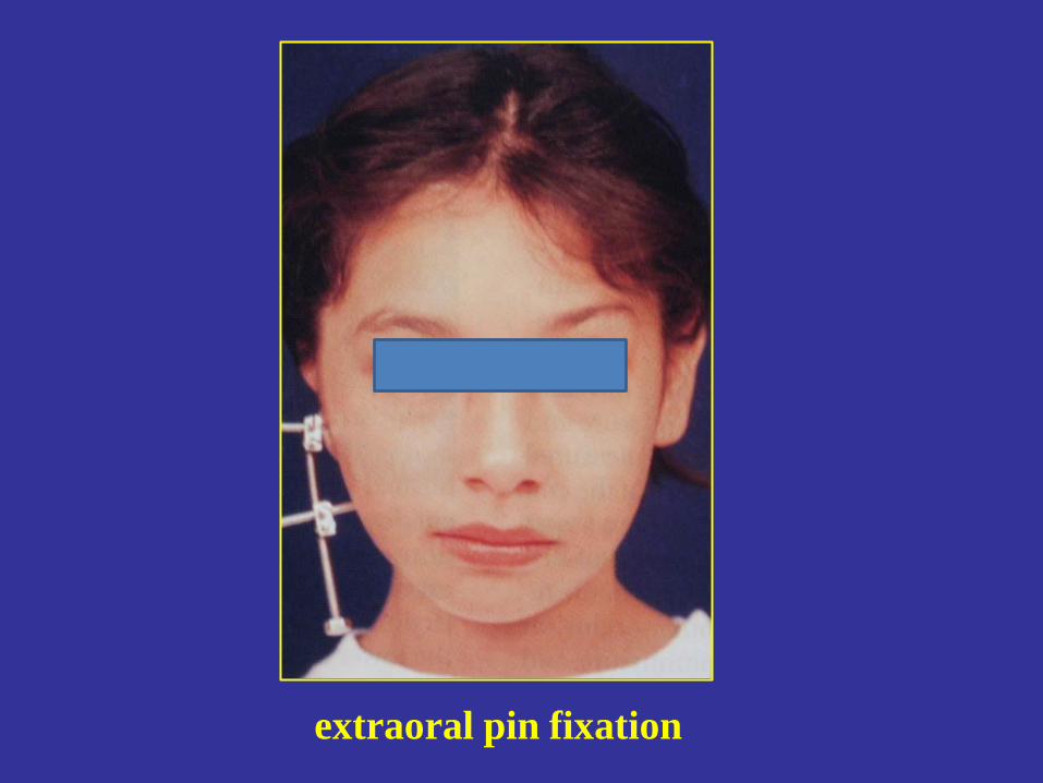
extraoral pin fixation
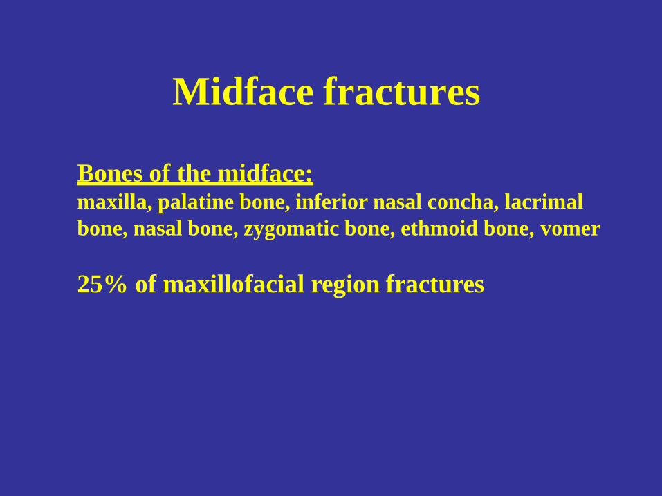
Midface fractures
Bones of the midface: maxilla, palatine bone, inferior nasal concha, lacrimal
bone, nasal bone, zygomatic bone, ethmoid bone, vomer
25% of maxillofacial region fractures
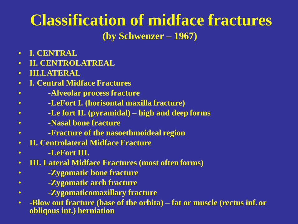
Classification of midface fractures (by Schwenzer – 1967)
• I. CENTRAL
• II. CENTROLATREAL
• III.LATERAL
• I. Central Midface Fractures
• -Alveolar process fracture
• -LeFort I. (horisontal maxilla fracture)
• -Le fort II. (pyramidal) – high and deep forms
• -Nasal bone fracture
• -Fracture of the nasoethmoideal region
• II. Centrolateral Midface Fracture
• -LeFort III.
• III. Lateral Midface Fractures (most often forms)
• -Zygomatic bone fracture
• -Zygomatic arch fracture
• -Zygomaticomaxillary fracture
• -Blow out fracture (base of the orbita) – fat or muscle (rectus inf. or obliqous int.) herniation
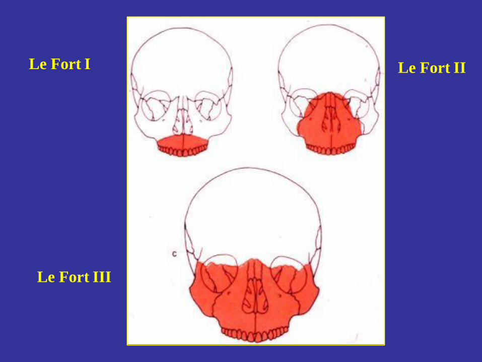
Le Fort I Le Fort II
Le Fort III
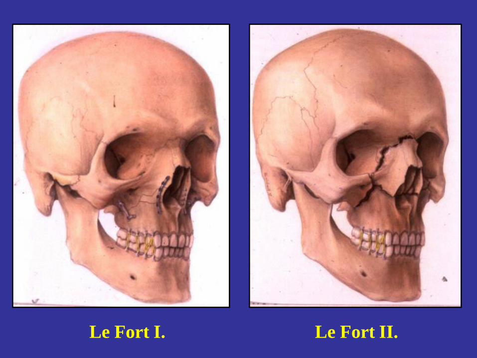
Le Fort II. Le Fort I.
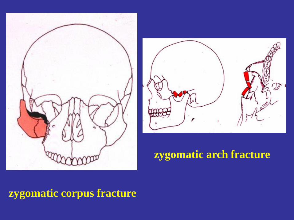
zygomatic arch fracture
zygomatic corpus fracture
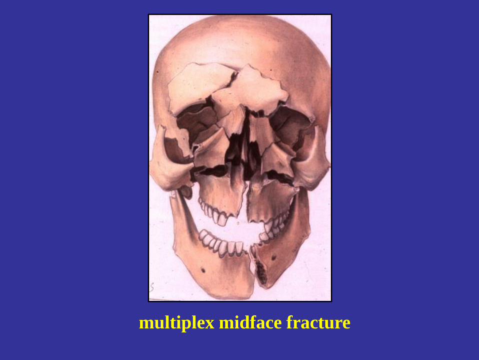
multiplex midface fracture
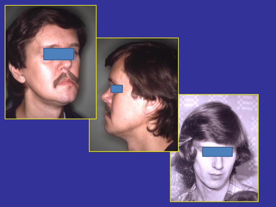
Diagnosis of midface fractures
• Physical examination (inspection, palpation)
– swelling, „flat face”, pain, pathologoc moves, step
formation, nose bleeding, periorbital emphysema,
malocclusion, diplopia
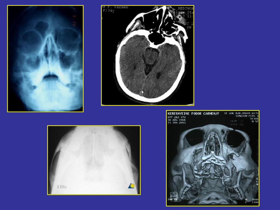
• Imaging methods
– X-ray. ( OP, PA, zygomatic arch- sinus-, overbiting x-
ray, etc.)
– CT, CBCT
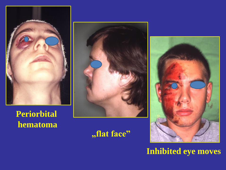
Periorbital
hematoma „flat face”
Inhibited eye moves
Therapy of midface fractures I.
Aim:
• Reconstruion of occlusion, functions and esthetics
Steps:
• reposition
• immobilisation (fixation)
• rehabilitation
Therapy of midface fractures II.
• conservative (rare)
• surgical
-Elevation with surgical hook or by elevator (Gillies)
without fixation in case of zygomatic bone fracture
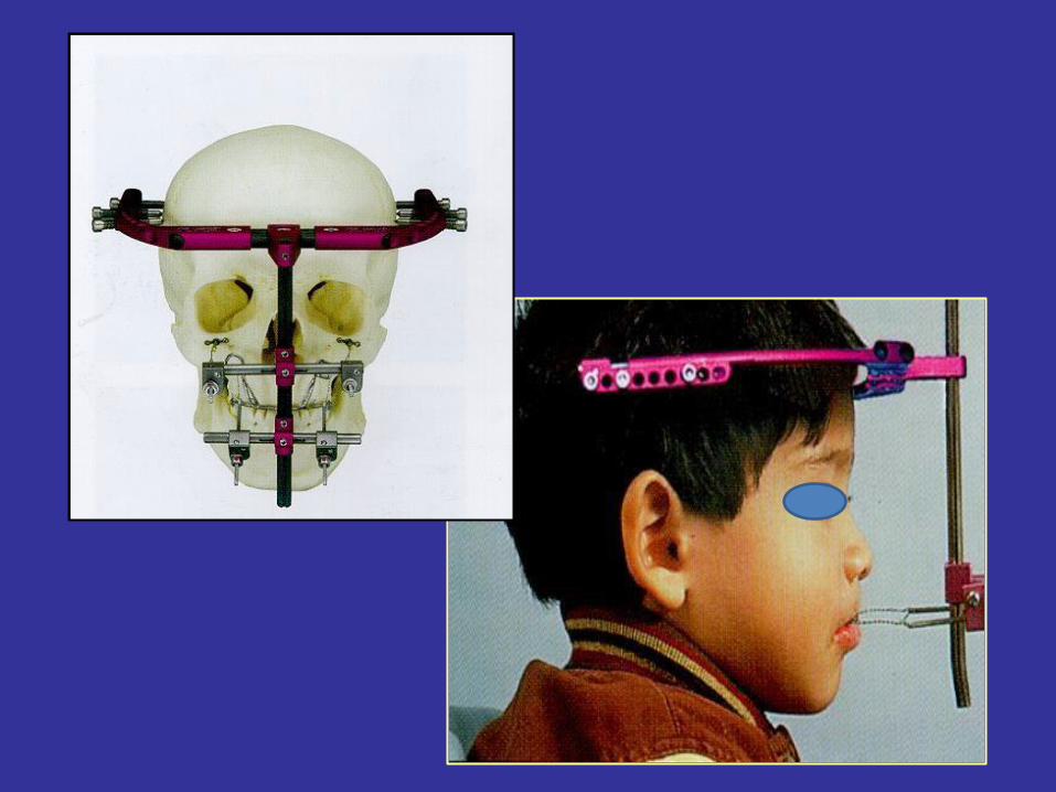
-External fixation: pin fixation, Halo instrument
-Internal fixation: miniplat-, microplate-, absorbable
plate osteosynthaesis, Addams wire ligature
-orbita base reconstruction with bone or liofilizated
dura, titanium net or with plastic plate (PDS)
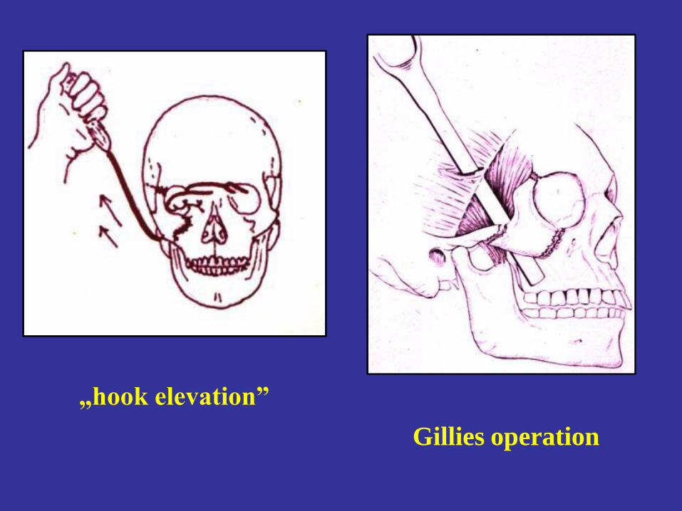
„hook elevation”
Gillies operation
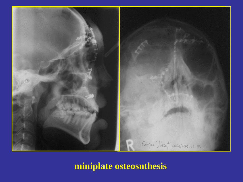
miniplate osteosnthesis
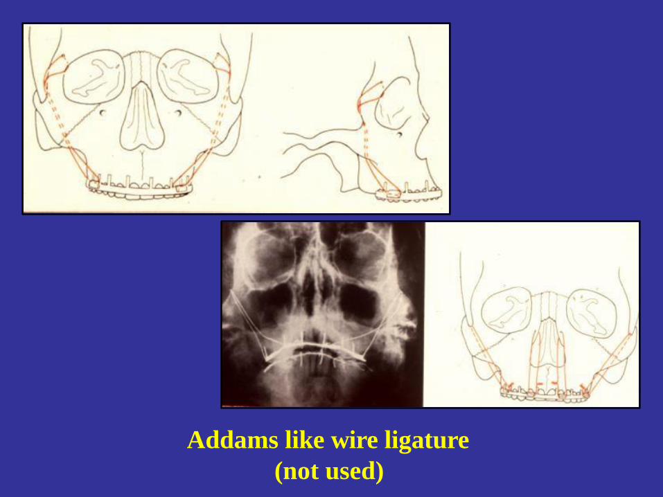
Addams like wire ligature
(not used)
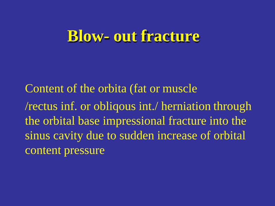
Blow- out fracture
Content of the orbita (fat or muscle
/rectus inf. or obliqous int./ herniation through
the orbital base impressional fracture into the
sinus cavity due to sudden increase of orbital
content pressure
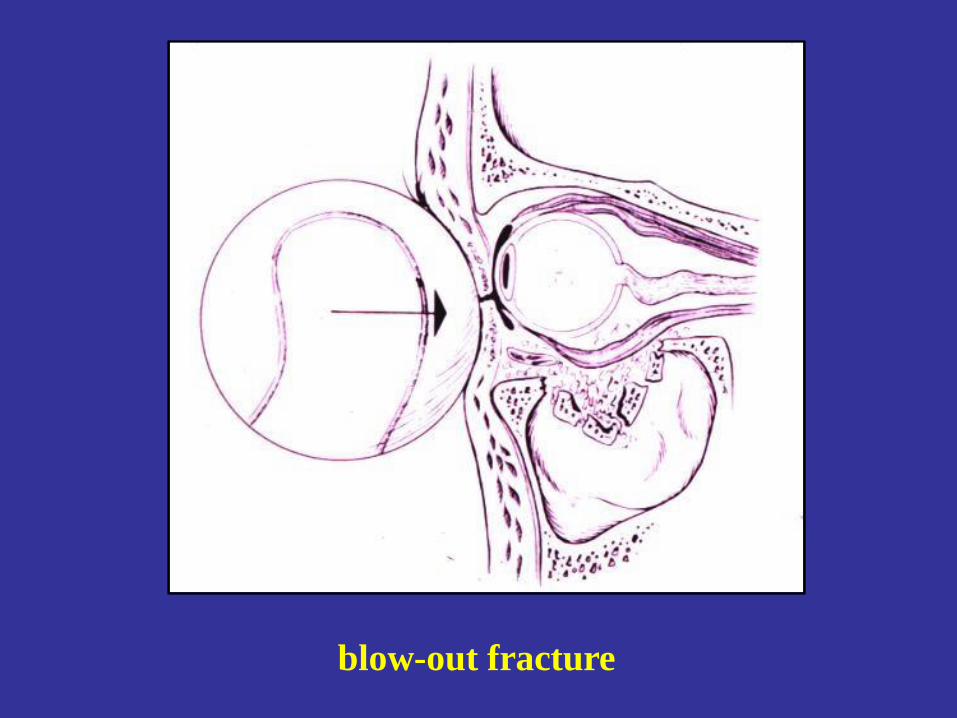
blow-out fracture
Symptoms
• decreased eye moves
• dyplopia
• enophtalmus
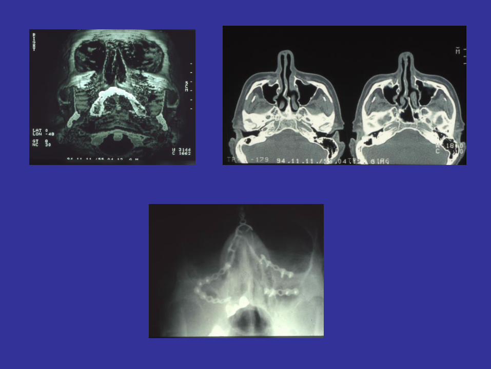
Diagnosis
• Physical examination
• Imaging methods
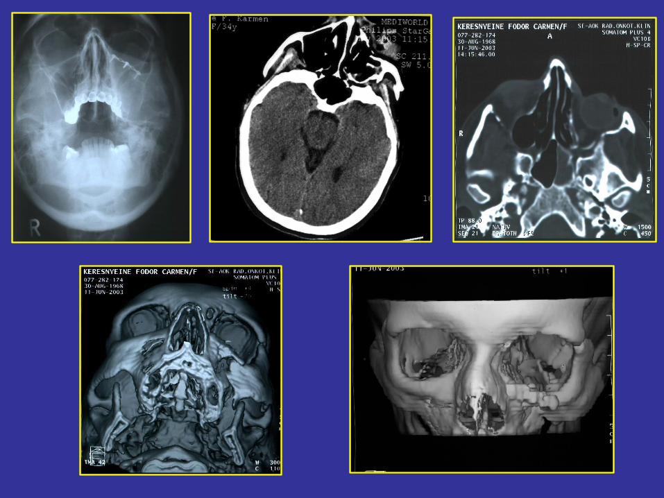
– PA skull x-ray, CT (coronal) !!!
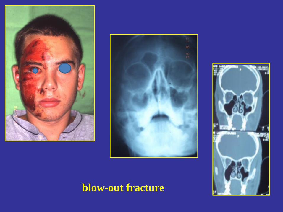
blow-out fracture
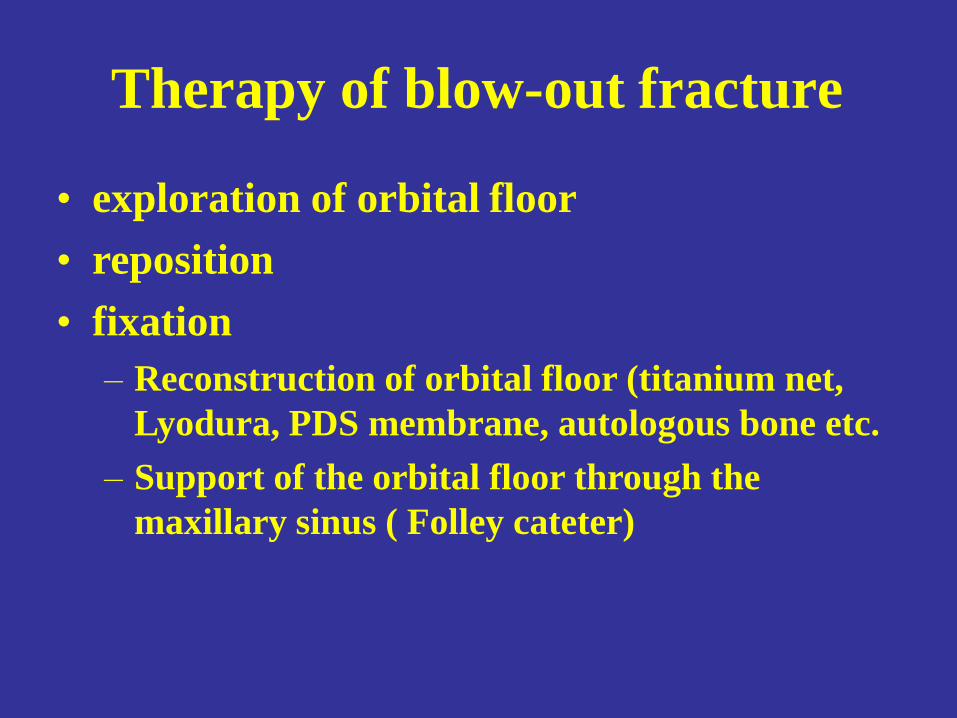
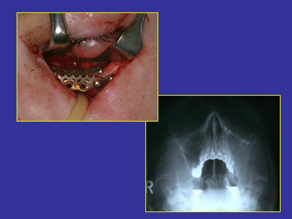
Therapy of blow-out fracture
• exploration of orbital floor
• reposition
• fixation
– Reconstruction of orbital floor (titanium net,
Lyodura, PDS membrane, autologous bone etc.
– Support of the orbital floor through the
maxillary sinus ( Folley cateter)
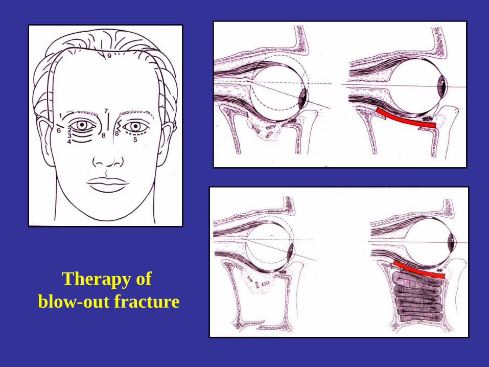
Therapy of
blow-out fracture
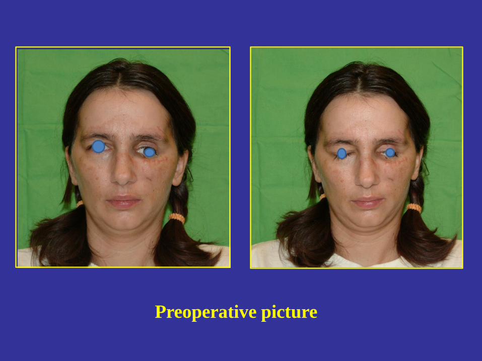
Preoperative picture
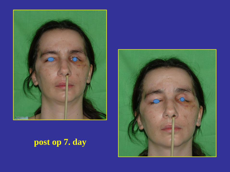
post op 7. day
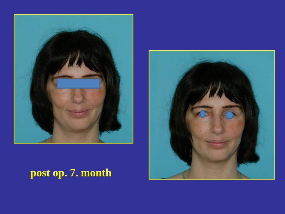
post op. 7. month
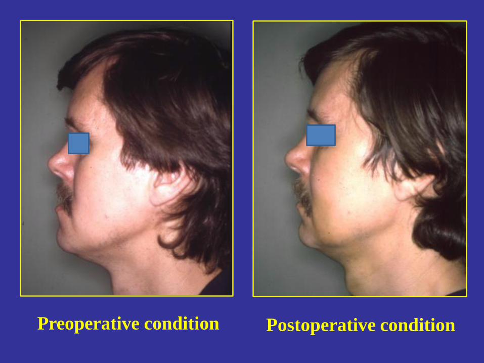
Postoperative condition Preoperative condition

THANK YOU!