Semi-invasive Diagnosis of Endometriosis
80
Semi-invasive Diagnosis of Endometriosis Doctoral Thesis Attila Bokor MD Semmelweis University Molecular Medical Sciences Doctoral School KU Leuven Medical Sciences Doctoral School Supervisor: Prof. Thomas D‘Hooghe MD, PhD Vilmos Fülöp MD, PhD, DSc Official reviewers: Prof. Péter Gőcze MD, PhD, DSc Attila Patócs MD, PhD Examination committee: Barna Vásárhelyi MD, PhD, DSc Pál Siklós MD, PhD Sándor Valent MD, PhD Budapest 2010
Transcript of Semi-invasive Diagnosis of Endometriosis

Semi-invasive Diagnosis of Endometriosis
Doctoral Thesis
Attila Bokor MD
Semmelweis University
Molecular Medical Sciences Doctoral School
KU Leuven
Medical Sciences Doctoral School
Supervisor: Prof. Thomas D‘Hooghe MD, PhD
Vilmos Fülöp MD, PhD, DSc
Official reviewers: Prof. Péter Gőcze MD, PhD, DSc
Attila Patócs MD, PhD
Examination committee: Barna Vásárhelyi MD, PhD, DSc
Pál Siklós MD, PhD
Sándor Valent MD, PhD
Budapest
2010

2
Content
2. ABBREVIATIONS ...................................................................................................... 4
3. LIST OF TABLES ....................................................................................................... 5
4. LIST OF FIGURES ...................................................................................................... 6
5. INTRODUCTION ........................................................................................................ 7
5.1. PATHOGENESIS ....................................................................................................... 7
5.2. FUTURE RESEARCH ............................................................................................... 10
5.3. PREVALENCE ........................................................................................................ 10
5.4. DIAGNOSIS ............................................................................................................ 11
5.5. PAIN...................................................................................................................... 11
5.6. SUBFERTILITY AND INFERTILITY ........................................................................... 12
5.7. ENDOCRINOLOGIC DISORDERS .............................................................................. 15
5.8. CLINICAL EXAMINATION ...................................................................................... 15
5.9. IMAGING AND ENDOMETRIOSIS ............................................................................. 16
5.10. LABORATORY TESTS ........................................................................................... 16
5.11. LAPAROSCOPIC FINDINGS.................................................................................... 17
5.12. HISTOLOGIC CONFÍRMATION .............................................................................. 20
5.13. SPONTANEOUS EVOLUTION DURING PREGNANCY ................................................ 21
5.14. PREVENTION ....................................................................................................... 21
5.15. THERAPY ............................................................................................................ 21
5.16. ENDOMETRIAL SENSORY NERVE FIBRES IN ENDOMETRIOSIS ............................... 22
5.16.1. Small diameter sensory nerve fibres in endometrium ................................ 22
5.16.2. Nerve growth factors .................................................................................. 23
5.17. NEED FOR NON-INVASIVE DIAGNOSIS .................................................................. 24
5.18. CELLS OF PERITONEAL CAVITY ........................................................................... 27
5.19. RETROGRADE MENSTRUATION ............................................................................ 30
5.19.1. Quality of viable endometrial cells ............................................................. 32
5.20. DIAGNOSTIC DELAY IN THE DIAGNOSIS OF ENDOMETRIOSIS ................................ 33
6. AIMS .......................................................................................................................... 35
7. MATERIAL AND METHODS.................................................................................. 36
7.1. TISSUE COLLECTION ............................................................................................. 36
7.2. HISTOLOGY ........................................................................................................... 37
7.3. IMMUNOHISTOCHEMISTRY .................................................................................... 37
7.4. STATISTICAL ANALYSIS ........................................................................................ 39
7.5. PATIENTS AND SAMPLE COLLECTION FOR THE PF STUDY ...................................... 40
7.6. CELL COUNTS ....................................................................................................... 41
7.7. IMMUNOCYTOCHEMISTRY ..................................................................................... 41
7.8. STATISTICAL ANALYSIS ........................................................................................ 42
8. RESULTS ................................................................................................................... 43
8.1. RESULTS OF THE ENDOMETRIAL SENSORY NERVE FIBRE STUDY ............................ 43
8.2. RESULTS OF THE PERITONEAL FLUID CYTOLOGY STUDY ....................................... 47
9. DISCUSSION ............................................................................................................. 50

3
10. CONCLUSION ........................................................................................................ 54
11. SUMMARY ............................................................................................................. 56
12. REFERENCES ......................................................................................................... 59
13. LIST OF PUBLICATIONS RELATED TO THE THESIS ..................................... 78
14. ACKNOWLEDGEMENT ........................................................................................ 80

4
2. Abbreviations
AFS: American Fertility Society
AHSG: Alpha-2 Heremans-Schmidt glycoprotein
Bcl-2: B-cell lymphoma/ leukemia-2
CA-125: Cancer antigen-125
CA-19-9: Cancer antigen-19-9
CCR1: Cognate chemokine receptor 1
CD44: Cluster of differentiation 44
CD68: Cluster of differentiation 68
CGRP: Calcitonine gene–related polypeptide
CK 7: Cytokeratin
CK 8/18: Cytokeratin 8/18
COX-2: Cyclooxygenase-2
CRP: C-reactive protein
HGF: Hepatocyte growth factor
ICAM-1: Intercellular cell adhesion molecule-1
IFN-gamma: Interferon-gamma
IGF-BP 3: Insulin-like growth factor binding protein-3
IL-1β, 2, 4, 6, 8, 10, 12: Interleukin
IL-1β: Interleukin-1β
LOO–CV: Leave–One–Out cross validation
LSSVMs: Least Squares Support Vector Machines
MCP-1: Monocyte chemotactic protein-1
M-CSF: Macrophage colony stimulating factor
MMP-1: Matrix metalloproteinase-1
MMP-2: Matrix metalloproteinase-2
MMP-3: Matrix metalloproteinase-3
MMP-7: Matrix metalloproteinase-7
MMP-9: Matrix metalloproteinase-9
NF: Neurofilament protein
NPY: Neuropeptide Y
PGP9.5: Protein gene product 9.5
RANTES: Regulated on activation normal T-cell expressed and secreted
sICAM-1: Soluble intercellular cell adhesion molecule-1
SP: Substance P
sVCAM-1: Soluble vascular cell adhesion molecule-1
TGF-β: Transforming growth factor-β
TNF alpha: Tumor necrosis factor alpha
TNFR2: Tumor necrosis factor receptor 2
T-plastin: T isoform plastin
VIP: Vasoactive intestinal peptide

5
3. List of tables
Table 1: Summary of representative proteins mainly cytokines,
angiogenic, adhesion and growth factors aberrantly expressed in
women with endometriosis. Page 25.
Table 2: Demographic Characteristics of the Study Population (n=40). Page 37.
Table 3: Demographic Characteristics of the Study Population (n=107). Page 40.
Table 4: Immunocytological markers for different cell populations
present in peritoneal fluid. Page 42.
Table 5: Quantitative assessment of the endometrial nerve fibre density
stained against different neural markers in patients with and without endometriosis.
Page 44.
Table 6: Univariate analysis of different endometrial neural markers for
the semi-invasive diagnosis of minimal and mild endometriosis. Page 46.
Table 7: Selecting the number of neural markers for LS–SVM
modelling based on Leave One Out–Cross Validation (LOO–CV). Page 47.
Table 8: Cell Counts in Menstrual vs. Nonmenstrual Phase of Cycle. Page 49.
Table 9: Prevalence of patients with PF cells stained positively by immunocytochemistry
for 7 different cell markers among patients with or without endometriosis. Page 50.

6
4. List of figures
Figure 1.: Black peritoneal laesion, gunshot laesions, hypervascularisation (typical
endometriosis). Page 18.
Figure 2.: Vesicular peritoneal endometriosis, filmy adhaesions and white plaques
(atypical appearence of endometriosis). Page 18.
Figure 3.: Deeply infiltrating endometriosis nodule. Page 19.
Figure 4.: Superficial ovarian endometriosis and typical gunshot laesions in cul de sac.
Page 20.
Figure 5.: Ovarian endometrioma. Page 20.
Figure 6.: Hystological aspect of endometriosis. Page 21.
Figure 7.: Small diameter nerve fibers in eutopic endometrium in women with minimal
and mild endometriosis. Eutopic endometrium stained with PGP9.5(A), with VIP(B),
with SP(C) with NPY(D) and with CGRP(E). Page 45.
Figure 8.: ROC curve of the LS–SVM model built using PGP9.5, VIP and SP. Page 48.

7
5. Introduction
5.1. Pathogenesis
Endometriosis is a common, benign, oestrogen-dependent, gynaecological disorder
associated with pelvic pain and infertility. While endometriosis has been described for
more than one hundred years, our current knowledge of its pathogenesis remains unclear.
There are a number of theories which have been proposed to explain the pathogenesis of
endometriosis:
Retrograde menstruation/transplantation
Coelomic metaplasia
The induction theory
Genetic background
Altered cellular immunity
Environmental basis
The ectopic transplantation of endometrial tissue, originally proposed by Sampson in
1924, is the most widely accepted theory on the pathogenesis of endometriosis. It claims
that the disorder originates from retrograde menstruation of endometrial tissue sloughed
through patent fallopian tubes into the peritoneal cavity [1]. Retrograde menstruation
occurs in 70% to 90% of women [2, 3], and may be more common in women with
endometriosis than in those without the disease [4]. The presence of endometrial cells in
the peritoneal fluid, indicating retrograde menstruation, has been reported in 59% to 79%
of women during menses or in the early follicular phase [5-7].
The development of endometriosis in the first few years after menarche has been
associated with a high rate of obstructing genital-tract anomalies. These include non-
communicating rudimentary uterine horns, cervical stenosis, cervical atresia, vaginal or
transverse septum agenesis, or an imperforate hymen [8, 9]. As a general rule, women
with a stricture at the level of the cervix have a higher incidence of endometriosis than
women with a stricture lower in the genital tract. Furthermore, women with shorter
intervals between menstruation and longer duration of menses are more likely to have
retrograde menstruation and are at higher risk for endometriosis [10, 11].
The transformation (metaplasia) of coelomic epithelium into endometrial tissue has
been proposed as a mechanism for the ontogenesis of endometriosis. This theory is best

8
supported by the fact that genetic induction of ovarian endometriosis is possible in mice
suggesting that ovarian endometriotic lesions may arise directly from the ovarian surface
epithelium through a metaplastic differentiation process induced by activation of an
onco-genic K-ras allele [12].
The induction theory is, in principle, an extension of the coelomic metaplasia theory. It
proposes that an endogenous biochemical factor in menstrual fluid present in the
peritoneum during menstruation can induce undifferentiated peritoneal cells to develop
into endometrial tissue. This theory has been somehow supported by experiments in
rabbits [13, 14] but has not been substantiated in women and primates.
Genetic basis
The risk or endometriosis is 7 times greater if a first-degree relative has been affected by
endometriosis [15, 16]. No specific Mendelian inheritance pattern has been identified,
therefore multifactorial inheritance has been postulated. A relative risk for endometriosis
of 7.2 has been found in mothers and sisters, and a 75% incidence has been noted in
homozygotic twins of patients with endometriosis [17]. Epidemiological research in the
Icelandic population has shown that endometriosis occurs concordantly in monozygotic
twins, that pain symptoms start at a similar age in affected nontwin sisters, and that the
prevalence of endometriosis is 6- to 9 times increased among first-degree relatives of
women with endometriosis when compared to the general population[17].
The induction of humanlike endometriosis in mice by genetic activation of an oncogenic
K-ras allele lends further support to the genetic basis of this disorder [12]. More recent
studies showed aneuploidy for chromosomes 11, 16, and 17 [18], increased heterogeneity
of chromosome 17 aneuploidy [19], and losses of lp and 22q (50%), 5p (33%), 6q (27%),
70 (22%), 9q (22%), and 16 (22%) of 18 selected endometriotic tissues [20].
Recent study shows relationship between the genetic basis of the reumatoid arthritis and
endometriosis [21].
The relationship between endometriosis and single gene polymorphisms is controversial
[22]. Positive correlations have been shown for single nucleotide polymorphisms linked
to cytokines and inflammation, steroid-synthesizing enzymes and detoxifying enzymes
and receptors, estradiol metabolism, other enzymes and metabolic systems, and adhesion
molecules and matrix enzymes. Apoptosis, cell-cycle regulation, and oncogenes seems
to be negatively correlated with the disease, whereas the group of hormone receptors,

9
growth factor systems, and especially groups of the HLA-system components show a
relatively strong correlation.
An ongoing web project (http://www.well.ox.ac.uk/krinaz/ ) describes allele and
genotype frequencies of SNPs in association studies and provides a useful tool for the
study of genetic variants and the pathogenesis of endometriosis[22].
Immunologic mechanisms are believed to be involved in the pathogenesis of
endometriosis. Several theories have been proposed to explain enhanced implantation
and defective clearing of endometrial cells from the pelvic cavity, facilitating the
development of endometriosis.
Although retrograde menstruation appears to be a common event in women, not all
women who have retrograde menstruation develop endometriosis. The immune system
may be altered in women with endometriosis, and it has been hypothesized that the
disease may develop as a result of reduced immunologic clearance of viable endometrial
cells from the pelvic cavity [23]. According to this hypothesis, endometriosis can be
caused by decreased clearance of peritoneal fluid endometrial cells resulting from
reduced natural killer (NK) cell and macrophage activity[24]. In contrast, endometriosis
can also be considered a condition of immunologic tolerance for ectopic endometrium,
which essentially is self-tissue.
Substantial evidence suggests that endometriosis is associated with a state of subclinical
peritoneal inflammation, marked by an increased peritoneal fluid volume, increased
peritoneal fluid white blood cell concentration (especially macrophages with increased
activation status), and increased inflammatory cytokines, growth factors, and
angiogenesis-promoting substances. It has been reported in baboons that subclinical
peritoneal inflammation occurs both during menstruation and after intrapelvic injection
of endometrium [25, 26]. Furthermore, there are data supporting that a higher basal
activation status of peritoneal macrophages in women with endometriosis may impair
fertility by reducing sperm motility, increasing sperm phagocytosis, or interfering with
fertilization [27, 28] possibly by increased secretion of cytokines such as tumor necrosis
factor-α(TNF-α) [11, 29].
Macrophages or other cells may promote the growth of endometrial cells by secretion of
growth and angiogenetic factors such as epidermal growth factor (EGF) [30],
macrophage-derived growth factor (MDGF) [31], fibronectin, and adhesion molecules

10
such as integrins [32]. After attachment of endometrial cells to the peritoneum,
subsequent invasion and growth appear to be regulated by matrix metalloproteinases
(MMP) and their tissue inhibitors [33, 34].
Aromatase activity is absent in normal endometrium. Contrarily, aromatase is expressed
aberrantly in endometriosis, which gives rise to very high levels of aromatase activity in
the endometriotic tissue. Both aromatase expression and activity are stimulated by
PGE2. This results in local production of estrogen, which induces PGE2 formation and
establishes a positive feedback cycle [35, 36].
The subclinical pelvic inflammatory status associated with endometriosis is also
reflected in the systemic circulation. Increased concentrations of C-reactive protein,
serum amyloid A (SAA), TNF-a, membrane cofactor protein-1, interleukin-6,
interleukin-8 and chemokine (C-C motif) receptor 1 (CCR1) have been observed in
peripheral blood samples of patients with endometriosis when compared with controls
[37].
Environmental factors
The links between reproductive health, infertility, and environmental pollution are
controversial. Attention has been directed to the potential role of dioxins in the
pathogenesis of endometriosis, but the issue remains controversial. A recent meta-
analysis concluded that currently there is insufficient evidence in women or in
nonhuman primates that endometriosis is caused by dioxin exposure [38].
5.2. Future research
Future research on the pathogenesis of endometriosis, should focus on the early
interactions between endometrial and peritoneal cells in the pelvic cavity at the time of
menstruation. Proteomic and genomic approaches can detect potential differences
between eutopic endometrium and myometrium in women with and without
endometriosis [39].
5.3. Prevalence
Based on the few reliable data, the prevalence of the condition can reasonably be
assumed to be around 10% [40]. Endometriosis is predominantly found in women of
reproductive age but can also be found in adolescents and in postmenopausal women

11
receiving hormonal replacement [40]. It is found in women of all ethnic and social
groups. In women with pelvic pain or infertility, a high prevalence of endometriosis (20
% - 90%) has been reported [41, 42]. In asymptomatic women undergoing tubal
ligation, the prevalence of endometriosis ranges from 3% to 43% [43].
5.4. Diagnosis
The current clinical opinion is that laparoscopy is required for definitive diagnosis of
endometriosis [44].
History and physical examination can yield a number of significant findings suggestive
for endometriosis including affected first degree relatives, chronic pelvic pain and
dysmenorrhea, retroverted uterus, adnexal masses, cul de sac nodularity and uterosacral
ligament thickening and tenderness, but none are diagnostic for endometriosis.
Well known risk factors for endometriosis include short cycle length [45], heavier
menstruation, and longer flow duration [46], probably related to a higher incidence of
retrograde menstruation. Patient height and weight are positively and negatively,
respectively, associated with the risk of endometriosis [47]. Although a higher
prevalence of endometriosis has been found among patients with coitus during menses
[48], more research is needed to address the role of sexual habits in the development of
endometriosis.
Endometriosis can be associated with significant gastrointestinal symptoms (pain,
nausea, vomiting, early satiety, bloating and distention, altered bowel habits) as well.
A characteristic motility change, along with bacterial overgrowth, has been documented
the bowel system of most women with endometriosis [49].
The average delay between the onset of pain symptoms and surgically confirmed
endometriosis is quite long: mean 8 years in the United Kingdom and 9 to 12 years in
the United States [50]. However over the past 20 years, there has been a steady
decrease in the delay in diagnosis and a decline in the prevalence of advanced
endometriosis at first diagnosis [51].
5.5. Pain
In adult women, dysmenorrhea is especially suggestive of endometriosis if it begins after

12
years of pain-free menses. Dysmenorrhea often starts before the onset of menstrual
bleeding and continues throughout the menstrual period. In adolescents, dysmenorrhee
may be present after menarche without an interval of pain-free menses. The distribution
of pain is variable but most often is bilateral [11].
Local symptoms can arise from rectal, ureteral, and bladder involvement, and lower back
pain can occur. Most studies have failed to detect a correlation between the degree of
pelvic pain and the severity of[52] endometriosis [42]. Some women with extensive
disease have no pain, whereas others with only minimal disease may experience severe
pelvic pain. Severe pelvic pain and dyspareunia may be associated with deep infiltrating
subperitoneal endometriosis [53].
Possible mechanisms causing pain in patients with endometriosis include local peritoneal
inflammation, deep infiltration with tissue damage, adhesion formation, fibrotic
thickening, and collection of shed menstrual blood in endometriotic implants, resulting in
painful traction with the physiologic movement of tissues [52, 53]. In rectovaginal
endometriotic nodules, a close histologic relationship has been observed between nerves
and endometriotic foci and between nerves and the fibrotic component of the nodule
[54].
5.6. Subfertility and Infertility
An association between endometriosis and subfertility is generally accepted, based on
epidemiological, retrospective or cross-sectional studies in women and on nonhuman
primate research[55]. In women with moderate or severe endometriosis, major pelvic
adhesions can impair oocyte release from the ovary or inhibit ovum pickup or
transport[56]. In women with mild endometriosis, the monthly fecundity rate (MFR) is
lower (5%-11%) than observed in a normally fertile population (25%,) [57]. The
association between minimal endometriosis and infertility has been explained by
numerous mechanisms (ovulatory dysfunction, luteal insufficiency, luteinized unruptured
follicle syndrome, recurrent abortion, altered immunity, and intraperitoneal
inflammation)[57], but remains controversial[55, 58]. It is possible that functional
disorders of the endometrium may both predispose to the development of endometriosis
and impair implantation mechanisms in affected women[11]. Both the prevalence of

13
endometriosis and the proportion of moderate to severe endometrisos are higher in
infertile women (27% and 71%) than in women of proven fertility women (3% and
43%)[55].
In a recent study authors have published a fundamental article about the disturbances of
implantation in endometriosis. The endometrium is receptive to embryonic implantation
during a well-defined ‗window of implantation‘ in the midsecretory phase, when
pinopods appear on the surface of the endometrium. The genes and gene products
aberrantly expressed in the endometrium from women with endometriosis include
aromatase, endometrial bleeding factor, hepatocyte growth factor, 17β-hydroxysteroid
dehydrogenase, homeobox genes A-10 and A-11, leukaemia inhibitory factor, matrix
metalloproteinase-7 and -11 and progesterone receptors [59].
Aromatase is only expressed in the eutopic and ectopic endometrium of women with
endometriosis, but not in the endometrium of healthy women. Endometrial bleeding
factor is downregulated during the window of implantation. It is a marker of uterine
non-receptivity and is abundantly expressed during the window of implantation in
women with endometriosis and infertility [60].
Matrix metalloproteinases degrade extracellular matrix components, and are normally
expressed in the endometrium during menstrual breakdown and subsequent oestrogen-
mediated endometrial growth, and are suppressed by progesterone during the secretory
phase. It has been shown that there is a persistent expression of MMP-7 and -11 during
the secretory phase, probably caused by complex interactions between progesterone and
local cytokines in endometriosis [61].
Lessey et al. [62] reported that the αVβ3 integrin chain is co-expressed with other
specific integrins only during the implantation window, and is a biomarker for
endometrial receptivity. In women with endometriosis, it appears that αVβ3 integrin
expression is reduced. It was suggested that a direct effect is exerted on the
endometrium by inflammatory factors contained in the peritoneal fluid of women with
endometriosis.
Giudice et al.[63] have screened 588 genes, and observed a marked up-regulation for
cytokines, growth factors and other gene products in endometriosis in decidualized
stromal cells, which supports a major role for paracrine interactions between the stroma
and other endogenous and transient cell populations within the endometrium and during
early pregnancy.

14
Kao et al. have applied paralleled gene expression profiling using high-density
oligonucleotide microarrays to investigate differentially regulated genes in endometrium
(obtained during the implantation window) from women with versus women without
endometriosis. By analysis of these data, they propose novel candidate genes for
implantation failure and infertility in endometriosis, such as GlcNAc6ST, olfactomedin,
C4BP, IL-15, Dickkopf-1, purinenucleoside phosphorylase, neuronal pentraxin II,
glycodelin S100E and BSEP. The other candidate genes may promote an inhospitable
endometrial milieu for embryonic implantation, due to embryo toxicity, immune
dysfunction, inflammatory or apoptotic responses [64].
Recently, Montgomery et al. reviewed gene mapping studies in endometriosis and the
prospects of finding gene pathways contributing to disease using the latest genome-wide
strategies. Genetic variants in 76 genes have been examined for association, but none
shows convincing evidence of replication in multiple studies. However, there is
evidence for genetic linkage to chromosomes 7 and 10, but the genes (or variants) in
these regions contributing to disease risk have yet to be identified [65].
The above mentioned data could suggest that the main reason for reproductive failure in
women with endometriosis lies in the endometrium and its altered characteristics during
the crucial implantation window. However, it has been shown [66] that patients with
endometriosis undergoing IVF with oocyte donation have the same chances of
implantation and pregnancy as other recipients when oocytes came from donors without
known endometriosis. At the same time, when the results of oocyte donation were
classified according to origin of the oocytes donated, patients who received embryos
derived from endometriotic ovaries showed a significantly reduced implantation rate as
compared with the remaining groups.
These observations may suggest that infertility in endometriosis patients may be
related to alterations within the oocyte, which in turn result in embryos with
decreased ability to implant. Some think that a good quality embryo may
overcome the slight decrease observed in endometrial receptivity [66].
Although much work has been done in identifying of cytokines, growth factors and gene
products, there is not yet a clear understanding of their importance in (recurrent)
pregnancy loss and implantation failure.

15
Based on controlled prospective studies, there is no evidence that endometriosis is
associated with pregnancy loss [67], or that medical or surgical treatment of
endometriosis reduces the spontaneous abortion rate [68, 69].
5.7. Endocrinologic disorders
Endometriosis has been associated with anovulation, abnormal follicular development,
reduced circulating E2 levels during the preovulatory phase, disturbed luteinizing
hormone (LH) surge patterns, premenstrual spotting, the luteinized unruptured follicle
syndrome, galactorrhea and hyperprolactinemia [11]. However, there are not sufficient
convincing data indicating that the prevalence of these endocrinologic abnormalities is
significantly increased in women with endometriosis [55].
5.8. Clinical Examination
In many women with endometriosis, no abnormality is detected during the clinical
examination. The vulva, vagina, and cervix should be inspected for any signs of
endometriosis, although the occurrence of endometriosis in these areas is rare (2). Other
possible signs of endometriosis include uterosacral or cul-de-sac nodularity, lateral or
cervical displacement caused by uterosacral scarring[70], painful swelling of the recto-
vaginal septum, and unilateral ovarian enlargement. In more advanced disease, the
uterus is often in fixed retroversion, and the mobility of the ovaries and fallopian tubes
is reduced. Evidence of deeply infiltrative endometriosis (deeper than 5 mm under the
peritoneum) in the rectovaginal septum with cul-de-sac obliteration or cystic ovarian
endometriosis should be suspected by clinical documentation of uterosacral nodularities
during menses, especially if CA125 serum levels are higher than 35 IU/mL [71, 72].
Extrapelvic endometriosis is rare (0,5 -2%) and may result from vascular or lymphatic
dissemination of endometrial cells to many gynecologic (vulva, vagina, cervix) and
non-gynecologic sites [11]. Although extrapelvic endometriosis is often asymptomatic,
it should be suspected when symptoms of pain or a palpable mass occur outside the
pelvis in a cyclic pattern. Endometriosis involving the intestinal tract, especially colon
and rectum, is the most common site of extrapelvic disease, and is associated with
cyclic or chronic abdominal and back pain, abdominal distention, cyclic rectal bleeding,

16
constipation, and obstruction. Ureteral involvement can lead to obstruction and result in
cyclic higher back pain, dysuria, and hematuria. Pulmonary endometriosis can manifest
as pneumothorax, hemothorax, or hemoptysis during menses. Umbilical endometriosis
should be suspected when a patient has a palpable mass and cyclic pain in the umbilical
area[11] .
Although clinical examination is a useful tool in the detection of endometriosis, the
diagnosis of endometriosis should be confirmed by biopsy of suspicious lesions that are
obtained laparoscopically.
5.9. Imaging and Endometriosis
Gynecologic transvaginal [73] or transrectal ultrasonography [74] is an important
diagnostic tool [75] in the assessment of ovarian endometriotic cysts and of rectovaginal
endometriosis (sensitivity, 97%; specificity, 96%). The presence of filling defects detected
by hysterosalpingography has also a significant positive correlation with endometriosis.
The positive predictive value of this finding is 84% and negatíve predictive value is 75%
[76]. Other imaging techniques, including computed tomography (CT) and MRI, can be
used to provide additional and confirmatory information, but they cannot be used for
primary diagnosis [77].
5.10. Laboratory tests
There is no blood test available for the diagnosis of endometriosis. Levels of CA125, a
coelomic epithelium marker common to most nonmucinous epithelial ovarian carcinomas,
have been found to be significantly higher in women with moderate or severe
endometriosis, but not in women with minimal or mild disease, when compared to women
with a normal pelvis [78]. Serum CA 125 levels are reported to increase during
menstruation according to some [79, 80] but not all [81] investigators, and this phenomenon
many be limited to patients with moderate to severe endometriosis [82]. Compared with
laparoscopy, measurement of serum CA125 levels has no value as a diagnostic tool [77]),
but serial CA125 determinations may be useful to predict the recurrence of endometriosis
after therapy [83].

17
5.11. Laparoscopic findings
Unless disease is visible in the vagina or elsewhere, most guidelines state that
laparoscopic visualization of suspicious lesions is the gold standard for the definitive
diagnosis of endometriosis [77]. During diagnostic laparoscopy, the pelvic and
abdominal cavity should be systematically investigated for the presence of
endometriosis. The type, location, and extent of all lesions and adhesions should be
documented in the operative notes ideally, should be recorded [77, 84]. Characteristic
findings include typical (―powder-burn‖ or ―gunshot‖) lesions on the serosal surfaces of
the peritoneum. These lesions are black, dark brown, or bluish nodules or small cysts
containing old hemorrhage surrounded by a variable degree of fibrosis (Fig.1.)
Figure 1. Peritoneal endometriotic laesions
Endometriosis can appear as subtle lesions also called ‘atypical‘, including red implants
(petechial, vesicular, polypoid, hemorrhagic, red flamelike), serous or clear vesicles and
sometimes white plaques or scarring, yellow-brown discoloration of the peritoneum,
and tubo-ovarian adhesions (Fig.2.)

18
Figure 2. Subtle lesions serous and clear vesicles
[85] Histologic confirmation of the laparoscopic impression is essential is important for
the diagnosis of endometriosis, not only for subtle lesions but also for typical lesions
reported to be histologically negative in 24% of cases [86], and should be considered as
ideal practice [77]. In the presence of ovarian endometrioma (greater than 3 cm in
diameter) and deeply infiltrating disease, histology should be obtained to identify
endometriosis and to exclude rare instances of malignancy [85].
Deeply invasive endometriosis is associated with reduced depth and volume of the
pouch of Douglas (Fig 3.) suggesting that this phenotype of endometriosis does not
develop in the recto-vaginal septum but is the consequence of intraperitoneally buried
anterior rectal wall adhesions [87].
Figure 3. Deeply infiltrating endometriosis of the rectum

19
Superficial ovarian endometriosis can present as both typical and subtle lesions (Fig.4.).
Figure 4. Superficial ovarian endometriosis
Ovarian endometriotic cysts are usually located on the anterior surface of the ovary and
are associated with retraction, pigmentation, and adhesions to the posterior peritoneum
(Fig.5.).
Figure 5. Ovarian endometriotic cysts
These ovarian endometriotic cysts often contain a thick, viscous dark brown fluid
(chocolate fluid) composed of hemosiderin derived from previous intraovarian
hemorrhage. Since such fluid may also be found in other conditions, such as in

20
hemorrhagic corpus luteum cysts or neoplastic cysts, biopsy and preferably removal of
the ovarian cyst for histologic examination is desirable [88].
5.12. Histologic Confírmation
Histologic confirmation is useful tool but not essential in the diagnosis of
endometriosis. In a study of 44 patients with chronic pelvic pain, endometriosis was
laparoscopically diagnosed in 36%, but histologic confirmation was obtained in only
18%. This approach resulted in a low diagnostic accuracy of laparoscopic inspection
with a positive predictive value of only 45%, explained by a specificity of only 77%
[11]. Microscopically, endometriotic implants consist of endometrial glands and stroma,
with or without hemosiderin-laden macrophages (Fig.6.).
Figure 6. Histologic aspect of endometriosis
Endometrioid stroma may be more characteristic of endometriosis than endometrioid
glands [89]. Vascularization, mitotic activity, and the three-dimensional structure of
endometriosis lesions are key factors.
The current classification system of endometriosis is Revised American Fertility Society
Classification which has been revised without major changes [90]. The classification is
based on the appearance, size, and depth of peritoneal and ovarian implants; the degree
of cul-de-sac obliteration and the presence, extent, and type of adnexal adhesions.

21
In the new classification system, the morphologic aspect of the lesions is additionally
categorized as red (red, red-pink, and clear lesions), white (white, yellow-brown, and
peritoneal defects), and black (black and blue lesions) [90].
Despite of the criticism of several authors, the revised classification of endometriosis is
the only internationally accepted system, it appears to be the best available tool to
describe objectively the extent of endometriosis.
5.13. Spontaneous evolution during pregnancy
The characteristics of endometriosis are variable during pregnancy, and lesions tend to
enlarge during the first trimester but regress thereafter [91]. Studies in baboons have
revealed no change in the number or surface area of endometriosis lesions during the first
two trimesters of pregnancy [92]. These results do not exclude a beneficial effect that
potentially may occur during the third trimester or in the immediate postpartum period.
5.14. Prevention
There are no successful strategies to prevent endometriosis. Although a reduced
incidence of endometriosis has been reported in women who engaged in aerobic activity
from an early age the possible protective effect of exercise has not been investigated
thoroughly [11]. There is insufficient evidence that oral contraceptive offers protection
against the development of endometriosis [93] .
5.15. Therapy
Treatment must be individualized, taking into consideration the clinical problem in its
entirety, including the impact of the disease and the effect of its treatment on quality of
life [44]. Complaints may persist despite adequate medical or surgical treatment of the
disease. In such circumstances, a multidisciplinary approach involving a pain clinic and
counseling should be considered early in the treatment plan [44].

22
5.16. Endometrial sensory nerve fibres in endometriosis
5.16.1. Small diameter sensory nerve fibres in endometrium
It has been known for some time that the myometrium, the endometrial–myometrial
interface and the deeper portion of the basal endometrium can be innervated by nerve
fibres, but that nerve fibres are absent from the superficial two-thirds of the endometrium
(the functional layer) in the normal human uterus [94]. The function of these nerve fibres
in normal basal endometrium is not well understood. However, some
acetylcholinesterase (AChE)-immunoreactive nerve fibres were detected in the basal
layer of normal human endometrium [94]. Neuropeptide - (NPY), substance P- (SP),
vasoactive intestinal peptide- (VIP) and neurotensin (NT)- immunoreactive nerve fibres
were also present in normal human endometrium [95]. Yet little is known about the
functions of these nerve fibres in human endometrium. Some researchers have studied
nerve fibres in normal endometrium in animals. Calcitonin gene-related peptide-(CGRP)
[96], AChE- and NPY-immunoreactive nerve fibres were detected in rat endometrium
[97] and many CGRP-immunoreactive nerve fibres were near the surface epithelium
[96]. SP and CGRP were also localized in myometrial nerves in a rat model [96].
A variety of clinical conditions including endometriosis, pelvic inflammatory disease,
uterine fibroids, adhesions and uterine contractions can cause pelvic pain. Various
investigators have demonstrated nerve fibres in endometriotic plaques, abdominal
adhesions and myometrium [54, 98-101]. Endometriotic plaques in a rat model were
innervated by SP, CGRP (sensory C and Aδ fibres) and vesicular monoamine transporter
(VMAT) fibres [101].
Nerve fibres were present in human peritoneal endometriotic plaques, and transforming
growth factor (TGF-β1) was expressed in the nerve fibres only in red and white lesions
[100]. Rectovaginal endometriotic nodules from women with severe dysmenorrhoea and
deep dyspareunia showed the presence of nerve fibres staining with the nonspecific
antibody, S-100 protein [54]. Human peritoneal adhesions contained synaptophysin-
CGRP-, SP-, vasoactive intestinal peptide- (VIP)- and tyrosine hydroxylase (TH)-
immunoreactive nerve fibres [99] and 78% of endometriosis-related adhesions showed

23
NF-immunoreactive nerve fibres [98]. SP- and CGRP-immunoreactive nerve fibres were
present in human myometrium [102] and SP-induced contractions were inhibited by
CGRP. It has been reported that women with endometriosis had uterine contractions with
higher frequency, amplitude and basal pressure tone during menses [103]; therefore, SP-
and CGRP-immunoreactive nerve fibres may play a role in the pain of women with
endometriosis.
5.16.2. Nerve growth factors
If the implantation theory is true, the nerve fibres in endometriotic plaques may originate
from nerve fibre progenitors in the functional layer of the endometrium or, more
probably, from ingrowth of local nerve fibres due to the secretion of nerve growth factors
(NGFs) and increased expression of two NGF receptors, namely Trk-A and p75, by the
implanting endometrium. It is not yet clear what neural stimuli may initiate the severe
pain sensations which some women experience with endometriosis. However, a number
of molecules that are capable of stimulating nerve fibres, such as tumour necrosis factor-
α (TNF-α) [104], prostaglandin I2 [105], prostaglandin E2 and F2α [106], and NGF
[107] can be released from endometriotic plaques. It seems highly probable that the
endometrium of women destined to develop endometriosis is functionally abnormal
[108-110]. It may also be programmed to secrete large amounts of nerve trophic factors,
resulting in the remarkable ingrowth of unmyelinated small nerve fibres demonstrated in
this study. This may also be translated into ingrowth of nerve fibres into new
endometriotic plaques on the peritoneum or ovary. These nerve fibres may have an
important role in the mediation of pain symptoms. No other satisfactory theory has yet
been advanced for the genesis of pain symptoms in women with endometriosis. This
demonstration of small nerve fibres in the functional layer of eutopic endometrium of
women with endometriosis is so striking in the present study that we believe it could
become a relatively simple surrogate marker of this condition using endometrial biopsies
[111]. It is even possible that an endometrial biopsy may demonstrate nerve fibres in
women destined to develop the condition later in life.
24
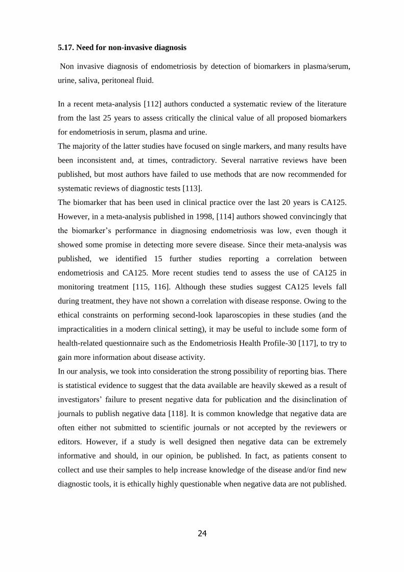
5.17. Need for non-invasive diagnosis
Non invasive diagnosis of endometriosis by detection of biomarkers in plasma/serum,
urine, saliva, peritoneal fluid.
In a recent meta-analysis [112] authors conducted a systematic review of the literature
from the last 25 years to assess critically the clinical value of all proposed biomarkers
for endometriosis in serum, plasma and urine.
The majority of the latter studies have focused on single markers, and many results have
been inconsistent and, at times, contradictory. Several narrative reviews have been
published, but most authors have failed to use methods that are now recommended for
systematic reviews of diagnostic tests [113].
The biomarker that has been used in clinical practice over the last 20 years is CA125.
However, in a meta-analysis published in 1998, [114] authors showed convincingly that
the biomarker‘s performance in diagnosing endometriosis was low, even though it
showed some promise in detecting more severe disease. Since their meta-analysis was
published, we identified 15 further studies reporting a correlation between
endometriosis and CA125. More recent studies tend to assess the use of CA125 in
monitoring treatment [115, 116]. Although these studies suggest CA125 levels fall
during treatment, they have not shown a correlation with disease response. Owing to the
ethical constraints on performing second-look laparoscopies in these studies (and the
impracticalities in a modern clinical setting), it may be useful to include some form of
health-related questionnaire such as the Endometriosis Health Profile-30 [117], to try to
gain more information about disease activity.
In our analysis, we took into consideration the strong possibility of reporting bias. There
is statistical evidence to suggest that the data available are heavily skewed as a result of
investigators‘ failure to present negative data for publication and the disinclination of
journals to publish negative data [118]. It is common knowledge that negative data are
often either not submitted to scientific journals or not accepted by the reviewers or
editors. However, if a study is well designed then negative data can be extremely
informative and should, in our opinion, be published. In fact, as patients consent to
collect and use their samples to help increase knowledge of the disease and/or find new
diagnostic tools, it is ethically highly questionable when negative data are not published.

25
It is worth noting that surgery often plays a vital role in the treatment of endometriosis.
Furthermore, it may also be of importance in the management of other conditions which
present in a similar manner (e.g. tubal infertility). The use of a biomarker may well be
tempered in these circumstances, but it may still help to reduce the need for diagnostic
surgery in some women, enable monitoring of the disease progression by non-surgical
methods, and potentially allow for better pre-operative assessment of women with
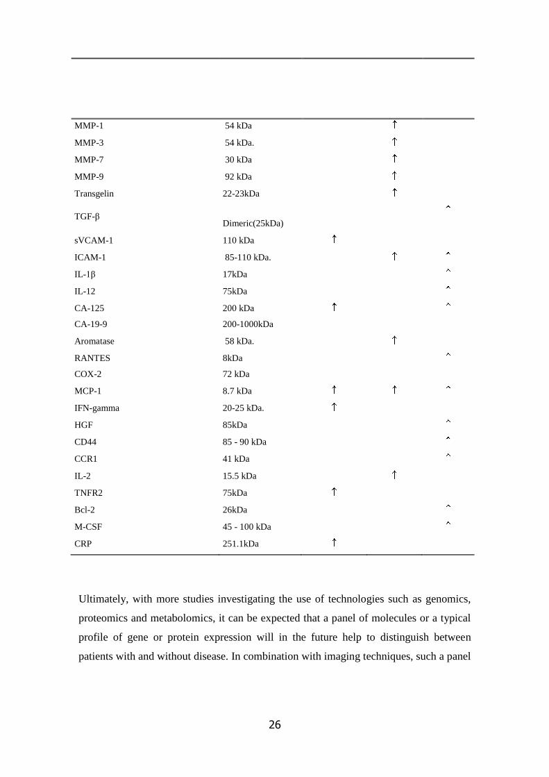
endometriosis. Finally, the majority of the studies included in our review focused on
assessing the diagnostic performance of single biomarkers. Realistically, however, a
reliable diagnostic tool for endometriosis is likely to consist of a panel of biomarkers, not
a single molecule. Table 1. shows the summary of representative proteins mainly
cytokines, angiogenic, adhesion and growth factors aberrantly expressed in women with
endometriosis.
Table 1. Representative proteins aberrantly expressed in women with endometriosis.
Proteins
Molecular
Weight(kDa)
Eutopic
endometrium
Peritoneal
Fluid
Endo-
lesions
IL-6 23-30 kDa
IL-8 8kDa
VEGF 32-42kDa
sICAM-1 90-95kDa
TNF alpha 17.5kDa
IGF-BP 3 17.7kDa
IL-4 10.8kDa
IL-10 39kDa
alpha-2Heremans-Schmidt
glycoprotein (AHSG) 64kDa
T-plastin 74kDa
Annexin II 38 kDa
Annexin V 36 kDa
26
Ultimately, with more studies investigating the use of technologies such as genomics,
proteomics and metabolomics, it can be expected that a panel of molecules or a typical
profile of gene or protein expression will in the future help to distinguish between
patients with and without disease. In combination with imaging techniques, such a panel
MMP-1 54 kDa
MMP-3 54 kDa.
MMP-7 30 kDa
MMP-9 92 kDa
Transgelin 22-23kDa
TGF-β Dimeric(25kDa)
sVCAM-1 110 kDa
ICAM-1 85-110 kDa.
IL-1β 17kDa
IL-12 75kDa
CA-125 200 kDa
CA-19-9 200-1000kDa
Aromatase 58 kDa.
RANTES 8kDa
COX-2 72 kDa
MCP-1 8.7 kDa
IFN-gamma 20-25 kDa.
HGF 85kDa
CD44 85 - 90 kDa
CCR1 41 kDa
IL-2 15.5 kDa
TNFR2 75kDa
Bcl-2 26kDa
M-CSF 45 - 100 kDa
CRP 251.1kDa

27
of biomarkers may indicate which women need a laparoscopy and eliminate countless
unnecessary operations.
5.18. Cells of peritoneal cavity
The pathogenesis of endometriosis can to a certain extent be explained by retrograde
menstruation of endometrial tissue sloughed through patent fallopian tubes into the
peritoneal cavity [1]. However, it has never been shown that the prevalence of
endometrial (EM) cells in peritoneal fluid (PF) is higher in women with endometriosis
than in controls during menstruation. In fact, the cytology of retrograde menstruation has
never been studied in depth.
Based on epidemiological and experimental data, it can be hypothesized that the quantity
of retrograde menstruation and the consecutively flushed endometrial cells play an
important role in the development of endometriosis [123, 179] In previous research,
retrograde menstruation, defined as red stained PF [172], has been observed during
culdoscopy in 50% [136], and during laparoscopy in 70-90% of patients at the time of
menstruation [3]. However, the presence of red blood cells in PF is not a proof of the
presence of viable EM cells at the time of menstruation. Furthermore, in most studies the
identification of PF EM cells has been limited to classical histological analsyis of cell
clumps present in PF [139, 180]. Not surprisingly, the presence of endometrial cells in
PF has been reported to vary between 0 - 59% [6]. Using more objective
immunocytochemical methods, some investigators [9] reported that PF contains single
epithelial cells, rather than endometrial tissue fragments, in women with patent tubes and
that these cells might be of endometrial origin. However, there is no evidence that the
EM cell concentration in PF is higher during menstruation than in other phases of the
menstrual cycle.
Erythrocytes represent a part of the cell population in PF which also contains other free
floating cells like macrophages, mesothelial cells, lymphocytes, eosinophil and mast
cells. Several studies show that there is an increase in erythrocyte count and
consecutively in hemoglobin content in the peritoneal fluid of women with peritoneal
endometriosis when compared with controls with a normal pelvis [10]. Hemoglobin
overload might have numerous cytotoxic effects in the peritoneal environment [181,
182]. Its nonprotein moeity, heme, and its ferrous iron core are known as pro-oxidant and

28
proinflammatory molecules [183] and might be involved in the pathogenesis of
endometriosis through several mechanisms including induction of oxidative stress,
stimulation of cell adhesion, and cytokine production by macrophages[10]. However, it is
not known if the PF concentration of red blood cells and hemoglobin is higher during
menstruation than during nonmenstrual phases of the cycle. Endometriosis is associated
with a state of subclinical peritoneal inflammation, marked by an increased PF volume,
increased PF white blood cell concentration (especially macrophages with increased
activation status), and increased inflammatory cytokines, growth factors, and
angiogenesis-promoting substances [3, 26, 119][5, 14-16 ]. Moreover, it has been
reported in baboons that subclinical peritoneal inflammation occurs both during
menstruation and after intrapelvic injection of endometrium and both the incidence and
recurrence of retrograde menstruation is increased in baboons with spontaneous
endometriosis when compared to healthy controls [25, 120, 184]. However, it is not
known if the PF concentration of white blood cells is higher during menstruation than
during nonmenstrual phases of the cycle.
The lack of knowledge regarding potential differences in the presence and distribution of
PF cell populations during menstruation between women with and without endometriosis
is a major obstacle with respect to the validity of the Sampson hypothesis. The aim of our
study was to test the hypothesis that menstruation is associated with a higher PF
concentration of RBCs, WBCs and EM cells when compared to nonmenstrual phases of
the cycle.
Women with early stages of endometriosis have the most pronounced increases of total
leukocytes, macrophages, helper T lymphocytes and natural killer cells compared to
fertile controls. There is compelling evidence for increased white blood cell populations
in the PF of women with endometriosis as compared to women without endometriosis
[119].
It has been proven that, the white blood cell concentration in peritoneal fluid is elevated
in primates in the subgroup with recent endometriosis when compared with the other
subgroups with a normal pelvis, long-term endometriosis or induced disease [120].
Moreover, it has been reported in baboons that subclinical peritoneal inflammation
occurs both during menstruation and after intrapelvic injection of endometrium
[7].Granulocytes, normally present in small numbers, are greatly increased with pelvic
inflammation [121].

29
These findings support an active immunological process.
Macrophages are attracted to the peritoneal environment more abundantly than any other
cell type [121-127]. These cells originate in the bone marrow, circulate as monocytes,
and then migrate to various body cavities where they function primarily as phagocytes
when activated. Macrophage-directed host defence mechanisms resulting in the
recognition, phagocytosis, and destruction of micro-organisms are well known.
Macrophages digest and process peritoneal debris such as spermatozoa and endometrial
tissue and present antigens to the T cells.
Recent studies [119, 128] suggests that activated macrophages are increased in the
peritoneal fluid of women with endometriosis. These activated macrophages secrete
numerous macromolecules that may contribute to the implantation of endometrial cells
and the progression of endometriosis [129-131]. Macrophages are capable of secreting
various substances, such as growth factors, cytokines, prostanoids, complement
components and hydrolytic enzymes [119, 132-134].
Macrophages also promote cellular growth and viability through secretion of growth
factors and cytokines. Furthermore, macrophages release low amounts of reactive oxygen
metabolites, such as superoxide anion, hydrogen peroxide and singlet oxygen.
Previous studies suggest that menstrual effluent contains factors inducing local
destruction of the peritoneal mesothelium, thereby creating adhesion sites for
endometrial cells. Experiments by Witz et al. [135] have given further support to this
theory, specifically demonstrating that endometrium—both stroma and epithelium—can
easily and rapidly adhere to an intact mesothelium.The experimental model involved
plating explants of peritoneum and culturing them in the presence of endometrium in
form of cellular aggregates or isolated epithelial and stromal cells or menstruated
fragments. The attachment process was evaluated by transmission electron microscopy
and a confocal laser-scanning microscope. The results indicate that endometrial
attachment to an intact mesothelium occurs within 1 hours and that transmesothelial
invasion occurs between 1 and 18–24 hours. Thus, and in contrast to previous
observations the intact mesothelium does not seem to constitute a defence barrier to the
adhesion of endometrial fragments and traumas to the mesothelial lining are not a
prerequisite for endometrial cell adhesion.
Several studies have shown that the attachment of endometrial cells is enhanced by the
induction of adhesion molecules and their receptors in case of endometriosis. After

30
adhesion, endometrial cells proliferate and gradually invade the peritoneal tissue and
some factors induce vascularization of endometriotic implants, allowing their further
development.
Cytokines and growth factors such as TGFβ, IL-8, IL-1, TNFα, IFNγ, vascular
endothelial cell growth factor (VEGF) and hepatocyte growth factor (HGF) and have
been implicated as inducers of these attachment, proliferation, and neovascularization.
The adhaesion phenomenon of the epithelial cells with possible endomterial origin may
affect the detection of the endometrial cells in PF.
5.19. Retrograde menstruation
Several investigations have shown that retrograde menstruation is quite common.
Polishuk et al. [136], performed culdoscopy during the menstrual period, found blood-
stained PF in 50% of the patients. Others have found that 90% of normal women
experience retrograde menstruation, with 70'% exhibiting grossly bloody PF during
menstruation [3]. However, the occurrence of red blood cells in PF during menstruation
is not proving retrograde transport of viable endomelrial cells. Studies analysing the
incidence of endometrial cells in PF are conflicting; the incidence varying from 0 to 59%
[137]. Several investigators have found endometrial tissue in the PF of women with or
without endometriosis with equal frequency [138, 139]. Only one study has identified
endometrial tissue more frequently in the PF of women with endometriosis [140].
The hypothesis that the quantity of endometrial cells deposited into the peritoneal cavity
during menstruation is higher among women who develop endometriosis is supported
by epidemiological evidence [3]. It is well known that women with short cycles and
long duration of menstrual flow are more likely to develop endometriosis [141].
Furthermore, outflow obstruction of the menstrual effluent, resulting in excessive
retrograde menstruation, has been associated with endometriosis both in humans and in
animal experiments[142]. The development of endometriosis in the first few years after
menarche has been associated with a high rate of obstructing genital-tract anomalies.
These include non-communicating rudimentary uterine horns, cervical stenosis, cervical
atresia, vaginal or transverse septum agenesis, or an imperforate hymen. As a general
rule, women with a stricture at the level of the cervix have a higher incidence of

31
endometriosis than women with a stricture lower in the genital tract. This explains the
observation that in women with Mullerian anomalies, those with outflow obstruction
were more likely to have endometriosis than those without (77 vs. 37%)[11] .
Furthermore, studies in baboons have demonstrated that supracervical ligation can cause
obstructed uterine outflow, resulting in decreased duration of antegrade menstruation
and increased retrograde menstruation. Endometriosis has been observed in animals as
early as 3 months after supracervical ligation suggesting that even modest degrees of
uterine outflow obstruction may play a role in the development of endometriosis [10].
These data suggest that retrograde transport of viable endometrial cells during
menstruation occurs in most women with patent tubes, suggesting that another factor(s)
aside from the presence of endometrial cell reflux is critical for the pathogenesis of
endometriosis.
The mesothelium is a simple squamous epithelium that lines the peritoneal cavity. It has
been shown that human peritoneal cells (mesothelial cells) are capable of producing
haematopoietic growth factors, either constitutively (IL- 1. IL-6. IL-8. MCP-I,
granulocyte colony-stimulating factor, macrophage colony-stimulating factor) or in
response to a variety of stimuli, including TNFα, IL- 1 and epidermal growth factor
(EGF) [143]. In addition, these cells secrete CA- 125 from their apical surfaces [144].
These findings suggest that mesothelial cells play an important role in the regulation of
peritoneal inflammation and tissue regeneration.
There is increasing evidence that local inflammation and secretion of prostaglandins
(PG) is related to differences in endometrial aromatase activity between women with and
without endometriosis [36]. The subclinical pelvic inflammatory status associated with
endometriosis is also reflected in the systemic circulation. Increased concentrations of C-
reactive protein, serum amyloid A (SAA), TNF-a, membrane cofactor protein-1,
interleukin-6, interleukin-8 and chemokine (C-C motif) receptor 1 (CCR1) have been
observed in peripheral blood samples of patients with endometriosis when compared
with controls [37].

32
5.19.1. Quality of viable endometrial cells
It has been hypothesised that the quality of endometrial cells in PF of women with
endometriosis is different from women with normal pelvis. Viable endometrial cells from
human endometriotic biopsies but not from human endometrial biopsies are invasive in
an in vitro collagen invasion assay, probably because they have a higher proportion of
potentially invasive E-cadherin-negative epithelial cells [145]. Inflammatory cytokines
(TNF-α, IL- 8 and IL-6) produced by endometrial cells probably contribute to this
adhesion process [146-148]. IL-8 has been shown to stimulate the adhesion of
endometrial cells to fibronectin [146]. TNF-α has been reported to also promote
endometrial stromal cell proliferation in vitro [147] and endometrial stromal cell
adhesion to extracellular matrix components [148]. TNF-α may induce IL-8 gene and IL-
8 protein expression in a dose-dependent manner, and the stimulating effect of TNF-α on
endometrial stromal cell proliferation can be reversed by adding anti-IL8 antibodies[145]
[30]. Does endometriosis then only occur among women with a high degree of
endometrial-peritoneal adhesion? This is unknown at present, since it is impossible to
study this process in women in vivo.
Debrock and colleagues [149] reported a 80–100% success rate of endometrial-peritoneal
adhesion in cultured explants after 48 hours, regardless of the presence or absence of
endometriosis. Witz et al [135] showed endometrial adhesion occurs within 1 hour and
transmesothelial invasion occurs within 18 hours. However, all these assays are merely
descriptive and there is a need to develop a quantitative in vitro assay to measure
endometrial-peritoneal adhesion. Endometrial quality can also be affected by local
estrogen production in eutopic/ectopic endometrium. Indeed, the expression of
uncontrolled aromatase mRNA in endometriotic lesions [150] suggests that a local
estrogenic milieu is important in the development of endometriosis. It is possible that
persistent expression of aromatase and 17β-hydroxysteroid dehydrogenase in
endometriotic lesions may also be driven by a T-like autoantibody response. Indeed,
autoantibodies recognising T-like antigens have been reported to be upregulated in
endometriosis and may trigger the synthesis of cytokines such as IL- 1, TNF-α and IL-6,

33
which in turn may induce the expression of aromatase and 17β-hydroxysteroid
dehydrogenase in endometriotic lesions [151].
5.20. Diagnostic delay in the diagnosis of endometriosis
Endometriosis is a common, chronic gynaecological disease defined by the ectopic
presence of endometrial glands and stroma, most commonly in the pelvis. It is
symptomatically associated with infertility and pelvic pain including dysmenorrhoea,
dyspareunia, dyschezia and chronic pelvic pain [50, 152]. Endometriosis–associated pain
can be caused by peritoneal inflammation, adhesion formation, and specific innervation
of endometriotic lesions and is correlated with the presence of deep infiltrating disease
[107, 153-155]. However, there is a poor correlation between pain and the degree of
endometriosis [53] (minimal–mild–moderate–severe), as determined according to the
revised staging system of American Society for Reproductive Medicine. For a definitive
diagnosis of endometriosis, visual inspection of the pelvis at laparoscopy is the ‗gold
standard‘ investigation, ideally combined with histological confirmation [44]. However,
laparoscopy is a surgical procedure with rare but significant potential risks for the
patients [156].
Due to the lack of a no–or semi–invasive diagnostic tool, the delay between onset of
pain symptoms and surgically confirmed endometriosis is as long as 8 years in the United
Kingdom and United States [50, 51]. The current delay in diagnosis and treatment
contributes to years of suffering and potential infertility if the disease is left untreated.
Clearly, a simple noninvasive diagnostic method may greatly help to reduce this delay,
especially for minimal–mild endometriosis which cannot be diagnosed by clinical
examination or ultrasound.
Attempts for noninvasive diagnosis of endometriosis based on analysis of biomarkers in
peripheral blood have been limited by insufficient sensitivity and specificity [157-159].
Based on the fact that eutopic endometrium from women with endometriosis is
biologically different from women with a normal pelvis [160-162], a semi–invasive
diagnostic test for endometriosis can potentially be developed in endometrium obtained
after transcervical endometrial biopsy. Whatever method is used, the most important
property of any diagnostic test is high sensitivity in order to ensure that no women with

34
endometriosis or other significant pelvic pathology are missed who might benefit from
surgery for infertility and/or pain [163].
In recent studies a higher density of small unmyelinated nerve fibres has been shown in
the functional layer of endometrium from women with confirmed endometriosis when
compared with women without endometriosis, especially in the secretory phase of the
cycle [164, 165]. Indeed, sensory nerve fibers can be identified in functional layer
endometrium by immunohistochemical analysis of various neural transmitters such as
substance P (SP), vasoactive intestinal polypeptide (VIP), or neural proteins like protein
gene product 9.5 (PGP9.5), neurofilament (NF), neuropeptide Y (NPY) and calcitonin
gene–related protein (CGRP). The detection of endometrial nerve fibers has been
proposed as a diagnostic tool for endometriosis in a recent pilot study [166]. However,
this study was limited by the lack of uniform histological confirmation of endometriosis,
inclusion of variable number of patients from all stages of the disease and by cycle phase
related changes of endometrium.
In the present study we tested the hypothesis that women with minimal and mild
endometriosis express a higher density of sensory small diameter nerve fibres in the
functional layer of endometrium than women with a normal pelvis in order to develop a
possible semi–invasive diagnostic tool for minimal to mild endometriosis.

35
6. Aims
In our experiments we aimed to develop a potential semi-invasive diagnostic test of
endometriosis by using eutopic endometrial samples and to assess the biological
changes in the peritoneal cavity during the menstrual cycle as a basis for biomarker
development. We were also focused on the study of retrograde menstruation a crucial
factor in understanding the pathogenesis of endometriosis.
Our specific aims were to:
1. Test the hypothesis that multiple sensory small diameter nerve fibers are present
in a higher density in endometrium from patients with endometriosis when
compared to women with a normal pelvis.
2. Prove that the assumed difference enables the development of a semi-invasive
diagnostic test for minimal-mild endometriosis.
3. Investigate whether menstruation is associated with a higher concentration of
endometrial cells in peritoneal fluid (PF).
4. Confirm that endometriosis is associated with an active immunologic process
with increased white and red blood cell concentration in PF when compared to
nonmenstrual phases of the cycle.

36
7. Material and Methods
7.1. Tissue collection
In this study 40 endometrial samples were selected from the biobank at the Leuven
University Fertility Centre where tissues from women undergoing laparoscopies for
infertility and/or pain have been stored since 1998. Endometrial biopsies were obtained
after hysteroscopy and before laparoscopy using a Pipelle (Pipelle de Cornier, Paris,
France), which is a sterile and disposable plastic cannula for sampling endometrium
[167]. All patients had signed a written informed consent before recruitment and the
study protocol had been approved by the Institutional Ethical and Review Board of
University Hospital Gasthuisberg.
Endometrial samples were selected based on cycle phase, on the presence/absence of
endometriosis, and on the absence of medical treatment for endometriosis within 3
months before sample collection. Menstrual cycle stage was reported as per the patient‘s
report of last menstrual period and by histological evaluation of the endometrial tissues
according to the criteria of Noyes [168].
Only samples collected during the secretory phase of the cycle were selected, since the
density of multiple small nerve fibers is higher during this phase than during other phases
of the cycle [164]. Twenty endometrial samples were selected from women with
laparoscopically and histologically confirmed minimal (n=10) or mild (n=10)
endometriosis (mean age 33 ±10 years), staged according to the revised staging system of
American Society for Reproductive Medicine (American Society for Reproductive
Medicine, 1996). Another 20 endometrial samples were selected from women with a
laparoscopically confirmed normal pelvis (mean age 32 ± 5 years). Demographic data of
our study population are shown in the Table 2.

37
Table 2. Demographic Characteristics of the Study Population (n=40)
Endometriosis (n=20) Controls
( n=20)
Age (years, mean±SD)
Gravidity/parity (mean±SD)
Primary/secondary infertility
[n(%)]
Chronic pelvic pain [n(%)]
Dysmenorrhoea [n(%)]
Dyspareunia [n(%)]
Concurrent hormonal medication
[n(%)]
Previous treatment for infertility
[n(%)]
Ovulation induction
Laparoscopic surgery
Indication for surgery [n(%)]
Infertility
Pelvic pain
Ethnicity [n(%)]
Caucasian
Asian
33±10
0.1±0.3 /0.05±0.22
18(90) /2(10)
0(0)
3(15)
0(0)
0(0)
3(15)
1(5)
2(10)
2(10)
0(0)
20(100)
0(0)
32±5
0.35±0.87/0.15±0.7
17(85) /3(15)
0(0)
2(10)
1(5)
0(0)
4(20)
0(0)
4(20)
4(20)
0(0)
19(95)
1(5)
7.2. Histology
All biopsies had been fixed in 10% neutral buffered formalin immediately after
collection for at least 24 hours, processed, paraffin embedded, and stored at room
temperature until further use. For this study, paraffin blocks were sectioned at 4µm
thickness on a Leica microtome (type 2055 Autocut; Nussloch, Germany). One hundred
serial sections were collected in sets of 4 subsequent sections on 25 silane-coated slides
and were air dried at 37°C. Every tenth slide of this series was stained with hematoxylin-
eosin for morphological evaluation. For immunohistochemical evaluation we selected
sections which exhibited clear histological features consistent with a normal secretory
phase.
7.3. Immunohistochemistry
Tissue sections were preheated for 2 hours at 55 oC, then deparaffinized and rehydrated.
After rinsing in 0.01M Tris buffered saline (TBS), the tissue sections were heat retrieved
in 0.01M TBS pH 9 with 0.001M EDTA. Serial sections were incubated overnight at 4°C
with monoclonal mouse antihuman NF (ready to use; Dako, Glostrup, Denmark)
polyclonal rabbit anti–PGP 9.5 (diluted 1:900; Dako), polyclonal rabbit anti–SP (diluted

38
1:2000; Serotec, Raleigh, NC, USA), monoclonal mouse anti–CGRP (diluted 1:2000;
Sigma, St. Louis, MO, USA), polyclonal rabbit anti–VIP (diluted 1:1400; Chemicon,
Temecula, CA, USA), and polyclonal rabbit anti–NPY (diluted 1:2000, Chemicon)
respectively. The antibodies were detected with REAL Detection System, Alkaline
Phosphatase/RED, Rabbit/Mouse (Dako) according to manufacturers instructions. Non–
specific immunoglobulin binding was blocked with a mixture of BSA (2%), Tween–80
(0.1%) and non–fat–dried milk (1%) applied for 15 to 45 minutes before the first and the
second antibody incubations. 0.01M TBS was used for all dilutions and rinsing steps
throughout the staining procedure and all steps were carried out at room temperature
except when state otherwise. Sections were counterstained lightly with Mayer‘s
hematoxylin and mounted in glycerine jelly. We used normal human skin as a positive
control as it reliably contains myelinated and unmyelinated nerve fibers expressing
PGP9.5, VIP, SP, CGRP NPY, and NF. Rabbit and mouse immunoglobulin fractions
were used as respective negative controls, the concentrations were matched with the
concentrations of the antibodies.
Assessment of nerve fibre density was performed using image analysis software KS400
3.0 (Zeiss, Göttingen, Germany) linked to a Zeiss microscope (Axioskop 50) fitted with a
Zeiss color camera (Axiocam MRc5). The evaluation of all immunohistochemical
stainings was done blindly by the evaluation of the whole surface of each section on high
power images (objective 40x, optovar 1, resolution 860x644 Px) of adjacent non
overlapping fields from left to right and from top to down. Each high power field (HPF)
covered a maximal area of 0.0789 mm2 from which all irrelevant zones (i.e. artefactual
or not belonging to the actual tissue) were subtracted before measurement of the actually
assessed field area. Within these HPFs, all nerve fibre profiles expressing neural markers
were counted with exclusion of those crossing the right or the bottom side of the field
frame respectively, thus avoiding to count these fibre profiles twice. After summation of
the nerve fibre counts and the HPF area values for the whole section, the total number of
nerve fibers was divided by the total surface area of the examined endometrium to obtain
the nerve fiber density for the current section. The average duration of screening of one
specimen was 30±10 minutes.

39
7.4. Statistical analysis
Data are presented as mean (SD) number of nerve fibres per mm2. Numerical data were
analysed using Excel (version 5.0; Microsoft Corporation, Redmond, WA, USA). The
K–S Lilliefors/Shapiro–Wilks test was used to test normality in order to determine
whether parametric or non–parametric tests were to be used in further analyses.
The differences of nerve fibre density between eutopic endometrium from women with
and without endometriosis were tested for significance by the Mann–Whitney U–test,
using the statistical package Prism 5.0 (GraphPad Software Inc., La Jolla, CA, USA).
Multivariate analysis was done using stepwise logistic regression (SAS 9.1.3 for
Windows, Cary, NC, USA) and Stepwise Logistic Regression and Least Squares Support
Vector Machines (LS–SVM) (MATLAB scripts were downloaded from LS–SVMlab
version 1.5 http://www.esat.kuleuven.ac.be/sista/lssvmlab/). For stepwise logistic
regression only variables with significant odds ratios (p–value <0.05) were allowed in the
model.
Both stepwise logistic regression and LS-SVM have been used frequently by our group
in developing models for diagnostic testing for clinical applications [169-171].
Moreover, both models can be implemented as a formula in excel to be used for
diagnostic testing.
For LS–SVM analysis feature selection was performed based on Leave–One–Out cross
validation (LOO–CV) analysis. Briefly, in each LOO–CV the neural markers were
ranked according to their p–value (Mann–Whitney U–test). Then the top ―n‖ –features
were selected where ―n‖ ranged from 1, 2,… to 6 (corresponding to all neural markers).
The ―n‖ with the lowest LOO–CV error was selected to build a model on the full data set.
The models were evaluated based on their Area Under the ROC curve (AUC) [172].
Additionally, an operating point on the ROC curve was chosen corresponding to the
maximum of the sum of sensitivity and specificity. Then models were also evaluated by
their sensitivity, specificity, accuracy, positive predictive value (PPV) and negative
predictive value (NPV).
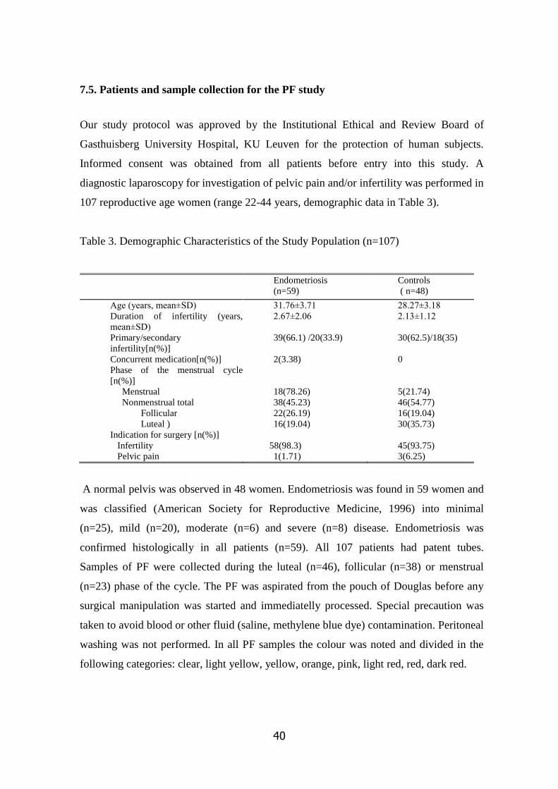
40
7.5. Patients and sample collection for the PF study
Our study protocol was approved by the Institutional Ethical and Review Board of
Gasthuisberg University Hospital, KU Leuven for the protection of human subjects.
Informed consent was obtained from all patients before entry into this study. A
diagnostic laparoscopy for investigation of pelvic pain and/or infertility was performed in
107 reproductive age women (range 22-44 years, demographic data in Table 3).
Table 3. Demographic Characteristics of the Study Population (n=107)
Endometriosis
(n=59)
Controls
( n=48)
Age (years, mean±SD)
Duration of infertility (years,
mean±SD)
Primary/secondary
infertility[n(%)]
Concurrent medication[n(%)]
Phase of the menstrual cycle
[n(%)]
Menstrual
Nonmenstrual total
Follicular
Luteal )
Indication for surgery [n(%)]
Infertility
Pelvic pain
31.76±3.71
2.67±2.06
39(66.1) /20(33.9)
2(3.38)
18(78.26)
38(45.23)
22(26.19)
16(19.04)
58(98.3)
1(1.71)
28.27±3.18
2.13±1.12
30(62.5)/18(35)
0
5(21.74)
46(54.77)
16(19.04)
30(35.73)
45(93.75)
3(6.25)
A normal pelvis was observed in 48 women. Endometriosis was found in 59 women and
was classified (American Society for Reproductive Medicine, 1996) into minimal
(n=25), mild (n=20), moderate (n=6) and severe (n=8) disease. Endometriosis was
confirmed histologically in all patients (n=59). All 107 patients had patent tubes.
Samples of PF were collected during the luteal (n=46), follicular (n=38) or menstrual
(n=23) phase of the cycle. The PF was aspirated from the pouch of Douglas before any
surgical manipulation was started and immediatelly processed. Special precaution was
taken to avoid blood or other fluid (saline, methylene blue dye) contamination. Peritoneal
washing was not performed. In all PF samples the colour was noted and divided in the
following categories: clear, light yellow, yellow, orange, pink, light red, red, dark red.

41
In the first 32 patients included in our study, PF cells were used for a detailed cytological
analysis using Papanicolau staining and immunocytochemical analysis. These 32 patients
included 13 controls and 19 patients with minimal (n=8), mild (n=5), moderate (n=2) or
severe (n=4) endometriosis. In these 32 patients, PF samples were collected during the
luteal (n=20), the follicular (n=5) or the menstrual (n=7) phase of the cycle.
7.6. Cell counts
Cell counts (leucocytes, erythrocytes) were determined on a Symex SE 9500 cell counter
(Sysmex Co., Kobe, Japan). Leucocyte counts below 0.6 x 10⁹
/L were redone by the
counting chamber method (Nageotte counting chamber, Marienfeld Laboratory
Glassware, Lauda-Königshofen, Germany).
7.7. Immunocytochemistry
PF specimens were centrifuged for 10 minutes at 3000 rpm. The pellet was processed
and fixed with CytoRich (Becton Dickinson, Franklin Lakes, NJ. USA ) then one thin-
layer (Papanicolau stain) was prepared with PrepStain (Becton Dickinson, Franklin
Lakes, NJ. USA ).
All PF samples underwent both Papanicolau and immunocytochemical staining with
monoclonal antibodies against CK 7 (1:100; Dako, Glostrup Denmark), CK 8/18 (1:20;
Novocastra, Newcastle upon Tyne, UK), Ber-Ep4 (1:200; Dako, Glostrup Denmark),
vimentin (1:100; Dako, Glostrup Denmark), calretinin (1:2000; Swant, Bellinzona,
Switzerland) and CD68 (1:10; Kp1, Dako, Glostrup Denmark).
These antibodies were selected since they are commonly used in effusion cytology and
they are relevant to identify endometrial epithelial or stromal cells, mesothelial cells and
macrophages in PF. Ber-Ep4 is a marker for cells with epithelial (in some cases
mesothelial) origin. CD68 is specific for cells from monocyte/macrophage lineage, CK7
and CK8/18 are markers for both endometrial epithelial and mesothelial cells, whereas
calretinin and vimentin are markers for both endometrial stromal and mesothelial cells
(Table 4).

42
Table 4. Immunocytological markers for different cell populations present in peritoneal
fluid
Marker
Endometrial
epithelial
/glandular
cells
Endometrial
stromal cells
Mesothelial
cells Macrophages NK cells Lymphocytes
Ber Ep4
Vimentin
Calretinin
Cytokeratin7
Cytokeratin8
Cytokeratin18
CD68
+
-
-
+
+
+
-
-
+
+
-
-
-
-
-
+
+
+
+
+
-/+
-
-
-
-
-
-
+
-
-
-
-
-
-
-
-
-
-
-
-
-
-
Immunocytochemistry was done according to our routinely used protocol with respect to
the dilution of the primary antibody, application of the heat-induced epitope retrieval and
visualization of the antibody complexes through Envision-HRP with DAB (Dako,
Glostrup, Denmark). Slides prepared by the PrepStain method were evaluated by an
experienced cytopathologist.
7.8. Statistical analysis
Numerical data were analysed using MS Office Excel (version 6.0; Microsoft
Corporation, Redmond, WA, USA). Kolomogorov-Smirnov/ Lilliefors and Shapiro–
Wilks test was used to test normality to determine whether parametric or non-parametric
tests were to be used in further analyses. For statistical analyses, Mann-Whitney and
Fisher exact tests were performed. Statistical calculations were performed by using the
GraphPad Prism version 5. 00 for Windows, (GraphPad Software, San Diego California
USA), and P ≤0.05 was considered significant.
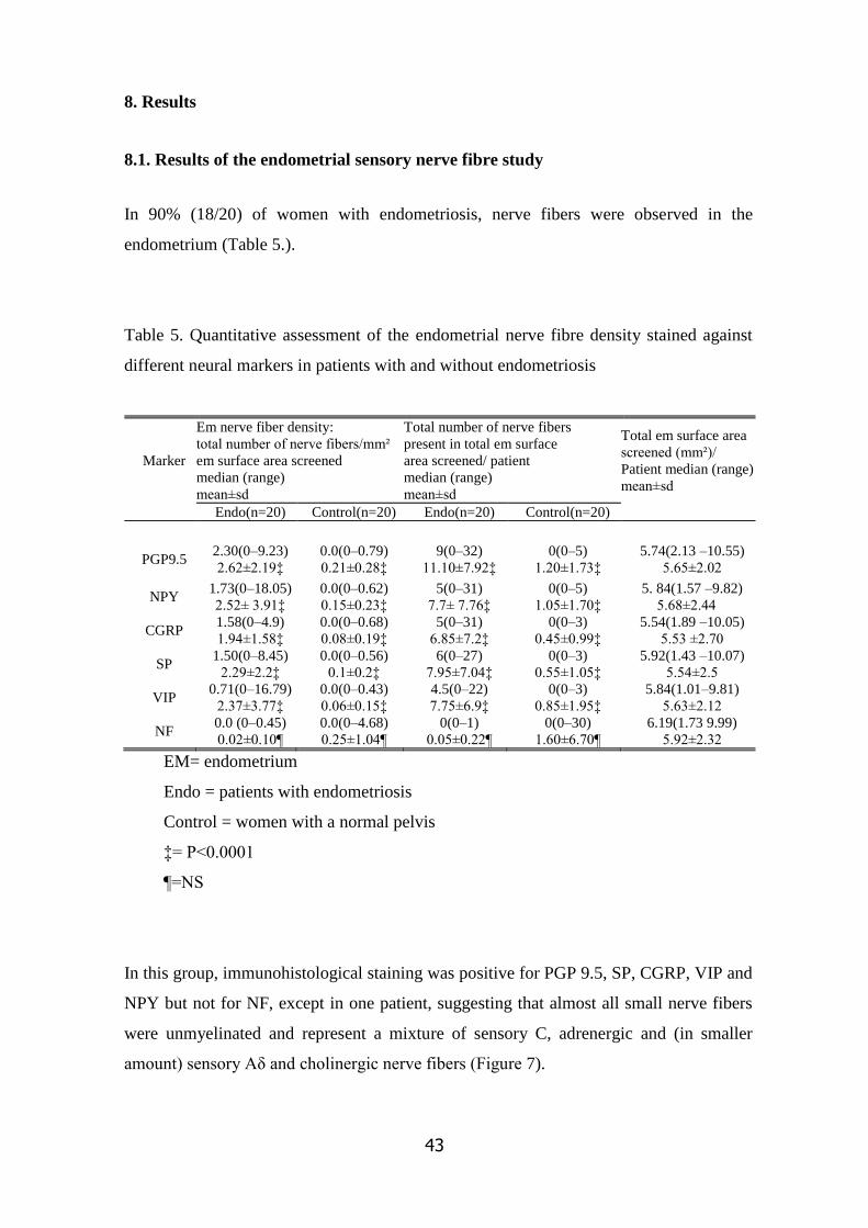
43
8. Results
8.1. Results of the endometrial sensory nerve fibre study
In 90% (18/20) of women with endometriosis, nerve fibers were observed in the
endometrium (Table 5.).
Table 5. Quantitative assessment of the endometrial nerve fibre density stained against
different neural markers in patients with and without endometriosis
Marker
Em nerve fiber density:
total number of nerve fibers/mm²
em surface area screened
median (range)
mean±sd
Total number of nerve fibers
present in total em surface
area screened/ patient
median (range)
mean±sd
Total em surface area
screened (mm²)/
Patient median (range)
mean±sd
Endo(n=20) Control(n=20) Endo(n=20) Control(n=20)
PGP9.5 2.30(0–9.23)
2.62±2.19‡
0.0(0–0.79)
0.21±0.28‡
9(0–32)
11.10±7.92‡
0(0–5)
1.20±1.73‡
5.74(2.13 –10.55)
5.65±2.02
NPY 1.73(0–18.05)
2.52± 3.91‡
0.0(0–0.62)
0.15±0.23‡
5(0–31)
7.7± 7.76‡
0(0–5)
1.05±1.70‡
5. 84(1.57 –9.82)
5.68±2.44
CGRP 1.58(0–4.9)
1.94±1.58‡
0.0(0–0.68)
0.08±0.19‡
5(0–31)
6.85±7.2‡
0(0–3)
0.45±0.99‡
5.54(1.89 –10.05)
5.53 ±2.70
SP 1.50(0–8.45)
2.29±2.2‡
0.0(0–0.56)
0.1±0.2‡
6(0–27)
7.95±7.04‡
0(0–3)
0.55±1.05‡
5.92(1.43 –10.07)
5.54±2.5
VIP 0.71(0–16.79)
2.37±3.77‡
0.0(0–0.43)
0.06±0.15‡
4.5(0–22)
7.75±6.9‡
0(0–3)
0.85±1.95‡
5.84(1.01–9.81)
5.63±2.12
NF 0.0 (0–0.45)
0.02±0.10¶
0.0(0–4.68)
0.25±1.04¶
0(0–1)
0.05±0.22¶
0(0–30)
1.60±6.70¶
6.19(1.73 9.99)
5.92±2.32
EM= endometrium
Endo = patients with endometriosis
Control = women with a normal pelvis
‡= P<0.0001
¶=NS
In this group, immunohistological staining was positive for PGP 9.5, SP, CGRP, VIP and
NPY but not for NF, except in one patient, suggesting that almost all small nerve fibers
were unmyelinated and represent a mixture of sensory C, adrenergic and (in smaller
amount) sensory Aδ and cholinergic nerve fibers (Figure 7).

44
Figure 7. Small diameter nerve fibers in eutopic endometrium in women with minimal
and mild endometriosis. Eutopic endometrium stained with PGP9.5(A), with VIP(B),
with SP(C) with NPY(D) and with CGRP(E). Arrows denote tiny positive multiple
nerve fibers. Eutopic endometrium from woman with endometriosis stained with NF(F).
Arrows denote perivascular myelinated nerve fibres. Magnification, x400. Scale bars
represent 25 μm.

45
However, these nerve fibers were not distributed homogeneously throughout the
endometrium. The density of nerve fibers was markedly skewed, with few specimens
showing counts above 30/mm2 and with most between 0–10 per mm2. There was no
significant difference between the nerve fibre densities in women with confirmed
minimal endometriosis (2.1±2.87) and those with mild endometriosis (1.84±2.59,
p=0.46).
In only 40% (8/20) of women without endometriosis, only small numbers of PGP9.5–
stained nerve fibers were present and were positive for SP, CGRP, VIP, and NPY but not
for NF, except in two patients (Table 6.).
Table 6. Univariate analysis of different endometrial neural markers for the semi-
invasive diagnosis of minimal and mild endometriosis
Data are presented as percentages. PPV= positive predictive value; NPV= negative
predictive value, AUC=area under the curve for diagnostic accuracy
Marker Sensitivity
(95% CI)
Specificity
(95% CI) PPV NPV
Cut off value
for nerve fiber
density
AUC
(95% CI)
PGP9.5 95(75.13–
99.87) 75(50.90–91.34) 79.19 93.75 0.49 0.94(0.86–1.02)
VIP 95+(75.13–
99.87) 80(56.34–94.27) 82.6 84.21 0.08 0.94(0.87–1.00)
CGRP 90(68.30–
98.77) 85(62.11–96.79) 85.71 89.47 0.23 0.92(0.83–1.01)
SP 95(75.13–
99.87) 80(56.34–94.27) 82.6 84.21 0.20 0.90(0.85–1.01)
NPY 95(75.13–
99.87) 65(40.78– 84.61) 72 86.66 0.13 0.90(0.80– 0.99)
NF 95(75.13–
99.80) 10(1.23–31.70) 0.33 48.64 0.19 0.52 (0.34–0.70)

46
Univariate and multivariate analysis is shown in Table 6 and in Table 7 respectively.
Table 7. Selecting the number of neural markers for LS–SVM modelling based on Leave
One Out–Cross Validation (LOO–CV).
Number of neural markers AUC (SE)
1 0.56 (0.10)
2 0.84 (0.07)
3 0.98 (0.02)
4 0.94 (0.05)
5 0.96 (0.03)
6(all) 0.94 (0.04)
AUC: Area under the ROC curve
SE: Standard error
No multivariate logistic regression model could be built that corresponded to our criteria
(p–value <0.05 on odds ratios). Using Leave–One–Out cross validation (LOO–CV)
analysis with LS–SVM modelling (Table 8.), the best result was obtained when selecting
the top 3 neural markers based on their p–value (Mann–Whitney U–test). A LS–SVM
model, built on the complete data set with the top 3 neural markers VIP, PGP9.5 and SP
had an AUC of 0.99, (SE 0.01). (Figure 8).
Figure 8. ROC curve of the LS–SVM model built using PGP9.5, VIP and SP
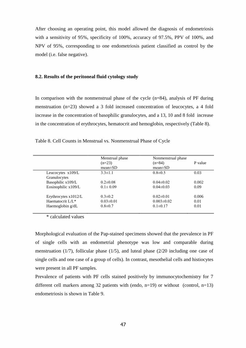
47
After choosing an operating point, this model allowed the diagnosis of endometriosis
with a sensitivity of 95%, specificity of 100%, accuracy of 97.5%, PPV of 100%, and
NPV of 95%, corresponding to one endometriosis patient classified as control by the
model (i.e. false negative).
8.2. Results of the peritoneal fluid cytology study
In comparison with the nonmenstrual phase of the cycle (n=84), analysis of PF during
menstruation (n=23) showed a 3 fold increased concentration of leucocytes, a 4 fold
increase in the concentration of basophilic granulocytes, and a 13, 10 and 8 fold increase
in the concentration of erythrocytes, hematocrit and hemoglobin, respectively (Table 8).
Table 8. Cell Counts in Menstrual vs. Nonmenstrual Phase of Cycle
Menstrual phase
(n=23)
mean±SD
Nonmenstrual phase
(n=84)
mean±SD
P value
Leucocytes x109/L
Granulocytes
Basophilic x109/L
Eosinophilic x109/L
Erythrocytes x1012/L
Haematocrit L/L*
Haemoglobin g/dL
3.3±1.1
0.2±0.08
0.1± 0.09
0.3±0.2
0.03±0.01
0.8±0.7
0.8±0.5
0.04±0.02
0.04±0.03
0.02±0.01
0.003±0.02
0.1±0.17
0.03
0.002
0.09
0.006
0.01
0.01
* calculated values
Morphological evaluation of the Pap-stained specimens showed that the prevalence in PF
of single cells with an endometrial phenotype was low and comparable during
menstruation (1/7), follicular phase (1/5), and luteal phase (2/20 including one case of
single cells and one case of a group of cells). In contrast, mesothelial cells and histiocytes
were present in all PF samples.
Prevalence of patients with PF cells stained positively by immunocytochemistry for 7
different cell markers among 32 patients with (endo, n=19) or without (control, n=13)
endometriosis is shown in Table 9.

48
Table 9. Prevalence of patients with PF cells stained positively by immunocytochemistry
Marker
Endometrial
epithelial
/glandular cells
Endometrial
stromal cells
Mesothelial
cells
Macrophages/
Monocytes
Ber Ep4
Total
Menstrual
Follicular
Luteal
Vimentin
Total
Menstrual
Follicular
Luteal
Calretinin
Total
Menstrual
Follicular
Luteal
Cytokeratin7
Total
Menstrual
Follicular
Luteal
Cytokeratin8
Total
Menstrual
Follicular
Luteal
Cytokeratin18
Total
Menstrual
Follicular
Luteal
CD68
Total
Menstrual
Follicular
Luteal
Endo Contr
Endo Contr Endo Contr Endo Contr
2/19
2/5
0/4
0/10
0/19
0/5
0/4
0/10
0/19
0/5
0/4
0/10
10/19
3/5
3/4
4/10
4/19
1/5
1/4
2/10
4/19
1/5
1/4
2/10
0/19
0/5
0/4
0/10
0/13
0/2
0/1
0/10
0/13
0/2
0/1
0/10
0/13
0/2
0/1
0/10
6/13
2/2
1/1
3/10
3/13
0/2
0/1
3/10
3/13
0/2
0/1
3/10
0/13
0/2
0/1
0/10
0/19
0/5
0/4
0/10
17/19
5/5
4/4
8/10
11/19
4/5
2/4
5/10
0/19
0/5
0/4
0/10
0/19
0/5
0/4
0/10
0/19
0/5
0/4
0/10
0/19
0/5
0/4
0/10
0/13
0/2
0/1
0/10
11/13
2/2
1/1
8/10
9/13
1/2
1/1
7/13
0/13
0/2
0/1
0/10
0/13
0/2
0/1
0/10
0/13
0/2
0/1
0/10
0/13
0/2
0/1
0/10
0/19
0/5
0/4
0/10
17/19
5/5
4/4
8/10
11/19
4/5
2/4
5/10
10/19
3/5
3/4
4/10
4/19
1/5
1/4
2/10
4/19
1/5
1/4
2/10
0/19
0/5
0/4
0/10
0/13
0/2
0/1
0/10
11/13
2/2
1/1
8/10
9/13
1/2
1/1
7/10
6/13
2/2
1/1
3/10
3/13
0/2
0/1
3/10
3/13
0/2
0/1
3/10
0/13
0/2
0/1
0/10
0/19
0/5
0/4
0/10
0/19
0/5
0/4
0/10
0/19
0/5
0/4
0/10
0/19
0/5
0/4
0/10
0/19
0/5
0/4
0/10
0/19
0/5
0/4
0/10
17/19
5/5
4/4
8/10
0/13
0/2
0/1
0/10
0/13
0/2
0/1
0/10
0/13
0/2
0/1
0/10
0/13
0/2
0/1
0/10
0/13
0/2
0/1
0/10
0/13
0/2
0/1
0/10
11/13
2/2
1/1
8/10

49
Mesothelial cells stained positively with CK7, CK8/18, vimentin, calretinin and
sometimes weakly with CD68. Cells positive for epithelial marker Ber-Ep4 were not
observed, except in two patients investigated during menses who had a few positive cells.
In 9/32 patients, the single cell population contained 10-50% cells with very weak,
probably nonspecific staining for calretinin.
In all patients 50-98% of single cells were strongly positive for both vimentin and CD68.
Most of the single cells present in PF were histiocytes or belonged to the
monocyte/macrophage lineage.
The prevalence of mesothelial cells and macrophages staining positively for the
antibodies tested (Table 9.) was comparable between menstrual and nonmenstrual phases
of the cycle and between patients with and without endometriosis.

50
9. Discussion
To the best of our knowledge, our study provides the first evidence that assessment of
nerve fiber density in eutopic secretory phase endometrium can be used as a diagnostic
test for minimal to mild endometriosis [90] with high sensitivity (95%) and high
specificity (100%). Only women with endometriosis who had not received any hormonal
treatment of endometriosis within 3 months of endometrial biopsy were included, since it
has been reported that hormonal medical treatment significantly decreases the multiple
small nerve fibre density in the functional layer of endometrium [173].
Our study design is superior to that of previous studies [166] for several reasons. Firstly,
only patients with the highest need for a non–invasive diagnostic test, i.e. patients with
minimal–mild endometriosis were included, whereas in previous studies [166] a mixed
population of women with minimal–severe endometriosis were studied. It is well
accepted that women with moderate–severe endometriosis are less in need of a non–
invasive diagnostic test since this degree of endometriosis can be diagnosed clinically
and by imaging methods fairly accurately [44].
Secondly, all cases with endometriosis were confirmed by both laparoscopy and
histology, whereas in the previous study histological confirmation was not available in
all cases [166].
Thirdly, only secretory phase endometrium was selected since the highest density of
nerve fibers is observed during this phase and since we wanted to rule out cycle phase
dependent changes in endometrial nerve fiber density [164], whereas in previous studies
endometrium from mixed unspecified phases of the cycle was studied [166].
Fourthly, taking into consideration our observation and previous reports showing that
nerve fibers are not distributed homogeneously in the functional layer of endometrium
[164-166], we examined the whole surface of all specimens to avoid inevitable bias
resulting from randomly chosen fields, whereas previous investigators only assessed
randomly chosen fields ([164-166].Using this meticulous approach, it is not surprising
that the range of nerve fiber densities was considerably lower in our study (0–18.05 /
mm2) than in previous studies (1.6–125 / mm2) [166], and that the mean nerve fibre
density for PGP 9.5 positive nerve fibres in secretory phase endometrium was 6 times
lower in our patients with minimal to mild endometriosis (2.62±2.19/ mm,2 mean±SD)
compared to the patients with mixed stages of endometriosis investigated in a previous

51
study (13±6/ mm,2 mean±SD)[164]. This methodology may also explain why our
diagnostic model could not confirm previously reported [166] results showing 100%
sensitivity and 100% NPV for the diagnosis of minimal–severe endometriosis.
Fifthly, the multivariate statistical methods used in our study were superior to the
univariate analyses described by previous investigators [166].
In our study, we selected a panel of neural biomarkers that are known [164-166], to
identify and differentiate nerve fibres. We used PGP 9.5 which is a panneuronal marker
[174, 175], whereas SP and CGRP are sensory nerve fiber markers which can be present
in both Aδ and C fibres [95]. VIP is a specific marker for parasympathetic neurons and
can be present in both sensory and cholinergic nerve fibres [176], while NPY is a specific
marker for sympathetic neurons and can be present in both sensory and adrenergic nerve
fibres [95]. NF is specific marker for myelinated nerve fibres [177]. Our data confirm
that the endometrium of patients with minimal to mild endometriosis is predominantly
innervated by multiple small diameter sensory (mostly C fibres), adrenergic and, in
smaller amount, Aδ and cholinergic nerve fibres, as reported previously [164, 165, 178].
For the understanding of the pathomechanism of endometriosis, in order to develop a
potential semi-invasive diagnostic tool, is essential to investigate the possible etiological
factors of the disease such as the retrograde menstruation.
To the best of our knowledge, our study presents for the first time evidence that
menstruation in women is associated with increased PF concentration of leucocytes and
erythrocytes when compared to nonmenstrual phases of the cycle. Our data indicate that
menstruation is associated with pelvic inflammation and are in agreement with our
previous observation in nonhuman primates demonstrating higher levels of white blood
cells in PF during menstruation [179].
Our data also show that menstruation is associated with a significant increase in
erythrocyte count and consecutively in free hemoglobin content in PF, and provide a
quantitative basis for the concept of retrograde menstruation. Hemoglobin accumulation
in the peritoneal cavity may be a consequence of increased influx caused by red blood
cell degradation, from retrograde menstrual reflux and/or a deficiency in the hemoglobin
inactivating system [182] and may be involved in the pathogenesis of endometriosis [10].
In the context of the pathogenesis of endometriosis, retrograde menstruation has to be
diagnosed not only as an increased presence of erythrocytes in PF but also as the
presence of endometrial cells in PF. Nevertheless, many investigators [3, 4, 185, 186]

52
have interpreted the presence of red stained PF at the time of menstruation as sufficient
evidence for retrograde menstruation of EM cells. However, it is well known that red-
stained PF can be observed not only during menstruation but also during the first 5 days
after ovulation [187] and during other phases of the cycle [3, 188] and that there is only a
weak correlation between the presence of endometrial cells and the color of PF [189].
In our study, we could not confirm the hypothesis that the prevalence and/or quantity of
EM cells in PF is increased in women with endometriosis (when compared to controls)
and increased during menstruation (when compared to nonmenstrual phases of the cycle).
Scientific evidence related to this hypothesis is controversial for several reasons.
Firstly, all studies addressing PF cytology are limited by small sample size, variable and
sometimes unspecified phases of the cycle studied, subjective methods to define or detect
EM cells or tissue in PF, and prior flushing of uterus and tubes via hysteroscopy before
laparoscopic aspiration of PF cells.
Secondly, most investigators have used Papanicolaou or Giemsa staining to detect EM
cells in PF and have considered clusters of cells in PF with positive staining to be EM
cells [3, 189]. However, there is no proof that PF clusters of cells represent EM cells,
since they may also include mesothelial cells or macrophages and hystiocytes. Indeed,
recent evidence [188] suggests that EM cells rarely agglutinate in PF to become
macroscopically visible tissue fragments containing endometrial glands and stroma
during menstruation (in only 17% or 3/18 cases and similar in women with (2/9) and
women without (1/9) endometriosis) or during all combined phases of the cycle (in only
16% or 16/99 cases and similar in women with (13/65 or 20%) and women without (3-34
or 9%) endometriosis).
Thirdly, immunocytological identification of EM cells in PF is not evident as reported in
our study and in 3 papers published by 3 others groups of investigators [188, 190][9].
Comparison between these 4 studies is difficult since native PF cells were analyzed in
our study and in only one other paper [9] whereas PF cell cultures were used in the 2
other reports [188, 190]. It is possible that immunocytological detection of EM cells is
facilitated after cell culture when compared to analysis of EM cells in native PF. In our
study the PrepStain method was used for the first time to study the cytology of native PF
in women with endometriosis. This liquid-based preparation technique is adequate for the
immunocytochemical study of numerous cell lines in effusion specimens [191] since
cells are transferred directly into the liquid medium at the time of collection and therefore

53
the artefact of air-drying artifact, a culprit in many limited or inadequate conventional
smears, is prevented. The results of our study are in line with a previously published
immunocytological analysis of PF cells [9] obtained during the early follicular phase
from 8 women with endometriosis and 8 with a normal pelvis. In that study [9] all PF
samples except one contained cells stained positively with monoclonal antibodies against
vimentin, cytokeratin 18 and 19, and 9 out of 16 PF samples contained cells that stained
positively with monoclonal antibody BW495/36, an epithelial marker present in
endometrium and absent in peritoneal epithelium. These data and our data are consistent
with the interpretation that EM cells are present in PF from patients with patent fallopian
tubes,and that their immunocytological staining profile is not different between women
with and without endometriosis. However, among PF cells obtained during the late
follicular phase prior to uterine flushing and cultured in vitro, other investigators could
not identify EM cells that were immunocytologically positive for cytokeratins
5/7/8/14/19, cytokeratin 7/18, epithelial marker: BW495/36, HMFG 2, EM epithelial
marker NEND-3 or ovarian carcinoma related markers OV-TL3, OV-TL10, OC-125
[190].
Fourthly, it is very difficult to identify with 100% certainty specific PF cell types by
specific immunocytological markers, since endometrial epithelial, endometrial stromal,
mesothelial cells and macrophages all stain positively for more than one marker (Table
3). For example, it was impossible to make a firm distinction in our study between
endometrial stromal cells and mesothelial cells, since both cell types stained positively
for vimentin and calretinin. It can be argued that our study was limited by the fact that
endometrial stromal cell marker CD10 was not used, but the problem is that also other
cell types stain positively for CD10, most importantly cervical stromal cells [192] and
other cell types (normal renal tubular and glomerular cells, renal carcinoma,
hepatocellular carcinoma lymphoid cells, mesonephric tumors, and acute lymphoblastic
leukemia and lymphoma).
Taking together the evidence from our study and other studies, it is remarkable to
conclude that even today there is no sound scientific evidence based on the analysis of
PF fluid cells that retrograde menstruation is associated with the increased presence of
endometrial cells in PF. Recent in vitro evidence demonstrating that attachment in vitro
of endometrial cells on mesothelial cells occurs within 1 hour [193] supports the possible
explanation that EM cells, refluxed via the Fallopian tube during menstruation into the

54
pelvic cavity, attach in a very short time to the peritoneal wall before they are detectable
as free floating cells or clusters in PF. However, it is also possible that endometriosis
does not (only) arise from retrograde menstruation, but may also be a consequence of
mesothelial metaplasia induced by menstruation or other factors. In support of this
metaplasia/induction theory, peritoneal endometriosis lesions appear to contain only
stromal endometriosis in 45% of the cases, and stromal endometriosis can be found in
about 7% of macroscopically normal peritoneum [194][31].

55
10. Conclusion
Our data have potentially high impact on the clinical diagnosis and management of
endometriosis. Transcervical endometrial biopsy represents an acceptable semi–invasive
technique, much less invasive than a laparoscopy, but still possibly associated with some
degree of pelvic pain at the time of biopsy. An early semi–invasive diagnosis of
minimal–mild endometriosis in women with or without pain who try to conceive should
enable gynaecologists to select them for laparoscopic excision of endometriosis which
improves pain and fertility and may prevent progression of endometriosis to a moderate
to severe stage, since endometriosis is a progressive disease in at least 50% of the cases.
We plan additional research to validate and confirm our results in a prospective
controlled study.
The results of our second study demonstrate for the first time that menstruation in women
is associated with an increased PF concentration of leucocytes, erythrocytes and
hemoglobin when compared to nonmenstrual phases of the cycle, supporting the concept
of retrograde menstruation. An increased PF concentration of PF endometrial cells was
not observed during menstruation when compared to nonmenstrual phases of the cycle.

56
11. Summary
In our experiments we were focused on the development of a potential non/semi-
invasive diagnostic test of endometriosis and on the assessment of retrograde
menstruation, a crucial factor in understanding the pathogenesis of endometriosis. The
aim of our first study was to test the hypothesis that multiple sensory small diameter
nerve fibers are present in a higher density in endometrium from patients with
endometriosis when compared to women with a normal pelvis, enabling the
development of a semi-invasive diagnostic test for minimal-mild endometriosis.
Secretory phase endometrium samples (n=40), obtained from women with
laparoscopically/histologically confirmed minimal-mild endometriosis (n=20) and from
women with a normal pelvis (n=20) were selected from the biobank at the Leuven
University Fertility Centre. Immunohistochemistry was performed to localise neural
markers for sensory C, Aδ, adrenergic and cholinergic nerve fibers in the functional
layer of the endometrium. Sections were immunostained with antihuman protein gene
product 9.5 (PGP9.5) anti-neurofilament protein (NF), anti-substance P (SP), anti-
vasoactive intestinal peptide (VIP), anti-neuropeptide Y (NPY), and anti-calcitonine
gene-related polypeptide (CGRP). Statistical analysis was done using Mann-Whitney
test, Receiver Operator Characteristic (ROC) analysis, Stepwise Logistic Regression
and Least Squares Support Vector Machines (LSSVMs). The density of small nerve
fibres was about 14 times higher in endometrium from patients with minimal-mild
endometriosis (1.96±2.73) when compared to women with a normal pelvis (0.14±0.46,
p <.0001). The combined analysis of neural markers PGP9.5, VIP, SP could predict the
presence of minimal-mild endometriosis with 95% sensitivity, 100% specificity, and
97.5 % accuracy. Since we believe that the detailed investigation of the pathogenesis of
endometriosis will create a solid background for future research with clinical relevance,
such as the development of a diagnostic method, we have investigated the biological
changes during the mentsrual cycle in the peritoneal cavity.
In our second experiment we have tested the hypothesis that menstruation is associated
with a higher concentration of endometrial cells in peritoneal fluid (PF) and with
increased white and red blood cell concentration in PF when compared to nonmenstrual
phases of the cycle. PF was obtained at laparoscopy from 107 women with
endometriosis (n= 59) and controls with a normal pelvis (n= 48) during the luteal

57
(n=46), follicular (n=38) or menstrual (n=23) phase of the cycle. Endometriosis was
classified according to the classification of the American Society for Reproductive
Medicine (rAFS into minimal (n=25), mild (n=20), moderate (n=6) and severe(n=8)
disease. Cell counts (leucocytes, erythrocytes, thrombocytes) were determined on a cell
counter. In a subset of 32 patients (13 controls and 19 women with endometriosis), PF
was fixed, processed and thinlayers were prepared and stained with Papanicolaou
method and with immunocytochemistry using monoclonal antibodies against
cytokeratin 7 (CK 7), CK 8/18, Ber-Ep4, vimentin, calretinin and CD68. Ber-Ep4 is a
marker for cells with epithelial origin (in some cases for mesothelial cells as well).
CD68 is specific for cells from monocyte/macrophage lineage; CK7 and CK8/18 are
markers for both endometrial epithelial and mesothelial cells, whereas calretinin and
vimentin are markers for both endometrial stromal and mesothelial cells. In comparison
with the nonmenstrual phase of the cycle, analysis of PF during menstruation showed an
increased concentration of leucocytes (3.3x10⁹/L vs 0.8x10⁹/L, P = 0.03), erythrocytes
(0.3x10¹²/L vs 0.02x10¹²/L, P = 0.006), hematocrit (0.03 L/L vs 0.003 L/L, P = 0.01)
and hemoglobin (0.8 g/dL vs 0.1 g/dL, P = 0.01). Mesothelial cells stained positively
with CK7, CK8/18, vimentin, and calretinin. Cells positive for Ber-Ep4 were not
observed, except in 2 patients with endometriosis we investigated during menstruation.
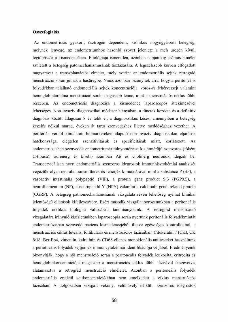
58
Összefoglalás
Az endometriosis gyakori, ösztrogén dependens, krónikus nőgyógyászati betegség,
melynek lényege, az endometriumhoz hasonló szövet jelenléte a méh üregén kívül,
legtöbbször a kismedencében. Etiológiája ismeretlen, azonban napjainkig számos elmélet
született a betegség patomechanizmusának tisztázására. A legszélesebb körben elfogadott
magyarázat a transzplantációs elmélet, mely szerint az endometriális sejtek retrográd
menstruácio során jutnak a hasüregbe. Nincs azonban bizonyíték arra, hogy a peritoneális
folyadékban található endometriális sejtek koncentrációja, vörös-és fehérvérsejt valamint
hemoglobintartalma menstruáció során magasabb lenne, mint a menstruációs ciklus többi
részében. Az endometriosis diagnózisa a kismedence laparoscopos áttekintésével
lehetséges. Non-invazív diagnosztikai módszer hiányában, a tünetek kezdete és a definitív
diagnózis között átlagosan 8 év telik el, a diagnosztikus késés, amennyiben a betegség
kezelés nélkül marad, éveken át tartó szenvedéshez illetve meddőséghez vezethet. A
perifériás vérből kimutatott biomarkereken alapuló non-invazív diagnosztikai eljárások
hatékonysága, elégtelen szenzitivitásuk és specificitásuk miatt, korlátozott. Az
endometriosisban szenvedők endometriumát túlnyomórészt kis átmérőjű szenzoros (főként
C-tipusú), adrenerg és kisebb számban Aδ és cholinerg neuronok idegzik be.
Transcervicálisan nyert endometriális szenzoros idegrostok immunhisztokémiai analízisét
végeztük olyan neurális transmitterek és fehérjék kimutatásával mint a substance P (SP), a
vasoactiv intestinalis polypeptid (VIP), a protein gene product 9.5 (PGP9.5), a
neurofilamentum (NF), a neuropeptid Y (NPY) valamint a calcitonin gene–related protein
(CGRP). A betegség pathomechanizmusának vizsgálata révén lehetőség nyílhat klinikai
jelentőségű eljárások kifejlesztésére. Ezért második vizsgálat sorozatunkban a peritoneális
folyadék ciklikus biológiai változásait tanulmányoztuk. A retrográd menstruáció
vizsgálatára irányuló kísérletünkben laparoscopia során nyertünk peritonális folyadékmintát
endometriózisban szenvedő páciens kismedencéjéből illetve egészséges kontrollokból, a
menstruációs ciklus luteális, follikuláris és menstruációs fázisaiban. Citokeratin 7 (CK), CK
8/18, Ber-Ep4, vimentin, kalretinin és CD68-ellenes monoklonális antitesteket használtunk
a periotnealis folyadék sejtjeinek immuncytokémiai identifikációja céljából. Eredményeink
bizonyítják, hogy a női menstruáció során a peritoneális folyadék leukocita, eritrocita és
hemoglobinkoncentrációja magasabb a menstruációs ciklus többi fázisával összevetve,
alátámasztva a retrográd menstruáció elméletét. Azonban a peritoneális folyadék
endometriális eredetű sejtkoncentrációjában nem emelkedett a ciklus menstruációs
fázisában. A dolgozatban vizsgált vékony, velőhüvely nélküli, szenzoros idegrostok

59
előfordulási gyakorisága nagyobb az igazoltan endometriosisban szenvedő nők
endometriumának funkcionális rétegében, mint az egészséges kontrollokéban.
Eredményeink alapját képezik egy potenciális semi-invazív tesztnek mellyel lehetővé válhat
az endometriosis korai klinikai diagnózisa.

60
12. References
1. Sampson JA. (1927) Metastatic or Embolic Endometriosis, due to the Menstrual
Dissemination of Endometrial Tissue into the Venous Circulation. Am J Pathol,
3: 93-110 43.
2. D'Hooghe TM, Debrock S. (2003) Evidence that endometriosis results from the
dislocation of basal endometrium? Hum Reprod, 18: 1130; author reply 1130-1.
3. Halme J, Hammond MG, Hulka JF, Raj SG, Talbert LM. (1984) Retrograde
menstruation in healthy women and in patients with endometriosis. Obstet
Gynecol, 64: 151-154.
4. Liu DT, Hitchcock A. (1986) Endometriosis: its association with retrograde
menstruation, dysmenorrhoea and tubal pathology. Br J Obstet Gynaecol,
93:859-862.
5. Koninckx PR, De Moor P, Brosens IA. (1980) Diagnosis of the luteinized
unruptured follicle syndrome by steroid hormone assays on peritoneal fluid. Br J
Obstet Gynaecol, 87:929-934.
6. Kruitwagen RF, Poels LG, Willemsen WNP, De Ronde IJY, Jap PHK. (1991)
Rolland R. Endometrial epithelial cells in peritoneal fluid during the early
follicular phase. Fertil Steril, 55:297-303.
7. D'Hooghe TM, Bambra CS, Raeymaekers BM, De Jonge I, Lauweryns JM,
Koninckx PR. (1995) Intrapelvic injection of menstrual endometrium causes
endometriosis in baboons (Papio cynocephalus and Papio anubis). Am J Obstet
Gynecol, 173:125-134.
8. Olive DL, Henderson DY. (1987) Endometriosis and mullerian anomalies.
Obstet Gynecol, 69:412-415.
9. Pinsonneault O, Goldstein DP. (1985) Obstructing malformations of the uterus
and vagina. Fertil Steril, 44:241-247.
10. D'Hooghe TM, Bambra CS, Suleman MA, Dunselman GA, Evers HL, Koninckx
PR. (1994) Development of a model of retrograde menstruation in baboons
(Papio anubis). Fertil Steril, 62:635-638.
11. Bokor A, Meuleman C, D'Hooghe T. Clinical Aspects of Endometriosis, in
Reproductive Endocrinology and Infertility:
Integrating Modern Clinical and Laboratory Practice.
CMP Douglas T Carrel, Editor. Springer, New York, 2010:191-207.
61
12. Dinulescu DM, Ince TA, Quade BJ, Shafer SA, Crowley D, Jacks T. (2005)
Role of K-ras and Pten in the development of mouse models of endometriosis
and endometrioid ovarian cancer. Nat Med, 11:63-70.
13. Levander G, Normann P. (1955) The pathogenesis of endometriosis; an
experimental study. Acta Obstet Gynecol Scand, 34:366-398.
14. Merrill JA. (1966) Endometrial induction of endometriosis across Millipore
filters. Am J Obstet Gynecol, 94:780-790.
15. Simpson JL, Elias S, Malinak LR, Buttram VC Jr. (1980) Heritable aspects of
endometriosis. I. Genetic studies. Am J Obstet Gynecol, 137:327-331.
16. Bulun SE. (2009) Endometriosis. N Engl J Med, 360:268-279.
17. Moen MH, Magnus P. (1993) The familial risk of endometriosis. Acta Obstet
Gynecol Scand, 72:560-564.
18. Shin JC, Ross HL, Elias S, Nguyen DD, Mitchell-Leef D, Simpson JL, Bischoff
FZ. (1997) Detection of chromosomal aneuploidy in endometriosis by multi-
color fluorescence in situ hybridization (FISH). Hum Genet, 100:401-406.
19. Kosugi Y, Elias S, Malinak LR, Nagata J, Isaka K, Takayama M, Simpson JL,
Bischoff FZ. (1999) Increased heterogeneity of chromosome 17 aneuploidy in
endometriosis. Am J Obstet Gynecol, 180:792-797.
20. Gogusev J, Bouquet de Jolinière J, Telvi L, Doussau M, du Manoir S, Stojkoski
A, Levardon M. (1999) Detection of DNA copy number changes in human
endometriosis by comparative genomic hybridization. Hum Genet, 105:444-451.
21. Sundqvist J, Falconer H, Seddighzadeh M, Vodolazkaia A, Fassbender A,
Kyama C, Bokor A, Stephansson O, Padyukov L, Gemzell-Danielsson K,
D'Hooghe TM. (2010) Endometriosis and autoimmune disease: association of
susceptibility to moderate/severe endometriosis with CCL21 and HLA-DRB1.
Fertil Steril, 10.1016/j.fertnstert.2010.07.1060 [doi]
22. Falconer H, D'Hooghe T, Fried G. (2007) Endometriosis and genetic
polymorphisms. Obstet Gynecol Surv, 62:616-628.
23. Dmowski WP, Steele RW, Baker GF. (1981) Deficient cellular immunity in
endometriosis. Am J Obstet Gynecol, 141:377-383.
24. Oosterlynck DJ, Cornillie FJ, Waer M, Vandeputte M, Koninckx PR. (1991)
Women with endometriosis show a defect in natural killer activity resulting in a
decreased cytotoxicity to autologous endometrium. Fertil Steril, 56:45-51.
62
25. D'Hooghe TM, Scheerlinck JP, Koninckx PR, Hill JA, Bambra CS. (1995) Anti-
endometrial lymphocytotoxicity and natural killer cell activity in baboons (Papio
anubis and Papio cynocephalus) with endometriosis. Hum Reprod, 10:558-562.
26. Hill JA. (1992) Immunology and endometriosis. Fertil Steril, 58:262-264.
27. Zeller JM, Henig I, Radwanska E, Dmowski WP. (1987) Enhancement of
human monocyte and peritoneal macrophage chemiluminescence activities in
women with endometriosis. Am J Reprod Immunol Microbiol, 13:78-82.
28. Halme J, Becker S, Haskill S. (1987) Altered maturation and function of
peritoneal macrophages: possible role in pathogenesis of endometriosis. Am J
Obstet Gynecol, 156:783-789.
29. Hill JA, Cohen J, Anderson DJ. (1989) The effects of lymphokines and
monokines on human sperm fertilizing ability in the zona-free hamster egg
penetration test. Am J Obstet Gynecol, 160:1154-1159.
30. Zhang RJ, Wild RA, Ojago JM. (1993) Effect of tumor necrosis factor-alpha on
adhesion of human endometrial stromal cells to peritoneal mesothelial cells: an
in vitro system. Fertil Steril, 59:1196-1201.
31. Halme J, White C, Kauma S, Estes J, Haskill S. (1988) Peritoneal macrophages
from patients with endometriosis release growth factor activity in vitro. J Clin
Endocrinol Metab, 66:1044-1049.
32. Kauma S, Clark MR, White C, Halme J. (1988) Production of fibronectin by
peritoneal macrophages and concentration of fibronectin in peritoneal fluid from
patients with or without endometriosis. Obstet Gynecol, 72:13-18.
33. Sharpe-Timms KL, Keisler LW, McIntush EW, Keisler DH. (1988) Tissue
inhibitor of metalloproteinase-1 concentrations are attenuated in peritoneal fluid
and sera of women with endometriosis and restored in sera by gonadotropin-
releasing hormone agonist therapy. Fertil Steril, 69:1128-1134.
34. Kokorine I, Nisolle M, Donnez J, Eeckhout Y, Courtoy PJ, Marbaix E. (1997)
Expression of interstitial collagenase (matrix metalloproteinase-1) is related to
the activity of human endometriotic lesions. Fertil Steril, 68:246-251.
35. Bulun SE, Yang S, Fang Z, Gurates B, Tamura M, Sebastian S. (2002) Estrogen
production and metabolism in endometriosis. Ann N Y Acad Sci, 955: 75-85;
discussion 86-8, 396-406.
63
36. Bulun SE, Zeitoun KM, Takayama K, Sasano H. (2000) Molecular basis for
treating endometriosis with aromatase inhibitors. Hum Reprod Update, 6:413-
418.
37. Agic A, Xu H, Finas D, Banz C, Diedrich K, Hornung D. (2006) Is
endometriosis associated with systemic subclinical inflammation? Gynecol
Obstet Invest, 62:139-147.
38. Guo SW. (2004) The link between exposure to dioxin and endometriosis: a
critical reappraisal of primate data. Gynecol Obstet Invest, 57:157-173.
39. D'Hooghe TM, Kyama C, Debrock S, Meuleman C, Mwenda JM. (2003) Future
directions in endometriosis research. Obstet Gynecol Clin North Am, 30:221-
244.
40. Giudice LC, Kao LC. (2004) Endometriosis. Lancet, 364:1789-1799.
41. Koninckx PR, Meuleman C, Demeyere S, Lesaffre E, Cornillie FJ. (1991)
Suggestive evidence that pelvic endometriosis is a progressive disease, whereas
deeply infiltrating endometriosis is associated with pelvic pain. Fertil Steril,
55:759-765.
42. Mahmood TA, Templeton A. (1991) Prevalence and genesis of endometriosis.
Hum Reprod, 6:544-549.
43. Moen MH. (1987) Endometriosis in women at interval sterilization. Acta Obstet
Gynecol Scand, 66:451-454.
44. Kennedy S, Bergqvist A, Chapron C, D'Hooghe T, Dunselman G, Greb R,
Hummelshoj L, Prentice A, Saridogan E. (2005) ESHRE Special Interest Group
for Endometriosis and Endometrium Guideline Development Group., ESHRE
guideline for the diagnosis and treatment of endometriosis. Hum Reprod,
20:2698-2704.
45. Arumugam K, Lim JM. (1997) Menstrual characteristics associated with
endometriosis. Br J Obstet Gynaecol, 104:948-950.
46. Vercellini P, De Giorgi O, Aimi G, Panazza S, Uglietti A, Crosignani PG.
(1997) Menstrual characteristics in women with and without endometriosis.
Obstet Gynecol, 90:264-268.
47. Matalliotakis IM, Cakmak H, Fragouli YG, Goumenou AG, Mahutte NG, Arici
A. (2008) Epidemiological characteristics in women with and without
endometriosis in the Yale series. Arch Gynecol Obstet, 277:389-393.

64
48. Filer RB, Wu CH. (1989) Coitus during menses. Its effect on endometriosis and
pelvic inflammatory disease. J Reprod Med, 34:887-890.
49. Mathias JR, Franklin R, Quast DC, Fraga N, Loftin CA, Yates L, Harrison V.
(1998) Relation of endometriosis and neuromuscular disease of the
gastrointestinal tract: new insights. Fertil Steril, 70:81-88.
50. Sinaii N, Plumb K, Cotton L, Lambert A, Kennedy S, Zondervan K, Stratton P.
(2008) Differences in characteristics among 1,000 women with endometriosis
based on extent of disease. Fertil Steril, 89:538-545.
51. Hadfield R, Mardon H, Barlow D, Kennedy S. (1996) Delay in the diagnosis of
endometriosis: a survey of women from the USA and the UK. Hum Reprod,
11:878-880.
52. Barlow DH, Glynn CJ. (1993) Endometriosis and pelvic pain. Baillieres Clin
Obstet Gynaecol, 7:775-789.
53. Chapron C, Fauconnier A, Dubuisson JB, Barakat H, Vieira M, Bréart G. (2003)
Deep infiltrating endometriosis: relation between severity of dysmenorrhoea and
extent of disease. Hum Reprod, 18:760-766.
54. Anaf V, Simon P, El Nakadi I, Fayt I, Buxant F, Simonart T, Peny MO, Noel
JC. (2000) Relationship between endometriotic foci and nerves in rectovaginal
endometriotic nodules. Hum Reprod, 15:1744-1750.
55. D'Hooghe TM, Debrock S, Hill JA, Meuleman C. (2003) Endometriosis and
subfertility: is the relationship resolved? Semin Reprod Med, 21:243-254.
56. Bokor A, Meuleman C, D'Hooghe T. The Role of the Fallopian Tubes in
Developement of Endometriosis, in The Fallopian Tube.
GA Ertan Saridogan, Ovrang Djahanbakhch, Editor. Anshan Ltd., Kent,
2009:449-457.
57. Haney AF. (1993) Endometriosis-associated infertility. Baillieres Clin Obstet
Gynaecol, 7:791-812.
58. D'Hooghe TM, Bambra CS, Raeymaekers BM, Koninckx PR. (1996) Serial
laparoscopies over 30 months show that endometriosis in captive baboons
(Papio anubis, Papio cynocephalus) is a progressive disease. Fertil Steril,
65:645-649.

65
59. Kaider AS, Kaider BD, Janowicz PB, Roussev RG. (1999) Immunodiagnostic
evaluation in women with reproductive failure. Am J Reprod Immunol, 42:335-
346.
60. Bulun SE, Mahendroo MS, Simpson ER. (1993) Polymerase chain reaction
amplification fails to detect aromatase cytochrome P450 transcripts in normal
human endometrium or decidua. J Clin Endocrinol Metab, 76:1458-1463.
61. Osteen KG, Keller NR, Feltus FA, Melner MH. (1999) Paracrine regulation of
matrix metalloproteinase expression in the normal human endometrium.
Gynecol Obstet Invest, 48:2-13.
62. Lessey BA, Damjanovich L, Coutifaris C, Castelbaum A, Albelda SM, Buck
CA. (1992) Integrin adhesion molecules in the human endometrium. Correlation
with the normal and abnormal menstrual cycle. J Clin Invest, 90:188-195.
63. Giudice LC, Telles TL, Lobo S, Kao L. (2002) The molecular basis for
implantation failure in endometriosis: on the road to discovery. Ann N Y Acad
Sci, 955:252-264; discussion 293-295, 396-406.
64. Kao LC, Germeyer A, Tulac S, Lobo S, Yang JP, Taylor RN, Osteen K, Lessey
BA, Giudice LC. (2003) Expression profiling of endometrium from women with
endometriosis reveals candidate genes for disease-based implantation failure and
infertility. Endocrinology, 144:2870-2881.
65. Montgomery GW, Nyholt DR, Zhao ZZ, Treloar SA, Painter JN, Missmer SA,
Kennedy SH, Zondervan KT. (2008) The search for genes contributing to
endometriosis risk. Hum Reprod Update, 14:447-457.
66. Bokor A, D'Hooghe T. Endometriosis and miscarriage: is there any association?
in Endometriosis: Current management and future trends. Rizk BRMB, Garcia-
Velasco JA, Editor. Jaypee Brothers Publishers, New Delhi, 2009:136-145.
67. Vercammen EE, D'Hooghe TM. (2000) Endometriosis and recurrent pregnancy
loss. Semin Reprod Med, 18:363-368.
68. Cahill DJ, Hull MG. (2000) Pituitary-ovarian dysfunction and endometriosis.
Hum Reprod Update, 6:56-66.
69. Marcoux S, Maheux R, Berube S. (1997) Laparoscopic surgery in infertile
women with minimal or mild endometriosis. Canadian Collaborative Group on
Endometriosis. N Engl J Med, 337:217-222.
66
70. Propst AM, Storti K, Barbieri RL. (1998) Lateral cervical displacement is
associated with endometriosis. Fertil Steril, 70:568-570.
71. Koninckx PR, Oosterlynck D, D'Hooghe T, Meuleman C. (1994) Deeply
infiltrating endometriosis is a disease whereas mild endometriosis could be
considered a non-disease. Ann N Y Acad Sci, 734:333-341.
72. Koninckx PR, Meuleman C, Oosterlynck D, Cornillie FJ. (1996) Diagnosis of
deep endometriosis by clinical examination during menstruation and plasma
CA-125 concentration. Fertil Steril, 65:280-287.
73. Guerriero S, Mais V, Ajossa S, Paoletti AM, Angiolucci M, Melis GB. (1996)
Transvaginal ultrasonography combined with CA-125 plasma levels in the
diagnosis of endometrioma. Fertil Steril, 65:293-298.
74. Fedele L, Bianchi S, Portuese A, Borruto F, Dorta M. (1998) Transrectal
ultrasonography in the assessment of rectovaginal endometriosis. Obstet
Gynecol, 91:444-448.
75. Moore J, Copley S, Morris J, Lindsell D, Golding S, Kennedy S. (2002) A
systematic review of the accuracy of ultrasound in the diagnosis of
endometriosis. Ultrasound Obstet Gynecol, 20:630-634.
76. McBean JH, Gibson M, Brumsted JR. (1996) The association of intrauterine
filling defects on hysterosalpingogram with endometriosis. Fertil Steril, 66:522-
526.
77. Bast RC Jr., Klug TL, St John E, Jenison E, Niloff JM, Lazarus H, Berkowitz
RS, Leavitt T, Griffiths CT, Parker L, Zurawski VR Jr, Knapp RC. (1983) A
radioimmunoassay using a monoclonal antibody to monitor the course of
epithelial ovarian cancer. N Engl J Med, 309:883-887.
78. Pittaway DE, Fayez JA. (1986) The use of CA-125 in the diagnosis and
management of endometriosis. Fertil Steril, 46:790-795.
79. Pittaway DE, Fayez JA. (1987) Serum CA-125 antigen levels increase during
menses. (1987) Am J Obstet Gynecol, 156:75-76.
80. Moloney MD, Thornton JG, Cooper EH. (1989) Serum CA 125 antigen levels
and disease severity in patients with endometriosis. Obstet Gynecol, 73:767-769.
81. O'Shaughnessy A, Check JH, Nowroozi K, Lurie D0. (1993) CA 125 levels
measured in different phases of the menstrual cycle in screening for
endometriosis. Obstet Gynecol, 81:99-103.

67
82. Pittaway DE. (1990) The use of serial CA 125 concentrations to monitor
endometriosis in infertile women. Am J Obstet Gynecol, 163:1032-1035;
discussion 1035-1037.
83. Nisolle M, Paindaveine B, Bourdon A, Berlière M, Casanas-Roux F, Donnez J.
(1990) Histologic study of peritoneal endometriosis in infertile women. Fertil
Steril, 53:984-988.
84. Berkes E, Bokor A, Rigo J Jr. (2010) [Current treatment of endometriosis with
laparoscopic surgery]. Orv Hetil, 151:1137-1144.
85. Moen MH, Halvorsen TB. (1992) Histologic confirmation of endometriosis in
different peritoneal lesions. Acta Obstet Gynecol Scand, 71:337-342.
86. Vercellini P, Aimi G, Panazza S, Vicentini S, Pisacreta A, Crosignani PG.
(2000) Deep endometriosis conundrum: evidence in favor of a peritoneal origin.
Fertil Steril, 73:1043-1046.
87. Redwine DB. (1999)Ovarian endometriosis: a marker for more extensive pelvic
and intestinal disease. Fertil Steril, 72:310-315.
88. Walter AJ, Hentz JG, Magtibay PM, Cornella JL, Magrina JF. (2001)
Endometriosis: correlation between histologic and visual findings at
laparoscopy. Am J Obstet Gynecol, 184:1407-1411; discussion 1411-1413.
89. Donnez J, Nisolle M, Casanas-Roux F. (1992) Three-dimensional architectures
of peritoneal endometriosis. Fertil Steril, 57:980-983.
90. ASRM Revised ASRM classification for endometriosis:1996. (1997) Fertil
Steril 67: 820.
91. McArthur JW, Ulfelder H. (1965) The effect of pregnancy upon endometriosis.
Obstet Gynecol Surv, 20:709-733.
92. D'Hooghe TM, Bambra CS, De Jonge I, Lauweryns JM, Raeymaekers BM,
Koninckx PR. (1997) The effect of pregnancy on endometriosis in baboons
(Papio anubis, Papio cynocephalus). Arch Gynecol Obstet, 261:15-9.
93. ESHRE Capri Workshop Group. (2005) Noncontraceptive health benefits of
combined oral contraception. Hum Reprod Update, 11:513-525.
94. Coupland RE. (1969) The distribution of cholinergic and other nerve fibres in
the human uterus. Postgrad Med J, 45:78-79.
95. Heinrich D, Reinecke M, Forssmann WG. (1986) Peptidergic innervation of the
human and guinea pig uterus. Arch Gynecol, 237:213-219.

68
96. Shew RL, Papka RE, McNeill DL. (1990) Calcitonin gene-related peptide in the
rat uterus: presence in nerves and effects on uterine contraction. Peptides,
11:583-589.
97. Papka RE, Cotton JP, Traurig HH. (1985) Comparative distribution of
neuropeptide tyrosine-, vasoactive intestinal polypeptide-, substance P-
immunoreactive, acetylcholinesterase-positive and noradrenergic nerves in the
reproductive tract of the female rat. Cell Tissue Res, 242:475-490.
98. Tulandi T, Chen MF, Al-Took S, Watkin K. (1998) A study of nerve fibers and
histopathology of postsurgical, postinfectious, and endometriosis-related
adhesions. Obstet Gynecol, 92:766-768.
99. Sulaiman H, Gabella G, Davis MSc C, Mutsaers SE, Boulos P, Laurent GJ,
Herrick SE. (2001) Presence and distribution of sensory nerve fibers in human
peritoneal adhesions. Ann Surg, 234:256-261.
100. Tamburro S, Canis M, Albuisson E, Dechelotte P, Darcha C, Mage G. (2003)
Expression of transforming growth factor beta1 in nerve fibers is related to
dysmenorrhea and laparoscopic appearance of endometriotic implants. Fertil
Steril, 80:1131-1136.
101. Berkley KJ, Dmitrieva N, Curtis KS, Papka RE. (2004) Innervation of ectopic
endometrium in a rat model of endometriosis. Proc Natl Acad Sci U S A,
101:11094-11098.
102. Samuelson UE, Dalsgaard CJ, Lundberg JM, Hökfelt T. (1985) Calcitonin gene-
related peptide inhibits spontaneous contractions in human uterus and fallopian
tube. Neurosci Lett, 62:225-230.
103. Bulletti C, De Ziegler D, Polli V, Del Ferro E, Palini S, Flamigni C. (2002)
Characteristics of uterine contractility during menses in women with mild to
moderate endometriosis. Fertil Steril, 77:1156-1161.
104. Bergqvist A, Bruse C, Carlberg M, Carlström K. (2001) Interleukin 1beta,
interleukin-6, and tumor necrosis factor-alpha in endometriotic tissue and in
endometrium. Fertil Steril, 75:489-495.
105. Ylikorkala O, Viinikka L. (1983) Prostaglandins and endometriosis. Acta Obstet
Gynecol Scand Suppl, 113:105-107.
106. De Leon DD, Bakker B, Wilson DM, Hintz RL, Rosenfeld RG. (1988)
Demonstration of insulin-like growth factor (IGF-I and -II) receptors and
69
binding protein in human breast cancer cell lines. Biochem Biophys Res
Commun, 152:398-405.
107. Anaf V, Simon P, El Nakadi I, Fayt I, Simonart T, Buxant F, Noel JC. (2002)
Hyperalgesia, nerve infiltration and nerve growth factor expression in deep
adenomyotic nodules, peritoneal and ovarian endometriosis. Hum Reprod,
17:1895-1900.
108. Lessey BA, Castelbaum AJ, Sawin SW, Buck CA, Schinnar R, Bilker W, Strom
BL. (1994) Aberrant integrin expression in the endometrium of women with
endometriosis. J Clin Endocrinol Metab, 79:643-649.
109. Ota H, Tanaka T. (1997) Integrin adhesion molecules in the endometrial
glandular epithelium in patients with endometriosis or adenomyosis. J Obstet
Gynaecol Res, 23:485-491.
110. Healy DL, Rogers PA, Hii L, Wingfield M. (1998) Angiogenesis: a new theory
for endometriosis. Hum Reprod Update, 4:736-740.
111. Bokor A, Kyama CM, Vercruysse L, Fassbender A, Gevaert O, Vodolazkaia A,
De Moor B, Fülöp V, D'Hooghe T. (2009) Density of small diameter sensory
nerve fibres in endometrium: a semi-invasive diagnostic test for minimal to mild
endometriosis. Hum Reprod, 24:3025-3032.
112. May KE, Conduit-Hulbert SA, Villar J, Kirtley S, Kennedy SH, Becker CM.
(2010) Peripheral biomarkers of endometriosis: a systematic review. Hum
Reprod Update,
113. Stroup DF, Berlin JA, Morton SC, Olkin I, Williamson GD, Rennie D, Moher
D, Becker BJ, Sipe TA, Thacker SB. (2000) Meta-analysis of observational
studies in epidemiology: a proposal for reporting. Meta-analysis Of
Observational Studies in Epidemiology (MOOSE) group. JAMA, 283:2008-
2012.
114. Mol BW, Bayram N, Lijmer JG, Wiegerinck MA, Bongers MY, van der Veen F,
Bossuyt PM. (1998) The performance of CA-125 measurement in the detection
of endometriosis: a meta-analysis. Fertil Steril, 70:1101-1108.
115. Chen FP, Soong YK, Lee N, Lo SK. (1998) The use of serum CA-125 as a
marker for endometriosis in patients with dysmenorrhea for monitoring therapy
and for recurrence of endometriosis. Acta Obstet Gynecol Scand, 77:665-670.

70
116. Matalliotakis IM, Arici A, Goumenou AG, Katassos T, Karkavitsas N,
Koumantakis EE. (2004) Comparison of the effects of leuprorelin acetate and
danazol treatments on serum CA-125 levels in women with endometriosis. Int J
Fertil Womens Med, 49:75-78.
117. Jones G, Kennedy S, Barnard A, Wong J, Jenkinson C. (2001) Development of
an endometriosis quality-of-life instrument: The Endometriosis Health Profile-
30. Obstet Gynecol, 98:258-264.
118. Dwan K, Altman DG, Arnaiz JA, Bloom J, Chan AW, Cronin E, Decullier E,
Easterbrook PJ, Von Elm E, Gamble C, Ghersi D, Ioannidis JP, Simes J,
Williamson PR. (2008) Systematic review of the empirical evidence of study
publication bias and outcome reporting bias. PLoS One, 3:3081.
119. Khan KN, Masuzaki H, Fujishita A, Kitajima M, Kohno T, Sekine I, Matsuyama
T, Ishimaru T. (2005) Regulation of hepatocyte growth factor by basal and
stimulated macrophages in women with endometriosis. Hum Reprod, 20:49-60.
120. D'Hooghe TM, Hill JA, Oosterlynck DJ, Koninckx PR, Bambra CS. (1996)
Effect of endometriosis on white blood cell subpopulations in peripheral blood
and peritoneal fluid of baboons. Hum Reprod, 11:1736-1740.
121. Hill JA, Faris HM, Schiff I, Anderson DJ. (1988) Characterization of leukocyte
subpopulations in the peritoneal fluid of women with endometriosis. Fertil
Steril, 50:216-222.
122. Haney AF, Muscato JJ, Weinberg JB. (1981) Peritoneal fluid cell populations in
infertility patients. Fertil Steril, 35:696-698.
123. Halme J, Becker S, Hammond MG, Raj S. (1982) Pelvic macrophages in normal
and infertile women: the role of patent tubes. Am J Obstet Gynecol, 142:890-
895.
124. Muscato JJ, Haney AF, Weinberg JB. (1982) Sperm phagocytosis by human
peritoneal macrophages: a possible cause of infertility in endometriosis. Am J
Obstet Gynecol, 144:503-510.
125. Haney AF, Misukonis MA, Weinberg JB. (1983) Macrophages and infertility:
oviductal macrophages as potential mediators of infertility. Fertil Steril, 39:310-
315.
126. Badawy SZ, Cuenca V, Marshall L. (1987) Peritoneal fluid prostaglandins in
patients with endometriosis. Contrib Gynecol Obstet, 16:60-65.

71
127. Badawy SZ, Cuenca V, Kaufman L, Stitzel A, Thompson M. (1989) The
regulation of immunoglobulin production by B cells in patients with
endometriosis. Fertil Steril, 51:770-773.
128. Montagna P, Capellino S, Villaggio B, Remorgida V, Ragni N, Cutolo M,
Ferrero S. (2008) Peritoneal fluid macrophages in endometriosis: correlation
between the expression of estrogen receptors and inflammation. Fertil Steril,
90:156-164.
129. Eisermann J, Gast MJ, Pineda J, Odem RR, Collins JL. (1988) Tumor necrosis
factor in peritoneal fluid of women undergoing laparoscopic surgery. Fertil
Steril, 50:573-579.
130. Buyalos RP, Funari VA, Azziz R, Watson JM, Martinez-Maza O. (1992)
Elevated interleukin-6 levels in peritoneal fluid of patients with pelvic
pathology. Fertil Steril, 58:302-306.
131. Donnez J, Smoes P, Gillerot S, Casanas-Roux F, Nisolle M. (1998) Vascular
endothelial growth factor (VEGF) in endometriosis. Hum Reprod, 13:1686-
1690.
132. Nathan CF. (1987) Secretory products of macrophages. J Clin Invest, 79:319-
326.
133. Fujishita A, Hasuo A, Khan KN, Masuzaki H, Nakashima H, Ishimaru T. (1999)
Immunohistochemical study of angiogenic factors in endometrium and
endometriosis. Gynecol Obstet Invest, 48:36-44.
134. Harada T, Iwabe T, Terakawa N. (2001) Role of cytokines in endometriosis.
Fertil Steril, 76:1-10.
135. Witz CA, Thomas MR, Montoya-Rodriguez IA, Nair AS, Centonze VE,
Schenken RS. (2001) Short-term culture of peritoneum explants confirms
attachment of endometrium to intact peritoneal mesothelium. Fertil Steril,
75:385-390.
136. Polishuk WZ, Sharf M. (1965) Culdoscopic findings in primary dysmenorrhea.
Obstet Gynecol, 26:746-748.
137. Kruitwagen RF, Poels LG, Willemsen WN, Jap PH, Thomas CM, Rolland R.
(1991) Retrograde seeding of endometrial epithelial cells by uterine-tubal
flushing. Fertil Steril, 56:414-420.
72
138. Koninckx PR, Heyns W, Verhoeven G, Van Baelen H, Lissens WD, De Moor P,
Brosens IA. (1980) Biochemical characterization of peritoneal fluid in women
during the menstrual cycle. J Clin Endocrinol Metab, 51:1239-1244.
139. Bartosik D, Jacobs SL, Kelly LJ. (1986) Endometrial tissue in peritoneal fluid.
Fertil Steril, 46:796-800.
140. Badawy SZ, Cuenca V, Marshall L, Munchback R, Rinas AC, Coble DA. (1984)
Cellular components in peritoneal fluid in infertile patients with and without
endometriosis. Fertil Steril, 42:704-708.
141. D'Hooghe TM, Bambra CS, Raeymaekers BM, Koninckx PR. (1996) Increased
prevalence and recurrence of retrograde menstruation in baboons with
spontaneous endometriosis. Hum Reprod, 11:2022-2025.
142. D'Hooghe TM, Bambra CS, Raeymaekers BM, Koninckx PR. (1996)
Development of spontaneous endometriosis in baboons. Obstet Gynecol,
88:462-466.
143. Lanfrancone L, Boraschi D, Ghiara P, Falini B, Grignani F, Peri G, Mantovani
A, Pelicci PG. (1992) Human peritoneal mesothelial cells produce many
cytokines (granulocyte colony-stimulating factor [CSF], granulocyte-monocyte-
CSF, macrophage-CSF, interleukin-1 [IL-1], and IL-6) and are activated and
stimulated to grow by IL-1. Blood, 80:2835-2842.
144. Zeillemaker AM, Verbrugh HA, Hoynck van Papendrecht AA, Leguit P. (1994)
CA 125 secretion by peritoneal mesothelial cells. J Clin Pathol, 47:263-265.
145. Gaetje R, Kotzian S, Herrmann G, Baumann R, Starzinski-Powitz A. (1997)
Nonmalignant epithelial cells, potentially invasive in human endometriosis, lack
the tumor suppressor molecule E-cadherin. Am J Pathol, 150:461-467.
146. Berkkanoglu M, Arici A. (2003) Immunology and endometriosis. Am J Reprod
Immunol, 50:48-59.
147. Iwabe T, Harada T, Tsudo T, Nagano Y, Yoshida S, Tanikawa M, Terakawa N.
(2000) Tumor necrosis factor-alpha promotes proliferation of endometriotic
stromal cells by inducing interleukin-8 gene and protein expression. J Clin
Endocrinol Metab, 85:824-829.
148. Witz CA, Monotoya-Rodriguez IA, Schenken RS. (1999) Whole explants of
peritoneum and endometrium: a novel model of the early endometriosis lesion.
Fertil Steril, 71:56-60.

73
149. Debrock S, Vander Perre S, Meuleman C, Moerman P, Hill JA, D'Hooghe TM.
(2002) In-vitro adhesion of endometrium to autologous peritoneal membranes:
effect of the cycle phase and the stage of endometriosis. Hum Reprod, 17:2523-
2528.
150. Nothnick WB, D'Hooghe TM. (2003) Medical management of endometriosis:
novel targets and approaches towards the development of future treatment
regimes. Gynecol Obstet Invest, 55:189-198.
151. Yeaman GR, Collins JE, Lang GA. (2002) Autoantibody responses to
carbohydrate epitopes in endometriosis. Ann N Y Acad Sci, 955:174-182;
discussion 199-200, 396-406.
152. Milingos S, Protopapas A, Drakakis P, Liapi A, Loutradis D, Kallipolitis G,
Milingos D, Michalas S. (2003)Laparoscopic management of patients with
endometriosis and chronic pelvic pain. Ann N Y Acad Sci, 997:269-273.
153. Mechsner S, Schwarz J, Thode J, Loddenkemper C, Salomon DS, Ebert AD.
(2007) Growth-associated protein 43-positive sensory nerve fibers accompanied
by immature vessels are located in or near peritoneal endometriotic lesions.
Fertil Steril, 88:581-587.
154. Berkley KJ, Rapkin AJ, Papka RE. (2005) The pains of endometriosis. Science,
308:1587-1589.
155. Wang G, Tokushige N, Markham R, Fraser IS. (2009) Rich innervation of deep
infiltrating endometriosis. Hum Reprod, 24:827-834.
156. Slack A, Child T, Lindsey I, Kennedy S, Cunningham C, Mortensen N,
Koninckx P, McVeigh E. (2007) Urological and colorectal complications
following surgery for rectovaginal endometriosis. BJOG, 114:1278-1282.
157. Kyama CM, T'Jampens D, Mihalyi A, Simsa P, Debrock S, Waelkens E,
Landuyt B, Meuleman C, Fulop V, Mwenda JM, D'Hooghe TM. (2006)
ProteinChip technology is a useful method in the pathogenesis and diagnosis of
endometriosis: a preliminary study. Fertil Steril, 86:203-209.
158. Kyama CM, Mihalyi A, Simsa P, Mwenda JM, Tomassetti C, Meuleman C,
D'Hooghe TM. (2008) Non-steroidal targets in the diagnosis and treatment of
endometriosis. Curr Med Chem, 15:1006-1017.
159. D'Hooghe TM, Kyama C, Debrock S, Meuleman C, Mwenda JM. (2004) Future
directions in endometriosis research. Ann N Y Acad Sci, 1034:316-325.

74
160. Berbic M, Schulke L, Markham R, Tokushige N, Russell P, Fraser IS. (2009)
Macrophage expression in endometrium of women with and without
endometriosis. Hum Reprod, 24:325-332.
161. Evans S, Moalem-Taylor G, Tracey DJ. (2007) Pain and endometriosis. Pain,
132:22-25.
162. Tran LV, Tokushige N, Berbic M, Markham R, Fraser IS. (2009) Macrophages
and nerve fibres in peritoneal endometriosis. Hum Reprod, 24:835-841.
163. D'Hooghe TM, Mihalyi AM, Simsa P, Kyama CK, Peeraer K, De Loecker P,
Meeuwis L, Segal L, Meuleman C. (2006) Why we need a noninvasive
diagnostic test for minimal to mild endometriosis with a high sensitivity.
Gynecol Obstet Invest, 62:136-138.
164. Tokushige N, Markham R, Russell P, Fraser IS. (2006) High density of small
nerve fibres in the functional layer of the endometrium in women with
endometriosis. Hum Reprod, 21:782-787.
165. Tokushige N, Markham R, Russell P, Fraser IS. (2007) Different types of small
nerve fibers in eutopic endometrium and myometrium in women with
endometriosis. Fertil Steril, 88:795-803.
166. Al-Jefout M, Andreadis N, Tokushige N, Markham R, Fraser I. (2007) A pilot
study to evaluate the relative efficacy of endometrial biopsy and full curettage in
making a diagnosis of endometriosis by the detection of endometrial nerve
fibers. Am J Obstet Gynecol, 197:578 e1-4.
167. Mutch DG, Powell MA, Allsworth JE, Taylor NP, Brooks RA. (2007) How
accurate is Pipelle sampling: a study by Huang et al. Am J Obstet Gynecol,
196:280-281.
168. Noyes RW, Hertig AT, Rock J. (1975) Dating the endometrial biopsy. Am J
Obstet Gynecol, 122:262-263.
169. Gevaert O, De Smet F, Kirk E, Van Calster B, Bourne T, Van Huffel S, Moreau
Y, Timmerman D, De Moor B, Condous G. (2006) Predicting the outcome of
pregnancies of unknown location: Bayesian networks with expert prior
information compared to logistic regression. Hum Reprod, 21:1824-1831.
170. Timmerman D, Testa AC, Bourne T, Ferrazzi E, Ameye L, Konstantinovic ML,
Van Calster B, Collins WP, Vergote I, Van Huffel S, Valentin L, International
Ovarian Tumor Analysis Group. (2005) Logistic regression model to distinguish

75
between the benign and malignant adnexal mass before surgery: a multicenter
study by the International Ovarian Tumor Analysis Group. J Clin Oncol,
23:8794-8801.
171. Pochet NL, Suykens JA. (2006) Support vector machines versus logistic
regression: improving prospective performance in clinical decision-making.
Ultrasound Obstet Gynecol, 27:607-608.
172. Hanley JA, McNeil BJ. (1982) The meaning and use of the area under a receiver
operating characteristic (ROC) curve. Radiology, 143:29-36.
173. Tokushige N, Markham R, Russell P, Fraser IS. (2008) Effects of hormonal
treatment on nerve fibers in endometrium and myometrium in women with
endometriosis. Fertil Steril, 90:1589-1598.
174. Lundberg LM, Alm P, Wharton J, Polak JM. (1988) Protein gene product 9.5
(PGP 9.5). A new neuronal marker visualizing the whole uterine innervation and
pregnancy-induced and developmental changes in the guinea pig.
Histochemistry, 90:9-17.
175. Quinn M. (2007) Uterine innervation in adenomyosis. J Obstet Gynaecol,
27:287-291.
176. Lynch EM, Wharton J, Bryant MG, Bloom SR, Polak JM, Elder MG. (1980)
The differential distribution of vasoactive intestinal polypeptide in the normal
human female genital tract. Histochemistry, 67:169-177.
177. Schlaepfer WW. (1987) Neurofilaments: structure, metabolism and implications
in disease. J Neuropathol Exp Neurol, 46:117-129.
178. Tariverdian N, Theoharides TC, Siedentopf F, Gutiérrez G, Jeschke U,
Rabinovich GA, Blois SM, Arck PC. (2007) Neuroendocrine-immune
disequilibrium and endometriosis: an interdisciplinary approach. Semin
Immunopathol, 29:193-210.
179. D'Hooghe TM, Debrock S. (2002) Endometriosis, retrograde menstruation and
peritoneal inflammation in women and in baboons. Hum Reprod Update, 8:84-
88.
180. Gomez-Fernandez CR, Ganjei-Azar P, Behshid K, Averette HE, Nadji M.
(2000) Normal endometrial cells in Papanicolaou smears: prevalence in women
with and without endometrial disease. Obstet Gynecol, 96:874-878.

76
181. Wagener FA, Feldman E, de Witte T, Abraham NG. (1997) Heme induces the
expression of adhesion molecules ICAM-1, VCAM-1, and E selectin in vascular
endothelial cells. Proc Soc Exp Biol Med, 216:456-463.
182. Defrere S, Lousse JC, González-Ramos R, Colette S, Donnez J, Van
Langendonckt A. (2008) Potential involvement of iron in the pathogenesis of
peritoneal endometriosis. Mol Hum Reprod, 14:377-385.
183. Balla J, Nath KA, Balla G, Juckett MB, Jacob HS, Vercellotti GM. (1995)
Endothelial cell heme oxygenase and ferritin induction in rat lung by
hemoglobin in vivo. Am J Physiol, 268:L321-327.
184. D'Hooghe TM, Bambra CS, Xiao L, Peixe K, Hill JA. (2001) Effect of
menstruation and intrapelvic injection of endometrium on inflammatory
parameters of peritoneal fluid in the baboon (Papio anubis and Papio
cynocephalus). Am J Obstet Gynecol, 184:917-925.
185. Blumenkrantz MJ, Gallagher N, Bashore RA, Tenckhoff H. (1981) Retrograde
menstruation in women undergoing chronic peritoneal dialysis. Obstet Gynecol,
57:667-670.
186. Koninckx PR, Ide P, Vandenbroucke W, Brosens IA. (1980) New aspects of the
pathophysiology of endometriosis and associated infertility. J Reprod Med,
24:257-260.
187. Scheenjes E, de Velde EL, Kremer J. (1990) Inspection of the ovaries and
steroids in serum and peritoneal fluid at various time intervals after ovulation in
fertile women: implications for the luteinized unruptured follicle syndrome.
Fertil Steril, 54:38-41.
188. Sharpe-Timms KL. (2005) Haptoglobin expression by shed endometrial tissue
fragments found in peritoneal fluid. Fertil Steril, 84:22-30.
189. Reti LL, Byrne GD, Davoren RA. (1983) The acute clinical features of
retrograde menstruation. Aust N Z J Obstet Gynaecol, 23:51-52.
190. Kruitwagen RF, Poels LG, Willemsen WN, Jap PH, de Ronde IJ, Hanselaar TG,
Rolland R. (1991) Immunocytochemical markerprofile of endometriotic
epithelial, endometrial epithelial, and mesothelial cells: a comparative study. Eur
J Obstet Gynecol Reprod Biol, 41:215-223.

77
191. Gabriel C, Achten R, Drijkoningen M. (2004) Use of liquid-based cytology in
serous fluids: a comparison with conventional cytopreparatory techniques. Acta
Cytol, 48:825-835.
192. McCluggage WG, Oliva E, Herrington CS, McBride H, Young RH. (2003)
CD10 and calretinin staining of endocervical glandular lesions, endocervical
stroma and endometrioid adenocarcinomas of the uterine corpus: CD10
positivity is characteristic of, but not specific for, mesonephric lesions and is not
specific for endometrial stroma. Histopathology, 43:144-150.
193. Lucidi RS, Witz CA, Chrisco M, Binkley PA, Shain SA, Schenken RS. (2005) A
novel in vitro model of the early endometriotic lesion demonstrates that
attachment of endometrial cells to mesothelial cells is dependent on the source
of endometrial cells. Fertil Steril, 84:16-21.
194. Boyle DP, McCluggage WG. (2009) Peritoneal stromal endometriosis: a
detailed morphological analysis of a large series of cases of a common and
under-recognised form of endometriosis. J Clin Pathol, 62:530-533.

78
13. List of publications related to the thesis
Articles
Bokor A, Kyama CM, Vercruysse L, Fassbender A, Gevaert O, Vodolazkaia A, De
Moor B, Fülöp V, D'Hooghe T. (2009) Density of small diameter sensory nerve fibres
in endometrium: a semi-invasive diagnostic test for minimal to mild endometriosis.
Hum Reprod, 24:827–834.
Bokor A, Debrock S, Drijkoningen M, Goossens W, Fulop V, D'Hooghe T. (2009)
Quantity and quality of retrograde menstruation: a case control study.
Reprod Biol Endocrinol, 7:123.
D'Hooghe TM, Kyama CM, Chai D, Fassbender A, Vodolazkaia A, Bokor A, Mwenda
JM. (2009) Nonhuman primate models for translational research in endometriosis.
Reprod Sci, 16:152-161.
Vodolazkaia A, Fassbender A, Kyama C, Bokor A, Stephansson O, Padyukov L,
Gemzell-Danielsson K, D'Hooghe TM. (2010) Endometriosis and autoimmune disease:
association of susceptibility to moderate/severe endometriosis with CCL21 and HLA-
DRB1. Fertil Steril, 10.1016/j.fertnstert.2010.07.1060 [doi]
Berkes E, Bokor A, Rigó J Jr. (2010) Current treatment of endometriosis with
laparoscopic surgery. Orv Hetil, 151:1137-1144. (In Hungarian)
Bokor A, Kyama CM, Vercruysse L, Fassbender A, Gevaert O, Vodolazkaia A, Rigó J,
De Moor B, Fülöp V, D'Hooghe T. The non-invasive diagnosis of endometriosis. Magy.
Nőorv.Lapja In Press (In Hungarian)

79
Textbook chapters
Bokor A, Meuleman C, D‘Hooghe T. The Role of the Fallopian Tube in the
Development of Endometriosis and Associated Infertility In: Allahbadia G N, Saridogan
E, Djahanbakhch O, (editors) The Fallopian Tube. Anshan Ltd., Kent, 2008: 449-457.
Bokor A, D‘Hooghe T. Endometriosis and Miscarriage: Is there any Association?
In: Garcia-Velasco J A, Rizk B R M B (editors) Endometriosis:current therapy and
future trends. Jaypee, New Delhi, 2009: 136-142.
Bokor A, Meuleman C, D‘Hooghe T.The Clinical Aspects of Endometriosis
In: Carrell D, Peterson CM, (editors) Reproductive Endocrinology and Infertility:
Integrating Modern Clinical and Laboratory Practice. Springer, New York, 2010:
191-207.

80
14. Acknowledgement
I would like to express my deep and sincere gratitude to my promoter, Professor
Thomas D‘Hooghe. His wide knowledge and his logical way of thinking have been of
great value for me. His understanding, encouraging and personal guidance have
provided a fundamental basis for the present thesis.
I wish to express my warm thanks to Professor János Rigó who introduced me to the
field of clinical research in obstetrics and gynaecology and highlighted the importance
of scientific thinking.
I am deeply grateful to my supervisor, Professor Vilmos Fülöp his detailed and
constructive comments, and for his important support throughout this work.
Lastly, I offer my regards and blessings to all of those who supported me in any respect
during the completion of the project: Professor Sophie Debrock, Lisbeth Vercruysse,
Cleophas Kyama, Amelie Fassbender, Alexandra Vodolazkaia and Steffi Mayer.