Rhabdomyosarcoma masquerading as lymphadenopathy in a ...
4
CASE REPORT Open Access Rhabdomyosarcoma masquerading as lymphadenopathy in a patient with newly diagnosed Hodgkin’ s lymphoma Joseph Dergan 1 , Sandeep Sirsi 1 , Armand Asarian 1 , Elizabeth Guevara 2 and Philip Xiao 3* Abstract Background: Hodgkin’s lymphoma (HL) is a rare malignancy which often presents with lymphadenopathy and classic “B symptoms” of weight loss, fever, and night sweats. Additional masses or nodes could easily be presumed to be a result of the initial diagnosis. On the other hand, adult rhabdomyosarcoma is a rare malignancy presenting with a new mass in a patient with previous diagnosis of Hodgkin’s lymphoma. In both cases, a tissue diagnosis should be obtained to appropriately confirm the diagnosis. Case presentation: We present a case of a 64-year-old male who presents with right axillary lymphadenopathy, diagnosed as Hodgkin’s lymphoma. He subsequently developed left inguinal lymphadenopathy without the classic B symptoms of HL. Excisional biopsy revealed rhabdomyosarcoma. Stage III Hodgkin’s lymphoma (lymph node involvement on both sides of the diaphragm) is not commonly seen without typical B symptoms. Once the diagnosis of two primary malignancies is made, the dilemma becomes determining the treatment course. In the case of Hodgkin’s lymphoma and rhabdomyosarcoma, there is some overlap in the chemotherapeutic regimen and use of radiation. Conclusions: This case illustrates the importance of careful examination of Hodgkin’s lymphoma patients and consideration of additional tissue diagnoses in atypical presentations of new masses or lymphadenopathy on the opposite side of the diaphragm. Keywords: Hodgkin’s lymphoma, Nodular sclerosing type, Lymphadenopathic rhabdomyosarcoma, Alveolar type Background Hodgkin’ s lymphoma (HL) is an uncommon malignancy of the lymphatic system, with an incidence of 2.5 per 100,000 [1]. It is well known to have a favorable progno- sis, with a 5-year survival greater than 85 % and is thus viewed as a potentially curable disease. There are five histologic types currently described: nodular sclerosing (most common), mixed cellularity, lymphocyte rich (best prognosis), lymphocyte depleted (worst prognosis), and nodular lymphocyte predominant. With the exception of nodular lymphocyte predominant, all of these are char- acterized by the presence of Reed-Sternberg (RS) cells (typically B lymphocytes with CD 15 and CD 30 antigen positivity). Nodular sclerosing type, which has a strong genetic component, commonly involves the mediastinum and has CD20 antigen positivity. Rhabdomyosarcoma (RMS) is a malignant tumor of striated muscle similar to other high-grade soft tissue sarcomas. It is the most common soft tissue sarcoma in children (90 % of cases are diagnosed under 25 years of age; 60–70 % of these are diagnosed under 10 years of age) but a rare diagnosis in adults, with only an approxi- mated 400 new cases in the USA each year [2]. Even more rare is the diagnosis of “lymphadenopathic” RMS, with only four prior cases described in the literature [3–5]. Overall 5-year survival is poor compared to that of HL, at 40 % [6]. Head and neck is the most common anatomic location for these tumors. Five histologic types are described: embryonal (most common), alveolar (worst prognosis; commonly found in the extremities, trunk, * Correspondence: [email protected] 3 Department of Pathology and Laboratory Medicine, The Brooklyn Hospital Center, Icahn School of Medicine at Mount Sinai, Brooklyn, NY 11201, USA Full list of author information is available at the end of the article © 2016 Dergan et al. Open Access This article is distributed under the terms of the Creative Commons Attribution 4.0 International License (http://creativecommons.org/licenses/by/4.0/), which permits unrestricted use, distribution, and reproduction in any medium, provided you give appropriate credit to the original author(s) and the source, provide a link to the Creative Commons license, and indicate if changes were made. The Creative Commons Public Domain Dedication waiver (http://creativecommons.org/publicdomain/zero/1.0/) applies to the data made available in this article, unless otherwise stated. Dergan et al. World Journal of Surgical Oncology (2016) 14:101 DOI 10.1186/s12957-016-0846-0 CORE Metadata, citation and similar papers at core.ac.uk Provided by Springer - Publisher Connector
Transcript of Rhabdomyosarcoma masquerading as lymphadenopathy in a ...
Dergan et al. World Journal of Surgical Oncology (2016) 14:101 DOI 10.1186/s12957-016-0846-0
CORE Metadata, citation and similar papers at core.ac.uk
Provided by Springer - Publisher Connector
CASE REPORT Open Access
Rhabdomyosarcoma masquerading aslymphadenopathy in a patient with newlydiagnosed Hodgkin’s lymphoma
Joseph Dergan1, Sandeep Sirsi1, Armand Asarian1, Elizabeth Guevara2 and Philip Xiao3*Abstract
Background: Hodgkin’s lymphoma (HL) is a rare malignancy which often presents with lymphadenopathy andclassic “B symptoms” of weight loss, fever, and night sweats. Additional masses or nodes could easily be presumedto be a result of the initial diagnosis. On the other hand, adult rhabdomyosarcoma is a rare malignancy presentingwith a new mass in a patient with previous diagnosis of Hodgkin’s lymphoma. In both cases, a tissue diagnosisshould be obtained to appropriately confirm the diagnosis.
Case presentation: We present a case of a 64-year-old male who presents with right axillary lymphadenopathy,diagnosed as Hodgkin’s lymphoma. He subsequently developed left inguinal lymphadenopathy without the classicB symptoms of HL. Excisional biopsy revealed rhabdomyosarcoma. Stage III Hodgkin’s lymphoma (lymph nodeinvolvement on both sides of the diaphragm) is not commonly seen without typical B symptoms. Once thediagnosis of two primary malignancies is made, the dilemma becomes determining the treatment course. In thecase of Hodgkin’s lymphoma and rhabdomyosarcoma, there is some overlap in the chemotherapeutic regimen anduse of radiation.
Conclusions: This case illustrates the importance of careful examination of Hodgkin’s lymphoma patients andconsideration of additional tissue diagnoses in atypical presentations of new masses or lymphadenopathy on theopposite side of the diaphragm.
Keywords: Hodgkin’s lymphoma, Nodular sclerosing type, Lymphadenopathic rhabdomyosarcoma, Alveolar type
BackgroundHodgkin’s lymphoma (HL) is an uncommon malignancyof the lymphatic system, with an incidence of 2.5 per100,000 [1]. It is well known to have a favorable progno-sis, with a 5-year survival greater than 85 % and is thusviewed as a potentially curable disease. There are fivehistologic types currently described: nodular sclerosing(most common), mixed cellularity, lymphocyte rich (bestprognosis), lymphocyte depleted (worst prognosis), andnodular lymphocyte predominant. With the exception ofnodular lymphocyte predominant, all of these are char-acterized by the presence of Reed-Sternberg (RS) cells(typically B lymphocytes with CD 15 and CD 30 antigen
* Correspondence: [email protected] of Pathology and Laboratory Medicine, The Brooklyn HospitalCenter, Icahn School of Medicine at Mount Sinai, Brooklyn, NY 11201, USAFull list of author information is available at the end of the article
© 2016 Dergan et al. Open Access This articleInternational License (http://creativecommonsreproduction in any medium, provided you gthe Creative Commons license, and indicate if(http://creativecommons.org/publicdomain/ze
positivity). Nodular sclerosing type, which has a stronggenetic component, commonly involves the mediastinumand has CD20 antigen positivity.Rhabdomyosarcoma (RMS) is a malignant tumor of
striated muscle similar to other high-grade soft tissuesarcomas. It is the most common soft tissue sarcoma inchildren (90 % of cases are diagnosed under 25 years ofage; 60–70 % of these are diagnosed under 10 years ofage) but a rare diagnosis in adults, with only an approxi-mated 400 new cases in the USA each year [2]. Evenmore rare is the diagnosis of “lymphadenopathic” RMS,with only four prior cases described in the literature[3–5]. Overall 5-year survival is poor compared to thatof HL, at 40 % [6]. Head and neck is the most commonanatomic location for these tumors. Five histologic typesare described: embryonal (most common), alveolar (worstprognosis; commonly found in the extremities, trunk,
is distributed under the terms of the Creative Commons Attribution 4.0.org/licenses/by/4.0/), which permits unrestricted use, distribution, andive appropriate credit to the original author(s) and the source, provide a link tochanges were made. The Creative Commons Public Domain Dedication waiverro/1.0/) applies to the data made available in this article, unless otherwise stated.

Fig. 2 Immunohistochemical stain result reveals that RS cells arepositive for CD30 (immunoperoxidase stain ×20)
Dergan et al. World Journal of Surgical Oncology (2016) 14:101 Page 2 of 4
perianal, and perirectal areas), botryoid embryonal, spindlecell embryonal, and anaplastic/undifferentiated [7]. Here,we report a case of lymphadenopathic rhabdomyosar-coma discovered in a patient with concurrent diagnosisof Hodgkin’s lymphoma.
Case presentationA 64-year-old Hispanic male presented to an outpatientsurgical setting at an inner-city community hospital com-plaining of a newly discovered right axillary mass. Thepatient had no past medical or surgical history other thana left inguinal hernia repair performed 6 years prior for areducible, asymptomatic, left inguinal hernia. On physicalexamination, the patient was noted to have a large, firm,irregular, fixed, non tender mass in the right axilla, con-sistent with a pathologic lymph node. He denied anyconstitutional or B symptoms. He also denied anyweight loss, sick contacts, or recent travel. The patientwas offered a short course of oral antibiotic therapyand was asked to return to the office in 2 weeks. Whenthe patient returned, the mass had slightly increased insize. The decision was made to perform a right axillarylymph node excisional biopsy. Microscopic examinationrevealed scattered Reed-Sternberg (RS) or mummifiedcells mixed with reactive inflammatory cells within fibrousnodular background (Fig. 1). Immunohistochemical stainresults revealed that RS cells were positive for CD15and CD30 (Fig. 2). The final pathology was reportedas Hodgkin’s lymphoma, nodular sclerosing type.He was immediately referred to a hematologist/oncolo-
gist, and the patient was prepared to initiate chemo-therapy. Various tumor markers, hepatitis panel, andHIV testing were all negative. During this time, thepatient developed right epitrochlear lymphadenopathydiscovered by the oncologist. When seen again by the
Fig. 1 Microscopic examination reveals scattered Reed-Sternberg(RS) or mummified cells mixed with reactive inflammatory cellswithin fibrous nodular background (HE ×40)
surgeon during a postoperative visit, the patient com-plained of left scrotal pain with an associated left inguinalmass, which he attributed to a recurrence of his previouslyrepaired left inguinal hernia. Upon close examination, itwas determined that the patient had left inguinallymphadenopathy, with characteristics similar to theright axillary contents that were previously excised. Theoncologist, once notified of this finding, requested aPET/CT scan. The scan revealed multiple enlargedright subclavian, right axillary, and left inguinal lymphnodes with moderate to significant FDG uptake. Basedon the nature of Hodgkin’s lymphoma, the recommen-dation was made for an excisional biopsy of the leftinguinal lymph node due to the low likelihood of themass having the same HL origin (as would be morelikely with non-Hodgkin’s lymphoma).A left inguinal lymph node excisional biopsy was per-
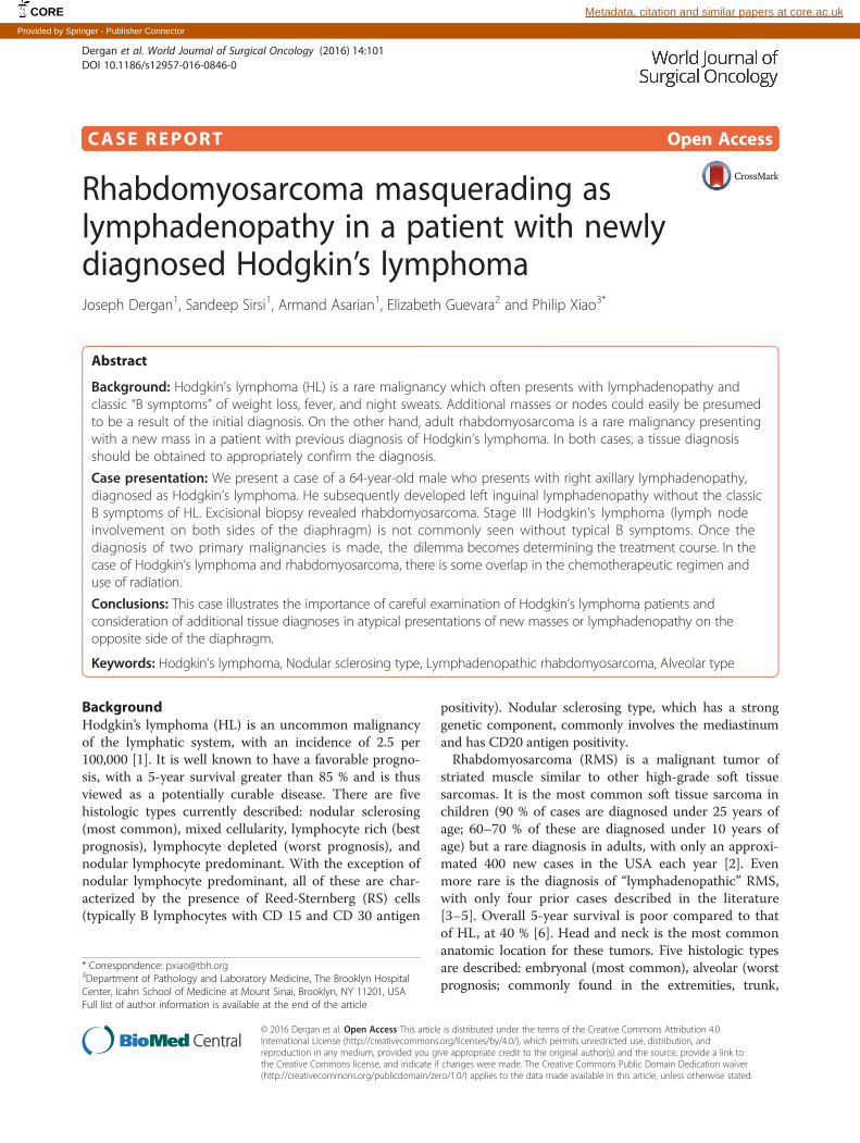
formed concurrently with placement of a left subclavianMediport. Grossly, the excised mass had a similar ap-pearance to the right axillary lymph nodes removed2 months earlier. It was an encapsulated, flesh coloredstructure. Unexpectedly, the microscopic examinationrevealed thin fibrous septae surrounded by small roundblue cells with an alveolar growth pattern. Immunohisto-chemical and molecular studies confirmed the diagnosisof rhabdomyosarcoma, alveolar type (Fig. 3). The patho-logic slides were sent to a tertiary center and reviewedfor a second opinion, and the diagnosis was concurred.The dilemma now faced is this: with two primary neo-plasms, which one should be treated first and whatmodalities of treatment would be most effective? Giventhat this presentation is exceedingly rare, the patient wasreferred to a highly specialized tertiary cancer center inorder to provide the best multidisciplinary approach tothis oncologic dilemma.
Fig. 3 Microscopic examination reveals thin fibrous septae surroundedby small round blue cells with an alveolar growth pattern (HE ×20)
Dergan et al. World Journal of Surgical Oncology (2016) 14:101 Page 3 of 4
DiscussionHodgkin’s lymphoma typically presents as painless lymph-adenopathy. The so called B symptoms, which includeweight loss, fever, and night sweats, may be present andimplicate a poorer prognosis. Due to mediastinal involve-ment, the nodular sclerosing type may present with chestpain, cough, and shortness of breath. Often, a plethora ofdiagnostic modalities aid in the diagnosis of Hodgkin’slymphoma, but ultimately, a tissue diagnosis is needed.This is usually obtained surgically with an excisionallymph node biopsy, as intact morphology is required foran accurate diagnosis. The one exception to this is in headand neck masses, where a fine needle aspiration (FNA)may be pursued in order to rule out squamous cell carcin-oma, which is very common in this anatomic location.Once the diagnosis is made, a clinical stage is deciphered,in order to orchestrate the treatment plan.Hodgkin’s lymphoma can be described in four stages
as follows: stage I (a single lymph node area or singleextranodal site), stage II (two or more lymph node areason the same side of the diaphragm), stage III (lymphnode areas on both sides of the diaphragm), and stageIV (disseminated or multiple involvement of the extra-nodal organs). Four treatments options are availabledepending on the nature of the disease: radiationtherapy, induction chemotherapy, salvage chemotherapy,and hematopoietic stem cell transplantation. An exampleof an induction chemotherapeutic regimen is cyclo-phosphamide, doxorubicin, vincristine, and prednisone(CHOP) [8]. When induction chemotherapy fails, orthe patient experiences relapse, a salvage chemothera-peutic regimen such as ifosfamide, carboplatin, andetoposide (ICE) can be initiated. Of note, HIV testing isobtained in all HL patients, as HAART therapy signifi-cantly improves outcomes in HIV positive patients withHodgkin’s lymphoma [7].
Rhabdomyosarcoma typically presents as a mass or areaof localized swelling, and less than half of these patientspresent with pain. However, symptoms can be specific tothe anatomic location of the lesion (e.g., cranial nerve com-pression in head/neck tumors invading the skull base).Diagnosis is made with a surgical biopsy (usually an inci-sional biopsy). CT and MRI are helpful for planning surgicalresection, as well as planning routes and doses for potentialradiation therapy post operatively. Locoregional and meta-static disease can be investigated with a bone marrowbiopsy, chest CT scan, technetium diphosphonate bone scan,and even a lumbar puncture (for parameningeal primaries).Mainstay therapy for rhabdomyosarcoma is surgical resec-tion. High-risk histology and advanced stage may warrantchemotherapy with or without radiation. For example, non-metastatic alveolar rhabdomyosarcoma is usually treatedwith the vincristine, dactinomycin, and cyclophosphamide(VAC) protocol with the addition of radiation therapy [9].Specifically relevant to this case, our patient initially
presented with two groups of enlarged lymph nodes onthe same side of the diaphragm and did not present withB symptoms. He subsequently developed involvement ofa new group of lymph nodes on the other side of thediaphragm (left inguinal) without development of Bsymptoms. This was a rare presentation which raised thesuspicion of the oncologist, prompting a second excisionalbiopsy. Specific to this case of Hodgkin’s lymphoma withsynchronous rhabdomyosarcoma (alveolar type), overlap-ping treatment is available, thus making treatment optionsless cumbersome. Margin negative (R0) resection is thegoal for the rhabdomyosarcoma. Given its aggressivehistology and the possibility of incomplete resection, che-moradiation will likely be pursued. The CHOP inductionchemotherapeutic regimen for Hodgkin’s lymphoma hassome degree of overlap with the VAC protocol for rhabdo-myosarcoma (vincristine being used for both). The chal-lenge for the chemotherapist is the dose frequencies andpotential toxicities that these patients may experience.
ConclusionsPatients with Hodgkin’s lymphoma that present with asecond primary tumor usually do so as a complication ofchemoradiation. Cancer screening is conducted regularlyin HL patients receiving treatment. However, presentingwith Hodgkin’s lymphoma and a second primary tumorsimultaneously is exceedingly rare and poorly described inthe literature. This can be a challenging diagnosis to make,and clinicians must be aware that such entities exist.When synchronous masses are present, or a new mass isdiscovered in a patient with Hodgkin’s lymphoma prior totreatment, one must be careful not to assume that theyare the same entity. When in doubt, a tissue diagnosisshould be pursued.
Dergan et al. World Journal of Surgical Oncology (2016) 14:101 Page 4 of 4
Ethics approval and consent to participateNot applicable.
Consent for publicationConsent was obtained from the patient for publicationof relevant medical information.
Availability of data and materialsNot applicable.
AbbreviationsCHOP: cyclophosphamide, doxorubicin, vincristine, and prednisone;HL: Hodgkin’s lymphoma; RMS: rhabdomyosarcoma; VAC: vincristine,dactinomycin, and cyclophosphamide.
Competing interestsThe authors declare that they have no competing interests.
Authors’ contributionsJD, PX, and SS designed the research. JD, AA, and EG analyzed the data. JDand PX wrote the paper. All authors read and approved the final manuscript.
AcknowledgementsThe authors acknowledge the editing and formatting support provided byArpit Patel, MD.
FundingNone applicable.
Author details1Department of Surgery, The Brooklyn Hospital Center, Icahn School ofMedicine at Mount Sinai, Brooklyn, NY 11201, USA. 2Department ofHematology/Oncology, The Brooklyn Hospital Center, Icahn School ofMedicine at Mount Sinai, Brooklyn, NY 11201, USA. 3Department ofPathology and Laboratory Medicine, The Brooklyn Hospital Center, IcahnSchool of Medicine at Mount Sinai, Brooklyn, NY 11201, USA.
Received: 9 December 2015 Accepted: 24 March 2016
References1. Surveillance, Epidemiology, and End Results (SEER) Program. (http://seer.
cancer.gov/statfacts/html/hodg.html) Research data (1973-2012), NationalCancer Institute, DCCPS, Surveillance Research Program, SurveillanceSystems Branch. April 2015, based on the November 2014 submission.
2. Hawkins WG, Hoos A, Antonescu CR, Urist MJ, Leung DH, Gold JS, et al.Clinicopathologic analysis of patients with adult rhabdomyosarcoma.Cancer. 2001;91(4):794–803.
3. Sharma T, Bhargava R, Sharma J, Sharma SP. Lymphadenopathic form ofsolid variant of alveolar rhabdomyosarcoma: a rare case report. J Cytol.2014;31(3):168–70.
4. Mekni A, Bouraoui S, Boussen H, el May A, Kchir N. Lymphadenopathic formof alveolar rhabdomyosarcoma: a case report. Tunis Med. 2004;82(2):241–4.
5. Ganesan P, Thulkar S, Rajan A, Bakhshi S. Solid variant of alveolarrhabdomyosarcoma mimicking non-Hodgkin lymphoma: case report andreview of literature. J Pediatr Hematol Oncol. 2008;30(10):772–4.
6. Ferrari A, Dileo P, Casanova M, Bertulli R, Meazza C, Gandola L, et al.Rhabdomyosarcoma in adults. A retrospective analysis of 171 patientstreated at a single institution. Cancer. 2003;98(3):571–80.
7. Newton Jr WA, Gehan EA, Webber BL, Marsden HB, van Unnik AJ, HamoudiAB, et al. Classification of rhabdomyosarcomas and related sarcomas.Pathologic aspects and proposal for a new classification—an IntergroupRhabdomyosarcoma Study. Cancer. 1995;70:1073–85.
8. National Comprehensive Cancer Network (NCCN). NCCN clinical practiceguidelines in oncology. Hodgkin Lymphoma Version 2.2015.Fort Washington, PA: National Comprehensive Cancer Network; 2015.
9. National Comprehensive Cancer Network (NCCN). NCCN clinicalpractice guidelines in oncology. Soft Tissue Sarcoma Version 1.2015.Fort Washington, PA: National Comprehensive Cancer Network; 2015.
• We accept pre-submission inquiries
• Our selector tool helps you to find the most relevant journal
• We provide round the clock customer support
• Convenient online submission
• Thorough peer review
• Inclusion in PubMed and all major indexing services
• Maximum visibility for your research
Submit your manuscript atwww.biomedcentral.com/submit
Submit your next manuscript to BioMed Central and we will help you at every step: