LYMPHADENOPATHY - APPROACH
47
Approach To Lymphadenopathy By Dr. Mahmoud Gaber
Transcript of LYMPHADENOPATHY - APPROACH

Approach To
Lymphadenopathy
By
Dr. Mahmoud Gaber

Introduction
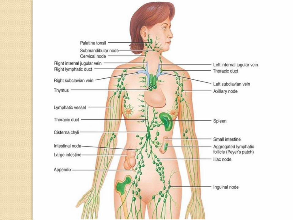
The lymphatic system is the part of the immune system comprising
lymphatic vessels that carry a clear fluid called lymph (from Latin lympha
"water") in a unidirectional pathway.
The components of the lymphatic system are :-
I. Lymph, the recovered fluid
II. Lymphatic vessels, which transport the lymph
III. Lymphatic tissue, composed of aggregates of lymphocytes and
macrophages that populate many organs of the body; and
IV. Lymphatic organs, in which these cells are especially concentrated
and which are set off from surrounding organs by connective tissue
capsules
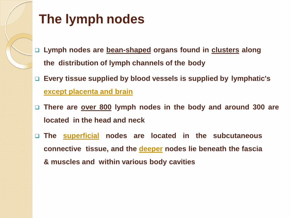
The lymph nodes
Lymph nodes are bean-shaped organs found in clusters along
the distribution of lymph channels of the body
Every tissue supplied by blood vessels is supplied by lymphatic's
except placenta and brain
There are over 800 lymph nodes in the body and around 300 are
located in the head and neck
The superficial nodes are located in the subcutaneous
connective tissue, and the deeper nodes lie beneath the fascia
& muscles and within various body cavities

Definition
Lymphadenopathy: refers to lymph nodes that are abnormal in:
Size
Consistency
Whether as a result of normal reactive process or pathology
(Abnormalities may be localized or generalized)
--------------------------------------------------------------------------------------------------------------------------------------------------------------------------
Generalized lymphadenopathy is defined as: -
Enlargement of ≥ 2 non-contiguous lymph node groups
Regional (localized) lymphadenopathy If :
Enlargement of a single node or multiple contiguous
nodal regions
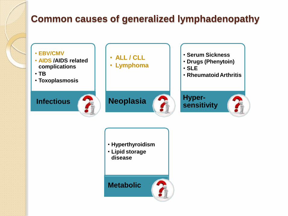
Common causes of generalized lymphadenopathy
• EBV/CMV
• AIDS /AIDS related complications
• TB
• Toxoplasmosis
Infectious
• ALL / CLL
• Lymphoma
Neoplasia
• Serum Sickness
• Drugs (Phenytoin)
• SLE
• RheumatoidArthritis
Hyper-sensitivity
• Hyperthyroidism
• Lipid storage disease
Metabolic
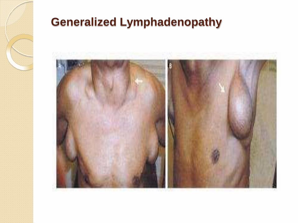
Generalized Lymphadenopathy
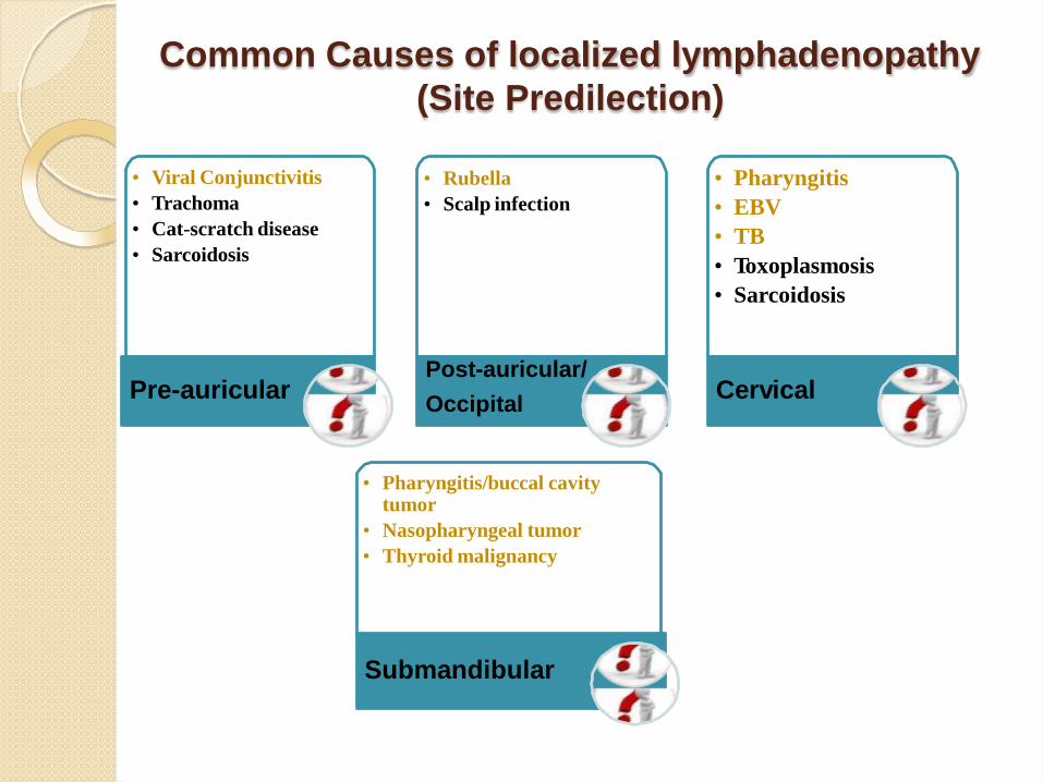
Common Causes of localized lymphadenopathy
(Site Predilection)
• Viral Conjunctivitis
• Trachoma
• Cat-scratch disease
• Sarcoidosis
Pre-auricular
• Rubella
• Scalp infection
Post-auricular/
Occipital
• Pharyngitis
• EBV
• TB
• Toxoplasmosis
• Sarcoidosis
Cervical
• Pharyngitis/buccal cavity tumor
• Nasopharyngeal tumor
• Thyroid malignancy
Submandibular
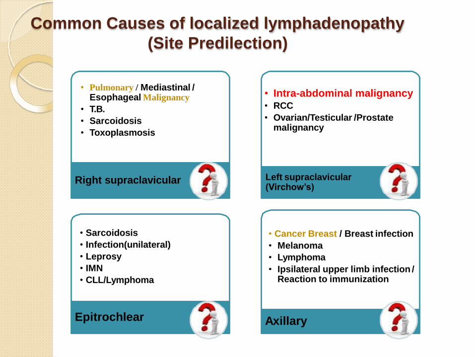
Common Causes of localized lymphadenopathy
(Site Predilection)
• Pulmonary / Mediastinal / Esophageal Malignancy
• T.B.
• Sarcoidosis
• Toxoplasmosis
Right supraclavicular
• Intra-abdominal malignancy• RCC
• Ovarian/Testicular /Prostate malignancy
Left supraclavicular (Virchow’s)
• Cancer Breast / Breast infection
• Melanoma
• Lymphoma
• Ipsilateral upper limb infection / Reaction to immunization
Axillary
• Sarcoidosis
• Infection(unilateral)
• Leprosy
• IMN
• CLL/Lymphoma
Epitrochlear

Common Causes of localized lymphadenopathy
(Site Predilection)
•Syphilis•Genital herpes
• Lower extremity/local infection
• Lymphoma
• Metastatic carcinoma from: rectum, genitalia or lower limb(melanoma)
Inguinal
• Lymphoma
• Bronchogenic Carcinoma
• T.B.
• Sarcoidosis
• Histiocytosis
Hilar
• GutAdenocarcinoma
• Hodgkin’s disease
• T.B.
• Lymphoma
• Bladder carcinoma
Abdominal
• Lymphomas
• Leukemias
• Cat-Scratch disease
• Metastasis
• Sarcoidosis
• Granulomas
Any region
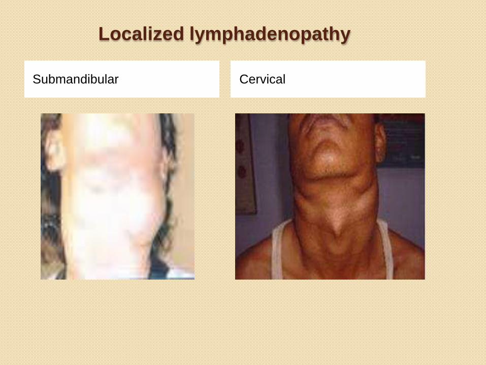
Localized lymphadenopathy
Submandibular Cervical

Localized lymphadenopathy
Right Supraclavicular Left Supraclavicular
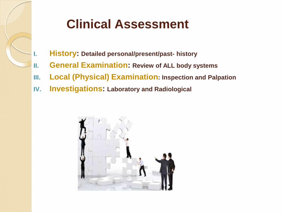
Clinical Assessment
I. History: Detailed personal/present/past- history
II. General Examination: Review of ALL body systems
III. Local (Physical) Examination: Inspection and Palpation
IV. Investigations: Laboratory and Radiological
History
Duration:
for < 2 weeks has a very low chance of representing a
malignant condition
for > 1 year and has been stable in size over the year,
less likely to be malignant (with exception of indolent
NHL and low-grade Hodgkin lymphomas)
The Exposure history as well as the Travel history may be
important for diagnosis:
Exposure to Animals/Pets and biting insects
Cat-scratch disease (bartonella)
Exposure to infectious contacts
Consuming Undercooked meat for possible Toxoplasmosis
Environmental exposure such as tobacco, alcohol, and
ultraviolet radiation may raise suspicion for metastatic
carcinoma of the internal organs, cancers of the head and
neck, and skin malignancies
Occupational exposure to silicon or beryllium
Sexual history is also important in determining potential sexually
transmitted causes of inguinal and cervical lymphadenopathy;
as: HIV, Syphilis, HBV, HSV, CMV
IV- Drug Users: for possible HIV, HBV
Drug history:
• Hydralazine• Phenytoin• Allopurinol• Atenolol
Blood Transfusion or recent transplant history: for possible
infections as CMV and HIV
• Carbamazepine• Cephalosporins• Quinidine• Sulfonamides
History

History and Examination
Constitutional symptoms such as: fever (nocturnal fever or Pel-Ebstein fever),
malaise, fatigue, cachexia, unexplained loss of weight (>10% of body eight)
and anorexia
Presence of non-pitting edema with inguinal LNs may suggest filariasis
Arthralgia, muscle weakness, unusual rashes may indicate possibility of
autoimmune diseases
compression symptoms as dyspnea & dysphagia due to pressure on
trachea or esophagus by the enlarged lymph nodes
Coexistence of splenomegaly implies a systemic disorders or a
hematological disorder as:
(IMN, Lymphoma, acute or chronic leukemia, SLE, sarcoidosis,
Toxoplasmosis, or cat-scratch disease)

RED FLAGS IN LYMPHADENOPATHY
1. Fever, night sweats, and unexplained weight loss
2. A supraclavicular node
3. Hard and tender L.N. with a significant size or draining an area with
a significant pathology
4. Matted or Fixed node(s)
5. Non-recessive node after 3 weeks period or after disappearance of
fever
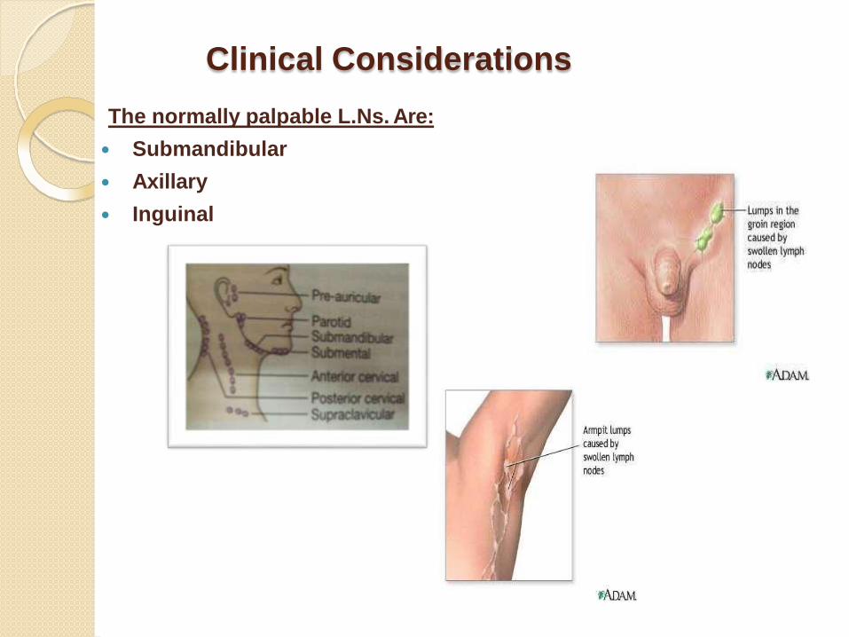
Clinical Considerations
The normally palpable L.Ns. Are:
Submandibular
Axillary
Inguinal
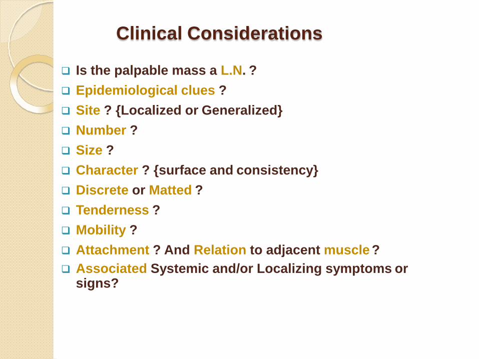
Clinical Considerations
Is the palpable mass a L.N. ?
Epidemiological clues ?
Site ? {Localized or Generalized}
Number ?
Size ?
Character ? {surface and consistency}
Discrete or Matted ?
Tenderness ?
Mobility ?
Attachment ? And Relation to adjacent muscle ?
Associated Systemic and/or Localizing symptoms or signs?

Local Examination
Mapping Examination
The physical examination
should be regionally directed
by knowledge of the lymphatic
drainage patterns
All the normal anatomic sites
should be inspected for any
obvious enlargements.
When lymphadenopathy is
localized, the clinician should
examine the region drained by
the nodes for evidence of
infection, lesions or tumors
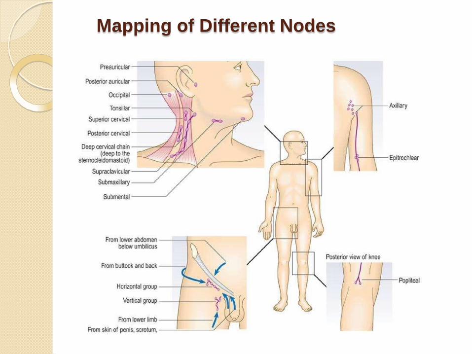
Mapping of Different Nodes
Node Palpation
*** Confirm that the palpable mass is indeed a L.N..
{ NOT something else as: Thyroglossal cyst, Abscess, Branchial cyst,
Enlarged parotid, salivary gland ..}
Exposure of the patient:
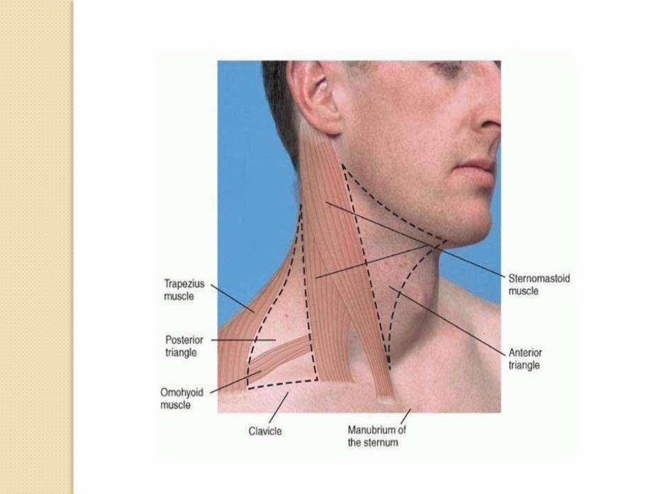
Cervical: whole head and neck to clavicles
Axillary: uncover to the waist
Inguinal: umbilicus to knee
Before performing palpation, ask the patient to identify painful
areas so that you can examine those areas last
During the procedure, pay attention to their facial expression to
assess for sign of discomfort
Technique: Use the pads of the index and middle finger to move
the skin in circular motions over the underlying tissues in each area

Axillary Node Palpation
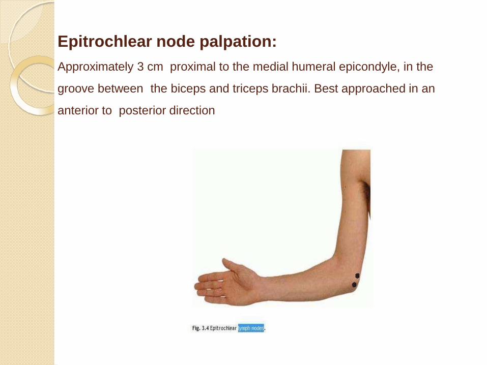
Epitrochlear node palpation:
Approximately 3 cm proximal to the medial humeral epicondyle, in the
groove between the biceps and triceps brachii. Best approached in an
anterior to posterior direction
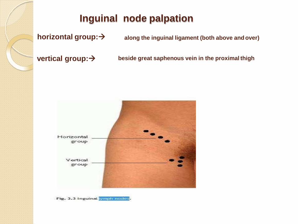
Inguinal node palpation
horizontal group: along the inguinal ligament (both above and over)
vertical group: beside great saphenous vein in the proximal thigh

Local examinationYou Have To Answer The Previous Questions of Clinical
Considerations ..
►►► Note for:
Number: (single or multiple), (localized or generalized)
Site: Anatomic location can narrow the D.D.
T.B. and Hodgkin’s ----- > cervical (earlier stages)
Cat-scratch disease ----- > cervical and axillary
IMN --- > cervical
Sexually-transmitted diseases ----- > Inguinal
Supraclavicular ----- > Highest risk of malignancy(90% in old patients)
Size (up to 1 cm is considered normal).. Except epitrochlear
:if >0.5cm
N.B.=The size is usually of little importance in adding information toestablish diagnosis; however increase in size on serial examination may be of value..
Local examination
Surface and Consistency (Soft, hard, firm, rubbery, fluctuant, or
variable)
Stony-hard nodes are typically a sign of cancer, usually metastatic
Firm, rubbery nodes suggest lymphoma
Softer nodes are the result of infections or inflammatory conditions
Suppurative nodes may be fluctuant
Discrete or Matted (T.B., Sarcoidosis) or amalgamated (metastatic carcinoma
or lymphomas)
Painless or Painful (when a lymph node increases in size its capsule
stretches and causes pain, or when there is hemorrhage into the necrotic
center of a malignant node)
N.B.=The presence or absence of tenderness does not necessarily differentiate benign from
malignant nodes..
Local examination
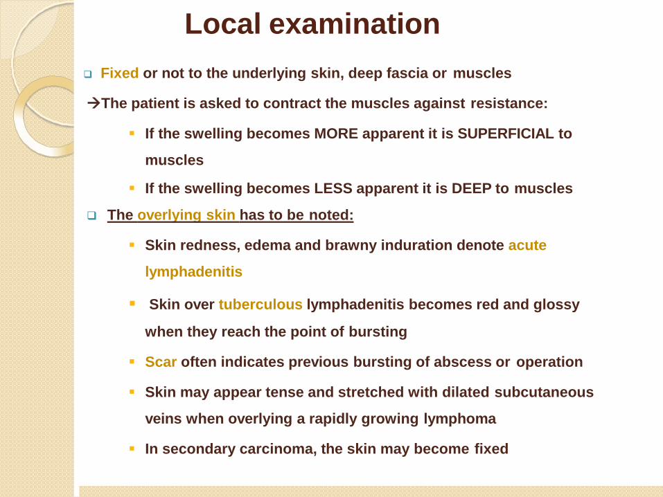
Fixed or not to the underlying skin, deep fascia or muscles
The patient is asked to contract the muscles against resistance:
If the swelling becomes MORE apparent it is SUPERFICIAL to
muscles
If the swelling becomes LESS apparent it is DEEP to muscles
The overlying skin has to be noted:
Skin redness, edema and brawny induration denote acute
lymphadenitis
Skin over tuberculous lymphadenitis becomes red and glossy
when they reach the point of bursting
Scar often indicates previous bursting of abscess or operation
Skin may appear tense and stretched with dilated subcutaneous
veins when overlying a rapidly growing lymphoma
In secondary carcinoma, the skin may become fixed

Investigations
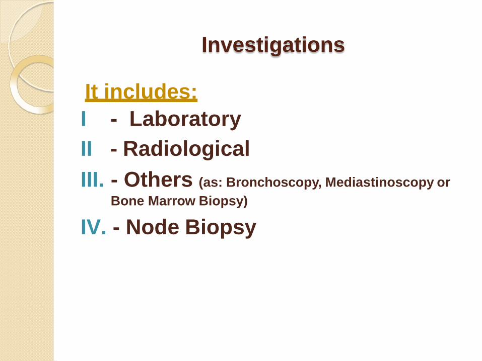
Investigations
It includes:
I - Laboratory
II - Radiological
III. - Others (as: Bronchoscopy, Mediastinoscopy or
Bone Marrow Biopsy)
IV. - Node Biopsy

Investigations
I - Laboratory:
CBC with differential count : provides useful data for the diagnosis of:
Acute or Chronic leukemia's
EBV or CMV mononucleosis(atypical lymphocytosis)
Pyogenic infections
Lymphoma with a leukemic component
Immune cytopenias (in illnesses such as SLE)
ESR
Serology:may demonstrate:
Antibodies specific to: components of EBV(viral Capsid Ag), CMV, HIV, Toxoplasma, Brucella, etc
PCR-for: CMV-DNA, T.B.
ANA/Anti-ds DNA antibody (SLE)
Others: In cases of hilar LAD, do:
Serum ACE
Tuberculin T.
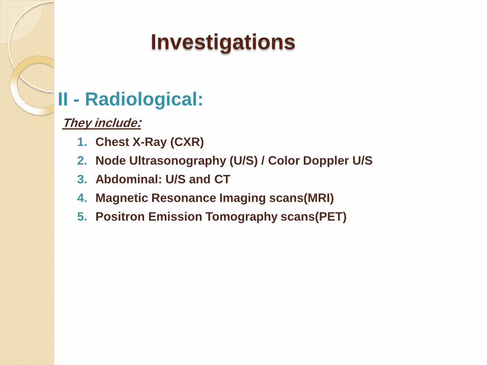
Investigations
II - Radiological:They include:
1. Chest X-Ray (CXR)
2. Node Ultrasonography (U/S) / Color Doppler U/S
3. Abdominal: U/S and CT
4. Magnetic Resonance Imaging scans(MRI)
5. Positron Emission Tomography scans(PET)
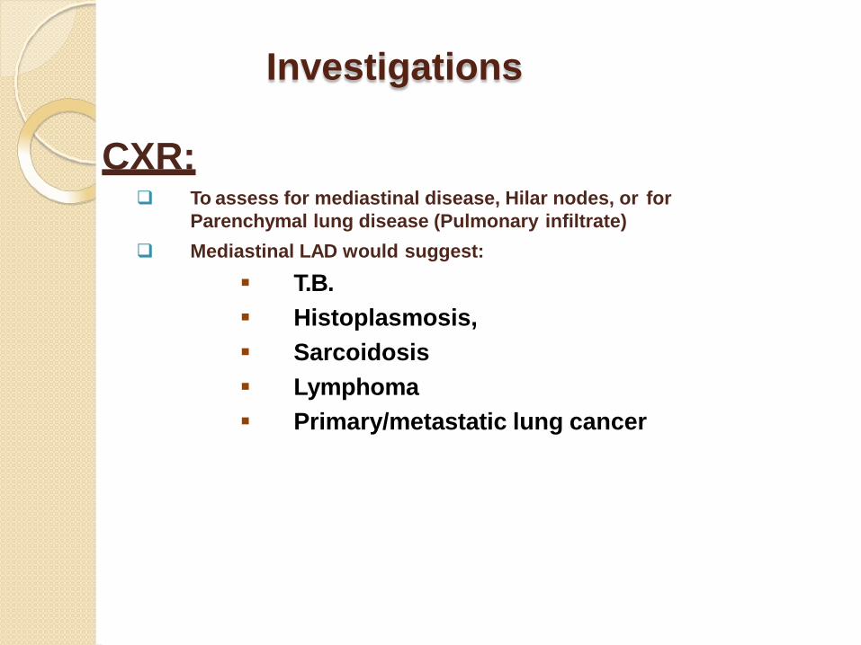
Investigations
CXR: To assess for mediastinal disease, Hilar nodes, or for
Parenchymal lung disease (Pulmonary infiltrate)
Mediastinal LAD would suggest:
T.B.
Histoplasmosis,
Sarcoidosis
Lymphoma
Primary/metastatic lung cancer
Investigations
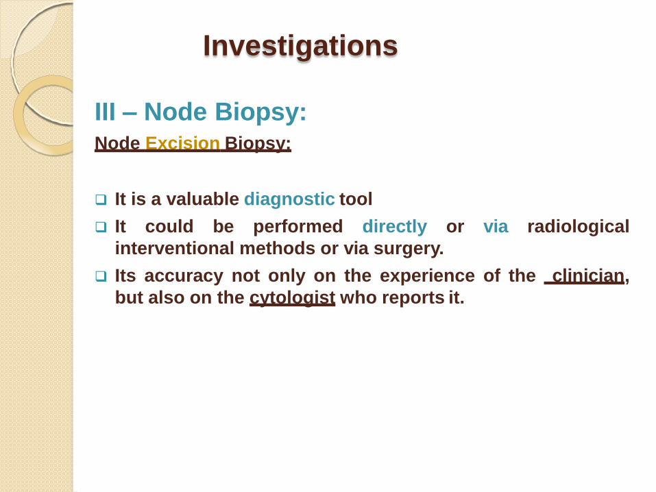
III – Node Biopsy:Node Excision Biopsy:
It is a valuable diagnostic tool
It could be performed directly or via radiological
interventional methods or via surgery.
Its accuracy not only on the experience of the clinician,
but also on the cytologist who reports it.
Self – Assessment Clinical Cases
ONE
Question
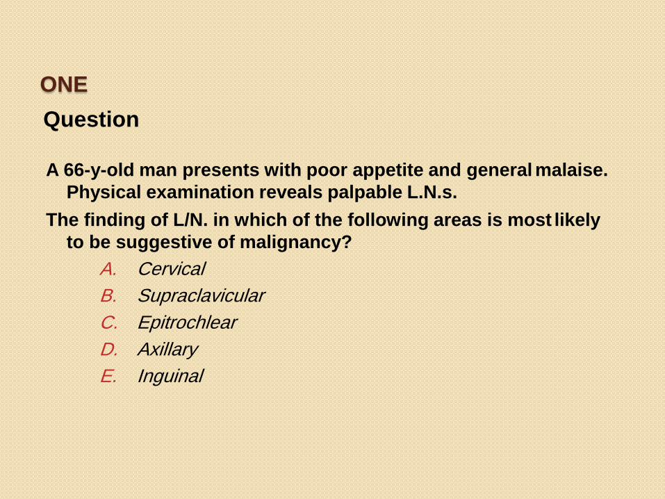
A 66-y-old man presents with poor appetite and general malaise.
Physical examination reveals palpable L.N.s.
The finding of L/N. in which of the following areas is most likely
to be suggestive of malignancy?
A. Cervical
B. Supraclavicular
C. Epitrochlear
D. Axillary
E. Inguinal

ONE
ANSWER
A 66-y-old man presents with poor appetite and general malaise.
Physical examination reveals palpable L.N.s.
The finding of L/N. in which of the following areas is most likely
to be suggestive of malignancy?
A. Cervical
B. Supraclavicular
C. Epitrochlear
D. Axillary
E. Inguinal

TWO
QUESTION
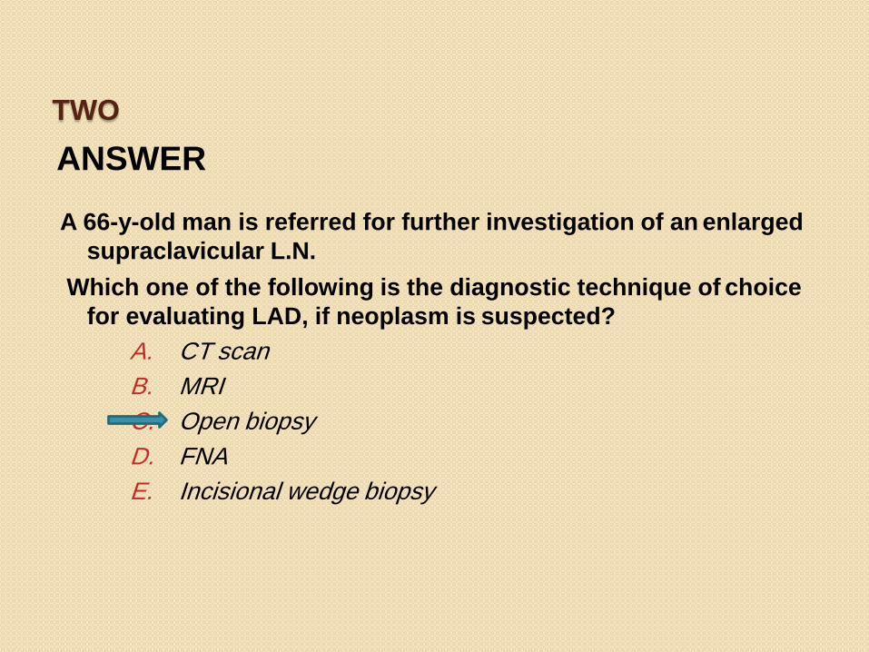
A 66-y-old man is referred for further investigation of an enlarged
supraclavicular L.N.
Which one of the following is the diagnostic technique of choice
for evaluating LAD, if neoplasm is suspected?
A. CT scan
B. MRI
C. Open biopsy
D. FNA
E. Incisional wedge biopsy
TWO
ANSWER
A 66-y-old man is referred for further investigation of an enlarged
supraclavicular L.N.
Which one of the following is the diagnostic technique of choice
for evaluating LAD, if neoplasm is suspected?
A. CT scan
B. MRI
C. Open biopsy
D. FNA
E. Incisional wedge biopsy
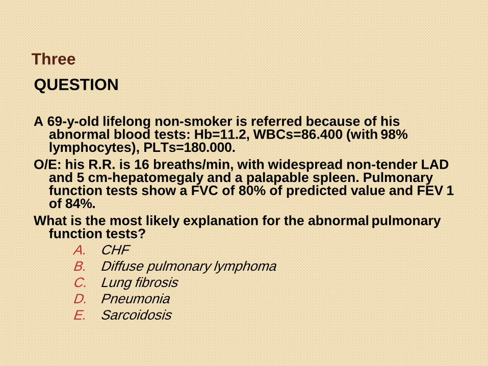
Three
QUESTION
A 69-y-old lifelong non-smoker is referred because of his abnormal blood tests: Hb=11.2, WBCs=86.400 (with 98% lymphocytes), PLTs=180.000.
O/E: his R.R. is 16 breaths/min, with widespread non-tender LAD and 5 cm-hepatomegaly and a palapable spleen. Pulmonary function tests show a FVC of 80% of predicted value and FEV 1 of 84%.
What is the most likely explanation for the abnormal pulmonary function tests?
A. CHFB. Diffuse pulmonary lymphomaC. Lung fibrosisD. PneumoniaE. Sarcoidosis
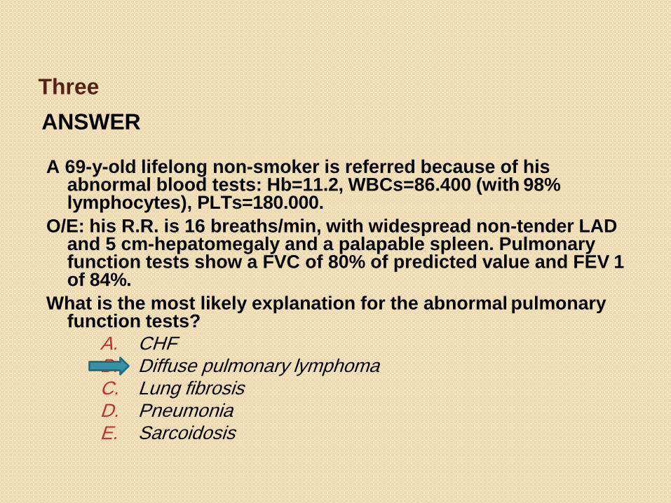
Three
ANSWER
A 69-y-old lifelong non-smoker is referred because of his abnormal blood tests: Hb=11.2, WBCs=86.400 (with 98% lymphocytes), PLTs=180.000.
O/E: his R.R. is 16 breaths/min, with widespread non-tender LAD and 5 cm-hepatomegaly and a palapable spleen. Pulmonary function tests show a FVC of 80% of predicted value and FEV 1 of 84%.
What is the most likely explanation for the abnormal pulmonary function tests?
A. CHFB. Diffuse pulmonary lymphomaC. Lung fibrosisD. PneumoniaE. Sarcoidosis
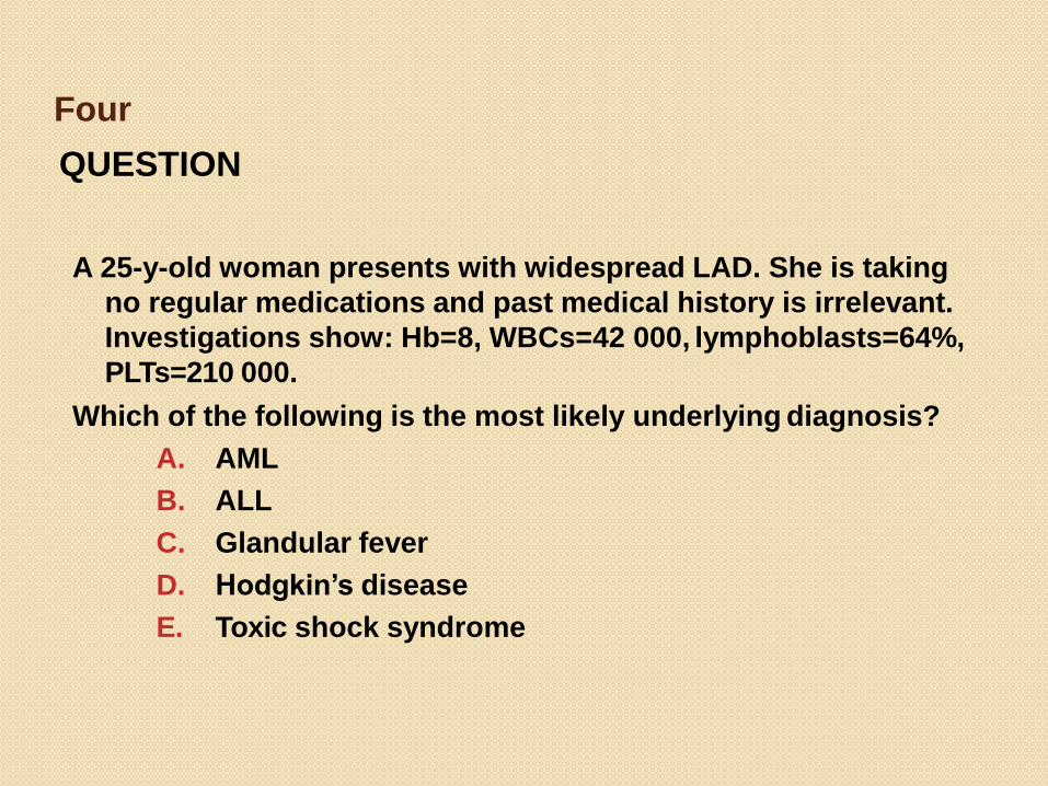
Four
QUESTION
A 25-y-old woman presents with widespread LAD. She is taking
no regular medications and past medical history is irrelevant.
Investigations show: Hb=8, WBCs=42 000, lymphoblasts=64%,
PLTs=210 000.
Which of the following is the most likely underlying diagnosis?
A. AML
B. ALL
C. Glandular fever
D. Hodgkin’s disease
E. Toxic shock syndrome
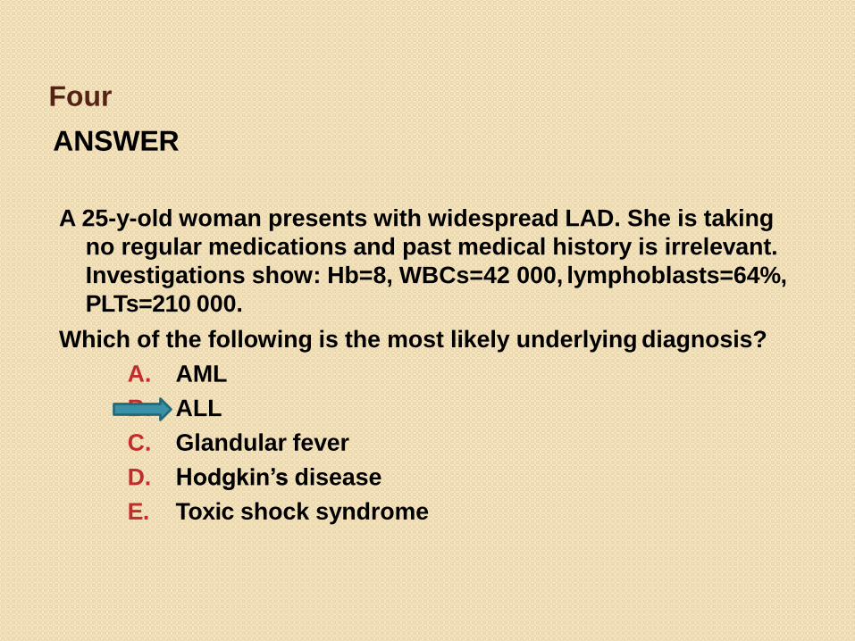
Four
ANSWER
A 25-y-old woman presents with widespread LAD. She is taking
no regular medications and past medical history is irrelevant.
Investigations show: Hb=8, WBCs=42 000, lymphoblasts=64%,
PLTs=210 000.
Which of the following is the most likely underlying diagnosis?
A. AML
B. ALL
C. Glandular fever
D. Hodgkin’s disease
E. Toxic shock syndrome
Five
QUESTION
A 24-y-old man has noted for the last 2 Ms that his face is swollen in the morning. He has lost 10-Kg in weight over 6-Ms. He has no other complaints.
O/E: The ext. jugular veins are dilated. CXR: shows a mediastinal mass.
Which one of the following is the most likely diagnosis of his SVC obstruction?
A. Adenocarcinoma of the lung
B. Hodgkin’s disease
C. Sarcoidosis
D. Seminoma
E. Tuberculosis
Five
ANSWER
A 24-y-old man has noted for the last 2 Ms that his face is swollen in the morning. He has lost 10-Kg in weight over 6-Ms. He has no other complaints.
O/E: The ext. jugular veins are dilated. CXR: shows a mediastinal mass.
Which one of the following is the most likely diagnosis of his SVC obstruction?
A. Adenocarcinoma of the lung
B. Hodgkin’s disease
C. Sarcoidosis
D. Seminoma
E. Tuberculosis