Ovarian embryonal rhabdomyosarcoma is a rare manifestation ... · Ovarian embryonal...
16
1 Ovarian embryonal rhabdomyosarcoma is a rare manifestation of the DICER1 syndrome AUTHORS: Leanne de Kock 1,2 , Harriet Druker 3 , Evan Weber 4 , Nancy Hamel 4 , Jeffrey Traubici 5 , David Malkin 6 , Jocelyne Arseneau 7 , Dorothée Bouron-Dal Soglio 8 , John R. Priest 9 , William D. Foulkes 1, 2, 4,10 AFFILIATIONS: 1 Department of Human Genetics, McGill University, Montreal, QC, Canada; 2 Lady Davis Institute, Segal Cancer Centre, Jewish General Hospital, Montreal, QC, Canada; 3 Division of Haematology/Oncology, The Hospital for Sick Children, Department of Molecular and Medical Genetics, The University of Toronto, Toronto, ON, Canada; 4 Department of Medical Genetics, Research Institute of the McGill University Health Centre, Montreal, QC, Canada; 5 Department of Diagnostic Imaging, Hospital for Sick Children, Toronto, ON, Canada; 6 Division of Haematology/Oncology, The Hospital for Sick Children, Departments of Pediatrics and Medical Biophysics, The University of Toronto, Toronto, ON, Canada; Department of Paediatrics, University of Toronto, Toronto, ON, Canada; 7 Department of Pathology, McGill University, Montreal, QC, Canada; 8 Department of Pathology, CHU-Sainte Justine and University of Montreal, Montréal, Quebec, Canada; 9 Minneapolis, Minnesota, USA; 10 Program in Cancer Genetics, Department of Oncology and Human Genetics, McGill University, Montreal, QC, Canada. Short Running Title: DICER1 mutations in an embryonal rhabdomyosarcoma of the ovary Correspondence to: Dr. William D. Foulkes at the Department of Medical Genetics, The Lady Davis Institute, Segal Cancer Centre, Jewish General Hospital, 3755 Cote St. Catherine Road, Montreal, QC, Canada, H3T 1E2. Email: [email protected]. Tel: (+1) 514-340-8222 Ext. 3213. All rights reserved. No reuse allowed without permission. (which was not peer-reviewed) is the author/funder, who has granted bioRxiv a license to display the preprint in perpetuity. The copyright holder for this preprint . http://dx.doi.org/10.1101/011304 doi: bioRxiv preprint first posted online Nov. 10, 2014;
Transcript of Ovarian embryonal rhabdomyosarcoma is a rare manifestation ... · Ovarian embryonal...
1
Ovarian embryonal rhabdomyosarcoma is a rare manifestation of the DICER1 syndrome
AUTHORS:
Leanne de Kock1,2
, Harriet Druker3, Evan Weber
4, Nancy Hamel
4, Jeffrey Traubici
5, David
Malkin6, Jocelyne Arseneau
7, Dorothée Bouron-Dal Soglio
8, John R. Priest
9, William D. Foulkes
1, 2,
4,10
AFFILIATIONS:
1 Department of Human Genetics, McGill University, Montreal, QC, Canada; 2 Lady Davis Institute,
Segal Cancer Centre, Jewish General Hospital, Montreal, QC, Canada; 3 Division of
Haematology/Oncology, The Hospital for Sick Children, Department of Molecular and Medical Genetics,
The University of Toronto, Toronto, ON, Canada; 4 Department of Medical Genetics, Research Institute
of the McGill University Health Centre, Montreal, QC, Canada; 5 Department of Diagnostic Imaging,
Hospital for Sick Children, Toronto, ON, Canada; 6 Division of Haematology/Oncology, The Hospital for
Sick Children, Departments of Pediatrics and Medical Biophysics, The University of Toronto, Toronto,
ON, Canada; Department of Paediatrics, University of Toronto, Toronto, ON, Canada; 7 Department of
Pathology, McGill University, Montreal, QC, Canada; 8 Department of Pathology, CHU-Sainte Justine
and University of Montreal, Montréal, Quebec, Canada; 9 Minneapolis, Minnesota, USA; 10 Program in
Cancer Genetics, Department of Oncology and Human Genetics, McGill University, Montreal, QC,
Canada.
Short Running Title: DICER1 mutations in an embryonal rhabdomyosarcoma of the ovary
Correspondence to: Dr. William D. Foulkes at the Department of Medical Genetics, The Lady Davis
Institute, Segal Cancer Centre, Jewish General Hospital, 3755 Cote St. Catherine Road, Montreal, QC,
Canada, H3T 1E2. Email: [email protected]. Tel: (+1) 514-340-8222 Ext. 3213.
All rights reserved. No reuse allowed without permission. (which was not peer-reviewed) is the author/funder, who has granted bioRxiv a license to display the preprint in perpetuity.
The copyright holder for this preprint. http://dx.doi.org/10.1101/011304doi: bioRxiv preprint first posted online Nov. 10, 2014;
2
Acknowledgements: We thank the family involved in this research for their consent to participation and
the clinicians for referring the case and providing samples. We also thank Dr. Steffen Albrecht for
assistance with the figures. This research was made possible thanks to the support of the Alex’s
Lemonade Stand Foundation grant to Dr. William D. Foulkes, and the CIHR/FRSQ training grant in
cancer research FRN53888 of the McGill Integrated Cancer Research Training Program and the McGill
Faculty of Medicine Internal Studentship Award (Awarded as a Ruth and Alex Dworkin Fellowship and a
Graduate Excellence Fellowship) to Leanne de Kock.
Conflicts of Interest: The authors have no conflicts of interest to disclose.
All rights reserved. No reuse allowed without permission. (which was not peer-reviewed) is the author/funder, who has granted bioRxiv a license to display the preprint in perpetuity.
The copyright holder for this preprint. http://dx.doi.org/10.1101/011304doi: bioRxiv preprint first posted online Nov. 10, 2014;
3
Ovarian embryonal rhabdomyosarcoma is a rare manifestation of the DICER1 syndrome
ABSTRACT
Embryonal rhabdomyosarcoma (ERMS), a malignant soft tissue sarcoma, is one of the most common
paediatric cancers. Certain ERMS tumours are associated with the DICER1 syndrome, a distinctive
tumour predisposition syndrome caused by germ-line mutations in the microRNA-maturation pathway
gene, DICER1. In addition to germ-line DICER1 mutations, highly characteristic somatic mutations have
been identified in several DICER1-associated tumour types. These so-called “hotspot” mutations affect
highly conserved amino acid residues central to the catalytic activity of the DICER1 ribonuclease IIIb
domain.
Primary ovarian ERMS (oERMS) is extremely rare. We present a case of a 6-year-old girl with an
oERMS found to harbour two mutations in DICER1. In addition to the oERMS, the girl also exhibited
other DICER1 phenotypes, including cystic nephroma (CN) and multinodular goitre. Somatic
investigations of the CN revealed the presence of a hotspot DICER1 mutation different from that in the
oERMS. Of particular interest is the CN presented at the age of 12 years, which is much older than
previously reported age range of susceptibility (birth to four years of age).
This report documents both germ-line and highly characteristic somatic DICER1 mutations in a case of
oERMS, adding to the expanding spectrum of rare childhood tumours in the DICER1 syndrome.
Key Words: DICER1, ovary, embryonal rhabdomyosarcoma, miRNA, pediatric tumours
All rights reserved. No reuse allowed without permission. (which was not peer-reviewed) is the author/funder, who has granted bioRxiv a license to display the preprint in perpetuity.
The copyright holder for this preprint. http://dx.doi.org/10.1101/011304doi: bioRxiv preprint first posted online Nov. 10, 2014;
4
Ovarian embryonal rhabdomyosarcoma is a rare manifestation of the DICER1 syndrome
Introduction
Embryonal rhabdomyosarcoma (ERMS) is a malignant tumour of mesenchymal origin and one of the
most common extra-cranial solid tumours of childhood. ERMS tends to affect children within the first
decade of life and the more common sites are orbit, nasopharynx, vagina and bladder. The majority of
ERMS arise sporadically, but associations between ERMS and inherited familial syndromes, such as the
Li-Fraumeni syndrome, neurofibromatosis and Beckwith-Weidemann syndrome suggest that a genetic
predisposition contributes to some cases1-5
.
Recently it has become evident that some ERMS are associated with germ-line DICER1 mutations. In
general, DICER1 mutations cause a distinctive tumour predisposition syndrome, the DICER1 syndrome,
(OMIM # 601200) which is characterized by pleuropulmonary blastoma (PPB) and other rare childhood
neoplasms6. PPB is a mixed pattern sarcoma in which ERMS is a predominant histologic subtype
7.
DICER1 mutations are strongly associated with ERMS of the uterine cervix (cERMS), a generally
uncommon site for ERMS, which occurs from the age of approximately 10-20 years5, 8, 9
. Both germ-line
and highly characteristic somatic “hotspot” DICER1 mutations have been reported in two cERMS8, 10
and
bladder ERMS has been observed in two DICER1 kindred7, 11
. DICER1 mutations were identified in two
of 52 sporadic ERMS cases7. No associations are evident between DICER1 mutations and alveolar
rhabdomyosarcoma (ARMS), a rhabdomyosarcoma subtype tending to affect older children and believed
to arise through biological mechanisms distinct from ERMS12
.
Like cERMS, ovarian ERMS (oERMS) is particularly rare13
; a case of oERMS, which was histologically
similar to cERMS and potentially associated with a DICER1 mutation, has been mentioned in the
literature5, 14
, but the child’s mutation status was not reported. We report detailed investigation of DICER1
gene in a six-year-old girl with oERMS and other DICER1 phenotypes.
All rights reserved. No reuse allowed without permission. (which was not peer-reviewed) is the author/funder, who has granted bioRxiv a license to display the preprint in perpetuity.
The copyright holder for this preprint. http://dx.doi.org/10.1101/011304doi: bioRxiv preprint first posted online Nov. 10, 2014;
5
Materials and Methods
The study was approved by the Institutional Review Board of the Faculty of Medicine of McGill
University no. A12-M117-11A and was performed with full informed parental consent. The tumours were
reviewed by pathologists at the institution from which the samples were acquired and by central reference
pathologists (JA and DB-DS).
Molecular Screening of DICER1: Somatic “hotspot” mutations affecting the RNase IIIa and RNase IIIb
domains were screened for by PCR amplification of gDNA extracted from each of the formalin-fixed,
paraffin-embedded (FFPE) tumours, followed by Sanger sequencing (McGill University and Genome
Quebec Innovation Centre (MUGQIC)), as previously described15-17
.
Immunohistochemistry (IHC): Immunostaining of desmin, myogenin and MyoD1 was performed on
deparaffinised 4 μm tissue sections using a DAKO Auto-stainer Link 48, following the manufacturer’s
instructions with only minor modifications (Dako Autostainer 48, CA). The MyoD1mouse monoclonal
antibody (Dako) was used at a dilution of 1:50. The desmin mouse monoclonal antibody (Dako) and
Myogenin mouse monoclonal antibody (Dako) were used without further dilution.
Case Report and results
A six-year-old girl of Ashkenazi Jewish and Anglo-Saxon descent presented with a three month history of
abdominal distension, rigidity and difficulty passing urine. A mass on the right ovary was discovered,
measuring 11.5 cm x 10 cm x 7 cm (Fig. 1a). Surgical resection of the right ovarian tumour, right
fallopian tube and part of the omentum was performed and she was treated with antinomycin D,
vincristine and cyclophosphamide in conjunction with etoposide and ifosfamide therapy. She also
received whole abdomen radiation 1800 cGy in 12 fractions. Pathology of the multiloculated mass
showed variability in architecture and differentiation: an edematous myxoid matrix containing small
All rights reserved. No reuse allowed without permission. (which was not peer-reviewed) is the author/funder, who has granted bioRxiv a license to display the preprint in perpetuity.
The copyright holder for this preprint. http://dx.doi.org/10.1101/011304doi: bioRxiv preprint first posted online Nov. 10, 2014;
6
pleomorphic spindle cells constituted the poorly differentiated areas of the tumour. The tumour cells
showed evidence of skeletal muscle differentiation. The more well-differentiated areas of the tumour
contained larger cells with abundant eosinophilic cytoplasm and oval to indented nuclei. Islands of
cartilaginous differentiation were focally present within the tumour and several mitotic figures were
noticeable (Fig. 2a, panel I and II). The capsule of the ovary was also focally thickened by a fibroblastic
proliferation. Five years post-radiation, she developed radiological focal nodular liver hyperplasia. At 12
years of age, she developed a cystic nephroma (CN) of the left kidney (Fig. 1b, panel II) and underwent a
partial nephrectomy. At 13 years of age, she developed multinodular goitre (MNG) (Supplementary
Figure S1 and S2) and at 16 years of age she developed a fibroadenoma of the breast.
The rarity of oERMS tumours makes their definitive diagnosis challenging. A full diagnostic workup of
the small, round blue cell tumour was thus performed using a number of ancillary tests. Light and electron
microscopy revealed skeletal muscle differentiation. Immunohistochemical (IHC) staining of myogenesis-
associated proteins including desmin, myogenin and MyoD1 was performed – focal positivity for all three
proteins was evident in the tumour cells (Fig. 2b, Panels I to III). Molecular genetic analyses were
performed at the primary institution to test for the presence of the t(1;13) and t(2;13) reciprocal
translocations known to be associated with ARMS4 - No hybrid transcripts were detected for either
translocation. Alternate diagnoses were considered during the differential diagnosis including a teratoma
or poorly differentiated Sertoli-Leydig cell tumour with heterologous elements. However, no endodermal
or ectodermal elements were found to support the former diagnosis, and no convincing Sertoli cell or
Leydig cell differentiation was identified. Following thorough pathological review, ERMS of the ovary
was the final diagnosis.
Molecular genetic sequencing of the patient’s germ-line gDNA revealed a deleterious mutation,
c.1196_1197dupAG, in exon 8 of DICER1 (Prevention Genetics, Marshfield, WI, USA). This mutation is
predicted to cause a translational frame-shift which would induce a premature stop codon within the
All rights reserved. No reuse allowed without permission. (which was not peer-reviewed) is the author/funder, who has granted bioRxiv a license to display the preprint in perpetuity.
The copyright holder for this preprint. http://dx.doi.org/10.1101/011304doi: bioRxiv preprint first posted online Nov. 10, 2014;
7
sequence coding for the trans-activating response RNA-binding protein binding domain (TRBP-BD)
[p.(Trp400Serfs*59)]. Translation of this mutant transcript, if not degraded by nonsense-mediated decay
(NMD), would result in the expression of a severely truncated protein consisting of only the DExD/H box
helicase domain and a portion of the TRBP-BD (Fig. 3a). We confirmed this mutation and also identified
an acquired somatic mutation in the oERMS tumour gDNA: c.5425G>A mutation, which is predicted to
result in p.(Gly1809Arg) at the protein level (Fig. 3b, Panel I). A different somatic DICER1 mutation,
c.5439G>T, predicted to cause p.(Glu1813Asp) at the protein level, was identified in the CN tumour
gDNA (Fig. 3b, Panel III). Both somatic mutations are predicted to be damaging by both SIFT and
PolyPhen2 with respective scores of 0 [0.02 for the p.(Gly1809Arg) mutation] and 1. No tissue was
available for molecular analysis from the MNG or the fibroadenoma.
Discussion
Ovarian tumours associated with the DICER1 syndrome are predominantly non-epithelial in origin. SLCT
(a non-epithelial ovarian sex-cord stromal tumour) is the most frequently observed ovarian tumour in
DICER1 mutation carriers14, 15
and somatic DICER1 missense mutations occurring within the RNase IIIb
domain have been identified in 60% of ovarian SLCTs10, 15
. Other ovarian sex cord-stromal tumours
reported with a DICER1 mutation include juvenile granulosa cell tumour and gynandroblastoma14, 15
. The
age of onset for DICER1-associated SLCT typically ranges from 2 to 45 years of age with a peak onset
between 10 and 15 years of age6. We sought to determine the approximate range for age of susceptibility
for oERMS tumours. Our literature search (summarised in Supplementary Table S1) revealed 32 well-
documented reports of ovarian rhabdomyosarcomas, nineteen of which are of the embryonal subtype. The
ages at first symptom report of the ERMS cases ranged from 6 to 86 years of age, with a median age of 25
years. The oERMS case we report presented at 6 years of age. It remains to be seen whether oERMS
occurring in association with a DICER1 mutation tend to present at an earlier age, as is the case for the
majority of the DICER1-associated diseases6. The cystic nephroma, a common DICER1 phenoptype, in
the patient we report is particularly notable because the 2.1 cm-diameter CN is documented to have
All rights reserved. No reuse allowed without permission. (which was not peer-reviewed) is the author/funder, who has granted bioRxiv a license to display the preprint in perpetuity.
The copyright holder for this preprint. http://dx.doi.org/10.1101/011304doi: bioRxiv preprint first posted online Nov. 10, 2014;
8
developed at age 12 years during a 7-month interval between surveillance magnetic resonance scans (Fig.
1b, panels I and II). Almost all DICER1-associated CN are diagnosed before age 4 years18, 19
; in rare CN
cases diagnosed at an older age, the age at which the CN actually developed is uncertain. Documented
development of CN at age 12 years is unprecedented and illustrates the challenges of disease screening in
DICER1 mutation carriers. The CN contained a somatic RNase IIIb mutation, discussed below, which is
typical for CN20
. This child had received 1800 cGy total abdominal radiation 6 years prior to development
of CN. There is, however, no suggestion in the literature that therapeutic doses of ionizing radiation might
predispose to development of CN or to DICER1 RNase IIIb mutations.
The somatic mutations identified within the oERMS (c.5439G>T) and CN (c.5425G>A) tumour samples
are typical of somatic mutations in other DICER1 phenotypes6 and occurred within the sequence encoding
the RNase IIIb domain of DICER1, affecting highly conserved amino acid residues. The DICER1 protein
is a small RNA processing endoribonuclease, which measures and cleaves microRNA (miRNA) hairpin
precursors to generate mature miRNAs, which subsequently regulate the translation of target-gene mRNA
transcripts. The amino acids altered by the mutations (Gly1809 and Glu1813) are metal-ion binding
residues central to the cleavage of 5p miRNAs from the 5′ arm of miRNA precursors (pre-miRNAs).
Mutations of these RNase IIIb sites disrupt the processing of pre-miRNAs and consequently reduce the
population of 5p miRNAs within the cell21, 22
. As has recently been reported23
, we noted the occurrence of
two different somatic DICER1 RNase IIIb mutations in two tumours that arose in the patient.
oERMS has striking histologic similarities, including primitive cartilage elements, to other distinctive
DICER1-related tumours like PPB and cERMS5, which points to a common histological origin and
genetic cause. The discovery of a somatic RNase IIIb DICER1 mutation in an oERMS arising in a germ-
line mutation carrier adds to the growing literature that these RNase IIIb mutations are key mutational
events in DICER1-related tumourigenesis. The downstream perturbations of miRNA biogenesis that arise
in cells in which DICER1 function is impaired are currently being explored and the underlying
All rights reserved. No reuse allowed without permission. (which was not peer-reviewed) is the author/funder, who has granted bioRxiv a license to display the preprint in perpetuity.
The copyright holder for this preprint. http://dx.doi.org/10.1101/011304doi: bioRxiv preprint first posted online Nov. 10, 2014;
9
mechanisms of tumourigenesis are being investigated. Sequencing of DICER1 in additional oERMS
tumours may further substantiate this tumours’ association with the DICER1 syndrome.
Conclusion
This report documents both germ-line and highly characteristic somatic DICER1 mutations in a case of
oERMS, suggesting that oERMS is among the growing list of rare childhood tumours associated with the
DICER1 syndrome. Screening for DICER1 mutations should be considered for children presenting with
this rare condition. This report also documents an unusually late presentation of CN at the age of 12 years.
Taken together with the primary finding of an oERMS, this finding shows that the clinical phenotype
associated with DICER1 mutations remains unsettled.
All rights reserved. No reuse allowed without permission. (which was not peer-reviewed) is the author/funder, who has granted bioRxiv a license to display the preprint in perpetuity.
The copyright holder for this preprint. http://dx.doi.org/10.1101/011304doi: bioRxiv preprint first posted online Nov. 10, 2014;
10
References
1. Li FP, Fraumeni JF, Jr. Soft-tissue sarcomas, breast cancer, and other neoplasms. A familial
syndrome? Ann Intern Med 1969;71:747-52.
2. Malkin D, Li FP, Strong LC, et al. Germ line p53 mutations in a familial syndrome of breast
cancer, sarcomas, and other neoplasms. Science 1990;250:1233-8.
3. Li M, Squire JA, Weksberg R. Molecular genetics of Beckwith-Wiedemann syndrome. Curr Opin
Pediatr 1997;9:623-9.
4. Dagher R, Helman L. Rhabdomyosarcoma: an overview. Oncologist 1999;4:34-44.
5. Dehner LP, Jarzembowski JA, Hill DA. Embryonal rhabdomyosarcoma of the uterine cervix: a
report of 14 cases and a discussion of its unusual clinicopathological associations. Mod Pathol
2012;25:602-14.
6. Foulkes WD, Priest JR, Duchaine TF. DICER1: mutations, microRNAs and mechanisms. Nat
Rev Cancer 2014;14:662-72.
7. Doros L, Yang J, Dehner L, et al. DICER1 mutations in embryonal rhabdomyosarcomas from
children with and without familial PPB-tumor predisposition syndrome. Pediatr Blood Cancer
2012;59:558-60.
8. Tomiak E, de Kock L, Grynspan D, et al. DICER1 mutations in an adolescent with cervical
embryonal rhabdomyosarcoma (cERMS). Pediatr Blood Cancer 2014;61:568-9.
9. Daya DA, Scully RE. Sarcoma botryoides of the uterine cervix in young women: a
clinicopathological study of 13 cases. Gynecol Oncol 1988;29:290-304.
10. Heravi-Moussavi A, Anglesio MS, Cheng SW, et al. Recurrent somatic DICER1 mutations in
nonepithelial ovarian cancers. N Engl J Med 2012;366:234-42.
11. Cross SF, Arbuckle S, Priest JR, et al. Familial pleuropulmonary blastoma in Australia. Pediatr
Blood Cancer 2010;55:1417-9.
All rights reserved. No reuse allowed without permission. (which was not peer-reviewed) is the author/funder, who has granted bioRxiv a license to display the preprint in perpetuity.
The copyright holder for this preprint. http://dx.doi.org/10.1101/011304doi: bioRxiv preprint first posted online Nov. 10, 2014;
11
12. Ognjanovic S, Linabery AM, Charbonneau B, et al. Trends in childhood rhabdomyosarcoma
incidence and survival in the United States, 1975-2005. Cancer 2009;115:4218-26.
13. Cribbs RK, Shehata BM, Ricketts RR. Primary ovarian rhabdomyosarcoma in children. Pediatr
Surg Int 2008;24:593-5.
14. Schultz KA, Pacheco MC, Yang J, et al. Ovarian sex cord-stromal tumors, pleuropulmonary
blastoma and DICER1 mutations: a report from the International Pleuropulmonary Blastoma
Registry. Gynecol Oncol 2011;122:246-50.
15. Witkowski L, Mattina J, Schonberger S, et al. DICER1 hotspot mutations in non-epithelial
gonadal tumours. Br J Cancer 2013;109:2744-50.
16. Wu MK, Sabbaghian N, Xu B, et al. Biallelic DICER1 mutations occur in Wilms tumours. J
Pathol 2013;230:154-64.
17. de Kock L, Sabbaghian N, Soglio DB, et al. Exploring the association Between DICER1
mutations and differentiated thyroid carcinoma. J Clin Endocrinol Metab 2014;99:E1072-7.
18. Bahubeshi A, Bal N, Rio Frio T, et al. Germline DICER1 mutations and familial cystic
nephroma. Journal of Medical Genetics 2010;47:863-6.
19. Boman F, Hill DA, Williams GM, et al. Familial association of pleuropulmonary blastoma with
cystic nephroma and other renal tumors: a report from the International Pleuropulmonary
Blastoma Registry. J Pediatr 2006;149:850-854.
20. Doros LA, Rossi CT, Yang J, et al. DICER1 mutations in childhood cystic nephroma and its
relationship to DICER1-renal sarcoma. Mod Pathol 2014;27:1267-80.
21. Anglesio MS, Wang Y, Yang W, et al. Cancer-associated somatic DICER1 hotspot mutations
cause defective miRNA processing and reverse-strand expression bias to predominantly mature
3p strands through loss of 5p strand cleavage. J Pathol 2013;229:400-9.
22. Pugh TJ, Yu W, Yang J, et al. Exome sequencing of pleuropulmonary blastoma reveals frequent
biallelic loss of TP53 and two hits in DICER1 resulting in retention of 5p-derived miRNA hairpin
loop sequences. Oncogene 2014;33:5295-302.
All rights reserved. No reuse allowed without permission. (which was not peer-reviewed) is the author/funder, who has granted bioRxiv a license to display the preprint in perpetuity.
The copyright holder for this preprint. http://dx.doi.org/10.1101/011304doi: bioRxiv preprint first posted online Nov. 10, 2014;
12
23. Schultz KA, Yang J, Doros L, et al. -pleuropulmonary blastoma familial tumor predisposition
syndrome: a unique constellation of neoplastic conditions. Pathol Case Rev 2014;19:90-100.
All rights reserved. No reuse allowed without permission. (which was not peer-reviewed) is the author/funder, who has granted bioRxiv a license to display the preprint in perpetuity.
The copyright holder for this preprint. http://dx.doi.org/10.1101/011304doi: bioRxiv preprint first posted online Nov. 10, 2014;
13
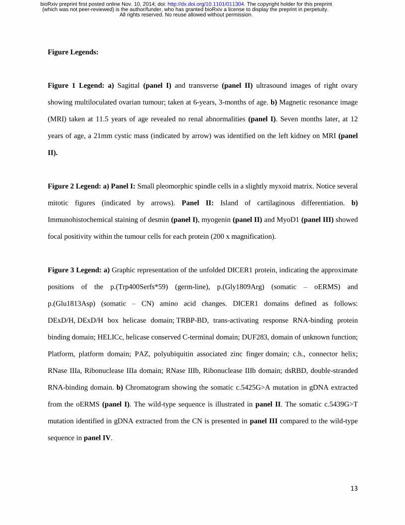
Figure Legends:
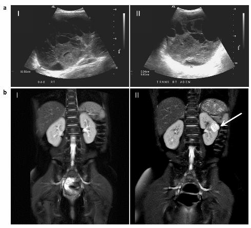
Figure 1 Legend: a) Sagittal (panel I) and transverse (panel II) ultrasound images of right ovary
showing multiloculated ovarian tumour; taken at 6-years, 3-months of age. b) Magnetic resonance image
(MRI) taken at 11.5 years of age revealed no renal abnormalities (panel I). Seven months later, at 12
years of age, a 21mm cystic mass (indicated by arrow) was identified on the left kidney on MRI (panel
II).
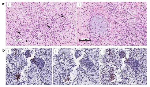
Figure 2 Legend: a) Panel I: Small pleomorphic spindle cells in a slightly myxoid matrix. Notice several
mitotic figures (indicated by arrows). Panel II: Island of cartilaginous differentiation. b)
Immunohistochemical staining of desmin (panel I), myogenin (panel II) and MyoD1 (panel III) showed
focal positivity within the tumour cells for each protein (200 x magnification).
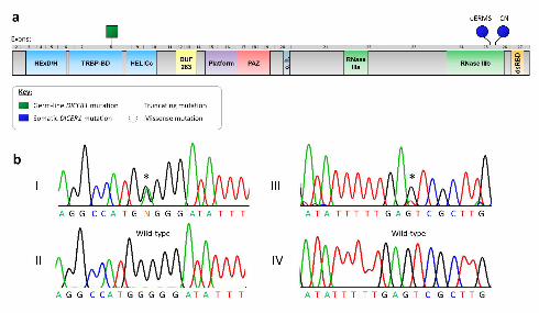
Figure 3 Legend: a) Graphic representation of the unfolded DICER1 protein, indicating the approximate
positions of the p.(Trp400Serfs*59) (germ-line), p.(Gly1809Arg) (somatic – oERMS) and
p.(Glu1813Asp) (somatic – CN) amino acid changes. DICER1 domains defined as follows:
DExD/H, DExD/H box helicase domain; TRBP-BD, trans-activating response RNA-binding protein
binding domain; HELICc, helicase conserved C-terminal domain; DUF283, domain of unknown function;
Platform, platform domain; PAZ, polyubiquitin associated zinc finger domain; c.h., connector helix;
RNase IIIa, Ribonuclease IIIa domain; RNase IIIb, Ribonuclease IIIb domain; dsRBD, double-stranded
RNA-binding domain. b) Chromatogram showing the somatic c.5425G>A mutation in gDNA extracted
from the oERMS (panel I). The wild-type sequence is illustrated in panel II. The somatic c.5439G>T
mutation identified in gDNA extracted from the CN is presented in panel III compared to the wild-type
sequence in panel IV.
All rights reserved. No reuse allowed without permission. (which was not peer-reviewed) is the author/funder, who has granted bioRxiv a license to display the preprint in perpetuity.
The copyright holder for this preprint. http://dx.doi.org/10.1101/011304doi: bioRxiv preprint first posted online Nov. 10, 2014;