REVIEW ARTICLE VASCULAR LIVER ANATOMY AND MAIN …€¦ · liver anatomy (4, 5). Therefore, the aim...
9
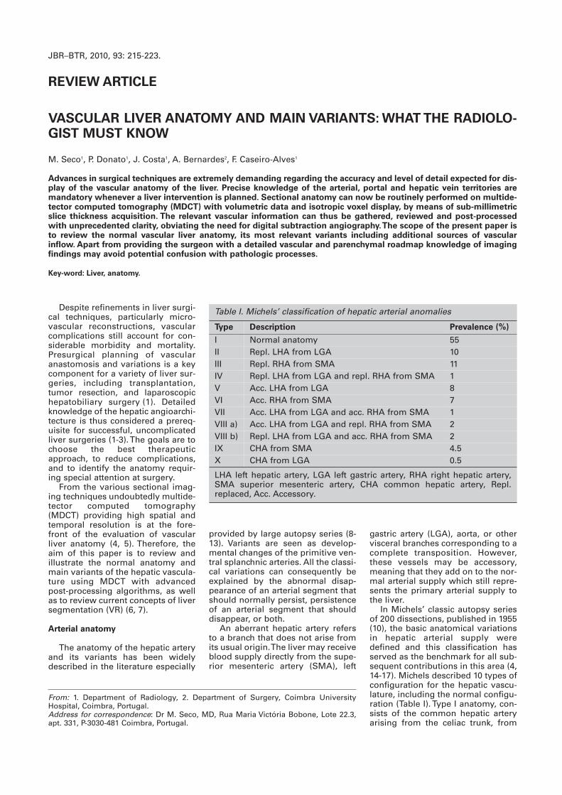
Despite refinements in liver surgi- cal techniques, particularly micro- vascular reconstructions, vascular complications still account for con- siderable morbidity and mortality. Presurgical planning of vascular anastomosis and variations is a key component for a variety of liver sur- geries, including transplantation, tumor resection, and laparoscopic hepatobiliary surgery (1). Detailed knowledge of the hepatic angioarchi- tecture is thus considered a prereq- uisite for successful, uncomplicated liver surgeries (1-3). The goals are to choose the best therapeutic approach, to reduce complications, and to identify the anatomy requir- ing special attention at surgery. From the various sectional imag- ing techniques undoubtedly multide- tector computed tomography (MDCT) providing high spatial and temporal resolution is at the fore- front of the evaluation of vascular liver anatomy (4, 5). Therefore, the aim of this paper is to review and illustrate the normal anatomy and main variants of the hepatic vascula- ture using MDCT with advanced post-processing algorithms, as well as to review current concepts of liver segmentation (VR) (6, 7). Arterial anatomy The anatomy of the hepatic artery and its variants has been widely described in the literature especially gastric artery (LGA), aorta, or other visceral branches corresponding to a complete transposition. However, these vessels may be accessory, meaning that they add on to the nor- mal arterial supply which still repre- sents the primary arterial supply to the liver. In Michels’ classic autopsy series of 200 dissections, published in 1955 (10), the basic anatomical variations in hepatic arterial supply were defined and this classification has served as the benchmark for all sub- sequent contributions in this area (4, 14-17). Michels described 10 types of configuration for the hepatic vascu- lature, including the normal configu- ration (Table I).Type I anatomy, con- sists of the common hepatic artery arising from the celiac trunk, from provided by large autopsy series (8- 13). Variants are seen as develop- mental changes of the primitive ven- tral splanchnic arteries. All the classi- cal variations can consequently be explained by the abnormal disap- pearance of an arterial segment that should normally persist, persistence of an arterial segment that should disappear, or both. An aberrant hepatic artery refers to a branch that does not arise from its usual origin. The liver may receive blood supply directly from the supe- rior mesenteric artery (SMA), left JBR–BTR, 2010, 93: 215-223. REVIEW ARTICLE VASCULAR LIVER ANATOMY AND MAIN VARIANTS: WHAT THE RADIOLO- GIST MUST KNOW M. Seco 1 , P. Donato 1 , J. Costa 1 , A. Bernardes 2 , F. Caseiro-Alves 1 Advances in surgical techniques are extremely demanding regarding the accuracy and level of detail expected for dis- play of the vascular anatomy of the liver. Precise knowledge of the arterial, portal and hepatic vein territories are mandatory whenever a liver intervention is planned. Sectional anatomy can now be routinely performed on multide- tector computed tomography (MDCT) with volumetric data and isotropic voxel display, by means of sub-millimetric slice thickness acquisition. The relevant vascular information can thus be gathered, reviewed and post-processed with unprecedented clarity, obviating the need for digital subtraction angiography. The scope of the present paper is to review the normal vascular liver anatomy, its most relevant variants including additional sources of vascular inflow. Apart from providing the surgeon with a detailed vascular and parenchymal roadmap knowledge of imaging findings may avoid potential confusion with pathologic processes. Key-word: Liver, anatomy. From: 1. Department of Radiology, 2. Department of Surgery, Coimbra University Hospital, Coimbra, Portugal. Address for correspondence: Dr M. Seco, MD, Rua Maria Victória Bobone, Lote 22.3, apt. 331, P-3030-481 Coimbra, Portugal. Table I. Michels’ classification of hepatic arterial anomalies Type Description Prevalence (%) I Normal anatomy 55 II Repl. LHA from LGA 10 III Repl. RHA from SMA 11 IV Repl. LHA from LGA and repl. RHA from SMA 1 V Acc. LHA from LGA 8 VI Acc. RHA from SMA 7 VII Acc. LHA from LGA and acc. RHA from SMA 1 VIII a) Acc. LHA from LGA and repl. RHA from SMA 2 VIII b) Repl. LHA from LGA and acc. RHA from SMA 2 IX CHA from SMA 4.5 X CHA from LGA 0.5 LHA left hepatic artery, LGA left gastric artery, RHA right hepatic artery, SMA superior mesenteric artery, CHA common hepatic artery, Repl. replaced, Acc. Accessory.
Transcript of REVIEW ARTICLE VASCULAR LIVER ANATOMY AND MAIN …€¦ · liver anatomy (4, 5). Therefore, the aim...
Despite refinements in liver surgi-cal techniques, particularly micro -vascular reconstructions, vascularcomplications still account for con-siderable morbidity and mortality.Presurgical planning of vascularanastomosis and variations is a keycomponent for a variety of liver sur-geries, including transplantation,tumor resection, and laparoscopichepatobiliary surgery (1). Detailedknowledge of the hepatic angioarchi-tecture is thus considered a prereq-uisite for successful, uncomplicatedliver surgeries (1-3). The goals are tochoose the best therapeuticapproach, to reduce complications,and to identify the anatomy requir-ing special attention at surgery.
From the various sectional imag-ing techniques undoubtedly multide-tector computed tomography(MDCT) providing high spatial andtemporal resolution is at the fore-front of the evaluation of vascularliver anatomy (4, 5). Therefore, theaim of this paper is to review andillustrate the normal anatomy andmain variants of the hepatic vascula-ture using MDCT with advancedpost-processing algorithms, as wellas to review current concepts of liversegmentation (VR) (6, 7).
Arterial anatomy
The anatomy of the hepatic arteryand its variants has been widelydescribed in the literature especially
gastric artery (LGA), aorta, or othervisceral branches corresponding to acomplete transposition. However,these vessels may be accessory,meaning that they add on to the nor-mal arterial supply which still repre-sents the primary arterial supply tothe liver.
In Michels’ classic autopsy seriesof 200 dissections, published in 1955(10), the basic anatomical variationsin hepatic arterial supply weredefined and this classification hasserved as the benchmark for all sub-sequent contributions in this area (4,14-17). Michels described 10 types ofconfiguration for the hepatic vascu-lature, including the normal configu-ration (Table I). Type I anatomy, con-sists of the common hepatic arteryarising from the celiac trunk, from
provided by large autopsy series (8-13). Variants are seen as develop-mental changes of the primitive ven-tral splanchnic arteries. All the classi-cal variations can consequently beexplained by the abnormal disap-pearance of an arterial segment thatshould normally persist, persistenceof an arterial segment that shoulddisappear, or both.
An aberrant hepatic artery refersto a branch that does not arise fromits usual origin. The liver may receiveblood supply directly from the supe-rior mesenteric artery (SMA), left
JBR–BTR, 2010, 93: 215-223.
REVIEW ARTICLE
VASCULAR LIVER ANATOMY AND MAIN VARIANTS: WHAT THE RADIOLO-GIST MUST KNOW
M. Seco1, P. Donato1, J. Costa1, A. Bernardes2, F. Caseiro-Alves1
Advances in surgical techniques are extremely demanding regarding the accuracy and level of detail expected for dis-play of the vascular anatomy of the liver. Precise knowledge of the arterial, portal and hepatic vein territories aremandatory whenever a liver intervention is planned. Sectional anatomy can now be routinely performed on multide-tector computed tomography (MDCT) with volumetric data and isotropic voxel display, by means of sub-millimetricslice thickness acquisition. The relevant vascular information can thus be gathered, reviewed and post-processedwith unprecedented clarity, obviating the need for digital subtraction angiography. The scope of the present paper isto review the normal vascular liver anatomy, its most relevant variants including additional sources of vascularinflow. Apart from providing the surgeon with a detailed vascular and parenchymal roadmap knowledge of imagingfindings may avoid potential confusion with pathologic processes.
Key-word: Liver, anatomy.
From: 1. Department of Radiology, 2. Department of Surgery, Coimbra UniversityHospital, Coimbra, Portugal.Address for correspondence: Dr M. Seco, MD, Rua Maria Victória Bobone, Lote 22.3,apt. 331, P-3030-481 Coimbra, Portugal.
Table I. Michels’ classification of hepatic arterial anomalies
Type Description Prevalence (%)
I Normal anatomy 55II Repl. LHA from LGA 10III Repl. RHA from SMA 11IV Repl. LHA from LGA and repl. RHA from SMA 1V Acc. LHA from LGA 8VI Acc. RHA from SMA 7VII Acc. LHA from LGA and acc. RHA from SMA 1VIII a) Acc. LHA from LGA and repl. RHA from SMA 2VIII b) Repl. LHA from LGA and acc. RHA from SMA 2IX CHA from SMA 4.5X CHA from LGA 0.5
LHA left hepatic artery, LGA left gastric artery, RHA right hepatic artery,SMA superior mesenteric artery, CHA common hepatic artery, Repl.replaced, Acc. Accessory.

216 JBR–BTR, 2010, 93 (4)
which the gastroduodenal artery andthe proper hepatic artery arise(Fig. 1). The proper hepatic arterybranches off the right hepatic artery(RHA) after the left hepatic artery(LHA). Further on RHA splits into itsanterior and posterior branches and
the LHA originates from the left gas-tric artery (LGA). The replaced arterycan be seen running through thelesser sac entering the liver via thefissure for the ligamentum venosum,into the umbilical fissure. This is thesecond most common arterial vari-ant, occurring with a frequency of10%.
In the type III Michels’ variant theRHA branches off from the SMA(Fig. 2). Whereas the right hepaticartery usually courses anterior to theright portal vein, the replaced righthepatic artery, runs posterior to themain portal vein in the portocavalspace, and classically ascends pos-terolateral to the common bile duct.This is the most common variantaccounting for 11% of cases. Type IVMichels’ variant corresponds to a sit-uation where type II and type vari-ants III coexist: both lobar arteriesare replaced with the RHA originat-ing from the SMA and the LHA fromthe LGA (Fig. 3). This kind of variantis rare with a reported incidence ofonly 1%. The remaining types of thisclassification are described in table 1.For illustrative purposes two differ-ent cases of type VIII (Fig. 4) and IX(Fig. 5) are shown attending to theirrarity accounting for 2 and 0.2% ofall arterial variants, respectively. Itshould be stressed that other vari-ants, not included in the originalMichels’s classification have alsobeen described such as a replacedRHA or the common hepatic arteryoriginating directly from the aorta.Of special interest is the anatomy ofthe artery (or arteries) that feed seg-ment IV, because of its importancefor surgical procedures involvingthis specific segment such as thecase of left liver donors for livertransplant in pediatric patients. Itsconfiguration is quite variable and itis possible to observe a single, dou-ble, and triple supply, originatingfrom RHA, LHA and/or proper hepat-ic artery (Fig. 6) . Usually the seg-ment IV artery originates from theLHA in 64-75% of patients and fromthe RHA in 25% corresponding towhat has been coined as the vascu-lar arcade (5, 18, 20).
Portal venous anatomy
The portal vein is formed in theretroperitoneum by the confluenceof the superior mesenteric vein andthe splenic vein, behind the neck ofthe pancreas and courses behind theduodenal bulb. In its most commonbranching pattern it divides at theporta hepatis into right and left por-tal veins (Fig. 7). The portal bifurca-
the LHA splits to feed segments IIand III. Segment IV is fed by one ormore branches originating from theLHA, RHA, or both. The frequency ofoccurrence of normal hepatic arterialanatomy ranges between 55-76% (4,18-19). In the type II Michels’ variant
Fig. 1. — Normal hepatic arteries. Coronal thick slab MIP image (A) and cadaveric dis-section (B). The common hepatic artery (CHA) arises from the celiac trunk (CT). Aftergiving rise to the gastroduodenal artery (GD), it continues as the proper hepatic artery(PHA), which divides into right hepatic artery (RHA) and left hepatic artery (LHA).
Fig. 2. — Michels’ type III anatomy. A: VR image. The right hepatic artery (arrow) aris-es from the superior mesenteric artery (arrowhead). B: VR image. C: Axial CT. When theright hepatic artery arises (arrow) from superior mesenteric artery it runs upwardsbehind the pancreas and dorsal to the portal vein in the portocaval space. On C an inva-sive gallbladder tumour (T) and a mestastatic node (n) are seen.
Fig. 3. — Michels’ type IV anatomy. A: Thick slab coronal MIP image. B: VR image. Theright hepatic artery (arrow) arises from the superior mesenteric artery and the lefthepatic artery (arrowhead) arises from left gastric artery.
A B
A B
A C
B
T
n

VASCULAR LIVER ANATOMY AND MAIN VARIANTS — SECO et al 217
tion may be extrahepatic (48% ofcases), intrahepatic (26%), or locatedright at the entrance of the liver(26%) (21, 22).
As it courses cranially, the rightportal vein first gives off collateralbranches to the caudate lobe andthen divides into anterior and poste-
vertical part), supplying the lateralsegments (segments II and III) of theleft lobe. It displays a wide anteriorconcavity ending up at the superiorand inferior segmental branches ofsegment IV. The segmental veinsdivide into subsegmental branches,and further on into small veins abut-ting at the portal triad at the level ofthe liver acinus.
Branching anomalies of the mainportal vein (PV) at the hepatic hilumare known to be less frequent (10-20% of cases) than those of thehepatic arteries and hepatic veins(23-26). Embryologically, the PV isformed during the second month ofgestation by selective involution ofvitelline veins, which have multiplebridging anastomoses, both anteriorand posterior to the duodenum.Modifications in the pattern of theseanastomoses end up in PV varia-tions.
According to the literature (25-27),the most common patterns are rep-resented by: a) trifurcation of themain portal vein (7.8%-10.8%); inthese cases, the main portal veindivides into three branches afterentering the porta hepatis; a rightanterior segment, a right posteriorsegment, and a left portal vein (Fig.8); b) origin of the right posteriorsegmental branch directly from themain portal vein (4.7%-5.8%), wherethe main portal vein gives rise to theright posterior segment, then contin-ues to the right for a short distance,and divides into the right anteriorsegmental branch and the left portalvein; c) origin of the right anteriorsegmental branch from the left por-tal vein (2.9%-4.3%); in these cases,the main portal vein divides into theright posterior segment and the leftportal vein. The right anterior seg-mental vein originates from the leftportal vein (Fig. 9).
Less well defined variations of the“normal” distribution of portal veinare commonly seen. These include ashort main right portal vein, a shorthorizontal portion of the left portalvein, disproportionate size of differ-ent segmental branches, and a smallaccessory branches (arising from themain portal vein) to the right posteri-or segment. Some of the latter varia-tions correlate with differences in thesize of some segments of the liver,where a hypoplastic segmentreceives small branches.
There may be cases of congenitalabsence of the portal vein, where allthe blood carried by the superiormesenteric and splenic veinsbypasses the liver draining directlyinto a systemic vein. This congenital
rior branches, which further subdi-vide into superior and inferior seg-mental branches to supply the rightlobe of the liver. The left portal veinfirst has a horizontal course (parshorizontalis) to the left and thenturns medially toward the ligamen-tum teres (pars umbilicalis, ie, the
Fig. 4. — Michels’ type VIII anatomy. A: Thick slab MIP. The replaced right hepaticartery (arrow) arises from the superior mesenteric artery and there is an acessory lefthepatic artery (arrowhead) that arises from the left gastric artery. B: Whenever the lefthepatic artery arises from the left gastric artery it runs in the lesser omentum andenters the liver via venous fissure.
Fig. 6. — Segment IV artery. A: VR image. The middle hepatic artery (arrow) origi-nates from proper hepatic artery. B: MIP image. In this case there is a double supply forsegment IV (arrows), one branch originating from the left hepatic artery and the otherbranch from the right hepatic artery.
Fig. 5. — Michels’ type IX anatomy. A: MIP image. B: VR image. The main hepaticartery (arrow) originates from the superior mesenteric artery (arrowhead).
A B
A B
A B

218 JBR–BTR, 2010, 93 (4)
malformation was first described byAbernathy in 1793 and is a clearexample of a portosystemic shunt(Fig. 10).
Morgan and Superina (28) subse-quently refined the classification ofportal shunting into two differenttypes: type I, when all portal venousblood is shunted to a systemic vein,
Hepatic veins anatomy
The three main hepatic veins(right, middle, and left) drain into theinferior vena cava (IVC) approxi-mately 1 cm below the diaphragmand 2 cm inferior to the lower borderof the right atrium (Fig. 11). The righthepatic vein (RHV) is the one widestsince it drains a larger volume ofliver parenchyma (segments V-VIII).The middle hepatic vein (MHV) runsalong the main portal fissure drain-ing segments IV, V and VIII. The lefthepatic vein (LHV) drains segmentsII and III and generally forms a com-mon trunk with the middle hepaticvein (MHV) in 85% of cases, ulti-
with complete bypass of the liver(e.g. congenital absence of the portalvein). This type of shunt has beenreferred to as a ‘total’ shunt or‘end/side’ shunt; type II shunt whenonly a portion of the portal venousflow is diverted from the liver corre-sponding to a ‘partial’ shunt or a‘side/side’ shunt.
Fig. 7. — Normal portal vein anatomy. MIP image. The mainportal vein (PV) divides into right portal vein (RPV) and left por-tal vein (LPV). The RPV bifurcates into anterior branch (AB) andposterior branch (PB), both of which bifurcate into ascendingand descending branches. Each of these four branches suppliesa segment of the right lobe. The left portal vein (LPV) divides intothree branches, one for each segment of the left lobe.
Fig. 9. — Right anterior segmental branch arising from the leftportal vein. MIP image. There is an absence of the right branchof the portal vein. The portal vein (PV) bifurcates into a left andposterior branch (to segments VI and VII). The anterior branch(arrow) to segments V and VIII arises from the left branch of theportal vein (LPV).
Fig. 10. — Congenital absence of the portal vein. The splanch-nic vein joins the inferior vena cava forming a porto-systemiccongenital shunt (arrow). Note a large macroregenerative nod-ule (asterisk) secondary to the perfusion abnormality of theliver.
Fig. 8. — Portal vein trifurcation. MIP image. Main portal vein(PV) divides into right anterior (AB) and posterior (PB) branchesand left portal vein (LPV). A right portal vein is not identified.

VASCULAR LIVER ANATOMY AND MAIN VARIANTS — SECO et al 219
mately draining into the anterior leftlateral aspect of the IVC (29,30). TheLHV is the smallest of these veins.Segment I has its own venousdrainage directly into the inferiorvena cava via a variable number ofindependent veins.
The anatomy of the major hepaticveins is quite variable. Also smalleraccessory veins (AHVs) may be rec-ognized, draining into the retrohep-atic portion of the inferior vena cavabetween the right adrenal vein andthe confluence of the main hepaticveins. These veins lack a precise sys-tematization except for the accesso-ry vein of segment VI, the largest andwell recognized of all. AHVs are
small and numerous, but their clini-cal relevance for liver surgery,should only be acknowledged whenits diameter exceeds 5 mm (Fig. 12).However, their pathophysiologicimportance may be greater when thevenous drainage of the liver is com-promised, such as in Budd-Chiarisyndrome or in cases of large centraltumors of the liver.
An accessory RHV occurs in52.5% of patients (Fig. 13), twoaccessory hepatic veins in 12%, anda dominant accessory vein drainingthe caudate lobe in 12%.
The most frequent variationsfound in literature are (31): a) a dom-inant right accessory hepatic veinseen in 3-5% of patients, which islarger in caliber than the right hepat-ic vein (which may be atretic or evenabsent in such a case); b) absence ofa common trunk formed by the mid-dle and the left hepatic veins; c) Thevein that drains the liver near the fal-ciform ligament is a tributary of themiddle hepatic vein instead of theleft hepatic vein.
Also of paramount importance isthe recognition of the territories ofliver drainage of the hepatic veins.This has major implications in thecontext of liver donor liver trans-plant (LDLT) procedures where seg-mental territories may be deprivedof their venous drainage ending upin venous congestion and reducingthe final graft volume (Fig. 14) (32).
Fig. 12. — Accessory hepatic vein (arrow). MIP image. Theyare significant for surgical purposes when their diameter issuperior to 5mm.
Fig. 13. — Large accessory right hepatic vein (arrow), drainingsegment VI.
Fig. 11. — Normal hepatic veins. A: MIP image. B: VR image. Normal hepatic veins,usually consisting of three main hepatic veins: right hepatic vein (RHV), middle hepat-ic vein (MHV) and left hepatic vein (LHV). The LHV forms a common trunk (arrow) withthe MHV in 85% of cases.
A B
220 JBR–BTR, 2010, 93 (4)
Non-portal venous supply to theliver
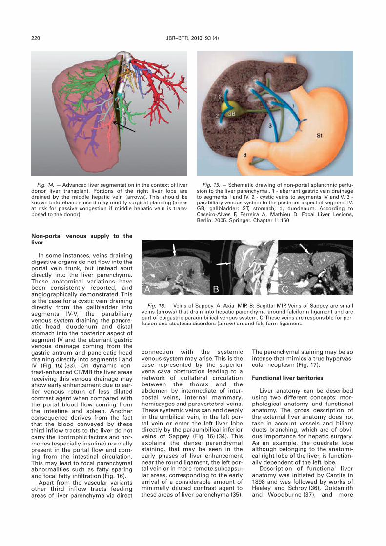
In some instances, veins drainingdigestive organs do not flow into theportal vein trunk, but instead abutdirectly into the liver parenchyma.These anatomical variations havebeen consistently reported, andangiographically demonstrated. Thisis the case for a cystic vein drainingdirectly from the gallbladder intosegments IV-V, the parabiliaryvenous system draining the pancre-atic head, duodenum and distalstomach into the posterior aspect ofsegment IV and the aberrant gastricvenous drainage coming from thegastric antrum and pancreatic headdraining directly into segments I andIV (Fig. 15) (33). On dynamic con-trast-enhanced CT/MR the liver areasreceiving this venous drainage mayshow early enhancement due to ear-lier venous return of less dilutedcontrast agent when compared withthe portal blood flow coming fromthe intestine and spleen. Anotherconsequence derives from the factthat the blood conveyed by thesethird inflow tracts to the liver do notcarry the lipotrophic factors and hor-mones (especially insuline) normallypresent in the portal flow and com-ing from the intestinal circulation.This may lead to focal parenchymalabnormalities such as fatty sparingand focal fatty infiltration (Fig. 16).
Apart from the vascular variantsother third inflow tracts feedingareas of liver parenchyma via direct
The parenchymal staining may be sointense that mimics a true hypervas-cular neoplasm (Fig. 17).
Functional liver territories
Liver anatomy can be describedusing two different concepts: mor-phological anatomy and functionalanatomy. The gross description ofthe external liver anatomy does nottake in account vessels and biliaryducts branching, which are of obvi-ous importance for hepatic surgery.As an example, the quadrate lobealthough belonging to the anatomi-cal right lobe of the liver, is function-ally dependent of the left lobe.
Description of functional liveranatomy was initiated by Cantlie in1898 and was followed by works ofHealey and Schroy (36), Goldsmithand Woodburne (37), and more
connection with the systemicvenous system may arise. This is thecase represented by the superiorvena cava obstruction leading to anetwork of collateral circulationbetween the thorax and theabdomen by intermediate of inter-costal veins, internal mammary,hemiazygos and paravertebral veins.These systemic veins can end deeplyin the umbilical vein, in the left por-tal vein or enter the left liver lobedirectly by the paraumbilical inferiorveins of Sappey (Fig. 16) (34). Thisexplains the dense parenchymalstaining, that may be seen in theearly phases of liver enhancementnear the round ligament, the left por-tal vein or in more remote subcapsu-lar areas, corresponding to the earlyarrival of a considerable amount ofminimally diluted contrast agent tothese areas of liver parenchyma (35).
Fig. 14. — Advanced liver segmentation in the context of liverdonor liver transplant. Portions of the right liver lobe aredrained by the middle hepatic vein (arrows). This should beknown beforehand since it may modify surgical planning (areasat risk for passive congestion if middle hepatic vein is trans-posed to the donor).
Fig. 15. — Schematic drawing of non-portal splanchnic perfu-sion to the liver parenchyma . 1 - aberrant gastric vein drainageto segments I and IV. 2 - cystic veins to segments IV and V. 3 -parabiliary venous system to the posterior aspect of segment IV.GB, gallbladder; ST, stomach; d, duodenum. According toCaseiro-Alves F, Ferreira A, Mathieu D. Focal Liver Lesions,Berlin, 2005, Springer. Chapter 11:160
Fig. 16. — Veins of Sappey. A: Axial MIP. B: Sagittal MIP. Veins of Sappey are smallveins (arrows) that drain into hepatic parenchyma around falciform ligament and arepart of epigastric-paraumbilical venous system. C: These veins are responsible for per-fusion and steatosic disorders (arrow) around falciform ligament.
BA C

VASCULAR LIVER ANATOMY AND MAIN VARIANTS — SECO et al 221
recently by Couinaud (38), andBismuth (39).
Couinaud (1957) proposed a liversegmentation system based on por-tal and hepatic veins. It includeseight segments divided by the sec-ond order branches of portal vein
is less prone to anatomical varia-tions.
Couinaud divided the liver intotwo functional parts: the left andright liver, separated by a main por-tal fissure containing the middlehepatic vein, known as Cantlie’s line.Surface markings of this line areinaccurate but grossly correspond toa plane joining the gallbladder fossaanteriorly to the left side of the infe-rior vena cava posteriorly. The leftand right hemilivers are further sub-divided by the left and right hepaticveins, lying in the left and right por-tal fissure, corresponding to the bedof the hepatic veins (Fig. 18).
The right hemiliver is subdividedinto two main sectors drawing a sec-ond line that vertically runs alongthe right portal vein bifurcation: aright lateral sector, lying posterolat-erally and a right paramedian sectorlying anteromedially. Each sector isformed by two segments: the rightlateral sector by segments VI and VIIand right paramedian sector by seg-ments V and VIII.
The left portal fissure lies in themiddle of left anatomical lobe andcorresponds to a plane passing fromthe confluence of the left hepaticvein with the inferior vena cavatowards the most lateral left lobe tip,dividing it into a left paramedian andlateral sectors. The left paramediansector consists of segments III andIV. The left lateral sector is comprisedonly of segment II, which is the pos-terior part of the left lobe (Fig. 19).Sectional imaging is not well suitedto determine the exact boundarybetween segments II and III. Its divi-sion can be assumed from a linedrawn from the middle portion ofvertical part of the left portal veinthat joins the left hepatic vein.Oblique reconstructions using MIPalgorithms can provide additionalhelp for its demonstration (Fig. 19).
From a functional point of view,the caudate lobe (or segment I) is anautonomous segment since its vas-cularization is independent from themain portal division and main hepat-ic veins.
Bismuth has brought together theCouinaud’s cadaveric system in situ(38) and the classification system ofGoldsmith and Woodburn in vivo(37). He distinguished three planes(scissurae), hosting the hepatic veinsand a transverse plane passingthrough the right and left portalbranches. The three hepatic veinsdivided the liver into four sectors.each supplied by its own portal pedi-cle (containing an arterial, portalvein, and bile duct branches).
with the argument that functionalsegmentation using the veins is pre-ferred over arteriobiliary segmenta-tion since portal vein branches offfirst, with arteriobiliary following thevein distribution. Portal segmenta-tion is also simpler to use because it
Fig. 17. — Two different patients with superior vena cava syn-drome. A: There is an area, localized at segment IV, of intenseand early enhancement, mimicking a hypervascular focal lesion.B: This case shows subcapsular venous collateral vessels at theright liver lobe again leading to a hyperdense pseudo-tumoralfocal lesion.
Fig. 18. — The liver can be divided into four sectors: left lateral, paramedial left, para-medial right and lateral right. The separation lines (arrows) between sectors are calledportal fissures and contain the hepatic veins.
Fig. 19. — The problem of separation between segment II and III on CT. A: On theaxial plane we trace a line that intersects both the middle portion of umbical segmentof portal vein and the left hepatic vein. Posterior to that line is segment II and anterioris segment III. B: On the sagittal plane the distinction between segment II and III isstraightforward. Posterior to the left hepatic vein is segment II and anterior is segment III.
BA
BA
222 JBR–BTR, 2010, 93 (4)
Although the above outlined con-cepts may be correct in somepatients, from a morphologic pointof view they can be questionable (40,41). Anatomists such as Platzer andMaurer (40) pointed out that the vari-ability of segmental boundaries istoo large to render any schemeviable. Fasel et al (42) confirmed thatthe vertical planes that intersect thetrunks of the hepatic veins do notcorrespond to the presumed inter-segmental boundaries. Radiologists,as well, have published observationsquestioning the radiologic methodscurrently used for delineation of thesegmental anatomy of the liver.Nelson et al (43) and Soyer et al (44)concluded that indirect landmarksare not reliable for the correct delin-eation of portal venous segmentsand subsegments. In opposition tothe traditional landmarks (using theplanes of the three major hepaticveins and the portal trunks as seg-mental boundaries), radiologists canlocalize lesions attributing them tothe nearest peripheral portal veinbranches. Rieker et al. (45) showeddifferences in segmental locations in16% of the lesions analysed. Thesedifferent locations were due to thepath of the portal trunks or of theperipheral portal branches crossingthe planes of the major hepaticveins. With current radiologic proce-dures based on indirect landmarks itis therefore not possible to exactlydetermine segmental and subseg-mental anatomy of the liver. Also,every concept of flat planes delineat-ing portal venous territories is anoversimplification which is not in fullagreement with anatomic reality.
True segmental and subsegmen-tal determination is possible onlywith methods that account for theactual anatomy of the portal venoustree, incorporating off-branching ofthird and fourth-order portal branch-es. This can be more readily appreci-ated when reading the 3D axialdataset in interactive cine mode dis-play.
Conclusion
Radiologists must be knowledge-able on liver vascular anatomy, itsvariants and the vascular landmarksallowing functional liver segmenta-tion. . Angio-MDCT of the liver isoften the standalone technique inthe preoperative evaluation ofpatients for a variety of different clin-ical scenarios. Routine use ofadvanced post-processing algo-rithms and segmentation techniquessuch as maximum intensity projec-
tion with three-dimensional CT arteri-ography. Radiology, 1995, 195: 363-370.
13. Winter T.C. III, Nghiem H.V., Freeny P.C.,Hommeyer S.C., Mack L.A. Hepaticarterial anatomy: demonstration ofnormal supply and vascular variantswith three-dimensional CT angio -graphy. RadioGraphics, 1995, 15: 771-780.
14. Alonso-Torres A., Fernandez-Cuadrado J., Pinilla I., et al. Multi -detector CT in the evaluation ofpotential living donors for liver trans-plantation. RadioGraphics, 2005, 25:1017-1030.
15. Stemmler B.J., Paulson E.K.,Thornton F.J., et al. Dual-phase 3D-MDCT angiography for the evaluationof the liver before hepatic resection.AJR, 2004, 183: 1551-1557.
16. De Santis M., Ariosi P., Calo G.F., et al.Hepatic arterial vascular anatomy andits variants in Italian. Radiol Med(Torino), 2000: 141-151.
17. Matsuki M., Tanikake M., Kani H., et al.Dual-Phase 3D CT angiography dur-ing a single breath hold using 16-MDCT: assessment of vascular anato-my before laparoscopic gastrectomy.AJR, 2006, 186: 1079-1085.
18. Saylisoy S., Atasoy C., Ersoz S., et al.Multislice CT angiography in the eval-uation of hepatic vascular anatomy inpotential right lobe donors. DiagnInterv Radiol, 2005, 11: 51-59.
19. Covey A.M., Brody L.A., MaluccioM.A. Variant hepatic arterial anatomyrevisited: digital subtraction angiog-raphy performed in 600 patients.Radiology, 2002, 224: 542-547.
20. Kapoor V., Brancatelli G., Federle M.P.,et al. Multidetector CT arteriographywith volumetric three-dimensionalrendering to evaluate patients withmetastatic colorectal disease. AJR,2003, 181: 455-463.
21. Schultz S.R., LaBerge J.M.,Gordon R.L., et al. Anatomy of theportal vein bifurcation: intra- versusextrahepatic location: implications fortransjugular intrahepatic portosys-temic shunts. J Vasc Intervent Radiol,1994, 5: 457-459.
22. Yamane T., Mori K., Sakamoto K.,Ikei S., Akagi M. Intrahepatic ramifica-tion of the portal vein in the right andcaudate lobes of the liver. Acta Anat(Basel), 1988, 133: 162-172.
23. Atri M., Bret P.M., Fraser-Hill M.A.Intrahepatic portal venous variations:prevalence with ultrasound. Radi -ology, 1992, 184: 523-526.
24. Couinaud C. Liver anatomy: portal(and suprahepatic segmentation) orbiliary segmentation. Dig Surg, 1999,16: 459-467.
25. Atri M., Bret P.M., Fraser-Hill M.A.Intrahepatic portal venous variations:prevalence with US. Radiology, 1992,184: 157-158.
26. Fraser-Hill M.A., Atri M., Bret P.M.,Aldis A.E., Illescas F.F., Herschorn S.D.Intrahepatic portal venous system:variations demonstrated with duplexand color Doppler US. Radiology,1990, 177: 523-526.
tions and three-dimensional volumerenderings provide high qualitygraphic display which assists in thedemonstration and understanding ofits variable liver blood supply. “Non-portal” venous supply to the livercan mimic focal liver pathology andthe exact location of the parenchy-mal abnormalities anticipates inter-pretative pitfalls. Although a single,worldwide-accepted classification ofthe liver anatomy does not exist,radiologists should favor the use ofthe terminology provided by thesegmentation-based functional anat -o my of the liver.
References
1. Van Thiel D.H., Wright H., Fagiuoli S.,et al. Preoperative evaluation of apatient for hepatic surgery. J SurgOncol Suppl, 1993, 349-51.
2. Ozeki Y., Uchiyama T., Katayama M., etal. Extended left hepatic trisegmen-tectomy with resection of main righthepatic vein and preservation of mid-dle and inferior right hepatic veins.Surgery, 1995, 117: 715-717.
3. Fan S., Lo C., Liu C., et al. Safety ofdonors in live donor liver transplan -tation using right lobe grafts. ArchSurg, 2000, 135: 336-340.
4. Sahani D., Mehta A., Blake M.,Prasad S., Harris G., Saini S.Preoperative hepatic vascular evalua-tion with CT and MR angiography:implications for surgery. Radio -Graphics, 2004, 24: 1367-1380.
5. Sahani D., D’souza R., Kadavigere R.,et al. Evaluation of living liver trans-plant donors: method for preciseanatomic definition by using a dedi-cated contrast-enhanced MR imagingprotocol. Radio-Graphics 2004, 24:957-967.
6. Marcus C.D., Ladam-Marcus V.J.,Bigot J.L. et al. Carotid artery steno-sis: evaluation at CT angiographywith the volume rendering technique.Radiology, 1999, 211: 775-780.
7. Corti R., Alerci M., Wyttenbach R., etal. Usefulness of multiplanar recon-structions in evaluation of carotidCT angiography. Radiology, 2003,226: 290-291.
8. Adachi B. Arterien system derJapaner. Kyoto: Kerkyusha, TokyoPress; 1928.
9. Flint E.R. Abnormalities of the righthepatic, cystic and gastroduodenalarteries and of the bile ducts. Br JSurg, 1923, 10: 509-519.
10. Michels N.A. Blood supply and anato-my of the upper abdominal organs.Philadelphia: JB Lippincott Co; 1955.
11. Gruttadauria S., Scotti Foglieni C.,Doria C., Luca A., Lauro A.,Marino I.R. The hepatic artery in livertransplantation and surgery: vascularanomalies in 701 cases. Clin Trans -plant, 2001, 15: 359-363.
12. Winter T.C. III, Freeny P.C., NghiemH.V., et al. Hepatic arterial anatomy intransplantation candidates: evalua-
VASCULAR LIVER ANATOMY AND MAIN VARIANTS — SECO et al 223
27. Chevallier P., Oddo F., Baldini E.,Peten E.P., Diaine B., Padovani B.Agenesis of the horizontal segment ofthe left portal vein demonstrated bymagnetic resonance imaging includ-ing phase-contrast magnetic reso-nance venography. Eur Radiol, 2000,10: 365-367.
28. Morgan G., Superina R. Congenitalabsence of the portal vein: two casesand a proposed classification systemfor portosystemic vascular anomalies.J Pediatr Surg, 1994, 29: 1239-1241.
29. Soyer P., Heath D., Bluemke D.A., etal. Three-dimensional helical CT ofintrahepatic venous structures: com-parison of three rendering tech-niques. J Comput Assist Tomogr,1996, 20: 122-127.
30. Lerut J.P., Mazza D., Van Leeuw V., etal. Adult liver transplantation andabnormalities of splanchnic veins:experience in 53 patients. Transpl Int,1997, 10: 125-132.
31. Donato P., Coelho P., Rodrigues H.,Vigia E., Fernandes J., Caseiro-Alves F., Bernardes A. Normal vascu-lar and biliary hepatic anatomy: 3Ddemonstration by multidetector CT.Surg Radiol Anat, 2007, 29: 575-82.
32. Erbay N., Raptopoulos V., Pomfret E.A.,Kamel I.R., Kruskal J.B. Living donorliver transplantation in adults: vascu-
41. Bernardes A., Coelho P., Ferreira S.,Patrício J. Branches segmentaires dela veine porte: variations et localisa-tion de l’origine – étude anatomiqueet par imagerie. E-mémoires del’Académie Nationale de Chirurgie,2007, 6: 72-76.
42. Fasel J.H.D., Gailloud P., Grossholz M.,Bidaut L., Probst P., Terrier F.Relationship between intrahepaticvessels and computer-generatedhepatic sissurae: an in vitro assay.Surg Radiol Anat, 1996, 18: 43-46.
43. Nelson R.C., Chezmar J.L.,Sugarbaker P.H., Murray D.R.,Bernardino M.E. (1990) Preoperativelocalization of focal liver lesions tospecific liver segments: utility of CTduring arterial portography. Radi -ology, 1990, 176: 89-94.
44. Soyer P, Roche A. Gad M et al.Preoperative segmental localizationof hepatic metastases: ulility of three-dimensional CT during arterial por-tography. Radiology, 180: 653-658.
45. Rieker O, Mildenberger P, Hintze C,Schunk K, Otto G, Thelen MSegmentanatomie der Leber in derComputertomographie: Lokalisierenwir die Lasionen richtig. Rofo, 2000,171: 147-152.
lar variants important in surgicalplanning for donors and recipients.AJR, 2003, 181: 109-114.
33. Itai Y., Matsui O. “Non-portal”splanchnic venous supply to the liver:abnormal findings on CT, US andMRI. Eur Radiol, 1999, 9: 237-243.
34. Hashimoto M., Heianna J., Tate E., etal. Small veins entering the liver. EurRadiol, 2002, 12: 2000-2005.
35. Maldjian P., Obolevich A., Cho K.Focal enhancement of the liver on CT.A sign of SVC obstruction. J ComputAssist Tomogr, 1995, 19: 316-318.
36. Healey J.E., Schroy P.C. Anatomy ofthe biliary ducts within the humanliver. Analysis of the prevailing pat-tern of branching and the major vari-ations of the biliary ducts. Am MedAssoc Arch Surg, 1953, 66: 599-616.
37. Goldsmith N.A., Woodburne R.T.Surgical anatomy pertaining to liverresection. Surg Gynecol Obstetr,1957, 195: 310-318.
38. Couinaud C. Le foie. Etudes anatomi-ques et chirurgicales. Paris: Masson;1957.
39. Bismuth H. Surgical anatomy andanatomical surgery of the liver. WorldJ Surg, 1982, 6: 3-9.
40. Platzer W., Maurer H. Zur Segment -einteilung der Leber. Acta Anat, 1966,63: 8-31.