Resting potential, action potential and electrical...
21
Resting potential, action potential and electrical excitibility. Measurement of membrane potential. Medical and Health Science Center, University of Debrecen Department of Biophysics and Cell Biology Tibor G. Szántó
-
Upload
truongminh -
Category
Documents
-
view
234 -
download
1
Transcript of Resting potential, action potential and electrical...

Resting potential, action potential and electrical excitibility. Measurement of membrane potential.
Medical and Health Science Center, University of Debrecen Department of Biophysics and Cell Biology
Tibor G. Szántó

Resting membrane potential
•
all
living
cells
exhibit
an
electrical
potential
difference
between
the
inner
and outer
surface
of the
cytoplasmic
membrane
• varies
among
different
cell
types, its
value
is typically
between
‐30 and ‐90 mV
(a
sensitive
potentiometer
indicates
an
electrical
potential
difference
between
the
two
electrodes
upon
insertion
of one
microelectrodes
into
the
nerve
cell)
(the
electrical
potential
of the
ic. space
is always
negative
compared
to
the
ec. space)
insert
microelectrode
into
axon
resting potential
time
mem
bran
epo
tential(mV)
resting potential

Resting membrane potential II.
‐
the
permeability
of
the
membrane
for
the
different
species
of monovalent
ions
is
different
(impermeable
for
larger
protein
and
phosphate‐anions)
• reasons: ‐
ion concentration‐differences
on
the
two
side
of the
membrane
‐
Donnan‐potential
• factors
determining
the
resting potential
(Em
):
‐
Diffusion
potential
‐
Pump
potential
(Na+/K+
ATP‐ase)
due
to
the
presence
of non‐permeable
protein anions
in
the
cytosolnegligible
contribution,
cells
continuously
fight
against
it
(thermodynamic
equilibrium
potential)
most significant
contribution
depending
on
the
cell
type: 2‐16 mV
(direct
contribution)

Resting membrane potential III.
•
the
Goldman‐Hodgkin‐Katz
voltage
equation
describes
the
resting state
of the membrane
as
the
result
of
a steady‐state
electrodiffusion
of the
permeant
ions
–
–
–
–ln Na K Clo o i
m
Na K Cli i o
p Na p K p ClRTEF p Na p K p Cl
• the
net passive
flux
of a given
ion is not zero at the resting membrane
potential
the ions on the two sides of the membrane of a living cell are not
in
thermodynamic
equilibrium
•
the
membrane
potential
depends
on
the
concentrations
of
the
permeating ions
and
the
membrane’s
permeability
for
these
ions,
which
is
mostly
determined by the opening / closing of ion channels specific for
that ion.
•
net ion fluxes flow across the membrane, only the sum of the charges moving is zero, i.e., IK
+INa
+ICl
=0, and IK
≠0, INa
≠0, ICl
≠
0

• at
a given
ion concentration
gradient
Ediff
is determined
by
Px
Resting membrane potential IV.
•
the
greater
the
permeability
for
a
given
ion,
the
closer
the
MP
will
be
to
the equilibrium
(Nernst)
potential
for
that
ion.
For
example,
opening
of
many
K+
channels
will
shift
the
MP
toward ‐89
mV,
while
opening
of
many
Na+
channels will shift the MP toward +60 mV.
the actual MP is the average of the Nernst-potentials of thepermeating ions weighted by the permeabilities
mem
bran
epo
tential(mV)
resting
potential
resting
potential

Resting membrane potential V.
• useful
relationship: Ediff
is determined
by
PNa
/PKmem
bran
epo
tential(mV)

Action potential
•
change
in
the
membrane
potential
with a
characteristic
time
course
and
voltage
values
•
two
major cell
types
that
are
able
to
fire action
potentials:
nerve‐
and
muscles
(endocrine
cells)
•
AP
transmits
information
in
neurons and initiates
contraction
in
muscles
•
all
impulses
look
alike, similar
in
shape, amplitude
and duration
•
strength
of
stimulus
is
coded in the frequency
• propagates
with
constant
amplitude

•
the
membrane
rapidly
depolarizes (depolarization
phase)
and
reaches
the
peak
potential.
This
is
followed
by
the repolarization
phase
during
which
the
membrane
potential
again
reaches
the resting potential
•
characteristic
phases
of
the
action potential
are
shown
in
figure
•
the
repolarization
phase
is
often followed
by
a
hyperpolarizing
afterpotential
•
once the membrane potential reaches the
threshold
potential
an action
potential
will
be
generated
(”all
or
nothing” response)
Action potential II.

• membrane
depolarization
to
reach
the
depolarizing
threshold
• depolarization
opens
voltage
gated
Na+
channels
• Na+‐ions
flow into
the
cell, which
causes
further
depolarizationpositive
feedback
•
the
Na
channels
enter
the
non‐conducting
inactivated
state
which
stops
the Na current
•delayed
opening
of voltage‐gated
K+
channels
inactivation
ball
sneaks
up
into
the
cavity
from
the
intracellular
side
(ball
and
chain mechanism)
channels only go back to the closed state at negative MP
•
K+
permeability
increases,
K+
ions
flow
from
the
cell.
K+
efflux
balances
Na+ influx
(repolarization)
•
though
depolarization
opens
the
K+
channels
the
K+
efflux
hiperpolarize renders
the
channels
to
the
closed
state
negative
feedback
Ionic currents during an action potential

•
closing
of
the
K+
channels
and
the
recovery
from
inactivation
of
the
Na+ channels
result
in
the
reestablishment
of the
resting potential
•
closing
of
the
K+
channels
is
slow,
the
relatively
large
K+
permeability
of
the membrane
results
in
a transient
hyperpolarization
•
the
membrane
potential
(measured
on
the
vertical
axis
of the
action
potential figure) reaches
a minimum and maximum value
if
the
transmembrane
potential
(Em
)
reaches
the
equilibrium
potential
for
the transported ion, no driving force, the transmembrane flux of ion
species Ix
= 0.
• the
theoretical
maximum of AP is the
equilibrium potential
of Na+
PNa
decreases
(the
driving
force
for
Na+
decreases)
PK
increases
before
reaching
the
peak
potential
(repolarization)
• in
reality
the
peak
potential
never
reaches
this
potential, because…
Ionic currents during an action potential II.

idõ
(ms)
resting potential
Na permeability
K permeability
time
(ms)
time
(ms)
perm
eability
mem
bran
epo
tential(mV)
Permeability changes during an action potential

•
absolute
refractory
period
(ARP):
Na channels
are
inactivated,
no
Na
conductance, no new
AP can
be initiated
•
the
amount
of
time
it
takes
for
an
excitable membrane to be
ready
for
a second
stimulus
once
it
returns
to
its
resting state
•
relative
refractory
period
(RRP):
Na channels
have
returned
to
the
closed
state,
but
the
MP
is
more
negative
than the
resting MP due
to
the
delayed
closing
of
the
K
channels,
so
stronger depolarization
would
be
needed
to
reach
the
firing
threshold
• two
types
of refractory
periods
occur
during
an AP
time
(ms)
mem
bran
epo
tential(mV)
resting potential
Refractory periods
• prevents the fusion of AP and allow the propagation of the separated APs

(Hodgkin
and Katz, 1949)
The first direct proof for the [Na+]ex
dependence of AP
•
The
amplitude
of
AP
is
determined
by
the extracellular
Na+
concentration
and
the duration
of sodium
channel
inactivation

Operation of ion channels during the action potential

•
a
plot
of
the
threshold
current
versus
pulse
duration
required
to
stimulate
excitable tissue
•
rheobase
is
the
minimal
current
amplitude
of
indefinite
duration
that
results
in
the depolarization
threshold
of the cell membranes being reached
•
the
chronaxie
is
the
minimum
time
over
which
an
electric
current
(double
the strength of the rheobase)
needs to be applied to stimulate a muscle
fiber or nerve cell
The strength-duration curve
the minimal charge that is capable
of firing an action potential
current×time duration
= constant
•
the
capacitive
elements
(ie.
capacitor
plates)
of
the
membrane
must
be
charged
to reach the necessary threshold potential
•
for
this
the
same
charge
is
required
in
case
of
any
duration
and
intensity
of
stimulus
(these
quantities
are
inversly
proportional to each other)
×
×
1
2
current
time duration
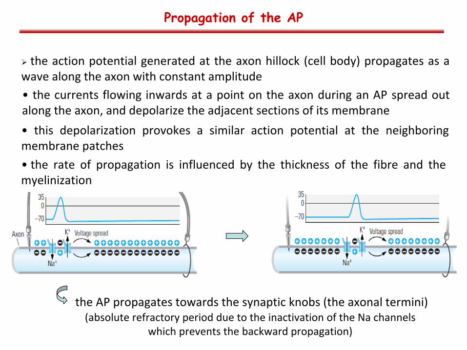
• the
currents
flowing
inwards
at
a
point
on
the
axon
during
an
AP
spread
out along the axon, and depolarize the adjacent sections of its membrane
•
the
rate
of
propagation
is
influenced
by
the
thickness
of
the
fibre
and
the myelinization
Propagation of the AP
the
action
potential
generated
at
the
axon
hillock
(cell
body)
propagates
as
a wave along the axon with constant amplitude
• this
depolarization
provokes
a
similar
action
potential
at
the
neighboring membrane patches
the AP propagates towards the synaptic knobs (the axonal termini) (absolute
refractory
period
due
to
the
inactivation
of the
Na channels
which
prevents
the
backward
propagation)

•
certain
neuronal
axons
are
covered
with myelin
sheaths
•
myelin
is
a
multilamellar
membrane
that enwraps
the
axon
in
segments
separated
by
intervals known as nodes of Ranvier•
increases
the
conduction
velocity
of
action
potentials •
myelin
behaves
as
an
insulator,
therefore
prevents
ions
from
entering
or
leaving
the axon along myelinated segments
•
the
ionic
current
from
an
action
potential at
one
node
of
Ranvier
provokes
another
action
potential
at
the
next
node
(saltatory conduction)
•
some
diseases
degrade
myelin
and
impair saltatory
conduction,
reducing
the
conduction
velocity
of
action
potentials
( multiple sclerosis)
Myelin and saltatory conduction

•
the
difference
between
the
command
and
the
actual
membrane
potentials
is calculated
and
a
current
is
injected
into
the
cell
through
the
second
electrode
with appropriate polarity and magnitude to cancel this difference
•
the
membrane
potential
of
a
cell
is
kept
constant
regardless
of
the
magnitude and direction of the ionic current flowing through the membrane
•
one electrode is used to measure the actual membrane potential and this value is compared to the command potential set by the researcher
The voltage-clamp technique.
Measurement of the membrane potential
• the method is capable of measuring individual ionic currents individuallyin
such
experiments,
specific
blockers
of
Na+
and
K+
channels
are
added
to
the extracellular fluid
(for example
tetrodotoxin or
tetra‐ethyl‐ammonium, respectively)
and the current remaining in the presence of a blocker is determined
TTX TEA

The voltage-clamp technique.
Measurement of the membrane potential II.

The voltage-clamp technique.
Measurement of the membrane potential III.

Application of fluorescent dyes/probes for measuring membrane potential.
some
dyes
change
their
fluorescent
properties
in
an
electric
field
and
can therefore be used to follow action potential propagation
another group of dyes does not change its spectroscopic properties in an electric field,
however,
due
to
their
net
negative
or
positive
charge
they
are
distributed
across
the
two
sides
of
the
membrane
according
to
their
solubility
and
the electrochemical gradient
•
changes
in
the
electrochemical
gradient
across
the
cell
membrane
results
in redistribution
of
the
dyes
leading
to
changes
in
the
fluorescence
intensity
emitted from the cells
•
the
more
negative
the
inside
of
the
cell
the
more
positively
charged
dye
is accumulated within the cell
•
negatively
charged
dye
molecules
are
not
accumulated
in
cells
having
a negative resting potential, their toxicity is negligible
Measurement of the membrane potential IV.