Research Article Free DNA in Cystic Fibrosis Airway Fluids ...
12
Research Article Free DNA in Cystic Fibrosis Airway Fluids Correlates with Airflow Obstruction Veronica Marcos, 1 Zhe Zhou-Suckow, 2 Ali Önder Yildirim, 3 Alexander Bohla, 3 Andreas Hector, 1,4 Ljubomir Vitkov, 5,6 Wolf Dietrich Krautgartner, 6 Walter Stoiber, 6 Matthias Griese, 1 Oliver Eickelberg, 3 Marcus A. Mall, 2 and Dominik Hartl 1,4 1 Department of Pediatric Pulmonology, Hauner Children’s Hospital, Ludwig Maximilians University, e German Center for Lung Research (DZL), 80377 Munich, Germany 2 Department of Translational Pulmonology, Translational Lung Research Center Heidelberg (TLRC), University of Heidelberg, e German Center for Lung Research (DZL), 69120 Heidelberg, Germany 3 Comprehensive Pneumology Center, Institute of Lung Biology and Disease (iLBD), University Hospital, Ludwig Maximilians University and Helmholtz Zentrum M¨ unchen, e German Center for Lung Research (DZL), 81377 Munich, Germany 4 Children’s Hospital and Interdisciplinary Center for Infectious Diseases, University of T¨ ubingen, 72076 T¨ ubingen, Germany 5 Department of Operative Dentistry & Periodontology, Saarland University, 66424 Homburg, Germany 6 Biomedical Ultrastructure Research Lab, Division of Animal Structure and Function, Department of Cell Biology, University of Salzburg, 5020 Salzburg, Austria Correspondence should be addressed to Dominik Hartl; [email protected] Received 11 January 2015; Revised 11 March 2015; Accepted 13 March 2015 Academic Editor: Pham My-Chan Dang Copyright © 2015 Veronica Marcos et al. is is an open access article distributed under the Creative Commons Attribution License, which permits unrestricted use, distribution, and reproduction in any medium, provided the original work is properly cited. Chronic obstructive lung disease determines morbidity and mortality of patients with cystic fibrosis (CF). CF airways are characterized by a nonresolving neutrophilic inflammation. Aſter pathogen contact or prolonged activation, neutrophils release DNA fibres decorated with antimicrobial proteins, forming neutrophil extracellular traps (NETs). NETs have been described to act in a beneficial way for innate host defense by bactericidal, fungicidal, and virucidal actions. On the other hand, excessive NET formation has been linked to the pathogenesis of autoinflammatory and autoimmune disease conditions. We quantified free DNA structures characteristic of NETs in airway fluids of CF patients and a mouse model with CF-like lung disease. Free DNA levels correlated with airflow obstruction, fungal colonization, and CXC chemokine levels in CF patients and CF-like mice. When viewed in combination, our results demonstrate that neutrophilic inflammation in CF airways is associated with abundant free DNA characteristic for NETosis, and suggest that free DNA may be implicated in lung function decline in patients with CF. 1. Introduction Cystic fibrosis (CF) is a fatal disorder characterized by chronic and progressive lung disease that determines mor- bidity and mortality of these patients [1]. Airways of CF patients show a chronic nonresolving neutrophilic inflamma- tion, which increases upon infection and disease progression. Neutrophil products, such as elastase, chitinase-like proteins and chemokines, have been identified as important risk factors of lung damage and lung function decline and are suggested as biomarkers based on both cross-sectional and longitudinal studies in patients with CF [2–7] and mice with CF-like lung disease [8]. Previous studies also provided evidence that free extracellular DNA is highly increased in CF airway specimen [9], initially referred to as DNA derived from necrotic cells. However, several studies have now established that CF airway secretions contain meshwork structures reminiscent of NETs [10–14]. Neutrophils represent the first line of cellular host defense against bacteria and fungi. Traditionally, neutrophils have Hindawi Publishing Corporation Mediators of Inflammation Volume 2015, Article ID 408935, 11 pages http://dx.doi.org/10.1155/2015/408935
Transcript of Research Article Free DNA in Cystic Fibrosis Airway Fluids ...

Research ArticleFree DNA in Cystic Fibrosis Airway Fluids Correlates withAirflow Obstruction
Veronica Marcos,1 Zhe Zhou-Suckow,2 Ali Önder Yildirim,3 Alexander Bohla,3
Andreas Hector,1,4 Ljubomir Vitkov,5,6 Wolf Dietrich Krautgartner,6 Walter Stoiber,6
Matthias Griese,1 Oliver Eickelberg,3 Marcus A. Mall,2 and Dominik Hartl1,4
1Department of Pediatric Pulmonology, Hauner Children’s Hospital, Ludwig Maximilians University,The German Center for Lung Research (DZL), 80377 Munich, Germany2Department of Translational Pulmonology, Translational Lung Research Center Heidelberg (TLRC), University of Heidelberg,The German Center for Lung Research (DZL), 69120 Heidelberg, Germany3Comprehensive Pneumology Center, Institute of Lung Biology and Disease (iLBD), University Hospital,Ludwig Maximilians University and Helmholtz Zentrum Munchen, The German Center for Lung Research (DZL),81377 Munich, Germany4Children’s Hospital and Interdisciplinary Center for Infectious Diseases, University of Tubingen, 72076 Tubingen, Germany5Department of Operative Dentistry & Periodontology, Saarland University, 66424 Homburg, Germany6Biomedical Ultrastructure Research Lab, Division of Animal Structure and Function, Department of Cell Biology,University of Salzburg, 5020 Salzburg, Austria
Correspondence should be addressed to Dominik Hartl; [email protected]
Received 11 January 2015; Revised 11 March 2015; Accepted 13 March 2015
Academic Editor: PhamMy-Chan Dang
Copyright © 2015 Veronica Marcos et al. This is an open access article distributed under the Creative Commons AttributionLicense, which permits unrestricted use, distribution, and reproduction in any medium, provided the original work is properlycited.
Chronic obstructive lung disease determines morbidity and mortality of patients with cystic fibrosis (CF). CF airways arecharacterized by a nonresolving neutrophilic inflammation. After pathogen contact or prolonged activation, neutrophils releaseDNA fibres decorated with antimicrobial proteins, forming neutrophil extracellular traps (NETs). NETs have been described toact in a beneficial way for innate host defense by bactericidal, fungicidal, and virucidal actions. On the other hand, excessiveNET formation has been linked to the pathogenesis of autoinflammatory and autoimmune disease conditions. We quantified freeDNA structures characteristic of NETs in airway fluids of CF patients and a mouse model with CF-like lung disease. Free DNAlevels correlated with airflow obstruction, fungal colonization, and CXC chemokine levels in CF patients and CF-like mice. Whenviewed in combination, our results demonstrate that neutrophilic inflammation in CF airways is associated with abundant freeDNA characteristic for NETosis, and suggest that free DNA may be implicated in lung function decline in patients with CF.
1. Introduction
Cystic fibrosis (CF) is a fatal disorder characterized bychronic and progressive lung disease that determines mor-bidity and mortality of these patients [1]. Airways of CFpatients show a chronic nonresolving neutrophilic inflamma-tion, which increases upon infection and disease progression.Neutrophil products, such as elastase, chitinase-like proteinsand chemokines, have been identified as important riskfactors of lung damage and lung function decline and are
suggested as biomarkers based on both cross-sectional andlongitudinal studies in patients with CF [2–7] and micewith CF-like lung disease [8]. Previous studies also providedevidence that free extracellular DNA is highly increasedin CF airway specimen [9], initially referred to as DNAderived from necrotic cells. However, several studies havenow established that CF airway secretions contain meshworkstructures reminiscent of NETs [10–14].
Neutrophils represent the first line of cellular host defenseagainst bacteria and fungi. Traditionally, neutrophils have
Hindawi Publishing CorporationMediators of InflammationVolume 2015, Article ID 408935, 11 pageshttp://dx.doi.org/10.1155/2015/408935

2 Mediators of Inflammation
been known to combat pathogens intracellularly by phago-cytosis, a paradigm that was extended and challenged by thefinding that neutrophils can immobilize and kill pathogensextracellularly through NET formation (NETosis) [15, 16].These released NETs consist of a nuclear DNA backboneequipped with characteristic granule and cytoplasmic pro-teins. While NETosis has been initially described as a novelform of cell death [17], recent studies demonstrated thatalso living neutrophils, eosinophils, and basophils can formextracellular traps (ETs) by expelling their mitochondrialDNA [18–23]. Viable/nonlytic rapid NET formation has beenfurther found in response to Staphylococcus aureus infection,where phagocytosis, chemotaxis, andNET formationworkedin a collaborative manner [24, 25].
In this study, we investigated CF airway inflammationwith a focus on the abundance of free DNA structurescharacteristic for NETs in different airway specimen (sputumand BAL) obtained from patients with CF and 𝛽ENaC-transgenic (𝛽ENaC-Tg) mice with CF-like lung disease [26–28] and correlated DNA levels with proinflammatory CXCchemokines, characteristic CF pathogens, andmeasurementsof lung function. Our results demonstrate that free airwayDNA levels correlate with obstructive lung disease andproinflammatory chemokines in CF patients and CF miceand could serve as therapeutic target and potential biomarkerin CF lung disease.
2. Methods
2.1. NET Characterization. NETs were visualized and char-acterized by staining of extracellular DNA, citrullinatedhistones, myeloperoxidase, or elastase. The quantification offree DNA was performed using the Quant-iT PicoGreenassay (Molecular Probes, Inc., Eugene, OR, USA) basedon a green fluorescent dye that binds DNA. For CLSM,samples were collected with poly-D-lysine-precoated coverslides placed on freshly harvested human sputum and wereleft in place for 5–10min in order to adhere. The cover slideswere washed in PBS at pH 7.4 and transferred into a fixativeof 4% paraformaldehyde for 2 hours. The fixed sampleswere washed with PBS, permeabilized (0.5% Triton X-100 inPBS), and blocked (10% normal goat serum, 10mM glycinein diluent containing 0.5% bovine serum albumin, 0.5%normal goat serum, and 0.5% Triton X-100 in PBS). DNAwas stained with DAPI (Sigma-Aldrich, Vienna, Austria). Forvisualisation of citrullinated histones, the samples were incu-bated with the rabbit anti-human citrullinated histone H3antibody (ab77164, Abcam, Cambridge, UK). This antibodywas detected in CLSM by means of a secondary anti-rabbitFITC antibody (ab6717, Abcam, Cambridge, UK). For visual-isation of neutrophil elastase ormyeloperoxidase, the sampleswere incubated with rabbit antineutrophil elastase (ab21595,Abcam, Cambridge, UK) or mouse antimyeloperoxidase(ab25989, Abcam, Cambridge, UK) antibodies. Negativecontrols were initially incubated in 500 U/mLDNase (DNaseI recombinant, grade I, Roche Diagnostics GmbH, Vienna,Austria) for 20min at room temperature (RT) and thenstoppedwith 50mMEDTA in excess and thereafter treated asmentioned above.The specimens were analysed with a CLSM
(Zeiss LSM 510meta UV, Carl Zeiss GmbH, Vienna, Austria).Cross talk between different channels was avoided by usingmultitracking modus. Relative fluorescence was quantifiedusing Zeiss LSM software application. For scanning electronmicroscopy (SEM) studies, sputum samples, collected asdescribed above, were fixed for two hours with Karnovskyfixative. The fixed samples were washed with 0.1M sodiumcacodylate at pH7.6 and blocked in 1% BSA for 20min at RT.Then, samples were dehydrated in ascending series of ethylalcohol, critical-point-dried, and subsequently sputteredwithgold. The specimens were examined in a scanning electronmicroscope ESEM XL30 (FEI Company, PHILIPS, Eind-hoven, Netherlands) operating at 20 kV.Thenegative controlswere digested with DNase and thereafter processed in thesame way. For transmission electron microscopy (TEM)studies, sputum samples were collected with formvar-coatedgrids placed on freshly harvested sputum and left in place for60 sec in order to adhere. The grids were then immediatelytransferred into a fixative of 4% paraformaldehyde in PBSat pH 7.4 for two hours. The fixed samples were washedwith PBS, permeabilized, and blocked (10% normal goatserum, 10mM glycine, 0.2% Tween 20 in diluent containing0.5% bovine serum albumin, and 0.5% Triton X-100 in PBS).For visualisation of citrullinated histone H3, the grids wereincubated with rabbit anti-human citrullinated histone H3(citrulline 2 + 8 + 17) antibody [CitH3] (ab77164, Abcam,Cambridge, UK) and a gold-conjugated secondary antibody(ab27237, Abcam, Cambridge, UK, gold sphere diameter20 nm or ab27235, Abcam, Cambridge, UK, for 5 nm goldsphere diameter). Finally, the grids were stained with 1%uranyl acetate (Sigma-Aldrich, Vienna, Austria).Thenegativecontrols were digested with DNase and thereafter processedin the same way. A second type of negative controls wasobtained by omitting the primary antibody. For ultrathinsputum sections, sputum samples were stained with theruthenium-red-osmium-tetroxide technique to enable thevisualization of NETs and bacterial glycocalyx. Briefly, thesamples were fixed with 1.2% glutaraldehyde (buffered at pH6.5 with 0.1M sodium cacodylate) with the addition of 0.05%ruthenium-red for 2 hours at RT. Postfixation was performedwith 1% osmium-tetroxide (buffered at pH 6.5 with 0.1Msodium cacodylate) and 0.05% ruthenium-red for 2 h at RT.The specimens were routinely embedded in Epon 812. Ultra-thin sections were examined with a transmission electronmicroscope LEO EM 910 (LEO Elektronenmikroskopie Ltd.,Oberkochen, Germany).
2.2. Quantification of CXCR2 Ligands. Levels of human ormurine CXCR2 ligands were quantified by commercial sand-wich ELISA kits according to the manufacturer’s instructionsas previously described [29].
2.3. Human Subjects. CF patients and healthy control sub-jects were included in the study (Table 1). The diagnosis ofCF was based on typical clinical symptoms and positivesweat tests or disease-causing mutations in the CFTR gene.Inclusion criteria for CF patients were stable concomitanttherapy at least two weeks prior to the study and a forcedexpiratory volume in 1 second (FEV
1) > 25% of predicted

Mediators of Inflammation 3
Table 1: Patient characteristics.
Cystic fibrosis Healthy controlsNumber total 80 10Age (years) 22 ± 10 28 ± 8Gender (M : F) 48/32 6/4FEV1 (% pred) 64.6 ± 15 n.d.FVC (% of pred) 73 ± 20 n.d.MEF25–75 (% pred) 33 ± 12 n.d.
Neutrophils (%) in sputum 88 ± 29 20 ± 9dF508 homozygous/heterozygous/other 38/27/15 n.d.
Statistical analysis was performed with ANOVA and the two-sided t-test. M:male, F: female; FEV1: forced expiratory volume in 1 second (% of predicted);results are expressed as means ± standard deviation; n.d. not determined.
value. Ten control subjects without pulmonary diseases wereselected as the control group. These subjects had no pul-monary disease and were free of respiratory tract infections.Chronic bacterial and fungal colonization were diagnosedusing the Leeds criteria [30], if the organism was present inmore than 50%of patient samples in the year prior to analysis.Bacterial and fungal species were analyzed using culture-based methods. The study was approved by the InstitutionalReview Board and by the Ethics Committees of the MedicalFaculty, Ludwig-Maximilians University, Munich, and theUniversity of Tubingen, Germany. Written informed consentwas obtained from all patients and control subjects prior tothe study. This study was conducted in accordance with theamended Declaration of Helsinki.
2.4. CFAirway Specimen. Induced sputumwas obtained afterinhalation of 5.85% hypertonic sodium chloride for 15min.Low-speed (4∘C, 500 g for 10min) supernatants obtainedfrom induced sputumwere further centrifuged at 4∘C, 4000 gfor 20min. Cell-free sputum supernatant was stored at −80∘Cuntil analysis. Bronchoscopy and BAL (4× 1mL of 0.9%NaClper kg body weight) were performed as described previously[31, 32]. Because of the high percentage of neutrophils the firstfraction of BAL was used for subsequent analyses.
2.5. Experimental Animals. All animal studies were approvedby the Regierungsprasidium Karlsruhe or Munich, Germany.The generation of 𝛽ENaC-Tg mice (line 6608) has beenpreviously described [26]. The colony was maintained on amixed genetic background (C3H/HeN × C57BL/6N), and𝛽ENaC-Tg mice were identified by PCR. Wild-type litter-mates served as controls in all experiments.Micewere housedin a pathogen-free animal facility and had free access to chowand water.
2.6. Bronchoalveolar Lavage and Cytokine Measurements.For bronchoalveolar lavage (BAL), mice were deeply anes-thetized via intraperitoneal injection of a combination ofketamine/xylazine (120mg/kg and 16mg/kg, resp.), the tra-chea was cannulated, and lungs were carefully lavagedtwice with 800𝜇L PBS. KC and MIP2 concentrations were
measured in BAL supernatant using ELISA according tomanufacturer’s instructions and total cell counts were deter-mined and differential cell counts performed on cytospinpreparations. Studies were performed by investigators whowere blinded with respect to the genotype and the treatmentof the mice.
2.7. Pulmonary Function Studies in Mice. We used inva-sive pulmonary function devices (Forced Maneuver System,Buxco Research Systems, Wilmington, NC). Mouse pul-monary function testing was performed and analyzed aspublished previously [33]. Allmicewere anesthetizedwith i.p.MMF (medetomidine, midazolam, and fentanyl), intubated,and placed in a forced pulmonary maneuver system. Ina heated plethysmograph chamber, mice were ventilatedat an average rate of 140 breaths per minute, and flow,mouth, esophageal pressure and heart rate were monitoredto measure forced expiratory volume at 100ms (FEV100).
2.8. Statistical Analysis. Comparisons among all groups wereperformed with ANOVA and comparisons between twopatient groups were performed with the two-sided 𝑡-test.Correlation analysis was performed by calculating the two-tailed Pearson correlation coefficient. Statistical analysis wasperformed with Prism 4.0 (GraphPad Software) and STATAversion 8.2 for Windows (STATA Corporation).
3. Results
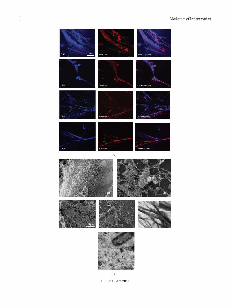
Increased extracellular DNA fibres with morphological NETcharacteristics were found in airway specimen from CFpatients (Figures 1(a) and 1(b)). Positive costaining forneutrophil elastase (Figure 1(a)) and citrullinated histones(Figures 1(a), 1(b), and 1(d)), as characteristic NET markers,and negative staining for F-actin (Figure 1(d)) supported thenotion that the CF airway DNA fibres represented NETsrather than necrosis-derived DNA. Treatment of CF airwayfluids with DNase dissolved these DNA-NET-like structuresfurther confirming their nature as DNA strands (data notshown). Ultrastructural imaging methods supported thesefindings and revealed a complex meshwork of DNA strandswith bacteria entangled (Figure 1(b)). Free DNA was presentin different CF airway compartments, in particular in sputum(Figure 1(a)), bronchoalveolar lavage fluid (BAL) (Figure 1(c),lower panel), and lung tissue (Figure 1(c) upper panel).Remarkably,most abundant freeDNAwas found inCF sputa,whereas in BAL and lung tissue, lower amounts of free DNAstrands were detected, which is consistent with the observa-tion that neutrophilic inflammation is most prominent in theproximal/bronchial airway compartments in CF lung disease.In BAL fluid, we observed lower amounts of DNA NET-like structures, but observed that elastase was associated tofree DNA structures and also appeared to colocalize withthe neutrophil’s cellular membrane (Figure 1(c), lower panel).In CF airway fluids, we found DNA/NET-like structuresassociated with bacteria (Figure 1(e)). However, associationsof free DNA structures with both live and dead bacteria werenoted (Figure 1(e)).
4 Mediators of Inflammation
(a)
(b)
Figure 1: Continued.

Mediators of Inflammation 5
(c)
(d)
(e)
Figure 1: Free NET-like DNA structures in CF lung disease. (a) Immunological characterization of free DNA structures in CF airway fluidsby CLSM. Upper two panel rows: NET-like DNA structures in induced CF sputum. Blue: DAPI stains DNA-NET backbone. Red: elastase.Lower two panel rows: Blue: DAPI, red: citrullinated histones. Scale bar: 20𝜇m. (b) Ultrastructure of free NET-like DNA structures. Upperand middle panels: SEM images of CF airway fluids. Arrow marks bacteria entrapped in DNA-NET-like structures. Scale bar: 2𝜇m; Middleright panel: TEM staining of citrullinated histones in CF airway fluids (sputa). Lower panel: Ultrathin sections of CF airway DNA-NET-like structures. The NETs and the bacterial extracellular polysaccharides are visualized by the ruthenium-red-osmium-tetroxide technique.Bacteria embedded in a dense wickerwork of NETs. Arrowheads mark NETs; asterisk: bacterial extracellular polysaccharide. (c)Upper panel:free DNA structures in CF lung tissue. Red: MPO (as characteristic NET component). Blue: DAPI (DNA). Inlays mark characteristic NET-areas. Lower panel: CF airway NETs in CF BAL fluids. Red: elastase (as characteristic NET component). Blue: DAPI (DNA). (d) Costainingsof DAPI, citrullinated histones, and F-actin. Scale bar: 20 𝜇m. (e) Dead/live staining of CF airway fluids (induced sputum). Free DNA anddead bacteria appear red; vital bacteria appear green.

6 Mediators of Inflammation
Stratifying CF patients for disease severity, we found thatCF patients with poor pulmonary function had higher levelsof free DNA in their airway fluids than patients with mildlung disease (Figure 2(a)). We further found that the airwayfree DNA levels were associated with fungal colonizationwith Aspergillus fumigatus but surprisingly not with bacterialinfection (Figure 2(a)). Representative NET-DNA (DAPI)staining of CF patient groups stratified for lung disease sever-ity is shown in Figure 2(b).Highly increased levels of theCXCchemokines CXCL1 (GRO-alpha), CXCL2 (GRO-beta), andCXCL8 (IL-8)were detected inCF airway fluids (Figure 2(c)).Since CXCR1 is proteolytically cleaved on CF airway neu-trophils [4, 32], CXCR2 remains the main binding sitefor these chemokines in the CF airway microenvironment.CF airway NETs correlated positively with levels of theproinflammatory chemokine CXCL2 (Figure 2(d)). Thesefindings provide evidence that CF lung disease features freeairwayDNA levels characteristic forNETosis and suggest thatincreased free DNA levels are associated with poor lungfunction of CF patients.
To investigate the role of free DNA/NET formation invivo, we used transgenic mice with airway-specific overex-pression of the amiloride-sensitive epithelial Na+ channel(𝛽ENaC-Tg) as amodel of CF-like lung disease [26, 27].Thesemice phenocopy airway surface liquid depletion, mucociliarydysfunction, and chronic airway disease with neutrophilicairway inflammation,mucus obstruction, and structural lungdamage [26, 27, 34]. Similar to human CF airway fluids,levels of the CXCR2 ligands CXCL1 and CXCL2 (Figure 3(a))and free DNA (Figure 3(b)) were highly increased in theBAL airway fluids of 𝛽ENaC-Tg mice compared to wild-typecontrols. Also consistent with the data obtained from humanCF lung disease samples, the extent of free DNA in murineCF airway fluids correlated positively with the levels of theCXCR2 ligand CXCL2 (Figure 3(c)) and with pulmonaryobstruction parameters (FEV100) (Figure 3(d)), whereas nocorrelationwas found between free DNA and levels of CXCL1or parameters of pulmonary restriction parameters (data notshown).
4. Discussion
NETosis is regarded as a double-edged sword in humandisease: on one hand, NETs can capture, immobilize, and killpathogens; on the other hand, uncontrolled and infection-independent NET formation has the potential to harm hosttissue through histones [35], proteases, or other mechanisms[21, 36, 37]. Related to the latter mechanism, NET formationhas recently been implicated in the pathogenesis of autoin-flammatory and autoimmune disease conditions, such aslupus, preeclampsia, septic shock, and autoimmune vasculitis[36–40]. Beyond these conditions, NETs were found, inconcert with platelets andmonocytes, to play a role in throm-bus formation and deep vein thrombosis [41, 42]. However,antihost defense and autoinflammatory conditions are notdichotomous in their nature and temporarily overlap uponresolution of infection or progression into chronic infec-tion, especially in immunocompromised conditions. Viewingthese findings in combination, the balance between targeted
antimicrobial host defense and nontargeted tissue damageof NET is delicate, but it is essential for the understandingand therapeutic potential ofNET formation in humandiseaseconditions.
The potential role of NETs in inflammatory lung diseaseshas just begun to evolve [40]. Several studies have now pro-vided evidence for NETosis or NETosis-like structures in CFlung disease and broadened the potential role of NET forma-tion in the complex pathogenesis of infective CF lung disease[43]. We and others have demonstrated previously that freeDNA NET-like structures are abundantly detectable in CFairway secretions [10–14]. Recently, a further study found thatCF sputum showed NETosis characteristics and implicatedmacrophage migration-inhibitory factor (MIF) in the forma-tion of NETs in the context of CF lung disease [12]. Here, weconfirm and extend these previous observations by showingthat CF airway free DNA levels correlate with pulmonaryobstruction in CF patients and mice. These observationsare reasonable, given the continuous and nonresolving neu-trophil recruitment and activation within CF airways, beingsupported by the beneficial effect of recombinant inhaledDNase (Dornase alpha) in CF patients with the strongestevidence in moderate and severe disease severity [44–46].Based on the increasing extent of neutrophilic inflammationand pulmonary obstruction, the evidence of free NET-likeDNA structures in CF airways tempts us to speculate that inearlier and milder stages of CF lung disease, NET formationmay act beneficial in providing extracellular antibacterial andantifungal host defense. At this time, DNasemay be usedwithmore caution, since encaptured pathogensmight be freed andcould cause additional host damage. On the other hand, inlatermoderate to severe stages of CF lung disease, the amountof mucus and DNA accumulation causes airway obstruction,rendering DNase efficient in cleaving DNA traps, and thiseffect probably overweighs the antimicrobial actions of NETformation, a hypothesis that awaits to be tested in further invivo studies and clinical trials. Our correlations between freeDNA levels and pulmonary obstruction parameters in humanpatients with CF and a mouse model of CF lung diseasesuggest that, at least in our CF cohort with a more advancedlung disease severity, NETs may cause more harm than goodto the host. The obvious limitations of our study and thewhole approach of free DNA analysis in CF airway fluidsremain that the free extracellular DNA could result fromdifferent forms of cell death in addition to NETosis, includingnecrosis, pyroptosis, and others. In addition, our studiesimage NET formation using fixation of biological samplesand are thereby limited by the fact that DNA structuresreminiscent of NETosis cannot be clearly attributed to activeNET formation, since this would require live cell imagingapproaches. Nevertheless, several other previous publicationshave provided indirect evidence indicating the presenceof NET structures in CF airway fluids [10–14], which issupported methodologically by our studies using confocallaser scanning, scanning electron microscopy, transmissionelectron microscopy, and atomic force microscopy, decreas-ing the probability of fixation/staining artefacts.
As CF lung disease is typically associated with chroniccolonizations and infections with Pseudomonas aeruginosa,

Mediators of Inflammation 7
∗
∗∗
∗ ∗
∗
∗
25000
20000
15000
10000
5000
00
20
40
60
80
100
120
0 5000 10000 15000 20000 25000
FEV
1 (%
of p
redi
cted
)
Free DNA (ng/mL)
Free
DN
A (n
g/m
L)
25000
20000
15000
10000
5000
0
Free
DN
A (n
g/m
L)
25000
20000
15000
10000
5000
0
Free
DN
A (n
g/m
L)
Controls CF
Controls CFControls CF
≤50 50–70 ≥70
0 1 2 3 0 1 2 3AspergillusPseudomonas
FEV1 FEV1 FEV1
r = −0.73, P < 0.01
(a)
CFHealthy
10𝜇m FEV1≤ 50FEV1 50–70FEV1≥ 70
(b)
100000
10000
1000
100
10
1CXCL1 CXCL2 CXCL8
∗∗
∗
(pg/
mL)
(c)
0
500
1000
1500
2000
2500
3000
0 5000 10000 15000 20000
CXCL
2 (p
g/m
L)
Free DNA (ng/mL)
r = 0.83, P < 0.01
(d)
Figure 2: Free DNA, disease severity, and infection in CF lung disease. (a) Stratification of CF patients (𝑛 = 80). Upper left: correlation offree DNA levels with FEV
1in sputum supernatants from CF patients. Upper right: free DNA levels in sputum supernatants from healthy
controls and CF patients, stratified by FEV1. Lower panels: free DNA levels in sputum supernatants from healthy controls and CF patients,
stratified by Pseudomonas aeruginosa orAspergillus fumigatus infection/colonization status (Leeds criteria, 0: never, 1: negative, 2: intermittent,and 3: chronic). (b) Representative CLSM images of airway fluids from one healthy individual and three different CF patients, stratified forlung function, are depicted. Scale bar: 10𝜇m; FEV
1: forced expiratory volume in 1 second (% of prediction). DAPI staining of nuclei and
extracellular DNA strands in CF sputa. (c) Chemokine levels in sputum supernatants from healthy controls (white, 𝑛 = 6) or CF patients(grey, 𝑛 = 50). (d) Correlation of free DNA with CXCL2 levels in airway fluids (cell-free sputum supernatants) from CF patients (𝑛 = 50).∗𝑃 < 0.05.

8 Mediators of Inflammation
0
100
200
300
400
500
600
WT
CXCL1 CXCL2
(pg/
mL)
𝛽ENaC-Tg
∗
∗
(a)
0
250
500
750
1000
WT
Free
DN
A (n
g/m
L)
𝛽ENaC-Tg
∗
(b)
0100200300400500600700
0 200 400 600 800 1000
CXCL
2 (p
g/m
L)
Free DNA (ng/mL)
WT
r = 0.6, P < 0.01
𝛽ENaC-Tg
(c)
00.20.40.60.8
11.21.4
0 200 400 600 800 1000
FEV
100
(mL/
s)
Free DNA (ng/mL)
r = 0.84, P < 0.01
WT𝛽ENaC-Tg
(d)
Figure 3: Free DNA in murine CF lung disease in vivo. (a) CXCR2 chemokines in BALF from 𝛽ENaC-transgenic (𝛽ENaC-Tg, 𝑛 = 19) andwild-type (WT, 𝑛 = 11) mice. Levels of CXCL1 (KC) and CXCL2 (MIP-2) were quantified by ELISA. (b) Free DNA in BALs from 𝛽ENaC-Tgmice. Free DNA was quantified in BALF from 𝛽ENaC-Tg (filled circles, 𝑛 = 19) and wild-type (empty circles, 𝑛 = 11) mice. (c) Correlationof free DNA with CXCL2 levels in BAL. (d) Correlation of free DNA with lung function (FEV100). ∗𝑃 < 0.05.
the effect of this pathogen on NET formation has beeninvestigated using different methodologies and modellingsystems [11, 13, 47–49]. The latter studies found that Pseu-domonas aeruginosa efficiently induced NET formation,particularly in solution in vitro, and demonstrated thatNETs can kill this pathogen through an extracellular DNA-mediatedmechanism.At single cell level, authors also showedthat neutrophils from CF patients had no cell intrinsicalteration in NET formation [11], supporting the notionthat the NET accumulation found in CF airway fluids isnot specific to CFTR mutation-based disease conditions,but rather represent a prototypical picture of severe andchronic neutrophilic inflammation in a body compartment.Serum DNases and/or antioxidants have been shown todampen/inhibit NET formation. As CF airway fluids showa low antioxidant activity and are substantially different intheir biochemical properties, we speculate that this particularmicroenvironment favors NET formation. While we did notfind a statistically significant association of Pseudomonasaeruginosa infection status with free DNA levels, a positivecorrelationwas found for fungal colonizationwithAspergillusfumigatus, which is in line with a recent study showing thatNETs are mainly formed in response to large pathogens, suchas fungi [50].
A recent study analyzing neutrophils from a patient withPapillon-Lefevre syndrome (PLS), lacking serine proteases,showed that neutrophils from this patient failed to produceNETs [51], corroborating the concept that serine proteases,particularly elastase, regulate NET formation [50, 52–54].Papayannopoulos and coworkers demonstrated that neu-trophil elastase, which is highly increased in CF airway fluidsand represents a promising therapeutic target [1, 4], enhancessputum solubilization by cleaving histones to enhance theaccess of exogenous nucleases to DNA [10]. The researchersalso showed that neutrophil elastase is bound to DNA, whichdownregulates its proteolytic activity and could restrict hosttissue damage. This study has been extended by a recentreport demonstrating that NETs represent a reservoir ofactive proteases and DNase treatment increases free pro-teolytic activities [55], suggesting that CF patients underDNase treatment could benefit from the addition of proteaseinhibitors [56].
Previous studies found a correlation between airwayDNA levels, neutrophilic inflammation, and lung functionparameters in CF patients [9, 57, 58]. Our study confirmsand extends these findings by showing a correlation betweenfree DNA levels with lung function, chemokine levels, and
Mediators of Inflammation 9
fungal colonization in CF patients. The correlation betweenthe neutrophil chemokine CXCL2 and free DNA levels in CFairway fluids could, on one hand, reflect increased CXCL2-mediated neutrophil chemotaxis or could also, on the otherhand, involve CXCL2 itself as potential trigger for DNArelease in neutrophils. As there is a lack of convincing datasupporting the latter hypothesis, the underlying mechanisticbasis for this correlation remains to be dissected in the future.
Besides NET-associated products, such as proteases, thatcan cause harm to the host, a recent study identified sur-factant protein D as key protein binding to NETs, therebypromotingNET-mediated trapping of P. aeruginosa [59].Thishas ample implications for CF lung disease, since surfactantprotein D has been found to be degraded/cleaved by bothserine and matrix metalloproteases in CF airway fluids[60–64], which, as a result, could impair the antibacterialNETosis-related effects of surfactant protein D in CF airwaysin vivo. Regarding surfactant protein D and beyond, proteinsthat may interfere with NET formation and/or NET activitiescould be attractive therapeutic targets for advanced CF lungdisease.
Our view of NET formation has been further extendedby the description of rapid NET formation in vitro [65] andin vivo [24, 25], unraveling a so far unappreciated modeof collaboration between NET formation, chemotaxis, andphagocytosis.While there is convincing data in skin infectionmodels in mice, evidence on human neutrophils is still lim-ited. The potential role of rapid NET formation for CF lungdisease remains to be dissected.
In summary, our study, in line with previous investiga-tions, demonstrates that free DNA with NET-like character-istics represents an extracellular component of CF airwayfluids. In advanced stages of CF lung disease, NETs seemto do more harm than good, but experimental data fora causative relationship is lacking. Approaches to interferewith NET formation or NET-associated products, such asDNases, antiproteases, supplementing surfactant protein D,targeting histones [66], or a combination thereof, couldrepresent promising therapeutic strategies forCF lung diseaseand other chronic lung diseases associated with sustainedneutrophilic inflammation [38, 40, 43].
Conflict of Interests
The authors declare that they have no competing financialinterests.
Acknowledgments
The authors thank both Barbara Lange-Sperandio and FriggaBeitinger, LMU, Munich, for lung sample preparations. Theythank Olivier Gires, LMU, Munich, and Lauren Mays, Uni-versity of Pennsylvania School ofMedicine, Philadelphia, PA,for the paper’s discussions. They thank Michaela Klappacher,Astrid Obermayer, andMirjam Franz for assistance in exper-imental workup and Stephanie Hirtz for breeding and geno-typing of experimental animals. This paper is supported bythe German Research Foundation (DFG, Emmy Noether
Programme HA 5274/3-1 to Dominik Hartl, MA 2081/3-3and MA 2081/4-1 to Marcus A. Mall), the German Society ofPediatric Pneumology (Dominik Hartl), PINA e.V. (DominikHartl), the Novartis Foundation (Dominik Hartl), and theErnest-Solvay-Foundation (Dominik Hartl).
References
[1] M. A. Mall and D. Hartl, “CFTR: cystic fibrosis and beyond,”EuropeanRespiratory Journal, vol. 44, no. 4, pp. 1042–1054, 2014.
[2] E. Fantino, C. L. Gangell, D. Hartl, P. D. Sly, and C. F. Arest,“Airway, but not serum or urinary, levels of YKL-40 reflectinflammation in early cystic fibrosis lung disease,” BMC Pul-monary Medicine, vol. 14, article 28, 2014.
[3] P. D. Sly, C. L. Gangell, L. Chen et al., “Risk factors forbronchiectasis in childrenwith cystic fibrosis,”TheNewEnglandJournal of Medicine, vol. 368, no. 21, pp. 1963–1970, 2013.
[4] D. Hartl, A. Gaggar, E. Bruscia et al., “Innate immunity in cysticfibrosis lung disease,” Journal of Cystic Fibrosis, vol. 11, no. 5, pp.363–382, 2012.
[5] S. D. Sagel,M. K. Sontag, J. S.Wagener, R. K. Kapsner, I. Osberg,and F. J. Accurso, “Induced sputum inflammatorymeasures cor-relate with lung function in children with cystic fibrosis,” Jour-nal of Pediatrics, vol. 141, no. 6, pp. 811–817, 2002.
[6] S. D. Sagel, R. Kapsner, I. Osberg, M. K. Sontag, and F. J.Accurso, “Airway inflammation in children with cystic fibrosisand healthy children assessed by sputum induction,”TheAmer-ican Journal of Respiratory and Critical Care Medicine, vol. 164,no. 8 I, pp. 1425–1431, 2001.
[7] A. Hector, M. S. D. Kormann, I. Mack et al., “The chitinase-likeprotein YKL-40 modulates cystic fibrosis lung disease,” PLoSONE, vol. 6, no. 9, Article ID e24399, 2011.
[8] S. Gehrig, J. Duerr,M.Weitnauer et al., “Lack of neutrophil elas-tase reduces inflammation, mucus hypersecretion, and emphy-sema, but notmucus obstruction, inmicewith cystic fibrosislikelung disease,” American Journal of Respiratory and CriticalCare Medicine, vol. 189, no. 9, pp. 1082–1092, 2014.
[9] K. K. Kirchner, J. S. Wagener, T. Z. Khan, S. C. Copenhaver, andF. J. Accurso, “Increased DNA levels in bronchoalveolar lavagefluid obtained from infants with cystic fibrosis,” AmericanJournal of Respiratory and Critical CareMedicine, vol. 154, no. 5,pp. 1426–1429, 1996.
[10] V. Papayannopoulos, D. Staab, and A. Zychlinsky, “Neu-trophil elastase enhances sputum solubilization in cystic fibrosispatients receiving dnase therapy,” PLoS ONE, vol. 6, no. 12,Article ID e28526, 2011.
[11] R. L. Young, K. C. Malcolm, J. E. Kret et al., “Neutrophil extra-cellular trap (NET)-mediated killing of pseudomonas aerug-inosa: evidence of acquired resistance within the CF airway,independent of CFTR,” PLoS ONE, vol. 6, no. 9, Article IDe23637, 2011.
[12] M. Dwyer, Q. Shan, S. D’Ortona et al., “Cystic fibrosis sputumDNA Has NETosis characteristics and neutrophil extracellulartrap release is regulated by macrophage migration-inhibitoryfactor,” Journal of Innate Immunity, vol. 6, no. 6, pp. 765–779,2014.
[13] D.-G. Yoo, M. Floyd, M. Winn, S. M. Moskowitz, and B. Rada,“NET formation induced by Pseudomonas aeruginosa cysticfibrosis isolates measured as release of myeloperoxidase-DNAand neutrophil elastase-DNA complexes,” Immunology Letters,vol. 160, no. 2, pp. 186–194, 2014.

10 Mediators of Inflammation
[14] R.Manzenreiter, F. Kienberger, V.Marcos et al., “Ultrastructuralcharacterization of cystic fibrosis sputum using atomic forceand scanning electron microscopy,” Journal of Cystic Fibrosis,vol. 11, no. 2, pp. 84–92, 2012.
[15] V. Brinkmann, U. Reichard, C. Goosmann et al., “Neutrophilextracellular traps kill bacteria,” Science, vol. 303, no. 5663, pp.1532–1535, 2004.
[16] V. Brinkmann and A. Zychlinsky, “Beneficial suicide: whyneutrophils die to make NETs,” Nature Reviews Microbiology,vol. 5, no. 8, pp. 577–582, 2007.
[17] T. A. Fuchs, U. Abed, C. Goosmann et al., “Novel cell deathprogram leads to neutrophil extracellular traps,”The Journal ofCell Biology, vol. 176, no. 2, pp. 231–241, 2007.
[18] S. Yousefi, J. A. Gold, N. Andina et al., “Catapult-like release ofmitochondrial DNA by eosinophils contributes to antibacterialdefense,” Nature Medicine, vol. 14, no. 9, pp. 949–953, 2008.
[19] S. Yousefi,D. Simon, andH.-U. Simon, “Eosinophil extracellularDNA traps: molecular mechanisms and potential roles indisease,”Current Opinion in Immunology, vol. 24, no. 6, pp. 736–737, 2012.
[20] S. Yousefi, C. Mihalache, E. Kozlowski, I. Schmid, and H. U.Simon, “Viable neutrophils release mitochondrial DNA to formneutrophil extracellular traps,”Cell Death&Differentiation, vol.16, no. 11, pp. 1438–1444, 2009.
[21] D. Simon, H.-U. Simon, and S. Yousefi, “Extracellular DNAtraps in allergic, infectious, and autoimmune diseases,” Allergy,vol. 68, no. 4, pp. 409–416, 2013.
[22] M. Morshed, R. Hlushchuk, D. Simon et al., “NADPHoxidase-independent formation of extracellular DNA traps bybasophils,”The Journal of Immunology, vol. 192, no. 11, pp. 5314–5323, 2014.
[23] R. Dworski, H.-U. Simon, A. Hoskins, and S. Yousefi,“Eosinophil and neutrophil extracellular DNA traps in humanallergic asthmatic airways,” Journal of Allergy and ClinicalImmunology, vol. 127, no. 5, pp. 1260–1266, 2011.
[24] A. Peschel and D. Hartl, “Anuclear neutrophils keep hunting,”Nature Medicine, vol. 18, no. 9, pp. 1336–1338, 2012.
[25] B. G. Yipp, B. Petri, D. Salina et al., “DynamicNETosis is carriedout by live neutrophils in human andmouse bacterial abscessesand during severe gram-positive infection,” Nature Medicine,vol. 6, no. 20, 2012.
[26] M. Mall, B. R. Grubb, J. R. Harkema, W. K. O’Neal, and R. C.Boucher, “Increased airway epithelial Na+ absorption producescystic fibrosis-like lung disease in mice,” Nature Medicine, vol.10, no. 5, pp. 487–493, 2004.
[27] Z. Zhou, J. Duerr, B. Johannesson et al., “The ENaC-overexpressingmouse as amodel of cystic fibrosis lung disease,”Journal of Cystic Fibrosis, vol. 10, supplement 2, pp. S172–S182,2011.
[28] M. A. Mall, S. Y. Graeber, M. Stahl, and Z. Zhou-Suckow, “Earlycystic fibrosis lung disease: Role of airway surface dehydrationand lessons from preventive rehydration therapies in mice,”TheInternational Journal of Biochemistry & Cell Biology, vol. 52, pp.174–179, 2014.
[29] M. A. Mall, J. R. Harkema, J. B. Trojanek et al., “Developmentof chronic bronchitis and emphysema in beta-epithelial Na+channel-overexpressing mice,” The American Journal of Respi-ratory and Critical Care Medicine, vol. 177, no. 7, pp. 730–742,2008.
[30] T. W. R. Lee, K. G. Brownlee, S. P. Conway, M. Denton, andJ. M. Littlewood, “Evaluation of a new definition for chronic
Pseudomonas aeruginosa infection in cystic fibrosis patients,”Journal of Cystic Fibrosis, vol. 2, no. 1, pp. 29–34, 2003.
[31] D. Hartl, V. Starosta, K. Maier et al., “Inhaled glutathionedecreases PGE
2and increases lymphocytes in cystic fibrosis
lungs,” Free Radical Biology andMedicine, vol. 39, no. 4, pp. 463–472, 2005.
[32] D. Hartl, P. Latzin, P. Hordijk et al., “Cleavage of CXCR1 onneutrophils disables bacterial killing in cystic fibrosis lungdisease,” Nature Medicine, vol. 13, no. 12, pp. 1423–1430, 2007.
[33] J. A. J. Vanoirbeek,M. Rinaldi, V. DeVooght et al., “Noninvasiveand invasive pulmonary function in mouse models of obstruc-tive and restrictive respiratory diseases,” The American Journalof Respiratory Cell and Molecular Biology, vol. 42, no. 1, pp. 96–104, 2010.
[34] M. A.Mall, B. Button, B. Johannesson et al., “Airway surface liq-uid volume regulation determines different airway phenotypesin liddle compared with 𝛽ENaC-overexpressing mice,” Journalof Biological Chemistry, vol. 285, no. 35, pp. 26945–26955, 2010.
[35] M. Saffarzadeh, C. Juenemann, M. A. Queisser et al.,“Neutrophil extracellular traps directly induce epithelial andendothelial cell death: a predominant role of histones,” PLoSONE, vol. 7, no. 2, Article ID e32366, 2012.
[36] V. Papayannopoulos and A. Zychlinsky, “NETs: a new strategyfor using old weapons,” Trends in Immunology, vol. 30, no. 11,pp. 513–521, 2009.
[37] A. Zychlinsky, “Neutrophil extracellular traps,” European Jour-nal of Clinical Investigation, vol. 39, p. 27, 2009.
[38] M. Zawrotniak andM. Rapala-Kozik, “Neutrophil extracellulartraps (NETs)—formation and implications,” Acta BiochimicaPolonica, vol. 60, no. 3, pp. 277–284, 2013.
[39] B. G. Yipp and P. Kubes, “NETosis: how vital is it?” Blood, vol.122, no. 16, pp. 2784–2794, 2013.
[40] O. Z. Cheng and N. Palaniyar, “NET balancing: a problem ininflammatory lung diseases,” Frontiers in Immunology, vol. 4,article 1, 2013.
[41] T. A. Fuchs, A. Brill, and D. D. Wagner, “Neutrophil extracel-lular trap (NET) impact on deep vein thrombosis,” Arterioscle-rosis, Thrombosis, and Vascular Biology, vol. 32, no. 8, pp. 1777–1783, 2012.
[42] M.-L. von Bruhl, K. Stark, A. Steinhart et al., “Monocytes,neutrophils, and platelets cooperate to initiate and propagatevenous thrombosis in mice in vivo,” Journal of ExperimentalMedicine, vol. 209, no. 4, pp. 819–835, 2012.
[43] S. Rahman and M. Gadjeva, “Does NETosis contribute tothe bacterial pathoadaptation in cystic fibrosis?” Frontiers inImmunology, vol. 5, article 378, 2014.
[44] P. A. Flume, B. P. O’Sullivan, K. A. Robinson et al., “Cystic fibro-sis pulmonary guidelines: chronicmedications formaintenanceof lung health,” American Journal of Respiratory and CriticalCare Medicine, vol. 176, no. 10, pp. 957–969, 2007.
[45] G. Doring, P. Flume, H.Heijerman, and J. S. Elborn, “Treatmentof lung infection in patients with cystic fibrosis: current andfuture strategies,” Journal of Cystic Fibrosis, vol. 11, no. 6, pp. 461–479, 2012.
[46] P. A. Flume andD. R. vanDevanter, “State of progress in treatingcystic fibrosis respiratory disease,”BMCMedicine, vol. 10, article88, 2012.
[47] Q. Shan, M. Dwyer, S. Rahman, and M. Gadjeva, “Distinctsusceptibilities of corneal Pseudomonas aeruginosa clinicalisolates to neutrophil extracellular trap-mediated immunity,”Infection and Immunity, vol. 82, no. 10, pp. 4135–4143, 2014.

Mediators of Inflammation 11
[48] D. G. Yoo, M. Winn, L. Pang et al., “Release of cystic fibrosisairway inflammatory markers from pseudomonas aeruginosa-stimulated human neutrophils involves NADPH oxidase-dependent extracellular DNA trap formation,” Journal ofImmunology, vol. 192, no. 10, pp. 4728–4738, 2014.
[49] B. Rada, M. A. Jendrysik, L. Pang et al., “Pyocyanin-enhancedneutrophil extracellular trap formation requires the NADPHoxidase,” PLoS ONE, vol. 8, no. 1, Article ID e54205, 2013.
[50] N. Branzk, A. Lubojemska, S. E. Hardison et al., “Neutrophilssense microbe size and selectively release neutrophil extracel-lular traps in response to large pathogens,”Nature Immunology,vol. 15, no. 11, pp. 1017–1025, 2014.
[51] O. E. Sørensen, S. N. Clemmensen, S. L. Dahl et al., “Papillon-Lefevre syndrome patient reveals species-dependent require-ments for neutrophil defenses,” The Journal of Clinical Investi-gation, vol. 124, no. 10, pp. 4539–4548, 2014.
[52] K.D.Metzler, C.Goosmann,A. Lubojemska, A. Zychlinsky, andV. Papayannopoulos, “A myeloperoxidase-containing complexregulates neutrophil elastase release and actin dynamics duringNETosis,” Cell Reports, vol. 8, no. 3, pp. 883–896, 2014.
[53] N. Branzk and V. Papayannopoulos, “Molecular mechanismsregulating NETosis in infection and disease,” Seminars inImmunopathology, vol. 35, no. 4, pp. 513–530, 2013.
[54] V. Papayannopoulos, K. D. Metzler, A. Hakkim, and A. Zych-linsky, “Neutrophil elastase and myeloperoxidase regulate theformation of neutrophil extracellular traps,”The Journal of CellBiology, vol. 191, no. 3, pp. 677–691, 2010.
[55] A. V. Dubois, A. Gauthier, D. Brea et al., “Influence of DNA onthe activities and inhibition of neutrophil serine proteases incystic fibrosis sputum,”American Journal of Respiratory Cell andMolecular Biology, vol. 47, no. 1, pp. 80–86, 2012.
[56] M. Griese, M. Kappler, A. Gaggar, and D. Hartl, “Inhibitionof airway proteases in cystic fibrosis lung disease,” EuropeanRespiratory Journal, vol. 32, no. 3, pp. 783–795, 2008.
[57] F. Carswell, D.W.Robinson,C.C. L.Ward, andM.R.Waterfield,“Deoxyribonucleic acid output in the sputum from cysticfibrosis patients,” European Journal of Respiratory Diseases, vol.65, no. 1, pp. 53–57, 1984.
[58] J.-S. Kim, K. Okamoto, and B. K. Rubin, “Pulmonary functionis negatively correlated with sputum inflammatorymarkers andcough clearability in subjects with cystic fibrosis but not thosewith chronic bronchitis,” Chest, vol. 129, no. 5, pp. 1148–1154,2006.
[59] D. N. Douda, R. Jackson, H. Grasemann, and N. Palaniyar,“Innate immune collectin surfactant protein D simultaneouslybinds both neutrophil extracellular traps and carbohydrate lig-ands and promotes bacterial trapping,”The Journal of Immunol-ogy, vol. 187, no. 4, pp. 1856–1865, 2011.
[60] P. E. Bratcher, N. M. Weathington, H. J. Nick, P. L. Jackson, R. J.Snelgrove, and A. Gaggar, “MMP-9 cleaves SP-D and abrogatesits innate immune functions in vitro,” PLoS ONE, vol. 7, no. 7,Article ID e41881, 2012.
[61] H. V. Olesen, U. Holmskov, P. O. Schiøtz, and G. L. Sørensen,“Serum-surfactant SP-D correlates inversely to lung function incystic fibrosis,” Journal of Cystic Fibrosis, vol. 9, no. 4, pp. 257–262, 2010.
[62] J. Cooley, B. McDonald, F. J. Accurso, E. C. Crouch, and E.Remold-O’Donnell, “Patterns of neutrophil serine protease-dependent cleavage of surfactant protein D in inflammatorylung disease,” Journal of Leukocyte Biology, vol. 83, no. 4, pp.946–955, 2008.
[63] D. Hartl and M. Griese, “Surfactant protein D in human lungdiseases,” European Journal of Clinical Investigation, vol. 36, no.6, pp. 423–435, 2006.
[64] C. von Bredow, A. Wiesener, and M. Griese, “Proteolysis ofsurfactant protein D by cystic fibrosis relevant proteases,” Lung,vol. 181, no. 2, pp. 79–88, 2003.
[65] F. H. Pilsczek, D. Salina, K. K. H. Poon et al., “A novel mech-anism of rapid nuclear neutrophil extracellular trap formationin response to Staphylococcus aureus,” Journal of Immunology,vol. 185, no. 12, pp. 7413–7425, 2010.
[66] M. Saffarzadeh and K. T. Preissner, “Fighting against the darkside of neutrophil extracellular traps in disease: manoeuvres forhost protection,” Current Opinion in Hematology, vol. 20, no. 1,pp. 3–9, 2013.

Submit your manuscripts athttp://www.hindawi.com
Stem CellsInternational
Hindawi Publishing Corporationhttp://www.hindawi.com Volume 2014
Hindawi Publishing Corporationhttp://www.hindawi.com Volume 2014
MEDIATORSINFLAMMATION
of
Hindawi Publishing Corporationhttp://www.hindawi.com Volume 2014
Behavioural Neurology
EndocrinologyInternational Journal of
Hindawi Publishing Corporationhttp://www.hindawi.com Volume 2014
Hindawi Publishing Corporationhttp://www.hindawi.com Volume 2014
Disease Markers
Hindawi Publishing Corporationhttp://www.hindawi.com Volume 2014
BioMed Research International
OncologyJournal of
Hindawi Publishing Corporationhttp://www.hindawi.com Volume 2014
Hindawi Publishing Corporationhttp://www.hindawi.com Volume 2014
Oxidative Medicine and Cellular Longevity
Hindawi Publishing Corporationhttp://www.hindawi.com Volume 2014
PPAR Research
The Scientific World JournalHindawi Publishing Corporation http://www.hindawi.com Volume 2014
Immunology ResearchHindawi Publishing Corporationhttp://www.hindawi.com Volume 2014
Journal of
ObesityJournal of
Hindawi Publishing Corporationhttp://www.hindawi.com Volume 2014
Hindawi Publishing Corporationhttp://www.hindawi.com Volume 2014
Computational and Mathematical Methods in Medicine
OphthalmologyJournal of
Hindawi Publishing Corporationhttp://www.hindawi.com Volume 2014
Diabetes ResearchJournal of
Hindawi Publishing Corporationhttp://www.hindawi.com Volume 2014
Hindawi Publishing Corporationhttp://www.hindawi.com Volume 2014
Research and TreatmentAIDS
Hindawi Publishing Corporationhttp://www.hindawi.com Volume 2014
Gastroenterology Research and Practice
Hindawi Publishing Corporationhttp://www.hindawi.com Volume 2014
Parkinson’s Disease
Evidence-Based Complementary and Alternative Medicine
Volume 2014Hindawi Publishing Corporationhttp://www.hindawi.com