Exhaled NO and small airway function in asthma and cystic ... · Exhaled NO and small airway...
106
Doctoral Thesis for the degree of Doctor of Medicine, the Sahlgrenska Academy, University of Gothenburg, Gothenburg, Sweden Exhaled NO and small airway function in asthma and cystic fibrosis Christina Keen Fredriksson Department of Paediatrics Institute of Clinical Sciences at Sahlgrenska Academy University of Gothenburg 2010
Transcript of Exhaled NO and small airway function in asthma and cystic ... · Exhaled NO and small airway...
Doctoral Thesis for the degree of Doctor of Medicine,
the Sahlgrenska Academy, University of Gothenburg,
Gothenburg, Sweden
Exhaled NO and small airway function
in asthma and cystic fibrosis
Christina Keen Fredriksson
Department of Paediatrics
Institute of Clinical Sciences at
Sahlgrenska Academy
University of Gothenburg
2010

Printed by
Intellecta Infolog, Göteborg, 2010
ISBN 978-91-628-8017-0

”The million dollar question” How to monitor and treat subjects
with multifaceted airway disease?

List of papers
This thesis is based on the following papers:
Paper I C Keen, A-C Olin, Å Edentoft, E Gronowitz and B Strandvik. Airway nitric
oxide in patients with cystic fibrosis is associated with pancreatic function,
pseudomonas infection and polyunsaturated fatty acids. CHEST, 2007;
131(6):1857-1864
Paper II C Keen, A-C Olin, S Eriksson, A Ekman, A Lindblad, S Basu, C Beermann and
B Strandvik. Supplementation with fatty acids influences the airway nitric oxide
and inflammatory markers in patients with cystic fibrosis. Journal of Pediatric
Gastroenterology and Nutrition, 2010; 50(5):537-544.
Paper III C Keen, P Gustafsson, A Lindblad, G Wennergren and A-C Olin. Low levels of
exhaled nitric oxide are associated with impaired lung function in cystic
fibrosis. Pediatric Pulmonology. 2010; 45(3):241-8.
Paper IV C Keen, A-C Olin, G Wennergren, F Aljassim and P Gustafsson:
Exhaled NO, small airway function and airway hyper-responsiveness in
paediatric asthma. (Submitted)
The papers will be referred to in the text by their Roman numerals.

Exhaled NO and small airway function
in asthma and cystic fibrosis
Christina Keen Fredriksson
Dept. of Paediatrics, Institute of Clinical Sciences,
Sahlgrenska Academy, University of Gothenburg, Gothenburg, Sweden
Background: Asthma and cystic fibrosis (CF) are chronic inflammatory airway
disorders known to involve the peripheral airways. Non-invasive tests sensitive
to peripheral airway function and inflammation are therefore of high priority.
Multiple breath inert gas washout (MBW) is a test sensitive to small airway
function and exhaled nitric oxide (NO) reflects airway inflammation in asthma.
Aim: To use exhaled NO in combination with MBW to assess the contribution
of small airway inflammation and dysfunction in paediatric asthma and CF in
order to potentially allow for earlier intervention and more successful
management of these conditions in the future.
Results: CF subjects had reduced levels of nasal and exhaled NO. All but three
children with CF had abnormally elevated LCI. Low levels of NO were
associated with impaired airway function, chronic infection with Ps.
Aeruginosa, severe genotypes and the fatty acid deficiency characteristic for CF
subjects. Low levels of alveolar NO, albeit not lower in CF than in healthy
controls, were associated with increased systemic inflammation and chronic
bacterial airway colonisation.
LCI, Scond, and Sacin were all significantly elevated in children with asthma and
Scond was the marker that most significantly differentiated the asthmatic children
from the healthy controls. Increased Scond was associated with increased levels
of exhaled NO and airway hyper-responsiveness and Sacin correlated with
alveolar NO.
Conclusions: This thesis provides further evidence of small airway
involvement in both paediatric asthma and CF. The findings of clinically
significant dysfunction of the small conducting airways in paediatric asthma and
the associations between small airway dysfunction, increased levels of exhaled
NO and airway hyper-responsiveness are novel findings. In CF, low levels of
exhaled NO are linked to small airway dysfunction. These findings provide new
exciting insights into the pathology and pathophysiology of paediatric asthma
and CF and may allow for earlier and better targeted interventions.
Keywords: asthma, children, cystic fibrosis, flow independent exhaled nitric
oxide, multiple breath inert gas washout, polyunsaturated fatty acids.
ISBN 978-91-628-8017-0
http://hdl.handle.net/2077/22183

Abbreviations
AA = arachidonic acid
ACT= Asthma Control Test
AHR = airway hyper-responsiveness
ALA = α-linolenic acid
ATS = American Thoracic Society
BMI = body mass index
cACT = child Asthma Control Test
CF = cystic fibrosis
Calv= alveolar NO concentration
cNOS = constitutive nitric oxide synthase
CFTR = cystic fibrosis transmembrane conductance regulator
DawNO = bronchial wall NO diffusion capacity
DHA = docosahexaenoic acid
eNOS = endothelial nitric oxide synthase
EDRF = endothelium dependent relaxing factor
EPA = eicosapentaenoic acid
ERS = European Respiratory Society
ESR = erythrocyte sedimentation rate
FA = fatty acid(s)
FENO = fraction of exhaled nitric oxide
FENO50 = fraction of exhaled nitric oxide at 50 ml/s
FEV1.0 = forced expiratory volume in one second
FRC = functional residual capacity
FVC = forced vital capacity
ICS =inhaled corticosteroid
IgG = immunoglobulin G
iNOS = inducible nitric oxide synthase
JawNO = bronchial NO flux
LA = linoleic acid
LCI = lung clearance index
LLN = lower limit of normal
MBW = multiple breath inert gas washout
MMEF = maximum mid expiratory flow
nNO = nasal nitric oxide
nNOS = neuronal nitric oxide synthase
NO = nitric oxide
NOS = nitric oxide synthase
OA = oleic acid
ppb = parts per billion
PUFA = polyunsaturated fatty acids
SF6 = sulphur hexafluoride
SnIII = normalized phase III slope
ULN = upper limit of normal
WBC = white blood cells
Vt = tidal volume

Contents
Introduction ________________________________________ 5
Airway inflammation _____________________________________ 5
Exhaled nitric oxide _____________________________________ 7 Background ________________________________________________ 7 Exhaled NO - how to measure? _________________________________ 9 Exhaled NO and allergic sensitization (atopy) _____________________ 11 Flow independent NO variables ________________________________ 12 Nasal NO _________________________________________________ 16 Exhaled NO and asthma _____________________________________ 17 Flow independent NO variables in asthma _______________________ 18 Exhaled NO and CF _________________________________________ 19 Flow independent NO variables in CF ___________________________ 20
Asthma _______________________________________________ 21 How to monitor asthma? _____________________________________ 22
Cystic fibrosis _________________________________________ 23 Fatty acids in CF and fatty acids and inflammation _________________ 25
Small airways _________________________________________ 28
Lung function tests _____________________________________ 30 Spirometry ________________________________________________ 30 Multiple breath inert gas washout ______________________________ 30 Airway challenge testing _____________________________________ 34
Asthma Control Test ____________________________________ 35
Aims _____________________________________________ 36
Specific aims in paper I-IV _______________________________ 36
Study concept _____________________________________ 37
Materials _________________________________________ 39
Subjects in study I-IV ___________________________________ 39 Healthy controls ____________________________________________ 42
Methods __________________________________________ 43
Exhaled and nasal NO __________________________________ 43 Exhaled NO _______________________________________________ 43 NO flow-independent variables ________________________________ 43 Nasal NO _________________________________________________ 44
2
Lung function tests _____________________________________ 44 Spirometry ________________________________________________ 44 Multiple breath inert gas washout ______________________________ 44 Airway challenge ___________________________________________ 46
Asthma Control Test ____________________________________ 46
Fatty acids ____________________________________________ 46
Urinary analysis _______________________________________ 46
Systemic inflammatory variables _________________________ 47
Statistics _____________________________________________ 47
Ethics ________________________________________________ 47
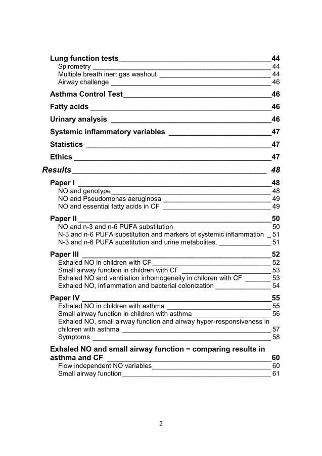
Results ___________________________________________ 48
Paper I _______________________________________________ 48 NO and genotype ___________________________________________ 48 NO and Pseudomonas aeruginosa _____________________________ 49 NO and essential fatty acids in CF _____________________________ 49
Paper II _______________________________________________ 50 NO and n-3 and n-6 PUFA substitution __________________________ 50 N-3 and n-6 PUFA substitution and markers of systemic inflammation _ 51 N-3 and n-6 PUFA substitution and urine metabolites. ______________ 51
Paper III ______________________________________________ 52 Exhaled NO in children with CF ________________________________ 52 Small airway function in children with CF ________________________ 53 Exhaled NO and ventilation inhomogeneity in children with CF _______ 53 Exhaled NO, inflammation and bacterial colonization _______________ 54
Paper IV ______________________________________________ 55 Exhaled NO in children with asthma ____________________________ 55 Small airway function in children with asthma _____________________ 56 Exhaled NO, small airway function and airway hyper-responsiveness in children with asthma ________________________________________ 57 Symptoms ________________________________________________ 58
Exhaled NO and small airway function − comparing results in asthma and CF ________________________________________ 60
Flow independent NO variables ________________________________ 60 Small airway function ________________________________________ 61

3
Discussion ________________________________________ 63
Exhaled NO and airway function in cystic fibrosis ___________ 64
Substitution with fatty acids in CF ________________________ 66
Exhaled NO and small airway function in asthma ____________ 67
Alveolar NO in asthma and CF ____________________________ 69
Methodological issues __________________________________ 70
Can exhaled NO be used as a biomarker in asthma and CF? __ 72
Conclusions and future studies _______________________ 73
FENO50 longitudinally in CF patients _______________________ 73
Multi centre study with n-3 fatty acids in CF ________________ 74
Multiple breath inert gas washout in asthma ________________ 74
Populärvetenskaplig sammanfattning _________________ 75
Utandat kväveoxid och funktion i små luftvägar vid astma och cystisk fibros __________________________________________ 75
Acknowledgements ________________________________ 78
References ________________________________________ 80
Paper I-IV ________________________________________ 100

4

5
Introduction
Asthma and cystic fibrosis (CF) are chronic inflammatory airway disorders
known to involve the peripheral airways1-6
. Asthma and CF are like diamonds;
multifaceted and expensive for the patient and the society. Safe, patient friendly,
non-invasive tests sensitive to peripheral airway function and inflammation are
therefore of high priority. A better understanding of the asthma spectrum could
help to better target treatment to obtain full asthma control. Early detection of
airway disease in CF is essential to start aggressive therapy, which might
prevent irreversible lung function impairment.
The aim of this thesis is to use exhaled nitric oxide (NO) and multiple breath
inert gas washout (MBW) to assess the contribution of airway inflammation and
small airway dysfunction in paediatric asthma and CF, to allow for potential
earlier intervention and treatment that is more successful in paediatric asthma
and CF in the future. Exhaled NO and MBW are safe and non-invasive methods
which are relatively easy to use, also in children.
Patients and society have much to gain if simple methods could be utilised to
better understand the pathology and pathophysiology of airway disease, in order
to target treatment. The ultimate objective of the studies would be to find ways
to find the right treatment to avoid personal suffering and the use of expensive,
sometimes ineffective or unnecessary interventions. This area of research is of
importance and deserves further studies.
Airway inflammation
Asthma and CF are characterized by airway inflammation, excessive airway
secretion and airway obstruction affecting people of all ages. Methods to assess
airway inflammation therefore need to be feasible in young children as well as
in older subjects (Table 1).
Bronchoscopy with biopsies and broncho- and bronchoalveolar lavage, has been
the gold standard for studying airway inflammation but bronchoscopy is an
invasive method requiring anaesthesia in children7. Induced sputum, also well
validated, less invasive but rather unpleasant for the patient, is frequently used
in research but difficult to use in clinical practice8. Several non-invasive
methods for investigating inflammatory markers in exhaled air have been

6
described during the last twenty years and in the early 1990s it was discovered
that NO was increased in exhaled air in asthmatics9,10
.
Table 1
Methods to measure airway inflammation
Method Advantages Disadvantages
Bronchial (transbronchial,
endobronchial) biopsies7
Gold standard Invasive
Requires specialist care
Results delayed
Specimens from large airways
Bronchoalveolar lavage7 Validated Requires bronchoscopy
Requires anaesthesia
Requires specialist care
Results delayed
Uncertain location
Induced sputum8 Possible to study several
different markers of
inflammation
Unpleasant for the patient
Results delayed
Samples requires expert handling
Exhaled NO11 Non-invasive
Patient friendly
Equipment for out patient
clinic is available
Immediate results
Uncertain value in non
eosinophilic inflammation
Influence of atopy
Influence of smoking
Breath condensate12 Non-invasive Insufficiently validated
Electronic nose13 Non-invasive Insufficiently validated
Not widely available
Exhaled particles14 Non-invasive Not validated
Blood, urine15 Non-invasive Indirect
Exhaled NO is validated and easy to use in children, but is it a useful marker of
airway inflammation? There are conflicting data, but many studies have shown
a correlation between exhaled NO and the eosinophil count in induced sputum
and bronchoalveolar lavage and the number of eosinophils in bronchial biopsies
in asthmatics16-20
. Several authors are in favour of using exhaled NO as a
marker of inflammation and steroid responsiveness in eosinophilic asthma 21-24
,
but this has been challenged by others, mainly due to the strong influence of

7
atopy and the conflicting data regarding the association between tissue airway
inflammation and exhaled NO25
. A recent Cochrane review concluded that
exhaled NO can not routinely be recommended for tailoring the dose of inhaled
corticosteroids in asthma26
.
In subjects with CF, exhaled NO is low in spite of often severe airway
inflammation27
, and it has been suggested that reduced levels of exhaled NO
could be associated with disease severity28
.
NO is produced all along the airway tree. More knowledge about the association
between exhaled NO from different airway compartments and airway function
could increase our understanding of the usefulness of exhaled NO for
monitoring airway disease.
Exhaled nitric oxide
Background
Historically NO was regarded as a noxious environmental pollutant destroying
the ozone layer but in 1992 NO was proclaimed the molecule of the year by the
journal “Science”. Why?
In 1980 R Furchgott and J V Zawadski showed that when strips of blood
vessels, nurtured in an organ bath, were chemically stimulated, the muscles
relaxed, a property that was lost if the inner layer of cells of an artery or vein,
the endothelium, was absent29
. This showed that a previously unrecognised
substance must exist that regulated the tone of the smooth muscles of blood
vessels. The mystery agent was referred to as endothelium dependent relaxing
factor, EDRF.
Curiosity provoked several laboratories to start searching among the body's
complex bio molecules to find a candidate for EDRF. Ferrige and Moncada
devised experiments to test whether NO could account for the actions of EDRF.
Equipment developed for their studies included a highly sensitive, miniaturised
version of an instrument used in the car industry to measure NO in the exhaust
gas of petrol engines. When linked to endothelial cells, repeated measurements
demonstrated that NO was indeed the relaxing factor released by these
cells30,31
. Moncada, Ignarro and Ferrige were awarded the Nobel Prize for this
discovery in 1998.
Nitric oxide is a small molecule of only 30 Daltons, involved in many different
biological functions in humans. Compared with the complexity of the hundreds

8
of other molecules that keep us ticking; the free radical form of nitric oxide is
simplicity itself: just two atoms, one atom of oxygen and one of nitrogen.
Nitric oxide is synthesized from L-arginine by NO synthase (NOS). Three
forms of the NOS enzyme have been described: endothelial NOS (eNOS),
neuronal NOS (nNOS), and the inducible form (iNOS)32
. These NOS have
been differentiated based on their constitutive (eNOS and nNOS) vs. inducible
(iNOS) expression33,34
. Lately it has become clear that this is too simplistic and
all three NOS can be induced, but by different stimuli35
. All three NOS are
expressed in the airways36
(Table 2).
Table 2
NOS located in the airways36
NOS Where NOS is expressed in the airways
eNOS
Endothelial cells in the pulmonary and bronchial vessels
Bronchial epithelial cells
Type II alveolar epithelial cells
iNOS
Respiratory epithelial cells
Type II alveolar epithelial cells
Endothelial cells
Macrophages, neutrophils, mast cells chondrocytes lung fibroblasts
Vascular smooth muscle cells
nNOS Airway nerve fibres, innervating smooth muscle and submucosal
glands
In addition to the enzymatic production of NO, a non-enzymatic production
occurs consisting of the reduction of nitrite to NO in the urine, oropharyngeal
and gastrointestinal tracts, and on the surface of the skin37
. The importance of
the NOS independent pathway for exhaled NO has been revealed by the
observations that exhaled NO levels can be reduced either by administration of
chlorhexidine mouthwash or by buffering the salivary pH38,39
.
Nitric oxide is an important signalling messenger in the cardiovascular
system30,40
. In inflammation, NO has multiple actions, both beneficial and
harmful41,42
. Constitutive low NO exerts protective effects against
microcirculatory damage and oedema formation. NO has many documented anti

9
microbial properties43
and the high NO levels prevailing in inflammation might
exert cytotoxic effects not only against invading microorganisms but also
against host cells44
.
Constitutive low levels of NO have important regulatory functions in the
airways. NO is a potent dilator of blood vessels in the bronchial circulation and
a bronchodilator 45
. NO also has a stimulatory effect on airway submucosal
gland secretion and ciliary beat frequency, hereby helping clearing the airways
of inhaled particles, including bacteria45,46
. High levels of NO (and reactive
nitrogen species), following upon increased iNOS expression may be associated
with cytotoxicity and potentiating of many detrimental events including pro-
inflammatory activities, such as vasodilatation and plasma extravasation of the
bronchial circulation; increased airway secretions; impaired ciliary motility;
promoting Th2 cell-mediated, eosinophilic inflammation; and necrosis and
apoptosis (which may also be protective!)45
. There is data supporting that the
biological effects of NO in the airways could be mediated through the formation
of S-nitrosothiols, which have a significant bronchodilating effect47
.
S-nitrosoglutathione is an endogenous bronchodilator regulated by
S-nitrosoglutathione reductase and it has been suggested that S-nitro glutathione
is of great importance to the NO signalling in the lungs48
. In summary, there is a
complex balance between the beneficial and harmful effects of NO in the
airways.
Exhaled NO - how to measure?
Nitric oxide was first found in exhaled air by Gustafsson and co workers in
199149
and two years later Alving et al. reported that exhaled NO was increased
in asthmatics9. The reported levels of NO in exhaled air varied considerably
between different studies and it was later found that exhaled NO was very
dependent on what technique and which exhalation flow rate that was used50,51
.
Joint guidelines on how to measure exhaled and nasal NO were therefore
presented by the European Respiratory Society (ERS) and the American
Thoracic Society (ATS) in 200552
. The recommendation is real-time
measurement of NO during a single slow exhalation. An inspiration of NO-free
air via a mouthpiece to total lung capacity should immediately be followed by a
full exhalation at an even rate (recommended exhalation flow rate 50 mL/s)
through the mouthpiece into the apparatus.
It was initially thought that exhaled NO derived mostly from the sinuses, which
contain high levels of NO53
. It has subsequently been shown that when exhaling
against a positive pressure in order to close the velum the lower airways

10
Back pressure
contribute to most of the exhaled NO (Fig 1)54
. This is the recommended
method today.
The gold standard for detecting NO in exhaled air is the chemiluminescence
method55
. It is based on a reaction between NO and ozone (O3), which is
generated from the ozone generator in the analyzer. NO and ozone form
nitrogen dioxide (NO2), part of which is the excited form, NO2*. When the
excited form of NO2 resumes its stable form, light is emitted and can be
quantified by a photomultiplier. The amount of light emitted is proportional to
the amount of NO in gas collected from the samples.
Figure 1 NO in exhaled air.
Figure adopted by Barnes.
Small handheld devices are widely used for measuring exhaled NO in the clinic,
e.g. Niox Mino™. Niox Mino™ is using an electrochemical technique for
measuring exhaled NO and has shown good repeatability and agreement with
devices using the chemiluminescence method56
. These small handheld devices
are used for the recommended exhalation flow of 50 mL/s only, while the large,
more expensive equipment using the chemiluminescence method can measure
exhaled NO at different exhalation flow rates.
Fraction of exhaled NO (FENO) is the term used for exhaled NO and in this
thesis FENO50 is the term used for exhaled NO at the recommended flow rate of
50 mL/s.
Exhaled NO is easy to measure and reproducible and well accepted by both
healthy and asthmatic subjects of most ages51,57
. In young children below the
age of 4-5 years, who can’t perform a slow exhalation, NO measurements can
be performed during tidal breathing58
, but this method is outside the scope of
this thesis.
Nasal NO
Soft palate closed
Exhaled NO
Exhalation against a resistance
NO analyzer
11
Data on reference values for FENO50 are rapidly increasing (Table 3). When
interpreting reference data it is important to take into consideration the method
used and whether the population is relevant to the study in question.
Table 3
FENO50 in healthy subjects
Author Subjects (n) FENO50 (ppb)
Children
Franklin et al.59 157 10.3*
Kharitonov et el.57 20 15.6±9.2*
Buchvald et al.60 405 9.7*
Malmberg et al.61 114 10.3*
Adults
Olivieri et al.62 204 10.8±4.7*
Olin et al.63 1131 16.6**
Travers et al.64 193 17.9**
Dressel et al.65 514 (♀) 17.5**
* Data presented as mean (± SD)
** Data presented geometric mean
Several factors, other than the investigation procedures, influence the FENO
levels and this could have implications on the interpretation of the results (Table
4). Atopy and smoking status are two main confounding factors when
evaluating FENO in the clinic. Atopy is sometimes defined as the genetic
predisposition to become IgE-sensitized to allergens commonly occurring in the
environment66
. However, in this thesis the word atopy is used synonymously
with allergic sensitization, as this definition is commonly used in the literature.
Exhaled NO and allergic sensitization (atopy)
Children with allergic (atopic) asthma have higher levels of FENO than children
with non-allergic asthma67-69
. There are studies showing no difference in FENO
levels between subjects with non-allergic asthma and healthy
controls61,70
. Moreover, there is evidence that some atopic individuals even
without asthma have abnormally high NO levels59,71-74
and that subjects with
persistent rhinitis sensitized to pollen can have a seasonal variation in
FENO75,76
.

12
Table 4
Factors affecting the FENO levels
Effect on FENO Clinical consequence
Age59-61,72 ↑ Different cut offs for normal levels in
children and adults
Height61,65,72 ↑ Se above. In adults no clinical
consequence
Gender57,62,64,65,71 Males ↑ None. Many studies show no difference
Atopy59-61,64,65,72,77 ↑ Risk of high FENO in non asthmatic
atopic individuals
Current smoking64,65,72,78,79 ↓ Uncertain value in smokers
Increased BMI80-82 ↑↓ ?
Oral pH38,39 ↑↓ Refrain from eating one hour prior to the
measurements
Nitrate rich meal83 ↑ Refrain from nitrate rich meal several
hours prior to measurements
Exercise84,85 ↓ Refrain from exercise one hour prior to
the measurements
Spirometry84-86 ↓ Perform spirometry after the NO
measurements
Respiratory tract infection65,87 ↑ Uncertain value of measurement during
infection.
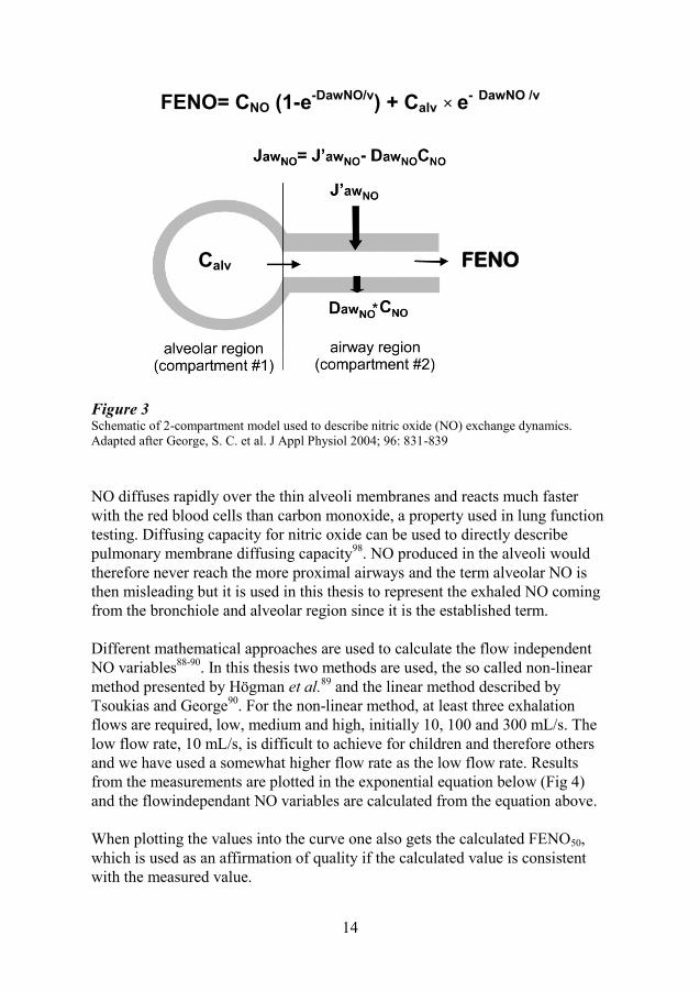
Flow independent NO variables
The FENO50 levels do not provide any information about the localisation of the
NO production (or inflammation) in the airways. Mathematical models of NO
dynamics suggest that the peripheral (alveolar) and the central (bronchial)
airway contribution to the FENO value can be calculated on the basis of NO
measurements at multiple exhalation flow rates88-91
. There is a strong inverse
relationship between the concentration of exhaled NO and the exhalation
flow92,93
. There is also a positive relationship between the elimination rate of
NO (product of concentration and flow) and exhalation flow93
. To explain these
observations Tsoukias and George presented the two-compartment model,
taking both peripheral and central airways into consideration90
(Fig 2).

13
Bronchial compartment
Alveolar compartment
In the two-compartment model a flexible expansible alveolar compartment
represents respiratory bronchiole and alveolar regions (generation 18 and
beyond according to Weibel94
, see fig 10). A single rigid cylindrical tube
bronchial compartment represents the conducting airways down to the
respiratory bronchioles (from trachea to generation 17 according to Weibel).
Exhaled NO originates from both these compartments.
Figure 2
The two-compartment model of the airways.
Nitric oxide is mainly produced in the airway wall and NOS is found in the
airway wall along the entire airway tree, including the bronchi, bronchioles and
alveoli95,96
.
In the two-compartment model, the final concentration of NO in exhaled air
depends on two mechanisms:
1) NO concentration in alveolar air and
2) conditioning of alveolar air while it travels through bronchial compartments.
The accumulation of bronchial NO from the bronchial wall to exhaled air while
it travels through the bronchial tree can be further modelled by dividing the
bronchial compartment into infinitely short units. When entering the bronchial
compartment the luminal air NO concentration equals the alveolar NO
concentration. In the first unit NO diffuses from the bronchial wall to the
luminal air and at the entry of the second unit the luminal NO concentration
equals the alveolar NO concentration + NO diffused in the first unit and so
forth. In every unit, the diffusion of NO from the bronchial wall to luminal air is
driven by the NO concentration gradient between these two. The diffusion rate
is determined by the bronchial diffusing capacity of NO, DawNO.
Conditioning of alveolar NO in the bronchial compartment thus depends on
transit time of the air through the conducting airways, the airway wall
concentration, CNO, and DawNO. By forming a differential equation based on
this model the final NO concentration in exhaled air (FENO) can be expressed
as an exponential function of exhalation flow rate88-90,97
(Fig 3).
Flowindependant
NO variables CNO= bronchial wall NO
concentration
Calv= avleolar NO concentration
v= flow rate
DawNO= bronchial wall NO
diffusion capacity
JawNO= bronchial NO flux
14
FENO= CNO (1-e-DawNO/v) + Calv × e- DawNO /v
Figure 3 Schematic of 2-compartment model used to describe nitric oxide (NO) exchange dynamics.
Adapted after George, S. C. et al. J Appl Physiol 2004; 96: 831-839
NO diffuses rapidly over the thin alveoli membranes and reacts much faster
with the red blood cells than carbon monoxide, a property used in lung function
testing. Diffusing capacity for nitric oxide can be used to directly describe
pulmonary membrane diffusing capacity98
. NO produced in the alveoli would
therefore never reach the more proximal airways and the term alveolar NO is
then misleading but it is used in this thesis to represent the exhaled NO coming
from the bronchiole and alveolar region since it is the established term.
Different mathematical approaches are used to calculate the flow independent
NO variables88-90
. In this thesis two methods are used, the so called non-linear
method presented by Högman et al.89
and the linear method described by
Tsoukias and George90
. For the non-linear method, at least three exhalation
flows are required, low, medium and high, initially 10, 100 and 300 mL/s. The
low flow rate, 10 mL/s, is difficult to achieve for children and therefore others
and we have used a somewhat higher flow rate as the low flow rate. Results
from the measurements are plotted in the exponential equation below (Fig 4)
and the flowindependant NO variables are calculated from the equation above.
When plotting the values into the curve one also gets the calculated FENO50,
which is used as an affirmation of quality if the calculated value is consistent
with the measured value.
Calv
FENO Calv

15
Figure 4 FENO values and exhalation flows plotted in the non-linear method according to Högman et al.89
At higher flow rates (>50ml/s) the exponential equation can be substituted with
a linear approximation where NO output is plotted against flow rate, the slope,
intercept model according to Tsoukias, or the linear method. The slope of the
regression line is an approximate for alveolar NO concentration and the
intercept is an approximate for bronchial NO flux, JawNO90
(Fig 5).
Figure 5 Example of the slope-intercept model (the linear method). Tsoukias et al. J Appl Physiol 85 (2):
653. (1998)
The two-compartment model is reproducible in healthy children99
as well as in
children with asthma100
and CF101
. The linear and the non-linear methods have
0
5
10
15
20
25
0 50 100 150 200 250 300
Flow (ml/s)
FENO50 measured (ppb)
FENO50 calculated (ppb):
ENO = Elimination rate of NO at end exhalation,
VE = Exhalation flow
FENO Graph
NO koncentration (ppb)

16
been compared in healthy children by Sepponen et al. and they found
significant differences in the calculated values for alveolar NO and bronchial
NO flux but the values were highly correlated99
. Data presented below
(Table 5).
Table 5
NO variables in healthy children presented as median (mean ± SD)99
NO variables Healthy children (n=66)
FENO50 mL/s (ppb) 10.3 (11.7±5.4)
Alveolar NO (ppb)1 1.9 (2.0±0.8)
Bronchial NO flux (pL/s)1 400 (500±300)
Alveolar NO (ppb)2 1.4 (1.5±0.7)
Bronchial NO flux (pL/s)2 500 (600±300)
Bronchial wall NO conc. (ppb)2 49.6 (68±53.3)
Bronchial NO diffusion capacity (pL/s/ppb)2 10.1.(8±7.5)
1= Calculated according to (the linear method) Tsoukias 90 2= Calculated according to (the non-linear method) Högman 89
Several authors have suggested additional improvements of the two-
compartment model. The airway tree has more of a trumpet shape (increasing
surface area per unit volume) and Condorelli et al. suggested a trumpet shaped
model. The importance of axial diffusion has been discussed by a few authors
and different adjustments have been proposed102,103
. The latest published
improvement is correction for ventilation inhomogeneity in a multicompartment
model104
. So far, these different models are used in the research setting and
there is no joint recommendation on which model to use.
Nasal NO
Nasal NO (nNO) is measured in a similar way to FENO and the ATS/ ERS
guidelines provide recommendations also for nNO52
. Nasal NO is measured by
sampling nasal air from one nostril through at catheter with a constant sample
rate of 50 ml/s, leaving the other nostril open. The measured NO concentration
varies with the flow rate through the nose and there is still need for a more
standardised method to measure nNO. Just as for FENO, a simultaneous
exhalation is recommended with a positive pressure >5 cm H2O in the mouth to
ensure closure of the velum to prevent pollution by NO from the lower airways.
17
Exhaled NO and asthma
There is a large number of studies showing increased levels of FENO in
children and adults with asthma, using first tidal breathing and later the
recommended single breath technique9,10,52,57,58,105-107
. High levels of FENO is
highly suggestive of asthma108
and a good predictor of response to treatment
with corticosteroids109
but maybe more so in adults than in children11
. Increased
FENO has been linked to enhanced expression of iNOS in the airway
epithelium in asthmatic subjects 110,111
. iNOS expression can be down regulated
by corticosteroids and there is substantial data showing that FENO decreases
after treatment with inhaled corticosteroids (ICS)112
. The reduction is rapid and
dose-dependent113-118
but so far there is not enough evidence of benefits using
FENO compared to clinical symptoms in tailoring the dose of ICS to support
the regular use of FENO for this purpose26
. Treatment with leuokotriene
receptor antagonists resulted in reduced levels of FENO in some studies119-121
,
but in other studies no change in FENO was seen after treatment with
leuokotriene receptor antagonists122,123
.
Asthma is a variable disease and FENO is a highly dynamic measurement in
asthma with a great intra individual variation over time124
. A single
measurement is therefore of little value in asthma but examples of FENO results
in asthmatic children are presented in Table 6.
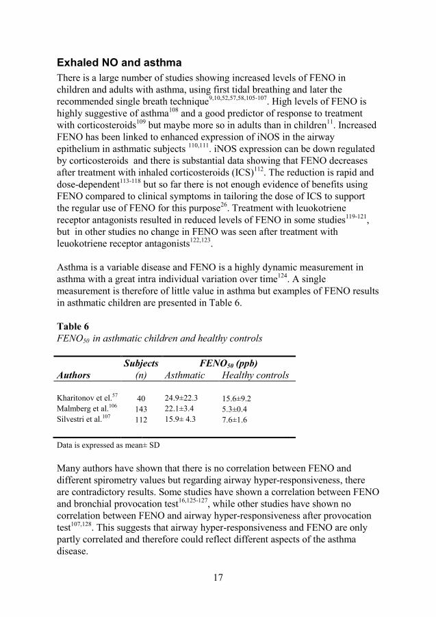
Table 6
FENO50 in asthmatic children and healthy controls
Subjects FENO50 (ppb)
Authors (n) Asthmatic Healthy controls
Kharitonov et el.57 40 24.9±22.3 15.6±9.2
Malmberg et al.106 143 22.1±3.4 5.3±0.4
Silvestri et al.107 112 15.9± 4.3 7.6±1.6
Data is expressed as mean± SD
Many authors have shown that there is no correlation between FENO and
different spirometry values but regarding airway hyper-responsiveness, there
are contradictory results. Some studies have shown a correlation between FENO
and bronchial provocation test16,125-127
, while other studies have shown no
correlation between FENO and airway hyper-responsiveness after provocation
test107,128
. This suggests that airway hyper-responsiveness and FENO are only
partly correlated and therefore could reflect different aspects of the asthma
disease.

18
Studies regarding the association between FENO and asthma control also show
contradictory results. Increased levels of FENO have been associated with a
deterioration in asthma control in some studies125,129
, while others found no
correlation between asthma control and FENO130,131
.
The discrepancy in the correlation between FENO and other markers of airway
disease could be due to different methodology used, or to the multifaceted
nature of the asthma disease with many different phenotypes within the asthma
population132,133
.
Flow independent NO variables in asthma
Many studies have shown that bronchial NO flux, (NO coming from the large
airways) is increased in asthma (Table 7) and that treatment with ICS can
reduce bronchial NO flux100,134-136
. For alveolar NO the results are more
contradictory, but there is data indicating that alveolar NO is increased in severe
asthma but no different compared to healthy controls in mild to moderate
asthma100,134,135
.
Table 7
Flow independent NO variables in asthma.
Alveolar NO (ppb) Bronchial NO flux (pL/s)
Authors age Asthma Controls Asthma Controls
Robroeks et al. 137
6–13 4.1±0.5 1093±251
*Paraskakis et al.100
4–18 2.2 (0.4–6.6) 1.63 (0.44–3.0) 1230 (8204–9236) 480 (196–1913)
Kerckx et al. 102
38±14 4.8±2.1 3.1±1.5 2254±1150 745±311
Lehtimäki et al. 138
32±2 1.1±0.2 1.1±0.2 2500±300 700±100
values presented as mean ±SD
*values presented as median (range)
There is an ongoing discussion that inhaled corticosteroids (ICS) cannot reach
the most peripheral airways and subsequently there would be no change in
alveolar NO after treatment with ICS. Systemic corticosteroids could better
reach the peripheral airways and thereby reduce alveolar NO. This is supported
by studies showing a reduction of alveolar NO after oral corticosteroids, but not
by ICS135,138,139
. There are new small-particle formulations of ICS that target
inflammation in the small airways, and there is data suggesting that treatment
with one of these ICS could result in decreased levels of alveolar NO140
.

19
Exhaled NO and CF
Airway inflammation starts early in life in subjects with CF, but in spite of often
marked airway inflammation, levels of FENO have either been reported not
different101,141,142
or decreased28,143-146
compared to control subjects (Table 8).
Table 8
FENO50 in children and young adults with CF.
CF FENO50 (ppb)
Authors subjects (n ) CF Controls
Suri et al.146 22 9.4 (2.7–26.9)* 10.4 (4.5–29.6)*
Robroeks et al.28 48 10±1.2** 15.4±1.4 **
Hubert et al.147 30 8.4 (6.2–16.2)***
* values presented as median (range)
** values presented as mean± s.e.m
*** values presented as median (25th–75th percentile
Nasal NO has consistently been reported to be low in CF subjects28,142,144,148
Several possible explanations have been presented for the low levels of FENO
and nNO seen in CF patients27,149
.
One explanation could be that the airway inflammation in CF is dominated by
neutrophils as opposed to the most common eosinophilic inflammation in
asthma which is associated increased FENO levels16,22,150
.
Second, there are studies showing polymorphisms in the genes coding for
constitutive NOS151,152
and decreased activity or expression of iNOS153,154
.
Inflammatory changes in the epithelial cells result in loss of epithelial cell
integrity and the respiratory epithelium is an important site for iNOS155
.
Consequently NO production could be lower in CF due to less or inactive NOS.
This is supported by a bronchoscopy study showing an inverse relationship
between neutrophilic airway inflammation and iNOS expression in the airway
epithelial cells and airway macrophages in children with CF, not seen in the
healthy controls156
.
Arginine is the substrate for NO production, and low bioavailability of arginine
has been shown in CF patients suggesting a third explanation for the low levels
of FENO157,158
. Grasemann and coworkers have shown an increase in FENO in

20
CF patients after giving arginine intravenously or orally159,160
and an increase in
both FENO and FEV1 shortly after an inhalation with arginine161
.
Mucus plugging and an asymmetric obstruction of the airways characterize CF
airway disease. This could result in low levels of NO in exhaled air due to poor
diffusion of NO into the gaseous phase148,162
and/or degradation of the produced
NO by bacteria found in the mucus, for example Ps. aeruginosa163
.
Low levels of nNO in CF subjects could be explained by blocked sinuses
(where the highest concentration of NO is found) which has been a big problem
in the CF population, but today blocked sinuses are much less prevalent due to
better treatment.
Up till today, these explanations are just speculations and there is still a big
uncertainty about the true cause and the clinical importance of the low levels of
NO found in the respiratory tract in patients with CF.
Flow independent NO variables in CF
There are only a few studies with extended NO measurements in CF, most of
them including a very small number of patients.
Table 9
Alveolar NO and bronchial NO flux in children and young adults with CF
(linear model90
)
Alveolar NO (ppb) Bronchial NO flux (pL/s)
Authors CF Controls CF Controls
Shin et al.101
1.96±1.18* 4.63±3.59* 607±648* 784±465*
Suri et al.146
2.2 (0.6–5.6)** 1.5(0.4–2.6)** 445 (64–1256)** 509 (197–1913)**
Hubert et al.147
3.3 (2.4–6.4)*** 283 (150–500)***
* values presented as mean ±SD
** values presented as median (range)
*** values presented as median (25th-75th percentile)
Shin et al. reported flow independent NO variables in 9 children with CF.
Bronchial NO flux was reduced and alveolar NO was no different from that in
healthy children101
. The same results were reported from a study in adults
(n=12)164
. In contrast to these findings Suri et al. found increased levels of
alveolar NO but no difference in bronchial NO levels in 22 children with CF
compared to healthy controls146
(Table 9).

21
INFLAMMATION
SYMPTOMS
VARIABLE
AIRWAY
OBSTRUCTION
AHREXACERBATIONS
Asthma
Asthma is a polygenetic disease where environmental factors are very important
for the clinical picture. It is still incompletely understood and continues to be a
significant management problem for clinicians, particularly as childhood
disease develops into chronic airflow obstruction in adults irrespective of
treatment165
. Asthma is the most common chronic disease of childhood and
accounts for a large proportion of paediatric hospitalisations, health care visits
and absenteeism from day care and school166
. There are many different asthma
phenotypes where the characteristic features (Fig 6) (Table 10), airway
obstruction, airway inflammation and airway hyper-responsiveness (AHR) are
more or less evident132,133,167
.
Figure 6 The different features in asthma:
symptoms, airway obstruction in the
small and large airways, airway
inflammation, airway hyper-
responsiveness (AHR) and
exacerbations are only partly
associated.
The treatment of choice today is ICS, used to treat the underlying airway
inflammation and the advantageous effects of ICS on symptoms, airway
function and inflammatory markers have been shown in a large number of
studies in children168-170
. However, epidemiological and clinical studies suggest
that many asthmatic children do not achieve sufficient asthma control in spite of
the availability of efficient drugs171,172
. Lack of compliance is one important
factor for not achieving asthma control. Another reason could be that the
inhaled corticosteroids do not reach the small airways or that the airway
inflammation is not steroid sensitive. Individuals with different asthma
phenotypes might benefit from different types of treatment, emphasizing the
need for a better understanding of the asthma disease and its underlying
mechanisms168
.

22
INFLAMMATION
•FENO
•Induced sputum
•Bronchoalveolar lavage
•Biopsy
SYMPTOMS
•Asthma Control Test
•Bronchodilator use
•Quality of life
•Activity impairment
AIRWAY
OBSTRUCTION/
STRUCTURAL
CHANGES
•Spirometry
•MBW
•Imaging
AIRWAY HYPER
RESPONSIVENESS
•Dry air challenge
•Excercise challenge
•Metacholine/histamine
challenge
•Mannitol test
EXACERBATION
•Days absent from school
•Emergency visits
•Days with oral steroids
How to monitor asthma?
Current guidelines recommend diagnosis and treatment based on symptoms and
spirometry (FEV1)168
. FEV1 is not sensitive to small airway dysfunction173,174
and small airways could be a missing link in the monitoring of asthma today.
There are several other possible methods to evaluate the different facets of the
asthma disease (Fig 7).
Figure 7 Possible asthma outcomes
Symptoms in children are often reported by the parents. Parents might over or
under report symptoms or signs of airway disease. The typical reversible large
airway obstruction may be absent during long periods and diagnosis can
therefore be difficult, especially in children. There is often little or no
correlation between symptoms and large airway obstruction (FEV1) or between
FEV1 and airway inflammation11,127,128,175
. Children with little or no symptoms
and normal lung function can suddenly deteriorate176
. To find these children we
need to have a better understanding of the underlying mechanisms and
pathophysiology.
Many new anti inflammatory drugs with more specific targets of inflammatory
mechanisms than the inhaled corticosteroids have are under development, and
also drugs targeting the small airways, emphasising the need to assess the
response to anti-inflammatory pharmacological treatment also in the small
airways.

23
Cystic fibrosis
CF is a rare, progressive disease, often leading to premature death. Airway
inflammation and infection are known to start early in life, invoking a
progressive decline in lung function, starting in the peripheral airways6,177-181
.
CF is one of the most common genetic diseases in the western world and in
Sweden; the incidence is around 1/5000-6000 newborn. CF is an important
differential diagnosis to asthma in young children with obstructive airway
disease. Airway inflammation plays a central role in both asthma and CF and
there are many similarities but also important differences (Table 10).
Thirty years ago, patients with CF were not expected to live into adulthood. Due
to remarkable improvements in the care of CF patients, more than 60% of the
Swedish CF population is today above 18 years of age182
. The mean age for
survival in Sweden is around 40-50 years. Nevertheless, CF is still a progressive
disease that often leads to chronic respiratory failure.
CF is caused by mutations in the CF transmembrane conductance regulator
(CFTR) gene leading to dysfunction of the CFTR protein183
. More than 1500
mutations have been identified (http//www.genet.sickkids.on.ca/cftr/) and they
are referred to six classes based on the function of the defective protein184,185
.
dF508 is the most common CFTR mutation in Sweden and elsewhere184,186
.
The CF diagnosis is based on the following criteria187
.
1.
Sweat chloride concentration of > 60 mmol/L on two occasions.
Two genetic mutations causing CF
Disturbed chloride transport measured as an epithelial potential difference
2.
Sibling with CF
Positive result at newborn screening (not done in Sweden)
CF is characterized by a wide variability of clinical expression and CFTR is
expressed in the epithelial cells in many different organ systems; the lungs and
pancreas but also in the salivary glands, sweat glands, kidneys, intestines,
gallbladder and uterus. Approximately 85 % of the CF subjects have pancreatic
insufficiency at diagnosis and it is associated with increased resting energy
expenditure and enzyme deficiency leading to fat malabsorption.
Supplementation with enzymes, especially lipase, is routinely prescribed and a
high energy intake is often recommended to children and adults with CF188
.

24
Table 10
Characteristics of the typical airway inflammation in asthma and cystic fibrosis
Asthma189-191
Cystic fibrosis177-179
Genetics Multiple genes Single gene
Clinical presentation Non productive cough
Reversible obstruction
Airway hyper-
responsiveness
Chest tightness
Wheeze
Productive cough
Irreversible obstruction
Respiratory failure
Variability Variable Chronic
Progressive Non progressive Progressive
Triggers Allergens
Infections, viral
Irritants
Infections, viral and bacterial
Signalling substances and mediators IL4
IL5
IL13
IgE
Leukotriens
Prostaglandins
IL-8
Proteases
Oxygen radicals
Leukotriene B4
Cells Eosinophils
Mast cells
CD4+ lymphocytes
Neutrophils
Macrophages
Typical characteristics Variable inflammation
triggered by typical
triggers
Bacterial infection with
intense inflammation
Oxidative stress
Small airways involved Conducting airways Intra acinar airways
Consequences of airway
inflammation
Remodelling and fixed
airway obstruction
Respiratory failure
Exhaled NO Increased Decreased

25
Liver disease is present in approximately 5-10 % of the CF population192
and
there is an increased risk of osteoporosis193,194
and diabetes with increasing
age195
.
It is the pulmonary abnormalities causing the greatest morbidity and mortality
in CF177
. The degree of pulmonary involvement is not related to the genotype
and therefore other modifying genes have been discussed, i.e. different genes
encoding for the inflammatory response in the airways. The NOS genes have
been suggested as possible disease modifying genes196
.
Persistent infection, often caused by Pseudomonas aeruginosa, and
inflammation, involving the peripheral (small) airways, begin at a very early
stage in patients with CF197,198
and continues throughout life199,200
. Chronic
inflammation directly damages the airway wall, ultimately leading to
bronchiectasis and progressive decline in pulmonary function, which often
accelerates during adolescence. Monitoring and controlling the infection and
inflammatory process early in the course of disease may limit the damaging
effects of excessive inflammation, thus delaying progression of pulmonary
deterioration and potentially decreasing morbidity and mortality201
.
Fatty acids in CF and fatty acids and inflammation
Fatty acid deficiency is an important feature in individuals with CF, who have
an impaired fatty acid metabolism with increased release and high turnover of
arachidonic acid (AA), and decreased levels of docosahexaenoic acid (DHA),
in plasma, erythrocyte membranes, platelets and tissue biopsies202-204
. The ratio
of AA (20:4n-6) to DHA (22:6n-3) is therefore often elevated. The high
turnover of AA results in low concentration of the essential fatty acid linoic acid
(LA). Eicosanoids, which are important inflammatory mediators, are
synthesized from AA and the high AA turnover results in a high eicosanoid
production205
. The fatty acid abnormality in CF is associated with the CFTR
mutation206
but the connection between the abnormalities in the fatty acids has
not been satisfactorily explained and the reason for the high turnover of AA in
CF is not known203,207
.
The fatty acid deficiency seen in CF could have implications on the airway
inflammation because AA is mainly associated with an increase of pro-
inflammatory eicosanoids, while DHA provide anti-inflammatory factors208
.
The fatty acids in our diet can be separated into three types; saturated,
monounsaturated and polyunsaturated fatty acids (PUFA)209
. Saturated fatty

26
20:4n6
20:5n3
22:6n3
acids do not contain any carbon double bonds, as the fatty acid is fully saturated
with hydrogen. Monounsaturated fatty acids, as the name suggests contain fatty
acids with one carbon double bond and likewise PUFA contain two or more
carbon double bonds. PUFA are further classified as n-9 (omega 9), n-6 (omega
6) or n-3 (omega 3) PUFA.
The difference between the PUFA is the location of the first double bond; on
carbon number 3 (n=omega-3), 6 (n=omega 6) or carbon number 9 (n=9) from
the methyl end of the molecule (Fig 8).
Figure 8 The carbon skeleton of the three main
long chain polyunsaturated fatty
acids (PUFA).
The chemical names and frequently used abbreviated names of the most
important PUFA are shown in Table 11. The so-called essential fatty acids, LA
and α-linolenic acids (ALA) have to come through our diet since humans cannot
synthesize them. The other fatty acids are either synthesized in the human body
from the essential fatty acids or ingested as part of our diet (Fig 9).
Table 11
N-3 and n-6 polyunsaturated fatty acids and their main food sources.
Name Chemical name Abbreviation Main food source
α-linolenic 18:3 n-3 ALA Flaxseeds, canola, walnuts
Eicosapentaenoic
acid
20:5n-3 EPA Fish and seafood, meat, eggs
Docosahexaenoic
acid
22:6n-3 DHA Fish and seafood, eggs
Linoleic acid 18:2 n-6 LA Sunflower oil, corn, poppy
seeds
Arachidonic acid 20:4n-6 AA Meat, egg, dairy products
Polyunsaturated fatty acids

27
Omega-6 Fatty Acids(e.g. Corn, sunflower oils)
Linoleic acid (LA)
Delta-6-desaturase
Gamma-Linolenic Acid
Dihomo-Gamma-LinolenicAcid
PGE1Delta-5-desaturase
Arachidonic Acid (AA)
Cyclooxygenase Lipoxygenase
PGE2 LTB4
(pro-inflammatory) (pro-inflammatory)
Lipoxins
(pro-inflammatory)
Omega-3 Fatty Acids(e.g. flaxseed oil, fish oils)
Alpha-Linolenic Acid (ALA)
Delta-6-desaturase
Stereodonic Acid
Eicosatetranoic Acid
Delta-5-desaturase
Eicosapentanoic acid (EPA)
Docosahexanoic acid (DHA)(e.g.fish oils)
Cyclooxygenase Lipoxygenase
PGE3 LTB5(anti-inflammatory) (anti-inflammatory)
Resolvins, Neuroprotectins
(anti-inflammatory)
Delta-6-desaturase
.
Figure 9
The long chain n-3 and n-6 polyunsaturated fatty acids
There is evidence suggesting that n-3 PUFA are associated with more health
benefits, including anti-inflammatory properties than n-6 PUFA, especially in
the western world where there is an unfavourably high n-6 to n-3 ratio in the
diet208
. An increased intake of n-3 PUFA, especially the long chain DHA and
EPA, may therefore have a beneficial role in the prevention and treatment of
inflammatory disorders208,210,211
.
There is a complicated cross talk between PUFA and NO production, where
PUFA have been shown to both increase and decrease NO production, partly
through influencing the different NOS212,213
. The changed fatty acid metbolism
in CF subjects could therefore influence the NO production in the airways.
There are several studies with n-3 PUFA substitution in different diseases, but
according to two recent reviews, so far not enough evidence for a beneficial role
to support broad clinical use of PUFA substitution in all CF patients214,215
.

28
0
1
2
3
4 5 6
16 17
18 19 20 21 22 23+
7 8 9
10 11 12 13 14 15
Conducting
airways
Peripheral
“small” airways
Acinus
Trachea
Small airways
In the 1960s Weibel proposed a structural model of the airways94
.The airways
are designed to optimize the gas exchange which is done over an inner surface
area the size of a tennis court. This is achieved by attaching the gas exchange
units at the end of a branched tree of conducting airways. A new airway
“generation” starts at each branching and there are around 23-26 generations
from the trachea down to the alveoli, which in adults corresponds to
approximately 25-26 cm216
. The first 16 generations are conducting airways
where the air is transported downwards. The acinus (generation 17 onwards)
contains airways designed for both air transport and gas exchange, and alveoli
primarily designed for gas exchange (Fig 10).
Figure 10 Schematic drawing of the airways with approximate distances in adults. Adapted from Henrik
Ljungberg.
The small airway zone (airways < 2mm inner diameter in adults), also referred
to as the peripheral airways, starts around the eighth generation. The large
airways are kept open by an outer wall of cartilage. The small airways have no
cartilage in the airway wall but instead the small airways are held open by the
balance between the elastic fibres and smooth muscle in the airway wall and the
lung’s elastic recoil and the lining fluid consisting of low surface tension
~ 24 cm (adults)
2-3 cm (adults)
½ mm (adults)

29
surfactant. In disease, these mechanisms might be in imbalance, resulting in
small airway obstruction.
Post-mortem lung specimens and in vivo transbronchial biopsies have
demonstrated that both large and small airways are engaged in asthma2,217
and
CF218
. Later supported by imaging studies showing airway wall thickening,
luminal obstruction, bronchiectasis and gas trapping even early on in the course
of CF airway disease219
. In asthma a direct challenge study has shown increased
airway resistance in the small airways measured directly through
bronchoscopy220
. These studies provide evidence of small airway involvement
in both asthma and CF, but the methods used are invasive or cause radiation,
making them less useful in the every day clinic. Different methods to assess the
small airways in airway disease are discussed in Table 12. Multiple breath inert
gas washout is one example of a non-invasive, relatively easy to perform
method to evaluate lung function even in the peripheral airways.
Table 12
Methods to assess the small airways.
Method Disadvantages
Histopathology of organ specimens221 Requires surgery
Bronchoscopy with biopsy, lavage222
Invasive
Bronchoscopy with ultrasound223 Invasive
CT scan224, 225 Radiation
MRI225 Availability, cost, patient collaboration
Spirometry (MMEF)226 Not sensitive, non specific , large interindividual
variability
Frequency dependence of Cdyn227 Invasive
MEFV-curves +/- Heliox228 Indirect, only suggestive
Forced oscillation (FOT)229 Dependant on operator technique
Alveolar NO230 Different models, conflicting results
Inert gas washout231 Cost, Time consuming

30
Lung function tests
Spirometry
Spirometry (meaning the measuring of breath) is the most commonly used
pulmonary function test. It measures the amount (volume) and/or speed (flow)
of air that can be inhaled and exhaled. The most commonly used indices are
forced expiratory volume in 1 second (FEV1), forced vital capacity (FVC) and
maximum mid expiratory flow (MMEF) reflecting the mechanical properties in
airway function. Disease causing obstruction of the larger airways will easily
affect the spirometry variables. Spirometry is, however, not sensitive enough to
detect early small airway involvement since the small airways are pathways of
very low resistance and only contribute to about 10% of the total airway
resistance174
. Small airway obstruction, however, affect how inspired air mixes
with the gas in the lungs and reduced efficiency in gas mixing is called
ventilation inhomogeneity. Many school age children with CF have FEV1
within the normal range even though they have signs of ventilation
inhomogeneity as evidence of residual airway disease180
. Early changes in the
small airways are also seen in mild asthma5,232
.
Multiple breath inert gas washout
Inert gas washout was first described over 60 years ago233
. It measures
ventilation inhomogeneity and accordingly it provides information about the
small (peripheral) airways173
. Inert gas washout is performed during a single
vital capacity breath or over a series of tidal breaths - the so-called MBW
method. This method requires minimal cooperation apart from maintaining an
adequate mouthpiece/face-mask seal and regular breathing pattern and MBW
can be successfully obtained in infants from very young age as well as in older
children where also within test repeatability is good234,235
. The single breath
method requires more cooperation from the subject. Recent reports suggest that
indices derived from MBW may be more sensitive than spirometry in detecting
lung disease in children with CF180
.
Inert gas washout is performed with different gases and most commonly used is
the N2 washout using 100% O2. In this thesis an inert gas mixture with 4% SF6,
4% He, 21% O2, balance N2 is used. Each test consists of two phases: a wash in
phase during which a dry gas mixture containing the gas, i.e. 4% SF6, 4% He,
21% oxygen (O2), and balance nitrogen (N2), is administered, and a washout
phase when the subject breaths normal air again in order to washout the inert
gas mixture (Fig 11). Wash in is continued until the inspiratory and expiratory
SF6 concentrations are stable and equal, plus another 30 s.

31
Figure 11 During the wash in
phase, the patient
breathes the gas
mixtures and during
the washout, the
patient is switched to
room air. Cartoon
adapted from Per
Gustafsson.
At this moment, the bias flow is stopped during expiration by disconnecting the
T-piece and the washout phase starts. The washout phase continues until the
end-tidal SF6 concentration is below 0.1% (i.e. 1/40th of the starting
concentration) (Fig 12).
Figure 12 Computer screen readout from
SF6 multiple breath washout.
Multiple breath inert gas washout provides data about the resting lung volume,
functional residual capacity (FRC, the volume of air left in the lungs after a tidal
expiration) and indices of ventilation distribution inhomogeneity. FRC is
calculated from the net amount of marker gas exhaled during washout
(reinspired gas subtracted), divided by the difference in marker gas
concentration at start and end of washouts235
. Several markers of ventilation
inhomogeneity have been reported in the literature236
. They all reflect

32
Convection Convection is the net transfer of gas molecules from areas of high pressure to areas of low pressure with a pressure gradient.
Diffusion Diffusion - the process by which gas molecules spread from areas of high concentration, to areas of low concentration with a concentration gradient
differences in specific ventilation (ventilation per unit volume) between lung
regions, which result in delayed washout of the marker gas from the more
poorly ventilated regions.
Lung clearance index
The lung clearance index (LCI) is the variable of ventilation inhomogeneity that
is the easiest to calculate and easiest for the patient/ parent and examiner to
understand. LCI is calculated as the number of lung volume turnovers that the
child must breathe to clear the lungs of the marker gas (to 1/40th of the starting
concentration) and it is calculated from the cumulative expired volume during a
washout divided by the FRC237
. An increase in LCI indicates increased
ventilation inhomogeneity. See Table 13 for LCI in healthy subjects.
Table 13
LCI in healthy controls and in subjects with asthma and CF.
LCI
Authors n age (years) Healthy Asthma CF
Gustafsson et al.180 71 3–18 6.33 (0.43) 8.33 (2.48)
Aurora et al.234 55 6–16 6.45 (0.49) 11.53 (2.86)
Horsley et al.238 45 6–49 6.3 (0.5) 13.1 (3.8)
Gustafsson 239 44 6–29 8.7 (1.3) 11.5 (3.3)
Macleod et al. 240 60 5–16 6.24 (0.47) 6.69 (0.91)
Downie et al. 241 40 18–66 8.16 (1.1)
Data presented as mean (±SD)
Phase III slope analysis from MBW
LCI from MBW reflect overall ventilation inhomogeneity and does not provide
any information about the location of the pathology.
More sophisticated analysis of the so called phase III (alveolar phase) slopes
generated during MBW has been proposed to provide more information about
the localisation of the ventilation inhomogeneity in the airway tree242,243
. This
method is based on the understanding that gas mixing in the lungs is based on
two mechanisms:
-convection which is predominant in the conducting airways
-diffusion which occurs in the intra-acinar airways

33
Conducting airways
Acinus
The zone in between where convection and diffusion occurs to the same extent
is called the diffusion-convection front. Ventilation inhomogeneity that takes
place proximal from the diffusion-convection front is called convection
dependant inhomogeneity and when it occurs in the vicinity of the diffusion-
convection front, it is called diffusion-convection interaction-dependent
inhomogeneity. The evolution of the phase III slope (Fig 13) from each breath
during the MBW enables the determination of the relative contribution of these
two mechanisms244,245
.
Figure 13 Phase III slope
Verbanck introduced Scond and Sacin (Fig 14) to reflect convection dependent
inhomogeneity and diffusion-convection interaction-dependent respectively245
.
Sacin
Approximate location for the diffusion convection front for SF6
0
1
2
3
4
5 6
1
6 17 18 19 20 21 22 23+
7 8 9
10 11 12 13 14 15
Figure 14 Scond and Sacin and the
corresponding airways
Scond

34
The phase III slope of each breath is analysed to calculate Scond and Sacin. To
compare phase III slopes from subsequent breaths, the effect of the dilution of
the marker gas is compensated for and the result is called the concentration
normalized phase III slope (SnIII). The SnIII for each breath is then plotted
against responding lung volume turnovers and Scond is defined as the calculated
SnIII increase between lung volume turnover 1.5 and 6.0. Sacin is defined as the
first breath SnIII value minus the convection dependant inhomogeneity
contribution to this value and constitutes the diffusion-convection interaction-
dependant contribution to the SnIII of the first washout breath.
To compensate for the subjects size and breathing pattern Scond and Sacin are
calculated from SnIII multiplied by tidal volume for each breath246
. Normative
data for Scond and Sacin has recently been published and the levels are similar for
children, teenagers and adults (Table 14)246
.
Table 14
Scond and Sacin in healthy subjects. Corrected for tidal volume.
MBW
Authors n age (years) Inert gas Scond (/L) Sacin (/L)
Gustafsson (unpubl) 44 41.1± 10.3 SF6 0.017 (0.006) 0.086 (0.026)
Horsley et al.238 17 31.1± 6.0 SF6 0.0101 (0.015) 0.112 (0.055)
Macleod et al. 240 28 11.1 (5–16) SF6 0.017 (0.020) 0.120 (0.06)
Aljassim et al. 247 18 18.0 (17–18) SF6 0.018 (0.006) 0.086 (0.025)
Verbanck et al. 248 63 31.1± 1.0 N2 0.028 (0.001) 0.072 (0.003)
Values presented as mean (SD)
Airway challenge testing
Bronchial hyper-responsiveness, often resulting in exercise-induced
bronchocontriction, is considered one of the key features in paediatric
asthma249-251
. The effect of exercise on ventilatory function in the child with
asthma was discussed already in the early 60s by Jones et al.252
.
The mechanism of action is thought to be respiratory heat and water loss with
transient hyperosmolarity in the respiratory mucosa causing the airways to
narrow. Whether the magnitude of airway obstruction is determined by airway
cooling alone or by evaporation of water from the respiratory mucosa
irrespective of cooling, is a matter of debate117,253-255
.

35
Isocapnoic hyperventilation with dry air mimics the drying of the airway
mucosa during exercise and has been used as a marker of airway hyper-
responsiveness and exercise induced airway narrowing in asthma256
. It is simple
to perform and easier to standardize than exercise testing even in young
children257
. The reaction probably reflects the pathophysiology of asthma better
than a pharmacological bronchoprovocation with histamine or metacholine255
.
Isocapnoic dry air hyperventilation challenge test is used in this thesis to
evaluate airway hyper-responsiveness. A 10% reduction in FEV1 is outside the
range for healthy subjects without asthma and this is therefore used as a cut off
level for a positive test for airway hyper- responsiveness256
.
Asthma Control Test
There are several validated patient reported outcomes developed for asthma.
Asthma Control Test (ACT) is a short questionnaire, which is validated in many
languages. ACT is a five-item tool for identifying patients with uncontrolled
asthma developed by Quality Metric, Inc.and GlaxoSmithKline. Responses for
each of the five items are summed to yield a score ranging from 5 (poor control
of asthma) to 25 (complete control of asthma). Validity, reliability, and
responsiveness have been established in subjects 12 years of age and
older258,259
. A paediatric version, the Childhood Asthma Control Test (C-ACT
has been developed for children 4-11 years old260
. C-ACT is a seven-item test
where one part is filled in by the parents and one part filled in by the children
themselves. The ACT and cACT were designed primarily as discriminatory
tools and a score of 19 or lower identifies patients with poorly controlled
asthma in both tests. C-ACT is complementary to, but not a substitute for, other
markers of disease control in asthmatic children, especially in the context of
follow-up visits261
. Patient reported outcomes, such as ACT, are recommended
by the GINA and other guidelines for monitoring subjects with asthma168
.

36
Aims
The overall aim of this thesis is to use exhaled NO in combination with MBW
to assess the contribution of airway inflammation and small airway dysfunction
in paediatric asthma and CF. This could potentially allow for earlier
intervention and more successful management of paediatric asthma and CF in
the future.
Specific aims in paper I-IV
Paper I The aim of study I was to assess FENO50 and nNO in subjects with CF and
healthy controls and to examine if low levels of FENO50 and nNO in CF are
associated with the CFTR genotype, pancreatic insufficiency, chronic
colonization with Ps. aeruginosa and the typical essential fatty acid imbalance
seen in CF patients.
Paper II Study II is a randomized double blind placebo controlled study aiming at
evaluating the influence of different blends of n-3 or n-6 PUFA compared to
placebo on FENO50, nNO and systemic markers of inflammation in subjects
with CF.
Paper III The aim of study III was to explore if there is an association between flow
independent NO variables, possibly reflecting airway inflammation in different
segments of the airways, and early signs of impaired airway function in children
with CF.
Paper IV The aim of study IV was to assess NO variables, indicating airway
inflammation, and LCI, Scond and Sacin, reflecting airway dysfunction, at
different depths of the airways, to search for an association between
inflammation and small airway dysfunction in children with allergic asthma. A
second objective was to explore if inflammation and impaired lung function in
the small airways is associated to airway hyper-responsiveness, or if the
presence of airway hyper-responsiveness is independent of small airway
dysfunction and airway inflammation and reflect another type of asthma.

37
Study concept
Four studies are reported in this thesis. Study design, inclusion and exclusion
criteria for study I-IV are reported in Table 15.
Table 15
Inclusion, exclusion criteria and study design in paper I-IV.
Study I Study II Study III Study IV
Patient category CF + controls CF CF + controls Asthma + controls
Patients (n)
59 35 45 47
Healthy controls (n) 114 35 74
Age ≥ 7 years ≥ 7 years 6 – 20 years 5 – 20 years
Study design Cross sectional
Consecutively
enrolled
Double blind
placebo controlled
study
Cross sectional
Consecutively
enrolled
Cross sectional
Explorative
Inclusion criteria Pathological
sweat test
Pathological sweat
test
Pancreas
insufficiency
Pathological sweat
test
Doctors diagnosed
asthma with allergic
sensitization
Exclusion criteria Acute
exacerbation
Smoking
Intake of n-3
capsules
Acute exacerbation
FEV1 ≤ 40% of
predicted
Severe liver
disease
Smoking
Acute exacerbation
Smoking
Acute airway
infection
Smoking
Study II is a double blind placebo controlled study where CF-subjects with
pancreas insufficiency were consecutively randomized into three parallel groups
(A, B and C). Each group was assigned to a fat blend with a different
composition of FA. Group A was given an experimental fat blend enriched by
n-3 fatty acids, group B a blend rich in saturated fatty acids (FA), corresponding

38
to an ordinary diet and used as placebo. Blend C was enriched by n-6 FA,
especially AA to see whether this could act as a feedback mechanism and
inhibit further transformation from LA. Blend C also contained more EPA and
DHA than the control, blend B, but less than blend A (Table 16).
Table 16
Fatty acid composition (mol %) of the three experimental fat blends
used in the different groups.
Group A Group B Group C 12:0 1.04 4.43 1.20
14:0 5.60 11.55 3.99
16:0 15.20 31.02 18.21
18:0 3.27 7.73 7.13
14:1n-5 0.43 1.18 0.32
16:1n-7 3.51 1.60 0.55
18:1n-9 (OA) 18.45 25.11 18.68
18:2n-6 (LA) 16.74 15.57 21.52
18:3n-3 (ALA) 5.71 1.28 0.39
18:3n-6 0.13 0.02 4.03
20:3n-6 0.09 0.05 1.59
18:4n-3 1.85 0.10 0.15
20:4n-6 (AA) 0.80 0.06 18.03
20:5n-3 (EPA) 21.27 0.06 1.95
22:6n-3 (DHA) 6.99 0.03 0.52
SFA 25.69 55.0 31.99
MUFA 22.47 27.91 19.76
PUFA 53.69 17.19 48.41
n6/n3 0.50 10.69 15.08
The study subjects received 50 mg/day/kg body weight of the FA blend and
they increased their pancreatic enzymes by 10-20% to keep “normal” stools.
Forty-five subjects were included but two subjects had to be excluded right
away due to acute exacerbations, thus forty-three patients were randomized.
Eight patients did not finish the study (distributed in all three groups) due to low
compliance, stomach pains and/or weight gain. Thus, thirty-five patients
completed the study and all results are based on these subjects.
The study subjects were examined at inclusion and after three months of
supplementation.

39
Materials
Subjects in study I-IV
The CF diagnosis was based on a pathological sweat test with sweat chloride
concentrations >60 mmol/L and genetic testing. Clinical characteristics and
baseline measurements of all CF subjects are reported in Table 17.
Table 17
Characteristics and baseline measurements of the CF subjects in study I-III.
Values are median (range) or numbers (proportions).
Study I Study II Study III
(n =59) (n = 35) (n = 45)
Age (yrs) 22.5 (7–55.3) 20.2 (8.1–41.2) 13.4 (6.2–18.3)
F/M 27/32 18/17 17/28
BMI (Z–score) -0.1 (-1.1–2.0) -0.24 (-1.61–1.0) -0.22 (-1.44–1.44)
Pancreas sufficiency (n [%]) 12 (20) 0 (0) 5 (11)
CF–related liver pathology treated
with ursodeoxycholic acid. (n [%])
9 (15) 6 (17) 9 (20)
CF associated diabetes (n [%]) 9 (15) 6 (17) 3 (7)
Chronic Ps. aeruginosa
colonization (n [%])
29 (49)
15 (43)
16 (36)
FVC (% predicted) 98 (34–132) 97 (69–142) 100 (73.4–132.1)
FEV1.0 (% predicted) 84 (13–127) 85 (47–129) 91 (54–122)
Current use of inhaled corticosteroids
(n [%]) 17 (29) 10 (28) 12 (27)
Long term antibiotics
(azitromycin or flucloxacillin) (n [%]) 38 (64) 22 (63) 22 (49)
Atopy (n [%]) 22 (37) 9 (26) 22 (49)
Pancreatic function was determined by measuring pancreatic enzymes in
duodenal secretions after stimulation with cholecystokinin and secretin or by
determining fecal elastase.

40
Chronic Ps. aeruginosa infection was defined as consecutive positive sputum
cultures during 6 months and raised serum titers of pseudomonas exotoxin A
antibodies262
.
CF subjects in study I were classified into four groups according to the CFTR
genotype were the patients in group 4 were pancreas insufficient and patients in
all other groups were pancreas insufficient (Table 18).
Table 18
Genotypes in the different groups in study I
Group Genotypes
1 homozygotes for dF508
2 heterozygotes for dF508 with one other “severe” mutation (394delTT,
3659delC, E60X, or 1112 delT) or homozygotes for that other mutation
3 compound heterozygotes for dF508 and V603F, R560T, or 621+1G-T
4 heterozygous for dF508, 3659del C or 394delTT and a mutation linked to
a “mild” phenotype (N1088D, R117C, R117H, R75Q, R658X, S945L,
1154insTC, or T338I)
Atopy (allergic sensitization) was in study I and II based on a clinical question
whether the patient had symptoms of allergic rhinitis after allergen exposure. In
study III-IV, atopy was based on a positive Phadiatop™ test (Phadia, Uppsala,
Sweden) which determines the presence of raised specific serum IgE antibodies
to common inhaled allergens (dog, cat, horse, timothy, birch, mugwort, house
dust mite and Cladosporium). It is expressed as positive or negative with a cut-
off level at 0.35kU/l.
Asthma was defined according to current guidelines based on clinical symptoms
of asthma and demonstrated reversible airway obstruction or a positive
bronchial challenge test168
. Clinical characteristics of the asthma subjects in
study IV are reported in Table 19.
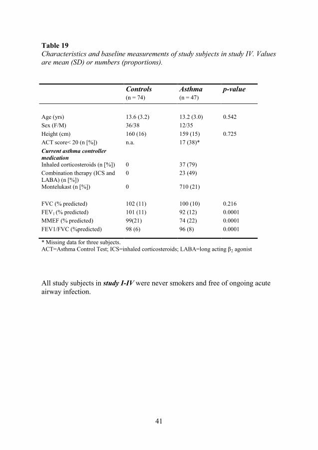
41
Table 19
Characteristics and baseline measurements of study subjects in study IV. Values
are mean (SD) or numbers (proportions).
Controls Asthma p-value
(n = 74) (n = 47)
Age (yrs) 13.6 (3.2) 13.2 (3.0) 0.542
Sex (F/M) 36/38 12/35
Height (cm) 160 (16) 159 (15) 0.725
ACT score< 20 (n [%]) n.a. 17 (38)*
Current asthma controller
medication
Inhaled corticosteroids (n [%]) 0 37 (79)
Combination therapy (ICS and
LABA) (n [%])
0 23 (49)
Montelukast (n [%]) 0 710 (21)
FVC (% predicted) 102 (11) 100 (10) 0.216
FEV1 (% predicted) 101 (11) 92 (12) 0.0001
MMEF (% predicted) 99(21) 74 (22) 0.0001
FEV1/FVC (%predicted) 98 (6) 96 (8) 0.0001
* Missing data for three subjects.
ACT=Asthma Control Test; ICS=inhaled corticosteroids; LABA=long acting β2 agonist
All study subjects in study I-IV were never smokers and free of ongoing acute
airway infection.

42
Healthy controls
In paper I, subjects examined within the scope of the European Community
Respiratory Health Survey II (ECRHSII)263
, were used as healthy controls
(Table 20).
In study III, healthy children were recruited from nearby schools, and they were
matched for age and atopy with the CF subjects (Table 20). These healthy
controls and a further thirty-nine children who only performed the MBW and
spirometry were in the control group in study IV (Table 19). Controls and
patients were examined with the same equipment and procedures in respective
study.
Table 20
Clinical characteristics of the controls in study I and III. Values are median
(range) or numbers (proportions)
Controls study I Controls study III
(n=104) (n = 35)
Age (yrs) 39.5 (30–54) 13.2 (7.7–18.5)
F/M 51/53 22/13
Height (cm) 159.0 (125–188)
Body mass index BMI (Z–score) –0.1 (–1.27–1.6)
Atopy (n [%]) 0 (0) 16 (43)

43
Methods
Exhaled and nasal NO
Exhaled NO
FENO50 was measured according to the ATS/ERS recommendations for on-line
NO measurements, as described earlier52
. The subjects were told not to eat or
drink and to avoid hard exercise four hours ahead of the NO measurements.
In study I and II nNO and FENO50 were measured with a chemiluminescence
analyzer (Ecophysics Breath Analyzer CLD 77 AM; Switzerland) with a
detection limit of 0.5 ppb. A visual feedback helped the subjects tested to keep
expiratory flow steady and close to target flow during the recordings
(Exhalation Breath Analyzer TM; Aerocrine A, Stockholm, Sweden). In study
III and IV the Eco Medics system was used and nitric oxide was measured with
a chemiluminescence analyzer (ANALYZER CLD 88 AM; Eco Medics AG,
Duernten, Switzerland) with a detection limit of 0.1 ppb. A two-point
calibration of the analyzer was performed weekly with a certified NO
calibration gas.
FENO50 was measured in triplets at a plateau of 3 seconds during a slow single
exhalation for 10 seconds. The mean FENO50 from three recordings within
10 % was reported.
In study III and IV, exhaled NO was measured at several exhalation flow-rates,
30, 50, 100, 150, 200 and 250 mL/s and in study IV in some patients 300 mL/s.
Different exhalation flows were obtained by using flow resistors. The
exhalation times varied between 6 and 15 seconds depending on the flow-rate.
Two exhalations that varied less than 5 % or three exhalations that varied less
than 10% were obtained at each flow-rate.
NO flow-independent variables
In study III and IV extended NO analysis was used, which have been described
and validated before90,134,264
. Two different models, the slope intercept (the
linear model)90
and the non-linear model were assessed134
. From the linear
model the two variables, alveolar NO and bronchial NO flux are obtained and

44
from the non-linear model the same two variables plus the airway wall NO
concentration and the airway NO diffusing capacity were calculated. Flow-rates
above 50 mL/s were used for the linear method and for the non-linear method,
all flow-rates were used. Two published methods were applied to correct for the
back diffusion of NO into the alveolar space when calculating alveolar NO with
the linear method.
A. Alveolar NO corr = Alveolar NO – bronchial NO flux/740 as
proposed by Condorelli et al.103
and
B. Alveolar NO corr = (Alveolar NO- 0.08 × FENO50) / 0.92 as proposed
by Kerckx et al. 102
.
Nasal NO
Nasal NO was recorded in study I and II with the Aerocrine system. Nasal NO
was measured according to guidelines by sampling nasal air from one nostril at
a constant sample rate of 50 ml/s, leaving the other nostril open52
. To ensure
closure of the velum a simultaneous exhalation was performed with a positive
pressure >5 cm H2O in the mouth. Both nostrils were examined in study I. The
mean level of nasal NO was determined during a plateau lasting more than three
seconds and the mean value calculated from three recordings was recorded.
Lung function tests
Spirometry
Spirometry was performed after the NO measurements and according to the
ATS/ERS standards265
using a Jaeger Masterscope spirometer (Erich Jaeger
AG, Würzburg, Germany). Results were expressed as percentage of predicted
values266,267
. In study IV results were expressed as percentage of predicted
values using the new global “all ages” reference values recently presented by
Stanojevic et al.268
.
Multiple breath inert gas washout
MBW was performed as described in the Introduction section. The children
were investigated in the sitting position and the younger subjects watched a

45
video while the older subjects watched a tidal volume trace on a computer
screen and were instructed to keep breathing regular with a tidal volume (VT)
between 10–15 mL/kg body weight. All subjects used a nose clip and breathed
through a Fleisch no.1 pneumotachometer (Metabo SA, Lausanne, Switzerland)
via a mouthpiece. A sampling tube from a mass spectrometer was introduced in
the middle of the air stream between the mouthpiece/mask and the
pneumotachometer through a short connecting piece. The external dead space
was 15 mL for the mouthpiece system and 30 mL for the mask system.
Single breath inert gas washout was also performed but a large proportion of the
children were not able to perform this according to the protocol and results are
therefore only reported from the MBW.
The dry gas mixture used for MBW in study III and IV contained 4% SF6, 4%
He, 21% oxygen (O2), and balance nitrogen (N2). The different gas
concentrations were measured by the mass spectrometer as dry gas
concentrations. Recorded inspiratory and expiratory flows and volumes were
converted to body temperature and ambient pressure, and saturated with water
vapour conditions. Gas samples and flow signals were aligned in time. The
sample flow of the mass spectrometer was 20 mL/min and the gas concentration
signals were updated at a rate of 33.3 Hz. All signals were recorded at 100 Hz
by a computer using software that corrected the flow signal sample-by-sample
for changes in dynamic viscosity caused by the variations in gas composition.
MBW calculations
LCI was calculated as the number of lung volume turnovers needed to lower the
end-tidal tracer gas concentration to 1/40th of the starting concentration. The
mean value, calculated from three recordings, was recorded.
The phase III slope of each breath during MBW was analysed to calculate Scond
and Sacin. To compensate for the subjects size and breathing pattern Scond and
Sacin were calculated from SnIII multiplied by tidal volume for each breath as
suggested by Robinson et al.246
. Quality criteria for SnIII analysis included
regular breathing with a tidal volume between 10 and 15 mL/kg body weight.
All asthma patients fulfilled this but less than half of the controls managed to do
so. LCI results are therefore reported from all the controls (n=74) while Scond
and Sacin are reported only from those individuals who fulfilled the quality
criteria (n=36).

46
Airway challenge
Airway challenge was performed by four minutes hyperventilation of
isocapnoic dry air from the Ailos Astmatest™ (Ailos Medical B, Karlstad,
Sweden). Flow was set at 75 of predicted maximum voluntary ventilation,
which was approximated as 26 x subject’s baseline FEV1. Hyperventilation was
guided by a target balloon on the air-administering arm of the apparatus.
Spirometry was measured at 2, 5 and 10 minutes post challenge. The response
was measured as the maximum percentage of fall in FEV1. AHR was defined as
maximum fall in FEV1 ≥ 10%. The bronchodilator response was measured as
the percentage rise from baseline FEV1 after inhalation of four puffs of
salbutamol (0.1 mg/dose) from a metered dose inhaler via an anti-static holding
chamber. Short acting bronchodilator treatment was withheld for 12 hours prior
to testing and long acting bronchodilators, inhaled corticosteroids and
montelukast were withheld for 24 hours.
Asthma Control Test
Symptoms in the asthmatic children were recorded using ACT and cACT in
children over and under twelve years of age respectively. The number of
children with an ACT score of 19 or less, the cut off value for well-controlled
asthma, is reported.
Fatty acids
Fatty acids in serum were analysed as previously described269
. Total lipids of
serum were extracted and serum phospholipids were fractionated and
transmethylated and analyzed by capillary gas-liquid chromatography.
Urinary analysis
8-iso-PGF2α (marker for oxidative stress) and 15-keto-dihydro-PGF2α (major
metabolite of cyclooxygenase catalyzed PGF2α) in urine were analyzed by
radioimmunoassay (RIA) as previously described270,271
.

47
Systemic inflammatory variables
Inflammatory markers in blood, used in the routine care, were analyzed at
Sahlgrenska University Hospital (Gothenburg, Sweden). In study II cytokines
(interleukin [IL]-10, IL-1b, IL-8, IL-6, and tumour necrosis factor [TNF]-a)
were analyzed in serum by enzyme-linked immunosorbent assay (Peli-Kine
Compact ELISA kits, Sanquin reagents, Amsterdam, the Netherlands).
Statistics
The statistical analyses were performed with the SPSS software package,
version 11 and version 15 (Chicago, IL, USA) and SAS (version 9.1, SAS
Institute, Cary, NC, USA, procedure Proc Mixed).
Results are given as median and range or mean (SD) if not otherwise stated.
Non-parametric tests were applied as the distributions of NO variables were not
normal distributed. Ln NO, which had a normal distribution, was used for the
multiple regression analysis. Mann-Whitney’s U-test and Kruskall-Wallis were
used to assess differences between groups and Student’s t-test was used for lung
function variables showing a normal distribution. Nominal data were compared
using the Yates corrected chi2 test.
Wilcoxon signed rank test was used to assess differences within one individual.
Spearman’s rank correlation and multiple linear regression analysis were
performed to assess the strength of the relationships between different variables.
The two-tailed significance level was set at p<0.05. Upper limit of normal is
defined as (mean + 1.96x SD) for reference population (healthy controls).
Ethics
The Ethics Committee of the University of Gothenburg approved the study
protocols for all four studies. Informed consent was obtained from all patients
and healthy controls and from the parents of children under 18 years old.

48
Results
Paper I
NO and genotype
Nasal NO and FENO50 were significantly lower in the CF subjects than in
healthy controls. There was no difference in nNO between the nostrils. CF
subjects homozygous for dF508 and other severe mutations had lower levels of
nNO and FENO50 compared to those with mutations associated with mild
disease (Fig 15).
Figure 15 Nasal nitric oxide (nNO) (left) and exhaled nitric oxide (FENO50) (right) in CF patients in relation
to different cystic fibrosis transmembrane conductance regulator (CFTR) mutations. Box plot
indicates median and 25th and 75th percentiles. Whiskers indicate 5th and 95th percentiles and
outliers are indicated (○).
Mild mutations
d508/d508
d508, 3659delC, 394del TT
d508 /less severe mutation Mild
mutations
nNO (ppb)
0
20
40
60
80
100
120
0
10
20
30
40
FENO50 (ppb)
d508/d508
d508, 3659delC, 394del TT
d508 /less severe mutation
p= 0.006 p= 0.05
Summary CF subjects with pancreatic insufficiency have lower nNO and FENO50 than
pancreas sufficient subjects. Low FENO50 and low nNO are associated with
chronic colonization with Ps aeruginosa, and with the lipid abnormalities
often seen in CF subjects.
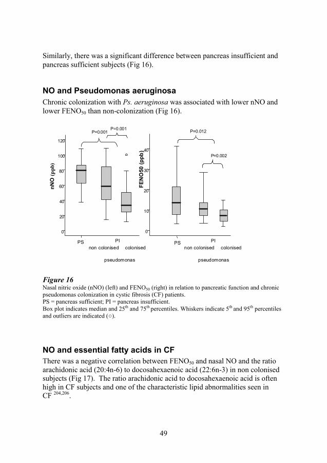
49
0
10
20
30
40
P=0.002
P=0.012
0
20
40
60
80
100
120
nN
O (
pp
b)
non colonised colonised
pseudomonas
PS
P=0.001P=0.001
PI
non colonised colonised
pseudomonas
PS PI
FE
NO
50
(p
pb
)
Similarly, there was a significant difference between pancreas insufficient and
pancreas sufficient subjects (Fig 16).
NO and Pseudomonas aeruginosa
Chronic colonization with Ps. aeruginosa was associated with lower nNO and
lower FENO50 than non-colonization (Fig 16).
Figure 16 Nasal nitric oxide (nNO) (left) and FENO50 (right) in relation to pancreatic function and chronic
pseudomonas colonization in cystic fibrosis (CF) patients.
PS = pancreas sufficient; PI = pancreas insufficient.
Box plot indicates median and 25th and 75th percentiles. Whiskers indicate 5th and 95th percentiles
and outliers are indicated (○).
NO and essential fatty acids in CF
There was a negative correlation between FENO50 and nasal NO and the ratio
arachidonic acid (20:4n-6) to docosahexaenoic acid (22:6n-3) in non colonised
subjects (Fig 17). The ratio arachidonic acid to docosahexaenoic acid is often
high in CF subjects and one of the characteristic lipid abnormalities seen in
CF 204,206
.

50
● = colonized with Ps. aeruginosa rs= -0.214, p= 0.27
○ = not colonized
rs = -0.412, p= 0.032
Figure 17 Nasal nitric oxide (nNO) in CF patients
in relation to the ratio between
arachidonic acid (AA) and
docosahexaenoic acid (DHA) in serum
phospholipids. Line represents the ratio
AA/DHA in reference population 269
Paper II
NO and n-3 and n-6 PUFA substitution
The serum phospholipid FA pattern changed significantly in all three groups,
reflecting the intake of fatty acids.
Univariate and multivariate analysis showed that nNO decreased significantly in
those subjects who received supplementation with n-6 fatty acids. There was no
change in FENO50 or nNO after intake of n-3 fatty acids.
The ratio of AA in serum phospholipids increased in 22 individuals (from group
B and C) and in those subjects, there was a significant decrease in FENO50 and
nNO (Fig 18)
Summary Supplementation with different mixtures of dietary FA influences serum FA
status and NO in the airways in patients with CF. Supplementation with n-3
FA attenuates the inflammatory response and n-6 supplemation aggravates
the inflammatory response.

51
Figure 18 Change from baseline in FENO50 in CF
patients where serum concentration of
arachidonic acid (=AA) increased and in
those were there was no AA increase.
Box plot indicates median and 25th and
75th percentiles. Whiskers indicate 5th
and 95th percentiles and outliers are
indicated (○).
N-3 and n-6 PUFA substitution and markers of systemic inflammation
Erythrocyte sedimentation rate (ESR) and IL-8, decreased after n-3 FA
supplementation and ESR increased after n-6 FA supplementation (Table 21).
Table 21
Erythrocyte sedimentation rate (ESR) mm/h and IL-8 ng/L at baseline and after
three months of FA supplementation. Values expressed as median (range).
* p=0.04 within group, **p=0.003 within group
†††p=0.05 between group C and B
N-3 and n-6 PUFA substitution and urine metabolites
There were no changes in the marker for oxidative stress and no change in 15-
keto-dihydro-PGF2a (a major metabolite of cyclooxygenase-catalyzed PGF2a)
in urine.
ESR IL 8
baseline 3 months diff baseline 3 months diff
Group A 7
(3–26)
6 *
(3–25)
–1
(-11–3)
17.5
(0.8–25.0)
9.3 **
(0.8–22.0)
-11.1
(-26.4–8.0)
Group B 9
(3–13)
6
(2–27)
0
(-3–16)
19.5
(6.6–69.0)
21.5
(0.8–80.0)
-3.0
(-48.0–64)
Group C 7
(3–31)
9 *
(3–32)
2 †††
(-1–7)
14.0
(6.0–88.0)
24.0
(4.7–61.0)
6.2 †††
(-32–29)
p=0.025
n=22 n=13

52
Paper III
Exhaled NO in children with CF
FENO50 and bronchial NO flux were lower in children with CF compared to
healthy controls. There were no significant differences in alveolar NO,
bronchial wall concentration and NO diffusion capacity between the groups
(Table 22). Bronchial NO flux and alveolar NO were calculated with two
different methods and the values differed significantly between the methods but
they were highly correlated (Table 22). There were no differences according to
gender in any of the NO variables in any of the groups.
Table 22
NO variables in CF and healthy children. Values are expressed as median
(range). P-value is given for differences between groups.
CF Controls p-
(n=45) (n=35) value
FENO50 mL/s (ppb) 9.1 (2.7–38.8) 12.4 (5.2–40.1) 0.029
Alveolar NO (ppb)1 1.2 (0–3.1) 1.4 (0.3–3.6) 0.147
Bronchial NO flux (pL/s)1 378 (97–1772) 578 (123–1993) 0.036
Alveolar NO (ppb)2 1.03 (0.1–2.9) 1.3 (0.1–2.9) 0.490
Bronchial NO flux (pL/s)2 412 (23–2101.) 664. (127–2310) 0.024
Bronchial wall NO conc. (ppb)2 54.8 (2.8–8209.6) 43.4 (9.3–1420.2) 0.813
Bronchial NO diffusion capacity
(nL/s)2 12.4 (0–97.2) 15.0 (1–51.9) 0.430
1 Calculated according to (the linear method) Tsoukias 2 Calculated according to (the non-linear method) Högman
Summary
FENO50 and bronchial NO flux are decreased in young subjects with CF
while alveolar NO is no different compared to healthy controls. Children with
CF have elevated LCI, indicating impaired small airway function. Low
FENO50 is associated with decreased airway function and low alveolar NO is
associated with elevated markers of systemic inflammation and pseudomonas
infection.

53
Small airway function in children with CF
LCI was significantly elevated in the children with CF compared to the healthy
controls, 8.33 (6.35–12.83) vs. 6.08 (5.05–6.96), p<0.001 (Fig 26). LCI was
significantly correlated with FEV1 but several CF subjects had increased LCI in
spite of normal FEV1, indicating that the children with CF had signs of small
airway involvement in spite of near to normal FEV1 (Fig 19).
Figure 19 FEV1 in relation to LCI in children
with CF. Line indicates upper limit of
normal for ref population.
Exhaled NO and ventilation inhomogeneity in children with CF
There was a significant negative correlation between FENO50 and LCI (Fig 20).
Alveolar NO also showed a negative correlation with LCI, r=-0.36, p=0.026.
Figure 20 FENO50 in relation to the lung
clearance index (LCI) in children
with CF.
rs = -0.585
p =0.0001
rs = -0.344 p = 0.021

54
● = colonization with Ps. Aeruginosa
rs = -0.286, p= 0.212
○ = no colonization
rs = -0.482, p=0.009
Exhaled NO, inflammation and bacterial colonization
Alveolar NO was negatively associated with several markers of systemic
inflammation (WBC, orosomucoid and IgG) (Fig 21). Subjects with chronic Ps.
aeruginosa colonization had lower levels of alveolar NO and higher LCI than
subjects who were not colonized (Table 23).
Figure 21 Alveolar nitric oxide (Högman)
concentration (ppb), in relation to
white blood cell count (WBC
x109/L).
Table 23
NO variables (non-linear method) and lung function in CF subjects with and
without chronic colonization with Ps. aeruginosa. Values are median (range).
Non-colonized Colonized p-
(n=29) (n=16) value
Age (years) 12.6 (6.2–17.8) 15.3 (7.8–18.3) 0.034
FEV1 (% predicted) 92.3 (64.6–122.5) 84.2 (54.0–115.7) 0.017
LCI 7.8 (6.5–10.7) 9.4 (7.1–12.9) 0.0001
FENO50 mL/s (ppb) 10.9 (3.5–35.6) 7.6 (2.7–38.8) 0.109
Alveolar NO (ppb) 1.3 (0.44–2.9) 0.6 (0–1.84) 0.002
Bronchial NO flux (pL/s) 421 (33–2101) 430 (23–1807) 0.864
Bronchial wall NO conc. (ppb) 53.1 (7.8–627.2) 28.6 (2.8–8210) 0.242
Bronchial NO diffusion capacity
(nL/s)
12.4 (0.6–34) 23.2 (0.2–97.2) 0.150
WBC x 109/L
rs = -0.407 p = 0.007

55
Summary
Children with allergic asthma have dysfunction in the small conducting and
acinar airways in spite of normal FEV1. FENO50 and bronchial NO flux are
increased and there is an association between the increased levels of exhaled
NO, the dysfunction in the small conducting airways and airway hyper-
responsiveness in paediatric asthma. Alveolar NO is correlated with
dysfunction in the small airways.
Paper IV
Exhaled NO in children with asthma
FENO50 and bronchial NO flux were significantly increased but alveolar NO
was no different in the asthmatic children compared to the healthy controls
(Table 24).
Table 24 Atopy status and nitric oxide measurements reported as numbers (proportion)
and median (range).
Controls Asthma p-value (n = 35) (n = 47)
Positive Phadiatop (n [%]) 16 (43) 47 (100)
FENO50 mL/s (ppb) 12.4 (5.2–40.1) 22.3 (4.8–184) 0.010
Alveolar NO (ppb)1 1.4(0.1–3.6) 1.2 (-1–6.1) 0.361
Bronchial NO flux (pL/s)1 664 (127–2310) 1217 (155–12 139) 0.041
Bronchial wall NO concentration (ppb)1 43.4 (9.3–496) 84.0 (11.7–696.3) 0.034
Bronchial NO diffusion capacity (nL/s)1 15.0 (1.0–51.9) 23.4(0.6–254.4) 0.221
1 Non-linear method

56
Small airway functionin children with asthma
The three variables derived from MBW (LCI, Scond and Sacin) were increased in
the asthmatic children (Table 25).
Table 25
Results of multiple breath inert gas washout presented as mean (SD).
Controls Asthma p- 95% CI for
(n = 36) (n = 47) value difference
LCI (lung
turnovers) *
6.07 (0.35) 6.44 (0.63) <0.0001 -0.55 to -0.19
Scond (Vt
corrected)
0.017 (0.004) 0.032 (0.018) <0.0001 -0.021 to -0.011
Sacin (Vt
corrected)
0.071 (0.019) 0.095 (0.030) 0.001 -0.033 to -0.012
* n=74
LCI was increased in the children with asthma compared to the healthy controls,
but it was Scond that most clearly separated children with asthma from healthy
controls (Fig 22).
Figure 22 (a) Scond (SF6) corrected for tidal volume and (b) Sacin (SF6) corrected for tidal volume in children
with asthma and healthy controls. Marker corresponds to mean value.
a) b)

57
Several asthmatic individuals had normal LCI but increased Scond and Sacin
(Fig 23).
Exhaled NO, small airway function and airway hyper-responsiveness in children with asthma
FENO50 was significantly correlated with Scond (Fig 24a), airway hyper-
responsiveness (Fig 24b) and bronchodilator response rs= 0.354, p< 0.015 in the
asthmatic children. Scond was also significantly correlated with bronchodilator
response to salbutamol (Fig 24c) and airway hyper-responsiveness rs= 0.479,
p= 0.001.
There were statistically significant correlations between alveolar NO and Scond
(rs=0.402, p=0.011), alveolar NO and Sacin (rs=0.402, p=0.015), alveolar NO and
bronchodilator response (rs=0.481, p=0.002), but there was no correlation
between alveolar NO and AHR (rs=0.212, p=0.194).
○ = Healthy controls ● = Children with asthma
a)
b)
Figure 23 Scond a) and Sacin b) in relation to
LCI in heatlhy and asthmatic
children. Lines represent upper
limit of normal for referene
population (healthy controls).
b)

58
b)
c)
Figure 24 Exhaled nitric oxide (ln FENO50 ppb) in
relation to a) Scond (SF6, corrected for tidal
volume), b) FEV1 % max fall after dry air
challenge and (c) Scond in relation to
bronchodilator response (% change in
FEV1).
Children with elevated Scond had higher alveolar NO and than children with
normal Scond but the difference in alveolar NO between those who had elevated
Sacin and those with normal Sacin did not reach significance (Table 26).
Symptoms
Subjects with well- and uncontrolled asthma (based on ACT score) are marked
separately in the figures above. The symptom score is only partly associated
with airway inflammation and lung function.
a)
● = ACT score < 20 ○ = ACT score ≥ 20 □ = ACT score missing
rs=0.420 p=0.003
rs=0.62, p<0.0001
rs=0.622 p<0.0001
rs=0.649 p<0.0001

59
Increased
LCI Scond Sacin
Mean (SD) or median, range Yes No Yes No Yes No
n=7 n=40 n=28 n=19 n=17 n=30
FEV1 (Z.-score) -2.61 (0.7) -0.57 (1.1) -0.69 (1.1) -0.76 (1.0) -1.01 (1.1) -0.56 (1.0)
p-value (95% CI) p=0.010 (0.28 to 1.69) p=0.799 (-0.72 to 0.56) p=0.182 (-0.22 to 1.11)
FEV1 (%pred) 85.0 (8.5) 93.3 (12.4) 92.2 (13.1) 90.9 (11.9) 88.2 (12.7) 93.6 (12.1)
p-value (95% CI) p=0.012 (3.03 to 19.6) p=0.731 (-8.70 to 6.15) p=0.163 (-2.31 to 13.14)
FEV1/FVC (%pred) 83.7 (8.5) 92.5 (9.0) 89.7 (8.7) 93.2 (10.1) 90.0 (10.5) 92.0 (8.7)
p-value (95% CI) p=0.035 (0.78 to16.75) p=0.225 (-2.26 to 9.27) p=0.448 (-3.84 to 8.45)
MMEF (%pred) 55.6 (17.7) 76.8 (21.9) 71.0 (21.6) 77.4 (23.6) 71.3 (26.7) 74.9 (20.0)
p-value (95% CI) 0.020 (4.21 to 38.06) p=0.349 (-7.30 to 20.17) p=0.632 (-11.66 to 18.85)
FENO50 (ppb) 118.8 17.6 27.3 16.1 26.8 22.1
range 22.3−183.8 4.8−145.3 6.1−183.8 4.8−145.3 5.4−178.3 4.8−183.8
p-value p=0.002 p=0.162 p=0.842
FENO300 (ppb) 29.3 5.1 6.5 4.0 6.4 5.1
range 6.9−39.7 1.3−34.0 2.5−39.7 1.3−3.4 1.6−39.7 1.3−37.7
p-value p=0-001 p=0.041 p=0.789
Alveolar NO (ppb) 1.64 1.14 1.48 0.6 0 1.63 1.17
range -1−6.1 -0.2−4.18 -1−6 -0.2−2.8 -1−6.1 -0.6−3.1
p-value p=0.531 p=0.007 p=0.311
AHR n(%) 6 (86) 14 (30) 17 (61) 3 (16) 8 (47) 12 (40)
p-value 0.037 p=0.006 0.870
Low ACT score n(%) 4 (57) 13 (35) 12 (48) 5 (26) 8 (47) 9 (33)
p-value p= 0.401 p=0.113 p=0.264
Table 26 Asthmatic children grouped according to MBW indices. Increased= > upper limit of normal for healthy controls. P-value and 95% CI are
given for the differences between groups. Values are given as mean (SD) or median, range.

60
○ healthy rs = 0.95
● CF rs =0.87
■ asthma rs = 0.96
p< 0.0001
Exhaled NO and small airway function − comparing results in asthma and CF
Flow independent NO variables
Bronchial NO flux was closely associated to FENO50 in all groups of children
(healthy, CF and asthma) (Fig 25). Bronchial NO flux was correlated with the
same lung function variables, as FENO50 in children with asthma and CF. There
was no correlation between alveolar NO and FENO50 in any of the paediatric
groups.
Figure 25 Bronchial NO flux in relation FENO50 in
children in study III-IV.
Bronchial wall NO concentration was increased in asthma and correlated to the
same parameters as FENO50 but also to FEV1, r=0.463, rs=0.002. Bronchial wall
NO concentration was not different in CF compared to healthy controls.
Bronchial NO diffusion capacity was not statistically different in asthma or CF
compared to healthy controls, however in CF, children with chronic
colonization with Ps. aeruginosa had higher NO diffusion capacity than non-
colonized children or healthy controls. There was a significant correlation
between NO diffusion capacity and LCI in CF, rs= 0.421, p=0.004
The two mathematical methods used, the linear and non-linear method,
produced highly correlated results in all three groups, r=0.99, r=0.97 and r=0.98
for bronchial No flux and r=0.82, r=0.90 and r=0.89 for alveolar NO in the
children with asthma, CF and healthy children respectively, p=0.0001 for all
correlations above. Alveolar NO obtained by the linear method was higher and
bronchial NO flux was lower compared to the levels obtained by the non-linear
method in all groups.

61
b)
Alveolar NO was corrected for axial back diffusion of NO to the alveolar region
with two different mathematical corrections103,272
. Both methods resulted in
negative values for alveolar NO in a majority of the asthmatic children, in a
third of the children with CF and in the same proportion in the healthy controls.
Some children with high FENO50 (n = 4) had negative alveolar NO according to
the non-linear method and about 10% of the children were difficult to fit into
the linear model (r2 <0.75). For the non-linear method there was a good
concordance between the estimated FENO50 and the measured FENO50 (± 10%).
Small airway function
LCI was significantly increased in children with asthma and CF but the increase
in asthma was very small. Scond could better differentiate between asthmatic
children and healthy controls (Fig 26).
Figure 26 a) LCI in healthy children and children with CF and asthma
b) Scond in healthy controls and in children with CF and asthma. Error bars show 95% of mean.
p <0.0001 between the groups.
A majority of children with asthma and CF had elevated (> upper limit of
normal) Scond. Sacin was increased in more than half of the children with CF and
one third of the children with asthma. There were significant differences in the
proportions of subjects with asthma and CF who had increased levels of the
MBW indices LCI, Scond and Sacin (Table 27).
a)

62
Table 27
Subjects with LCI, Scond and Sacin above upper limit of normal
(ULN; mean+1.96 SD for reference population, healthy controls) and those
with reduced FEV1 (below lower limit of normal; LLN).
CF Asthma p-value
(n=45) (n=47) (between groups)
LCI increased (>ULN) n (%) 42 (94) 7 (15) <0.0001
Scond increased (>ULN) n (%) 42 (94) 31 (66) 0.003
Sacin increased (>ULN) n (%) 33 (74) 18 (38) 0.002
FEV1 reduced (<LLN) n (%) 9 (20) 7 (15) 0.511

63
Discussion
When analyzing the results in study I-IV there are five topics that raise new
questions and warrant further discussion and possibly even future studies.
1) Exhaled NO and airway function in CF: Compared to healthy controls,
subjects with CF have reduced levels of exhaled NO. Low FENO50 is correlated
with reduced airway function in spite of inflammation often being the cause of
the decline in airway function seen in CF.
Is low FENO50 a risk factor in CF or can reduced FENO50 be used as an early
marker of airway pathology?
2) Substitution with fatty acids in CF: Subjects with CF often have an
impaired FA metabolism with an increased turnover of AA and low levels of
DHA 204
and results from study II suggest that supplementation with n-3 FA can
increase levels of n-3 FA in serum and attenuate systemic inflammation while
AA supplementation leads to increased systemic inflammation and even more
reduced levels of FENO50.
Can FA supplementation prevent airway inflammation and maybe result in an
increase of airway NO in CF?
3) Exhaled NO and small airway function in asthma: The results from study
IV suggest that the small conducting airways are a major site for pathology in
paediatric allergic asthma where the functional impairment is associated with
airway inflammation and airway hyper-responsiveness.
Are FENO50 and MBW useful methods to understand more about the different
facets of paediatric asthma?
4) Alveolar NO: Alveolar NO is suggested as a marker of airway inflammation
in the small acinar airways. Although alveolar NO is no different in children
with asthma or CF compared to healthy controls, alveolar NO is correlated with
acinar airway function in asthma and inversely correlated with systemic
inflammation in CF. Children with Ps. aeruginosa have lower levels of alveolar
NO indicating that alveolar NO is inversely correlated with inflammation in the
airways in CF.
Is alveolar NO a useful marker of peripheral airway disease in asthma and CF?
5) Methodological questions about exhaled NO and MBW: are these
methods reliable and can they be used in the clinic?

64
Exhaled NO and airway function in cystic fibrosis
Three studies in CF subjects are presented in this thesis and all three studies
show that CF subjects, children as well as adults, have lower levels of exhaled
and nasal NO than age matched healthy controls, supporting data from several
other studies28,143-146,148
.
CF subjects homozygous for dF508 and other severe mutations associated with
pancreas insufficiency have lower levels of FENO50 and nNO compared to
patients with mutations associated with mild disease. In the literature there is
conflicting data regarding exhaled NO and genotype. Franklin et al. reported a
non significant trend for exhaled NO being lower in young children
homozygous for dF508 compared to other genotypes273
. Thomas et al. reported
no difference in FENO between subjects homozygous and heterozygous for
dF508144
. In study II all patients included were pancreas insufficient and there
was no difference in nNO or FENO50 between those homozygous and those
heterozygous for dF508, indicating that it is not the genotype per se that cause
the low airway NO. None of the above studies can provide evidence on whether
low airway NO is caused by malabsorption due to pancreas insufficiency or
another modifying gene or if is the result of the more severe clinical disease
seen in subjects with severe mutations.
Clinical airway disease in CF is associated with chronic colonization with Ps
aeruginosa and airway obstruction. Colonized CF patients have significantly
lower NO levels than non-colonized patients. In the paediatric population (paper
III), this difference did no reach significance but children colonized with Ps.
aeruginosa had lower levels of alveolar NO than non-colonized subjects did.
Other groups have studied the association between pseudomonas infection and
exhaled NO with contradictory results. Some groups have found an association
between pseudomonas and low levels of exhaled NO151,274
, while others have
reported no difference in exhaled NO between infected and non infected
individuals144,147,164
.
Increased NO production is associated with host defense in both animal models
and humans43
and subjects with high airway NO may therefore be protected
from chronic infection. Impairment in NO production could predispose for
pseudomonas infection but low levels of NO could on the other hand be caused
by consumption by the bacteria, for example Ps. aeruginosa163
. The studies in
this thesis cannot conclude whether the low airway NO is the cause or the result
off bacterial colonization and there is support in the literature for both theories.
Elphick et al. reported reduced NO in the airways in non-infected CF infants145
,
suggesting that low airway NO comes before the infection. Franklin et al.

65
reported normal levels of exhaled NO in CF infants273
, thus suggesting that the
reduced levels seen in CF is the result of bacterial colonization rather than the
cause.
Forty-two out of the forty-five children with CF in study III had increased LCI,
while only nine had pathological FEV1. This supports other studies showing that
MBW is more sensitive to early changes in the airways in CF180, 234, 238
. Both
Scond and Sacin were affected providing evidence of small airway dysfunction
early on in the CF airway disease.
Reduced levels of FENO50 in children with CF are associated with impaired
airway function and FENO50 was not reduced in children with CF who had LCI
within normal range (n=15), suggesting that the occurrence of small airway
disease in young CF subjects is accompanied by reduced expiratory NO levels,
also reported by Hubert et al.147
. The present study showed that increased LCI
was also correlated with increased bronchial NO diffusion capacity, speaking
against NO being low due to mucus causing bad diffusion capacity.
Whichever come first, the low FENO50 or CF airway disease? Our studies are
exploratory and not mechanistic and whether the reduced levels of exhaled NO
are the result or the cause of the airway pathology in CF is outside the scope of
this thesis. The results in this thesis clearly show that the reduced levels of
exhaled NO seen in subjects with CF are associated with a worse outcome and
different interventions have been tried to increase exhaled NO in CF subjects.
Grasemann et al. showed that inhaled arginine (the substrate for NO production)
results in an increase in exhaled NO and FEV1 shortly after the inhalation161
.
The most interesting clinical question if a reduction in FENO50 is an indicator of
airway disease remains unanswered. A large prospective study could provide
further information whether reduced NO is an inborn risk factor of airway
disease or a useful marker of reversible airway pathology. FENO50 is not a good
marker of airway inflammation in CF but atopic children with CF have
significantly higher FENO50 compared to non-atopic CF subjects. Many CF
subjects report asthma like symptoms and some of them have higher levels of
FENO50, which subsequently are reduced by treatment with ICS. FENO50 could
therefore possibly indicate when young CF individuals might benefit from ICS,
but this needs to be confirmed in a randomized study. The combined picture
with low FENO50 and increased LCI, which is common in CF, would also
differentiate CF from asthma where FENO50 often is elevated and LCI normal
or just slightly increased.

66
Substitution with fatty acids in CF
There is a negative correlation between FENO50 and nNO and the lipid
abnormalities often seen in CF patients, an association which has not been
reported elsewhere. In study II short term supplementation with dietary PUFA
influences serum phospholipid FA pattern and to some extent the immune
response, FENO50 and nNO. Supplementation with n-6 FA results in aggravated
inflammation and decreased levels of FENO50 and nNO while the
supplementation with n-3 FA leads to attenuated systemic inflammation and no
change in airway NO. Exhaled and nNO are therefore not good markers of
airway inflammation. The supplementation period is too short to expect any
change in lung function and there are no differences in the number of positive
sputum cultures before and after PUFA supplementation.
It has been suggested that AA, the substrate of proinflammatory eicosanoids,
may play a role in the aggravated inflammation that is characteristic of CF
patients. There is evidence that PUFA can have effects on NOS expression in
macrophages. NO production has been shown to both increase and decrease
under the influence of PUFA, possibly in a concentration dependent manner212
.
In a recent paper AA significantly reduced eNOS expression and NO
production in human platelets275
. AA has also been shown to reduce nNOS276
.
Nuclear factor kappa B (NFB) has been found to mediate the anti-
inflammatory properties of PUFA277
. iNOS and NO production are also
activated by NFB, suggesting a link between PUFA and NO278
.
Another possible link between airway NO and PUFA may be the profound
effect essential fatty acids have on gene expression279
suggesting that essential
fatty acids might also influence the gene expression of NOS. Fatty acids interact
with the genome through several mechanisms. They regulate nuclear receptor
activity of several transcription factors or act indirectly through their effects on
several specific enzyme mediated pathways or pathways involving changes in
lipid membrane composition.
There are other intervention studies with n-3 PUFA in asthma and CF showing
that n-3 PUFA inhibit the inflammatory response but also studies showing no
such effect (for reviews see 211,214,215
). In spite of the short duration of study II,
the study shows significant changes in inflammatory markers. The positive
results with n-3 PUFA substitution regarding the systemic inflammatory
response should encourage larger studies with PUFA supplementation.
67
Exhaled NO and small airway function in asthma
Study IV is the first paediatric study to show clear evidence of airway
dysfunction in the small conducting airways in paediatric asthma. FENO50 is
significantly increased in the asthmatic children in study IV and correlates with
the conducting airway function. Alveolar NO, although not increased, is also
correlated with ventilation inhomogeneity in the small airways, both the small
conducting airways and the acinar airways. These results provide evidence that
airway inflammation is of importance in allergic paediatric asthma, not only in
the large airways but also in the small airways, all the way down to the most
peripheral acinar zone.
Based on our findings, it is important to monitor the peripheral airways in
paediatric asthma. Anti asthmatic treatment should target the small airways.
FEV1 is within normal limits in most subjects with asthma, clearly showing that
this is not sensitive enough to detect early changes in the asthmatic airways in
children.
The symptom score cannot differentiate between children with and without
small airway dysfunction and inflammation, and symptom scores are not
enough to support treatment decisions.
Study IV confirms the finding of residual small airway disease in asthmatic
children reported by Macleod et al.240
who reported increased LCI in children
with asthma. We now provide clear evidence that the small conducting airways
are the main site for disease process in asthma. Macleod et al. found no
correlation between FENO50 and LCI, but only just over half of the asthma
subjects in the study were atopic, which could have influence the results
There are a few studies in adults assessing the association between small airway
function and exhaled NO in asthma. Battaglia et al.280
showed a correlation
between exhaled NO and overall ventilation inhomogeneity. Verbanck et al.
found signs of abnormal function in the conducting airways that was only
poorly associated with bronchial NO flux281
. Downie et al. showed that
ventilation inhomogeneity arising in the small conducting airways is a major
determinant of AHR in asthma, irrespective of airway inflammation measured
as exhaled NO241
. Our study is the first study in the paediatric age group
showing clear associations between airway dysfunction in the small conducting
airways, exhaled NO and AHR. This suggests that airway inflammation is a
more important determinant of small airway dysfunction and AHR in children
with allergic asthma than in adults where airway remodelling and other

68
AHRAHR
SScondcond
FENOFENO5050
structural changes could play a bigger role. However, it should be noted that
there are individuals in the present study who have signs of small airway
disease without increased levels of FENO50, indicating that the functional
changes can occur without ongoing inflammation. This could maybe be a sign
of early airway remodeling since others have shown that the ventilation
inhomogeneity is not fully reversible after acute treatment with β2 agonists or
long term treatment with inhaled corticosteroids240,281
. Some subjects in the
present study have AHR without increased FENO50 indicating that AHR can
also be present without concomitant allergic airway inflammation.
Figure 27 The correlation between FENO50,
AHR and Scond
This absence of a strong correlation between Scond, FENO50 and AHR could
represent different phenotypes that would benefit from different kinds of
treatment.
Since the small conducting airways are a major site for disease process in
paediatric asthma and spirometry is not sensitive enough to detect pathology in
this region, it is important to add methods sensitive to small airway dysfunction
and/or inflammation. LCI is increased in CF but not sensitive enough in the
asthma population. Scond, although not as simple to measure as LCI, is more
sensitive to changes also in young asthmatic subjects. The findings in study IV
suggest that adding FENO50 and MBW to the current guidelines recommending
symptoms and FEV1 as a base for asthma monitoring could better characterize
the asthma disease in children. Gaining more knowledge about asthma might
change our view of treatment from “one drug fits all” to more personalized
medicine where treatment is individualized. FENO50 and MBW can add
valuable information in those children who do not fully respond to the given
treatment.

69
Alveolar NO in asthma and CF
Alveolar NO, presumably representing inflammation in the acinar airways, is
correlated with acinar airway function and airway function in the small
conducting airways in asthma. Low alveolar NO is correlated with increased
markers of systemic inflammation and chronic bacterial airway colonization in
the children with CF in study III. Alveolar NO was not different in children
with asthma or CF on a group level compared to the healthy controls. However,
some asthmatic children have high alveolar NO and increased Sacin, indicating
that in a subpopulation within the asthmatic group dysfunction and
inflammation in the distal airways could be of importance. The commonly used
inhaled corticosteroids do not reach this airway zone.
Other studies have also shown “normal” levels of alveolar NO in mild to
moderate asthma but increased levels in severe and nocturnal asthma100,134-136
.
Alveolar NO could be an important outcome in asthma studies evaluating the
most peripheral airways and possibly as an outcome in treatment studies aiming
at treating pathology in this region. However, there are still many
methodological questions, discussed below, to solve before alveolar NO is used
as an outcome in larger studies.
In CF, alveolar NO is significantly lower in children with chronic colonization
with Ps. Aeruginosa than in non-colonized children. Suri et al. presented a
study with somewhat contradictory results from the results presented here. They
showed that alveolar NO was higher in children with CF compared to healthy
controls and suggested that alveolar NO could be a marker of distal airway
inflammation in CF146
. It is difficult to explain why our results differ from the
results reported by Suri et al. The present study has the same number of atopic
individuals in both groups while Suri et al. included children with atopy only in
the CF group and not in the healthy group, which could have affected the
results. Data on alveolar NO in CF has also been presented by Shin et al. who
reported somewhat lower levels in children with CF compared to healthy
controls101
and by Hofer et al. who presented data in adults164
where the CF
subjects had similar levels of alveolar NO compared to healthy controls. Based
on these results one can argue against alveolar NO being a good marker of
inflammation in the peripheral airways in subjects with CF.
Bronchial NO flux is significantly reduced in children with CF, and
significantly increased in children with asthma. These results indicate that the
divergent NO excretion/ production is located mainly in the conducting airway
zone.

70
Methodological issues
FENO50 is the method used in all studies in this thesis and just like others we
find that FENO50 is easy to measure and reproducible in healthy individuals
from the age of six years as well as in subjects with asthma and CF 52,60,62
. All
subjects in our studies were able to perform the NO measurements and perform
three measurements for FENO50 that were within ±10%. Nasal NO is also easy
to measure, although the youngest children have difficulties with the airflow
blowing into the one nostril.
Exhaled NO at multiple flows was assessed in all children however, the low
flows (≤ 30mL/s) and high flows (≥ 250mL/s) are more difficult for the
youngest children. Alveolar NO is derived from a mathematical model and
small changes in the measured values results in large variations in the modelled
variables. The calculated alveolar NO is negative in four of the subjects with
FENO50 above 100 ppb and subjects with high FENO50 are more difficult to fit
into the mathematical NO models. This has also been observed by Suresh and
Shelley et al.104,282
, who suggested that patchy ventilation inhomogeneity could
explain this. They proposed a new model based on several airway compartments
as opposed to the two-compartment model used in this thesis. When modelling
alveolar NO with this new model some individuals have much higher alveolar
NO and this would suggest regional high NO production in the small airways.
Three of the asthmatic individuals with negative alveolar NO in paper IV have
increased LCI and Scond. It would be interesting to introduce the multi
compartment model into this group of asthmatic children.
The importance of retrograde axial diffusion on alveolar NO has been discussed
by several authors who have suggested different mathematical equations to
compensate for this102, 103
. When applying two of these mathematical corrections
to the alveolar NO levels in paper III and IV the corrected alveolar NO is
negative in more than half of the asthmatic children and about one third of the
children with CF and the same proportion in the healthy group. These
corrections therefore are not applicable to this set of children.
Alveolar NO and bronchial NO flux derived from the linear and non-linear
method are highly correlated even though the numbers differ significantly. Both
methods can be used in healthy children, as also shown by Sepponen el al.99
,
and in children with asthma and CF. When comparing results from different
studies it is very important to know what method was used to calculate the NO
variables.

71
Bronchial NO flux is closely correlated with FENO50 in children with and
without airway disease in paper III and IV and bronchial NO flux does not seem
to provide more information compared to FENO50. Alveolar NO is not
correlated with FENO50 in any of the groups and alveolar NO could therefore
add information in conjunction with FENO50; however, the methods used in this
thesis are not robust enough.
Bronchial NO diffusion capacity and bronchial wall NO concentration show
large inter and intra individual differences. Whether this is due to real
differences or whether the models are not robust enough is still an open
question. NO concentration in the airway wall is increased in children with
asthma and it is correlated with Scond and airway hyper-responsiveness.
Bronchial wall NO diffusion capacity is not significantly increased in the
asthmatic children and there is no correlation between bronchial wall NO
diffusion capacity and airway function or airway hyper-responsiveness.
Bronchial wall NO diffusion capacity is not significantly increased in the CF
group compared to the healthy controls but in the CF group, bronchial wall
diffusion capacity is associated with ventilation inhomogeneity. This shows that
diffusion capacity increases while overall FENO50 decrease with increased
airway pathology indicating that NO diffusing capacity could be of importance
in CF.
The multiple breath inert gas washout method is also a user-friendly method.
All children in our studies perform MBW well enough to calculate LCI and
others have shown that it is reproducible in most age groups234
. Scond and Sacin
add information about the location of airway pathology, however these
variables require specially trained personal to acquire measurements of good
quality, especially difficult in healthy children since they are not used to the
equipment and lab settings. Several of the healthy children in study IV have
difficulties in fulfilling the quality criteria for evaluating Scond and Sacin. Training
could help this problem but it is time consuming. Evaluation of the SnIII curve is
done manually and therefore very dependent on the investigator. When
presenting Scond and Sacin it is recommended that they are adjusted for volume
and breathing pattern, especially in children246
. The equipment required for
MBW is still very expensive and not commercially fully available which put
limits to the usefulness of MBW in the clinic today.

72
Can exhaled NO be used as a biomarker in asthma and CF?
What are our and others findings in the case of the usefulness of exhaled NO as
a biomarker in asthma and CF (Table 28)?
Table 28
Requirements for a biomarker and corresponding findings in regards ot exhaled
NO in asthma and CF.
Requirement
Exhaled NO in asthma
Exhaled NO in CF
It should provide complimentary
information- not the same
information we already have.
FENO50 complimentary
information to other markers
such as AHR, LCI, ACT.
Alveolar NO?
FENO50 could be a marker of
airway disease?
Alveolar NO??
It should respond to an
intervention.
Treatment with ICS decrease
FENO50.
Alveolar NO can respond to po.
steroid treatment.
FENO50 decreases after
substitution with AA. Others
have shown an increase after
inhalation with arginine.
No intervention study with
alveolar NO
It should be relevant to the
disease outcome.
FENO can predict steroid
responsiveness.
?
It should be feasible.
FENO is very patient friendly.
Small relatively inexpensive
equipment is accessible .
Alveolar NO patient friendly but
equipment expensive.
See asthma
So yes - FENO50 fulfils the requirements for a biomarker in asthma and possibly
also in CF. Prospective longitudinal studies are needed to evaluate whether
FENO50 is relevant to disease outcome in CF. Others have suggested that
FENO50 could be used to predict the response to inhaled corticosteroids in
asthma and to differentiate asthma from CF11
. However, maybe FENO50 can
also be used in CF to diagnose asthma within the CF population and to predict a
positive response to inhaled corticosteroids within this population.
Better methods for calculating alveolar NO are required before this variable is
useful in the asthma clinic.

73
Conclusions and future studies
This thesis provides further evidence of early changes in the small airways in
paediatric asthma and CF. The findings of dysfunction in the small airways
associated with increased levels of NO in exhaled air in paediatric asthma are
completely new. In CF, it is instead decreased levels of NO in exhaled air that is
linked to declining lung function. The clinical significance of this is unknown.
Alveolar NO could provide additional information when assessing treatment
directed against the smallest airways in patients with asthma, but the models
used to calculate alveolar NO need to be standardised.
Treatment of inflammation is of paramount importance to the patient with CF
and treatment with omega-3 fatty acids could lead to reduced inflammation.
Asthma and CF are like diamonds - multifaceted and expensive. They have a
complex clinical picture and they constitute a major cost burden for both
patients and society. The findings in this thesis provide new exciting insights
into the pathology and pathophysiology of paediatric asthma and CF, which
may allow for earlier and better targeted interventions.
Longitudinal data from prospective studies is required before implementing
these findings in the clinic. Three such studies are proposed below.
FENO50 longitudinally in CF patients
We have consistently found that CF patients with more severe disease have
lower levels of exhaled NO than those with a less severe disease. It would
therefore be of interest to see whether low levels of NO can predict further
deterioration or if reduction of exhaled NO is an early result of infection,
inflammation and damage in the airways and thereby maybe an early marker of
airway disease. A future longitudinal study would be necessary to provide the
answers to these questions.

74
Multi centre study with n-3 fatty acids in CF
Our data suggests that supplementation with n-3 fatty acids in CF can attenuate
the inflammatory response. These findings need to be verified by larger multi
centre randomized studies. In these studies, it would also be of interest to
monitor the signs of pulmonary involvement. I would suggest the use of MBW
in such a study since LCI has been shown to be a sensitive marker of airway
disease in CF.
Multiple breath inert gas washout in asthma
We have shown that Scond and, to a lesser extent, Sacin are increased in children
with mild to moderate asthma as a sign of small airway involvement. It would
be interesting to use this technique in combination with FENO50 in the
evaluation of new treatment targeting small airways in asthma to evaluate the
effects on the peripheral airways. When methods for alveolar NO have been
standardised this variable could also be included in evaluating anti-
inflammatory treatment targeting the most peripheral airways.
MMBBWW
FFEENNOO
FFEENNOO

75
Populärvetenskaplig sammanfattning
Utandat kväveoxid och funktion i små luftvägar vid astma och cystisk fibros
Bakgrund Astma och cystisk fibros (CF) karaktäriseras av inflammation i luftvägarna som
orsakar andningssvårigheter och hosta. Forskarna har under senare år insett att
de riktigt små luftvägarna som ligger långt ut i lungorna, nära lungblåsorna, är
av betydelse vid både astma och CF. Det är svårt att komma åt de små
luftvägarna med dagens undersökningsmetoder och de har därför kallats för
”den tysta zonen”. Hos individer med CF startar inflammationen längst ut i
lungorna redan tidigt i livet och denna inflammation leder sedan till icke
reparerbara skador i lungorna. Astma är efter allergi den vanligaste kroniska
sjukdomen hos barn i västvärlden. Stora studier har visat, att många barn med
astma inte uppnår full astmakontroll trots behandling. Möjligen kan en
bidragande orsak vara, att dagens behandling med inhalationsläkemedel inte når
ut till de minsta luft-vägarna och därmed uppnår man inte önskad effekt fullt ut.
De små luftvägarna definieras hos vuxna som de luftvägar som har en diameter
på mindre än 2 mm. Idag mäts lungfunktion med så kallad spirometri men
denna metod är inte tillräckligt känslig för att påvisa förändringar i de små luft-
vägarna. Luftvägsinflammation kan mätas i lungsköljvätska och i vävnadsprov
från lungorna, men dessa metoder är besvärliga och kräver narkos, då de ska ut-
föras på barn. Det är därför av stor vikt att hitta andra metoder som kan påvisa
försämrad funktion och/ eller inflammation i de allra minsta luftvägarna, och
som är enkla att utföra.
Inertgasutsköljning är en metod för att mäta hur effektivt gasblandningen sker i
lungorna. Ojämn gasblandning uppstår om olika luftvägsavsnitt ventileras
ojämnt, dvs. om lungorna fungerar sämre. Denna metod är känslig för av-
vikelser i hela luftvägsträdet, inklusive de allra minsta luftvägarna. Metoden är
lätt att utföra på barn i alla åldrar, dvs den kräver inte att barnet ska följa svåra
instruktioner och den innebär ingen smärta eller obehag för patienten.
Koncentrationen av kväveoxid (NO) i utandningsluft är förhöjd hos individer
med astma. Förhöjda NO nivåer har relaterats till markörer för allergisk
inflammation i lungsköljvätska och vävnadsprover från luftvägarna. Utandat

76
NO avspeglar därför med stor sannolikhet allergisk inflammation i luftvägarna
vid astma. Denna metod är lätt att använda på barn från cirka sex års ålder.
Syfte med avhandlingen Syftet med studierna i denna avhandling är att undersöka om NO i
utandningsluft och inert gasutsköljning kan bidra till ökad förståelse beträffande
utbredning och svårighetsgrad av sjukdomsprocessen i luftvägarna vid astma
och CF.
Studier i avhandlingen De tre första artiklarna i avhandlingen är studier på individer med CF, där NO i
utandningsluft karaktäriserades med hjälp av olika modeller för att avgöra från
var i luftvägarna NO härstammar. NO i utandningsluft relaterades också till den
genetiska defekten bakom CF, avsaknad av bukspottskörtel funktion, fettsyror i
blodet och kronisk bakterieinfektion i lungorna. Lungfunktionen mättes med
inert gas utsköljning och spirometri.
Resultaten av dessa studier visar att NO i utandningsluft är sänkt hos individer
med CF trots att dessa har att pågående inflammation i luftvägarna. Orsaken är
fortfarande oklar. CF individer med svårare genetisk defekt, icke fungerande
bukspottskörtel och kronisk bakteriell infektion i lungorna har lägre halter av
NO i utandningsluft än individer med något mindre allvarlig sjukdom utan
kronisk infektion.
Majoriteten av de undersökta barnen med CF har en försämrad funktion i de
små luftvägarna mätt med inertgasutsköljning medan endast ett fåtal av barnen
har en försämrad spirometri. Inertgasutsköljning är därför en mer tillförlitlig
metod för att påvisa påverkan på luftvägarna hos barn med CF än vad
spirometri är. Ju sämre lungfunktion barnen med CF har desto lägre nivå av
utandat NO.
Fettsyresammansättningen är påverkad hos individer med CF. Dessa har låga
nivåer av omega-3 fettsyror och en ökad omsättning av omega-6 fettsyror.
Omega-3 fettsyror har en antiinflammatorisk effekt. Den störda fettsyre
sammansättningen kan därför ha betydelse för inflammationen i luftvägarna hos
CF patienter. Mot bakgrund av detta fick en grupp CF patienter tillskott av olika
fettsyror i kapselform under tre månader. Behandling med omega-3 fettsyror
resulterade i sänkt inflammation medan behandling med omega-6 fettsyror
förvärrade inflammationen och sänkte de redan låga NO nivåerna i
utandningsluft även ytterligare. Kväveoxid har en antibakteriell effekt och de

77
låga nivåerna i utandningsluft hos CF patienter kan därför kanske utgöra en
riskfaktor för kroniska infektioner i lungorna.
Den fjärde studien rör barn med allergisk astma, där NO från olika
luftvägavsnitt mättes i förhållande till lungfunktion i samma luftvägsavsnitt.
NO nivåerna i utandningsluft är förhöjda hos många av barnen med astma trots
pågående behandling, vilket är känt sedan tidigare. Vad som är dock är ny
kunskap i denna avhandling är att 2/3 av barnen med astma i vår studie har
påverkan på de små luftvägarna påvisad med inertgasutsköljning trots normal
eller nära normal spirometri. NO nivåerna i utandningsluft ifrån de små
luftvägarna korrelerar till dessa förändringar och talar för att inflammation är en
viktig bakomliggande orsak till astma även ute i de perifera luftvägarna.
Betydelse Resultaten i denna avhandling visar att genom att mäta NO i utandningsluft och
undersöka lungfunktionen men hjälp av inertgasutsköljning kan man få en ökad
förståelse för den bakomliggande luftvägssjukdomen vid astma och CF.
Fynden från studien på barn med astma är unika i det att denna studie för första
gången visar, att de små luftvägarna är påverkade hos barn med allergisk astma
med normal spirometri. Detta kan få konsekvenser på hur vi i framtiden
behandlar barn och ungdomar med astma.
De låga NO nivåerna hos patienter med CF är av oklar betydelse men eventuellt
skulle sänkta nivåer av utandat NO kunna vara en markör för tidig luftvägs-
påverkan. Genom att följa NO nivåerna och se om de stiger igen efter be-
handling skulle detta kunna utvärderas i studier där man följer individer med CF
under en längre tid.
Fynden från studien med omega-3 fettsyror till individer med CF talar för att
större studier bör göras för att undersöka huruvida dessa resultat kan upprepas
och ligga till grund för en generell rekommendation om omega-3 tillskott till
individer med CF.

78
Acknowledgements
First, I would like to thank all the patients with asthma and CF and their
families who have taken part in our studies. You have all been very patient, full
of good questions and you have inspired me to continue my work!
I also would like to thank all the healthy controls who have spent several hours
with us and who have shown a great deal of patience when different parts of the
equipment have decided not to cooperate. It has been a lot of fun working
together with all of you!
My particular thanks to:
My supervisor Anna-Carin Olin who has spurred me on and never placed any
doubts in me! You got me started on this project and you made me finish it!
My co-supervisor Göran Wennergren who has supported me through many
years and who has been a staunch supporter during hard times!
My co-supervisor Per Gustafsson who has helped me tremendously with his
scientific knowledge and who has inspired me through his never-ending
enthusiasm.
My special thanks also to:
- Professor Birgitta Strandvik who is the most enthusiastic woman I know
and who is the only person who can inspire you in just half an hour to go
on with your research even when you are ready to throw it all away!
- To all my wonderful colleagues at the CF centre and paediatric allergy
department in Gothenburg for your help, support, encouragement,
laughter and shared tears!
- To Käthe Strandner and Kerstin Herlitz for getting all the CF patients to
their appointments on time and for taking all blood samples.
- To Anders Lindblad for your willingness to share your experience.
- To all my collaborators at the Department of Paediatric Physiology, and
a very special thanks to Kristina Hellgren, for your help with all the
investigations and your flexibility and readiness to change your schedule
on a short notice!
- To Susanne Eriksson for keeping track of all fat capsules!
- To Eva Gronowitz- my coauthor for guiding me in how to create tables
and figures.
- To my coauthors Samar Basu and Christopher Beermann.

79
- To Anna Ekman, my coauthor, for your help with statistics.
- To Berit Holmberg for analysing all the fatty acids and to Marianne
Andersson for introducing me into the wonders of how the “NO-
equipment” really works!
- To Emilia Wiklund for all your help with the healthy controls.
- To my friends and colleagues at Queen Silva children’s hospital for
sharing all the fun and frustration during my years in the hospital.
- To Kjell Torén and all colleagues in the respiratory group at the
Department of Occupational and Environmental Medicine for your
support.
- Sofi Johansson who has been my colleague and friend.
- Kristina, my best friend through life, who has been a fantastic organizer.
- All my dear friends who have supported me through life and research!
Annika and Caroline for sharing other parts of life apart from research
and Berndt for helping me putting research in the right perspective!
- My parents and brother, Anders, who have challenged me to always do
my best and never, give up.
Last and most:
My beloved husband, Olle, who has made this work possible with your loving
support and “vaktmästarjobb” and to my fantastic children, Viktor and Samuel
who still love me in spite of all the hours spent in front of the computer instead
of together with you! You are the real joy of my life and I will now “return” to
you!
This project was supported by the Sahlgrenska Academy at the University of
Gothenburg, the Research Foundation of the Swedish Asthma and Allergy
Association, the Swedish Foundation for Health Care Sciences and Allergy
Research, the Swedish Cystic Fibrosis Association, and DANONE Research.

80
References
1. Kraft M, Djukanovic R, Wilson S, Holgate ST, Martin RJ. Alveolar tissue
inflammation in asthma. Am J Respir Crit Care Med. 1996;154:1505-1510.
2. Hamid Q, Song Y, Kotsimbos TC, et al. Inflammation of small airways in
asthma. J Allergy Clin Immunol. 1997;100:44-51.
3. Gelfand EW, Kraft M. The importance and features of the distal airways in
children and adults. J Allergy Clin Immunol. 2009;124:S84-87.
4. Hyde DM, Hamid Q, Irvin CG. Anatomy, pathology, and physiology of the
tracheobronchial tree: emphasis on the distal airways. J Allergy Clin Immunol.
2009;124:S72-77.
5. Tulic MK, Christodoulopoulos P, Hamid Q. Small airway inflammation in
asthma. Respir Res. 2001;2:333-339.
6. Tiddens HA, Donaldson SH, Rosenfeld M, Pare PD. Cystic fibrosis lung
disease starts in the small airways: can we treat it more effectively? Pediatr
Pulmonol. 2010;45:107-117.
7. Jarjour NN, Peters SP, Djukanovic R, Calhoun WJ. Investigative use of
bronchoscopy in asthma. Am J Respir Crit Care Med. 1998;157:692-697.
8. Pizzichini E, Pizzichini MM, Efthimiadis A, et al. Indices of airway
inflammation in induced sputum: reproducibility and validity of cell and fluid-
phase measurements. Am J Respir Crit Care Med. 1996;154:308-317.
9. Alving K, Weitzberg E, Lundberg JM. Increased amount of nitric oxide in
exhaled air of asthmatics. Eur Respir J. 1993;6:1368-1370.
10. Kharitonov SA, Yates D, Robbins RA, Logan-Sinclair R, Shinebourne EA,
Barnes PJ. Increased nitric oxide in exhaled air of asthmatic patients. Lancet.
1994;343:133-135.
11. Pijnenburg MW, De Jongste JC. Exhaled nitric oxide in childhood asthma: a
review. Clin Exp Allergy. 2008;38:246-259.
12. Hoffmeyer F, Raulf-Heimsoth M, Bruning T. Exhaled breath condensate and
airway inflammation. Curr Opin Allergy Clin Immunol. 2009;9:16-22.
13. Montuschi P, Santonico M, Pennazza G, et al. Diagnostic Performance of an
Electronic Nose, Fractional Exhaled Nitric Oxide and Lung Function Testing
in Asthma. Chest.
14. Almstrand AC, Bake B, Ljungstrom E, et al. Effect of airway opening on
production of exhaled particles. J Appl Physiol. 2010;108:584-588.

81
15. Wolthers OD. Eosinophil granule proteins in the assessment of airway
inflammation in pediatric bronchial asthma. Pediatr Allergy Immunol.
2003;14:248-254.
16. Jatakanon A, Lim S, Kharitonov SA, Chung KF, Barnes PJ. Correlation
between exhaled nitric oxide, sputum eosinophils, and methacholine
responsiveness in patients with mild asthma. Thorax. 1998;53:91-95.
17. Silkoff PE. Noninvasive measurement of airway inflammation using exhaled
nitric oxide and induced sputum. Current status and future use. Clin Chest
Med. 2000;21:345-360.
18. Payne DN, Adcock IM, Wilson NM, Oates T, Scallan M, Bush A.
Relationship between exhaled nitric oxide and mucosal eosinophilic
inflammation in children with difficult asthma, after treatment with oral
prednisolone. Am J Respir Crit Care Med. 2001;164:1376-1381.
19. Warke TJ, Fitch PS, Brown V, et al. Exhaled nitric oxide correlates with
airway eosinophils in childhood asthma. Thorax. 2002;57:383-387.
20. Lex C, Ferreira F, Zacharasiewicz A, et al. Airway eosinophilia in children
with severe asthma: predictive values of noninvasive tests. Am J Respir Crit
Care Med. 2006;174:1286-1291.
21. Pavord ID, Shaw D. The use of exhaled nitric oxide in the management of
asthma. J Asthma. 2008;45:523-531.
22. Taylor DR, Pijnenburg MW, Smith AD, De Jongste JC. Exhaled nitric oxide
measurements: clinical application and interpretation. Thorax. 2006;61:817-
827.
23. Smith AD, Cowan JO, Brassett KP, Herbison GP, Taylor DR. Use of exhaled
nitric oxide measurements to guide treatment in chronic asthma. N Engl J Med.
2005;352:2163-2173.
24. Turner S. Exhaled nitric oxide in the diagnosis and management of asthma.
Curr Opin Allergy Clin Immunol. 2008;8:70-76.
25. Franklin PJ, Stick SM. The value of FeNO measurement in asthma
management: the motion against FeNO to help manage childhood asthma--
reality bites. Paediatr Respir Rev. 2008;9:122-126.
26. Petsky HL, Cates CJ, Li A, Kynaston JA, Turner C, Chang AB. Tailored
interventions based on exhaled nitric oxide versus clinical symptoms for
asthma in children and adults. Cochrane Database Syst Rev. 2009:CD006340.
27. de Winter-de Groot KM, van der Ent CK. Nitric oxide in cystic fibrosis. J
Cyst Fibros. 2005;4 Suppl 2:25-29.
28. Robroeks CM, Rosias PP, van Vliet D, et al. Biomarkers in exhaled breath
condensate indicate presence and severity of cystic fibrosis in children. Pediatr
Allergy Immunol. 2008;19:652-659.

82
29. Furchgott RF, Zawadzki JV. The obligatory role of endothelial cells in the
relaxation of arterial smooth muscle by acetylcholine. Nature. 1980;288:373-
376.
30. Palmer RM, Ferrige AG, Moncada S. Nitric oxide release accounts for the
biological activity of endothelium-derived relaxing factor. Nature.
1987;327:524-526.
31. Ignarro LJ, Buga GM, Wood KS, Byrns RE, Chaudhuri G. Endothelium-
derived relaxing factor produced and released from artery and vein is nitric
oxide. Proc Natl Acad Sci U S A. 1987;84:9265-9269.
32. Knowles RG, Moncada S. Nitric oxide synthases in mammals. Biochem J.
1994;298 ( Pt 2):249-258.
33. Forstermann U, Boissel JP, Kleinert H. Expressional control of the
'constitutive' isoforms of nitric oxide synthase (NOS I and NOS III). Faseb J.
1998;12:773-790.
34. Kleinert H, Pautz A, Linker K, Schwarz PM. Regulation of the expression of
inducible nitric oxide synthase. Eur J Pharmacol. 2004;500:255-266.
35. Alderton WK, Cooper CE, Knowles RG. Nitric oxide synthases: structure,
function and inhibition. Biochem J. 2001;357:593-615.
36. Ricciardolo FL, Sterk PJ, Gaston B, Folkerts G. Nitric oxide in health and
disease of the respiratory system. Physiol Rev. 2004;84:731-765.
37. Lundberg JO, Weitzberg E. NO generation from nitrite and its role in
vascular control. Arterioscler Thromb Vasc Biol. 2005;25:915-922.
38. Zetterquist W, Pedroletti C, Lundberg JO, Alving K. Salivary contribution to
exhaled nitric oxide. Eur Respir J. 1999;13:327-333.
39. Malinovschi A, Janson C, Holm L, Nordvall L, Alving K. Basal and induced
NO formation in the pharyngo-oral tract influences estimates of alveolar NO
levels. J Appl Physiol. 2009;106:513-519.
40. Napoli C, Ignarro LJ. Nitric oxide and pathogenic mechanisms involved in the
development of vascular diseases. Arch Pharm Res. 2009;32:1103-1108.
41. Szabo C. Pathophysiological roles of nitric oxide in inflammation Nitric oxide:
Biology and pathobiology: LJ Ignarro, Academic Press, San Diego; 2000:841-
872.
42. Hollenberg SM, Cinel I. Bench-to-bedside review: nitric oxide in critical
illness--update 2008. Crit Care. 2009;13:218.
43. Fang FC. Perspectives series: host/pathogen interactions. Mechanisms of nitric
oxide-related antimicrobial activity. J Clin Invest. 1997;99:2818-2825.
44. Vincent JL, Zhang H, Szabo C, Preiser JC. Effects of nitric oxide in septic
shock. Am J Respir Crit Care Med. 2000;161:1781-1785.

83
45. Ricciardolo FL. Multiple roles of nitric oxide in the airways. Thorax.
2003;58:175-182.
46. Barnes PJ, Belvisi MG. Nitric oxide and lung disease. Thorax. 1993;48:1034-
1043.
47. Gaston B, Reilly J, Drazen JM, et al. Endogenous nitrogen oxides and
bronchodilator S-nitrosothiols in human airways. Proc Natl Acad Sci U S A.
1993;90:10957-10961.
48. Que LG, Yang Z, Stamler JS, Lugogo NL, Kraft M. S-nitrosoglutathione
reductase: an important regulator in human asthma. Am J Respir Crit Care
Med. 2009;180:226-231.
49. Gustafsson LE, Leone AM, Persson MG, Wiklund NP, Moncada S.
Endogenous nitric oxide is present in the exhaled air of rabbits, guinea pigs and
humans. Biochem Biophys Res Commun. 1991;181:852-857.
50. Byrnes CA, Dinarevic S, Busst CA, Shinebourne EA, Bush A. Effect of
measurement conditions on measured levels of peak exhaled nitric oxide.
Thorax. 1997;52:697-701.
51. Silkoff PE, McClean PA, Slutsky AS, et al. Marked flow-dependence of
exhaled nitric oxide using a new technique to exclude nasal nitric oxide. Am J
Respir Crit Care Med. 1997;155:260-267.
52. ATS/ERS Recommendations for Standardized Procedures for the Online and
Offline Measurement of Exhaled Lower Respiratory Nitric Oxide and Nasal
Nitric Oxide, 2005. Am J Respir Crit Care Med. 2005;171:912-930.
53. Lundberg JO, Farkas-Szallasi T, Weitzberg E, et al. High nitric oxide
production in human paranasal sinuses. Nat Med. 1995;1:370-373.
54. Kharitonov SA, Barnes PJ. Nasal contribution to exhaled nitric oxide during
exhalation against resistance or during breath holding. Thorax. 1997;52:540-
544.
55. Hampl V, Walters CL and Archer SL. Determination of nitric oxide by the
chemiluminescence reaction with ozone: Eds. M Feelisch and JS Stamler, John
Wiley & Sons ltd.; 1996:309-318.
56. Alving K, Janson C, Nordvall L. Performance of a new hand-held device for
exhaled nitric oxide measurement in adults and children. Respir Res.
2006;7:67.
57. Kharitonov SA, Gonio F, Kelly C, Meah S, Barnes PJ. Reproducibility of
exhaled nitric oxide measurements in healthy and asthmatic adults and
children. Eur Respir J. 2003;21:433-438.
58. Baraldi E, de Jongste JC. Measurement of exhaled nitric oxide in children,
2001. Eur Respir J. 2002;20:223-237.
59. Franklin PJ, Taplin R, Stick SM. A community study of exhaled nitric oxide
in healthy children. Am J Respir Crit Care Med. 1999;159:69-73.

84
60. Buchvald F, Baraldi E, Carraro S, et al. Measurements of exhaled nitric oxide
in healthy subjects age 4 to 17 years. J Allergy Clin Immunol. 2005;115:1130-
1136.
61. Malmberg LP, Petays T, Haahtela T, et al. Exhaled nitric oxide in healthy
nonatopic school-age children: determinants and height-adjusted reference
values. Pediatr Pulmonol. 2006;41:635-642.
62. Olivieri M, Talamini G, Corradi M, et al. Reference values for exhaled nitric
oxide (reveno) study. Respir Res. 2006;7:94.
63. Olin AC, Bake B, Toren K. Fraction of exhaled nitric oxide at 50 mL/s:
reference values for adult lifelong never-smokers. Chest. 2007;131:1852-1856.
64. Travers J, Marsh S, Aldington S, et al. Reference ranges for exhaled nitric
oxide derived from a random community survey of adults. Am J Respir Crit
Care Med. 2007;176:238-242.
65. Dressel H, de la Motte D, Reichert J, et al. Exhaled nitric oxide: independent
effects of atopy, smoking, respiratory tract infection, gender and height. Respir
Med. 2008;102:962-969.
66. Johansson SG, Bieber T, Dahl R, et al. Revised nomenclature for allergy for
global use: Report of the Nomenclature Review Committee of the World
Allergy Organization, October 2003. J Allergy Clin Immunol. 2004;113:832-
836.
67. Brussee JE, Smit HA, Kerkhof M, et al. Exhaled nitric oxide in 4-year-old
children: relationship with asthma and atopy. Eur Respir J. 2005;25:455-461.
68. Frank TL, Adisesh A, Pickering AC, et al. Relationship between exhaled
nitric oxide and childhood asthma. Am J Respir Crit Care Med.
1998;158:1032-1036.
69. Franklin PJ, Turner SW, Le Souef PN, Stick SM. Exhaled nitric oxide and
asthma: complex interactions between atopy, airway responsiveness, and
symptoms in a community population of children. Thorax. 2003;58:1048-1052.
70. Prasad A, Langford B, Stradling JR, Ho LP. Exhaled nitric oxide as a
screening tool for asthma in school children. Respir Med. 2006;100:167-173.
71. Nordvall SL, Janson C, Kalm-Stephens P, Foucard T, Toren K, Alving K.
Exhaled nitric oxide in a population-based study of asthma and allergy in
schoolchildren. Allergy. 2005;60:469-475.
72. Olin AC, Rosengren A, Thelle DS, Lissner L, Bake B, Toren K. Height, age,
and atopy are associated with fraction of exhaled nitric oxide in a large adult
general population sample. Chest. 2006;130:1319-1325.
73. Horvath I, Barnes PJ. Exhaled monoxides in asymptomatic atopic subjects.
Clin Exp Allergy. 1999;29:1276-1280.
74. Olin AC, Alving K, Toren K. Exhaled nitric oxide: relation to sensitization
and respiratory symptoms. Clin Exp Allergy. 2004;34:221-226.

85
75. Downie SR, Andersson M, Rimmer J, et al. Symptoms of persistent allergic
rhinitis during a full calendar year in house dust mite-sensitive subjects.
Allergy. 2004;59:406-414.
76. Roberts G, Hurley C, Bush A, Lack G. Longitudinal study of grass pollen
exposure, symptoms, and exhaled nitric oxide in childhood seasonal allergic
asthma. Thorax. 2004;59:752-756.
77. Cibella F, Cuttitta G, La Grutta S, Passalacqua G, Viegi G. Factors that
influence exhaled nitric oxide in Italian schoolchildren. Ann Allergy Asthma
Immunol. 2008;101:407-412.
78. Kharitonov SA, Robbins RA, Yates D, Keatings V, Barnes PJ. Acute and
chronic effects of cigarette smoking on exhaled nitric oxide. Am J Respir Crit
Care Med. 1995;152:609-612.
79. Malinovschi A, Janson C, Holmkvist T, Norback D, Merilainen P, Hogman
M. Effect of smoking on exhaled nitric oxide and flow-independent nitric
oxide exchange parameters. Eur Respir J. 2006;28:339-345.
80. Tsang KW, Ip SK, Leung R, et al. Exhaled nitric oxide: the effects of age,
gender and body size. Lung. 2001;179:83-91.
81. Maniscalco M, de Laurentiis G, Zedda A, et al. Exhaled nitric oxide in severe
obesity: effect of weight loss. Respir Physiol Neurobiol. 2007;156:370-373.
82. Barros R, Moreira A, Fonseca J, et al. Obesity and airway inflammation in
asthma. J Allergy Clin Immunol. 2006;117:1501-1502.
83. Olin AC, Aldenbratt A, Ekman A, et al. Increased nitric oxide in exhaled air
after intake of a nitrate-rich meal. Respir Med. 2001;95:153-158.
84. Silkoff PE, Wakita S, Chatkin J, et al. Exhaled nitric oxide after beta2-agonist
inhalation and spirometry in asthma. Am J Respir Crit Care Med.
1999;159:940-944.
85. Gabriele C, Pijnenburg MW, Monti F, Hop W, Bakker ME, de Jongste JC.
The effect of spirometry and exercise on exhaled nitric oxide in asthmatic
children. Pediatr Allergy Immunol. 2005;16:243-247.
86. Persson MG, Wiklund NP, Gustafsson LE. Endogenous nitric oxide in single
exhalations and the change during exercise. Am Rev Respir Dis.
1993;148:1210-1214.
87. Kharitonov SA, Yates D, Barnes PJ. Increased nitric oxide in exhaled air of
normal human subjects with upper respiratory tract infections. Eur Respir J.
1995;8:295-297.
88. Silkoff PE, Sylvester JT, Zamel N, Permutt S. Airway nitric oxide diffusion in
asthma: Role in pulmonary function and bronchial responsiveness. Am J Respir
Crit Care Med. 2000;161:1218-1228.

86
89. Högman M, Drca N, Ehrstedt C, Merilainen P. Exhaled nitric oxide
partitioned into alveolar, lower airways and nasal contributions. Respir Med.
2000;94:985-991.
90. Tsoukias NM, George SC. A two-compartment model of pulmonary nitric
oxide exchange dynamics. J Appl Physiol. 1998;85:653-666.
91. Hyde RW, Geigel EJ, Olszowka AJ, et al. Determination of production of
nitric oxide by lower airways of humans--theory. J Appl Physiol.
1997;82:1290-1296.
92. Högman M, Stromberg S, Schedin U, Frostell C, Hedenstierna G, Gustafsson
LE. Nitric oxide from the human respiratory tract efficiently quantified by
standardized single breath measurements. Acta Physiol Scand. 1997;159:345-
346.
93. Tsoukias NM, Tannous Z, Wilson AF, George SC. Single-exhalation profiles
of NO and CO2 in humans: effect of dynamically changing flow rate. J Appl
Physiol. 1998;85:642-652.
94. Weibel ER. Morphometry of the human lung: the state of the art after two
decades. Bull Eur Physiopathol Respir. 1979;15:999-1013.
95. Watkins DN, Peroni DJ, Basclain KA, Garlepp MJ, Thompson PJ. Expression
and activity of nitric oxide synthases in human airway epithelium. Am J Respir
Cell Mol Biol. 1997;16:629-639.
96. Gutierrez HH, Pitt BR, Schwarz M, et al. Pulmonary alveolar epithelial
inducible NO synthase gene expression: regulation by inflammatory mediators.
Am J Physiol. 1995;268:L501-508.
97. Jörres RA. Modelling the production of nitric oxide within the human
airways. Eur Respir J. 2000;16:555-560.
98. Borland CD, Higenbottam TW. A simultaneous single breath measurement of
pulmonary diffusing capacity with nitric oxide and carbon monoxide. Eur
Respir J. 1989;2:56-63.
99. Sepponen A, Lehtimaki L, Huhtala H, Kaila M, Kankaanranta H, Moilanen E.
Alveolar and bronchial nitric oxide output in healthy children. Pediatr
Pulmonol. 2008;43:1242-1248.
100. Paraskakis E, Brindicci C, Fleming L, et al. Measurement of bronchial and
alveolar nitric oxide production in normal children and children with asthma.
Am J Respir Crit Care Med. 2006;174:260-267.
101. Shin HW, Rose-Gottron CM, Sufi RS, et al. Flow-independent nitric oxide
exchange parameters in cystic fibrosis. Am J Respir Crit Care Med.
2002;165:349-357.
102. Kerckx Y, Michils A, Van Muylem A. Airway contribution to alveolar nitric
oxide in healthy subjects and stable asthma patients. J Appl Physiol.
2008;104:918-924.

87
103. Condorelli P, Shin HW, Aledia AS, Silkoff PE, George SC. A simple
technique to characterize proximal and peripheral nitric oxide exchange using
constant flow exhalations and an axial diffusion model. J Appl Physiol.
2007;102:417-425.
104. Suresh V, Shelley DA, Shin HW, George SC. Effect of heterogeneous
ventilation and nitric oxide production on exhaled nitric oxide profiles. J Appl
Physiol. 2008;104:1743-1752.
105. Horvath I, Donnelly LE, Kiss A, et al. Combined use of exhaled hydrogen
peroxide and nitric oxide in monitoring asthma. Am J Respir Crit Care Med.
1998;158:1042-1046.
106. Malmberg LP, Pelkonen AS, Haahtela T, Turpeinen M. Exhaled nitric oxide
rather than lung function distinguishes preschool children with probable
asthma. Thorax. 2003;58:494-499.
107. Silvestri M, Sabatini F, Spallarossa D, et al. Exhaled nitric oxide levels in non-
allergic and allergic mono- or polysensitised children with asthma. Thorax.
2001;56:857-862.
108. Smith AD, Cowan JO, Filsell S, et al. Diagnosing asthma: comparisons
between exhaled nitric oxide measurements and conventional tests. Am J
Respir Crit Care Med. 2004;169:473-478.
109. Smith AD, Cowan JO, Brassett KP, et al. Exhaled nitric oxide: a predictor of
steroid response. Am J Respir Crit Care Med. 2005;172:453-459.
110. Hamid Q, Springall DR, Riveros-Moreno V, et al. Induction of nitric oxide
synthase in asthma. Lancet. 1993;342:1510-1513.
111. Saleh D, Ernst P, Lim S, Barnes PJ, Giaid A. Increased formation of the potent
oxidant peroxynitrite in the airways of asthmatic patients is associated with
induction of nitric oxide synthase: effect of inhaled glucocorticoid. Faseb J.
1998;12:929-937.
112. Radomski MW, Palmer RM, Moncada S. Glucocorticoids inhibit the
expression of an inducible, but not the constitutive, nitric oxide synthase in
vascular endothelial cells. Proc Natl Acad Sci U S A. 1990;87:10043-10047.
113. Kharitonov SA, Yates DH, Barnes PJ. Inhaled glucocorticoids decrease nitric
oxide in exhaled air of asthmatic patients. Am J Respir Crit Care Med.
1996;153:454-457.
114. Lim S, Jatakanon A, John M, et al. Effect of inhaled budesonide on lung
function and airway inflammation. Assessment by various inflammatory
markers in mild asthma. Am J Respir Crit Care Med. 1999;159:22-30.
115. van Rensen EL, Straathof KC, Veselic-Charvat MA, Zwinderman AH, Bel
EH, Sterk PJ. Effect of inhaled steroids on airway hyperresponsiveness,
sputum eosinophils, and exhaled nitric oxide levels in patients with asthma.
Thorax. 1999;54:403-408.

88
116. Kharitonov SA, Donnelly LE, Montuschi P, Corradi M, Collins JV, Barnes
PJ. Dose-dependent onset and cessation of action of inhaled budesonide on
exhaled nitric oxide and symptoms in mild asthma. Thorax. 2002;57:889-896.
117. Jones SL, Herbison P, Cowan JO, et al. Exhaled NO and assessment of anti-
inflammatory effects of inhaled steroid: dose-response relationship. Eur Respir
J. 2002;20:601-608.
118. Silkoff PE, McClean P, Spino M, Erlich L, Slutsky AS, Zamel N. Dose-
response relationship and reproducibility of the fall in exhaled nitric oxide after
inhaled beclomethasone dipropionate therapy in asthma patients. Chest.
2001;119:1322-1328.
119. Bisgaard H, Loland L, Oj JA. NO in exhaled air of asthmatic children is
reduced by the leukotriene receptor antagonist montelukast. Am J Respir Crit
Care Med. 1999;160:1227-1231.
120. Bratton DL, Lanz MJ, Miyazawa N, White CW, Silkoff PE. Exhaled nitric
oxide before and after montelukast sodium therapy in school-age children with
chronic asthma: a preliminary study. Pediatr Pulmonol. 1999;28:402-407.
121. Wilson AM, Dempsey OJ, Sims EJ, Lipworth BJ. A comparison of topical
budesonide and oral montelukast in seasonal allergic rhinitis and asthma. Clin
Exp Allergy. 2001;31:616-624.
122. Yamauchi K, Tanifuji Y, Pan LH, et al. Effects of pranlukast, a leukotriene
receptor antagonist, on airway inflammation in mild asthmatics. J Asthma.
2001;38:51-57.
123. Dempsey OJ, Kennedy G, Lipworth BJ. Comparative efficacy and anti-
inflammatory profile of once-daily therapy with leukotriene antagonist or low-
dose inhaled corticosteroid in patients with mild persistent asthma. J Allergy
Clin Immunol. 2002;109:68-74.
124. Pijnenburg MW, Floor SE, Hop WC, De Jongste JC. Daily ambulatory
exhaled nitric oxide measurements in asthma. Pediatr Allergy Immunol.
2006;17:189-193.
125. Jones SL, Kittelson J, Cowan JO, et al. The predictive value of exhaled nitric
oxide measurements in assessing changes in asthma control. Am J Respir Crit
Care Med. 2001;164:738-743.
126. Porsbjerg C, Brannan JD, Anderson SD, Backer V. Relationship between
airway responsiveness to mannitol and to methacholine and markers of airway
inflammation, peak flow variability and quality of life in asthma patients. Clin
Exp Allergy. 2008;38:43-50.
127. Steerenberg PA, Janssen NA, de Meer G, et al. Relationship between exhaled
NO, respiratory symptoms, lung function, bronchial hyperresponsiveness, and
blood eosinophilia in school children. Thorax. 2003;58:242-245.

89
128. Strunk RC, Szefler SJ, Phillips BR, et al. Relationship of exhaled nitric oxide
to clinical and inflammatory markers of persistent asthma in children. J Allergy
Clin Immunol. 2003;112:883-892.
129. Robroeks CM, van de Kant KD, Jobsis Q, et al. Exhaled nitric oxide and
biomarkers in exhaled breath condensate indicate the presence, severity and
control of childhood asthma. Clin Exp Allergy. 2007;37:1303-1311.
130. Rosias PP, Dompeling E, Dentener MA, et al. Childhood asthma: exhaled
markers of airway inflammation, asthma control score, and lung function tests.
Pediatr Pulmonol. 2004;38:107-114.
131. Quaedvlieg V, Sele J, Henket M, Louis R. Association between asthma
control and bronchial hyperresponsiveness and airways inflammation: a cross-
sectional study in daily practice. Clin Exp Allergy. 2009.
132. Wenzel SE. Asthma: defining of the persistent adult phenotypes. Lancet.
2006;368:804-813.
133. Bush A, Menzies-Gow A. Phenotypic differences between pediatric and adult
asthma. Proc Am Thorac Soc. 2009;6:712-719.
134. Högman M, Holmkvist T, Wegener T, et al. Extended NO analysis applied to
patients with COPD, allergic asthma and allergic rhinitis. Respir Med.
2002;96:24-30.
135. Berry M, Hargadon B, Morgan A, et al. Alveolar nitric oxide in adults with
asthma: evidence of distal lung inflammation in refractory asthma. Eur Respir
J. 2005;25:986-991.
136. Brindicci C, Ito K, Barnes PJ, Kharitonov SA. Differential flow analysis of
exhaled nitric oxide in patients with asthma of differing severity. Chest.
2007;131:1353-1362.
137. Robroeks CM, van de Kant KD, van Vliet D, et al. Comparison of the anti-
inflammatory effects of extra-fine hydrofluoroalkane-beclomethasone vs
fluticasone dry powder inhaler on exhaled inflammatory markers in childhood
asthma. Ann Allergy Asthma Immunol. 2008;100:601-607.
138. Lehtimäki L, Kankaanranta H, Saarelainen S, Turjanmaa V, Moilanen E.
Inhaled fluticasone decreases bronchial but not alveolar nitric oxide output in
asthma. Eur Respir J. 2001;18:635-639.
139. Gelb AF, Taylor CF, Nussbaum E, et al. Alveolar and airway sites of nitric
oxide inflammation in treated asthma. Am J Respir Crit Care Med.
2004;170:737-741.
140. Cohen J, Douma WR, ten Hacken NH, Vonk JM, Oudkerk M, Postma DS.
Ciclesonide improves measures of small airway involvement in asthma. Eur
Respir J. 2008;31:1213-1220.
141. Ho LP, Innes JA, Greening AP. Exhaled nitric oxide is not elevated in the
inflammatory airways diseases of cystic fibrosis and bronchiectasis. Eur Respir
J. 1998;12:1290-1294.

90
142. Lundberg JO, Nordvall SL, Weitzberg E, Kollberg H, Alving K. Exhaled
nitric oxide in paediatric asthma and cystic fibrosis. Arch Dis Child.
1996;75:323-326.
143. Grasemann H, Michler E, Wallot M, Ratjen F. Decreased concentration of
exhaled nitric oxide (NO) in patients with cystic fibrosis. Pediatr Pulmonol.
1997;24:173-177.
144. Thomas SR, Kharitonov SA, Scott SF, Hodson ME, Barnes PJ. Nasal and
exhaled nitric oxide is reduced in adult patients with cystic fibrosis and does
not correlate with cystic fibrosis genotype. Chest. 2000;117:1085-1089.
145. Elphick HE, Demoncheaux EA, Ritson S, Higenbottam TW, Everard ML.
Exhaled nitric oxide is reduced in infants with cystic fibrosis. Thorax.
2001;56:151-152.
146. Suri R, Paraskakis E, Bush A. Alveolar, but not bronchial nitric oxide
production is elevated in cystic fibrosis. Pediatr Pulmonol. 2007;42:1215-
1221.
147. Hubert D, Aubourg F, Fauroux B, et al. Exhaled nitric oxide in cystic fibrosis:
relationships with airway and lung vascular impairments. Eur Respir J.
2009;34:117-124.
148. Balfour-Lynn IM, Laverty A, Dinwiddie R. Reduced upper airway nitric
oxide in cystic fibrosis. Arch Dis Child. 1996;75:319-322.
149. Grasemann H, Ratjen F. Cystic fibrosis lung disease: the role of nitric oxide.
Pediatr Pulmonol. 1999;28:442-448.
150. Lim S, Jatakanon A, Meah S, Oates T, Chung KF, Barnes PJ. Relationship
between exhaled nitric oxide and mucosal eosinophilic inflammation in mild to
moderately severe asthma. Thorax. 2000;55:184-188.
151. Grasemann H, Knauer N, Buscher R, Hubner K, Drazen JM, Ratjen F.
Airway nitric oxide levels in cystic fibrosis patients are related to a
polymorphism in the neuronal nitric oxide synthase gene. Am J Respir Crit
Care Med. 2000;162:2172-2176.
152. Grasemann H, Storm van's Gravesande K, Buscher R, et al. Endothelial nitric
oxide synthase variants in cystic fibrosis lung disease. Am J Respir Crit Care
Med. 2003;167:390-394.
153. Meng QH, Springall DR, Bishop AE, et al. Lack of inducible nitric oxide
synthase in bronchial epithelium: a possible mechanism of susceptibility to
infection in cystic fibrosis. J Pathol. 1998;184:323-331.
154. Kelley TJ, Drumm ML. Inducible nitric oxide synthase expression is reduced
in cystic fibrosis murine and human airway epithelial cells. J Clin Invest.
1998;102:1200-1207.
155. Asano K, Chee CB, Gaston B, et al. Constitutive and inducible nitric oxide
synthase gene expression, regulation, and activity in human lung epithelial
cells. Proc Natl Acad Sci U S A. 1994;91:10089-10093.

91
156. Wooldridge JL, Deutsch GH, Sontag MK, et al. NO pathway in CF and non-
CF children. Pediatr Pulmonol. 2004;37:338-350.
157. Grasemann H, Schwiertz R, Matthiesen S, Racke K, Ratjen F. Increased
arginase activity in cystic fibrosis airways. Am J Respir Crit Care Med.
2005;172:1523-1528.
158. Grasemann H, Schwiertz R, Grasemann C, Vester U, Racke K, Ratjen F.
Decreased systemic bioavailability of L-arginine in patients with cystic
fibrosis. Respir Res. 2006;7:87.
159. Grasemann H, Gartig SS, Wiesemann HG, Teschler H, Konietzko N, Ratjen
F. Effect of L-arginine infusion on airway NO in cystic fibrosis and primary
ciliary dyskinesia syndrome. Eur Respir J. 1999;13:114-118.
160. Grasemann H, Grasemann C, Kurtz F, Tietze-Schillings G, Vester U, Ratjen
F. Oral L-arginine supplementation in cystic fibrosis patients: a placebo-
controlled study. Eur Respir J. 2005;25:62-68.
161. Grasemann H, Kurtz F, Ratjen F. Inhaled L-arginine improves exhaled nitric
oxide and pulmonary function in patients with cystic fibrosis. Am J Respir Crit
Care Med. 2006;174:208-212.
162. Grasemann H, Ioannidis I, Tomkiewicz RP, de Groot H, Rubin BK, Ratjen F.
Nitric oxide metabolites in cystic fibrosis lung disease. Arch Dis Child.
1998;78:49-53.
163. Gaston B, Ratjen F, Vaughan JW, et al. Nitrogen redox balance in the cystic
fibrosis airway: effects of antipseudomonal therapy. Am J Respir Crit Care
Med. 2002;165:387-390.
164. Hofer M, Mueller L, Rechsteiner T, Benden C, Boehler A. Extended nitric
oxide measurements in exhaled air of cystic fibrosis and healthy adults. Lung.
2009;187:307-313.
165. Strunk RC. Childhood Asthma Management Program: lessons learned. J
Allergy Clin Immunol. 2007;119:36-42.
166. Sennhauser FH, Braun-Fahrlander C, Wildhaber JH. The burden of asthma in
children: a European perspective. Paediatr Respir Rev. 2005;6:2-7.
167. Stein RT, Martinez FD. Asthma phenotypes in childhood: lessons from an
epidemiological approach. Paediatr Respir Rev. 2004;5:155-161.
168. GINA. Global strategy for asthma management and prevention.
2009.Available at
http://www.ginasthma.com/Guidelineitem.asp??l1=2&l2=1&intId=1561.
169. Bacharier LB, Boner A, Carlsen KH, et al. Diagnosis and treatment of asthma
in childhood: a PRACTALL consensus report. Allergy. 2008;63:5-34.
170. Brand PL, Baraldi E, Bisgaard H, et al. Definition, assessment and treatment
of wheezing disorders in preschool children: an evidence-based approach. Eur
Respir J. 2008;32:1096-1110.

92
171. Rabe KF, Adachi M, Lai CK, et al. Worldwide severity and control of asthma
in children and adults: the global asthma insights and reality surveys. J Allergy
Clin Immunol. 2004;114:40-47.
172. Gustafsson PM, Watson L, Davis KJ, Rabe KF. Poor asthma control in
children: evidence from epidemiological surveys and implications for clinical
practice. Int J Clin Pract. 2006;60:321-334.
173. Cosio M, Ghezzo H, Hogg JC, et al. The relations between structural changes
in small airways and pulmonary-function tests. N Engl J Med. 1978;298:1277-
1281.
174. Macklem PT. The physiology of small airways. Am J Respir Crit Care Med.
1998;157:S181-183.
175. Shirai T, Furuhashi K, Suda T, Chida K. Relationship of the asthma control
test with pulmonary function and exhaled nitric oxide. Ann Allergy Asthma
Immunol. 2008;101:608-613.
176. Frey U, Suki B. Complexity of chronic asthma and chronic obstructive
pulmonary disease: implications for risk assessment, and disease progression
and control. Lancet. 2008;372:1088-1099.
177. Ratjen F, Doring G. Cystic fibrosis. Lancet. 2003;361:681-689.
178. De Rose V. Mechanisms and markers of airway inflammation in cystic
fibrosis. Eur Respir J. 2002;19:333-340.
179. Elizur A, Cannon CL, Ferkol TW. Airway inflammation in cystic fibrosis.
Chest. 2008;133:489-495.
180. Gustafsson PM, Aurora P, Lindblad A. Evaluation of ventilation
maldistribution as an early indicator of lung disease in children with cystic
fibrosis. Eur Respir J. 2003;22:972-979.
181. Armstrong DS, Hook SM, Jamsen KM, et al. Lower airway inflammation in
infants with cystic fibrosis detected by newborn screening. Pediatr Pulmonol.
2005;40:500-510.
182. Lannefors L, Lindgren A. Demographic transition of the Swedish cystic
fibrosis community--results of modern care. Respir Med. 2002;96:681-685.
183. Riordan JR, Rommens JM, Kerem B, et al. Identification of the cystic fibrosis
gene: cloning and characterization of complementary DNA. Science.
1989;245:1066-1073.
184. Rowe SM, Miller S, Sorscher EJ. Cystic fibrosis. N Engl J Med.
2005;352:1992-2001.
185. Zielenski J. Genotype and phenotype in cystic fibrosis. Respiration.
2000;67:117-133.
186. Strandvik B, Björck E, Fällstrom M, et al. Spectrum of mutations in the CFTR
gene of patients with classical and atypical forms of cystic fibrosis from
93
southwestern Sweden: identification of 12 novel mutations. Genet Test.
2001;5:235-242.
187. Rosenstein BJ, Cutting GR. The diagnosis of cystic fibrosis: a consensus
statement. Cystic Fibrosis Foundation Consensus Panel. J Pediatr.
1998;132:589-595.
188. Strandvik B. Nutritional management of cystic fibrosis. Annales Nestlé.
1991:38-46.
189. Djukanovic R, Roche WR, Wilson JW, et al. Mucosal inflammation in
asthma. Am Rev Respir Dis. 1990;142:434-457.
190. Bousquet J, Jeffery PK, Busse WW, Johnson M, Vignola AM. Asthma. From
bronchoconstriction to airways inflammation and remodeling. Am J Respir Crit
Care Med. 2000;161:1720-1745.
191. Vignola AM, Mirabella F, Costanzo G, et al. Airway remodeling in asthma.
Chest. 2003;123:417S-422S.
192. Yankaskas J, Knowles MR. Cystic fibrosis in adults. Philadelphia: Lippincott-
Raven; 1999.
193. Gronowitz E, Garemo M, Lindblad A, Mellström D, Strandvik B. Decreased
bone mineral density in normal-growing patients with cystic fibrosis. Acta
Paediatr. 2003;92:688-693.
194. Sermet-Gaudelus I, Castanet M, Retsch-Bogart G, Aris RM. Update on cystic
fibrosis-related bone disease: a special focus on children. Paediatr Respir Rev.
2009;10:134-142.
195. Moran A, Dunitz J, Nathan B, Saeed A, Holme B, Thomas W. Cystic fibrosis-
related diabetes: current trends in prevalence, incidence, and mortality.
Diabetes Care. 2009;32:1626-1631.
196. Buscher R, Grasemann H. Disease modifying genes in cystic fibrosis:
therapeutic option or one-way road? Naunyn Schmiedebergs Arch Pharmacol.
2006;374:65-77.
197. Dakin CJ, Numa AH, Wang H, Morton JR, Vertzyas CC, Henry RL.
Inflammation, infection, and pulmonary function in infants and young children
with cystic fibrosis. Am J Respir Crit Care Med. 2002;165:904-910.
198. Long FR, Williams RS, Castile RG. Structural airway abnormalities in infants
and young children with cystic fibrosis. J Pediatr. 2004;144:154-161.
199. Konstan MW, Wagener JS, VanDevanter DR. Characterizing aggressiveness
and predicting future progression of CF lung disease. J Cyst Fibros. 2009;8
Suppl 1:S15-19.
200. Chmiel JF, Davis PB. State of the art: why do the lungs of patients with cystic
fibrosis become infected and why can't they clear the infection? Respir Res.
2003;4:8.

94
201. Bush A. Treatment of cystic fibrosis: time for a new paradigm? Chest.
2009;136:1197-1199.
202. Strandvik B. Fatty acid metabolism in cystic fibrosis. N Engl J Med.
2004;350:605-607.
203. Carlstedt-Duke J, Brönnegard M, Strandvik B. Pathological regulation of
arachidonic acid release in cystic fibrosis: the putative basic defect. Proc Natl
Acad Sci U S A. 1986;83:9202-9206.
204. Freedman SD, Blanco PG, Zaman MM, et al. Association of cystic fibrosis
with abnormalities in fatty acid metabolism. N Engl J Med. 2004;350:560-569.
205. Strandvik B, Svensson E, Seyberth HW. Prostanoid biosynthesis in patients
with cystic fibrosis. Prostaglandins Leukot Essent Fatty Acids. 1996;55:419-
425.
206. Strandvik B, Gronowitz E, Enlund F, Martinsson T, Wahlström J. Essential
fatty acid deficiency in relation to genotype in patients with cystic fibrosis. J
Pediatr. 2001;139:650-655.
207. Bhura-Bandali FN, Suh M, Man SF, Clandinin MT. The deltaF508 mutation
in the cystic fibrosis transmembrane conductance regulator alters control of
essential fatty acid utilization in epithelial cells. J Nutr. 2000;130:2870-2875.
208. Calder PC. N-3 polyunsaturated fatty acids and inflammation: from molecular
biology to the clinic. Lipids. 2003;38:343-352.
209. Calder PC. Dietary fatty acids and the immune system. Nutr Rev.
1998;56:S70-83.
210. Fetterman JW, Jr., Zdanowicz MM. Therapeutic potential of n-3
polyunsaturated fatty acids in disease. Am J Health Syst Pharm. 2009;66:1169-
1179.
211. Galli C, Calder PC. Effects of fat and fatty acid intake on inflammatory and
immune responses: a critical review. Ann Nutr Metab. 2009;55:123-139.
212. de Lima TM, de Sa Lima L, Scavone C, Curi R. Fatty acid control of nitric
oxide production by macrophages. FEBS Lett. 2006;580:3287-3295.
213. Mariotto S, Suzuki Y, Persichini T, Colasanti M, Suzuki H, Cantoni O. Cross-
talk between NO and arachidonic acid in inflammation. Curr Med Chem.
2007;14:1940-1944.
214. Al-Turkmani MR, Freedman SD, Laposata M. Fatty acid alterations and n-3
fatty acid supplementation in cystic fibrosis. Prostaglandins Leukot Essent
Fatty Acids. 2007;77:309-318.
215. McKarney C, Everard M, N'Diaye T. Omega-3 fatty acids (from fish oils) for
cystic fibrosis. Cochrane Database Syst Rev. 2007:CD002201.
216. Weibel ER, Sapoval B, Filoche M. Design of peripheral airways for efficient
gas exchange. Respir Physiol Neurobiol. 2005;148:3-21.

95
217. Kuwano K, Bosken CH, Pare PD, Bai TR, Wiggs BR, Hogg JC. Small
airways dimensions in asthma and in chronic obstructive pulmonary disease.
Am Rev Respir Dis. 1993;148:1220-1225.
218. Hamutcu R, Rowland JM, Horn MV, et al. Clinical findings and lung
pathology in children with cystic fibrosis. Am J Respir Crit Care Med.
2002;165:1172-1175.
219. Brody AS, Kosorok MR, Li Z, et al. Reproducibility of a scoring system for
computed tomography scanning in cystic fibrosis. J Thorac Imaging.
2006;21:14-21.
220. Wagner EM, Bleecker ER, Permutt S, Liu MC. Direct assessment of small
airways reactivity in human subjects. Am J Respir Crit Care Med.
1998;157:447-452.
221. Carroll N, Cooke C, James A. The distribution of eosinophils and
lymphocytes in the large and small airways of asthmatics. Eur Respir J.
1997;10:292-300.
222. Balzar S, Wenzel SE, Chu HW. Transbronchial biopsy as a tool to evaluate
small airways in asthma. Eur Respir J. 2002;20:254-259.
223. Anantham D, Koh MS, Ernst A. Endobronchial ultrasound. Respir Med.
2009;103:1406-1414.
224. Lee YM, Park JS, Hwang JH, et al. High-resolution CT findings in patients
with near-fatal asthma: comparison of patients with mild-to-severe asthma and
normal control subjects and changes in airway abnormalities following steroid
treatment. Chest. 2004;126:1840-1848.
225. Tashkin DP, de Lange EE. Imaging of the distal airways. J Allergy Clin
Immunol. 2009;124:S78-83.
226. Contoli M, Bousquet J, Fabbri LM, et al. The small airways and distal lung
compartment in asthma and COPD: a time for reappraisal. Allergy.
2010;65:141-151.
227. de la Hoz RE, Berger KI, Klugh TT, Friedman-Jimenez G, Goldring RM.
Frequency dependence of compliance in the evaluation of patients with
unexplained respiratory symptoms. Respir Med. 2000;94:221-227.
228. van Haren EH, Lammers JW, Festen J, van Herwaarden CL. Bronchodilator
response in adult patients with cystic fibrosis: effects on large and small
airways. Eur Respir J. 1991;4:301-307.
229. Goldman MD, Saadeh C, Ross D. Clinical applications of forced oscillation to
assess peripheral airway function. Respir Physiol Neurobiol. 2005;148:179-
194.
230. Puckett JL, George SC. Partitioned exhaled nitric oxide to non-invasively
assess asthma. Respir Physiol Neurobiol. 2008;163:166-177.

96
231. Horsley A. Lung clearance index in the assessment of airways disease. Respir
Med. 2009;103:793-799.
232. Corren J. Small airways disease in asthma. Curr Allergy Asthma Rep.
2008;8:533-539.
233. Darling RC, Cournand A, Mansfield JS, Richards DW. Studies on the
Intrapulmonary Mixture of Gases. I. Nitrogen Elimination from Blood and
Body Tissues During High Oxygen Breathing. J Clin Invest. 1940;19:591-597.
234. Aurora P, Gustafsson P, Bush A, et al. Multiple breath inert gas washout as a
measure of ventilation distribution in children with cystic fibrosis. Thorax.
2004;59:1068-1073.
235. Beydon N, Davis SD, Lombardi E, et al. An official American Thoracic
Society/European Respiratory Society statement: pulmonary function testing in
preschool children. Am J Respir Crit Care Med. 2007;175:1304-1345.
236. Larsson A, Jonmarker C, Werner O. Ventilation inhomogeneity during
controlled ventilation. Which index should be used? J Appl Physiol.
1988;65:2030-2039.
237. Cumming G, Jones JG. The construction and repeatability of lung nitrogen
clearance curves. Respir Physiol. 1966;1:238-248.
238. Horsley AR, Gustafsson PM, Macleod KA, et al. Lung clearance index is a
sensitive, repeatable and practical measure of airways disease in adults with
cystic fibrosis. Thorax. 2008;63:135-140.
239. Gustafsson PM. Peripheral airway involvement in CF and asthma compared
by inert gas washout. Pediatr Pulmonol. 2007;42:168-176.
240. Macleod KA, Horsley AR, Bell NJ, Greening AP, Innes JA, Cunningham S.
Ventilation heterogeneity in children with well controlled asthma with normal
spirometry indicates residual airways disease. Thorax. 2009;64:33-37.
241. Downie SR, Salome CM, Verbanck S, Thompson B, Berend N, King GG.
Ventilation heterogeneity is a major determinant of airway
hyperresponsiveness in asthma, independent of airway inflammation. Thorax.
2007;62:684-689.
242. Verbanck S, Paiva M. Model simulations of gas mixing and ventilation
distribution in the human lung. J Appl Physiol. 1990;69:2269-2279.
243. Dutrieue B, Vanholsbeeck F, Verbanck S, Paiva M. A human acinar structure
for simulation of realistic alveolar plateau slopes. J Appl Physiol.
2000;89:1859-1867.
244. Crawford AB, Makowska M, Paiva M, Engel LA. Convection- and diffusion-
dependent ventilation maldistribution in normal subjects. J Appl Physiol.
1985;59:838-846.

97
245. Verbanck S, Schuermans D, Van Muylem A, Paiva M, Noppen M, Vincken
W. Ventilation distribution during histamine provocation. J Appl Physiol.
1997;83:1907-1916.
246. Robinson PD, Goldman MD, Gustafsson PM. Inert gas washout: theoretical
background and clinical utility in respiratory disease. Respiration.
2009;78:339-355.
247. Aljassim F, Robinson PD, Sigurs N, Gustafsson PM. A whisper from the silent
lung zone. Pediatr Pulmonol. 2009;44:829-832.
248. Verbanck S, Schuermans D, Meysman M, Paiva M, Vincken W. Noninvasive
assessment of airway alterations in smokers: the small airways revisited. Am J
Respir Crit Care Med. 2004;170:414-419.
249. Anderson SD. Exercise-induced asthma in children: a marker of airway
inflammation. Med J Aust. 2002;177 Suppl:S61-63.
250. Warner JO, Naspitz CK. Third International Pediatric Consensus statement
on the management of childhood asthma. International Pediatric Asthma
Consensus Group. Pediatr Pulmonol. 1998;25:1-17.
251. Deal EC, Jr., McFadden ER, Jr., Ingram RH, Jr., Breslin FJ, Jaeger JJ. Airway
responsiveness to cold air and hyperpnea in normal subjects and in those with
hay fever and asthma. Am Rev Respir Dis. 1980;121:621-628.
252. Jones RS, Buston MH, Wharton MJ. The effect of exercise on ventilatory
function in the child with asthma. Br J Dis Chest. 1962;56:78-86.
253. Argyros GJ, Phillips YY, Rayburn DB, Rosenthal RR, Jaeger JJ. Water loss
without heat flux in exercise-induced bronchospasm. Am Rev Respir Dis.
1993;147:1419-1424.
254. McFadden ER, Jr. Exercise-induced asthma. Assessment of current etiologic
concepts. Chest. 1987;91:151S-157S.
255. Zach M. Cold dry air challenge for measuring bronchial responsiveness--
where do we stand? Pediatr Pulmonol. 1995;19:323-325.
256. Joos GF, O'Connor B, Anderson SD, et al. Indirect airway challenges. Eur
Respir J. 2003;21:1050-1068.
257. McLaughlin FJ, Dozor AJ. Cold air inhalation challenge in the diagnosis of
asthma in children. Pediatrics. 1983;72:503-509.
258. Nathan RA, Sorkness CA, Kosinski M, et al. Development of the asthma
control test: a survey for assessing asthma control. J Allergy Clin Immunol.
2004;113:59-65.
259. Schatz M, Sorkness CA, Li JT, et al. Asthma Control Test: reliability, validity,
and responsiveness in patients not previously followed by asthma specialists. J
Allergy Clin Immunol. 2006;117:549-556.

98
260. Liu AH, Zeiger R, Sorkness C, et al. Development and cross-sectional
validation of the Childhood Asthma Control Test. J Allergy Clin Immunol.
2007;119:817-825.
261. Piacentini GL, Peroni DG, Bodini A, et al. Childhood Asthma Control Test
and airway inflammation evaluation in asthmatic children. Allergy.
2009;64:1753-1757.
262. Hollsing AE, Granström M, Vasil ML, Wretlind B, Strandvik B. Prospective
study of serum antibodies to Pseudomonas aeruginosa exoproteins in cystic
fibrosis. J Clin Microbiol. 1987;25:1868-1874.
263. Janson C, Anto J, Burney P, et al. The European Community Respiratory
Health Survey: what are the main results so far? European Community
Respiratory Health Survey II. Eur Respir J. 2001;18:598-611.
264. Lehtimäki L, Kankaanranta H, Saarelainen S, et al. Extended exhaled NO
measurement differentiates between alveolar and bronchial inflammation. Am J
Respir Crit Care Med. 2001;163:1557-1561.
265. Miller MR, Hankinson J, Brusasco V, et al. Standardisation of spirometry. Eur
Respir J. 2005;26:319-338.
266. Solymar L, Aronsson PH, Bake B, Bjure J. Nitrogen single breath test, flow-
volume curves and spirometry in healthy children, 7-18 years of age. Eur J
Respir Dis. 1980;61:275-286.
267. Quanjer PH, Tammeling GJ, Cotes JE, Pedersen OF, Peslin R, Yernault JC.
Lung volumes and forced ventilatory flows. Report Working Party
Standardization of Lung Function Tests, European Community for Steel and
Coal. Official Statement of the European Respiratory Society. Eur Respir J
Suppl. 1993;16:5-40.
268. Stanojevic S, Wade A, Cole TJ, et al. Spirometry centile charts for young
Caucasian children: the Asthma UK Collaborative Initiative. Am J Respir Crit
Care Med. 2009;180:547-552.
269. Gronowitz E, Mellström D, Strandvik B. Serum phospholipid fatty acid
pattern is associated with bone mineral density in children, but not adults, with
cystic fibrosis. Br J Nutr. 2006;95:1159-1165.
270. Basu S. Radioimmunoassay of 15-keto-13,14-dihydro-prostaglandin F2alpha:
an index for inflammation via cyclooxygenase catalysed lipid peroxidation.
Prostaglandins Leukot Essent Fatty Acids. 1998;58:347-352.
271. Basu S. Radioimmunoassay of 8-iso-prostaglandin F2alpha: an index for
oxidative injury via free radical catalysed lipid peroxidation. Prostaglandins
Leukot Essent Fatty Acids. 1998;58:319-325.
272. Kerckx Y, Van Muylem A. Axial distribution heterogeneity of nitric oxide
airway production in healthy adults. J Appl Physiol. 2009;106:1832-1839.

99
273. Franklin PJ, Hall GL, Moeller A, Horak F, Jr., Brennan S, Stick SM. Exhaled
nitric oxide is not reduced in infants with cystic fibrosis. Eur Respir J.
2006;27:350-353.
274. Texereau J, Fajac I, Hubert D, et al. Reduced exhaled NO is related to
impaired nasal potential difference in patients with cystic fibrosis. Vascul
Pharmacol. 2005;43:385-389.
275. Signorello MG, Segantin A, Leoncini G. The arachidonic acid effect on
platelet nitric oxide level. Biochim Biophys Acta. 2009;1791:1084-1092.
276. Palomba L, Persichini T, Mazzone V, Colasanti M, Cantoni O. Inhibition of
nitric-oxide synthase-I (NOS-I)-dependent nitric oxide production by
lipopolysaccharide plus interferon-gamma is mediated by arachidonic acid.
Effects on NFkappaB activation and late inducible NOS expression. J Biol
Chem. 2004;279:29895-29901.
277. Zhao G, Etherton TD, Martin KR, et al. Anti-inflammatory effects of
polyunsaturated fatty acids in THP-1 cells. Biochem Biophys Res Commun.
2005;336:909-917.
278. Jia Y, Turek JJ. Inducible nitric oxide synthase links NF-kappaB to PGE2 in
polyunsaturated fatty acid altered fibroblast in-vitro wound healing. Lipids
Health Dis. 2005;4:14.
279. Jump DB. Fatty acid regulation of gene transcription. Crit Rev Clin Lab Sci.
2004;41:41-78.
280. Battaglia S, den Hertog H, Timmers MC, et al. Small airways function and
molecular markers in exhaled air in mild asthma. Thorax. 2005;60:639-644.
281. Verbanck S, Schuermans D, Vincken W. Inflammation and airway function in
the lung periphery of patients with stable asthma. J Allergy Clin Immunol.
2010;125:611-616.
282. Shelley DA, Puckett JL, George SC. Quantifying proximal and distal sources
of NO in asthma using a multi-compartment model. J Appl Physiol.[Epub
ahead of print].

100
Paper I-IV