Regulation of sleep and wakefulness
36
Sleep and Wakefulness
Transcript of Regulation of sleep and wakefulness
Sleep and Wakefulness
Topics
• definition and phenomenology of sleep– invertebrates– vertebrates (humans)
• regulation of sleep– circadian– homeostatic – humoral regulation – the function of sleep
– “luxury sleep” – neuronal regulation
3/30
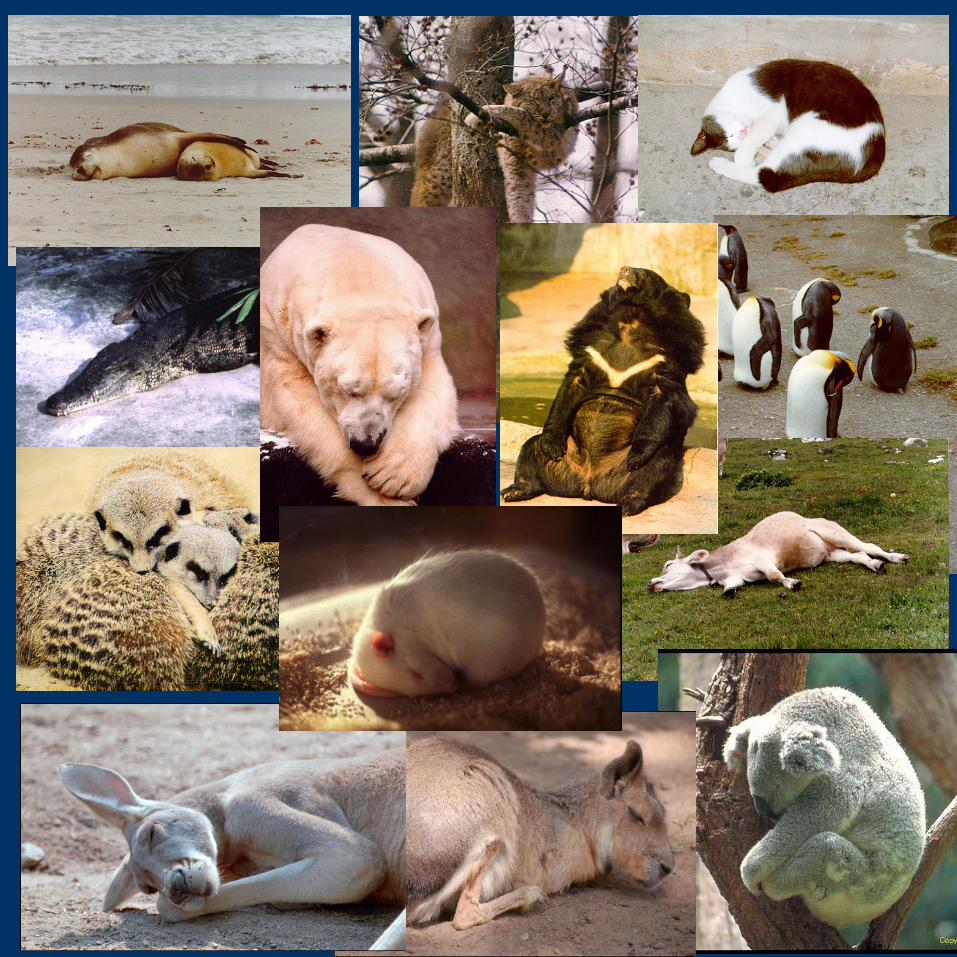
Definition of sleep• rest-activity NOT= sleep-wakefulness
• general criteria of sleep– lack of movements– elevated sensory threshold– full reversibility– stereotypic posture– specific resting place– circadian organization– homeostatic regulation: deprivation –rebound
• mammals (and birds) - polygraphic criteria
4/30
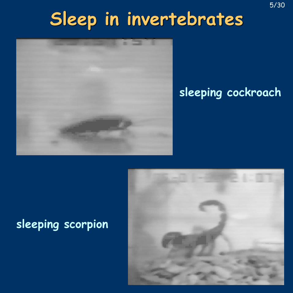
Sleep in invertebrates
sleeping cockroach
sleeping scorpion
5/30
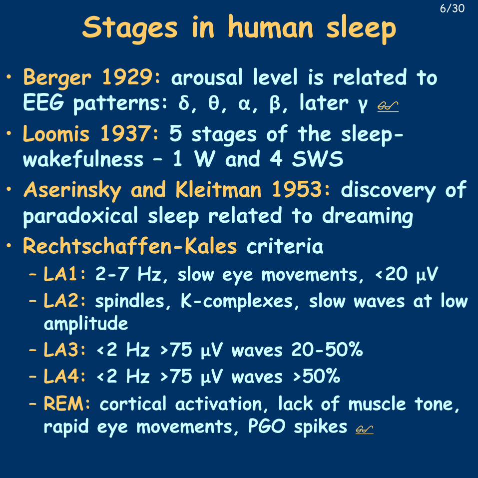
Stages in human sleep• Berger 1929: arousal level is related to EEG patterns: δ, θ, α, β, later γ $
• Loomis 1937: 5 stages of the sleep-wakefulness – 1 W and 4 SWS
• Aserinsky and Kleitman 1953: discovery of paradoxical sleep related to dreaming
• Rechtschaffen-Kales criteria– LA1: 2-7 Hz, slow eye movements, <20 μV– LA2: spindles, K-complexes, slow waves at low amplitude
– LA3: <2 Hz >75 μV waves 20-50%– LA4: <2 Hz >75 μV waves >50%– REM: cortical activation, lack of muscle tone, rapid eye movements, PGO spikes $
6/30
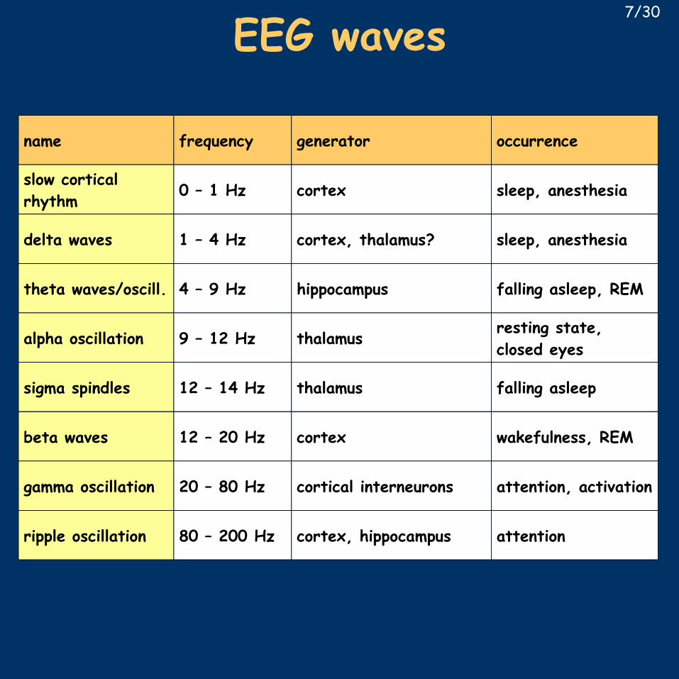
name frequency generator occurrence
slow cortical rhythm
0 – 1 Hz cortex sleep, anesthesia
delta waves 1 – 4 Hz cortex, thalamus? sleep, anesthesia
theta waves/oscill. 4 – 9 Hz hippocampus falling asleep, REM
alpha oscillation 9 – 12 Hz thalamus resting state, closed eyes
sigma spindles 12 – 14 Hz thalamus falling asleep
beta waves 12 – 20 Hz cortex wakefulness, REM
gamma oscillation 20 – 80 Hz cortical interneurons attention, activation
ripple oscillation 80 – 200 Hz cortex, hippocampus attention
7/30
EEG waves
Steriade, M., et al., J. Neurophysiol. 85 (2001): 1969-1985
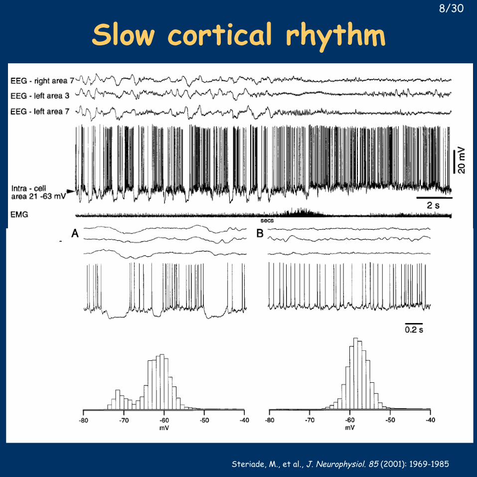
Slow cortical rhythm8/30
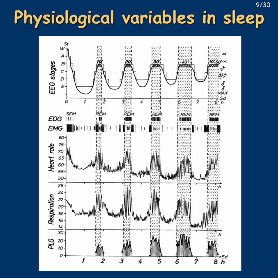
Physiological variables in sleep9/30
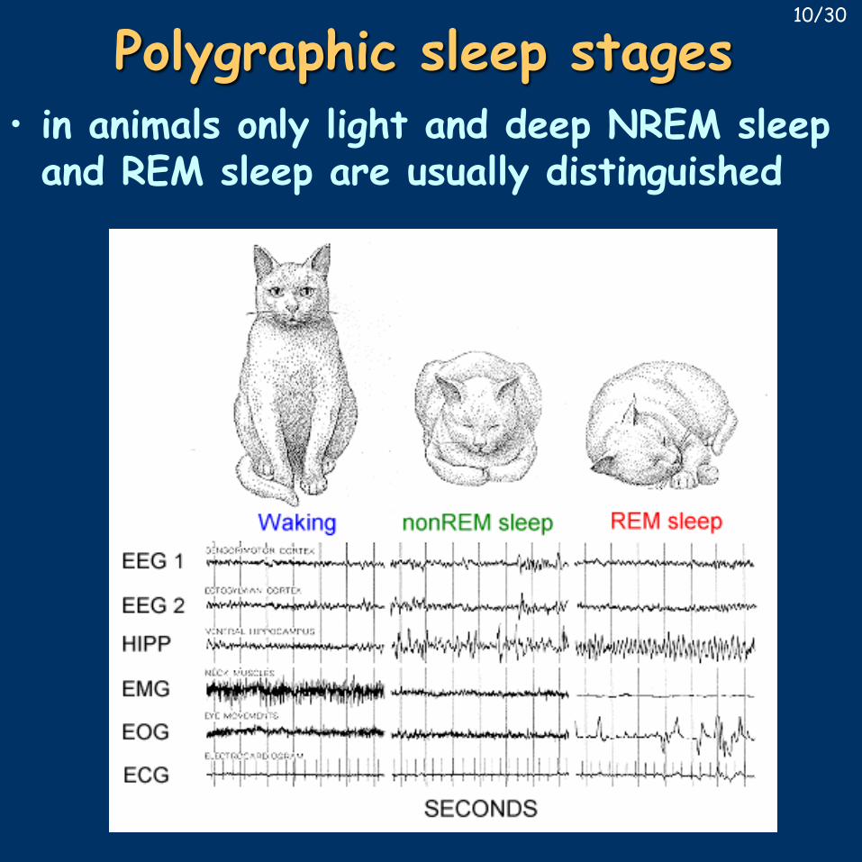
Polygraphic sleep stages• in animals only light and deep NREM sleep and REM sleep are usually distinguished
10/30
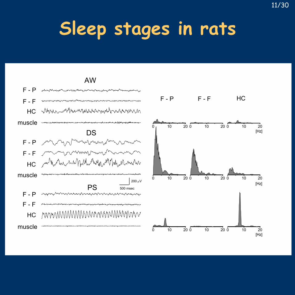
Sleep stages in rats
F - P
F - F
F - P
F - P
PS
DS
AW
muscle
muscle
F - F
HC
HC
muscle
HCF - F
0 10 20
[Hz]
[Hz]
[Hz]
F - P
0 10 20
F - F
0 10 20
HC
0 10 20
0 10 20 0 10 20
0 10 20
500 msec
200 µV
0 10 20 0 10 20
11/30
REM sleep in cats12/03
REM sleep in humans13/30
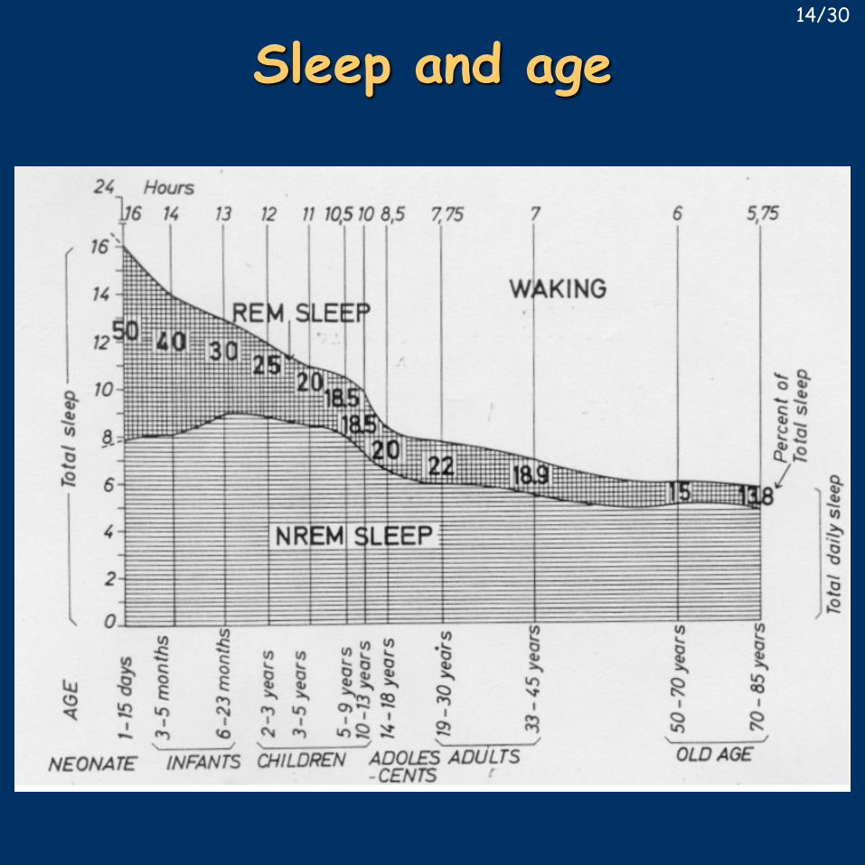
Sleep and age14/30
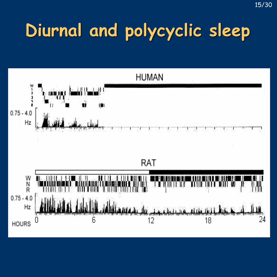
Diurnal and polycyclic sleep15/30
Humoral regulation of sleep• closely related to homeostatic regulation• something is being accumulated or used up• sleep can be easily disturbed, but difficult to induce, appropriate control is a main issue
• two approaches:– harmful effects of sleep deprivation
• stress is difficult to eliminate• motivation to sleep is almost as strong as motivation to avoid pain - torture
– isolation of sleep factors• following sleep deprivation• during natural or experimentally evoked sleep• testing prospective signal molecules normally present in our body
16/30
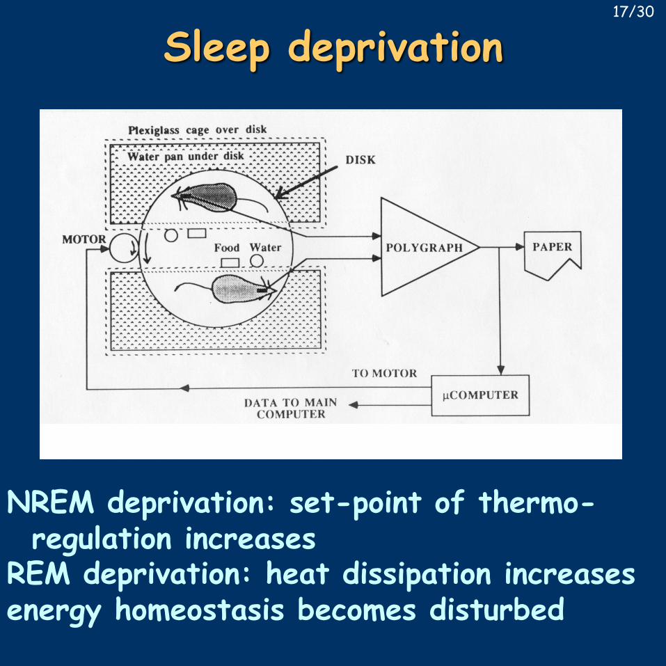
Sleep deprivation
NREM deprivation: set-point of thermo-regulation increases
REM deprivation: heat dissipation increasesenergy homeostasis becomes disturbed
17/30
Sleep factors• Ishimori, Pieron, ~1910: dogs kept awake by forced walking for 10 days – successful sleep transfer
• methodological problems – repeated with positive results in goat-rat experiments
• deprivation is not needed for the effect –collection of human urine
• end result: muramyl peptide• Uchinozo extractions from the brainstem of sleep deprived rats - uridine, oxidized glutathione (glu-cys-gly)
• Monnier sleep induced by thalamic stimulation in rabbits: DSIP (9 aa-s)
• these are not natural sleep factors• natural signal molecules: GHRH, adenosine, interleukin-1, TNFα, PGD2
18/30
Transfer of natural sleep• parabiotic animals: Matsumoto, 1972 –higher synchronity of NREM and REM sleep than between animals joined by their skin only
• de Andres, 1976 – transplantation of an additional head to dogs – independent sleep, 108 h survival
• Siamese twins – independent sleep is possible, but contradicting results exist
• Mukhametov, 1985-87 sleep in dolphins –the two hemispheres can sleep separately
• described in other animals as well: birds, whale, etc. – complete decussation of thevisual pathway is a prerequisite
19/30
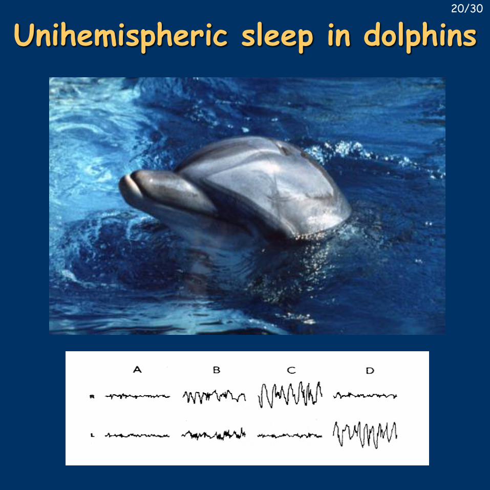
Unihemispheric sleep in dolphins20/30
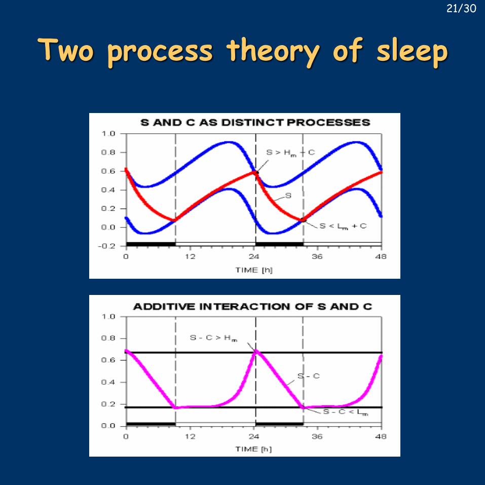
Two process theory of sleep21/30
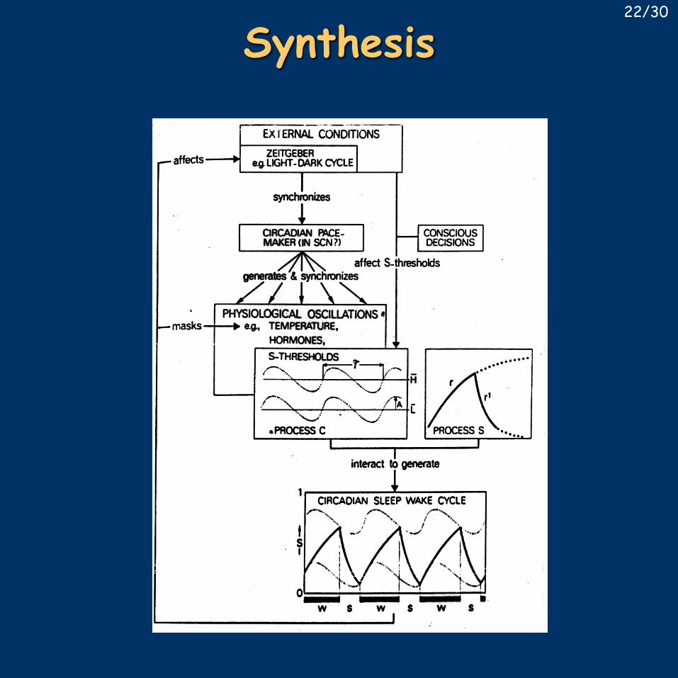
Synthesis22/30
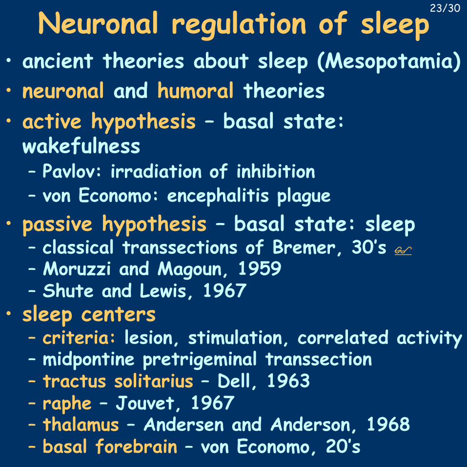
Neuronal regulation of sleep• ancient theories about sleep (Mesopotamia)• neuronal and humoral theories• active hypothesis – basal state:wakefulness– Pavlov: irradiation of inhibition– von Economo: encephalitis plague
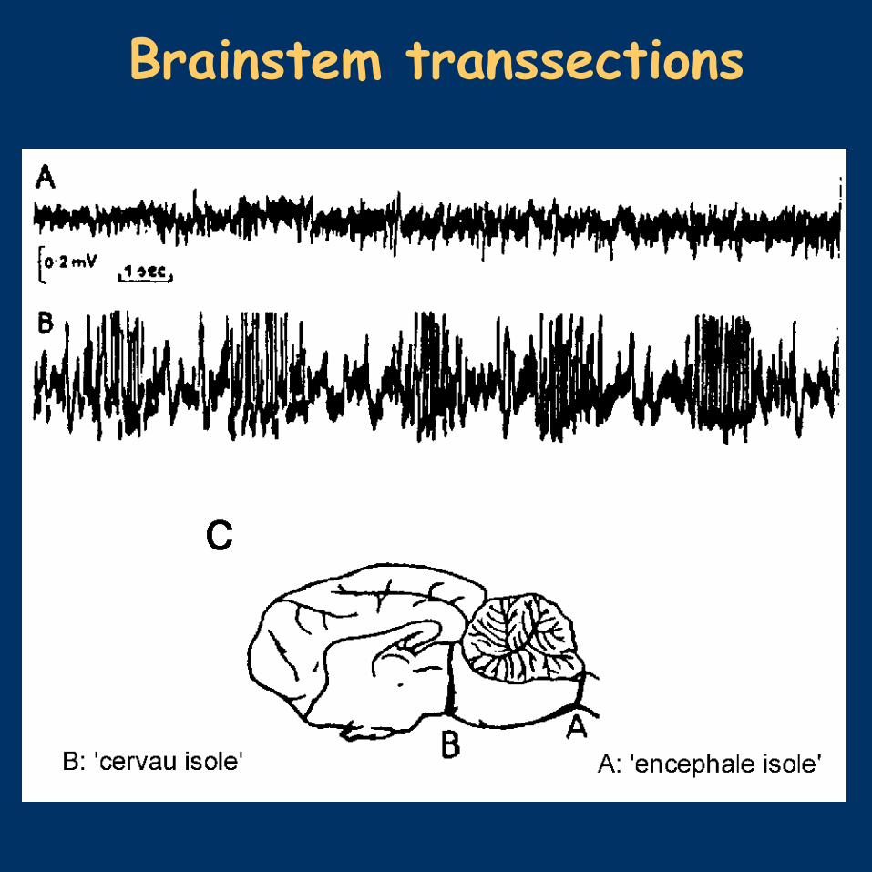
• passive hypothesis – basal state: sleep– classical transsections of Bremer, 30’s $
– Moruzzi and Magoun, 1959– Shute and Lewis, 1967
• sleep centers– criteria: lesion, stimulation, correlated activity– midpontine pretrigeminal transsection– tractus solitarius – Dell, 1963– raphe – Jouvet, 1967– thalamus – Andersen and Anderson, 1968– basal forebrain – von Economo, 20’s
23/30
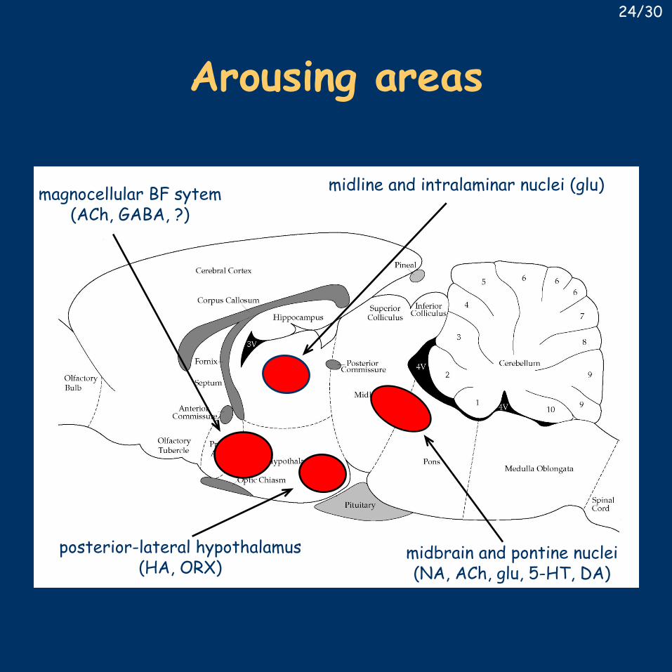
Arousing areas
midline and intralaminar nuclei (glu)
midbrain and pontine nuclei(NA, ACh, glu, 5-HT, DA)
posterior-lateral hypothalamus(HA, ORX)
magnocellular BF sytem(ACh, GABA, ?)
24/30
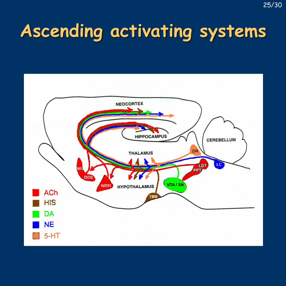
Ascending activating systems25/30
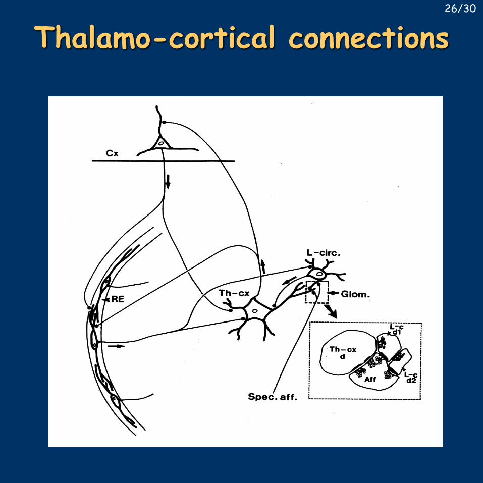
Thalamo-cortical connections26/30
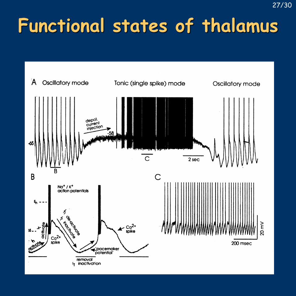
Functional states of thalamus27/30
Role of the basal forebrain• von Economo: BF-POA promotes sleep, posterior HT promotes wakefulness
• Sterman and Clemente 1962- lesion causes decreased or fragmented sleep
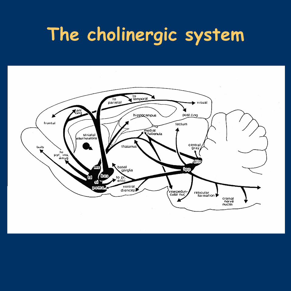
• stimulation – sleep (also at high frequency!)• conditioned response to sounds• warming, ACh crystals – sleep• late 70’s, early 80’s – description of the cholinergic system $
• cholinergic cells disappear or shrink in Alzheimer’s disease
• electrical – excitotxic – selective lesion• corticopetal projection is not exclusively cholinergic
• SCN, thermoregulation, proximity of HT, VLPO, prefrontal cortex – high importance
28/30
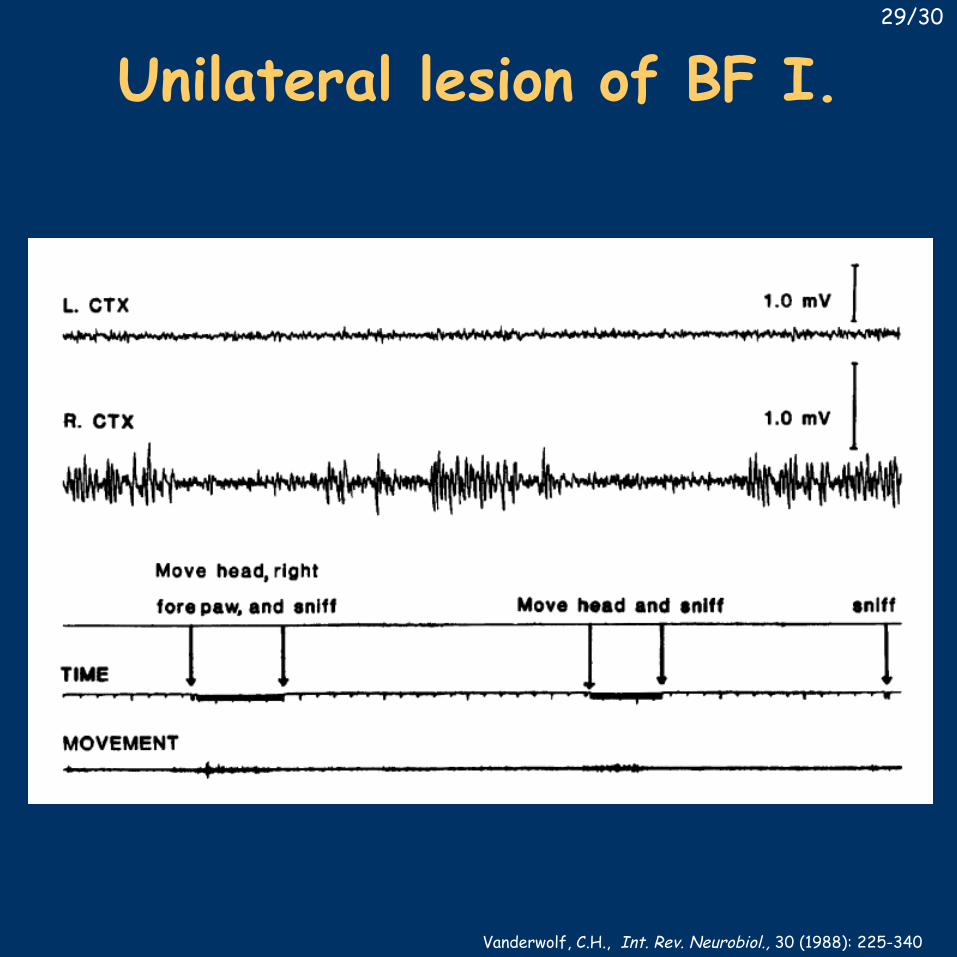
Unilateral lesion of BF I.
Vanderwolf, C.H., Int. Rev. Neurobiol., 30 (1988): 225-340
29/30
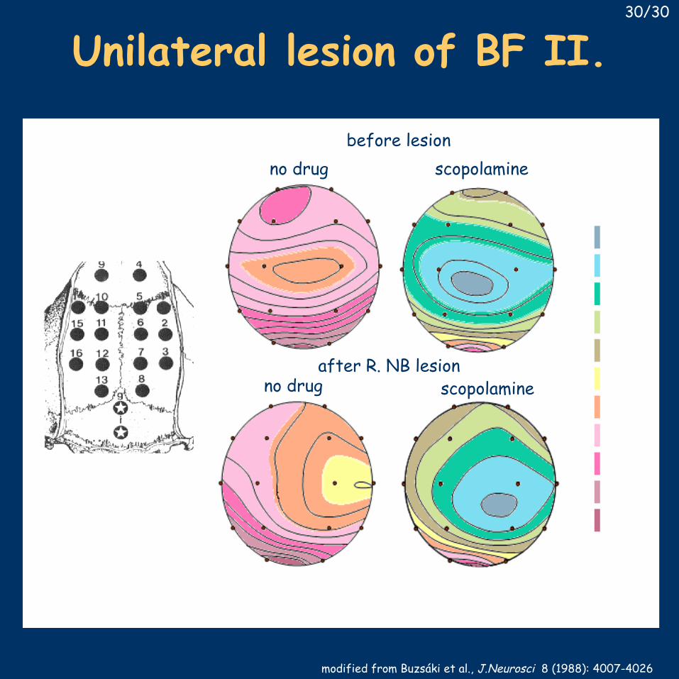
scopolamine
scopolamine
no drug
no drug
before lesion
after R. NB lesion
modified from Buzsáki et al., J.Neurosci 8 (1988): 4007-4026
Unilateral lesion of BF II.30/30


Berger - 1929
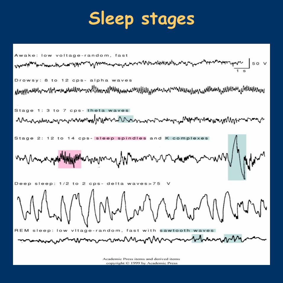
Sleep stages
Brainstem transsections
The cholinergic system