Rapid emergence and mechanisms of resistance by U87 ... benefit of therapeutic agents to the short...
6
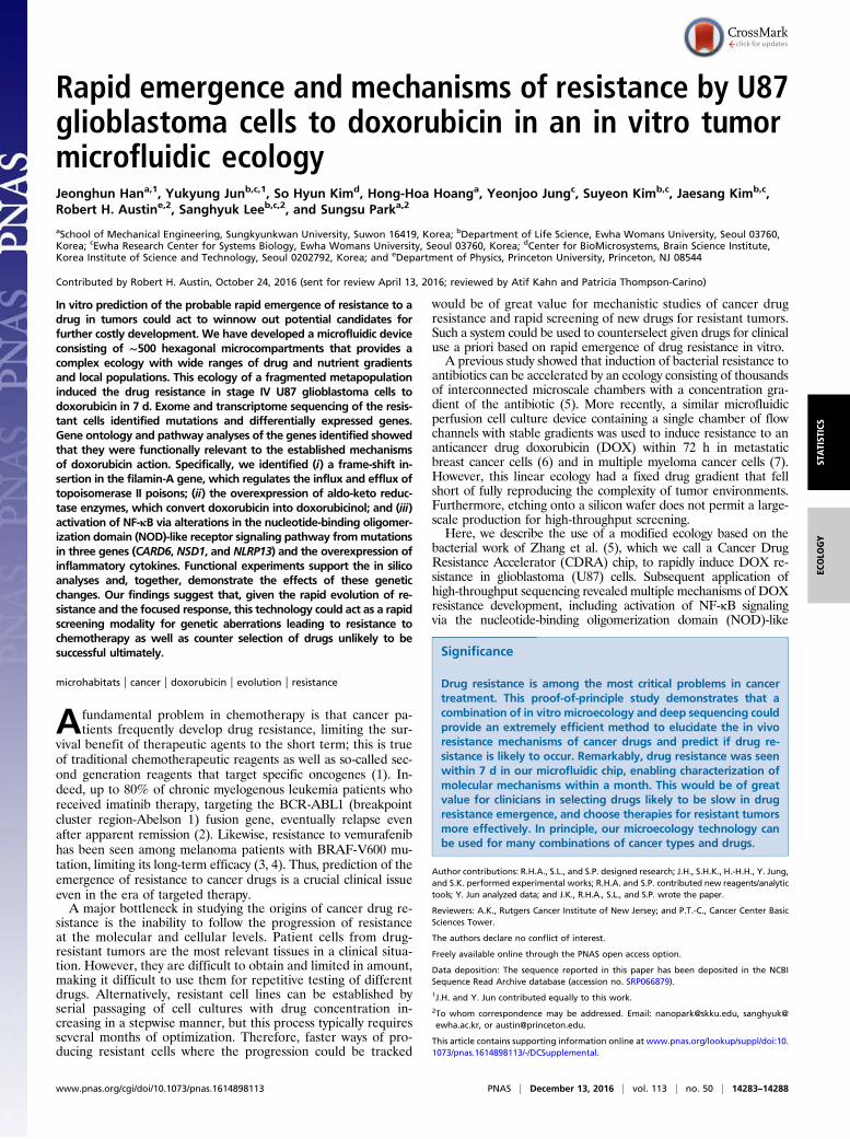
Rapid emergence and mechanisms of resistance by U87 glioblastoma cells to doxorubicin in an in vitro tumor microfluidic ecology Jeonghun Han a,1 , Yukyung Jun b,c,1 , So Hyun Kim d , Hong-Hoa Hoang a , Yeonjoo Jung c , Suyeon Kim b,c , Jaesang Kim b,c , Robert H. Austin e,2 , Sanghyuk Lee b,c,2 , and Sungsu Park a,2 a School of Mechanical Engineering, Sungkyunkwan University, Suwon 16419, Korea; b Department of Life Science, Ewha Womans University, Seoul 03760, Korea; c Ewha Research Center for Systems Biology, Ewha Womans University, Seoul 03760, Korea; d Center for BioMicrosystems, Brain Science Institute, Korea Institute of Science and Technology, Seoul 0202792, Korea; and e Department of Physics, Princeton University, Princeton, NJ 08544 Contributed by Robert H. Austin, October 24, 2016 (sent for review April 13, 2016; reviewed by Atif Kahn and Patricia Thompson-Carino) In vitro prediction of the probable rapid emergence of resistance to a drug in tumors could act to winnow out potential candidates for further costly development. We have developed a microfluidic device consisting of ∼500 hexagonal microcompartments that provides a complex ecology with wide ranges of drug and nutrient gradients and local populations. This ecology of a fragmented metapopulation induced the drug resistance in stage IV U87 glioblastoma cells to doxorubicin in 7 d. Exome and transcriptome sequencing of the resis- tant cells identified mutations and differentially expressed genes. Gene ontology and pathway analyses of the genes identified showed that they were functionally relevant to the established mechanisms of doxorubicin action. Specifically, we identified (i ) a frame-shift in- sertion in the filamin-A gene, which regulates the influx and efflux of topoisomerase II poisons; (ii ) the overexpression of aldo-keto reduc- tase enzymes, which convert doxorubicin into doxorubicinol; and (iii ) activation of NF-κB via alterations in the nucleotide-binding oligomer- ization domain (NOD)-like receptor signaling pathway from mutations in three genes (CARD6, NSD1, and NLRP13) and the overexpression of inflammatory cytokines. Functional experiments support the in silico analyses and, together, demonstrate the effects of these genetic changes. Our findings suggest that, given the rapid evolution of re- sistance and the focused response, this technology could act as a rapid screening modality for genetic aberrations leading to resistance to chemotherapy as well as counter selection of drugs unlikely to be successful ultimately. microhabitats | cancer | doxorubicin | evolution | resistance A fundamental problem in chemotherapy is that cancer pa- tients frequently develop drug resistance, limiting the sur- vival benefit of therapeutic agents to the short term; this is true of traditional chemotherapeutic reagents as well as so-called sec- ond generation reagents that target specific oncogenes (1). In- deed, up to 80% of chronic myelogenous leukemia patients who received imatinib therapy, targeting the BCR-ABL1 (breakpoint cluster region-Abelson 1) fusion gene, eventually relapse even after apparent remission (2). Likewise, resistance to vemurafenib has been seen among melanoma patients with BRAF-V600 mu- tation, limiting its long-term efficacy (3, 4). Thus, prediction of the emergence of resistance to cancer drugs is a crucial clinical issue even in the era of targeted therapy. A major bottleneck in studying the origins of cancer drug re- sistance is the inability to follow the progression of resistance at the molecular and cellular levels. Patient cells from drug- resistant tumors are the most relevant tissues in a clinical situa- tion. However, they are difficult to obtain and limited in amount, making it difficult to use them for repetitive testing of different drugs. Alternatively, resistant cell lines can be established by serial passaging of cell cultures with drug concentration in- creasing in a stepwise manner, but this process typically requires several months of optimization. Therefore, faster ways of pro- ducing resistant cells where the progression could be tracked would be of great value for mechanistic studies of cancer drug resistance and rapid screening of new drugs for resistant tumors. Such a system could be used to counterselect given drugs for clinical use a priori based on rapid emergence of drug resistance in vitro. A previous study showed that induction of bacterial resistance to antibiotics can be accelerated by an ecology consisting of thousands of interconnected microscale chambers with a concentration gra- dient of the antibiotic (5). More recently, a similar microfluidic perfusion cell culture device containing a single chamber of flow channels with stable gradients was used to induce resistance to an anticancer drug doxorubicin (DOX) within 72 h in metastatic breast cancer cells (6) and in multiple myeloma cancer cells (7). However, this linear ecology had a fixed drug gradient that fell short of fully reproducing the complexity of tumor environments. Furthermore, etching onto a silicon wafer does not permit a large- scale production for high-throughput screening. Here, we describe the use of a modified ecology based on the bacterial work of Zhang et al. (5), which we call a Cancer Drug Resistance Accelerator (CDRA) chip, to rapidly induce DOX re- sistance in glioblastoma (U87) cells. Subsequent application of high-throughput sequencing revealed multiple mechanisms of DOX resistance development, including activation of NF-κB signaling via the nucleotide-binding oligomerization domain (NOD)-like Significance Drug resistance is among the most critical problems in cancer treatment. This proof-of-principle study demonstrates that a combination of in vitro microecology and deep sequencing could provide an extremely efficient method to elucidate the in vivo resistance mechanisms of cancer drugs and predict if drug re- sistance is likely to occur. Remarkably, drug resistance was seen within 7 d in our microfluidic chip, enabling characterization of molecular mechanisms within a month. This would be of great value for clinicians in selecting drugs likely to be slow in drug resistance emergence, and choose therapies for resistant tumors more effectively. In principle, our microecology technology can be used for many combinations of cancer types and drugs. Author contributions: R.H.A., S.L., and S.P. designed research; J.H., S.H.K., H.-H.H., Y. Jung, and S.K. performed experimental works; R.H.A. and S.P. contributed new reagents/analytic tools; Y. Jun analyzed data; and J.K., R.H.A., S.L., and S.P. wrote the paper. Reviewers: A.K., Rutgers Cancer Institute of New Jersey; and P.T.-C., Cancer Center Basic Sciences Tower. The authors declare no conflict of interest. Freely available online through the PNAS open access option. Data deposition: The sequence reported in this paper has been deposited in the NCBI Sequence Read Archive database (accession no. SRP066879). 1 J.H. and Y. Jun contributed equally to this work. 2 To whom correspondence may be addressed. Email: [email protected], sanghyuk@ ewha.ac.kr, or [email protected]. This article contains supporting information online at www.pnas.org/lookup/suppl/doi:10. 1073/pnas.1614898113/-/DCSupplemental. www.pnas.org/cgi/doi/10.1073/pnas.1614898113 PNAS | December 13, 2016 | vol. 113 | no. 50 | 14283–14288 STATISTICS ECOLOGY
Transcript of Rapid emergence and mechanisms of resistance by U87 ... benefit of therapeutic agents to the short...
Rapid emergence and mechanisms of resistance by U87glioblastoma cells to doxorubicin in an in vitro tumormicrofluidic ecologyJeonghun Hana,1, Yukyung Junb,c,1, So Hyun Kimd, Hong-Hoa Hoanga, Yeonjoo Jungc, Suyeon Kimb,c, Jaesang Kimb,c,Robert H. Austine,2, Sanghyuk Leeb,c,2, and Sungsu Parka,2
aSchool of Mechanical Engineering, Sungkyunkwan University, Suwon 16419, Korea; bDepartment of Life Science, Ewha Womans University, Seoul 03760,Korea; cEwha Research Center for Systems Biology, Ewha Womans University, Seoul 03760, Korea; dCenter for BioMicrosystems, Brain Science Institute,Korea Institute of Science and Technology, Seoul 0202792, Korea; and eDepartment of Physics, Princeton University, Princeton, NJ 08544
Contributed by Robert H. Austin, October 24, 2016 (sent for review April 13, 2016; reviewed by Atif Kahn and Patricia Thompson-Carino)
In vitro prediction of the probable rapid emergence of resistance to adrug in tumors could act to winnow out potential candidates forfurther costly development. We have developed a microfluidic deviceconsisting of ∼500 hexagonal microcompartments that provides acomplex ecology with wide ranges of drug and nutrient gradientsand local populations. This ecology of a fragmented metapopulationinduced the drug resistance in stage IV U87 glioblastoma cells todoxorubicin in 7 d. Exome and transcriptome sequencing of the resis-tant cells identified mutations and differentially expressed genes.Gene ontology and pathway analyses of the genes identified showedthat they were functionally relevant to the established mechanismsof doxorubicin action. Specifically, we identified (i) a frame-shift in-sertion in the filamin-A gene, which regulates the influx and efflux oftopoisomerase II poisons; (ii) the overexpression of aldo-keto reduc-tase enzymes, which convert doxorubicin into doxorubicinol; and (iii)activation of NF-κB via alterations in the nucleotide-binding oligomer-ization domain (NOD)-like receptor signaling pathway frommutationsin three genes (CARD6, NSD1, and NLRP13) and the overexpression ofinflammatory cytokines. Functional experiments support the in silicoanalyses and, together, demonstrate the effects of these geneticchanges. Our findings suggest that, given the rapid evolution of re-sistance and the focused response, this technology could act as a rapidscreening modality for genetic aberrations leading to resistance tochemotherapy as well as counter selection of drugs unlikely to besuccessful ultimately.
microhabitats | cancer | doxorubicin | evolution | resistance
Afundamental problem in chemotherapy is that cancer pa-tients frequently develop drug resistance, limiting the sur-
vival benefit of therapeutic agents to the short term; this is trueof traditional chemotherapeutic reagents as well as so-called sec-ond generation reagents that target specific oncogenes (1). In-deed, up to 80% of chronic myelogenous leukemia patients whoreceived imatinib therapy, targeting the BCR-ABL1 (breakpointcluster region-Abelson 1) fusion gene, eventually relapse evenafter apparent remission (2). Likewise, resistance to vemurafenibhas been seen among melanoma patients with BRAF-V600 mu-tation, limiting its long-term efficacy (3, 4). Thus, prediction of theemergence of resistance to cancer drugs is a crucial clinical issueeven in the era of targeted therapy.A major bottleneck in studying the origins of cancer drug re-
sistance is the inability to follow the progression of resistanceat the molecular and cellular levels. Patient cells from drug-resistant tumors are the most relevant tissues in a clinical situa-tion. However, they are difficult to obtain and limited in amount,making it difficult to use them for repetitive testing of differentdrugs. Alternatively, resistant cell lines can be established byserial passaging of cell cultures with drug concentration in-creasing in a stepwise manner, but this process typically requiresseveral months of optimization. Therefore, faster ways of pro-ducing resistant cells where the progression could be tracked
would be of great value for mechanistic studies of cancer drugresistance and rapid screening of new drugs for resistant tumors.Such a system could be used to counterselect given drugs for clinicaluse a priori based on rapid emergence of drug resistance in vitro.A previous study showed that induction of bacterial resistance to
antibiotics can be accelerated by an ecology consisting of thousandsof interconnected microscale chambers with a concentration gra-dient of the antibiotic (5). More recently, a similar microfluidicperfusion cell culture device containing a single chamber of flowchannels with stable gradients was used to induce resistance to ananticancer drug doxorubicin (DOX) within 72 h in metastaticbreast cancer cells (6) and in multiple myeloma cancer cells (7).However, this linear ecology had a fixed drug gradient that fellshort of fully reproducing the complexity of tumor environments.Furthermore, etching onto a silicon wafer does not permit a large-scale production for high-throughput screening.Here, we describe the use of a modified ecology based on the
bacterial work of Zhang et al. (5), which we call a Cancer DrugResistance Accelerator (CDRA) chip, to rapidly induce DOX re-sistance in glioblastoma (U87) cells. Subsequent application ofhigh-throughput sequencing revealed multiple mechanisms of DOXresistance development, including activation of NF-κB signalingvia the nucleotide-binding oligomerization domain (NOD)-like
Significance
Drug resistance is among the most critical problems in cancertreatment. This proof-of-principle study demonstrates that acombination of in vitro microecology and deep sequencing couldprovide an extremely efficient method to elucidate the in vivoresistance mechanisms of cancer drugs and predict if drug re-sistance is likely to occur. Remarkably, drug resistance was seenwithin 7 d in our microfluidic chip, enabling characterization ofmolecular mechanisms within a month. This would be of greatvalue for clinicians in selecting drugs likely to be slow in drugresistance emergence, and choose therapies for resistant tumorsmore effectively. In principle, our microecology technology canbe used for many combinations of cancer types and drugs.
Author contributions: R.H.A., S.L., and S.P. designed research; J.H., S.H.K., H.-H.H., Y. Jung,and S.K. performed experimental works; R.H.A. and S.P. contributed new reagents/analytictools; Y. Jun analyzed data; and J.K., R.H.A., S.L., and S.P. wrote the paper.
Reviewers: A.K., Rutgers Cancer Institute of New Jersey; and P.T.-C., Cancer Center BasicSciences Tower.
The authors declare no conflict of interest.
Freely available online through the PNAS open access option.
Data deposition: The sequence reported in this paper has been deposited in the NCBISequence Read Archive database (accession no. SRP066879).1J.H. and Y. Jun contributed equally to this work.2To whom correspondence may be addressed. Email: [email protected], [email protected], or [email protected].
This article contains supporting information online at www.pnas.org/lookup/suppl/doi:10.1073/pnas.1614898113/-/DCSupplemental.
www.pnas.org/cgi/doi/10.1073/pnas.1614898113 PNAS | December 13, 2016 | vol. 113 | no. 50 | 14283–14288
STATIST
ICS
ECOLO
GY

receptor (NLR, a subset of pattern recognition receptors) pathway.Our results indicate the potential utility of CDRA chips in selectingand counterselecting therapeutic agents in a variety of cancer types.
ResultsMicrofluidic Chip Design and Cell Culture for Resistance Induction. TheCDRA chip contained 488 hexagonal microchambers (diameter200 μm, height 40 μm) surrounded by two microchannels (width 150μm) for long-term cell culture under antiparallel overlapping gra-dients of DOX and nutrients (Fig. 1A). The concentration gradientson the chip were successfully shown by flowing fluorescent materialfrom one of the inlets and measuring the fluorescence intensity (Fig.1B). The chip operates without the aid of additional instrumentationfor the generation of concentration gradient in typical cell cultureincubators. Culture requires minimal manipulation with dailyresupply of DOX and nutrients whose flow is maintained by gravityonly. The handy operation is schematically depicted along with theactual image of the CDRA chip (SI Appendix, Fig. S1). The chip,made from polydimethylsiloxane (PDMS), can be produced in largescale at low cost via routine soft-microfabrication replica processes.U87 cancer cells were distributed homogeneously throughout
the microchambers before flowing DOX and media, and cellnumbers were counted over 7 d from microscopic images of the
CDRA chip (SI Appendix, Fig. S2). We observed a dramaticdecrease in cell numbers in the chambers near the DOX channelat day 3, and, apparently, three-quarters of the chambers becameempty at day 5 owing to the cytotoxic effect of DOX (Fig. 1C).Cell repopulation was evident by day 7 (Fig. 1C), indicating thedevelopment of resistance and migration of resistant cells toareas with higher DOX concentration.We performed several experiments to validate the DOX resis-
tance. Cells harvested from the chip at day 7 (U87-DR) showed∼30 times greater resistance to DOX than wild-type U87 cells(U87-WT) as indicated by IC50 (Fig. 2A), although the prolifera-tion activity was reduced by half in the resistant cells comparedwith the WT cells (Fig. 2B). One of the main causes of drug re-sistance is the change in drug pump activity. In a Rhodamine 123efflux assay, reduction in fluorescence intensity reflecting dye ex-clusion was ∼2.5-fold higher in the resistant cells (Fig. 2C). Also, adrug efflux assay using flow cytometry showed a substantial in-crease in efflux activity after acquisition of the resistance (Fig. 2D).Together, these data indicate that the cells acquired distinct at-tributes as DOX-resistant cells within 7-d-long drug exposure.
Identification of Gene Alterations for DOX Resistance. We used acombination of exome and transcriptome sequencing to elucidateresistance mechanisms and shed light on the development of DOXresistance in cancer cells. We note that, like many cancers, themutational spectrum of the U87 cells before incubation in the chip isextraordinarily complex, with hundreds of single-nucleotide variants(SNVs), small indels, large microdeletions, and interchromosomaltranslocations (8). Despite this complexity, the U87 cells come intoour chip highly sensitive to DOX, but come out highly resistant.To identify genomic aberrations responsible for the development
of DOX resistance, we performed exome sequencing of WT andDOX-resistant U87 cells obtained after 7 d of growth on a CDRAchip and compared the results. We combined the results from foursoftware programs [MuTect (9), Strelka (10), VarScan2 (11), andVirmid (12)] for detecting somatic mutations and identified 107variants with high confidence (i.e., called by multiple programs),including 2 indels, 5 stopgain mutations, and 54 missense mutations
Fig. 1. Design and properties of the CDRA chip. (A) Schematic diagram of thechip and a microscopic image showing chip dimensions. Filled arrow indicatesthe flow of 5 μMDOX and nutrients, whereas empty arrow represents the flowof nutrients only. (B) A fluorescence image of the concentration gradient. Theplot shows the fluorescence intensity profile along two horizontal lines.(C) Distribution of viable cells in the chip over a period of 7 d. Cell numbers ineach hexagonal are indicated in red color coded in six steps.
Fig. 2. Characterization of DOX-resistant cells from the CDRA chip. (A) Cellviability of resistant (U87-DR) and WT (U87-WT) cells at various concentra-tions (0 μM to 1 μM) of DOX. IC50 values were estimated to be 0.05 μM and1.47 μM for the WT and resistant cells, respectively. Note that the x-axis scaleis logarithmic. (B) Cell proliferation activity of the resistant and WT cells in adrug-free condition. (C) Relative efflux rate measured by the reduced fluo-rescence intensity of Rhodamine 123 due to dye efflux (P value = 0.008 withone-tailed t test, from triplicate experiments). (D) Estimation of drug effluxactivities of the WT and resistant cells. Cell counts were measured before(gray line) and after (blue line) the 1 h-incubation in flow cytometry.
14284 | www.pnas.org/cgi/doi/10.1073/pnas.1614898113 Han et al.

(SI Appendix, Table S1; see Materials and Methods for details). Astatistical functional analysis of these nonsynonymous variants usingWeb-based Gene Set Analysis Toolkit (WebGestalt) (13) showedthat three representative terms were enriched in the Gene On-tology (GO) molecular function category, namely “carbohydratederivative binding,” “nucleoside binding,” and “ATP-dependenthelicase activity” (SI Appendix, Table S2). These findings furthersupport the findings of previous studies that DOX inhibits theenzyme topoisomerase II by intercalating between two base pairsof the DNA double helix (14).The helicase activity of topoisomerase II was shown to be
potently inhibited by anthracycline antibiotics such as DOX (15).Moreover, we observed a mutation in the CHD1 (chromodomainhelicase DNA binding protein 1) gene, which is a known target ofepirubicin, the 4′-epi-isomer of DOX (16). Another notablemutation was a frame-shift insertion in the filamin-A (FLNA)gene, whose loss is known to promote DOX resistance by reg-ulating the influx and efflux of topoisomerase II poisons (17).
Transcriptome Sequencing and Pathway Analysis for ResistanceDevelopment. To further identify the altered pathways and molecu-lar processes responsible for emergence of drug resistance, we gen-erated RNA-seq data for the U87 WT and resistant cell linesobtained after 7 d of growth on a CDRA chip. Our computationalpipeline identified 83 differentially expressed genes (DEGs), of which77 were up-regulated and 6 were down-regulated (SI Appendix, TableS3; seeMaterials and Methods for details). A gene set analysis of GOterms for these 83 DEGs yielded many terms related to immuneresponses, including “response to wounding,” “inflammatory re-sponse,” “leukocyte chemotaxis,” “response to hypoxia,” “regulationof cell proliferation,” and “cytokine activity’ (SI Appendix, Table S4).The most relevant term was “DOX metabolic process” [false dis-covery rate (FDR) = 3.89E-6], which was represented by four aldo-keto reductases (AKRs; AKR1B1, AKR1C1, AKR1C2, and AKR1C3)that convert DOX to doxorubicinol. Compared with DOX, doxor-ubicinol shows dramatically reduced cytotoxicity, reduced DNA-binding activity, and strong localization to extranuclear lysosomes(18).The Kyoto Encyclopedia of Genes and Genomes pathway anal-
ysis of 83 DEGs with WebGestalt (13) yielded the NOD-like re-ceptor signaling pathway (FDR = 1.33E-6) and cytokine−cytokinereceptor interaction pathway (FDR = 2.46E-8). The DEGs involvedin these pathways were mostly chemokines and proinflammatorycytokines (e.g., CCL2, CXCL1, CXCL2, CXCL3, IL1B, IL6, IL8,and IL11) that were regulated by the nuclear factor kappa B(NF-κB) pathway.Notably, NF-κB activation is known to be essential for the
cytotoxic effects of DOX and its analogs (19). From the exomesequencing data, we further identified three mutated genes(CARD6, NSD1, and NLRP13) that are associated with NF-κBactivation. CARD6 (caspase activation and recruitment domainfamily, member 6) negatively regulates NF-κB activation byNOD1 (nucleotide-binding oligomerization domain containing1) receptor in human embryonic kidney-derived HEK293T cells(20, 21) whereas the effect is reversed in Huh7 cells of hep-atocarcinoma origin (22), thus indicating that the role ofCARD6 in NF-κB activation may be cell-type specific.
Experimental Validation for Key Regulators of DOX Resistance. We nextsought to carry out preliminary validation of our in silico analyses.First, we attempted to mimic the effect of the frame-shift mutationin FLNA by knocking down with specific siRNAs. Consistent withpredicted loss of function from a frame-shift mutation, it was verifiedthat the down-regulation of FLNA by siRNA caused a 1.5-fold in-crease in DOX resistance at the 10 nM concentration in the U87cancer cell line (Fig. 3A). We also observed a stopgain muta-tion in CARD6 in resistant cells (c.G2014T of NM_032587encoding p.Gly672*) and performed a similar knockdown experi-ment for CARD6. The treatment of U87 cells with siRNA specificfor CARD6 elevated DOX resistance consistent with the down-regulation of NF-κB pathway signaling in these cells (Fig. 3B). In
Fig. 3. Knockdown of FLNA, CARD6, and NSD1 by siRNAs in U87 cells leads toincreased resistance to DOX. Cell survival curves of 72-h DOX-treated cellstransfected with control siRNAs (NC) or gene-specific siRNAs are shown. Graphsshow the representative results of three independent cell viability assays afterknockdown of each of three genes, (A) FLNA, (B) CARD6, and (C) NSD1. Knock-down of FLNA, CARD6, and NSD1 led to increased resistance to DOX, where theaverage *P = 0.023, 0.004, and 0.004, respectively, at 0.01 μMDOX concentration.The knockdown efficiency of each siRNA is shown in SI Appendix, Fig. S3.
Han et al. PNAS | December 13, 2016 | vol. 113 | no. 50 | 14285
STATIST
ICS
ECOLO
GY
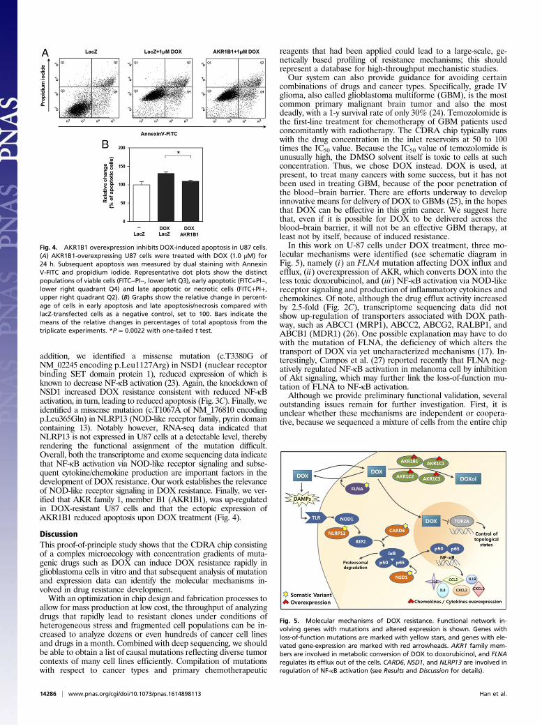
addition, we identified a missense mutation (c.T3380G ofNM_02245 encoding p.Leu1127Arg) in NSD1 (nuclear receptorbinding SET domain protein 1), reduced expression of which isknown to decrease NF-κB activation (23). Again, the knockdown ofNSD1 increased DOX resistance consistent with reduced NF-κBactivation, in turn, leading to reduced apoptosis (Fig. 3C). Finally, weidentified a missense mutation (c.T1067A of NM_176810 encodingp.Leu365Gln) in NLRP13 (NOD-like receptor family, pyrin domaincontaining 13). Notably however, RNA-seq data indicated thatNLRP13 is not expressed in U87 cells at a detectable level, therebyrendering the functional assignment of the mutation difficult.Overall, both the transcriptome and exome sequencing data indicatethat NF-κB activation via NOD-like receptor signaling and subse-quent cytokine/chemokine production are important factors in thedevelopment of DOX resistance. Our work establishes the relevanceof NOD-like receptor signaling in DOX resistance. Finally, we ver-ified that AKR family 1, member B1 (AKR1B1), was up-regulatedin DOX-resistant U87 cells and that the ectopic expression ofAKR1B1 reduced apoptosis upon DOX treatment (Fig. 4).
DiscussionThis proof-of-principle study shows that the CDRA chip consistingof a complex microecology with concentration gradients of muta-genic drugs such as DOX can induce DOX resistance rapidly inglioblastoma cells in vitro and that subsequent analysis of mutationand expression data can identify the molecular mechanisms in-volved in drug resistance development.With an optimization in chip design and fabrication processes to
allow for mass production at low cost, the throughput of analyzingdrugs that rapidly lead to resistant clones under conditions ofheterogeneous stress and fragmented cell populations can be in-creased to analyze dozens or even hundreds of cancer cell linesand drugs in a month. Combined with deep sequencing, we shouldbe able to obtain a list of causal mutations reflecting diverse tumorcontexts of many cell lines efficiently. Compilation of mutationswith respect to cancer types and primary chemotherapeutic
reagents that had been applied could lead to a large-scale, ge-netically based profiling of resistance mechanisms; this shouldrepresent a database for high-throughput mechanistic studies.Our system can also provide guidance for avoiding certain
combinations of drugs and cancer types. Specifically, grade IVglioma, also called glioblastoma multiforme (GBM), is the mostcommon primary malignant brain tumor and also the mostdeadly, with a 1-y survival rate of only 30% (24). Temozolomide isthe first-line treatment for chemotherapy of GBM patients usedconcomitantly with radiotherapy. The CDRA chip typically runswith the drug concentration in the inlet reservoirs at 50 to 100times the IC50 value. Because the IC50 value of temozolomide isunusually high, the DMSO solvent itself is toxic to cells at suchconcentration. Thus, we chose DOX instead. DOX is used, atpresent, to treat many cancers with some success, but it has notbeen used in treating GBM, because of the poor penetration ofthe blood−brain barrier. There are efforts underway to developinnovative means for delivery of DOX to GBMs (25), in the hopesthat DOX can be effective in this grim cancer. We suggest herethat, even if it is possible for DOX to be delivered across theblood–brain barrier, it will not be an effective GBM therapy, atleast not by itself, because of induced resistance.In this work on U-87 cells under DOX treatment, three mo-
lecular mechanisms were identified (see schematic diagram inFig. 5), namely (i) an FLNA mutation affecting DOX influx andefflux, (ii) overexpression of AKR, which converts DOX into theless toxic doxorubicinol, and (iii) NF-κB activation via NOD-likereceptor signaling and production of inflammatory cytokines andchemokines. Of note, although the drug efflux activity increasedby 2.5-fold (Fig. 2C), transcriptome sequencing data did notshow up-regulation of transporters associated with DOX path-way, such as ABCC1 (MRP1), ABCC2, ABCG2, RALBP1, andABCB1 (MDR1) (26). One possible explanation may have to dowith the mutation of FLNA, the deficiency of which alters thetransport of DOX via yet uncharacterized mechanisms (17). In-terestingly, Campos et al. (27) reported recently that FLNA neg-atively regulated NF-κB activation in melanoma cell by inhibitionof Akt signaling, which may further link the loss-of-function mu-tation of FLNA to NF-κB activation.Although we provide preliminary functional validation, several
outstanding issues remain for further investigation. First, it isunclear whether these mechanisms are independent or coopera-tive, because we sequenced a mixture of cells from the entire chip
Fig. 4. AKR1B1 overexpression inhibits DOX-induced apoptosis in U87 cells.(A) AKR1B1-overexpressing U87 cells were treated with DOX (1.0 μM) for24 h. Subsequent apoptosis was measured by dual staining with AnnexinV-FITC and propidium iodide. Representative dot plots show the distinctpopulations of viable cells (FITC−PI−, lower left Q3), early apoptotic (FITC+PI−,lower right quadrant Q4) and late apoptotic or necrotic cells (FITC+PI+,upper right quadrant Q2). (B) Graphs show the relative change in percent-age of cells in early apoptosis and late apoptosis/necrosis compared withlacZ-transfected cells as a negative control, set to 100. Bars indicate themeans of the relative changes in percentages of total apoptosis from thetriplicate experiments. *P = 0.0022 with one-tailed t test.
Fig. 5. Molecular mechanisms of DOX resistance. Functional network in-volving genes with mutations and altered expression is shown. Genes withloss-of-function mutations are marked with yellow stars, and genes with ele-vated gene-expression are marked with red arrowheads. AKR1 family mem-bers are involved in metabolic conversion of DOX to doxorubicinol, and FLNAregulates its efflux out of the cells. CARD6, NSD1, and NLRP13 are involved inregulation of NF-κB activation (see Results and Discussion for details).
14286 | www.pnas.org/cgi/doi/10.1073/pnas.1614898113 Han et al.

in this study. Sequencing cells in each compartment separately,which requires modification of the chip design to allow access tocells in individual compartments, would be useful to determine theindependence or cooperativeness of resistance mechanisms. An-other important issue in the resistance development is the origin ofthe resistant clones. Two contrasting scenarios are clonal expan-sion of resistant clones that were present in the initial populationin extreme minor population and acquisition of de novo mutationsafter drug treatment and subsequent clonal expansion. Single-cellsequencing of resistant products from the CDRA chip would be aviable option to investigate the evolutionary process of tumor cellsfor developing resistance.The mechanisms involved in the development of drug resistance
vary greatly, depending on the cancer types and cancer drugs (28).This, in turn, implies that there may be some level of specificity dueto personal variations, including the genotype and clinical andtreatment history as well as the cancer type in question (29). In thisregard, as well, our chip system could be of value, as our systemcould be also applied to patient-derived primary tumor cells. Cul-turing patient-derived primary cells on the CDRA chip shouldmake it possible to investigate the drug resistance mechanisms thatare specific to the patient within a few weeks. In addition, the re-quirement for a small number of cells per chip implies that multipleparallel prospective studies can be carried out in clinical settings toidentify the likely response/resistance course over time and themechanisms of tumor cell adaption. Driver mutations thus identi-fied may serve as predictive markers for monitoring of resistancedevelopment and/or as targets for therapeutic agents that can by-pass the relevant drug resistance mechanisms. This represents thepotential to greatly advance studies of the molecular mechanisms ofresistance development and thereby to allow the development ofmore targeted, personalized, and effective cancer treatments.
Materials and MethodsFabrication and Operation of a CDRA Chip. The chip was designed by AutoCADsoftware (student version 2012) and fabricated in a chrome mask. Soft li-thography was used to make the chip in PDMS (Sylgard 184 silicone elastomer;Dow Corning Co.) (30). In detail, the negative-tone epoxy photoresist, SU-82025 (MicroChem), was spin-coated on a 4-inch silicon wafer for 30 s at2,000 rpm to obtain a 40-μm-thick layer. The SU-8-coated wafer was exposedto UV through the mask by a contact aligner (MDA-400M; MIDAS). The ex-posed wafer was developed by immersing it into a glass container filled withSU-8 developer (Microchem). The patterned mold was coated withtrichloro(1H,1H,2H,2H-perfluorooctyl)silane (Sigma-Aldrich) under vacuumconditions at room temperature for 2 h. To make a PDMS replica of the mold,a mixture of the PDMS prepolymer and curing agent in a 10:1 (w:w) ratio wascast onto the mold and cured at 80 °C for 2 h. Then, the polymerized PDMSlayer was peeled off of the mold. A single cell loading hole (0.9 mm diameter)and four reservoirs (6 mm diameter) were punched through the cured PDMSlayer (31). Afterward, the layer was treated with O2 plasma for 30 s at 90 wattsand then bound to a freshly cleaned glass slide (76 mm × 26 mm ×1 mm) (Marienfeld).
Dimensions of the completed chip are 32mm (length)×20mm (width)×9mm(height). The CDRA chip contained an array of 488 hexagonal microchambers(diameter 200 μm, height 40 μm). The peripheral microchambers had sides ofmicroslits with a gap of 5 μm to allow drugs and nutrients to flow into the in-terior microchambers, and the other interior microchambers had sides with threeposts with the largest gap of 30 μm to allow cells to freely migrate into theirneighboring microchambers.
Cell Culture in CDRA Chip. The human glioma cell line U87 was obtained fromthe commercial provider ATCC (American Type Culture Collection) and didnot require institutional review board (IRB) approval. It was cultured in MEM(minimum essential media) (Life Technologies) supplemented with 10% (vol/vol) FBS (HyClone Laboratories), 100 units per milliliter of penicillin (LifeTechnologies) and 100 μg/mL of streptomycin (Life Technologies).
For sanitization, 70% ethanol was carefully injected into the chip via thecell loading hole in the center of the chip using a needle-free syringe; 70%ethanol was then rinsed with PBS and MEM in a sequential manner. Theinterior surface of the chip was filled with PBS containing 10 μg/mL of fi-bronectin (Sigma-Aldrich) and incubated at 37 °C for 1 h, and unbound fi-bronectin was washed away three times with PBS.
At first, 100 μL of MEM were individually loaded into the four reservoirs(DOX+ and DOX− inlets, and waste 1 and waste 2 outlets) of the chip. About50,000 cells in MEM were then loaded into the chip through the cell loadinghole in the center of the chip using a pipette. Once the array of hexagonalmicrochambers was filled with cells, the hole was sealed with a metal pin.DOX+ and DOX− inlet reservoirs were filled with 150 μL of MEM, and thewaste outlet reservoirs were filled with 50 μL of MEM. A pressure differencebetween each pair of the inlet and outlet induces gravity-driven perfusion to feedcells in themicrochambers with nutrients and oxygen (32). The chip was incubated24 h. The next day, the media in all of the reservoirs was removed using a pipette.DOX+ and DOX− inlet reservoirs were filled with 150 μL of MEM containing 5 μMDOX and MEM, respectively, and waste 1 and waste 2 outlet reservoirs were filledwith 50 μL of MEM containing 5 μM DOX and MEM, respectively. This generatedoverlapping gradients of DOX and nutrients over the hexagonal chambers. Thereservoir contents were replaced daily with freshly preparedMEM containing 5 μMDOX or MEM in the same manner and incubated for 7 d.
Once the culture was finished, cells were trypsinized, collected, andtransferred to a new culture dish to keep culturing them for 1 wk under MEMto obtain sufficient numbers of cells for further characterization.
Demonstration of Concentration Gradients in the Chip. The cell loading hole inthe chip was sealed with the metal pin. DOX+ and DOX− inlet reservoirs in adiagonal position were filled with 150 μL of MEM containing 1 μM fluores-cein isothiocyanate (FITC) (Sigma-Aldrich) and MEM, and waste 1 and waste2 outlet reservoirs were filled with 50 μL of MEM containing 1 μM FITC andMEM, respectively. Fluorescence images were taken under an SMZ-1500stereoscope with fluorescence filters (Olympus Co.) 6 h after loading MEMcontaining 1 μM FITC into the reservoirs. Fluorescence intensity profiles overthe chip were analyzed by Image J program.
Cell Viability Analysis for U87 Cells. To assess viability of U87-WT and U87-DRcells, 3,000 cells were seeded in 96 wells without or with DOX. After 72-hincubation, viable cells were counted in each well. The percentage of cellviability was calculated by dividing the viable cell number at each drugconcentration by the number of cells cultured without drugs.
Drug Efflux Assay. Multidrug resistance was determined by assaying the abilityof the cell to extrude fluorescent dye, Rhodamine 123 (Sigma-Aldrich), which isa substrate of MDR1 (multidrug resistance protein 1) and MRP1 (multidrugresistance-associated protein 1) genes. Cells were preloaded with Rhodamine123 for 30 min on ice, and then incubated in a 37 °C for 1 h to allow MDRprotein-mediated efflux of the dye. The cells were stained with propidiumiodide (PI; Sigma-Aldrich) and maintained on ice until analysis. The dye effluxwas analyzed by flow cytometry (BD Biosciences) excluding PI-positive deadcells from the analysis. We also evaluated the efflux rate of Rhodamine 123 bya fluorometric plate reader (Molecular Devices). Cell suspensions were dis-pensed in the wells of a 96-well plate before and after dye efflux. Fluorescenceintensities were measured at 485 nm (excitation) and 530 nm (emission).
Cell Proliferation Assay in Drug-Free Condition. Cell proliferation of U87-WTand U87-DR cells was measured using EZ-Cytox cell viability assay kit (DaeillabService). Briefly, the cells (1 × 10³ to 3 × 10⁴) were seeded in 96 wells containing180 μL of media and cultured for 6 h. Then, 20 μL of EZ-Cytox reagents wereadded to each well and incubated for 1 h. Absorbance of water-soluble tet-razolium salt (WST)-formazan produced by mitochondrial dehydrogenase wasmeasured at 450 nm using a microplate reader (Molecular Devices).
Gene-Specific siRNAs and siRNA Transfection. For the knockdown experimentsof FLNA, predesigned siRNAs and the scrambled negative control siRNAswere obtained from Genolution Pharmaceuticals. For the knockdown ex-periments of CARD6 and NSD1, predesigned siRNAs (FlexiTube GeneSolutionGS84674 for CARD6 with IDs of Hs_CARD6_5 and Hs_CARD6_6; FlexiTubeGeneSolution GS64324 for NSD1 with IDs of Hs_NSD1_3 and Hs_NSD1_8) andthe scrambled negative control siRNAs were purchased from Qiagen. ThesiRNA duplexes were transfected to U87 cells using Lipofectamine RNAiMAX(Invitrogen) according to the manufacturer’s instructions.
Cell Proliferation Assay for the Gene Knockdown. The cell proliferation wasdetermined by using the EZ-Cytox cell viability assay kit (Daeillab Service).Briefly, 3,000 cells of U87 were seeded to each well of a 96-well plate. The nextday, 20 nMof target siRNAswere transfected. After 6 h, cells were treated withDOX in culture medium and incubated for 3 d. EZ-Cytox solution was added toeach well and incubated at 37 °C for 3 h. Absorbance was measured at 450 nm
Han et al. PNAS | December 13, 2016 | vol. 113 | no. 50 | 14287
STATIST
ICS
ECOLO
GY

using a microplate reader (Molecular Devices). The percentage viability wasdetermined by normalizing the value at the condition without DOX.
Annexin V Apoptosis Analysis. For apoptosis analysis, pcDNA3.1-AKR1B1 orpcDNA3.1-lacZ (a negative control) was transfected into U87 cells in a 35-mmdish using Lipofectamine 3000 (Life Technologies). At 24 h posttransfection,the cells were treated with 1.0 μM DOX for 24 h. Apoptosis was determinedby dual staining with Annexin V-FITC and propidium iodide using a kitpurchased from BD Biosciences. Flow cytometric analysis was carried out ona BD LSRFortessa cell analyzer (BD Biosciences). The fluorescence distributionof a total of 10,000 nuclei was analyzed using BD FACSDiva software. Therelative proportion of Annexin V-positive cells was determined and countedas apoptotic cells.
Exome Sequencing Data Processing and Quality Control. Total DNA wasextracted from U87-WT and U87-DR cells with a genomic DNA extraction kit(Qiagen) following manufacturer’s instructions. Exome enrichment wasachieved using Agilent SureSelect Human All Exon V4 with postcapture kit(Agilent). Exome sequencing was carried out with Illumina HiSeq-2000 se-quencing platform (Illumina) using 101 base pair paired-end runs. Fastx-Toolkit(version 0.0.13.2) (hannonlab.cshl.edu/fastx_toolkit/) was used for preprocess-ing of the raw sequencing data such as trimming the adapter sequences, fil-tering artifacts, and discarding low-quality reads with a quality score less than20. The reads were aligned to the human reference genome (hg19) usingbowtie2 (version 2.1.0) with the default options (33). Genome Analysis Toolkit(version 2.5.2) (34) and Picard (version 1.92) (broadinstitute.github.io/picard/)were used for removal of duplicate reads, local realignment, and recalibrationof base quality scores. The average number of aligned reads was 81 million,and the mean read depth of the target region was 187×.
Mutation Calling and Annotation. To identify variations in DOX-resistant cells,we used four programs for identifying somatic mutations using the U87-WT
cells as a control sample: (i) MuTect (version 1.1.4) (9) with final judgment ofsite, KEEP, (ii) Strelka (version 1.0.7) (10) with default parameters, (iii) Var-Scan2 (version 2.3.5) (11) with somatic P value < 0.05, and (iv) Virmid (version1.1.0) (12) with passed variants. Somatic variants identified by multiple (≥2)programs were cataloged as reliable substitutions, and we have obtained107 highly confident variants that included 2 indels, 5 stopgain mutations,and 54 missense mutations (SI Appendix, Table S1). Insertions and deletionswere identified by Strelka and VarScan2. Functional variants were annotatedusing the ANNOVAR program (35), and Integrative Genomics Viewer wasused for visual examination of read alignment (36).
RNA-Seq Data Processing. Total RNA was extracted from U87-WT and U87-DRcells using aDirect-zol RNAMiniPrep kit (ZymoResearch) followingmanufacturer’sinstructions. RNA sequencing was performed with Illumina HiSeq-2500 sequenc-ing platform (Illumina) using 101-bp paired-end runs. Fastx-Toolkit (version0.0.13.2) was used for preprocessing of the raw sequencing data. The reads werealigned to the human reference genome (hg19) using MapSplice 2 (version 2.1.7)(37). RNA Sequencing by Expectation Maximization (RSEM) (version 1.2.12) (38)was used to quantify the mRNA abundance. Differential expressions betweenU87 and U87-DR cells were obtained by edgeR program (version 3.8.6) (39). DEGswere selected under the threshold of P value < 0.01 (SI Appendix, Table S3).
GO and Pathway Analysis. Gene set analysis for GO terms and pathways wasperformed using WebGestalt web server (13). Somatic mutations and DEGswere included in the analysis (SI Appendix, Tables S2 and S4, respectively).
ACKNOWLEDGMENTS. This work was supported by the Technology Innova-tion Program of the Ministry of Trade, Industry and Energy, Republic of Korea(Grant 10050154 to S.L. and S.P.), the National Research Foundation of Korea(Grant NRF-2014M3C9A3065221 to S.L. and Grant NRF-2015K1A4A3047851 toJ.K. and S.L.) funded by the Ministry of Science, ICT, and Future Planning.
1. Holohan C, Van Schaeybroeck S, Longley DB, Johnston PG (2013) Cancer drug resis-tance: An evolving paradigm. Nat Rev Cancer 13(10):714–726.
2. Hochhaus A, La Rosée P (2004) Imatinib therapy in chronic myelogenous leukemia:Strategies to avoid and overcome resistance. Leukemia 18(8):1321–1331.
3. Chapman PB, et al.; BRIM-3 Study Group (2011) Improved survival with vemurafenibin melanoma with BRAF V600E mutation. N Engl J Med 364(26):2507–2516.
4. Wagle N, et al. (2011) Dissecting therapeutic resistance to RAF inhibition in melanomaby tumor genomic profiling. J Clin Oncol 29(22):3085–3096.
5. Zhang Q, et al. (2011) Acceleration of emergence of bacterial antibiotic resistance inconnected microenvironments. Science 333(6050):1764–1767.
6. Wu A, et al. (2013) Cell motility and drug gradients in the emergence of resistance tochemotherapy. Proc Natl Acad Sci USA 110(40):16103–16108.
7. Wu A, et al. (2015) Ancient hot and cold genes and chemotherapy resistance emer-gence. Proc Natl Acad Sci USA 112(33):10467–10472.
8. Clark MJ, et al. (2010) U87MG decoded: The genomic sequence of a cytogeneticallyaberrant human cancer cell line. PLoS Genet 6(1):e1000832.
9. Cibulskis K, et al. (2013) Sensitive detection of somatic point mutations in impure andheterogeneous cancer samples. Nat Biotechnol 31(3):213–219.
10. Saunders CT, et al. (2012) Strelka: Accurate somatic small-variant calling from se-quenced tumor-normal sample pairs. Bioinformatics 28(14):1811–1817.
11. Koboldt DC, et al. (2012) VarScan 2: Somatic mutation and copy number alterationdiscovery in cancer by exome sequencing. Genome Res 22(3):568–576.
12. Kim S, et al. (2013) Virmid: Accurate detection of somatic mutations with sampleimpurity inference. Genome Biol 14(8):R90.
13. Wang J, Duncan D, Shi Z, Zhang B (2013) WEB-based GEne SeT AnaLysis Toolkit(WebGestalt): Update 2013. Nucleic Acids Res 41(Web Server issue):W77–W83.
14. Yang F, Teves SS, Kemp CJ, Henikoff S (2014) Doxorubicin, DNA torsion, and chro-matin dynamics. Biochim Biophys Acta 1845(1):84–89.
15. Bachur NR, et al. (1992) Helicase inhibition by anthracycline anticancer agents. MolPharmacol 41(6):993–998.
16. Rask-Andersen M, Almén MS, Schiöth HB (2011) Trends in the exploitation of noveldrug targets. Nat Rev Drug Discov 10(8):579–590.
17. Yue J, Lan S, Yuan C, Shen Z (2012) Prognostic values of filamin-A status fortopoisomerase II poison chemotherapy. Int J Biol Sci 8(4):442–450.
18. Heibein AD, Guo B, Sprowl JA, Maclean DA, Parissenti AM (2012) Role of aldo-ketoreductases and other doxorubicin pharmacokinetic genes in doxorubicin resistance,DNA binding, and subcellular localization. BMC Cancer 12:381.
19. Ashikawa K, Shishodia S, Fokt I, Priebe W, Aggarwal BB (2004) Evidence that acti-vation of nuclear factor-kappaB is essential for the cytotoxic effects of doxorubicinand its analogues. Biochem Pharmacol 67(2):353–364.
20. Strober W, Murray PJ, Kitani A, Watanabe T (2006) Signalling pathways and molec-ular interactions of NOD1 and NOD2. Nat Rev Immunol 6(1):9–20.
21. Stehlik C, Hayashi H, Pio F, Godzik A, Reed JC (2003) CARD6 is a modulator of NF-kappa B activation by Nod1- and Cardiak-mediated pathways. J Biol Chem 278(34):31941–31949.
22. Dufner A, Pownall S, Mak TW (2006) Caspase recruitment domain protein 6 is a
microtubule-interacting protein that positively modulates NF-κB activation. Proc
Natl Acad Sci USA 103(4):988–993.23. Lu T, et al. (2010) Regulation of NF-κB by NSD1/FBXL11-dependent reversible lysine
methylation of p65. Proc Natl Acad Sci USA 107(1):46–51.24. Central Brain Tumor Registry of the United States (2008) Statistical Report: Primary
Brain Tumors in the United States, 2000–2004 (Central Brain Tumor Reg United States,
Hinsdale, IL).25. Whittle JR, et al. (2015) First in human nanotechnology doxorubicin delivery system to
target epidermal growth factor receptors in recurrent glioblastoma. J Clin Neurosci
22(12):1889–1894.26. Thorn CF, et al. (2011) Doxorubicin pathways: Pharmacodynamics and adverse effects.
Pharmacogenet Genomics 21(7):440–446.27. Campos LS, et al. (2015) Filamin A expression negatively regulates sphingosine-1-
phosphate-induced NF-κB activation in melanoma cells by inhibition of Akt signaling.
Mol Cell Biol 36(2):320–329.28. Garraway LA, Jänne PA (2012) Circumventing cancer drug resistance in the era of
personalized medicine. Cancer Discov 2(3):214–226.29. Sequist LV, et al. (2011) Genotypic and histological evolution of lung cancers ac-
quiring resistance to EGFR inhibitors. Sci Transl Med 3(75):75ra26.30. Xia Y, Whitesides GM (1998) Soft lithography. Angew Chem Int Ed Engl 37(5):
550–575.31. Choi J, et al. (2011) Wnt5a-mediating neurogenesis of human adipose tissue-derived
stem cells in a 3D microfluidic cell culture system. Biomaterials 32(29):7013–7022.32. Chen SY, Hung PJ, Lee PJ (2011) Microfluidic array for three-dimensional perfusion
culture of human mammary epithelial cells. Biomed Microdevices 13(4):753–758.33. Langmead B, Salzberg SL (2012) Fast gapped-read alignment with Bowtie 2. Nat
Methods 9(4):357–359.34. McKenna A, et al. (2010) The Genome Analysis Toolkit: A MapReduce framework for
analyzing next-generation DNA sequencing data. Genome Res 20(9):1297–1303.35. Wang K, Li M, Hakonarson H (2010) ANNOVAR: Functional annotation of genetic
variants from high-throughput sequencing data. Nucleic Acids Res 38(16):e164.36. Thorvaldsdóttir H, Robinson JT, Mesirov JP (2013) Integrative Genomics Viewer (IGV):
High-performance genomics data visualization and exploration. Brief Bioinform
14(2):178–192.37. Wang K, et al. (2010) MapSplice: Accurate mapping of RNA-seq reads for splice
junction discovery. Nucleic Acids Res 38(18):e178.38. Li B, Dewey CN (2011) RSEM: Accurate transcript quantification from RNA-Seq data
with or without a reference genome. BMC Bioinformatics 12:323.39. Robinson MD, McCarthy DJ, Smyth GK (2010) edgeR: A bioconductor package for
differential expression analysis of digital gene expression data. Bioinformatics 26(1):
139–140.
14288 | www.pnas.org/cgi/doi/10.1073/pnas.1614898113 Han et al.