Quantitative ultrasound
5
l g!i;. • ++ -+ ELSEVIER European Journal of Radiology 20 (1995) 188-192 EUROPEAN JOURNAL OF RADIOLOGY Quantitative ultrasound * Thomas Fuerst, Claus C. Gliier, Harry K. Genant* Department of Radiology, University of California, San Francisco, CA 94143. USA Received 26 June 1995; accepted 30 June 1995 Keywords: Osteoporosis, US studies; Ultrasound (US), osteoporosis 1. Introduction The use of quantitative ultrasound (QUS) for the assessment of the skeletal status has seen continued in- terest in recent years. The attractiveness of QUS lies in its low cost, portability, ease of use and its freedom from ionizing radiation. These benefits combined with pre- liminary clinical results showing good diagnostic sensi- tivity for fracture discrimination have encouraged further basic investigation and commercial develop- ment. Currently there are more than half a dozen com- mercially available QUS devices, although none has been approved for clinical use by the FDA. For this rea- son QUS devices for bone assessment are found primar- ily at research centers in the United States but have a much wider distribution in Europe and Asia. Currently there are approximately 1700 ultrasound devices in use world-wide. These devices measure ultrasound transmis- sion velocity (UTV) and/or the attenuation of ultra- sound signal, called broadband ultrasound attenuation (BUA). Ultrasound properties can be measured by reflection or transmission [1]. Current commercial sys- tems are of the transmission type, using two ultrasound transducers (a transmitter and receiver) positioned on each side of the tissue to be measured. These devices measure ultrasound parameters in primarily trabecular bone at the calcaneus and patella, cortical bone at the tibia and integral bone at the phalanges. Presented at the Symposium'Current Assessmentof Osteoporo- sis', Vienna, 5 March 1995. * Corresponding author. Attenuation of the ultrasound signal occurs as energy is removed from the wave by absorption and scattering in the bone and soft tissue. The attenuation parameter BUA was first proposed by Langton et al. [2]. It is deter- mined at the calcaneus and is a measure of the frequency dependence of the attenuation of ultrasound. This de- pendence is approximately linear over the range 200-600 kHz. BUA is defined as the slope of attenua- tion versus frequency in this range and is reported in units of dB/MHz. Another attenuation parameter that has been investigated is ultrasound attenuation in bone (UAB) [3,4]. It is computed as the mean attenuation of ultrasound signal at a select number of frequencies be- tween 200 and 600 kHz. Precision of the BUA measure- ment is not as high as UTV. Measured values of the coefficient of variation (CV) reported for the various de- vices range from 0.9 to 6.3% [5-12]. In studies of age- related changes calcaneal BUA has been shown to have a significant negative correlation with age. BUA appears to be stable from the age of 20 to approximately 50 years and then shows a steady decline beginning around the time of the menopause [13]. The annual rate of change in BUA has been reported to range from 0.4 to 0.8 dB/MHz (0.4-1.0%) per year [6,7,13]. Large changes in BUA have been observed immediately following the menopause (2.5% per year between 0-5 years post- menopausal) followed by a period of slower change (0.5% per year) [71. The other QUS parameter common- ly measured is velocity. UTV is commonly measured at the calcaneus, tibia, patella and phalanges. Ultrasound velocity is determined as the quotient of transit time and body part width or length and is quoted in meters per 0720-048X/95/$09.50 © 1995 ElsevierScience Ireland Ltd. All rights reserved SSDI 0720-048X(95)00650-F
Transcript of Quantitative ultrasound
l g!i;. • + + - +
E L S E V I E R European Journal of Radiology 20 (1995) 188-192
EUROPEAN JOURNAL OF RADIOLOGY
Quantitative ultrasound *
Thomas Fuerst, Claus C. Gliier, Harry K. Genant*
Department of Radiology, University of California, San Francisco, CA 94143. USA
Received 26 June 1995; accepted 30 June 1995
Keywords: Osteoporosis, US studies; Ultrasound (US), osteoporosis
1. Introduction
The use of quantitative ultrasound (QUS) for the assessment of the skeletal status has seen continued in- terest in recent years. The attractiveness of QUS lies in its low cost, portability, ease of use and its freedom from ionizing radiation. These benefits combined with pre- liminary clinical results showing good diagnostic sensi- tivity for fracture discrimination have encouraged further basic investigation and commercial develop- ment. Currently there are more than half a dozen com- mercially available QUS devices, although none has been approved for clinical use by the FDA. For this rea- son QUS devices for bone assessment are found primar- ily at research centers in the United States but have a much wider distribution in Europe and Asia. Currently there are approximately 1700 ultrasound devices in use world-wide. These devices measure ultrasound transmis- sion velocity (UTV) and/or the attenuation of ultra- sound signal, called broadband ultrasound attenuation (BUA). Ultrasound properties can be measured by reflection or transmission [1]. Current commercial sys- tems are of the transmission type, using two ultrasound transducers (a transmitter and receiver) positioned on each side of the tissue to be measured. These devices measure ultrasound parameters in primarily trabecular bone at the calcaneus and patella, cortical bone at the tibia and integral bone at the phalanges.
Presented at the Symposium 'Current Assessment of Osteoporo- sis', Vienna, 5 March 1995.
* Corresponding author.
Attenuation of the ultrasound signal occurs as energy is removed from the wave by absorption and scattering in the bone and soft tissue. The attenuation parameter BUA was first proposed by Langton et al. [2]. It is deter- mined at the calcaneus and is a measure of the frequency dependence of the attenuation of ultrasound. This de- pendence is approximately linear over the range 200-600 kHz. BUA is defined as the slope of attenua- tion versus frequency in this range and is reported in units of dB/MHz. Another attenuation parameter that has been investigated is ultrasound attenuation in bone (UAB) [3,4]. It is computed as the mean attenuation of ultrasound signal at a select number of frequencies be- tween 200 and 600 kHz. Precision of the BUA measure- ment is not as high as UTV. Measured values of the coefficient of variation (CV) reported for the various de- vices range from 0.9 to 6.3% [5-12]. In studies of age- related changes calcaneal BUA has been shown to have a significant negative correlation with age. BUA appears to be stable from the age of 20 to approximately 50 years and then shows a steady decline beginning around the time of the menopause [13]. The annual rate of change in BUA has been reported to range from 0.4 to 0.8 dB/MHz (0.4-1.0%) per year [6,7,13]. Large changes in BUA have been observed immediately following the menopause (2.5% per year between 0-5 years post- menopausal) followed by a period of slower change (0.5% per year) [71. The other QUS parameter common- ly measured is velocity. UTV is commonly measured at the calcaneus, tibia, patella and phalanges. Ultrasound velocity is determined as the quotient of transit time and body part width or length and is quoted in meters per
0720-048X/95/$09.50 © 1995 Elsevier Science Ireland Ltd. All rights reserved SSDI 0720-048X(95)00650-F

T. Fuerst et aL / European Journal of Radiology 20 (1995) 188-192 189
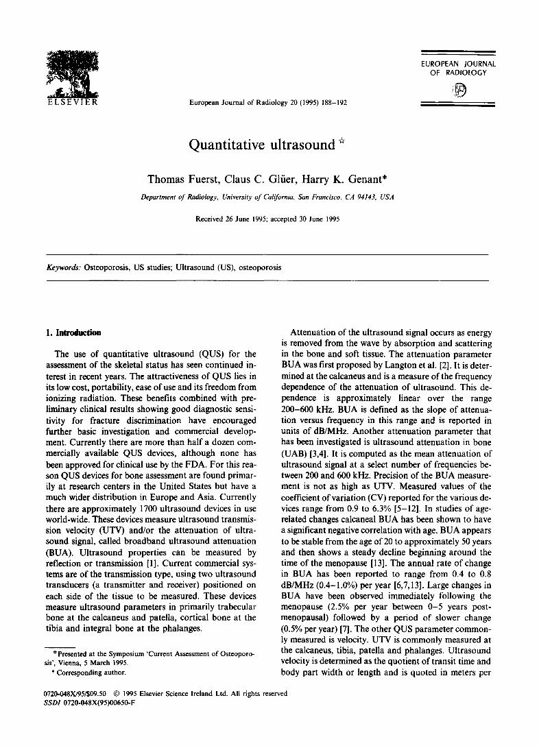
Siclnal sont 1st received sianal
SOS BUA UTV
Calcaneus
Fig. 1. A schematic diagram showing the measurement of ultrasound attenuation (BUA) and velocity (UTV) at the calcaneus. This is a transmission measurement which uses a water bath to provide acoustic coupling between the transducers and the heel.
second (m/s). At the calcaneus the width is either the overall heel width (bone and soft tissue) or the width of bone alone [14]. The first gives a measure of UTV called the speed of sound (SOS) while the second is referred to as ultrasound velocity through bone (UVB). Values of UTV measured at the calcaneus range from 1400 to 1900 m/s [6,13]. There is considerable overlap of SOS and UVB in this range with UVB generally being higher than SOS. Patellar velocity is typically 1600-2200 m/s [15]. Velocity measured in the cortical bone at the mid-tibia is higher and ranges from 3300 to 4300 m/s. The accu- racy of these measurements is difficult to assess as the complex structure of bone and its inhomogeneity result in variable conduction paths and transit times and make determination of true velocity ambiguous. However estimates of patellar velocity accuracy suggest that the measured value underestimates true ultrasound velocity by approximately 100 m/s [16]. The precision of UTV measurements is high with coefficients of variation (CV) of 0.3-1.5% being reported in the literature [8,14,16-18]. Cross-sectional investigations of age- related changes in calcaneal SOS in healthy women have shown reductions in velocity at annual rates of 1.3-4.9 m/s (0.1-0.3%) per year [6,13]. One study which includ- ed data from young normal women showed a steady decline in velocity from 20 to 90 years of age [13]. Stud- ies of ultrasound velocity in the tibia have found similar reductions in velocity of approximately 1.2 m/s per year while velocity at the patella decreases by 3 m/s per year [16].
Ultrasound has long been used to assess mechanical properties in engineering and industrial applications. The mechanical properties of a material or tissue are determined by its material and structural properties.
Material properties are independent of geometry and ar- chitecture while structural properties are determined by these factors. DXA and QCT are used to determine bone mineral density, a material property. The parame- ters measured with ultrasound, BUA and UTV, are in- fluenced not only by bone density but also bone's structure and composition. It is generally believed that BUA is determined by bone density and bone microar- chitecture (trabecular number, connectivity and orienta- tion) while UTV is influenced by the elasticity of bone as well as bone density. The promise of QUS may lie in this apparent dependence of the ultrasound parameters on bone quality. It has been shown that osteoporosis in- volves a change in bone architecture as well as BMD [19]. These architectural changes can significantly influ- ence the mechanical competence of bone.
The theoretical dependence of QUS on both bone density and architecture raises a question about the po- tential role of ultrasound in the screening for osteoporo- sis and fracture risk assessment. If QUS parameters correlate strongly with BMD, ultrasound could poten- tially be used as an inexpensive, radiation-free screening tool for osteoporosis. Alternatively, if BUA and UTV do not correlate so closely with BMD but provide infor- mation beyond that obtained by current bone density measurement techniques, ultrasound could serve as a tool to more accurately determine fracture risk as it would provide information about bone structure which is a component of strength not. revealed by density measurements alone. These two roles are mutually ex- clusive. As a screening tool, QUS is required to be high- ly correlated with BMD. However, if this is true, there is little likelihood that ultrasound can provide informa- tion to augment densitometric techniques.
Various studies have compared BUA at the calcaneus to bone density measured by various techniques. Corre- lation coefficients have ranged from 0.33 to 0.83 for lumbar spine BMD and 0.30-0.87 for the femoral neck [8-10,20,21]. Site-matched comparisons of BUA and BMD at the calcaneus yielded r-values in the range 0.56-0.75 [5,1 l]. Similar relationships have been found between UTV and BMD [6,8,13,22]. However the cor- relations are at best moderate and the errors in predic- ting lumbar spine or femoral neck BMDare too high to allow QUS to be a surrogate for DXA [8,11,23,24]. From these in vivo investigations it appears that 50% of the variability of BUA is unexplained by BMD. Whether this unexplained 50% is related to bone strength or structure or to some parameter unrelated to osteoporosis has yet to be determined.
Many in vitro and in vivo studies have been under- taken to elucidate the nature of the information derived from ultrasound measurement. Ultrasound velocity has been used to study the elastic properties of human and bovine cortical bone [25,26]. Both investigations showed high correlations between mechanically and ultra-

190 T. Fuerst et al. /European Journal of Radiology 20 (1995) 188-192
sonically determined elastic moduli. In other studies with trabecular bone samples velocity correlated well with ultimate strength (r = 0.71-0.75) [27,28]. McCar- thy et al. have recently investigated the relationship be- tween velocity and specimen orientation, density, porosity and temperature [29]. They demonstrated sig- nificant correlations between ultrasound velocity and bone specific gravity and porosity. In a study with bovine bone cubes Gliier et al. examined the relation- ship between ultrasound velocity and trabecular bone structure determined by analysis of microcomputed to- mography images [4]. They found that velocity was largely influenced by trabecular separation. In this same investigation attenuation parameters BUA and UAB were shown to be influenced by connectivity and trabec- ular separation. Moreover these associations with bone structure were independent of the associations between the QUS parameters and BMD. Other work by Gliier et al. has shown that BUA is dependent on trabecular ori- entation, with BUA being as much as 50% higher along the axis parallel to the principle orientation of the trabeculae [30]. UAB has also been shown to have a negative linear correlation (r = 0.90) with trabecular plate separation as determined by histomorphometry [311. Other work has shown that a combination of BUA and velocity can be used to estimate Young's modulus in trabecular bone [31]. These results strongly suggest a relationship between QUS and bone structure and strength beyond that which can be explained by bone mineral density.
Clinical investigations of the ability of QUS to dis- criminate between fractured and non-fractured popula- tions have shown promising results. Several studies have demonstrated that velocity measurements at the patella, tibia or phalanges can identify patients with prevalent vertebral fractures with the same effectiveness as con- ventional bone mass measurements at the spine, hip or forearm [16,17,32-34]. A recent report also showed that patellar ultrasound velocity could predict incident verte- bral fracture [35]. Measurements of QUS parameters at the calcaneus have also shown strong association with fracture risk. Several small case-control studies have established that BUA is significantly lower in patients who have suffered previous hip fracture compared to age-matched controls [36,37]. Two reports showed BUA was as powerful as DXA of the proximal femur to dis- criminate the fracture group as measured by z-scores or ROC analysis [37,38]. Recent results from the Study of Osteoporotic Fractures have shown that calcaneal QUS gives odds ratios for hip fracture equivalent to and inde- pendent of femoral DXA [39]. One prospective study has shown that BUA at the calcaneus is strongly associ- ated with incident hip fracture [40]. Other investigations have shown the association of calcaneal QUS with verte- bral fracture. These studies have found the diagnostic sensitivity of either BUA and SOS was equal to or great-
er than that of spine and hip DXA [39,41-43]. Using models which combine DXA and QUS measurements, these studies have also been able to show BUA and SOS maintain their ability to discriminate patients with frac- tures even after adjusting for the effect of BMD. This provides additional evidence that QUS measures characteristics of bone strength which are independent of density.
The use of quantitative ultrasound for skeletal assess- ment has seen many advances in recent years in the mea- surement technique, our understanding of the relationship between ultrasound parameters and bone structure and strength and in the clinical application of QUS for risk assessment. However further developments will be required to make QUS a more ef- fective tool in the provision of health care. From a tech- nical standpoint the development of a dry system would improve the acceptance of the technique for bone assess- ment. In addition efforts should be made to improve the precision of the measurements to allow better discrimination of patients at risk for fracture and to allow longitudinal assessment to monitor disease pro- gression or to determine the efficacy of therapy. Because of the inhomogeneity of the calcaneus it may be necessary to develop an imaging QUS system [441 to allow reproducible measurement of the same region. Another technical improvement would entail the de- velopment of a suitable standard to allow adequate quality control and cross-calibration of the ultrasound devices. Current standards either do not represent a typical heel geometry or are not stable. Despite much evidence to suggest that ultrasound measures characteristics of bone beyond density, more research is required to elucidate the true nature of the QUS measurements and how they relate to bone structure and strength. This would include theoretical developments on the interaction of ultrasound with the trabecular net- work [45] as well as experimental investigation of the re- lationship between QUS parameters and bone strength, both in vitro and in situ. In addition the usefulness of other attenuation parameters (like UAB) and other fre- quencies (those greater than 600 kHz) deserve further in- vestigation. Moreover combinations of attenuation, velocity and/or BMD should be examined for enhanced discriminatory and predictive power [46]. Finally to fur- ther assess the usefulness of QUS to predict fracture risk, additional prospective studies should be conducted which will allow more accurate interpretation of the QUS results and comparison of the effectiveness of QUS to currently accepted means of risk assessment.
References
ill Antich PP, Anderson JA, Ashman RB, Dowdey JE, Gonzales J, Murry RC, Zerwekh JE, Pak CY. Measurement of mechani- cal properties of bone material in vitro by ultrasound reflec-

T. Fuerst et al./ European Journal of Radiology 20 (1995) 188-192 191
tion: methodology and comparison with ultrasound transmission. J Bone Miner Res 1991; 6: 417-426.
[2] Langton CM, Palmer SB, Porter RW. The measurement of broadband ultrasound attenuation in cancellous bone. Eng Med 1984; 13: 89-91.
[3] Evans JA, Tavakoli MB. Ultrasonic attenuation and velocity in bone. Phys Med Biol 1990; 35: 1387-1396.
[4] Glfier CC, Wu CY, Jergas M, Goldstein SA, Genant HK. Three quantitative ultrasound parameters reflect bone struc- ture. Caleif Tissue Int 1994; 55: 46-52.
[5] Gl~er CC, Vahlensieek M, Faulkner KG, Engelke K, Black D, Genant HK. Site-matcbed calcaneal measurements of broad- band ultrasound attenuation and single x-ray absorptiometry: do they measure different skeletal properties? J Bone Miner Res 1992; 7: 1071-1079.
[6] Waud C, Lew R, DT, B. The relationship between ultrasound and densitometric measurements of bone mass at the calcaneus in women. Calcif Tissue Int 1992; 51: 415-418.
[71 Herd RJM, Ramalingham T, Ryan PJ, Fogelman I, Blake GM. Measurements of broadband ultrasound attenuation in the cal- caneus in premenopausal and postmenopausal women. Osteo- porosis Int 1992; 2: 247-251.
[8] Faulkner KG, McClung MR, Coleman L J, Kingston-Sandahl E. Quantitative ultrasound of the heel: correlation with den- sitometric measurements at different skeletal sites. Osteoporo- sis Int 1994; 4: 42-47.
[9] Agren M, Kareilas A, Leahey D, Marks S, Baran D. Ultra- sound attenuation of the calcaneus: a sensitive and specific discriminator of osteopenia in postmenopausal women. Calcif Tissue Int 1991; 48: 240-244.
[10] Baran DT, McCarthy CK, Leahey D, Lew R. Broadband ultra- sound attenuation of the calcaneus predicts lumbar and femoral neck density in caucasian women: a preliminary study. Osteoporosis Int 1991; 1: 110-113.
[11] Salamone LM, Krall EA, Harris S, Dawson-Hugbes B. Com- parison of broadband ultrasound attenuation to single x-ray absorptiometry measurements at the calcaneus in postmenopausal women. Calcif Tissue Int 1994; 54: 87-90.
[12] Herd RJ, Blake GM, Ramalinham T, Miller CG, Ryan PJ, Fogeiman I. Measurements of postmenopausal bone loss with a new contact ultrasound system. Calcif Tissue Int 1993; 53: 153-157.
[13] Schott AM, Hans D, Sornay-Rendu E, Delmas PD, Meunier PJ. Ultrasound measurements on os ealcis: precision and age- related changes in a normal female population. Osteoporosis lnt 1993; 3: 249-254.
[14] Miller CG, Herd RJM, Ramalingam T, Fogelman I, Blake GM. Ultrasonic velocity measurements through the calcaneus: which velocity should be measured? Osteoporosis Int 1993; 3: 31-35.
[15] Stegman MR, Heaney RP, Recker RR, Travers-Gustafson D, Leist J. Velocity of ultrasound and its association with fracture history in a rural population. Am J Epidemiol 1994; 139: 1027-1034.
[16] Heaney RP, Avioli LV, Chestnut CH, Lappe J, Recker RR, Brandburger GH. Osteoporotic bone fragility: detection by ul- trasound transmission velocity. J Am Med Assoc 1989; 261: 2986-2990.
[17] Orgee J, McCloskey EV, Foster H, Coombes G, Khan S, Kanis JA. Tibial ultrasound velocity - - a useful clinical measure of skeletal status. J Bone Miner Res 1994; 9: S156.
[18] Foldes J, Rimon A, Popovit.zer M. Ultrasonic measurement of the tibia: initial evaluation of a novel approach. Bath Con- ference on Osteoporosis and Bone Mineral Measurement. Bath, UK: The British Institute of Radiology, 1994; 58.
[19] Parfitt AM, Mattbews C, Villanueva A. Relationships between
surface, volume, and thickness of iliac trabecular bone in aging and in osteoporosis. J Clin Invest 1983; 72: 1396-1409.
[20] Zagzebski JA, Rossmann PJ, Mesina C, Mazess RB, Madsen EL. Ultrasound transmission measurements through the os calcis. Calcif Tissue Int 1991; 49:107-111.
[21] Massie A, Reid DM, Porter RW. Screening for osteoporosis: comparison between dual energy X-ray absorptiometry and broadband ultrasound attenuation in 1000 perimenopausal women. Osteoporosis Int 1993; 3: 107-110.
[22] Lees B,Stevenson JC. Evaluation of a new ultrasound bone densitometer. 4th International Symposium on Osteoporosis and Consensus Development Conference, 1993: Hong Kong; 53.
[23] Herd R J, Blake GM, Miller CG, Parker JC, Fogelman 1. The ultrasonic assessment of osteopenia as defined by dual X-ray absorptiometry. Br J Radiol 1994; 67: 631-635.
[24] Young H, Howey S, Purdie DW. Broadband ultrasound at- tenuation compared with dual-energy X-ray absorptiometry in screening for postmenopausal low bone density. Osteoporosis Int 1993; 3: 160-164.
[25] Abendschein W,Hyatt GW. Ultrasonics and selected physical properties of bone. Clin Orthop Rel Res 1970; 69: 294-301.
[26] Ashman RB, Cowin SC, van Buskirk WC, Rice JC. A con- tinuous wave technique for the measurement of the elastic pro- perties of cortical bone. J Biomechanics 1984; 17: 349-361.
[27] Ashman RB, Rho JY. Elastic modulus of trabecular bone ma- terial. J Biomechanics 1988; 21: 177-181.
[28] Turner CH, Eich M. Ultrasonic velocity as a predictor of strength in bovine cancellous bone. Calcif Tissue Int 1991; 49: i16-119.
[29] McCarthy RN, JeffCott LB, McCartney RN. Ultrasound speed in equine cortical bone: effects of orientation, density, porosity, and temperature. J Biomechanics 1990; 23: 1139-1143.
[30] Gliier CC, Wu CY, G-cnant HK. Broadband ultrasound at- tenuation signals depend on trabecular orientation: an in-vitro study. 9th International Workshop on Bone Densitometry. Traverse City, USA [abstract]. Calcif Tissue Int 1992. In press.
[31] Grimm MJ, Williams JL. Use of ultrasound attenuation and velocity to estimate Young's modulus in trabecular bone. Nineteeth IEEE Annula Northeast Bioengineering Conference. Newark, NJ: IEEE, 1993; 62-63.
[32] Stegman MR, Heaney RP, Reeker RR. Comparison of speed of sound ultrasound with single photon absorptiometry for determining fracture odds ratios. J Bone Miner Res 1995; 10: 346-352.
[33] Foldes AJ,Popovtzer MM. Ultrasonic measurement of the tibia: clinical evaluation. Perth International Bone Meeting. Fremantle, Australia, 1995; 52.
[34] Lenfeld F, Wiister C, Beck C, Ziegler R. Validity of ultrasound measurements of bone mineral density on the phalanges of the hand. Perth International Bone Meeting. Fremantle, Australia, 1995; 54.
[35] Heaney RP, Avioli LV, Cbesnut Ili CH, Lappe J, Reeker RR, Brandenburger GH. Ultrasound velocity through bone predicts incident vertebral deformity. J Bone Miner Res 1995; 10: 341-345.
[36] Baran DT, Kelly AM, Karellas A, Gionet M, Price M, Leahy D, Steuterrnan S, McSherry B, Roche J. Ultrasound attenua- tion of the os calcis in women with osteoporosis and hip frac- tures. Calcif Tissue Int 1988; 43: 138-142.
[37] Stewart A, Reid DM, Porter RW. Broadband ultrasound at- tenuation and dual energy X-ray absorptiometry in patients with hip fractures: which technique discriminates fracture risk. Calcif Tissue Int 1994; 54: 466-469.
[38] Schott AM, WeilI-Engerer S, Hans D, Duboeuf F, Delmas PD,

192 T. Fuerst et al. / European Journal of Radiology 20 (1995) 188-192
Meunier PJ. Ultrasound discriminates patients with hip frac- ture equally well as dual energy X-ray absorptiometry and in- dependently of bone mineral density. J Bone Miner Res 1995; 10: 243-249.
[39] Gliier CC, Cummings SR, Bauer DC, Stone K, Pressman A, Genant HK. Associations between quantitative ultrasound and recent fractures. J Bone Miner Res 1994; 9 SI: 153.
[40] Porter R, Miller C, Grainger D, Palmer S. Prediction of hip fracture in elderly women: a prospective study. BMJ 1990; 301: 638-641.
[41] Wiister C, Paetzoid W, Scheidt-Nave C, Brandt K, Ziegler R. Equivalent diagnostic validity of ultrasound and dual X-ray ab- sorptiometry in a clinical case-comparison study of women with vertebral osteoporosis. J Bone Min Res 1994; 9:$211.
[42] Turner CH, Peacock M, Schaefer CA, Timmerman L, Johnston CC. Ultrasonic measurements discriminate hip frac-
tures independently of bone mass. J Bone Min Res 1994; 9: S157.
[43] Ross P, Huang C, Davis J, lmose K, Yates J, Vogel J, Wasnich R. Predicting vertebral deformity using bone densitometry at various skeletal sites and calcaneus ultrasound. Bone 1995; 16: 325-332.
[44] Laugier P, Giat P, Berger G. Broadband ultrasonic attenuation imaging: a new imaging technique. Calcif Tissue lnt 1994. In press.
[45] Biot MA. Generalized theory of acoustic propagation in porous dissipative media. J Acoust Soc Am 1962; 34: 1254-1264.
[461 Antich P, Mason R, McColl R, Zerwech J, Pak C. Trabecular architecture studies by 3D MRI microscopy in bone biopsies. J Bone Min Res 1994; 9S1: 327.
![A Pilot Comparative Study of Quantitative Ultrasound ...AJR) 2017.pdf · W2 AJR:208, May 2017 Paige et al. purpose [4]. Conventional ultrasound (CUS) is the most widely used imaging](https://static.fdocuments.in/doc/165x107/60918a22fb79fb61351d50a5/a-pilot-comparative-study-of-quantitative-ultrasound-ajr-2017pdf-w2-ajr208.jpg)