Purification and Immunological Characterization ofa...
6
INFECTION AND IMMUNITY, Apr. 1991, p. 1417-1422 Vol. 59, No. 4 0019-9567/91/041417-06$02.00/0 Copyright C 1991, American Society for Microbiology Purification and Immunological Characterization of a GroEL-Like Protein from Bordetella pertussis DRUSILLA L. BURNS,'* JEANINE L. GOULD-KOSTKA,1 MARTIN KESSEL,2 AND JUAN L. ARCINIEGA' Division of Bacterial Products, Center for Biologics Evaluation and Research,1 and Laboratory of Structural Studies Research, National Institute of Arthritis and Musculoskeletal and Skin Diseases,2 Bethesda, Maryland 20892 Received 26 December 1990/Accepted 31 January 1991 A GroEL-like protein from Bordetella pertussis was purified. This protein was found to have the tetradecameric subunit structure typical of the GroEL family of proteins and to contain epitopes similar to those of other members of this family, including a human GroEL-like protein. Active immunization of neonatal mice with the B. pertussis GroEL-like protein provided little protection against an aerosol challenge with B. pertussis. Antibodies to this protein were elicited in mice by a standard diphtheria-tetanus-pertussis (DTP) vaccine but not by an experimental aceliular pertussis vaccine. Since the Bordetella GroEL-like protein was found to contain epitopes similar to those on the mammalian analog, the potential exists that vaccination with standard DTP vaccines may induce antibodies which react with the mammalian GroEL analog. Recently, a family of related proteins has been identified in many species of bacteria as well as in the mitochondria and chloroplasts of eucaryotes (17, 23, 34, 35). The prototype of this family is the GroEL protein of Escherichia coli, which is a tetradecamer of subunits, each subunit having a molecular weight of about 65,000 (18, 22). GroEL consists of two seven-member rings of subunits stacked together. The bio- chemical role of GroEL has recently been elucidated. This protein has been identified as a molecular chaperone which aids in the proper folding and assembly of polypeptide chains, probably by binding to unfolded polypeptide chains, either as they are synthesized or very shortly thereafter, and thereby preventing the formation of incorrectly folded inter- mediates (13, 15, 31). Increased expression of GroEL and many other members of this family of proteins often occurs under conditions which promote denaturation of proteins such as high tem- peratures (11, 28). Increased levels of GroEL-like proteins would prevent formation of denatured protein aggregates within the cell by inhibiting incorrect interactions that would cause the formation of these aggregates (13). The members of the GroEL family of proteins are potent immunogens in a number of infections including those caused by Mycobacterium tuberculosis, Mycobacterium le- prae, and Chlamydia spp. (30, 35, 39). These antigens have been postulated to be involved in the survival of the bacteria in the host (7), pathogenesis of the organism (30), and induction of autoimmunity (38). Recently, antibodies to a 63-kDa protein of Bordetella pertussis were found in sera from patients diagnosed with culture-confirmed pertussis and in sera from children vacci- nated with standard diphtheria-tetanus-pertussis (DTP) vac- cine (2). In the same study, this 63-kDa protein was found to react with an antibody elicited by the mycobacterial GroEL analog, suggesting that the protein from B. pertussis may be a member of the GroEL family of proteins. We have purified this protein and have further characterized it in order to demonstrate that it is structurally related to the GroEL family of proteins. In addition, we have examined some immunological properties of this protein. * Corresponding author. MATERIALS AND METHODS Purification of the GroEL-like protein. B. pertussis To- hama I, a virulent strain, was grown in liquid Cohen-Wheeler medium at 37°C as previously described (6). A cell pellet was obtained and resuspended in deionized H2Q to a concentra- tion of 3.8 x 10⁹ bacteria per ml (132 ml total). The cell suspension was sonicated with four cycles (2-min pulsed sonication followed by 2 min off) at 50% duty cycle, using a Heat-Systems-Ultrasonics cell disrupter model W-225 (Farmingdale, N.Y.). The sonicate was centrifuged at 27,000 x g for 40 min, and the supernatant was collected. (In other instances, cells have also been broken by using a French press). A portion of the supernatant (39 ml) was processed further. This portion was dialyzed against buffer A (20 mM Tris-HCI [pH 7.5], 5 mM ,B-mercaptoethanol, 0.4 M NaCI) and was applied to a column (1.5 by 7.5 cm) containing DEAE-Sepharose (Pharmacia, Uppsala, Sweden) in order to remove contaminating nucleic acids. The column, which had been equilibrated with buffer A, was eluted with the same buffer at a flow rate of 54 ml/h. The GroEL-like protein which does not bind to the column under these buffer conditions was collected. The pooled fractions containing the GroEL-like protein (51 mg of protein) were dialyzed against buffer B (20 mM Tris-HCl [pH 7.5], 5 mM ,-mercap- toethanol) and were applied to a second DEAE-Sepharose column which had been equilibrated with the same buffer. The column (5 by 4.7 cm) was eluted at a rate of 54 ml/h with two consecutive linear gradients of NaCl in buffer B (0 to 0.2 and 0.2 to 0.4 M NaCl). The first gradient removed some contaminating protein, and the second gradient eluted the GroEL-like protein. Portions of each fraction (trichloroace- tic acid was added to a 100-,ul portion of each fraction to precipitate the protein) were subjected to sodium dodecyl sulfate (SDS)-polyacrylamide gel electrophoresis (SDS- PAGE), and the GroEL-like protein was identified by its characteristic monomeric molecular mass. Fractions con- taining the GroEL-like protein were pooled. The pooled fractions were dialyzed against buffer B and applied to a third DEAE-Sepharose column (1.5 by 3.5 cm) which had been equilibrated with buffer B in order to concentrate the protein. The GroEL-like protein (21 mg of protein) was eluted with buffer B (6.5 ml) containing 0.5 M NaCl and was identified in the eluted fractions by SDS-PAGE. After dial- 1417 on June 10, 2018 by guest http://iai.asm.org/ Downloaded from
Transcript of Purification and Immunological Characterization ofa...

INFECTION AND IMMUNITY, Apr. 1991, p. 1417-1422 Vol. 59, No. 40019-9567/91/041417-06$02.00/0Copyright C 1991, American Society for Microbiology
Purification and Immunological Characterization of a GroEL-LikeProtein from Bordetella pertussis
DRUSILLA L. BURNS,'* JEANINE L. GOULD-KOSTKA,1 MARTIN KESSEL,2 AND JUAN L. ARCINIEGA'Division of Bacterial Products, Center for Biologics Evaluation and Research,1 and Laboratory of Structural Studies
Research, National Institute of Arthritis and Musculoskeletal and Skin Diseases,2 Bethesda, Maryland 20892
Received 26 December 1990/Accepted 31 January 1991
A GroEL-like protein from Bordetella pertussis was purified. This protein was found to have thetetradecameric subunit structure typical of the GroEL family of proteins and to contain epitopes similar tothose of other members of this family, including a human GroEL-like protein. Active immunization of neonatalmice with the B. pertussis GroEL-like protein provided little protection against an aerosol challenge with B.pertussis. Antibodies to this protein were elicited in mice by a standard diphtheria-tetanus-pertussis (DTP)vaccine but not by an experimental aceliular pertussis vaccine. Since the Bordetella GroEL-like protein wasfound to contain epitopes similar to those on the mammalian analog, the potential exists that vaccination withstandard DTP vaccines may induce antibodies which react with the mammalian GroEL analog.
Recently, a family of related proteins has been identified inmany species of bacteria as well as in the mitochondria andchloroplasts of eucaryotes (17, 23, 34, 35). The prototype ofthis family is the GroEL protein of Escherichia coli, which isa tetradecamer of subunits, each subunit having a molecularweight of about 65,000 (18, 22). GroEL consists of twoseven-member rings of subunits stacked together. The bio-chemical role of GroEL has recently been elucidated. Thisprotein has been identified as a molecular chaperone whichaids in the proper folding and assembly of polypeptidechains, probably by binding to unfolded polypeptide chains,either as they are synthesized or very shortly thereafter, andthereby preventing the formation of incorrectly folded inter-mediates (13, 15, 31).
Increased expression of GroEL and many other membersof this family of proteins often occurs under conditionswhich promote denaturation of proteins such as high tem-peratures (11, 28). Increased levels of GroEL-like proteinswould prevent formation of denatured protein aggregateswithin the cell by inhibiting incorrect interactions that wouldcause the formation of these aggregates (13).The members of the GroEL family of proteins are potent
immunogens in a number of infections including thosecaused by Mycobacterium tuberculosis, Mycobacterium le-prae, and Chlamydia spp. (30, 35, 39). These antigens havebeen postulated to be involved in the survival of the bacteriain the host (7), pathogenesis of the organism (30), andinduction of autoimmunity (38).
Recently, antibodies to a 63-kDa protein of Bordetellapertussis were found in sera from patients diagnosed withculture-confirmed pertussis and in sera from children vacci-nated with standard diphtheria-tetanus-pertussis (DTP) vac-cine (2). In the same study, this 63-kDa protein was found toreact with an antibody elicited by the mycobacterial GroELanalog, suggesting that the protein from B. pertussis may bea member of the GroEL family of proteins. We have purifiedthis protein and have further characterized it in order todemonstrate that it is structurally related to the GroELfamily of proteins. In addition, we have examined someimmunological properties of this protein.
* Corresponding author.
MATERIALS AND METHODS
Purification of the GroEL-like protein. B. pertussis To-hama I, a virulent strain, was grown in liquid Cohen-Wheelermedium at 37°C as previously described (6). A cell pellet wasobtained and resuspended in deionized H2Q to a concentra-tion of 3.8 x 10⁹ bacteria per ml (132 ml total). The cellsuspension was sonicated with four cycles (2-min pulsedsonication followed by 2 min off) at 50% duty cycle, using a
Heat-Systems-Ultrasonics cell disrupter model W-225(Farmingdale, N.Y.). The sonicate was centrifuged at 27,000x g for 40 min, and the supernatant was collected. (In otherinstances, cells have also been broken by using a Frenchpress). A portion of the supernatant (39 ml) was processedfurther. This portion was dialyzed against buffer A (20 mMTris-HCI [pH 7.5], 5 mM ,B-mercaptoethanol, 0.4 M NaCI)and was applied to a column (1.5 by 7.5 cm) containingDEAE-Sepharose (Pharmacia, Uppsala, Sweden) in order toremove contaminating nucleic acids. The column, which hadbeen equilibrated with buffer A, was eluted with the samebuffer at a flow rate of 54 ml/h. The GroEL-like proteinwhich does not bind to the column under these bufferconditions was collected. The pooled fractions containingthe GroEL-like protein (51 mg of protein) were dialyzedagainst buffer B (20 mM Tris-HCl [pH 7.5], 5 mM ,-mercap-toethanol) and were applied to a second DEAE-Sepharosecolumn which had been equilibrated with the same buffer.The column (5 by 4.7 cm) was eluted at a rate of 54 ml/h withtwo consecutive linear gradients of NaCl in buffer B (0 to 0.2and 0.2 to 0.4 M NaCl). The first gradient removed some
contaminating protein, and the second gradient eluted theGroEL-like protein. Portions of each fraction (trichloroace-tic acid was added to a 100-,ul portion of each fraction toprecipitate the protein) were subjected to sodium dodecylsulfate (SDS)-polyacrylamide gel electrophoresis (SDS-PAGE), and the GroEL-like protein was identified by itscharacteristic monomeric molecular mass. Fractions con-
taining the GroEL-like protein were pooled. The pooledfractions were dialyzed against buffer B and applied to a
third DEAE-Sepharose column (1.5 by 3.5 cm) which hadbeen equilibrated with buffer B in order to concentrate theprotein. The GroEL-like protein (21 mg of protein) was
eluted with buffer B (6.5 ml) containing 0.5 M NaCl and was
identified in the eluted fractions by SDS-PAGE. After dial-
1417
on June 10, 2018 by guesthttp://iai.asm
.org/D
ownloaded from

1418 BURNS ET AL.
ysis against buffer C (0.1 M Tris-HCl [pH 7.8], 0.5 M NaCI),the concentrated protein (5.5 mg of protein in 6.3 ml) wasapplied to a column (1.6 by 100 cm) of Sepharose 4B(Pharmacia) which had been equilibrated with 2.5 columnvolumes of the same buffer and was eluted at a flow rate of18.5 ml/h. Fractions were collected, and portions of eachfraction (trichloroacetic acid was added to a 100-,u portionof each fraction to precipitate the protein) were subjected toSDS-PAGE. Fractions containing the GroEL-like proteinwere pooled. The pooled fractions (1.2 mg of protein in 18.5ml) were dialyzed against 20 mM potassium phosphate buffer(pH 7.3) (buffer D) and applied to a column (0.7 by 5.5 cm)of Phenyl Sepharose (Pharmacia) equilibrated with 3 columnvolumes of buffer D at a flow rate of 9.5 ml/h. The columnwas first washed with 1 column volume of buffer D followedby 2 column volumes of 2 mM potassium phosphate buffer,pH 7.3, in order to remove contaminating protein. TheGroEL-like protein was then eluted with 1 column volume of1 mM potassium phosphate buffer, pH 7.3, containing 2%(wt/vol) 3-[(3-cholamidoprophyl)-dimethylammonio]-1-pro-panesulfonate (Calbiochem Corp., La Jolla, Calif.).The preparation of the GroEL-like protein purified from
strain Tohama I had less than 0.6 ng of pertussis toxin per p.gof protein and contained 62.5 endotoxin units per pg ofprotein. The preparation of the GroEL-like protein fromstrain 10901, an avirulent strain which does not producepertussis toxin, contained 12.5 endotoxin units per jig ofprotein.SDS-PAGE. SDS-PAGE was performed essentially as
described by Laemmli (27), using either 12 or 15% polyacryl-amide gels.
Antibodies. Monoclonal antibody MLIIH9 (ascitic fluid),reactive with a mycobacterial GroEL-like protein (14), waskindly provided by Thomas M. Buchanan (University ofWashington, Seattle, Wash.) and was used at a dilution of1:4,000. Anti-Pl serum, reactive with a GroEL-like mito-chondrial protein of CHO cells (16), was a gift of Radhey S.Gupta (McMaster University, Hamilton, Ontario, Canada)and was used at a dilution of 1: 1,000. A polyclonal antiserumwhich is reactive with a 57-kDa chlamydial GroEL-likeprotein (8, 9, 30) was generously provided by Richard P.Morrison (Rocky Mountain Laboratories, National Insti-tutes of Health, Hamilton, Mont.) and was used at a dilutionof 1:1,000.Immunization procedures. BALB/c mice were immunized
twice, intraperitoneally, with 0.5 ml of DTP vaccine (ad-sorbed), U.S. Standard Pertussis Vaccine (lot 9) (nonad-sorbed), JNIH-6 (1), or 8 [ig of purified B. pertussis GroEL-like protein adsorbed onto an equivalent weight ofAlhydrogel (Superfos a/s, Vedbaek, Denmark), at a 4-weekinterval. Mice were bled 2 weeks after the second immuni-zation, and sera were prepared. For immunoblot analysis,the, sera were utilized at a 1:100 dilution unless otherwiseindicated.Immunoblot analysis. Antigens which had been subjected
to SDS-PAGE were electrophoretically transferred to nitro-cellulose sheets (pore size, 0.45 ,um; Schleicher & Schuell,Keene, N.H.) at 100 V for 1 h in a solution consisting of 25mM Tris, 192 mM glycine, and 5% (vol/vol) methanol. Thenitrocellulose sheets were incubated overnight with 10%(wt/vol) skim milk in distilled water. Nitrocellulose stripswere washed three times with phosphate-buffered saline, pH7.4, containing 0.2% gelatin (PBS-gelatin) and were incu-bated with the corresponding serum or monoclonal antibodydiluted in PBS-gelatin. After three additional washes with
PBS-gelatin, the strips were incubated for 1 h with eitheranti-mouse or anti-rabbit antibody conjugated to horseradishperoxidase (Bio-Rad Laboratories, Richmond, Calif.). Thenitrocellulose strips were washed with PBS, and the bandswere visualized by the addition of the color reagent (15 mg of4-chloro-1-naphthol in 5 ml of methanol combined with 25 mlof PBS containing 0.06% hydrogen peroxide).
Electron microscopy. A sample of the GroEL-like proteinwas applied to a carbon-coated collodion-covered grid for 30s and then negatively stained with 1% aqueous uranylacetate. Preparations were examined in a Philips EM420electron microscope operating at 80 kV. Micrographs atnominal magnifications of x36,000 and x60,000 were re-corded on Kodak emulsion SO-163.Image processing. Fields of GroEL-like particles selected
for image processing were digitized by using a Perkin-Elmer1010 MG scanning microdensitometer at a sampling ratecorresponding to 0.34 nm/pixel. Images were processed byusing the PIC system of programs (36).
Particles were selected from the digitized fields into 40 by40 pixel windows and centered by using a rotationallysymmetrized reference. The images were then converted topolar coordinates, and the rotational power spectrum wascalculated. Particles showing clear sevenfold rotational sym-metry were averaged after translational and rotational cor-relation alignment.
Protein determination. Protein concentration was deter-mined by either the BCA protein assay method (PierceChemical Co., Rockford, Ill.) or the method of Lowry et al.(29), using bovine serum albumin as the standard, or by themethod of Bradford (5) using ovalbumin as the standard.Mouse aerosol challenge. The ability of the GroEL-like
protein to protect neonatal mice against lethal B. pertussisrespiratory challenge was analyzed as previously described(33). Mice were immunized on days 5 and 12 postpartumwith 20 p.g of purified protein adsorbed to Alhydrogeladministered intraperitoneally.
Assay for pertussis toxin content. The Chinese hamsterovary (CHO) cell assay was used to measure pertussis toxincontent of GroEL-like protein preparations. This assay wasperformed as previously described (19).
Assay for endotoxin content. The Limulus amebocyte ly-sate assay was used to measure endotoxin content ofGroEL-like protein preparations. This assay was performedas previously described (20).
Isolation of mitochondria. Mitochondria from HeLa cellswere isolated by using a modification of previously publishedprocedures (4, 21). Cells were trypsinized, suspended in 50ml of modified Hanks balanced salt solution (Flow Labora-tories, Costa Mesa, Calif.), and pelleted by centrifugation at750 x g for 15 min. Approximately 50 million cells werewashed twice, each time with 15 ml of Hanks balanced saltsolution, by centrifugation for 15 min at 750 x g. The pelletof cells was gently dispersed with the tip of a glass rod.Washed cells were resuspended in 2 ml of ice-cold 0.88 Msucrose and disrupted in a glass Dounce homogenizer for 15min. The homogenate was centrifuged twice at 750 x g for 20min. The supernatant was then centrifuged for 20 min at11,000 x g. The pellet consisted mainly of mitochondria, asrevealed by staining with 0.005% (wt/vol) Janus green B in0.88 M sucrose.
RESULTS
Previously, a protein with a subunit molecular mass of 63kDa from B. pertussis was described (2). This protein, which
INFECT. IMMUN.
on June 10, 2018 by guesthttp://iai.asm
.org/D
ownloaded from

CHARACTERIZATION OF THE GroEL ANALOG OF B. PERTUSSIS
..
-63 kDa
FIG. 1. Purification of the GroEL-like protein of B. pertussis.Samples taken after each step of the purification procedure (4 p.g ofprotein) were analyzed by SDS-PAGE, followed by staining withCoomassie blue R250. Lane 1, molecular mass markers: 97, 66, 43,31, and 22 kDa; lane 2, supernatant from Tohama I cell culturesonicate; lane 3, fraction from the first DEAE-Sepharose column;lane 4, pooled fractions from the second DEAE-Sepharose column;lane 5, fraction from the third DEAE-Sepharose column; lane 6,pooled fractions from Sepharose 4B column; lane 7, pufified proteinafter Phenyl Sepharose column. Arrow indicates position of theGroEL-like protein.
represents approximately 2% of the total cytoplasmic pro-tein of B. pertussis, was purified from both Tohama I, avirulent strain, and the avirulent strain 10901. The purity ofthe preparation from Tohama I was determined by SDS-PAGE and densitometry to be 91%. The purity of thepreparation from the avirulent strain was determined to be98%. An SDS gel showing the protein obtained from TohamaI at various stages of the purification process is shown in Fig.1.While the subunit molecular mass of this protein is 63 kDa
(Fig. 1), the native molecular weight as determined bymolecular sieve chromatography was found to be greaterthan 660,000 (data not shown). The subunit structure of theprotein was therefore examined by electron microscopy.Two distinct views of the particles are evident (Fig. 2). Anend-on projection view looking down the particle axis exhib-its a sevenfold symmetry. The diameter of the molecule is 15nm. The side view shows a rectangular-shaped particle withfour equally spaced transverse stain-excluding striationssimilar to those seen in electron micrographs of GroEL (22).Occasionally particles in side view can be seen attached toeach other along the particle axis. The similar structures ofthis molecule and GroEL, which is a tetradecamer (22),would suggest that the Bordetella protein probably consistsof two tiers of seven subunits and is a tetradecamer. Theappearance of four stain-excluding striations in the side viewsuggests that each subunit has a shape approximating adumbbell.
This B. pertussis protein was previously shown to bind toa monoclonal antibody elicited by a 65-kDa protein from M.tuberculosis (2) which is a member of the GroEL family ofproteins. We have found that the B. pertussis protein alsobinds to polyclonal antibodies which were elicited by thechlamydial member of the GroEL family (Fig. 3). Moreover,polyclonal antibodies raised against the purified B. pertussisprotein react with a protein found in E. coli extracts (Fig. 3).
14 I~ ..4<.
'S,
.Ss,ld,l,Xi
s, 4(14f: 4; 4* 4
4- - 4 4.4
X4',,
At.}^
4: 4tE
t.; ... s...
;. SM j
v ..
*}f.ir tr
i'>wS. , w S-SX V
< % k - f sr*e } {--. Xs
Z.4 .4
4,,)~~~~~~~~~1
St4t ' ¶¢'"7 t .. ^ '*~~~~~~~~~~4r.4i,
FIG. 2. A field of GroEL-like particles from B. pertussis nega-tively stained with 1% aqueous uranyl acetate showing the two basicorientations of the particle. In the end-on view (E), the sevenfoldsymmetry can be recognized in many of the particles. The side view(S) shows the four prominent stain excluding striations. The insetshows a correlation average of 15 translationally and rotationallyaligned particles in an end-on view. The particles were chosen foraveraging on the basis of sevenfold symmetry determined by therotational power spectrum. The strong enhancement of the seven-fold symmetry is evident. Bar = 50 nm.
This E. coli protein is likely GroEL since it has the expectedmolecular size and since antibodies to the mycobacterialGroEL-like protein also react with a protein of identicalmolecular size. It should be noted that in this experiment,the monoclonal antibody raised to the mycobacterialGroEL-like protein also reacted with an additional protein(Mr = 40,000). While the identity of this protein is notknown, it may represent a breakdown product of GroEL.
Previously, antibodies to this B. pertussis protein werefound in sera from children immunized with DTP vaccine(2). Since the possibility existed that these antibodies werenot elicited by the DTP vaccine but rather by bacteria otherthan B. pertussis to which the children were exposed, weimmunized mice with a DTP vaccine. A standard DTPvaccine was used which contained diphtheria and tetanustoxoids as well as killed, whole B. pertussis cells. Whilenormal mouse serum did not contain antibodies to the B.
VOL. 59, 1991 1419
I ,
m
on June 10, 2018 by guesthttp://iai.asm
.org/D
ownloaded from

1420 BURNS ET AL.
a b
97 -_66 -
,9 7 -44-3G -44 3 --
a 31 -
43 -. 22 -.
1 431 -.
22 #
1 2 1FIG. 3. Serological similarities between the Gro
from B. pertussis and other procaryotic proteins.protein from B. pertussis (1.6 ,ug) was subjected tosubsequent immunoblot analysis. Bands were eitheramido black (lane 1) or with a polyclonal antiserumchlamydial GroEL-like protein (lane 2). (b) E. cMG1655 cells were solubilized in buffer containingmM dithiothreitol and were subsequently subjectedand immunoblot analysis. Bands were visualized wiblack (lane 1), a monoclonal antibody (MLIIH9) rtuberculosis GroEL-like protein (lane 2), or a polycfrom mice immunized with the B. pertussis GroEL-li3). The positions of molecular mass markers (in Iindicated.
pertussis protein, sera from mice immunizvaccine showed strong reactivity toward th(protein when examined by immunoblot anaMoreover, killed whole B. pertussis cells, sii
A BB,i
.43e;
; -
a - 97 -
66 -
43 -
22 -e
TABLE 1. Protection of neonatal mice from challengewith B. pertussisa
No. of survivors/total no.Immunogen
a Expt. 1 Expt. 2
GroEL-like protein 3/10 2/12Pertussis toxoid 9/10 5/7Tetanus toxoid 0/12 0/12
a Mice were immunized twice with either GroEL-like protein (20 jig),pertussis toxoid (8 pg), or tetanus toxoid (8 ,ug) as previously described (33).On day 0, mice were challenged with an aerosol containing approximately 109B. pertussis organisms (strain 18323) per ml. The number of mice whichsurvived 21 days postchallenge is reported. In experiment 1, the GroEL-likeprotein purified from B. pertussis Tohama I was utilized, whereas in experi-ment 2, the GroEL-like protein purified from B. pertussis 10901 was used.
2 3oEL-like protein found in the DTP vaccine, elicited antibodies to the B.(a) GroEL-like pertussis proteins, suggesting that the antibodies may havevSu-PAlE anw been elicited by the pertussis component of the DTP vac-
svisualizedwfth cine. An acellular pertussis vaccine (JNIH-6) consisting ofscli K-12 strain purified filamentous hemagglutinin and pertussis toxoid (1)2%l KSDS strain
did not elicit an antibody response to this protein. Several2% SDS and 10 ddnteii natbd epnet hspoen eeaI to SDS-PAGE proteins other than the 63-kDa monomer are also visible onith either amido the immunoblots shown in Fig. 4. The identities of theseaised to the M. proteins are unknown but may represent contaminating,lonal antiserum proteins in the preparation which are reactive with antibod-ike protein (lane ies in the various sera. Alternatively, these proteins may bekilodaltons) are degradation products of the GroEL-like protein.
Since the GroEL-like protein antigen is present in a DTPvaccine and since it can elicit an antibody response, weexamined the ability of this protein to protect mice against
ed with DTP aerosol challenge with B. pertussis. In each of two experi-e B. pertussis ments, the majority of mice which had been immunized withlysis (Fig. 4). the GroEL-like protein from B. pertussis did not survive pastmilar to those 21 days postchallenge (Table 1). In contrast, most of the
mice immunized with pertussis toxoid survived.GroEL-like proteins are not only found in bacteria but are
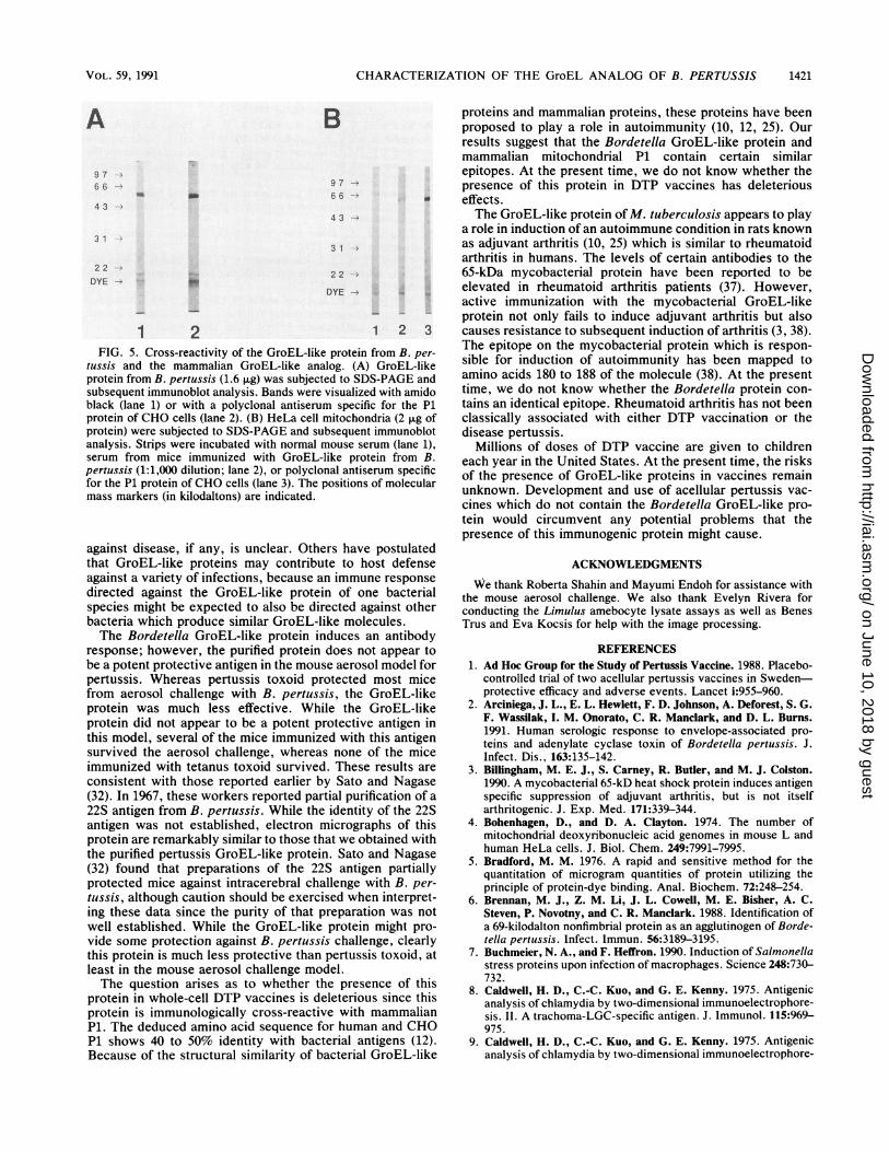
present in chloroplasts of plants and mitochondria of eucary-otes. The mitochondrial protein has been named P1 (12). Weexamined whether the B. pertussis GroEL-like proteinshares epitopes with mitochondrial P1. Polyclonal antibodieselicited by P1 isolated from CHO cells bound to epitopes
- found on the GroEL-like protein from B. pertussis (Fig. 5).Moreover, polyclonal antibodies raised to the B. pertussisprotein bind a protein of 65 kDa found in preparations ofHeLa cell mitochondria.
31 -
22 -
1 2
DISCUSSION
1 2 1 2 1 2
a b cFIG. 4. Ability of vaccines to induce antibodies to the GroEL-
like protein from B. pertussis. GroEL-like protein from B. pertuissis(either 2.8 [A] or 1.6 [B] ,ug) was subjected to SDS-PAGE andsubsequent immunoblot analysis. (A) Strips were incubated eitherwith amido black (lane 1) or with serum from mice immunized withstandard DTP vaccine (lane 2). (B) Strips were incubated with amidoblack (lanes a, b, and c, strips 1), normal mouse serum (lane a, strip2), serum from mice immunized with U.S. Standard Vaccine, lot 9(lane b, strip 2), or serum from mice immunized with the acellularpertussis vaccine, JNIH-6 (lane c, strip 2). The positions of molec-ular mass markers (in kilodaltons) are indicated. Position of theGroEL-like protein is indicated by the arrow. (Bands migrating atthe very top of the strips and as species of <22 kDa are pyronin dyewhich was used as a marker.)
The Bordetella protein described in this study exhibitsboth immunological and structural similarities to the GroELfamily of proteins. This protein has a sevenfold symmetryidentical to that described for GroEL and, like GroEL,appears to be a tetradecamer (22). Recently, a similarmolecule was described from Neurospora crassa mitochon-dria (24). The size of the Bordetella protein is 15 nm which isclose to that measured for N. crassa and is slightly largerthan GroEL from E. coli.GroEL-like proteins are immunodominant antigens from a
number of infectious agents including M. leprae, M. tuber-culosis, Coxiella burnetti, and Legionella pneumophila (7).While these proteins can be major immunogens, as indicatedby the finding that 10 to 20% of all T cells which respond toM. tuberculosis are specific for the 65-kDa GroEL-likeprotein (26), the role that these antigens play in protection
INFECT. IMMUN.
on June 10, 2018 by guesthttp://iai.asm
.org/D
ownloaded from
CHARACTERIZATION OF THE GroEL ANALOG OF B. PERTUSSIS
A
9 766
4 3
31
2 2 ---
DYE -
B
9 7
6664 3-
331
22e
DYE ..
1 2 1 2 3FIG. 5. Cross-reactivity of the GroEL-like protein from B. per-
tussis and the mammalian GroEL-like analog. (A) GroEL-likeprotein from B. pertussis (1.6 Fxg) was subjected to SDS-PAGE andsubsequent immunoblot analysis. Bands were visualized with amidoblack (lane 1) or with a polyclonal antiserum specific for the P1protein of CHO cells (lane 2). (B) HeLa cell mitochondria (2 ,ug ofprotein) were subjected to SDS-PAGE and subsequent immunoblotanalysis. Strips were incubated with normal mouse serum (lane 1),serum from mice immunized with GroEL-like protein from B.pertussis (1:1,000 dilution; lane 2), or polyclonal antiserum specificfor the P1 protein of CHO cells (lane 3). The positions of molecularmass markers (in kilodaltons) are indicated.
against disease, if any, is unclear. Others have postulatedthat GroEL-like proteins may contribute to host defenseagainst a variety of infections, because an immune response
directed against the GroEL-like protein of one bacterialspecies might be expected to also be directed against otherbacteria which produce similar GroEL-like molecules.The Bordetella GroEL-like protein induces an antibody
response; however, the purified protein does not appear tobe a potent protective antigen in the mouse aerosol model forpertussis. Whereas pertussis toxoid protected most micefrom aerosol challenge with B. pertussis, the GroEL-likeprotein was much less effective. While the GroEL-likeprotein did not appear to be a potent protective antigen inthis model, several of the mice immunized with this antigensurvived the aerosol challenge, whereas none of the miceimmunized with tetanus toxoid survived. These results are
consistent with those reported earlier by Sato and Nagase(32). In 1967, these workers reported partial purification of a
22S antigen from B. pertussis. While the identity of the 22Santigen was not established, electron micrographs of thisprotein are remarkably similar to those that we obtained withthe purified pertussis GroEL-like protein. Sato and Nagase(32) found that preparations of the 22S antigen partiallyprotected mice against intracerebral challenge with B. per-
tussis, although caution should be exercised when interpret-ing these data since the purity of that preparation was notwell established. While the GroEL-like protein might pro-
vide some protection against B. pertussis challenge, clearlythis protein is much less protective than pertussis toxoid, atleast in the mouse aerosol challenge model.The question arises as to whether the presence of this
protein in whole-cell DTP vaccines is deleterious since thisprotein is immunologically cross-reactive with mammalianP1. The deduced amino acid sequence for human and CHOP1 shows 40 to 50% identity with bacterial antigens (12).Because of the structural similarity of bacterial GroEL-like
proteins and mammalian proteins, these proteins have beenproposed to play a role in autoimmunity (10, 12, 25). Ourresults suggest that the Bordetella GroEL-like protein andmammalian mitochondrial P1 contain certain similarepitopes. At the present time, we do not know whether thepresence of this protein in DTP vaccines has deleteriouseffects.The GroEL-like protein ofM. tuberculosis appears to play
a role in induction of an autoimmune condition in rats knownas adjuvant arthritis (10, 25) which is similar to rheumatoidarthritis in humans. The levels of certain antibodies to the65-kDa mycobacterial protein have been reported to beelevated in rheumatoid arthritis patients (37). However,active immunization with the mycobacterial GroEL-likeprotein not only fails to induce adjuvant arthritis but alsocauses resistance to subsequent induction of arthritis (3, 38).The epitope on the mycobacterial protein which is respon-sible for induction of autoimmunity has been mapped toamino acids 180 to 188 of the molecule (38). At the presenttime, we do not know whether the Bordetella protein con-tains an identical epitope. Rheumatoid arthritis has not beenclassically associated with either DTP vaccination or thedisease pertussis.
Millions of doses of DTP vaccine are given to childreneach year in the United States. At the present time, the risksof the presence of GroEL-like proteins in vaccines remainunknown. Development and use of acellular pertussis vac-cines which do not contain the Bordetella GroEL-like pro-tein would circumvent any potential problems that thepresence of this immunogenic protein might cause.
ACKNOWLEDGMENTSWe thank Roberta Shahin and Mayumi Endoh for assistance with
the mouse aerosol challenge. We also thank Evelyn Rivera forconducting the Limulus amebocyte lysate assays as well as BenesTrus and Eva Kocsis for help with the image processing.
REFERENCES1. Ad Hoc Group for the Study of Pertussis Vaccine. 1988. Placebo-
controlled trial of two acellular pertussis vaccines in Sweden-protective efficacy and adverse events. Lancet i:955-960.
2. Arciniega, J. L., E. L. Hewlett, F. D. Johnson, A. Deforest, S. G.F. Wassilak, I. M. Onorato, C. R. Manclark, and D. L. Burns.1991. Human serologic response to envelope-associated pro-teins and adenylate cyclase toxin of Bordetella pertussis. J.Infect. Dis., 163:135-142.
3. Billingham, M. E. J., S. Carney, R. Butler, and M. J. Colston.1990. A mycobacterial 65-kD heat shock protein induces antigenspecific suppression of adjuvant arthritis, but is not itselfarthritogenic. J. Exp. Med. 171:339-344.
4. Bohenhagen, D., and D. A. Clayton. 1974. The number ofmitochondrial deoxyribonucleic acid genomes in mouse L andhuman HeLa cells. J. Biol. Chem. 249:7991-7995.
5. Bradford, M. M. 1976. A rapid and sensitive method for thequantitation of microgram quantities of protein utilizing theprinciple of protein-dye binding. Anal. Biochem. 72:248-254.
6. Brennan, M. J., Z. M. Li, J. L. Cowell, M. E. Bisher, A. C.Steven, P. Novotny, and C. R. Manclark. 1988. Identification ofa 69-kilodalton nonfimbrial protein as an agglutinogen of Borde-tella pertussis. Infect. Immun. 56:3189-3195.
7. Buchmeier, N. A., and F. Heffron. 1990. Induction of Salmonellastress proteins upon infection of macrophages. Science 248:730-732.
8. Caldwell, H. D., C.-C. Kuo, and G. E. Kenny. 1975. Antigenicanalysis of chlamydia by two-dimensional immunoelectrophore-sis. II. A trachoma-LGC-specific antigen. J. Immunol. 115:969-975.
9. Caldwell, H. D., C.-C. Kuo, and G. E. Kenny. 1975. Antigenicanalysis of chlamydia by two-dimensional immunoelectrophore-
VOL. 59, 1991 1421
on June 10, 2018 by guesthttp://iai.asm
.org/D
ownloaded from
1422 BURNS ET AL.
sis. I. Antigenic heterogeneity between C. trachomatis and C.psittaci. J. Immunol. 115:963-968.
10. Cohen, I. R. 1986. Regulation of autoimmune disease; physio-logical and therapeutic. Immunol. Rev. 94:5-21.
11. Craig, E. A. 1985. The heat shock response. Crit. Rev. Bio-chem. 18:239-280.
12. Dudani, A. K., and R. S. Gupta. 1989. Immunological charac-terization of a human homolog of the 65-kilodalton mycobacte-rial antigen. Infect. Immun. 57:2786-2793.
13. Ellis, R. J. 1990. The molecular chaperone concept. Semin. CellBiol. 1:1-9.
14. Gillis, T. P., and T. M. Buchanan. 1982. Production and partialcharacterization of monoclonal antibodies to Mycobacteriumleprae. Infect. Immun. 37:172-178.
15. Goloubinoff, D., J. R. Christeller, A. A. Gatenby, and G. H.Lorimer. 1989. Reconstitution of active dimeric ribulosebisphosphate carboxylase from an unfolded state depends ontwo chaperonin proteins and Mg-ATP. Nature (London) 342:884-889.
16. Gupta, R. S., and A. K. Dudani. 1987. Mitochondrial binding ofa protein affected in mutants resistant to the microtubuleinhibitor podophyllotoxin. Eur. J. Cell Biol. 44:278-285.
17. Hemmingsen, S. M., C. Woolford, S. M. van der Vies, K. Tilly,D. T. Dennis, C. P. Georgopoulos, R. W. Hendrix, and R. J.Ellis. 1988. Homologous plant and bacterial proteins chaperoneoligomeric protein assembly. Nature (London) 333:330-334.
18. Hendrix, R. 1979. Purification and properties of groE, a hostprotein involved in bacteriophage assembly. J. Mol. Biol. 129:375-392.
19. Hewlett, E. L., K. T. Sauer, G. A. Myers, J. L. Cowell, andR. L. Guerrant. 1983. Induction of a novel morphologicalresponse in Chinese hamster ovary cells by pertussis toxin.Infect. Immun. 40:1198-1203.
20. Hochstein, H. D. 1981. The LAL test versus the rabbit pyrogentest for endotoxin detection. Pharmacol. Tech. 5:37-42.
21. Hogeboom, G. H., W. C. Schneider, and G. E. Palade. 1948.Cytochemical studies of mammalian tissues. 1. Isolation ofintact mitochondria from rat liver; some biochemical propertiesof mitochondria and submicroscopic particulate material. J.Biol. Chem. 172:619-635.
22. Hohn, T., B. Hohn, A. Engel, M. Wurtz, and P. R. Smith. 1979.Isolation and characterization of the host protein groE involvedin bacteriophage lambda assembly. J. Mol. Biol. 129:359-373.
23. Hoiby, N., J. B. Hertz, and V. Anderson. 1976. Cross-reactionsbetween Bordetella pertussis and twenty-eight other bacterialspecies. Acta Pathol. Microbiol. Scand. Sect. B 84:395-400.
24. Hutchison, E. G., W. Tichelaar, G. Hofhaus, H. Weiss, and K. R.Leonard. 1989. Identification and electron microscopic analysisof a chaperonin oligomer from Neurospora crassa mitochon-dria. EMBO J. 8:1485-1490.
25. Kaufman, S. H. E. 1990. Heat shock proteins and the immune
response. Immunol. Today 11:129-136.26. Kaufman, S. H. E., V. Vath, J. E. R. Thole, J. D. A. van
Embden, and F. Emmrich. 1987. Enumeration ofT cells reactivewith Mycobacterium tuberculosis organisms and specific for therecombinant mycobacterial 64 kDa protein. Eur. J. Immunol.17:351-357.
27. Laemmli, U. K. 1970. Cleavage of the structural proteins duringthe assembly of the head of bacteriophage T4. Nature (London)227:680-685.
28. Lindquist, S. 1986. The heat shock response. Annu. Rev.Biochem. 5:1151-1191.
29. Lowry, 0. H., J. J. Rosebrough, A. L. Farr, and R. J. Randall.1951. Protein measurement with the Folin phenol reagent. J.Biol. Chem. 193:265-275.
30. Morrison, R. P., K. Lyng, and H. D. Caldwell. 1989. Chlamydialdisease pathogenesis: ocular hypersensitivity elicited by a genusspecific 57-kD protein. J. Exp. Med. 169:663-675.
31. Rothman, J. E. 1989. Polypeptide chain binding proteins: cata-lysts of protein folding and related processes in cells. Cell59:591-601.
32. Sato, Y., and K. Nagase. 1967. Isolation of protective antigenfrom Bordetella pertussis. Biochem. Biophys. Res. Commun.27:195-201.
33. Shahin, R. D., M. J. Brennan, Z. M. Li, B. D. Meade, and C. R.Manclark. 1989. Characterization of the protective capacity andimmunogenicity of the 69-kD outer membrane protein of Bor-detella pertussis. J. Exp. Med. 171:63-73.
34. Shinnick, T. M., M. H. Vodkin, and J. C. Williams. 1988. TheMycobacterium tuberculosis 65-kilodalton antigen is a heatshock protein which corresponds to common antigen and to theEscherichia coli GroEL protein. Infect. Immun. 56:446-451.
35. Thole, J. E. R., P. Hindersson, J. de Bruyn, F. Cremers, J. vander See, H. de Cock, J. Tommassen, and W. van Eden. 1988.Antigenic relatedness of a strongly immunogenic 65-kDa myco-bacterial protein antigen with a similarly sized ubiquitous bac-terial common antigen. Microb. Pathog. 4:71-83.
36. Trus, B., and A. C. Steven. 1981. Digital image processing ofelectron micrographs-the PIC system. Ultramicroscopy 23:39-52.
37. Tsoulfa, G., G. A. W. Rook, J. D. A. van Embden, D. B. Young,A. Mehlert, D. A. Isenberg, F. C. Hay, and P. M. Lydyard. 1989.Raised serum IgG and IgA to mycobacterial antigens in rheu-matoid arthritis. Ann. Rheum. Dis. 48:118-123.
38. Van Eden, W., J. E. R. Thole, R. van der Zee, A. Noordzij, J. D.A. van Embden, E. J. Hensen, and I. R. Cohen. Cloning of themycobacterial epitope recognized by T lymphocytes in adjuvantarthritis. 1988. Nature (London) 331:171-173.
39. Young, D. B., R. Lathigra, R. Hendrix, D. Sweetser, and R. A.Young. 1988. Stress proteins are immune targets in leprosy andtuberculosis. Proc. Natl. Acad. Sci. USA 82:4267-4270.
INFECT. IMMUN.
on June 10, 2018 by guesthttp://iai.asm
.org/D
ownloaded from