PUNICA GRANATUM - University of Georgia · INHIBITION OF NON-ENZYMATIC PROTEIN GLYCATION BY...
126
INHIBITION OF NON-ENZYMATIC PROTEIN GLYCATION BY POMEGRANATE (PUNICA GRANATUM) POLYPHENOLICS By PAMELA GARNER DORSEY (Under the Direction of Phillip Greenspan) ABSTRACT Protein glycation is the non-enzymatic reaction of a reducing sugar or sugar derivative with amino acids, peptides, or proteins resulting in advanced glycation endproducts (AGEs) and crosslinking of proteins. This process is accelerated in diabetes mellitus, and detrimental for proteins that are not readily recycled, such as collagen and lens crystallins. The current study investigates the effect of commercially available pomegranate juice, major phytochemicals found in pomegranate juice (ellagic acid, punicalagin), whole pomegranate fruit and various fractions of pomegranate fruit (peel, membrane, aril) on the in vitro fructose mediated glycation of bovine serum albumin (BSA). Total phenolic content and antioxidant capacity for all juices and pomegranate extracts were determined by the Folin-Ciocalteu method and the FRAP (ferric reducing antioxidant potential) assay, respectively. AGE formation was detected by a fluorescence spectroscopy assay. The effect of pomegranate juice on the inhibition of glycation for other proteins (gelatin, IgG, ribonuclease, lysozyme) was also investigated to determine whether pomegranate polyphenolics are generalized inhibitors of this process.
Transcript of PUNICA GRANATUM - University of Georgia · INHIBITION OF NON-ENZYMATIC PROTEIN GLYCATION BY...
INHIBITION OF NON-ENZYMATIC PROTEIN GLYCATION BY POMEGRANATE
(PUNICA GRANATUM) POLYPHENOLICS
By
PAMELA GARNER DORSEY
(Under the Direction of Phillip Greenspan)
ABSTRACT
Protein glycation is the non-enzymatic reaction of a reducing sugar or sugar derivative
with amino acids, peptides, or proteins resulting in advanced glycation endproducts
(AGEs) and crosslinking of proteins. This process is accelerated in diabetes mellitus, and
detrimental for proteins that are not readily recycled, such as collagen and lens
crystallins. The current study investigates the effect of commercially available
pomegranate juice, major phytochemicals found in pomegranate juice (ellagic acid,
punicalagin), whole pomegranate fruit and various fractions of pomegranate fruit (peel,
membrane, aril) on the in vitro fructose mediated glycation of bovine serum albumin
(BSA). Total phenolic content and antioxidant capacity for all juices and pomegranate
extracts were determined by the Folin-Ciocalteu method and the FRAP (ferric reducing
antioxidant potential) assay, respectively. AGE formation was detected by a fluorescence
spectroscopy assay. The effect of pomegranate juice on the inhibition of glycation for
other proteins (gelatin, IgG, ribonuclease, lysozyme) was also investigated to determine
whether pomegranate polyphenolics are generalized inhibitors of this process.
Pomegranate juice produced the greatest inhibition of BSA glycation on the basis of
volume, phenolic content, and antioxidant capacity when compared to other juices.
Punicalagin and ellagic acid produced similar results to that of pomegranate juice. Whole
pomegranate had a much greater inhibitory activity than whole apple and the membrane
extract of pomegranate fruit was the most potent inhibitor of BSA glycation compared to
the aril and peel pomegranate extracts.
Pomegranate juice, incubated with gelatin, IgG, ribonuclease A (RNase), and lysozyme
inhibited the glycation of these proteins in the presence of fructose, similar to that
observed with BSA. SDS PAGE analysis of BSA and RNase revealed that pomegranate
juice prevented chemical modifications to the native protein structure associated with
glycation. This work suggests that the pomegranate is a robust inhibitor of protein
glycation in various model proteins, owing the majority of its inhibitory activity to the
antioxidant capacity of phytochemicals found within the membrane of the fruit.
INDEX WORDS: Protein glycation, Pomegranate, Fluorescence, SDS-PAGE, Bovine
Serum Album, Ribonuclease A, Fruit, Juice
INHIBITION OF NON-ENZYMATIC PROTEIN GLYCATION BY POMEGRANATE
(PUNICA GRANATUM) POLYPHENOLICS
BY
PAMELA GARNER DORSEY
B.S., North Carolina Agricultural and Technical State University, 2003
M.S., North Carolina Agricultural and Technical State University, 2006
A Dissertation Submitted to the Graduate Faculty of The University of Georgia in Partial
Fulfillment of the Requirements for the Degree
DOCTOR OF PHILOSOPHY
ATHENS, GEORGIA
2012
© 2012
Pamela Garner Dorsey
All Rights Reserved

INHIBITION OF NON-ENZYMATIC PROTEIN GLYCATION BY POMEGRANATE
(PUNICA GRANATUM) POLYPHENOLICS
BY
PAMELA GARNER DORSEY
Major Professor: Phillip Greenspan Committee: Ronald Pegg Warren Beach Anthony Capomacchia Raj Govindarajan Electronic Version Approved: Maureen Grasso Dean of the Graduate School May 2012
iv
DEDICATION
I lovingly dedicate this dissertation to my parents:
Edward and Wilhelmenia Garner
Jeremiah 29:10-14
v
ACKNOWLEDGEMENTS
There are so many people that have been instrumental in helping me to achieve this
degree. To begin, I would like to thank Dr. Phillip Greenspan. Six years ago I had no idea
that I would have the opportunity to work with such a wonderful advisor. His patience,
insight, and overall support have not only helped me to develop professionally but
personally as well. I have humorous and fond memories of my time under his
supervision, and look forward to continuing our relationship in the future. Secondly I
would like to thank my graduate committee: Diane Hartle, Ronald Pegg, Warren Beach,
Raj Govindarajan, and Anthony Capomacchia. I am indebted to Dr. Anthony
Capomacchia, who not only recommended me for the Sloan Fellowship, but recruited me
from North Carolina A&T State University. Thirdly I would like to thank all of the
labmates, professors, technicians, and staff that have become a village of support during
my time at UGA: Dr. Susan Elrod, Dr. Johnetta Walters, Dr. Amy Brackman, Dr. Eve
Bralley, Dr. Deborah Elder, Dr. Julie Coeffield, Dr. Xiaqing “Ellen” Li, Dr. Aaron
Beedle, Kimberly Freeman, Linda Duncan, Kathie Wickwire, Vivia Hill-Silcott, Joy
Wilson, Mary Eubanks, and Demetrius Smith. Last but certainly not least, I am eternally
grateful to my family who has supported me through all of my educational endeavors. I
could not have come this far without the love and support of my husband Joe, my parents,
and my sister Angela.
vi
TABLE OF CONTENTS
Page
ACKNOWLEDGEMENTS .................................................................................................v
CHAPTER
1 INTRODUCTION AND LITERATURE
REVIEW ...........................................................................................................1
Pomegranate ......................................................................................................4
Health Benefits of Pomegranate .......................................................................7
Glycation of Proteins ......................................................................................10
Inhibition of AGE formation ..........................................................................22
References .......................................................................................................25
2 INHIBITION OF NON-ENZYMATIC PROTEIN GLYCATION
BY (PUNICA GRANATUM) JUICE ...............................................................39
Abstract ...........................................................................................................40
Introduction .....................................................................................................41
Materials and Methods ....................................................................................42
Results .............................................................................................................45
Discussion .......................................................................................................49
References .......................................................................................................54
Tables and Figures ..........................................................................................60
vii
3 INHIBITION OF NON-ENZYMATIC PROTEIN GLYCATION BY
WHOLE POMEGRANATE (PUNICA GRANATUM) AND
COMPONENTS OF THE PERICARP ...........................................................70
Abstract ...........................................................................................................71
Introduction .....................................................................................................72
Materials and Methods ....................................................................................73
Results .............................................................................................................76
Discussion .......................................................................................................79
References .......................................................................................................82
Tables and Figures ..........................................................................................88
4 POMEGRANATE (PUNICA GRANATUM) JUICE: A NON-SPECIFIC
INHIBITOR OF PROTEIN GLYCATION ....................................................96
Abstract ...........................................................................................................97
Introduction .....................................................................................................98
Materials and Methods ....................................................................................99
Results ...........................................................................................................101
Discussion .....................................................................................................104
References .....................................................................................................107
Tables and Figures ........................................................................................111
5 CONCLUSIONS AND RECOMMENDATIONS ..........................................115
6 APPENDIX ......................................................................................................117
1
CHAPTER 1
INTRODUCTION AND LITERATURE REVIEW
Epidemiological studies demonstrate a link between consumption of plant derived foods
and various health benefits. These benefits have been linked to the phytochemcials found
within these foodstuffs. Epidemiological studies also demonstrate a relationship between
dietary habits and disease risk. Clinical trials investigating the metabolism,
bioavailability, and tissue distribution of phytochemicals in humans are rare. Most tests
involve animal models or in vitro assays. After stomach hydrolysis, polyphenolic
compounds undergo extensive metabolism while they transit through the GI tract with
few exceptions. Metabolism begins in the lumen of the small intestine where sugar
moieties are cleaved, and the aglycone undergoes glucuronidation, sulfation, and/or
methylation. The majority of polyphenols are not absorbed in the small intestine in their
native form; they are however, structurally modified by colonic microflora (1).
Secondary metabolites from primary compounds exist at low concentrations in the
plasma; however, they can produce physiological effects (2). After circulation in the
bloodstream, these secondary metabolites (low molecular weight phenolic acids) are
excreted in the urine.
The mechanism by which a majority of phenolic compounds provides beneficial
health benefits is scavenging free radicals. The bioactivity of phenolic compounds has
been correlated to their antioxidant properties (ability to scavenge free radicals). Free
radicals have been involved in the development of many chronic diseases such as LDL
2
oxidation in cardiovascular disease and DNA oxidation in cancer. Recently many
bioactives from food have been marketed in the form of pharmaceutical products
including but not limited to pills, capsules, powders, and aqueous solutions. This class of
products is called nutraceuticals. Nutraceuticals are dietary supplements that provide a
concentrated bioactive from a food source in a non-food matrix.
Many nutraceuticals currently available on the market are associated with health
claims. Scientific evidence supporting their health benefits is lacking because supporting
studies are performed in vitro or in animal model assays. The measurements of
antioxidant capacity utilizing in vitro assays are used all the time to determine how
“potent” an antioxidant is. FRAP, ABTS, DPPH, and lipid peroxidation evaluate the
phytochemical’s ability to scavenge artificially made radical species. Antioxidant
capacity may not reflect its activity in vivo; however, in vitro antioxidant activity is used
to characterize the compound. Phytochemicals are arranged in five different categories:
carotenoids, phenolics, alkaloids, nitrogen-containing compounds, and organosulfur
compounds. Phenolics encompass: (a) phenolic acids, (b) flavonoids, (c) stilbenes, (d)
coumarins, and (e) tannins.
Ellagic acid and ellagitannins are phenolics that belong to the phenolic acid and
tannin category respectively. Ellagic acid was first studied in the 1960s for its effect on
blood pressure (3). The dietary administration of ellagitannin containing foods such as
strawberries and raspberries to rats has shown to inhibit events associated with the
initiation and promotion/progression of chemically induced colon and esophageal cancers
(4, 5). A more recent study showed that there was no effect on the number or size of
adenomas in the small intestine of Apc-mutated Min mice after the administration of

3
ellagic acid and cloudberry (high ellagitannin content). This could suggest that the
chemoprotective effect of ellagic acid is specific to the type of tumor and animal model.
Pomegranate juice is recognized as one of the most powerful in vitro antioxidant
foods. Its activity is due to punicalagin, a very potent ellagitannin. In general,
ellagitannins are not absorbed (6) however small amounts of punicalagin were detected in
rats’ plasma following long-term administration at high doses. Ellagitannins are
hydrolyzed to ellagic acid in the small intestine (7). Reports show that ellagic acid is
absorbed 30-90 minutes after ingestion suggesting absorption from the stomach (8).
Various factors may affect the absorption of ellagic acid including the influence of the
food matrix, the dose of free ellagic acid, and individual variability.
Ellagic acid can bind readily to the intestine epithelium also affecting its
absorption. When ellagitannins or ellagic acid reach the distal part of the small intestine
they are metabolized by gut microflora to render urolithin A and B, or hydroxyl-6H-
dibenzo [b,d] pyran-6-one derivatives. Urolithin A and B are absorbed, conjugated, and
detected in plasma at an approximate concentration of 10μM (9). These metabolites enter
the enterohepatic circulation before becoming excreted in the urine. Ellagic acid methyl
ether glucuronides have been detected in human plasma and urine. Extracts from red
raspberry leaves and seeds and pomegranates are available in capsules, powders, tablets,
and liquid form. These products have a GRAS status from the federal government;
however, more toxicity studies need to be performed in humans. One study in humans
shows an improvement in neutropenia after chemotherapy with patients ingesting 180mg
of ellagic acid daily for 6 weeks (10). In the mid-1990s, ellagic acid and ellagic acid
containing supplements were advertised as cancer preventing or cancer therapy products.
4
Based on initial studies, ellagic acid and ellagitannins are beneficial phytochemicals for
use as supplements or within functional foods.
Pomegranate
Over 1000 cultivars exist of the pomegranate (Punica granatum) (11), which arose
from the Middle East and extend throughout the Mediterranean, China, India, the
American Southwest, and Mexico. References to pomegranates can be found in
Christianity, Judaism, Islam, and Buddhism. Pomegranates are even displayed on the
coat of arms of several British medical societies (12). Throughout history, the
pomegranate has symbolized life, longevity, health, femininity, knowledge, morality,
immorality, and spirituality (13). In Ayurvedic medicine, the pomegranate is considered
a pharmacy contained within a fruit. Ancient medicine has utilized the pomegranate to
cure diarrhea (14), and as a remedy for diabetes mellitus (15). Recent health benefits
have been attributed to pomegranate’s antioxidant, anti-inflammatory, and anti-cancer
activity.
Pomegranate Fruit Composition
The pomegranate plant is considered a large tree or shrub, which bears a large
berry as fruit. The fruit is surrounded by a tough, leathery exocarp, which contains
numerous arils. Each aril is a translucent sac containing tart juice and a single seed.
These arils are suspended within a matrix known as the membrane (Figure 1.1).

5
FIGURE 1.1 Punica granatum
Pomegranate seed is comprised of approximately 12-20% oil. It consists of 80%
conjugated octadecatrienoic acid mainly comprised of cis 9, trans 11, and cis 13. Minor
components of pomegranate seed oil include sterols, steroids, and cerebroside. On a
phenolic basis pomegranate, seed oil also contains hydroxybenzoic acids (ellagic acid and
3, 3´-Di-O-methylellagic acid).
Pomegranate juice contains anthocyanins (cyanidin 3-O-glucoside, delphinidin 3-
O-glucoside, and pelargonidin 3-O-glucoside) which provide the juice and the fruit with
an intense red color. The concentration of anthocyanins increased during ripening (16),
however the concentration declined after the fruit was pressed for juice (17).
Pomegranate juice that comes from squeezing the whole fruit contains several
polyphenolics and sugars. These polyphenolics included: (a) punicalagin, a type of
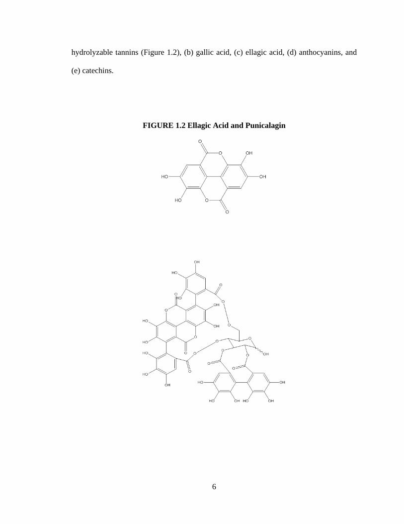
6
hydrolyzable tannins (Figure 1.2), (b) gallic acid, (c) ellagic acid, (d) anthocyanins, and
(e) catechins.
FIGURE 1.2 Ellagic Acid and Punicalagin
Ellagic Acid
Punicalagin
7
Pomegranate juice polyphenols can be placed in 4 major groups. The groups are
anthocyanin pigments (i.e. cyanidin 3-glucoside), hydrolyzable tannins of the gallagyl
type (i.e. punicalagin isomers and punicalin), ellagic acid and its glucosides, and complex
hydrolyzable tannins (which degrade to ellagic acid after hydrolysis). Hydrostatic
pressure from the juicing process crushes the entire fruit releasing juice from the arils and
water-soluble ellagitannins from the rind (18). Despite their inherent size, pomegranate
tannins can be absorbed by the intestines (19). It has been reported that ellagitannins of
pomegranate are hydrolyzed extensively in mice, which excrete ellagic acid in their feces
and urine (20).
The pericarp (peel and membrane) of pomegranate fruit contain abundant
amounts of flavonoids and tannins. Pomegranate leaves contain hydroxybenzoic acids
(gallic acid and ellagic acid), hydroxycinnamic acids (caffeic acid, chlorogenic acids, and
ρ-coumaric acid), flavan-3-ols (catechin and epicatechin), flavonols (quercetin and
kaempferol), flavones (luteolin and apigenin), and ellagitannins (punicalin, punicalagin).
Health Benefits of Pomegranate
Inflammation
Acute and chronic inflammation is the body’s response to injury or tissue damage;
if it has not been resolved in a timely matter chronic disorders of the immune system
(rheumatoid arthritis and inflammatory bowel disease) and even cancer can arise (21, 22).
Inhibition of cyclooxygenase by non-steroidal anti-inflammatory drugs is a conventional
mode of therapy however, this treatment can provide an array of side effects. Whole
pomegranate extract applied to mouse skin inhibited cyclooxygenase expression (23), and
8
compared to other commonly consumed juices it increased the anti-thrombotic prostanoid
PGI2, in human subjects.
Changes in phosphorylation of pro-inflammatory cytokines can prompt
inflammatory cascades. Whole pomegranate fruit extract inhibited the phosphorylation
of several cytokines in UV-B irradiated keratinocytes. Whole pomegranate fruit extract
also inhibited the activation of NF-κB and MAPK, and cytokine formation in mouse skin
exposed to 12-O-tetradecanoylphorbol-13-acetate (23). Patients with periodontitis that
received intragingival chips containing pomegranate peel extract had reduced levels of
inflammatory cytokines several months post treatment (24). Matrix metalloproteinases
(MMP) are enzymes important in cancer progression (25). The activity of MMP in
human chondrocytes was inhibited by whole pomegranate fruit extracts (26).
Heart Disease
There are several studies demonstrating the health benefits of pomegranate juice.
Consumption of pomegranate juice by healthy individuals for two weeks significantly
reduced oxidation of LDL and HDL and increased HDL associated paraoxonase1
(PON1) activity (27). Patients with carotid artery stenosis that consumed pomegranate
juice for three years reduced serum oxidative stress, increased serum PON1 activity, and
reduced the atherosclerotic lesion size (28). Fresh pomegranate juice can ameliorate the
vasomotor effects of sheer stress in hypercholesterolemic mice (29).
Cancer
Cancer is an uncontrolled growth of malignant cells that can be propagated by
inflammation. In female CD-1 mice with skin tumors induced by 7,12-dimethyl-
benz[a]anthracene and promoted by 12-O-tetradecanoylphorbol 13-acetate, treatment
9
with 5% pomegranate seed oil produced significant decreases in tumor incidence and
multiplicity. Human Burkitt’s lymphoma cells exposed to pomegranate peel extract
experienced cell cycle changes (30) due to modulation of the cell signaling molecules.
Carbonic anhydrase catalyzes the reversible hydration of carbon dioxide to bicarbonate,
of which 14 isoforms are known in mammals. Carbonic anhydrase inhibitors strongly
inhibit cancer cell growth in vitro and in vivo. Pomegranate peel extract inhibited the de-
esterification of ρ-nitrophenyl acetate catalyzed by carbonic anhydrase, which establishes
it as a carbonic anhydrase inhibitor. Aromatase catalyzes the formation of estrone and
estradiol from androstenedione and testosterone, respectively (31). This enzyme can
propagate hormone dependent cancers such as estrogen sensitive breast cancers. Both
fermented pomegranate juice and pomegranate peel extract significantly inhibit
aromatase. The growth of new blood vessels (angiogenesis) is necessary to supply
oxygen and nutrients for tumor growth and metastasis. Angiogenesis in chicken
chorioallantoic membrane in vivo was significantly suppressed by fermented
pomegranate juice. Pro-angiogenic vascular endothelial growth factor (VEGF) was
significantly downregulated in MCF-7 extrogen dependent cells by fermented
pomegranate juice. Pomegranate peel extract led to apoptotic DNA fragmentation and
suppression of growth in two human Burkitt’s lymphoma cell lines, Raji and P3HR-1
(30). It has been demonstrated in many studies (32-34) that the anti-proliferative activity
of polyphenolic compounds in pomegranate collectively is superior to singular
compounds. There is a paucity of clinical work involving anti-cancer benefits from
pomegranate; however, in a recent clinical trial, 46 men who consumed 8 oz. of

10
pomegranate juice daily experienced an increase in PSA ( a clinical biomarker for
prostate cancer mortality) doubling time from 15 to 37 months (35).
Glycation of Proteins
History
Protein glycation involves the non-enzymatic reaction of a reducing sugar or
sugar derivative to amino acids, peptides, or proteins (36). This non-enzymatic reaction
was first studied by L.C. Maillard in the early 1900s (37). Food chemists have studied
the Maillard reaction as it relates to flavor, color, and texture in cooked, processed, and
stored foods. In the 1970s and 1980s, researchers discovered that this process also
occurred in vivo. Studies in the 1970s demonstrated that hemoglobin A1c (HbA1c),
naturally occurring minor human hemoglobin, is elevated in diabetics. Koenig and
coworkers (38) found that the carbohydrate in HbA1c was attached as a 1-deoxy-1-
fructosyl residue to the N-terminal valine nitrogen. They were also the first to propose
using HbA1c as a means to monitor glycemic control in diabetic patients (39). In the
1980s, researchers began to understand the significance of Maillard reaction products in
diabetic complications (40) and aging (41). These products were given the name
glycated proteins to distinguish them from enzymatically glycosylated proteins.
Complex pigments and crosslinks formed from glycated protein were termed advanced
glycation end-products (AGEs).
Formation of Glycated Proteins
Reducing sugars (glucose, fructose, galactose, mannose, and ribose) are extremely
reactive with nucleophillic nitrogen bases. Glucose is the least reactive of the common
sugars, possibly leading to its selection as the principal free sugar in vivo (40). A
11
reducing sugar reacts with a free amino group forming a glycosylamine, which degrades
to a Schiff base. In a Schiff base, the aldehydic carbon-oxygen double bond of the sugar
is converted to a carbon-nitrogen double bond with the amine. The open-chain double
bonded form of the Schiff base adducts of hexoses or pentoses are thermodynamically
disfavored as opposed to the pyranose of furanose forms (glycosylamines) (42).
Formation of the Schiff base from the reducing sugar and amine is fast and reversible.
The Amadori rearrangement of a Schiff base to the Amadori product occurs via an open-
chain end form, which tends to be slower than Schiff base formation. However, the
reverse reaction (Amadori rearrangement) is much slower than the formation; therefore,
Amadori products tend to accumulate on proteins. In the 1950s, it was realized that
Amadori products (i.e. fructosamine) could form from aliphatic amines such as amino
acids and not just aromatic amines (43). These adducts (Schiff base and Amadori
products) are known as early stage glycation products. Despite continued research, these
processes are still not completely understood. Adducts formed include fluorescent
chromophores and browning pigments.
Recently it has become clear that α-dicarbonyl compounds are intermediates in
the formation of AGEs. Free α-dicarbonyl glyoxal compounds such as 3-
deoxyglucosone, methylgloxal, and glycoxal are formed from degraded glucose and
Amadori products (44). Dicarbonyls can crosslink proteins forming AGEs directly and
have been detected in vivo (45). Amadori products can also dehydrate at the 4-position to
form 1-amino-4-deoxy-2,3 dione (Amadori dione) (46,47) and subsequently dehydrate at
the 5-position to yield an unsaturated dione (Amadori ene-dione) (48). Amadori ene
12
diones are also responsible for crosslinking proteins. This suggests that AGEs can form
in the beginning or ending stages of the glycation process.
Fluorescent AGEs
The characteristic brown color and fluorescence are properties used to estimate
AGE formation. Fluorescent AGE crosslinks include: (a) pentosidine, (b) crossline, (c)
AGE-XI,(d) pentodilysine, (e) vesperlysine A, B,&C, and (f) FPPC. Pentosidine was
first identified in dura matter collagen by Sell and coworkers (49). Pentosidine can be
formed from the reaction of lysine or arginine with glucose, ribose, ascorbic acid, or 3-
deoxyglucosone. Pentosidine has been found in human tissues not limited to the skin,
tracheal cartilage, cortical bone, aorta, cardiac muscle, lung, liver, kidney, and eye lens.
Studies have shown that there is a direct correlation between skin pentosidine levels and
the severity of diabetic complications in patients with diabetes mellitus (49). Crosslines
(50) and vesperlysines (51) have been detected in vivo.
Non-Fluorescent AGEs
Fluorescent AGE crosslinks are an easy marker for AGE formation due to their
detection but they only account for one percent of the total amount of crosslinking
structures in vivo (52). AGE structures mostly responsible for protein-protein
crosslinking in vivo are non-fluorescent and have not been fully identified yet. Non-
fluorescent AGE crosslinks include pyrraline imine, AFGP (alky formyl glycosyl
pyrrole) imine, amadori dione, α-amino acid amides, imidazolium dilysine,
aminoimidazoline imine, glucosepan, and ALI (arginine-lysine-imidazole).
13
Imidazolium dilysine, also known as GOLD/MOLD crosslinks, have been
isolated from the reaction of two glyoxal derivative molecules with two lysine residues in
vitro, and they have been detected in vivo (53). Imidazole dilysine are present at levels
10 to 50 fold higher than pentosidine in tissue (54).
AFGP imines form from two sugar molecules with one alkylamine molecule (56).
The α positions of the side chains attached to the pyrrole ring carbons in AFGP imines
are susceptible to nucleophilic attack by thiols (57) and lysine amine groups (56). This
indicates that AFGPs are cross-linked proteins.
The pyrraline crosslink (N-alkyl-5-hydroxymethyl-2-pyrrolaldehyde) is a
monomeric AGE that forms on lysine residues in vivo. The aldehyde of pyrraline can
form a Schiff base with another amino group, which has the possibility to form lysine-
lysine crosslinking in vivo. Sugar and Amadori compounds react with primary amines to
form N,N’-dialkyl-alanine or α amino acid amide crosslinks. Gloxal derivatives from
Amadori product breakdown are possible intermediates for their formation (58). They
are difficult to isolate because they are cleaved under protein hydrolysis to yield
carboxymethylysine.
Aminoimidazoline imine crosslinks are derived from the reaction between
arginine and an α-oxoaldimine Schiff base of a lysine residue or glyoxal derivatives (i.e.
methylgloxal, 3-deoxyglucosone) (59).
ALI (arginine-lysine-imidazole) crosslink is derived from the reaction between an
Amadori dione and an arginine residue (60). ALI is immunochemically close to AGE
structures on glucose modified bovine serum albumin (61). Amadori dione crosslink is a
14
conjugate addition of a nucleophilic protein side chain to a protein bound Amadori ene-
dione, which results in a protein-protein crosslink containing an α-diketone structure in
the linker.
A number of non-crosslinking AGE structures have a profound effect on protein
structure and function in vivo. They serve as precursors to crosslinks or biological
receptor ligands, which induce adverse cellular and tissue changes. These non-crosslink
structures include: (a) pyrraline, (b) 1-carboxy-alkyllysine, and (c) imidazolone A and B.
Pyrraline is a pyrrole aldehyde AGE found in vivo (62). It is derived from a reaction
between 3-deoxyglucosone and lysine residues, and is known to form crosslinks between
proteins. 1-carboxyalkyllysines involves a 1-carboxyalkyl group attached to a free amino
group of an amino acid residue (i.e. NЄ-(carboxymethyl) lysine and NЄ-(1-carboxyethyl)
lysine). These compounds have been found in vivo. They may form from reactions of
lysine residues with glyoxal derivatives (63) or from autoxidation of early stage AGEs
(i.e. Amadori products) (53). Imidazolones form from the reaction of glyoxal,
methylglyoxal, or 3-deoxyglucosone with the guanidino group of arginine.
AGE Formation in Various Human Tissues
Collagen is a prime target for AGE formation due to its low turnover rate. AGEs
damage vascular collagen, which contributes to atherosclerosis, coronary disease, kidney
damage, retinal pathology, and poor peripheral circulation. Lens crystallins are also a
prime target for AGE formation, which can lead to cataracts. Collagen lysine residues
and hydroxylysine residues are oxidized by lysl oxidase, which converts Є-amino groups
to aldehydes, which crosslinks with lysine or hydroxylysine residues in adjacent collagen
15
molecules. AGEs mediate crosslinking in collagen, which causes loss of bulk elasticity,
flexibility, and increased brittleness. Collagen can also react with exogenous molecules
such as albumin, immunoglobulin, and LDL (64), resulting in the thickening of the
basement membrane and the development of atherosclerotic lesions
Human lens crystalline can last for the human lifetime. Yellow brown pigments
that have the spectral and fluorescent properties of AGEs form in the lens as a function of
age (65). Glycation can modify amino groups in crystallins resulting in conformational
changes. These changes expose sulfhydryl groups which autoxidize to form
intermolecular disulfide bonds (66). These aggregates are substantial enough in size to
scatter light and produce a cataract.
Protein Glycation in DNA
The presence of AGEs on DNA can cause unusual transpositional rearrangements
(67). Like protein, DNA contains amino groups. The 2-amino group of guanosine is the
most reactive. In mammalian cells, AGE formation on DNA may be responsible for
insertions containing repetitive sequences of the Alu family that disrupt human genes
(68). Protein glycation in DNA can cause congenital malformations in infants of poorly
controlled, insulin dependent diabetic mothers.
Implications of Glycated Protein in Cardiovascular Disease
The Amadori product and later stage AGEs undergo autoxidation and have pro-
oxidant effects on other molecules as well (69). The AGE radical may extract a hydrogen
atom from a biomolecule nearby converting it to a radical, leading to its autoxidation.
This effect is demonstrated in the glycation of lipoproteins like LDL (70). The glycation
16
of hemoglobin increases its oxygen affinity and makes it more susceptible to oxidation
(71). AGE modification of LDL can occur on amino groups on the apoprotein (72) and
the aminolipid (i.e. phosphatidylethanolamine) (73). AGE formation on the apoprotein
can cause crosslinking of LDL to the collagen layer of the blood vessel wall (64)
increasing the half-life of LDL in serum by impeding the recognition site for its receptor
mediated uptake (72). This will increase the probability of autoxidation of the lipid
component (73). Oxidation of LDL can lead to loss of recognition by cellular LDL
receptors and induce uptake by macrophage scavenger receptors (74). Macrophages
attracted to AGEs on vessel wall collagen may accumulate modified LDL, resulting in
their conversion to foam cells, which are thought to be key in the atherosclerotic process.
Inactivated oxygen does not normally react with most organic compounds; however,
redox chemistry can allow iron or copper ions to induce the addition of oxygen to olefinic
bonds of unsaturated fatty acids to form hydroperoxides. Further reactions of these
hydroperoxides with metals can form hydroxyl and peroxy radicals that will cleave fatty
acids to form aldehydes. Free metals are usually not present in significant amounts in
vivo; therefore, the ability of AGEs to initiate oxidative reactions in the absence of metals
offers a mechanism for lipoprotein lipid peroxidation in vivo (70).
Implications of Glycated Protein in Alzheimer’s disease
There is evidence that AGEs play a role in abnormal amyloid aggregation in
Alzheimer’s disease. Analysis of the plaque revealed that Alzheimer’s disease patients
had three times the amount of AGE content per mg of amyloid in comparison to control
subjects (75). The amyloid plaque continues to increase as the disease progresses, its
AGE content increasing crosslinking, and resisting proteolytic degradation and removal.
17
Implications of Glycated Protein in Diabetes Mellitus
Bookchin and Gallop (76) first characterized glycated hemoglobin (HbA1c), and
its increase in persons with diabetes was reported by Rahbar (77). Long term monitoring
of diabetes mellitus is currently performed by self-monitoring blood glucose (SMBG)
and HbA1c levels every 3-6 months. A 2007 Freemantle study of 1286 type 2 diabetes
patients over 5 years found that neither SMBG testing nor its frequency was associated
with glycemic benefit in type 2 diabetes patients . A 2006 study of 3000 type 2 diabetes
patients on oral antidiabetic drugs or a restricted diet found no benefit from SMBG in
glycemic control for either group (78). Hemoglobin resides in the red blood cell, which
has a half-life of 120 days, therefore the amount of HbA1c in a patient’s blood becomes a
record of glycemic control over a 3-6 month period. HbA1c is a verified standard for
being able to predict the risk of having diabetic complications. Another short-term
marker is apolipoprotein B (a component to LDL) that becomes glycated, and is involved
in atherogenesis. LDL is recycled every three to five days; representing glycemic control
over the preceding few days. Glycated albumin (GA) has also been used as a method to
assess glycemic control.
Albumin is the largest component of plasma proteins, representing more than 60%
of total plasma protein concentration. Albumin is responsible for maintenance of oncotic
pressure. The structure of albumin is divided into three domains, and each domain is
divided into two subdomains, which are held together by disulfide bonds. In a healthy
human the amount of glycated albumin is roughly 1-10%, however with diabetes mellitus
this amount can increase by threefold (79).
18
Lysine, arginine, and cysteine are predisposed to non-enzymatic glycation in
serum albumin. Of the 29-glycation sites found, 18 of them are lysine residues. Lysine-
525 is main site for non-enzymatic glycation in serum albumin; and it accounts for 30%
of the overall glycation to albumin (80). Arginine-410 is predominantly responsible for
albumin glycation mediated by methylglyoxal (81). Kisugi and coworkers (82) have
demonstrated that there is a strong correlation between the amount of glycated albumin
and the number of glycation sites. In a diabetic patient with no glycemic control there
were 10 glycated sites, however after insulin therapy there were only three glycation
sites.
Glycation of albumin is measured by: (a) colorimetric assay, (b) enzymatic assay,
(c) HPLC and affinity chromatography, (d)immunoassay, and (e) ELISA. The
thiobarbituric acid assay (TBA) and the fructosamine assay are common colorimetric
methods used to measure glycation. TBA measures the amount of ketoamine bound to
albumin based on the release of glucose from albumin in the form of 5-
hydroxymethylfurfural (5-HMF). However, the 5-HMF is heat sensitive and free glucose
in the assay can interfere with results (83). The fructosamine assay involves the
reduction of nitro blue tetrazolium with ketoamines to form Formazan (chromophore).
Thiol groups, uric acid, and lipemia can interfere with this assay (84). An enzymatic
assay has been developed to measure the amount of glycated albumin using albumin-
specific proteinase, keto amine oxidase, and bromocresolpurple reagent. This method
provides an easier system to measure the amount of glycated albumin. (85).
Glycation of albumin is greater in people with coronary artery disease and unlike
HbA1c is a predictor of coronary artery disease in type 2 diabetes. A recent study (86)
19
found that glycation of albumin decreased with improved glycemic control in comparison
to HbA1c. The reaction rate of non-enzymatic glycation of albumin is nine times greater
than human hemoglobin. There is evidence to use glycated albumin to detect short-term
glycemic control, which is highly recommended in gestational diabetes.
Receptors for Advanced Glycation Endproducts
Scavenger receptors with an affinity for AGEs have been found on many cells
including macrophages, lymphocytes, and barrier cells (i.e. endothelial and mesangial
cells) (87,88). Phagocytic cells that express these recpetors can endocytose old, AGE
modified proteins, releasing AGE modified peptides similar to those absorbable from
food and excreted by the kidneys. The cellular uptake of AGE proteins signals synthesis
and release of certain cytokines and growth factors that stimulate the re-synthesis of
proteins that have been removed (89).
RAGE is the most widely characterized receptor, which is distributed among
endothelial cells, smooth muscle cells, and macrophages. Diabetic erythrocytes can
induce oxidative stress in endothelial cells, however the effects are dampened by RAGE
(90). Interaction between AGEs and RAGE can up-regulate the expression of adhesion
molecules, including VCAM-1, which is responsible for atherosclerotic lesion formation
(91). There are other receptors for AGEs besides RAGE, however they are less selective.
The activity and function of RAGE and other AGE receptors in vivo is still unclear.
Tissue overgrowth can occur if this process is not regulated. For example, high
levels of AGEs on matrix proteins in the diabetic kidney signals mesangial cells to
20
produce large amounts of matrix proteins, resulting in a thickened basement membrane
incapable of normal kidney filtration (92).
Controversy over Diabetic Complications
Diabetic complications are hypothesized to originate from the following sources
collectively: AGES, aldose reductase (93), oxidative stress (94), pseudohypoxia (95), true
hypoxia (96), carbonyl stress (97), altered lipoprotein metabolism (97), increased protein
kinase C activity (98), and altered growth factor expression (99). There has been
controversy as to whether oxidative stress occurs in early stages of diabetes or is it a
consequence of tissue damage. Increase in reactive carbonyls derived from oxidative and
nonoxidative (carbonyl stress) leads to increased chemical modification of proteins,
oxidative stress, and eventually tissue damage.
Carbonyl stress is usually caused by an increase in the concentration of reactive
carbonyl precursors of AGEs (glycoxidation and lipoxidation products), or an increase in
substrate stress. Baynes and coworkers (69) propose that carbonyl stress in diabetes is
the result of a deficient or overloaded detox pathway. Carbonyl trapping may be a more
efficient means to inhibit the progression of diabetes in comparison to antioxidants. This
is evident in certain vitamins or coenzymes such as glutathione, carotene and vitamin E.
Controversy also surrounds the role AGEs play in the formation of diabetic
complications (69). Primarily, AGEs are detectable at trace amounts in tissue proteins
therefore discounting the idea that they have a quantitative effect in the development of
complications. These moieties tend to become very unstable in conditions required for
isolation and analysis making characterization impossible. Secondly, the concentration
21
of AGEs in older adults is similar to that of younger diabetic patients with severe
complications, which would suggest there is little direct correlation. However, older
adults with high concentrations of AGEs are at increased risk for cardiovascular and
Alzheimer’s disease. Thirdly, AGE concentrations are low in tissues from diabetic
animal models compared to those in humans, which could suggest AGEs are not a
common cause of diabetic complications in certain mammalian species.
Development of AGE Inhibitors
The development of AGE inhibitors involves two different approaches. First
inhibition by carbonyl blocking agents such as aminoguanidine, and second the cleavage
of already formed AGE protein-protein crosslinks (i.e. DPTC). The ketone group of the
1-amino-1-deoxyfructose residue in the Amadori product is the key in AGE forming
reactions. Aminoguanidine, a low molecular weight compound, chemically deactivates
the ketone group making it inert. Aminoguanidine is highly nucleophilic, since it reacts
with ketones and aldehydes. Research has demonstrated the effectiveness of
aminoguanidine at inhibiting AGE formation in vivo in a wide array of systems and
tissues (100-102). The limitation to aminoguanidine is that it cannot repair or undue
previous AGE damage or crosslinking. Aminoguanidine is an Amadorin, which can only
inhibit the formation of Amadori products.
A new class of anti-AGE agents that contain a thiazolium structure can
chemically break α-dicarbonyl compounds (i.e. glyoxals, α-diketones) by cleaving the
carbon-carbon bond between the carbonyls. For example, 4, 5 dimethyl-3-
phenacylthiazolium chloride (DPTC) is a promising compound that reversed large artery
22
stiffness and crosslinking of tail collagen in streptozocin diabetic rats (1 mg/kg/day) after
1 to 3 weeks (103).
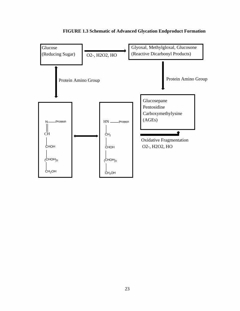
Inhibition of AGE formation with polyphenols
There are three mechanisms by which phenolics may inhibit glycation. They are
antioxidants (blocking glucose autooxidation), metal chealation, or trapping reactive
dicarbonyl compounds (Figure 1.3).
23
FIGURE 1.3 Schematic of Advanced Glycation Endproduct Formation
O2-, H2O2, HO
Protein Amino Group
O2-, H2O2, HO
Protein Amino Group
Glucose (Reducing
Sugar)
Glyoxal, Methylgloxal, Glucosone, (Reactive Dicarbonyl Products)
Glucosepane Pentosidine
Carboxymethylysine (Advanced Glycation
Endproducts)
Oxidative Fragmentation
Schiff Base
CH
N
CHOH
(CHOH)3
CH2OH
Protein
Amadori Product
HN
CH2
Protein
CHOH
(CHOH)3
CH2OH
Glucosepane Pentosidine Carboxymethylysine (AGEs)
Glyoxal, Methylgloxal, Glucosone (Reactive Dicarbonyl Products)
Glucose (Reducing Sugar)
Protein Amino Group Protein Amino Group
O2-, H2O2, HO
Oxidative FragmentationO2-, H2O2, HO
24
AGES tend to accumulate in proteins with a long half-life such as the proteins found in
the neurological system. Epidemiological observations have linked flavonoid intake to a
reduced risk of neurodegenerative disease (104).
Inhibition of AGE formation with pomegranate polyphenols
Verzelloni and coworkers (1) investigated the inhibitory activity of select colonic
microbiota derived polyphenol catobolites against advanced glycation endproducts
formation in vitro and their ability (at physiological concentrations) to counteract mild
oxidative stress in cultured human neuronal cells. Three groups were tested for their
inhibition against protein glycation: ellagitanin group (pyrogallol, urolithin A, urolithin
B), coffee group (dihydro caffeic acid, dihydroferulic acid, and feruloylglycine),
berry/red wine anthocyanin group (3-hydroxyphenylacetic acid, 3,4-dihydroxyphenyl
acetic acid, 3-methoxy-4hydroxyphenylacetic acid). The ellagitannin group was
extremely effective in protecting albumin from glycation at a concentration of 2μmol/L;
reducing AGE formation by almost 50% compared to untreated albumin. Rout and
Banerjee (105) explored the effect of a polysaccharide fraction from pomegranate rind on
the glycation of BSA (10 mg/ml) in the presence of fructose and glucose (25 mM). The
polysaccharide fraction from pomegranate rind inhibited AGE formation by 28% at a
concentration of 10μg/ml.
25
References
1) Verzelloni E, Pellacani C, Tagliazucchi D, Tagliaferri S, Calani L, Costa LG,
Brighenti F, Borges G, Crozier A, Conte A, Del Rio D. Antiglycative and neuroprotective
activity of colon-derived polyphenol catabolites. Molecular Nutrition and Food Research
55: 1-9.2011.
2) Espίn JC, Garcίa-Conesa MT, Tomás-Barberán FA. Nutraceuticals: Facts and fiction.
Phytochemistry 68:2986-3008.2007.
3) Botti RE, Ratnoff OD. Studies of pathogenesis of thrombosis. Experimental
hypercoagulable state induced by intravenous injection of ellagic acid. Journal of
Laboratory and Clinical Medicine 64: 385-398.1964.
4) Harris GK, Gupta AM, Nines RG, Kresty LA, Habib SG, Frankel WL, LaPerle K,
Gallaher DD, Schwartz SJ, Stoner GD. Effects of lyophilized black raspberries on
azoxymetane-induced colon cancer and 8-hydroxy-2´-deoxyguanosine levels in the
Fischer 344 rat. Nutrition and Cancer 40: 125-133. 2001.
5) Chen T, Hwang HJ, Rose ME, Nines RG, Stoner GD. Chemopreventive properties of
black raspberries in N-nitrosomethylbenzylamine-induced rat esophageal
tumorigenesis:down-regulation of cyclooxygenase-2, inducible nitric oxide synthase, and
c-Jun. Cancer Research 66:2853-2859. 2006.
6) Cerdá B, Cerón JJ, Espίn JC, Tomás-Barberán FA. The repeated oral administration
of high doses of the pomegranate ellagitannin punicalagin to rats for 37 days is not toxic.
Journal of Agricultural and Food Chemistry 51: 3493-3501.2003.
7) Larrosa M, Tomás-Barberán FA, Espίn JC. The hydrolysable tannin punicalagin
releases ellagic acid which induces apoptosis in human colon adenocarcinoma Caco-2
26
cells by using the mitochondrial pathway. The Journal of Nutritional Biochemistry 17:
611-625. 2006.
8) Seeram NP, Lee R, Heber D. Bioavailability of ellagic acid in human plasma after
consumption of ellagitannins from pomegranate (Punica granatum L.) juice. Clinica
Chimica Acta 348: 63-68.2004.
9) Cerdá B, Espίn JC, Parra A, Martίnez P, Tomás-Barberán FA. The potent in vitro
antioxidant ellagitannins from pomegranate juice are metabolized into bioavailable but
poor antioxidant hydroxy-6H-dibenzopyran-6-one derivatives by the colonic microflora
of healthy humans. European Journal of Nutrition 43: 205-220. 2004.
10) Falsaperla M, Morgia G, Tartarote A, Ardito R, Romano G. Support ellagic acid
therapy in patients with hormone refractory prostate cancer (HRPC) on standard
chemotherapy using vinorelbine and estramustine phosphate. European Urology 47: 449-
455.2005.
11) Levin GM. Pomegranate (Punica granatum) plant genetic resources in Turkmenistan.
Plant Genetic Resources Newsletter 97: 31-37. 1994.
12) Langley P. Why a pomegranate? British Medical Journal 321: 1153-1154. 2000.
13) Mahdihassan S. Outline of the beginnings of alchemy and its antecedents. American
Journal of Chinese Medicine 12: 32-42. 1984.
14) Arseculeratne SN, Gunatilaka AL, Panabokke RG. Studies on medicinal plants of Sri
Lanka. Part 14. Toxicity of some traditional medicinal herbs. Journal of
Ethnopharmacology 13: 323-335. 1985.
27
15) Saxena A, Vikram NK. Role of selected Indian plants in management of type 2
diabetes: a review. Journal of Alternative and Complementary Medicine 10: 369-378.
2004.
16) Hernandez F, Melgarejo P, Tomas-Barberan FA, Artes F. Evolution of juice
anthocyanins during ripening of new selected pomegranate (Punica granatum) clones.
Euopean Food Research Technology 210: 39-42. 1999.
17) Perez-Vicente A, Gil-Izquierdo A, Garcia-Viguera C. In vitro gastrointestinal study
of pomegranate juice phenolic compounds, anthocyanins and Vitamin C. Journal of
Agricultural and Food Chemistry 50: 2308-2312. 2002.
18) Gil MI, Toms-Barbern FA, Hess-Pierce B, Holcroft DM, Kader AA. Antioxidant
Activity of Pomegranate Juice and Its Relationship with Phenolic Composition and
Processing. Journal of Agricultural and Food Chemistry 48: 4581-4589. 2000.
19) Filippich LJ, Zhu J, Oelrichs P, Alsalami MT, Doig AJ, Cao GR, English PB.
Hepatotoxic and nephrotoxic principles in Terminalia oblongata. Research in Veterinary
Science 50: 170-177. 1991.
20) Castonguay A, Boukharta M, Jalbert G. Comparitive study of ellagic acid and its
analogues as chemopreventive agents against lung tumorigenesis. ACS Symposium
Series 546. American Chemical Society : 294-302. 1994.
21) Balkwill F, Charles KA, Mantovani A. Smoldering inflammation in the initiation and
promotion of malignant disease. Cancer Cell 7: 211-217. 2005.
22) Simmons DL, Buckley CD. Some new and not so new, anti-inflammatory targets.
Current Opinion in Pharmacology 5: 394-397. 2005.
28
23) Afaq F, Saleem M, Krueger CG, Reed JD, Mukhtar H. Anthocyanin- and
hydrolyzable tannin-rich pomegranate fruit extract modulates MAPK and NF-kappa B
pathways and inhibits skin tumorigenesis in CD-1 mice. International Journal of Cancer
113: 423-433. 2005.
24) Sastravaha G, Gassmann G, Sangtherapitikul P, Grimm WD. Adjunctive periodontal
treatment with Centella asiatica and Punica granatum extracts in supportive periodontal
therapy. Journal of the International Academy of Periodontology 7: 70-79. 2005.
25) Shapiro SD. Mighty mice: transgenic technology “knocks out” questions of matrix
metalloproteinase function. Matrix Biology 15: 527-533. 1997.
26) Ahmed S, Wang N, Hafeez BB, Cheruvu VK, Haqqi TM. Punica granatum L.
extract inhibits IL-1 beta-induced expression of matrix metalloproteinases by inhibiting
the activation of MAP kinases and NF-kappa B in human chondrocytes in vitro. Journal
of Nutrition 135: 2096-2102. 2005.
27) Aviram M, Dornfeld L, Rosenblat M, Volkova N, Kaplan M, Coleman R, Hayek T,
Presser D, Fuhrman B. Pomegranate juice consumption reduces oxidative stress,
atherogenic modifications to LDL, and platelet aggregation: studies in humans and in
atherosclerotic apolipoprotein E-deficient mice. American Journal of Clinical Nutrition
71: 1062-1076. 2000.
28) Aviram M, Rosenblat M, Gaitini D, Nitecki S, Hoffman A, Dornfeld L, Volkova N,
Presser D, Attias J, Liker H, Hayek T. Pomegranate juice consumption for 3 years by
patients with carotid artery stenosis reduces common carotid intima-media thickness,
blood pressure and LDL oxidation. Clinical Nutrition 23: 423-433. 2004.
29
29) de Nigris F, Williams-Ignarro S, Botti C, Sica V, Ignarro LJ, Napoli C. Pomegranate
juice reduces oxidized low-density lipoprotein downregulation of endothelial nitric oxide
synthase in human coronary endothelial cells. Nitric Oxide 15: 259-263. 2006.
30) Settheetham W, Ishida T. Study of genotoxic effects of antidiarrheal medicinal herbs
on human cells in vitro. The Southeast Asian Journal of Tropical Medicine and Public
Health 26: 306-310. 1995.
31) Karaer O, Oruc S, Koyuncu FM. Aromatase inhibitors: possible future applications.
Acta Obstetricia et Gynecologica Scandinavica 83: 699-706. 2004.
32) Kim ND, Mehta R, Yu W, Neeman I, Livney T, Amichay A, Poirier D, Nicholls P,
Kirby A, Jiang W, Mansel R, Ramachandran C, Rabi T, Kaplan B, Lansky E.
Chemopreventive and adjuvant therapeutic potential of pomegranate (Punica granatum)
for human breast cancer. Breast Cancer Research and Treatment 71: 203-217. 2002.
33) Albrecht M, Jiang W, Kumi-Diaka J, Lansky EP, Gommersall LM, Patel A, Mansel
RE, Neeman I, Geldof AA, Campbell MJ. Pomegranate extracts potently suppress
proliferation xenograft growth, and invasion of human prostate cancer cells. Journal of
Medicinal Food 7: 274-283. 2004.
34) Seeram NP, Adams LS, Henning SM, Niu Y, Zhang Y, Nair MG, Heber D. In vitro
antiproliferative, apoptotic, and antioxidant activities of punicalagin, ellagic acid and a
total pomegranate tannin extract are enhanced in combination with other polyphenols as
found in pomegranate juice. Journal of Nutritional Biochemistry 16: 360-367. 2005.
35) Pantuck AJ, Leppert JT, Zomorodian N, Aronson W, Hong J, Barndard RJ, Seera M,
Liker H, Wang H, Elashoff R, Heber D, Aviram M, Ignarro L, Belldegrun A. Phase II
30
study of pomegranate juice formen with rising prostate-specific antigen following surgery
or radiation for prostate cancer. Clinical Cancer Research 12: 4018-4026. 2006.
36) Rabbani N, Varma CM, Bodmer CW, Zehnder D, Ceriello A, Thornalley PJ.
Increased glycation and oxidative damage to apolipoprotein B100 of LDL in patients
with type 2 diabetes and effect of metformin. Diabetes 59: 1038-1045. 2010.
37) Maillard LC: Action des acides amines sur les sucres: Formation des melanoidines
par voie methodologique: Comptes Rendus de l’Academie des Sciences 156: 148-149.
1912.
38) Koenig RJ, Blobstein SH, Cerami A. Structure of carbohydrate of hemoglobin AIc.
The Journal of Biological Chemistry 252: 2992-2997. 1977.
39) Koenig RJ, Peterson CM, Jones RL, Saudek C, Lehrman M, Cerami A. Correlation of
glucose regulation and hemoglobin AIc in diabetes mellitus. The New England Journal
of Medicine 295: 417-420. 1976.
40) Bunn HF, Gabbay KH, Gallop PM. The glycosylation of hemoglobin: relevance to
diabetes mellitus. Science 200: 21-27. 1978.
41) Monnier VM and Cerami A. Nonenzymatic browning in vivo: possible process for
aging of long-lived proteins. Science 211: 491-493. 1981.
42) Pigman W. “The Carbohydrates: Chemistry and Biochemistry”. Volume 2.
Academic Press, New York, NY. 1970.
43) Hodge JE. The Amadori rearrangement. Advances in Carbohydrate Chemistry 10:
169-205. 1955.
31
44) Thornalley PJ, Langborg A, Minhas HS. Formation of glyoxal, methylglyoxal and 3-
deoxyglucosone in the glycation of proteins by glucose. Biochemical Journal 344: 109-
116. 1999.
45) Ulrich P, Cerami A. Protein Glycation, Diabetes, and Aging. Recent Progress in
Hormone Research 56: 1-22. 2001.
46) Huber B, Ledl F. Formation of 1-amino, 1,4 dideoxy-2,3-hexodiuloses and maillard
reaction. Carbohydrate Research 204: 215-220.1990.
47) Jyun H, Chen C, Cerami A. Mechanism of inhibition of advanced glycosylation by
aminoguanidine in vitro. Journal of Carbohydrate Chemistry 12: 731-742.1993.
48) Estendorfer S, Ledl F, Severin T. Formation of an aminoreduction from glucose.
Angewandte Chemie 29: 536-537.1990.
49) Sell D, Nagaraj R, Grandhee S, Odetti P, Lapolla A, Fogarty J, Monnier V.
Pentosidine: a molecular marker for the cumulative damage to proteins in diabetes, aging
and uremia. Diabetes Metabolism Research and Reviews 7: 239-251.1991.
50) Obayashi H, Nakano K, Shigeta H, Yamaguchi M, Yoshimori K, Fukui M, Fuji M,
Kitagawa Y, Nakamura N, Nakamura K, Nakazawa Y, lenaga K, Ohta M, Nishimura M,
Fukui I, Kondo M. Formation of crossline as a fluorescent advanced glycation end
product in vitro and in vivo. Biochemical and Biophysical Research Communications
226: 37-41. 1996.
51) Tessier F, Obrenovich M, Monnier V. Structure and mechanism of formation of
human lens fluorophore LM-1. Relationship to vesperlysine A and the advanced maillard
reaction in aging, diabetes, and cataractogenesis. The Journal of Biological Chemistry
274: 20796-20804. 1999.
32
52) Dyer DG, Blackledge JA, Katz BM, Hull CJ, Addison HD, Thorpe SR, Lyons TJ,
Baynes JW. The maillard reaction in vivo. Zeitschrift Für Ernährungswissenschaft 30:
29-45.1991.
53) Frye EB, Degenhardt TP, Thorpe SR, Baynes JW. Role of the maillard reaction in
aging of tissue proteins. Advanced glycation end product-dependent increase in
imidazolium cross-links in human lens proteins. The Journal of Biological Chemistry
273: 18714-18719.1998.
54) DegenhardtTP, Thorpe SR, Baynes JW. Chemical modification of proteins by
methylglyoxal. Cellular and Molecular Biology 44: 1139-1145.1998.
56) Farmer JG, Ulrich PC, Cerami A. Novel pyrroles from sulfite-inhibited maillard
reactions: insight into the mechanism of inhibition. The Journal of Organic Chemistry
53: 2346-2349. 1988.
57) Klein E, Ledl F, Bergmüller W, Severin T. Reactivity of maillard products with a
pyrrole structure. Zeitschrift Für Lebensmitteluntersuchung und-Forschung A 194: 556-
560. 1992.
58) Büttner U, Gerum F, Severin T. Formation of α-amino acid amides and α-hydroxy-
acid amides by degradation of sugars with primary amines. Carbohydrate Research 300:
265-269. 1997.
59) Lederer MO, Klaiber RG. Cross-linking of proteins by maillard processes:
characterization and detection of lysine-arginine cross-links derived from glyoxal and
methylglyoxal. Bioorganic and Medicinal Chemistry 7: 2499-2507.1999.
33
60) Al-Abed Y, Bucala R. Structure of a synthetic glucose derived advanced glycation
end-product that is immunologically cross-reactive with its naturally occurring
counterparts. Bioconjugate Chemistry 11: 39-45.2000.
61) Makita Z, Vlassara H, Cerami A, Bucala R. Immunochemical detection of advanced
glycosylation end products in vivo. The Journal of Biological Chemistry 267: 5133-
5138.1992.
62) Hayase F, Nagaraj RH, Miyata S, Njoroge FG, Monnier VM. Aging of proteins:
immunological detection of a glucose-derived pyrrole formed during maillard reaction in
vivo. The Journal of Biological Chemistry 264: 3758-3764. 1989.
63) Glomb MA, Monnier VM. Mechanism of protein modification by glyoxal and
glycolaldehyde, reactive intermediates of the maillard reaction. The Journal of Biological
Chemistry 270: 10017-10026. 1995.
64) Brownlee M, Pongor S, Cerami A. Covalent attachment of soluble proteins by
nonenzymatically glycosylated collagen. Role in the in situ formation of immune
complexes. The Journal of Experimental Medicine 158: 1739-1744.1983.
65) Monnier VM, Cerami A. Detection of nonenzymatic browning products in the human
lens. Biochimica et Biophysica Acta 760: 97-103. 1983.
66) Monnier VM, Stevens VJ, Cerami A. Nonenzymatic glycosylation, sulfhydryl
oxidation, and aggregation of lens proteins in experimental sugar cataracts. The Journal
of Experimental Medicine 150: 1098-1107. 1979.
67) Lee AT, Cerami A. In vitro and in vivo reactions of nucleic acids with reducing
sugars. Mutation Research/Reviews in Genetic Toxicology 238: 185-191.1990.
34
68) Bucala R, Lee AT, Rourke L, Cerami A. Transposition of an Alu-containing element
induced by DNA-advanced glycosylation endproducts. Proceedings of the National
Academy of Sciences 90: 2666-2670.1993.
69) Baynes JW, Thorpe SR. Role of oxidative stress in diabetic complications: a new
perspective on an old paradigm. Diabetes 48: 1-9.1999.
70) Bucala R, Makita Z, Vega G, Grundy S, Koschinsky T, Cerami A, Vlassara H.
Modification of low density lipoprotein by advanced glycation end products contributes
to the dyslipidemia of diabetes and renal insufficiency. Proceedings of the National
Academy of Sciences 91: 9441-9445.1994.
71) Richard B, Cerami A. Advanced glycosylation: chemistry, biology and implications
for diabetes and aging. August JT, Anders MW (eds.) Advances in Pharmacology.
Academic Press, Inc. San Diego, CA. 1-26.1992.
72) Bucala R, Mitchell R, Arnold K, Innerarity T, Vlassara H, Cerami A. Identification of
the major site of apolipoprotein b modification by advanced glycosylation end products
blocking uptake by the low density lipoprotein receptor. The Journal of Biological
Chemistry 270: 10828-10832. 1995.
73) Bucala R, Makita Z, Koschinsky T, Cerami A, Vlassara H. Lipid advanced
glycosylation: pathway for lipid oxidation in vivo. Proceedings of the National Academy
of Sciences 90: 6434-6438.1993.
74) Witztum JL, Steinberg D. Role of oxidized low density lipoprotein in atherogenesis.
The Journal of Clinical Investigation 88: 1785-1792.1991.
75) Vitek MP, Bhattacharaya K, Glendening JM, Stopa E, Vlassara H, Bucala R,
Manogue K, Cerami A. Advanced glycation endproducts contribute to amyloidosis in
35
Alzheimer disease. Proceedings of the National Academy of Sciences 91: 4766-
4770.1994.
76) Bookchin RM, Gallop PM. Structure of hemoglobin A1c: Nature of the N-terminal β
chain chain blocking group. Biochemical and Biophysical Research Communications 32:
86-93. 1968.
77) Rahbar S. An abnormal hemoglobin in red cells of diabetics. Clinica Chimica Acta
22: 296-298. 1968.
78) Schütt M, Kern W, Krause U, Busch P, Dapp A, Grziwotz R, Mayer I, Rosenbauer J,
Wagner C, Zimmermann A, Kerner W, Holl RW. Is the frequency of self-monitoring of
blood glucose related to long-term metabolic control? Multicenter analysis including
24,500 patients from 191 centers in Germany and Austria. Experimental and Clinical
Endocrinology and Diabetes 114: 384-388. 2006.
79) Bourdon E, Loreau N, Blache D. Glucose and free radicals impair the antioxidant
properties of serum albumin. The Journal of the Federation of American Societies for
Experimental Biology 13: 233-244.1999.
80) Garlick RL, Mazer JS. The principal site of nonenzymatic glycosylation of human
serum albumin in vivo. The Journal of Biological Chemistry 258: 6142-6146.1983.
81) Ahmed N, Dobler D, Dean M, Thornalley PJ. Peptide mapping identifies hotspot site
of modification in human serum albumin by methylglyoxal involved in ligand binding
and esterase activity. The Journal of Biological Chemistry 280: 5724-5732. 2005.
82) Kisugi R, Kouzuma T, Yamamoto T, Akizuki S, Miyamoto H, Someya Y, Yokoyama
J, Abe I, Hirai N, Ohnishi A. Structural and glycation site changes of albumin in diabetic
patient with very high glycated albumin. Clinica Chimica Acta 382: 59-64. 2007.

36
83) Elder E, Kennedy L. Rapid, accurate colorimetric assay of non-enzymatically
glycosylated serum proteins. Diabetologia 24:70-71.1983.
84) Mashiba S, Uchida K, Okuda S, Tomita S. Measurement of glycated albumin by the
nitroblue tetrazolium colorimetric method. Clinica Chimica Acta 212: 3-15. 1992.
85) Kouzuma T, Uemastu Y, Usami T, Imamura S. Study of glycated amino acid
elimination reaction for an improved enzymatic glycated albumin measurement method.
Clinica Chimica Acta 346: 135-143. 2004.
86) Takahashi S, Uchino H, Shimizu T, Kanazawa A, Tamura Y, Sakai K, Watada H,
Hirose T, Kawamori R, Tanaka Y. Comparison of glycated albumin (GA) and glycated
hemoglobin (HbA1c) in type 2 diabetic patients: usefulness of GA for evaluation for
short-term changes in glycemic control. Endocrine Journal 54: 139-144. 2007.
87) Esposito C, Gerlach H, Brett J, Stern D, Vlassara H. Endothelial receptor-mediated
binding of glucose-modified albumin is associated with increased monolayer
permeability and modulation of cell surface coagulant properties. The Journal of
Experimental Medicine 170: 1387-1407.1989.
88) Skolnik EY, Yang Z, Makita Z, Radoff S, Kristein M, Vlassara H. Human and rat
mesangial cell receptors for glucose-modified proteins: potential role in kidney tissue
remodeling and diabetic nephropathy. The Journal of Experimental Medicine 174: 931-
939.1991.
89) Vlassara H, Brownlee M, Manogue KR, Dinarello CA, Pasagian A. Cachectin/TNF
and IL-1 induced by glucose-modified proteins: role in normal tissue remoldeling.
Science 240: 1546-1548.1988.
37
90) Wautier JL, Wautier MP, Schmidt AM, Anderson GM, Hori O, Zoukourian C,
Capron L, Chappey O, Yan SD, Brett J. Advanced glycation end products (AGEs) on the
surface of diabetic erythrocytes bind to the vessel wall via a specific receptor inducing
oxidant stress in the vasculature: a link between surface-associated AGEs and diabetic
complications. Proceedings of the National Academy of Sciences 91: 7742-7746.
91) Vlassara H, Fuh H, Donnelly T, Cybulsky M. Advanced glycation endproducts
promote adhesion molecule (VCAM-1, ICAM-1) expression and atheroma formation in
normal rabbits. Molecular Medicine 1: 447-456.1995.
92) Makita Z, Radoff S, Rayfield EJ, Yang Z, Skolnik E, Delaney V, Friedman EA,
Cerami A, Vlassara H. Advanced glycosylation end products in patients with diabetic
nephropathy. The New England Journal of Medicine 325: 836-842.1991.
93) Witzum JL. Role of modified lipoproteins in diabetic macroangiopathy. Diabetes 46:
S112-S114.1997.
94) Baynes JW. Role of oxidative stress in development of complications in diabetes.
Diabetes 40: 405-412.1991.
95) Williamson JR, Chang K, Frangos M, Hasan KS, Ido Y, Kawamura T, Nyengaard
JR, van den Eden M, Kilo C, Tilton RG. Hyperglycemic pseudohypoxia and diabetic
complications. Diabetes 42: 801-813.1993.
96) Cameron NE, Cotter MA. Metabolic and vascular factors in pathogenesis of diabetic
neuropathy. Diabetes 46: S31-S37. 1997.
97) Lyons TJ, Jenkins AJ. Glycation, oxidation, and lipoxidation in the development of
the complications of diabetes: a carbonyl stress hypothesis. Diabetes Review 5: 365-391.
1997.
38
98) Ishii H, Daisuke K, King GL. Protein kinase C activation and its role in the
development of vascular complications in diabetes mellitus. Journal of Molecular
Medicine 76: 21-31.1998.
99) Pfeiffer A, Schatz H. Diabetic microvascular complications and growth factors.
Experimental and Clinical Endocrinology Diabetes 103: 7-14.1995.
100) Ellis En, Good BH. Prevention of glomerular basement membrane thickening by
aminoguanidine in experimental diabetes mellitus. Metabolism 40: 1016-1019.1991.
101) Hammes HP, Martin S, Federlin K, Geisen K, Brownlee M. Aminoguanidine
treatment inhibits the development of experimental diabetic retinopathy. Proceedings of
the National Academy of Sciences 88: 11555-11558.1991.
102) Huijberts MS, Wolffenbuttel BH, Boudier HA, Crijns FR, Kruseman AC, Poitevin
P, Levy BI. Aminoguanidine treatment increases elasticity and decreases fluid filtration
of large arteries from diabetic rats. The Journal of Clinical Investigation 92: 1407-
1411.1993.
103) Wolffenbuttel BH, Boulanger CM, Crijns FR, Huijberts MS, Poitevin P, Swennen
GN, Vasan S, Egan JJ, Ulrich P, Cerami A, Levy BI. Proceedings of the National
Academy of Sciences 95: 4630-4634.1998.
104) Beking R, Vieira A. Flavonoid intake and disability-adjusted life years due to
Alzheimer’s and related dementias: a population-based study involving twenty-three
developed countries. Public Health Nutrition 13: 1403-1409.2010.
105) Rout S, Banerjee R. Free radical scavenging, anti-glycation and tyrosinase inhibition
properties of a polysaccharide fraction isolated from the rind from Punica granatum.
Biosource Technology 98: 3159-3163.2007.
39
CHAPTER 2
INHIBITION OF NON-ENZYMATIC PROTEIN GLYCATION BY POMEGRANATE
(PUNICA GRANATUM) JUICE
_______________________
Pamela Garner Dorsey1, Phillip Greenspan1 1Department of Pharmaceutical and Biomedical Science University of Georgia, Athens, GA 30602 To be submitted to Phytochemistry
40
Abstract
Diabetes affects nearly eight percent of the United States population, and is the eighth
leading cause of death according to the Centers for Disease Control and Prevention.
Complications from this chronic disease encompass kidney failure, non-traumatic lower
limb amputations, blindness, heart disease, and stroke. Elevated glucose levels generate
non-enzymatic glycation of proteins and the formation of advanced glycation
endproducts; this ultimately leads to the crosslinking of proteins which are thought to be
responsible for diabetic complications. Since many antioxidants and polyphenolic
compounds have been shown to inhibit protein glycation both in vitro and in vivo, our
study sought to demonstrate the effect of commonly consumed ready to drink (RTD)
juices (pomegranate, cranberry, black cherry, pineapple, apple, and Concord grape) on
the fructose mediated glycation of albumin. Pomegranate juice exhibited the highest total
phenolic content and antioxidant potential compared to the other RTD juices. The extent
of albumin glycation after a 72 hour incubation of albumin (10 mg/ml) and fructose (250
mM) was ascertained by measuring fluorescence intensity at the wavelength pair of
370/440 nm. Albumin glycation decreased by 98% in the presence of 10 μl of
pomegranate juice/ml while other RTD juices (cranberry, black cherry, pineapple, and
concord grape) inhibited glycation by only 25%. Pomegranate juice produced the greatest
inhibition of protein glycation when compared to other RTD juices incubated at the same
phenolic concentration and the same antioxidant potential (FRAP units). Major phenolic
constituents of pomegranate fruit, punicalagin and ellagic acid, significantly inhibited the
glycation of albumin by 92 and 94%, respectively, at 5μg GAE/ml. These results
demonstrate that pomegranate juice and two of its major phenolic constituents are potent
inhibitors of protein glycation mediated by fructose.
41
Introduction
Protein glycation, also known as the Maillard reaction, is a complex series of
sequential and parallel steps that begin with the non-enzymatic binding of a reducing
sugar or sugar derivative to an amine group of a protein (1). Molecular rearrangements
(Schiff base formation and Amadori rearrangements) lead to the formation of advanced
glycation endproducts (AGEs) and ultimately the crosslinking of proteins. This is clearly
observed in proteins that are not readily recycled in the body such as collagen and
crystallins in the lens of the eye. AGEs, extremely reactive compounds, increase in
proteins with time; however this process is accelerated in diabetes (2). AGEs formation
has now been recognized to participate in the pathogenesis of retinopathy (3),
nephropathy (4), neuropathy (5), and atherosclerosis (6).
Long term consumption of foods that are rich in polyphenols can ameliorate or
provide prevention against certain disease states (7, 8). One such fruit, the pomegranate,
has been used for medicinal purposes since ancient times (9). The high phenolic content
of the pomegranate is a natural defense against environmental stressors in the very arid
regions of the world which include Iran, Afghanistan, and northern India. The antioxidant
potential of pomegranate juice has been reported to exceed that of red wine and tea (two
phenolically rich beverages), and phenolically rich ready to drink (RTD) juices (Concord
grape, blueberry, black cherry, acaί, and cranberry) (10). Pomegranate juice contains
significant amounts of hydrolyzable ellagitannins, gallotannins, ellagic acid and various
flavonoids (11). Hydrolyzable tannins account for more than 90% of the antioxidant
potential of pomegranate fruit; the major phytochemical contributor is punicalagin (12).
In recent years, studies have documented that punicalagin has significant anti-
inflammatory, antiproliforative, and apoptotic properties (13). Punicalagin and other
42
hydrolyzable tannins originate from the peel of pomegranate fruit, and are found with
ellagic acid, in commercially available juices (14). This would suggest that the juicing of
pomegranates with hydrostatic pressure leaches hydrolyzable tannins from the
pomegranate peel.
Numerous polyphenolic compounds have been shown to inhibit non-enzymatic
glycation of proteins (15-19) both in vitro and in vivo. In this study, we investigated the
effect of pomegranate juice and its major phenolics (punicalagin and ellagic acid) on the
formation of AGEs after incubation of bovine serum albumin with fructose. The
inhibition observed by pomegranate juice was compared to other commonly consumed
ready to drink (RTD) juices.
Materials and Methods Materials
Bovine serum albumin (essentially fatty acid free), D-(-) fructose, Chelex 100
(sodium form), Folin-Ciocalteu reagent, TPTZ (2, 4, 6-tri[2-pyridyl]-s-triazine, ferrous
sulfate heptahydrate, ellagic acid, anhydrous ferric chloride, and 2-mercaptoethanol were
purchased from Sigma Chemical Company (St. Louis, MO). Punicalagin was obtained
from Chengdu Biopurify Phytochemicals Ltd (Chengdu, Sichuan China). The RTD
juices, Organic Apple (R.W. Knudsen), Black Cherry (R.W. Knudsen), Concord Grape
(R.W. Knudsen), Organic Cranberry (Lakewood), Organic Pineapple (Lakewood), and
100% Pomegranate Juice (POM Wonderful), were purchased locally from an Earth Fare
Supermarket. Laemmli sample buffer, Bio-Safe Coomassie blue, and Criterion precast
gels (4-15% in Tris HCl, pH 8.6 ) were purchased from Bio-Rad (Hercules, CA).
43
Total Phenolic Content
The total phenolic content for all RTD juices was determined by the Folin-
Ciocalteu method as described by Slinkard and coworkers (20), utilizing gallic acid as a
standard. Briefly, gallic acid standards (20 μl) and each RTD juice (20 μl) was combined
with distilled water (1580 μl), Folin Ciocalteu reagent (100 μl), and 300μl of sodium
carbonate (1.6M). Distilled water (20 μl) was used as a blank. This mixture was allowed
to incubate for 45 minutes at room temperature, after which absorbance was read at 765
nm on a Beckman DU 600 series spectrophotometer. The results are expressed as gallic
acid equivalents (GAE/ml).
Ferric Reducing Antioxidant Potential (FRAP) Assay
The Ferric Reducing Antioxidant Potential of all RTD juices was determined by a
modified method described by Benzie and coworkers (21), where iron (II) sulfate
heptahydrate was the standard. Briefly iron (II) sulfate heptahydrate standards (10 μl) and
each RTD juice (10 μl) were combined with distilled water (30 μl), and 300 μl of reagent
(25 ml acetate buffer (300 mM, pH 3.6), 25 ml of 10 mM TPTZ solution, and 2.5 ml of
20 mM ferric chloride solution). Distilled water (10 μl) was used as a blank. The mixture
was allowed to incubate for six minutes after which each sample was combined with
distilled water (340 μl). Absorbance was read at 593nm on a Beckman DU 600 series
spectrophotometer. The results are expressed as mM FeSO4 equivalents/ml.
Modification of Albumin by Fructose
The glycation of bovine serum albumin was determined by a method described by
Farrar and coworkers (22). Bovine serum albumin (10 mg/ml) was incubated in the
presence of D-(-) fructose (250 mM) and various concentrations of RTD juices, ellagic
44
acid, and punicalagin in 200 mM potassium phosphate buffer (pH 7.4) at 37ºC for 72
hours. Potassium phosphate buffer was treated with Chelex 100 prior to use. Ellagic acid
and punicalagin were dissolved in 99.5% H2O/0.5% 1N NaOH and 50% EtOH
respectively. All samples were corrected for the native fluorescence of incubated albumin
with RTD juices, ellagic acid or punicalagin. The fluorescence intensity was measured at
an excitation/emission wavelength pair of 370/440 nm using a Perkin-Elmer LS 55
Luminescence Spectrometer with slit widths set at 3nm.
The fluorescence emission spectra of the albumin/fructose solution incubated for
72 hours in the absence and presence of pomegranate juice was determined by setting the
excitation wavelength at 370nm. The emission spectra was scanned from 380 to 550 nm
with the slit width set at 3nm.
Modification of Albumin by Methylglyoxal
The glycation of bovine serum albumin by methylglyoxal was performed using a
method described by Lee and coworkers (23). Bovine serum albumin (100 μM) was
incubated in the presence of methylglyoxal (1mM) in Chelex 100 treated 0.1 M sodium
phosphate, pH 7.0. After 96 hours, the fluorescence was measured at a wavelength pair of
350/409 nm. All samples were corrected for the native fluorescence of albumin incubated
with pomegranate juice.
Analysis of Protein Modifications
Bovine serum albumin (10 mg/ml) was incubated in the presence of D-(-) fructose
(250 mM), in the presence and absence of pomegranate and apple juice (2.5 and 5 μg
GAE/ml) at 37ºC for 14 days. In this experiment 10μl of apple and pomegranate juice
were added to the incubations every 3 days. Protein modifications induced by protein
45
glycation were assessed by sodium dodecyl polyacrylamide gel electrophoresis (SDS-
PAGE) (24). Aliquots for each sample were diluted 1:1 with Laemmli sample buffer
containing 62.5mM Tris-HCl (pH 6.8), 25% (v/v) glycerol, 2% (w/v) SDS, 0.01% (v/v)
Bromophenol Blue, and 5% (v/v) 2-mercaptoethanol. Samples were boiled at 100ºC for
five minutes after dilution and then centrifuged. Centrifuged samples were loaded onto a
15% resolving polyacrylamide gel with a 4 % stacking polyacrylamide gel and subjected
to electrophoresis for 45 minutes. Gels were stained with Bio-safe Coomassie Blue for
one hour and de-stained with distilled water. All gels were scanned on a FluorChem HD2
system with Alpha View software.
Statistical Analysis
Experiments were performed in triplicate and expressed as mean ± SEM. Data
was analyzed utilizing a one-way analysis of variance (ANOVA) and multiple
comparisons were performed employing Tukey’s test. Statistical significance was set at
p< 0.05.
Results
Locally purchased RTD fruit juices were initially tested for their phenolic content
employing the Folin Ciocalteu assay (20). Pomegranate juice contained the highest
concentration of total phenols (Table 1), nearly 4 mg GAE/ml, which is in agreement
with the results of Aviram and coworkers (25). Concord grape and black cherry juice
contained the second and third highest concentration of total phenols. The RTD juices
were also analyzed for their antioxidant capacity as determined by the FRAP assay
(Table 1). Pomegranate juice had the highest antioxidant capacity of all the RTD juices
examined, approximately 3 times greater than the next highest RTD juice. Similar to that
46
observed with the content of phenolic compounds, Concord grape and black cherry juice
possessed the second and third highest antioxidant capacity. These results are in
agreement with Seeram and coworkers (10). It is interesting to note that while
pomegranate juice contained approximately five times the phenolic content of apple
juice, the FRAP values of pomegranate juice were nearly twenty times greater in
comparison to apple juice.
To examine the effect of the RTD juices on protein glycation, each juice (10
μl/ml) was incubated with a solution containing bovine serum albumin (10 mg/ml) and
fructose (250 mM). After 72 hours, the control incubation of BSA and fructose in the
absence of any RTD juice resulted in a significant increase in fluorescence (Figure 1).
When BSA and fructose were incubated in the presence of the RTD juices (10 μl of
juice/ml), pomegranate juice produced the greatest inhibition; the fluorescence intensity
observed in the presence of POM juice was 2% of control. Black cherry, concord grape,
cranberry, and pineapple juice all resulted in a decrease in fluorescence of approximately
20%. The least effective inhibitor of protein glycation was apple juice.
The emission spectrum of the albumin/fructose solution after 72 hour incubation
in the absence of any RTD juice is shown in Figure 2. With the excitation set at 370 nm,
a broad fluorescence spectra is observed having an emission maximum at 440 nm. When
pomegranate juice was present at a concentration of 10 μl/ml during the course of the 72
hour incubation, the resulting emission spectrum resembles a plateau with no major
emission maximum observed at 440 nm, the emission spectrum is quite similar to
spectrum observed when albumin is incubated with pomegranate juice (Figure 2). These
47
results demonstrate the near absence of fluorescent AGE products in the presence of
pomegranate juice.
The superior inhibition observed with pomegranate juice on protein glycation
when performed on the basis of volume was not unexpected due to the fact it contains the
highest concentration of phenolic compounds (Table 1). To further examine the relative
inhibition of RTD juices on protein glycation, the effect of RTD juices was studied when
the juices were present at the same phenolic concentration. In this experiment, the
phenolic content for each juice was normalized to 5 μg GAE/ml of the incubation
mixture. When pomegranate juice was incubated at this concentration, it still provided the
greatest inhibition of protein glycation; the decrease in fluorescence intensity was
approximately 90% (Figure 3). Pineapple yielded the next greatest decrease in
fluorescence intensity (66%). The other RTD juices produced lesser levels of inhibition.
The resulting inhibition pattern among RTD juices in this in vitro assay varies greatly,
and demonstrates that not all phenolic compounds inhibit glycation equally. The phenols
present in pomegranate juice are clearly the most potent inhibitors when compared to the
other RTD juices (apple, cherry, concord grape, cranberry, pineapple).
To further characterize the relative uniqueness of pomegranate juice inhibition of
protein glycation, an experiment was performed in which the different RTD juices were
added to the albumin/fructose mixture at a single antioxidant capacity value based on the
FRAP assay. In this experiment, the RTD juices were added at a concentration of 0.20
mmolFeSO4 equivalents/ml (Figure 4). Under these conditions, pomegranate juice still
produced the greatest decrease in fluorescence intensity (98%); black cherry juice was the
second best inhibitor with an approximately 60% decrease in fluorescence intensity.
48
These results demonstrate that the degree of inhibition of protein glycation among these
juices can vary significantly even when they are present at the same antioxidant capacity.
Pomegranate juice, prepared by squeezing the whole fruit, is rich in hydrolyzable
tannins, namely punicalagin, the major ellagitannin found in the commercial pomegranate
fruit juices (14). The concentration dependent inhibition of protein glycation by
punicalagin is shown in Figure 5. Similar to that seen with pomegranate juice,
punicalagin was an extremely effective inhibitor at a concentration of 5μg/ml. Both at
5μg/ml and at 2.5μg/ml, the punicalagin inhibition of protein glycation was greater than
90%. At 1μg/ml, inhibition of protein glycation was not observed. Ellagic acid, a
metabolite of ellagitannins (i.e. punicalagin), is formed after digestion (26); the data in
Figure 6 demonstrates that ellagic acid inhibits protein glycation to the same extent as
punicalagin.
Methylglyoxal is a reactive α-dicarbonyl which is formed from the auto-oxidation
of glucose. Reactive carbonyls have the ability to bind with amino groups of proteins to
form AGEs directly. When methylglyoxal was incubated with bovine serum albumin for
96 hours, a dramatic inhibition of fluorescence intensity was not observed by
pomegranate juice at phenolic concentrations of 5 and 10 μg GAE/ml (Figure 7). These
results suggest that pomegranate juice does not appear to be an effective inhibitor of
glycation mediated by reactive α-dicarbonyls such as methylgloxal.
The effect of pomegranate juice was also examined (Figure 8) on protein
modifications. Albumin (10mg/ml) incubated in potassium phosphate buffer for 14 days
(column A) resulted in a distinct protein band at 65KDa. Albumin (10mg/ml) incubated
49
in the presence of fructose (250mM) for 14 days (column B) had a noticeable widening
of the albumin band. These results are similar to those of Yamagishi and coworkers (27)
who observed a widening of the BSA band incubated in the presence of glucose.
Albumin incubated in the presence of fructose and apple juice (5μg GAE/ml) (column C)
resulted in the same band widening as column B. Interestingly, albumin incubated in the
presence of fructose and 5μg GAE/ml pomegranate juice (column D) resulted in a sharp
65 KDa band, similar to that of albumin incubated in the absence of fructose (column A).
Thus, pomegranate juice prevented the band widening of albumin resulting from
incubation with fructose. SDS-PAGE analysis revealed that pomegranate juice, and not
apple juice, prevented protein modifications associated with albumin glycation.
Discussion
Glucose or fructose can react with amino groups of proteins to initiate the
Maillard reaction forming a glycosylamine which degrades to a Schiff base. The Schiff
base undergoes an Amadori rearrangement to form fructosamine (28), after which the
degradation of fructosamine forms stable products known as AGEs. However, α-
oxoaldehydes (carbonyls) formed from degraded glucose, fructose, or Schiff bases can
react with amino groups of proteins to form AGEs directly; therefore AGEs can be
formed in early or late stages of glycation. The mechanism of action of phenolic
compounds on glycation is thought to be related to their antioxidant properties (29); they
prevent the oxidation of Amadori products and the subsequent formation of AGEs,
sometimes referred to as glycooxidation. Some polyphenols may inhibit the later stages
of glycation by preventing the binding of dicarbonyls to protein amino groups (30).
Verzelloni and coworkers (31) suggest that phenolic compounds may inhibit the
50
formation of Amadori products because of their specific binding to albumin. The data in
this paper suggests pomegranate juice is an effective natural inhibitor of protein glycation
independent of trapping dicarbonyl species for there was no effect on AGEs formed from
α-oxoaldehydes (methylgloxal) (Figure 7). In addition, the inhibitory effects of
pomegranate on glycation appears independent of protein binding of phenolic
compounds; a similar pattern of inhibition by pomegranate juices was found in proteins
other than albumin, such as gelatin and immunoglobulin G (data not shown).
In this study pomegranate juice had a higher phenolic content and antioxidant
capacity when compared to other commonly consumed RTD juices (apple, cherry,
Concord grape, cranberry, pineapple) (Table 1). However, when pomegranate juice and
the other RTD juices were normalized on the basis of phenolic content (5 μg GAE/ml of
mixture) (Figure 3) and antioxidant capacity (0.20 mmol FeSO4/100ml) (Figure 4),
pomegranate juice was superior to other RTD juices in inhibiting albumin glycation. This
would suggest that pomegranate juice contains phenolic compounds that are more potent
inhibitors of protein glycation than phenolic compounds found in other RTD juices.
Yokozawa and cowokers (16) demonstrated that different phenols inhibit protein
glcyation to varying degrees and similar studies have been performed to illustrate this
concept (18). The major polyphenols present in each RTD juice examined in this study
are listed in Table 2. Concord grape, black cherry and cranberry juice contain
anthocyanins, while Concord grape, cranberry, and apple contain proanthocyanidins.
Matsuda and coworkers (29) examined the relationship between flavonoid
structure, the inhibition of protein glycation, and radical scavenging properties of sixty-
two flavonoids. Flavonoids with a strong scavenging activity (i.e. DPPH radical
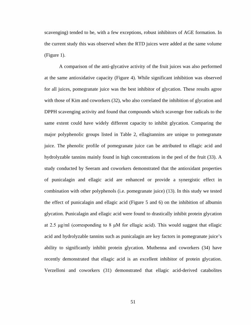
51
scavenging) tended to be, with a few exceptions, robust inhibitors of AGE formation. In
the current study this was observed when the RTD juices were added at the same volume
(Figure 1).
A comparison of the anti-glycative activity of the fruit juices was also performed
at the same antioxidative capacity (Figure 4). While significant inhibition was observed
for all juices, pomegranate juice was the best inhibitor of glycation. These results agree
with those of Kim and coworkers (32), who also correlated the inhibition of glycation and
DPPH scavenging activity and found that compounds which scavenge free radicals to the
same extent could have widely different capacity to inhibit glycation. Comparing the
major polyphenolic groups listed in Table 2, ellagitannins are unique to pomegranate
juice. The phenolic profile of pomegranate juice can be attributed to ellagic acid and
hydrolyzable tannins mainly found in high concentrations in the peel of the fruit (33). A
study conducted by Seeram and coworkers demonstrated that the antioxidant properties
of punicalagin and ellagic acid are enhanced or provide a synergistic effect in
combination with other polyphenols (i.e. pomegranate juice) (13). In this study we tested
the effect of punicalagin and ellagic acid (Figure 5 and 6) on the inhibition of albumin
glycation. Punicalagin and ellagic acid were found to drastically inhibit protein glycation
at 2.5 μg/ml (corresponding to 8 μM for ellagic acid). This would suggest that ellagic
acid and hydrolyzable tannins such as punicalagin are key factors in pomegranate juice’s
ability to significantly inhibit protein glycation. Muthenna and coworkers (34) have
recently demonstrated that ellagic acid is an excellent inhibitor of protein glycation.
Verzelloni and coworkers (31) demonstrated that ellagic acid-derived catabolites
52
(urolithins A and B) are also effective in protecting albumin from glycation at a
concentration of 10 μM.
At the present time, there are no FDA approved medications that directly inhibit
the glycation of proteins in diabetes, with the subsequent slowing in the progression of
diabetic complications. The only way to pharmacologically arrest protein glycation is to
lower plasma glucose levels. Unfortunately, even with many drug therapy options, too
many diabetics are unable to maintain plasma glucose concentrations at near normal
levels; this leads to an elevated rate of glycation. For this reason, phenolic compounds
appear to offer another approach to slow the glycation process. In 2003, Nagasawa and
coworkers (35) illustrated this concept in a diabetic rat study. They studied the effect of
G-rutin (0.2% of the diet) on the protein glycation in plasma and kidney in these animals.
After one month, G-rutin treated rats had a much lower content of glycation products in
the serum and kidney. However, in this experimental model, G-rutin did not significantly
lower the elevated blood glucose levels found in the diabetic rats. Therefore, at least in
this animal model of diabetes, addition of a phenolic compound to the diet suppressed the
extent of glycation of tissue proteins independently of blood glucose concentration.
There have been two studies that determined the levels of hemoglobin A1c after
pomegranate juice consumption in humans. Sumner and coworkers (36) studied the effect
of three month pomegranate juice consumption on myocardial perfusion in twenty-six
patients with coronary heart disease with normal levels of hemoglobin A1c. Rosenblat
and coworkers (37) studied the effect of three month pomegranate juice consumption on
the oxidative status in ten diabetic patients. In both studies, no effect of pomegranate
juice consumption was found on serum blood glucose levels or hemoglobin A1c
53
concentrations. Unfortunately, based on the number of subjects, neither study was
designed to determine if pomegranate juice would significantly lower serum glucose or
hemoglobin A1c. It is interesting to note that consumption of pomegranate juice was not
associated with significant increases in plasma blood glucose concentrations in either
study.
In summary, pomegranate juice and its constituents were found to be effective
inhibitors of protein glycation. Unlike many studies examining the effects of various
phenolic compounds on protein glycation, pomegranate juice employed in these
experiments is commercially available. Therefore, proper studies, employing a sufficient
number of subjects, can be readily performed to ascertain whether the anti-glycative
properties described in this communication is also observed in experimental animals with
diabetes and in diabetic patients.
54
References
1) Rabbani N, Varma CM, Bodmer CW, Zehnder D, Ceriello A, Thornalley PJ. Increased
glycation and oxidative damage to apolipoprotein B100 of LDL in patients with type 2
diabetes and effect of metformin. Diabetes 59: 1038-1045. 2010.
2) Wells-Knecht KJ, Brinkmann E, Wells-Knecht MC, Ahmed MU, Zyzak DV, Thorpe
SR, Baynes JW, New Biomarkers of Maillard reaction damage to proteins. Nephrology
Dialysis Transplantation 11:41-47. 1996.
3) Okumura A, Mitamura Y, Namekata K, Nakamura K, Harada C, Harada T. Glycated
albumin includes activation of activator-protein-1 in retinal glial cells. Japanese Journal
of Ophthalmology 51: 236-237. 2007.
4) Roohk H, Zaidi A. A review of glycated albumin as an intermediate glycation index
for controlling diabetes. Journal of Diabetes Science and Technology 2: 1114-1121.
2008.
5) Vitek MP, Bhattacharya K, Glendening JM, Stopa E, Vlassara H, Bucala R, Manogue
K, Cerami A. Advanced glycation endproducts contribute to amyloidosis in Alzheimer
disease. Proceedings of the National Academy of Sciences 91: 4766-4770. 1994.
6) Farougue H, O’Brien R, Meredith I. Diabetes mellitus and coronary heart disease-from
prevention to intervention: Part II. Australian and New Zealand Journal of Medicine 30:
608-617. 2000.
7) Espίn JC, Garcίa-Conesa MT, Tomás-Barberán FA. Nutraceuticals: Facts and fiction.
Phytochemistry 68. 2986-3008. 2007.
8) Sargeant LA, Khaw KT, Bingham S, Day NE, Luben RN, Oakes S, Welch A,
Wareham NJ. Fruit and vegetable intake and population glycosylated haemoglobin
55
levels: the EPIC-Norfolk study. European Journal of Clinical Nutrition 55: 342-348.
2001.
9) Seeram NP, Schulman RN, Heber D. Pomegranates: Ancient Roots Modern Medicine;
Medicinal and Aromatic Plant Series; CRC Press/Taylor and Francis Group. Boca Raton,
FL. 2006.
10) Seeram N, Aviram M, Zhang Y, Henning S, Feng L, Dreher M, Heber D.
Comparison of antioxidant potency of commonly consumed polyphenol-rich beverages in
the United States. Journal of Agricultural and Food Chemistry 56: 1415-1422. 2008.
11) Seeram NP, Henning SM, Zhang Y, Suchard M, Li Z, Herber D. Pomegranate juice
ellagitannin metabolites are present in human plasma and some persist in urine for up to
hours. The Journal of Nutrition 136: 2481-2485. 2006.
12) Tzulker R, Glazer I, Bar-Ilan I, Holland D, Aviram M, Amir R. Antioxidant activity,
polyphenol content, and related compounds in different fruit juices and homogenates
prepared from 29 different pomegranate accessions. Journal of Agricultural and Food
Chemistry 55: 9559-9570. 2007.
13) Seeram N, Adams L, Henning S, Niu Y, Zhang Y, Nair M, Heber D. In vitro
antiproliferative, apoptotic and antioxidant activities of punicalagin, ellagic acid, and a
total pomegranate tannin extract are enhanced in combination with other polyphenols as
found in pomegranate juice. The Journal of Nutritional Biochemistry 16: 360-367. 2005.
14) Gil MI, Toms-Barbern FA, Hess-Pierce B, Holcroft DM, Kader AA. Antioxidant
activity of pomegranate juice and its relationship with phenolic composition and
processing. Journal of Agricultural and Food Chemistry 48: 4581-4589. 2000.
56
15) Ahmad M, Pischetsrieder M, Ahmed N. Aged garlic extract and s-allyl cysteine
prevent formation of advanced glycation endproducts. European Journal of
Pharmacology 561: 32-38. 2007.
16) Yokozawa T, Nakagawa T. Inhibitory effects of Luobuma tea and its components
against glucose-mediated protein damage. Food and Chemical Toxicology 42: 975-981.
2004.
17)Yamaguchi F, Ariga T, Yoshimura Y, Nakazawa H. Antioxidative and anti-glycation
activity of garcinol from Garcinia indica fruit rind. Journal of Agricultural and Food
Chemistry 48: 180-185. 2000.
18) Wu C, Yen G. Inhibitory effect of naturally occurring flavonoids on the formation of
advanced glycation endproducts 53: 3167-3173. 2005.
19) Tsuji-Naito K, Saeki H, Hamano M. Inhibitory effects of chrysanthemum species
extracts on formation of advanced glycation end products. Food Chemistry 116: 854-859.
2009.
20) Slinkard K, Singleton VL. Total phenolic analysis: automation and comparison with
manual methods. American Journal of Enology and Viticulture 28: 49-55. 1977.
21) Benzie IF, Strain JJ. The ferric reducing ability of plasma (FRAP) as a measure of
‘antioxidant power’: the FRAP assay. Analytical Biochemistry 239: 70-76. 1996.
22) Farrar JL Hartle DK, Hargrove JL, Greenspan P. A novel nutraceutical property of
select sorghum (sorghum bicolor) brans: inhibition of protein glycation. Phytotherapy
Research 22: 1052-1055. 2008.
57
23) Lee C, Yim M, Chock P, Yim H, Kang S. Oxidation reduction properties of
methylglyoxal-modified protein in relation to free radical generation. The Journal of
Biological Chemistry. 273: 25272-25278. 1998.
24) Laemmli UK. Cleavage of structural proteins during the assembly of the head of
bacteriophage T4. Nature 227:680-685. 1970.
25) Aviram M, Volkova N, Raymond C, Dreher M, Reddy MK, Ferreira D, Rosenblat M.
Pomegranate phenolics from the peels, arils, and flowers are antiatherogenic: studies in
vivo in atherosclerotic apolipoprotein E-deficient (E°) mice and in vitro in cultured
macrophages and lipoproteins. Journal of Agricultural and Food Chemistry 56: 1148-
1157. 2008.
26) Larrosa M, Tomás-Barberán FA, Espίn JC. The dietary hydrolysable tannin
punicalagin releases ellagic acid that induces apoptosis in human colon adenocarcinoma
Caco-2 cells by using the mitochondrial pathway. The Journal of Nutritional
Biochemistry 17: 611-625. 2006.
27) Yamagishi S, Yonekura H, Yamamoto Y, Katsuno K, Sato F, Mita I, Ooka H,
Satozawa N, Kawakami T, Nomura M, Yamamoto H. Advanced glycation end-products-
driven angiogenesis in vitro: Induction of the growth and tube formation of human
microvascular endothelial cells through autocrine vascular endothelial growth factor. The
Journal of Biological Chemistry 272: 8723-8730. 1997.
28) Hodge JE. The Amadori rearrangement. Advanced Carbohydrate Chemistry 10: 169-
205. 1955.
58
29) Matsuda H, Wang T, Managi H, Yoshikawa M. Structural requirements of flavonoids
for inhibition of protein glycation and radical scavenging activities. Bioorganic and
Medicinal Chemistry 11: 5317-5323. 2003.
30) Sang S, Shao X, Bai N, Lo CY, Yang CS, Ho CT. Tea polyphenol (-)-
epigallocatechin-3-gallate: a new trapping agent of reactive dicarbonyl species. Chemical
Research in Toxicology 20: 1862-1870. 2007.
31) Verzelloni E, Pellacani C, Tagliazucchi D, Tagliaferri S, Calani L, Costa LG,
Brighenti F, Borges G, Crozier A, Conte A, Del Rio D. Antiglycative and neuroprotective
activity of colon-derived polyphenol catabolites. Molecular Nutrition and Food Research
55: 1-9. 2011.
32) Kim HY and Kim K. Protein glycation inhibitory and antioxidative activities of some
plant extracts in vitro. Journal of Agricultural and Food Chemistry 51: 1586-1591. 2003.
33) Heber D, Seeram NP, Wyatt H, Henning SM, Zhang Y, Ogden LG, Drehar M, Hill
JO. Safety and antioxidant activity of a pomegranate ellagitannin-enriched polyphenol
dietary supplement in overweight individuals with increased waist size. Journal of
Agricultural and Food Chemistry 55: 10050-10054. 2007.
34) Muthenna P, Akileshwari C, Reddy GB. Ellagic acid-a new antiglycating agent:
Inhibition of Nε-(carboxymethyl) lysine. Biochemical Journal: 1-24. 2011.
35) Nagasawa T, Tabata N, Ito Y, Aiba Y, Nishizawa N, Kitts DD. Dietary G-rutin
suppresses glycation in tissue proteins of streptozotocin-induced diabetic rats. Molecular
and Cellular Biochemistry 252: 141-147. 2003.
36) Sumner MD, Elliott-Eller M, Weidner G, Doubenmier JJ, Chew MH, Marlin R,
Raisin CJ, Ornish D. Effects of pomegranate juice consumption on myocardial perfusion
59
in patients with coronary heart disease. The American Journal of Cardiology 96: 810-814.
2005.
37) Rosenblat M, Hayek T, Aviram M. Anti-oxidative effects of pomegranate juice (PJ)
consumption by diabetic patients on serum and on macrophages. Atherosclerosis 187:
363-371. 2006.
38) Mattivi F, Zulian C, Nicolini G, Valenti L. Wine, biodiversity, technology, and
antioxidants. Annals of The New York Academy of Sciences 957: 37-56. 2002.
39) Bermudez-Soto MJ, Tomás-Barberán FA. Evaluation of commercial red fruit juice
concentrates as ingredients for anti-oxidant functional juices. European Food Research
and Technology 219: 133-141. 2004.
40) Mullen W, Marks SC, Crozier A. Evaluation of phenolic compounds in commercial
fruit juices and fruit drinks. Journal of Agricultural and Food Chemistry 55: 3148-3157.
2007.
41) Lee KW, Kim YJ, Kim DO, Lee HJ, Lee CY. Major phenolics in apple and their
contribution to the total antioxidant capacity. Journal of Agricultural and Food Chemistry
51: 6516-6520. 2003.
42) Wen L, Wrolstad RE. Phenolic composition of authentic pineapple juice. Journal of
Food Science 67: 155-161. 2002.
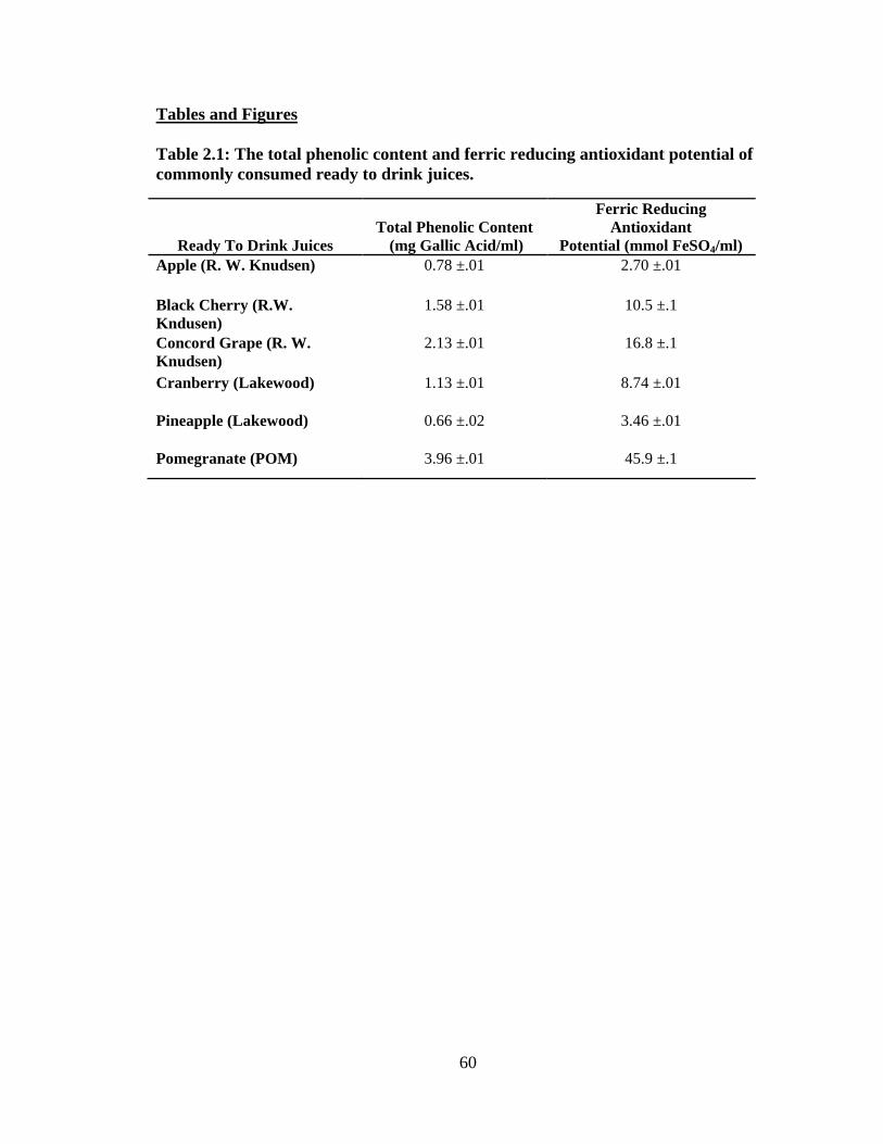
60
Tables and Figures
Table 2.1: The total phenolic content and ferric reducing antioxidant potential of commonly consumed ready to drink juices.
Ready To Drink Juices
Total Phenolic Content
(mg Gallic Acid/ml)
Ferric Reducing Antioxidant
Potential (mmol FeSO4/ml) Apple (R. W. Knudsen) 0.78 ±.01 2.70 ±.01
Black Cherry (R.W. Kndusen)
1.58 ±.01 10.5 ±.1
Concord Grape (R. W. Knudsen)
2.13 ±.01 16.8 ±.1
Cranberry (Lakewood) 1.13 ±.01 8.74 ±.01
Pineapple (Lakewood) 0.66 ±.02 3.46 ±.01
Pomegranate (POM) 3.96 ±.01 45.9 ±.1
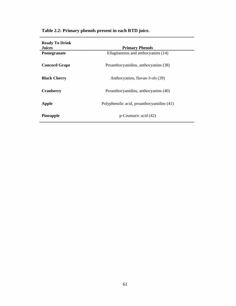
61
Table 2.2: Primary phenols present in each RTD juice.
Ready To Drink Juices Primary Phenols Pomegranate Ellagitannins and anthocyanins (14)
Concord Grape Proanthocyanidins, anthocyanins (38)
Black Cherry Anthocyanins, flavan-3-ols (39)
Cranberry Proanthocyanidins, anthocyanins (40)
Apple Polyphenolic acid, proanthocyanidins (41)
Pineapple p-Coumaric acid (42)
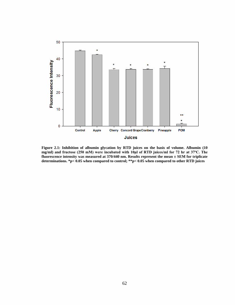
62
Figure 2.1: Inhibition of albumin glycation by RTD juices on the basis of volume. Albumin (10 mg/ml) and fructose (250 mM) were incubated with 10μl of RTD juices/ml for 72 hr at 37ºC. The fluorescence intensity was measured at 370/440 nm. Results represent the mean ± SEM for triplicate determinations. *p< 0.05 when compared to control; **p< 0.05 when compared to other RTD juices
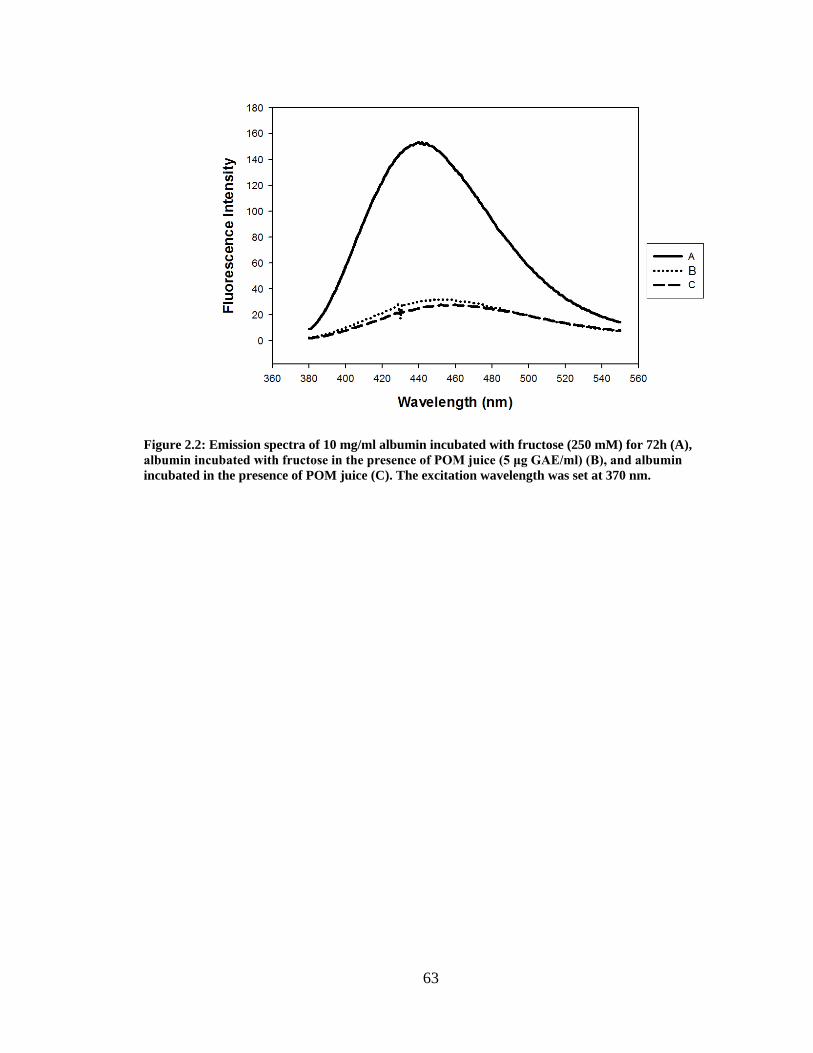
63
Figure 2.2: Emission spectra of 10 mg/ml albumin incubated with fructose (250 mM) for 72h (A), albumin incubated with fructose in the presence of POM juice (5 μg GAE/ml) (B), and albumin incubated in the presence of POM juice (C). The excitation wavelength was set at 370 nm.
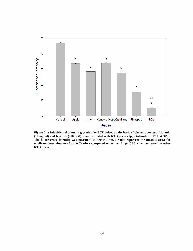
64
Figure 2.3: Inhibition of albumin glycation by RTD juices on the basis of phenolic content. Albumin (10 mg/ml) and fructose (250 mM) were incubated with RTD juices (5μg GAE/ml) for 72 h at 37ºC. The fluorescence intensity was measured at 370/440 nm. Results represent the mean ± SEM for triplicate determinations.* p< 0.05 when compared to control;** p< 0.05 when compared to other RTD juices
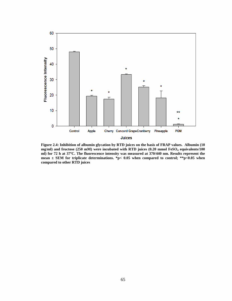
65
Figure 2.4: Inhibition of albumin glycation by RTD juices on the basis of FRAP values. Albumin (10 mg/ml) and fructose (250 mM) were incubated with RTD juices (0.20 mmol FeSO4 equivalents/100 ml) for 72 h at 37ºC. The fluorescence intensity was measured at 370/440 nm. Results represent the mean ± SEM for triplicate determinations. *p< 0.05 when compared to control; **p<0.05 when compared to other RTD juices

66
Figure 2.5: Effect of punicalagin on albumin glycation. Albumin (10 mg/ml) and fructose (250 mM) were incubated with punicalagin for 72h at 37°C. The fluorescence intensity was measured at 370/440 nm. Results represent the mean ± SEM for triplicate determinations. *p< 0.05 when compared to control value
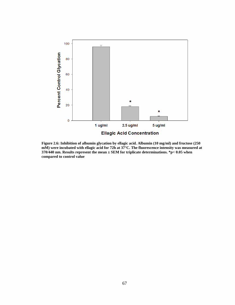
67
Figure 2.6: Inhibition of albumin glycation by ellagic acid. Albumin (10 mg/ml) and fructose (250 mM) were incubated with ellagic acid for 72h at 37°C. The fluorescence intensity was measured at 370/440 nm. Results represent the mean ± SEM for triplicate determinations. *p< 0.05 when compared to control value
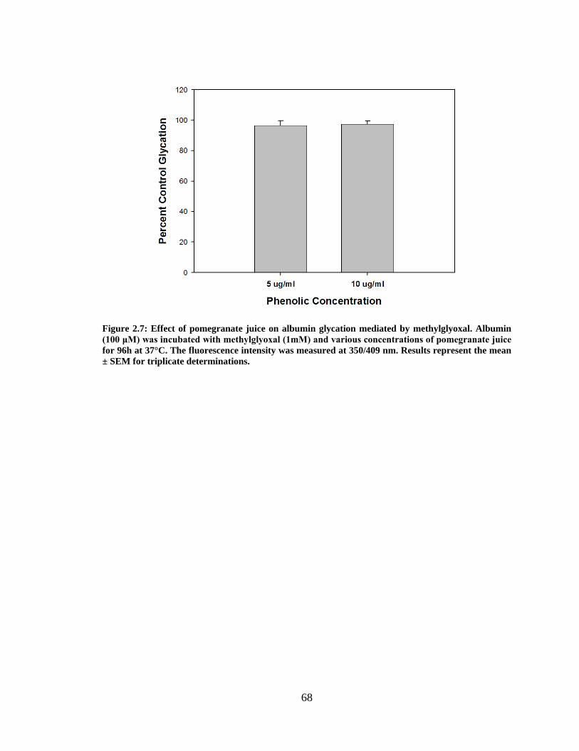
68
Figure 2.7: Effect of pomegranate juice on albumin glycation mediated by methylglyoxal. Albumin (100 μM) was incubated with methylglyoxal (1mM) and various concentrations of pomegranate juice for 96h at 37°C. The fluorescence intensity was measured at 350/409 nm. Results represent the mean ± SEM for triplicate determinations.
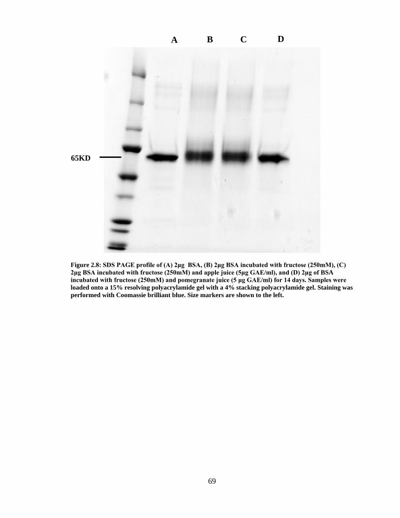
69
Figure 2.8: SDS PAGE profile of (A) 2μg BSA, (B) 2μg BSA incubated with fructose (250mM), (C) 2μg BSA incubated with fructose (250mM) and apple juice (5μg GAE/ml), and (D) 2μg of BSA incubated with fructose (250mM) and pomegranate juice (5 μg GAE/ml) for 14 days. Samples were loaded onto a 15% resolving polyacrylamide gel with a 4% stacking polyacrylamide gel. Staining was performed with Coomassie brilliant blue. Size markers are shown to the left.
65KD
A B C D
70
CHAPTER 3
INHIBITION OF NON-ENZYMATIC PROTEIN GLYCATION BY WHOLE
POMEGRANATE (PUNICA GRANATUM) AND COMPONENTS OF THE PERICARP
_______________________
Pamela Garner Dorsey1, Ronald B. Pegg2, Phillip Greenspan1 1Department of Pharmaceutical and Biomedical Science 2Department of Food Science and Technology University of Georgia, Athens, GA 30602 To be submitted to Journal of Agricultural and Food Chemistry
71
Abstract
The non-enzymatic glycation of proteins, an oxidative dependent process, initiates
the formation of advanced glycation endproducts (AGEs) which eventually leads to the
crosslinking of proteins. Over the past decade many studies have focused on the
antioxidant potential of pomegranate fruit; however little work has been performed on the
relationship between its antioxidant activity and ameliorating the diabetic state. This
study investigates the effect of a phenolic whole pomegranate fruit extract and various
fractions (aril, peel, and membrane) of pomegranate fruit on the in vitro fructose
mediated glycation of albumin. Compared to apple, whole pomegranate fruit exhibited a
much higher total phenolic content and antioxidant potential. Pomegranate fruit
decreased glycation by 80% and 90% when incubated at a phenolic concentration of 2.5
and 5 μg GAE/ml respectively; apple, at these phenolic concentrations, inhibited protein
glycation by only 20% and 40% respectively. Pomegranate membrane exhibited the
highest total phenolic content and antioxidant potential compared to the pomegranate aril
and peel. At 2.5 μg GAE/ml, the membrane fraction decreased glycation by 85%
compared to the aril (42%), and peel (75%). Pomegranate membrane also produced the
greatest decrease in glycation when these fractions were incubated at the same
antioxidant capacities (FRAP values). These results demonstrate that pomegranate fruit is
a potent inhibitor of fructose mediated albumin glycation when compared to whole apple.
The inhibition observed is attributed to the presence of ellagitannins, which are not found
in whole apple.
72
Introduction
Complications stemming from diabetes mellitus such as neuropathy (1),
nephropathy (2), and retinopathy (3) are initiated by the accumulation of advanced
glycation endproducts (AGEs) in various tissues. AGEs are formed from the non-
enzymatic binding of a protein, nucleic acid, or lipid to a reducing carbohydrate (4).
Hyperglycemia is known to cause oxidative damage and an imbalance between reactive
oxygen species and antioxidant detoxification pathways. Diabetic individuals are more
susceptible to oxidative processes due to an increased production of reactive oxygen
species (5), and a lower concentration of inherent antioxidants (vitamins C and E) (6).
Many mechanisms are involved in hyperglycemia mediated oxidative stress including the
oxidation of glucose, protein glycation, and the formation of AGEs; free radicals and
oxidation reactions are involved in the glucose-mediated modification of proteins (7).
Diets rich in fruits and vegetables reduce the risk of cancer and other chronic
diseases (8). Fruits and vegetables are also an important dietary source of polyphenols
(9), and the typical human intake of flavonoids (a major group of polyphenols) is
reported to be 1g per day (8). Diets rich in fruits and vegetables (5 or more servings a
week), as demonstrated by Sargeant and coworkers (10), significantly reduced HbA1C
levels in a cohort of over 5,000 adults. In the United States, apples (33.1%) are the largest
contributors of polyphenols to the American diet followed by oranges (14.0%), grapes
(12.8%), and strawberries (9.8%) (11). Polyphenols found in various botanical extracts
(12-14) have been shown to inhibit the formation of AGEs both in vitro and in vivo;
flavonoids have the greatest inhibitory effect among phenolics examined (15).
Pomegranate fruit is rich in polyphenols and found in the arid regions of the
world. Ancient medicine has utilized the pomegranate to treat many maladies (16, 17),
73
however the health benefits of pomegranate stem from its anti-oxidative and anti-
inflammatory properties (18,19). Previously we have shown that polyphenols found in
commercially available pomegranate juice are superior inhibitors of fructose mediated
protein glycation when compared to commonly consumed juices at the same phenolic
content and antioxidant capacity (20). This suggests that the major polyphenols contained
within the pomegranate are unique inhibitors of the glycation process. The major group
of polyphenols, ellagitannins, is found in the peel and membrane of the fruit (21). During
the juicing process, pomegranate is squeezed whole, leaching ellagitannins from the peel
and membrane into the resultant juice. The current study examined the effect of extracts
from whole pomegranate fruit and its various components (peel, membrane, arils) on the
glycation of bovine serum albumin mediated by fructose. Apple was employed as a
control fruit, for we previously demonstrated that pomegranate juice was superior to
apple juice in inhibiting protein glycation (20).
Materials and Methods Materials
Bovine serum albumin (essentially fatty acid free), D-(-) fructose, Chelex 100
(sodium form), Folin-Ciocalteu reagent, TPTZ (2, 4, 6-tri[2-pyridyl]-s-triazine, ferrous
sulfate heptahydrate, anhydrous ferric chloride, and Amberlite XAD-16 were purchased
from Sigma Chemical Company (St. Louis, MO). Pomegranates (POM Wonderful
variety) and apples (Red Delicious) were purchased locally from Publix Supermarket.
74
Extraction of Pomegranate and its Pericarp Components
Pomegranates are characterized as large berries covered by a leathery exocarp
(peel). A white, spongy, and very bitter tissue connected to the peel extends into the
interior of the fruit (membrane). The membrane provides a matrix for edible seeds
surrounded by a sack of juice (arils) (22). POM Wonderful pomegranates were cut into
quarters (exposing the pericarp of the fruit), and placed in distilled water to facilitate the
separation of the arils, membrane, and peel. Arils, membrane, peel, and the fruit as a
whole were macerated in a Super 5000 Vitamix (Cleveland,OH) until a slurry was
formed. The resultant slurries were subjected to a hot water extraction at 100ºC for two
hours, after which they were vacuumed and filtered twice. A burette (20ml) packed with
Amberlite XAD-16 was washed four times with deionized water, and the eluent was
disposed of. The fruit slurries were loaded onto the burette and washed several times with
deionized water, to achieve a Brix level of zero. The Brix level was measured with a
pocket PAL-1 Atago pocket refractometer (Tokyo, Japan). Phenolic compounds were
eluted from the Amberlite XAD-16 using methanol. The samples were placed on a A-210
Buchi rotovapor (Switzerland) at 200 MPa for one hour, after which they were freeze
dried with a FreeZone 2.5 Labconco freeze dry system (Kansas City,MO). All sample
extracts were reconstituted in 50% EtOH at a concentration of 1mg/ml.
Total Phenolic Content
The total phenolic content for all fruit extracts was determined by the Folin-
Ciocalteu method as described by Slinkard and coworkers (23), utilizing gallic acid as a
standard. Briefly, gallic acid standards (20 μl) and each fruit extract (20 μl) were
combined with distilled water (1580 μl), Folin Ciocalteu reagent (100 μl), and 300 μl of
75
1.6 M sodium carbonate. Distilled water (20 μl) was used as a blank. The mixtures were
incubated for 45 minutes at room temperature, after which absorbance was read at 765
nm on a Beckman DU 600 series spectrophotometer.
Ferric Reducing Antioxidant Potential (FRAP) Assay
The Ferric Reducing Antioxidant Potential of all fruit extracts was determined by
a modified method described by Benzie and coworkers (24), where iron (II) sulfate
heptahydrate was the standard. Briefly iron (II) sulfate heptahydrate standards (10 μl) and
each fruit extract (10 μl) were combined with distilled water (30 μl), and 300 μl of
reagent (25 ml of 300 mM acetate buffer (pH 3.6), 25 ml of TPTZ solution, and 2.5 ml of
20 mM ferric chloride solution). Distilled water (10 μl) was used as a blank. The mixtures
were allowed to incubate for six minutes after which each sample was combined with
distilled water (340 μl). Absorbance was read at 593nm on a Beckman DU 600 series
spectrophotometer.
Modification of Albumin by Fructose
The glycation of bovine serum albumin was determined by a method described by
Farrar and coworkers (25). Bovine serum albumin (10 mg/ml) was incubated in the
presence of D-(-) fructose (250 mM) and various concentrations of the fruit extracts in
200 mM potassium phosphate buffer (pH 7.4) at 37ºC for 72 hours. Potassium phosphate
buffer was treated with Chelex 100 prior to use. All samples were corrected for the
fluorescence of albumin incubated with each fruit extract. The fluorescence intensity was
measured at an excitation/emission wavelength pair of 370/440 nm using a Perkin-Elmer
LS 55 luminescence spectrometer with slit widths set at 3nm.

76
Modification of Albumin by Methylgloxal
The glycation of bovine serum albumin by methylgloxal was performed using a
method described by Lee and coworkers (26). Bovine serum albumin (100 μM) was
incubated in the presence of methylgloxal (1mM) in Chelex 100 treated 0.1 M sodium
phosphate buffer, pH 7.0. After 96 hours, the fluorescence was measured at an
excitation/emission wavelength pair of 350/409 nm using a Perkin-Elmer LS 55
luminescence spectrometer with slit widths set at 3 nm. All samples were corrected for
the fluorescence of albumin incubated with each extract.
Statistical Analysis
Experiments were performed in triplicate and expressed as mean ± SEM. Data
was analyzed utilizing a one-way analysis of variance (ANOVA) and multiple
comparisons were performed employing Tukey’s test. Statistical significance was set at
p< 0.05.
Results
The phenolic fractions of pomegranates, the various components of pomegranates
and apples were freeze dried and resuspended in 50% ethanol at a concentration of 1
mg/ml. Whole pomegranate fruit had a higher content of total phenols than whole apple
(Table 1), which is in agreement with a study by Martin and coworkers (27). Whole apple
and pomegranate fruit were also analyzed for their antioxidant capacity as determined by
the FRAP assay (Table 1); pomegranate exhibited an antioxidant capacity that was
approximately four times greater than that of apple.
Total phenolic content and antioxidant capacity for components of pomegranate’s
pericarp were analyzed. Peel and membrane extracts exhibited total phenolic content of
77
0.79 and 0.81 mg GAE/ml respectively, slightly greater than the phenolic content of
whole fruit (Table 2). The aril extract had a much lower phenolic content, which is in
agreement with work by Tzulker and coworkers (28). It is noteworthy to mention that
peel and membrane extracts exhibited an antioxidant capacity that was approximately
three times greater than the aril extract (Table 2).
To examine the effect of pomegranates and apples on protein glycation,
pomegranate and apple extracts (2.5 and 5 μg GAE/ml) were incubated with a solution
containing bovine serum albumin (10 mg/ml) and fructose (250 mM). When the
BSA/fructose mixture was incubated in the presence of whole pomegranate extract (2.5
µg GAE/ml), glycation was inhibited by approximately 80% (Figure 1), however the
same concentration of apple phenolic compounds resulted in only a 20% decrease in
control glycation. Increasing the concentration of whole pomegranate extract to 5 μg
GAE/ml resulted in a further decrease in glycation to approximately 10% of control
values. However, apple phenolics, at 5 μg GAE/ml did not produce the degree of
inhibition observed with pomegranate phenolics at 2.5 μg GAE/ml. These results are in
agreement with our previous findings on the effect of pomegranate and apple juices on
protein glycation (20).
The effect of extracts from various components of the pomegranate pericarp
(arils, peel, membrane) on protein glycation was also examined. In this study (Figure 2),
the phenolic content for each component was normalized to 5 μg GAE/ml in the
incubation mixture. At this concentration, all components inhibited protein glycation by
over 80%; similar to that observed with the whole pomegranate extract. The membrane
extract produced the greatest decrease in fluorescence intensity to approximately 5% of

78
control fluorescence. When the extracts from the various components were incubated at
both 2.5 and 5 µg GAE/ml, the membrane extract again produced the greatest decrease in
glycation followed by the peel extract (Figure 3).
The effect of each pomegranate component extract, set at two different
antioxidant capacities, on protein glycation was then examined (Figure 4). At an
antioxidant capacity of 0.04 mmol FeSO4/100 ml, the membrane extract produced the
greatest inhibition in glycation followed by the peel extract, with arils still showing
significant inhibitory activity. At 0.05 mmol FeSO4/ 100 ml all extracts inhibited
glycation by over 90%, with the membrane extract still producing the greatest inhibition
among the components. Combined with data presented in Tables 2 and 3, it is apparent
that the type of phenols found in the peel and membrane of the fruit is the determining
factor for the inhibition of protein glycation. Ellagitannins (the major phenolic group in
pomegranate fruit) are mainly located in the peel and outer pericarp of pomegranate fruit
(28) which includes the pomegranate membrane.
Reactive dicarbonyls, such as methylglyoxal, bind to proteins to form AGEs
directly bypassing earlier stages of protein glycation (Schiff bases and Amadori products)
(4). When pomegranate and apples extracts (2.5 and 5 µg GAE/ml) were incubated with
albumin (100 μM) in the presence of methylgloxal (1mM), there was only a slight
decrease in glycation observed (Figure 5). Pomegranate aril, peel, and membrane extracts
were also examined for their effect on methylglyoxal mediated glycation (Figure 6). Only
the membrane extract, at 5μg GAE/ml, significantly inhibited glycation. The extent of
inhibition (approximately 20%) at 5 μg GAE/ml was quite weak compared to that
observed with fructose mediated albumin glycation (Figure 2). These results demonstrate

79
that the dramatic inhibition of fructose mediated protein glycation by pomegranates is not
observed in other pathways of protein glycation.
Discussion
There are numerous studies centered on the antiglycating activity of natural
products; however there are few focused on the inhibition of this process by functional
foods. The antioxidant and anti-inflammatory effects of pomegranate fruit are well
documented but its effect on protein glycation has not been investigated. Previously we
explored the effect of pomegranate juice and other commonly consumed juices (black
cherry, concord grape, pineapple, apple, and cranberry) on the formation of AGEs (20).
While all fruit juices inhibited protein glycation at a concentration of 1% (v/v),
pomegranate juice was the best inhibitor of the group. Based on these findings, we
explored the effect of the pomegranate fruit and the various components of its pericarp on
the inhibition of protein glycation. The extract of whole pomegranate produced a greater
inhibition of protein glycation when compared to whole apple extract (Figure 1). These
results agree with our previous work (20); apple juice was much less effective in
inhibiting protein glycation when compared to pomegranate juice. This is not the first
report on the effect of the apple on protein glycation. Lavelli and coworkers (29) reported
that dehydrated apple extracts were effective inhibitors of protein glycation. Based on
molar concentration, procyanidin B2 was found to be the most effective phenolic
inhibitor, followed by catechin and epicatechin.
In contrast, pomegranates and other berries (strawberries, blackberries, and
raspberries) are phenolically rich in ellagitannins and ellagic acid (hydrolytic product of
ellagitannins) (30). The highest concentrations of ellagitannins are found in the
80
membrane and peel of the pomegranate (21) and these phenolic compounds provide the
majority of the anitoxidative properties of the fruit. The peels of many different fruits
contain a high concentration of antioxidants which provide protection against
environmental stressors (31,32). In a similar matter, Yamaguchi and coworkers (33)
incubated 10μM of garcinol (purified product from Garcina indica fruit rind) with BSA
(20 mg/ml) and D-fructose (500 mM); glycation was inhibited by 50%. The findings in
the current study in regards to the pomegranate peel and membrane agree with previous
results where the major phenolics found in pomegranate, punicalagin (ellagitannin) and
ellagic acid, inhibited protein glycation by over 90% at 2.5 μg GAE/ml (20). Thus, the
rather high content of ellagitannins in pomegranate fruit appears to be responsible for the
dramatic effect on protein glycation.
Shao and coworkers (32) documented that apple phenolics, phloretin and
phloridzin were capable of trapping reactive dicarbonyl species such as methylglyoxal
and glyoxal. Based on the results presented in Figure 5, the phenolics present in both
pomegranates and apples did not appreciably inhibit methylgloxal mediated protein
glycation at a concentration of 5 μg GAE/ml. Phloretin and phloridzin represent
approximately 22% of the total major apple phenolics (33). The concentration of
methylglyoxal in the current study mixture was 1mM, which is approximately 100 times
the concentration of phenolic compounds. In contrast, Shao and coworkers (32) used
nearly equivalent concentrations of dicarbonyl and phenolic species to demonstrate the
carbonyl trapping properties of apple phenolics.
Hyperglycemia has been associated with the overproduction of the superoxide
anion radical from the mitochondrial electron transport chain (34). These free radicals
81
damage many biomolecules (lipids, proteins, DNA), that can lead to many chronic
diseases and diabetic complications. Flavonoids with a strong scavenging activity
(antioxidant capacity) tend to have a greater inhibitory effect on AGE formation than
weaker scavenging phenolics (35). However, as shown in Figure 4, different
pomegranate fractions inhibit protein glycation to varying degrees even at the same
antioxidant capacity. This would suggest that antioxidant capacity is not the sole factor in
determining inhibition of AGE formation.
Pomegranate has been extolled for many years due to its medicinal properties.
However, there are few studies that highlight the inhibitory properties of pomegranate
fruit on protein glycation. A recent study involving a water soluble extract of
pomegranate rind (10μg/ml) inhibited glucose and fructose mediated glycation of BSA by
28%; however vitamin C (10μg/ml) inhibited protein glycation by 41% (36). The extracts
from the whole fruit and various components of the whole fruit (arils, peel, membrane) in
the current study demonstrated inhibition of protein glycation at over 90% at 5 ug
GAE/ml (Figure 3). The current study is the first to demonstrate the inhibitory properties
of whole pomegranate fruit and various components of its pericarp on protein glycation.

82
References
1) Vitek MP, Bhattacharya K, Glendening JM, Stopa E, Vlassara H, Bucala R, Manogue
K, Cerami A. Advanced glycation endproducts contribute to amyloidosis in Alzheimer
disease. Proceedings of the National Academy of Sciences 91: 4766-4770. 1994.
2) Roohk H, Zaidi A. A review of glycated albumin as an intermediate glycation index
for controlling diabetes. Journal of Diabetes Science and Technology 2. 1114-1121.
2008.
3) Okumura A, Mitamura Y, Namekata K, Nakamura K, Harada C, Harada T. Glycated
albumin includes activation of activator-protein-1 in retinal glial cells. Japanese Journal
of Ophthalmology 51: 236-237. 2007.
4) Ulrich P, Cerami A. Protein glycation, diabetes, and aging. Recent Progress in
Hormone Resaerch 56: 1-22. 2001.
5) Wolff SP, Jiang ZY, Hunt JV. Protein glycation and oxidative stress in diabetes
mellitus and aging. Free Radical Biology and Medicine 10: 339-352. 1991.
6) Wen Y, Skidmore JC, Porter-Turner MM, Rea CA, Khokher MA, Singh BM.
Relationship of glycation, antioxidant status and oxidative stress to vascular endothelial
damage in diabetes. Diabetes, Obesity, and Metabolism 4: 305-308. 2002.
7) Bonnefont-Rousselot D. Glucose and reactive oxygen species. Current Opinion in
Clinical Nutrition and Metabolic Care 5: 561-568. 2002.
8) Chun OK, Kim DO, Smith N, Schroeder D, Han JT, Lee CY. Daily consumption of
phenolics and total antioxidant capacity from fruit and vegetables in the American diet.
Journal of the Science of Food and Agriculture 85: 1715-1724. 2005.
83
9) Prior RL. Fruits and vegetables in the prevention of cellular oxidative damage. The
American Journal of Clinical Nutrition 78: 570S-578S. 2003.
10) Sargeant LA, Khaw KT, Bingham S, Day NE, Luben RN, Oakes S, Welch A,
Wareham NJ. Fruit and vegetable intake and population glycosylated haemoglobin
levels: the EPIC-Norfolk Study. European Journal of Clinical Nutrition 55: 342-348.
2001.
11) Wolfe KL, Kang X, He X, Dong M, Zhang Q, Liu RH. Cellular antioxidant activity
of common fruits. Journal of Agricultural and Food Chemistry 56: 8418-8426. 2008.
12) Urios P, Grigorova-Borsos AM, Sternberg M. Flavonoids inhibit the formation of the
cross-linking AGE pentosidine in collagen incubated with glucose, according to their
structure. European Journal of Nutrition 46: 139-146. 2007.
13) Ahmad MS, Pischetsrieder M, Ahmed N. Aged garlic extract and S-allyl cysteine
prevent formation of advanced glycation endproducts. European Journal of
Pharmacology 561: 32-38. 2007.
14) Yokozawa T, Nakagawa T. Inhibitory effects of Luobuma tea and its components
against glucose-mediated protein damage. Food and Chemical Toxicology 42: 975-981.
2004.
15) Wu CH, Yen GC. Inhibitory effect of naturally occurring flavonoids on the formation
of advanced glycation endproducts. Journal of Agricultural and Food Chemistry 53:
3167-3173. 2005.
16) Arseculeratne SN, Gunatilaka AA, Panabokke RG. Studies on medicinal plants of Sri
Lanka. Part 14. Toxicity of some traditional medicinal herbs. Journal of
Ethnopharmacology 13: 323-335. 1985.
84
17) Saxena A, Vikram NK. Role of selected Indian plants in management of type 2
diabetes: a review. Journal of Alternative and Complementary Medicine 10: 369-378.
2004.
18) Rosenblat M, Hayek T, Aviram M. Anti-oxidative effects of pomegranate juice (PJ)
consumption by diabetic patients on serum and on macrophages. Atherosclerosis 187:
363-371. 2006.
19) Adams LS, Zhang Y, Seeram NP, Heber D, Chen S. Pomegranate ellagitannin-
derived compounds exhibit antiproliferative and antiaromatase activity in breast cancer
cells in vitro. Cancer Prevention Research 3: 108-113. 2010.
20) Dorsey P. The inhibition of non-enzymatic fructose mediated protein glycation by
pomegranate polyphenolics. Department of Phamaceutical and Biomedical Science,
University of Georgia.2012.
21) Gil MI, Toms-Barbern FA, Hess-Pierce B, Holcroft DM, Kader AA. Antioxidant
activity of pomegranate juice and its relationship with phenolic composition and
processing. Journal of Agricultural and Food Chemistry 48: 4581-4589. 2000.
22) Lansky EP, Newman RA. Punica granatum (pomegranate) and its potential for
prevention and treatment of inflammation and cancer. Journal of Ethnopharmacology
109: 177-206. 2007.
23) Slinkard K, Singleton VL. Total phenolic analysis: automation and comparison with
manual methods. American Journal of Enology and Viticulture 28: 49-55. 1977.
24) Benzie IF, Strain JJ. The ferric reducing ability of plasma (FRAP) as a measure of
‘antioxidant power’: the FRAP assay. Analytical Biochemistry 239: 70-76. 1996.
85
25) Farrar JL, Hartle DK, Hargrove JL, Greenspan P. A novel nutraceutical property of
select sorghum (sorghum bicolor) brans: inhibition of protein glycation. Phytotherapy
Research 22: 1052-1055. 2008.
26) Lee C, Yim M, Chock P, Yim H, Kang S. Oxidation reduction properties of
methylglyoxal modified protein in relation to free radical generation. The Journal of
Biological Chemistry 273: 25272-25278. 1998.
27) Martin KR, Krueger CG, Rodriquez G, Dreher M, Reed JD. Development of a novel
pomegranate standard and new method for the quantitative measurement of pomegranate
polyphenols. Journal of the Science of Food and Agriculture 89: 157-162. 2008.
28) Tzulker R, Glazer I, Ban-Ilan I, Holland D, Aviram M, Amir R. Antioxidant activity,
polyphenol content, and related compounds in different fruit juices and homogenates
prepared from 29 different pomegranate accessions. Journal of Agricultural and Food
Chemistry 55: 9559-9570. 2007.
29) Lavelli V. Combined effect of storage temperature and water activity on the
antiglycoxidative properties and color of dehydrated apples. Journal of Agricultural and
Food Chemistry 57:11491-11497. 2009.
30) Bakkalbaşi E, Menteş Ö, Artik N. Food ellagitannins-occurrence, Effects of
processing and storage. Critical Reviews in Food Science and Nutrition 49: 283-298.
2008.
31) Łata B, Przeradzka M. Changes of antioxidant content in fruit peel and flesh of
selected apple cultivars during storage. Journal of Fruit and Ornamental Plant Research
10: 1-9.2002.
86
32) Guo C, Yang J, Wei J, Li Y, Xu J, Jiang Y. Antioxidant activities of peel, pulp and
seed fractions of common fruits as determined by FRAP assay. Nutrition Research 23:
1719-1726.2003.
33) Yamaguchi F, Ariga T, Yoshimura Y, Nakazawa H. Antioxidative and anti-glycation
activity of garcinol from Garcinia indica fruit rind. Journal of Agricultural and Food
Chemistry 48: 180-185.2000.
34) Shao X, Bai N, He K, Ho CT, Yang CS, Sang S. Apple polyphenols, phloretin and
phloridzin: new trapping agents of reactive dicarbonyl species. Chemical Research in
Toxicology 21: 2042-2050. 2008.
35) Lu Y, Foo Y. Identification and quantification of major polyphenols in apple pomace.
Food Chemistry 59: 187-194. 1997.
36) Nishikawa T, Edelstein D, Du XL, Yamagishi S, Matsumura T, Kaneda Y, Yorek
MA, Beebe D, Oates PJ, Hammes HP, Giardino I, Brownlee M. Normalizing
mitochondrial superoxide production blocks three pathways of hyperglycaemic damage.
Nature 404: 787-790. 2000.
37) Matsuda H, Wang T, Hiromi M, Yoshikawa M. Structural requirements of flavonoids
for inhibition of protein glycation and radical scavenging activities. Bioorganic and
Medicinal Chemistry 11: 5317-5323. 2003.
38) Rout S, Banerjee R. Free radical scavenging, anti-glycation and tyrosinase inhibition
properties of a polysaccharide fraction isolated from the rind from Punica granatum.
Bioresource Technology 98: 3159-3163. 2007.
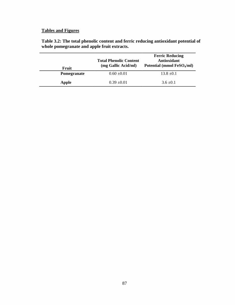
87
Tables and Figures
Table 3.2: The total phenolic content and ferric reducing antioxidant potential of whole pomegranate and apple fruit extracts.
Fruit
Total Phenolic Content
(mg Gallic Acid/ml)
Ferric Reducing Antioxidant
Potential (mmol FeSO4/ml)
Pomegranate 0.60 ±0.01 13.8 ±0.1
Apple 0.39 ±0.01 3.6 ±0.1
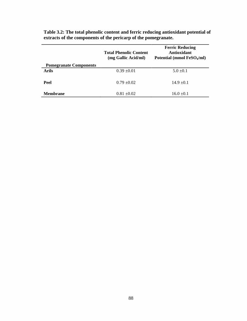
88
Table 3.2: The total phenolic content and ferric reducing antioxidant potential of extracts of the components of the pericarp of the pomegranate.
Pomegranate Components
Total Phenolic Content
(mg Gallic Acid/ml)
Ferric Reducing Antioxidant
Potential (mmol FeSO4/ml)
Arils 0.39 ±0.01 5.0 ±0.1
Peel 0.79 ±0.02 14.9 ±0.1
Membrane 0.81 ±0.02 16.0 ±0.1
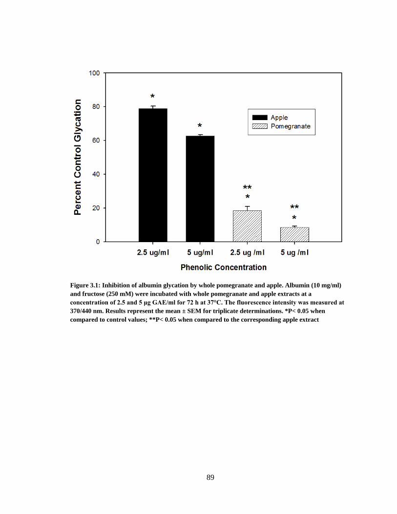
89
Figure 3.1: Inhibition of albumin glycation by whole pomegranate and apple. Albumin (10 mg/ml) and fructose (250 mM) were incubated with whole pomegranate and apple extracts at a concentration of 2.5 and 5 μg GAE/ml for 72 h at 37°C. The fluorescence intensity was measured at 370/440 nm. Results represent the mean ± SEM for triplicate determinations. *P< 0.05 when compared to control values; **P< 0.05 when compared to the corresponding apple extract
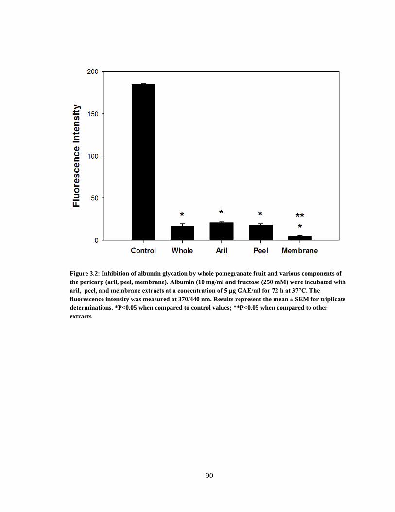
90
Figure 3.2: Inhibition of albumin glycation by whole pomegranate fruit and various components of the pericarp (aril, peel, membrane). Albumin (10 mg/ml and fructose (250 mM) were incubated with aril, peel, and membrane extracts at a concentration of 5 μg GAE/ml for 72 h at 37°C. The fluorescence intensity was measured at 370/440 nm. Results represent the mean ± SEM for triplicate determinations. *P<0.05 when compared to control values; **P<0.05 when compared to other extracts
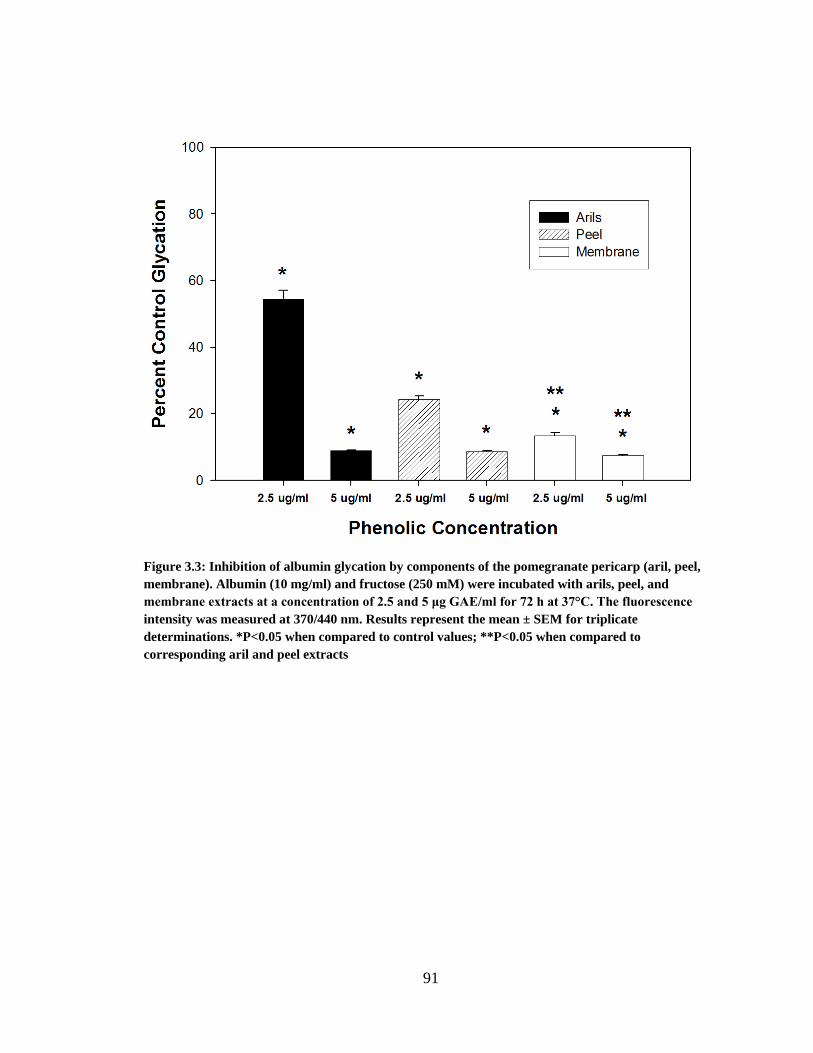
91
Figure 3.3: Inhibition of albumin glycation by components of the pomegranate pericarp (aril, peel, membrane). Albumin (10 mg/ml) and fructose (250 mM) were incubated with arils, peel, and membrane extracts at a concentration of 2.5 and 5 μg GAE/ml for 72 h at 37°C. The fluorescence intensity was measured at 370/440 nm. Results represent the mean ± SEM for triplicate determinations. *P<0.05 when compared to control values; **P<0.05 when compared to corresponding aril and peel extracts
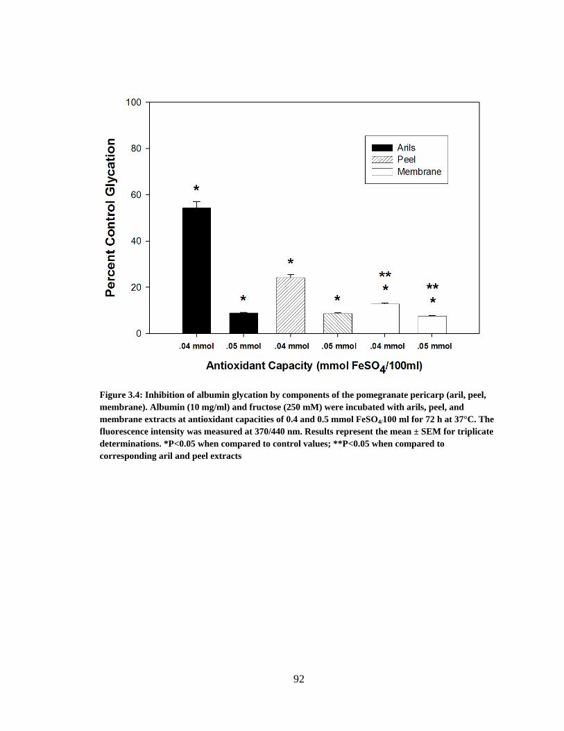
92
Figure 3.4: Inhibition of albumin glycation by components of the pomegranate pericarp (aril, peel, membrane). Albumin (10 mg/ml) and fructose (250 mM) were incubated with arils, peel, and membrane extracts at antioxidant capacities of 0.4 and 0.5 mmol FeSO4/100 ml for 72 h at 37°C. The fluorescence intensity was measured at 370/440 nm. Results represent the mean ± SEM for triplicate determinations. *P<0.05 when compared to control values; **P<0.05 when compared to corresponding aril and peel extracts
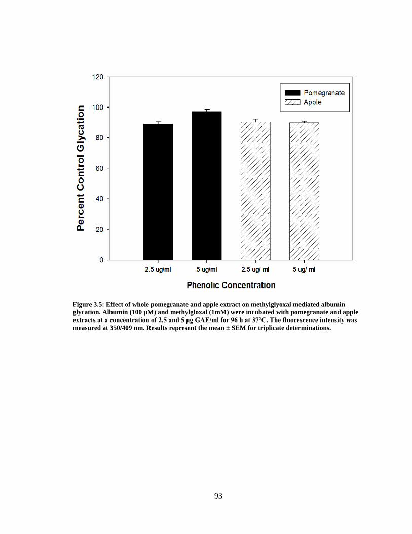
93
Figure 3.5: Effect of whole pomegranate and apple extract on methylglyoxal mediated albumin glycation. Albumin (100 µM) and methylgloxal (1mM) were incubated with pomegranate and apple extracts at a concentration of 2.5 and 5 μg GAE/ml for 96 h at 37°C. The fluorescence intensity was measured at 350/409 nm. Results represent the mean ± SEM for triplicate determinations.
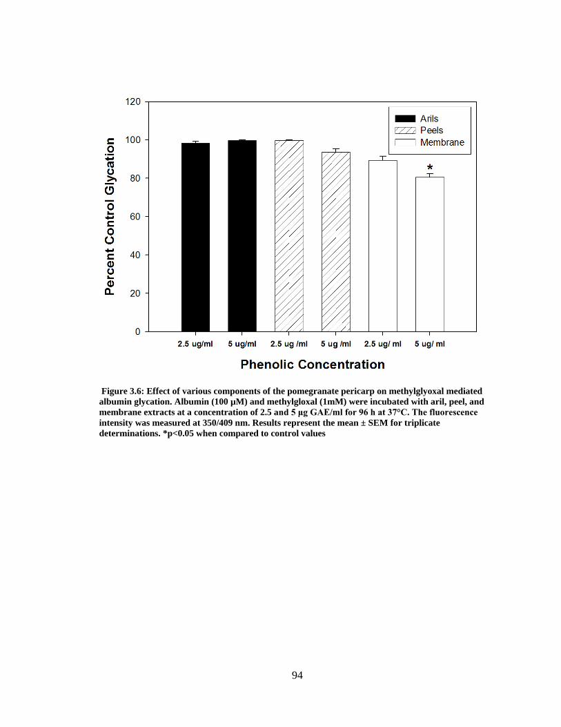
94
Figure 3.6: Effect of various components of the pomegranate pericarp on methylglyoxal mediated albumin glycation. Albumin (100 µM) and methylgloxal (1mM) were incubated with aril, peel, and membrane extracts at a concentration of 2.5 and 5 μg GAE/ml for 96 h at 37°C. The fluorescence intensity was measured at 350/409 nm. Results represent the mean ± SEM for triplicate determinations. *p<0.05 when compared to control values
95
CHAPTER 4
POMEGRANATE (PUNICA GRANATUM) JUICE: A NON-SPECIFIC INHIBITOR OF
PROTEIN GLYCATION
_______________________
Pamela Garner Dorsey1, Phillip Greenspan1 1Department of Pharmaceutical and Biomedical Science University of Georgia, Athens, GA 30602 To be submitted to Phytochemistry
96
Abstract
Oxygen drives the Maillard reaction between a reducing sugar and protein at
physiological pH and temperature. This reaction yields fluorescent and non-fluorescent
glycoxidation products that contribute to the chemical modification and crosslinking of
proteins. The Maillard reaction is a key component in the development of diabetic
complications. In previous studies we found that bovine serum albumin incubated in the
presence of fructose yielded fluorescent glycoxidation products and pomegranate juice
was highly effective in arresting the progression of this reaction. In the current study we
sought to examine the effect of pomegranate juice on the in-vitro fructose mediated
glycation of four different proteins (gelatin, IgG, lysozyme, and ribonuclease A).
Pomegranate juice, at 2.5 and 5 µg GAE/ml, reduced the formation of fluorescent
glycoxidation products by 87 and 92% respectively when gelatin was incubated in the
presence of fructose. Pomegranate juice also produced a significant reduction in AGE
formation when IgG was incubated in the presence of fructose. Lysozyme incubated in
the presence of fructose did not yield an appreciable amount of fluorescent glycoxidation
products, possibly due to lysozyme’s lysine residue content. SDS-PAGE analysis
revealed that ribonuclease A incubated in the presence of fructose resulted in the
formation of many oligomeric products visible at different molecular weights.
Pomegranate juice (5µg GAE/ml), when incubated with ribonuclease A and fructose,
prevented the formation of oligomeric products. In contrast, apple juice at the same
phenolic concentration did not alter the modifications to ribonuclease A observed in the
presence of fructose. This work demonstrates that pomegranate juice is a natural and
effective in vitro inhibitor of glycation of various mammalian proteins.
97
Introduction
Proteins in the body are exposed to many factors which can result in their
modification; oxidation and glycation are two major non-enzymatic alterations that affect
a protein’s native structure (1). The alterations to these proteins are often cleared by
receptors on neighboring cells; one important receptor is RAGE (receptor for advanced
glycation endproduct). However, in people who suffer from chronic diseases (who are
more susceptible to oxidation), these altered proteins may accumulate, leading to
pathological conditions. In diabetes mellitus, these alterations are a hallmark for many
diabetic complications such as retinopathy.
Albumin is the most abundant protein found in blood plasma with a concentration
of 35 to 50g/l (2). Albumin’s tertiary structure allows it to bind to small molecules such
as metal ions, fatty acids, bilirubin, and various pharmaceutical agents (3). Due to its long
half-life (21 days) and its high concentration in blood plasma, albumin is sensitive to
non-enzymatic modification by carbohydrates. Many structural modifications occur as a
result of the glycation of albumin including an increase in molecular weight. This
increase in molecular weight can be attributed to one or several glucose units attached to
certain amino acid residues such as arginine, lysine and cysteine and the eventual
crosslinking of albumin molecules.
Modifications to the native protein structure are also correlated to changes in the
binding properties of albumin (1). Nonetheless, there are many contradictory studies
involving the binding affinity of modified albumin. One study indicated that the
alteration of certain albumin binding sites by glycation causes a decrease in binding
affinity for certain fatty acids (4). Conversely, in another study lengthy incubations (60
98
days) of albumin in the presence of glucose (9 mol glucose/mol albumin) enhanced the
binding of warfarin (5); however Okabe and coworkers (6) reported no effect on warfarin
binding by human serum albumin glycated by glucose at a lower glucose concentration (2
mol glucose/mol albumin).
In previous studies (data not shown), we have demonstrated that pomegranate juice and
certain parts of pomegranate fruit are effective inhibitors of the glycation of bovine serum
albumin mediated by fructose. These inhibitory properties are attributed to ellagitannins
and ellagic acid found within the membrane and peel of the fruit. Based on these data,
we wanted to demonstrate that the inhibitory effect of pomegranate’s major
polyphenolics on albumin glycation is not unique to albumin, but also is observed with
other model proteins. Glycation occurs to many proteins in-vivo, specifically those that
have a long half-life such as collagen and laminin (7). Bovine serum albumin is
commonly used to study in vitro protein glycation and it has an 80% structural similarity
to human albumin (8). Other investigators (9,10) have employed other proteins to study
glycation. In this study, gelatin, IgG, lysozyme, and ribonuclease A were incubated in
the presence of fructose and the effect of pomegranate juice on glycoxidation was
determined. Ribonuclease A was subjected to SDS-PAGE analysis to demonstrate the
inhibition of glycative modifications to the native protein structure by pomegranate juice.
Materials and Methods
Materials
Bovine serum album (Fraction V, essentially fatty acid free), gelatin (bovine skin),
ribonuclease A (bovine pancreas), lysozyme (chicken egg white), Chelex 100 (sodium
form), and 2-mercaptoethanol were purchased from Sigma Chemical Company (St.
Louis, MO). Bovine IgG was purchased from Equitech-Bio (Kerville, TX). Laemmli
99
sample buffer, Bio-Safe Coomassie blue and Criterion precast gels (8-16% Tris HCl)
were purchased from Bio-Rad (Hercules, CA). Apple juice (R.W. Knudsen) and
pomegranate juice (POM Wonderful) were purchased locally from an Earthfare
Supermarket.
Modification of Albumin by Fructose
A modified method described by Farrar and coworkers (11) was used to
determine the degree of glycation for gelatin, IgG, and lysozyme. Gelatin, IgG, and
lysozyme (2 mg/ml) were incubated in the presence of D-(-) fructose (125 mM) and
pomegranate juice (2.5 and 5 μg GAE/ml) in 200 mM potassium phosphate buffer at
37ºC for 72 hours. The potassium phosphate buffer was treated with Chelex 100 prior to
use. All incubation mixtures were corrected for the native fluorescence of incubated
gelatin, IgG, and lysozyme, with pomegranate juice. The fluorescence intensity was
measured at an excitation/emission wavelength pair of 370/440 nm using a Perkin-Elmer
LS 55 Luminescence Spectrophotometer with slit widths set at 3nm.
Analysis of Protein Modifications
Ribonuclease A (10mg/ml) and D-(-) fructose (250mM) were incubated in the
presence and absence of pomegranate juice (5μg GAE/ml) and apple juice (5 μg
GAE/ml) in 200 mM potassium phosphate buffer at 37ºC for 14 days. In this experiment
10μl of apple and pomegranate juice were added to the 3ml incubations every 3 days.
Modifications induced by protein glycation were assessed by sodium dodecyl
polyacrylamide gel electrophoresis (SDS-PAGE) (12). Aliquots for each sample were
diluted 1:1 with Laemmli sample buffer containing 62.5mM Tris-HCl (pH 6.8), 25%
(v/v) glycerol, 2% (w/v) SDS, 0.01% (v/v) Bromophenol Blue, and 5% (v/v) 2-
100
mercaptoethanol. Samples were boiled at 100ºC for five minutes after dilution and then
centrifuged. Centrifuged samples were loaded onto a 16% resolving polyacrylamide gel
with an 8 % stacking polyacrylamide gel and subjected to electrophoresis for 45 minutes.
Gels were stained with Bio-safe Coomassie Blue for one hour and de-stained with
distilled water. All gels were scanned on a FluorChem HD2 system with Alpha View
software.
Statistical Analysis
Experiments were performed in triplicate and expressed as mean ± SEM. Data
was analyzed utilizing a one-way analysis of variance (ANOVA) and multiple
comparisons were performed employing Tukey’s test. Statistical significance was set at
p< 0.05.
Results
In previous studies, the Folin Ciocalteu method was used to determine the
phenolic content of pomegranate juice (data not shown). The concentration of total
phenols was approximately 4mg GAE/ml. On the basis of phenolic content we were able
to determine that a normalized concentration of 5μg GAE/ml inhibited the formation of
fluorescent glycoxidation products in albumin incubated in the presence of fructose by
90%.
Gelatin is a denatured protein that originates from the processing of collagen. In
vivo, collagen is extremely sensitive to glycooxidation due to its long half-life, and is
involved in the development of diabetic complications. In this study gelatin, (2mg/ml)
was incubated in the presence of fructose (125 mM) for 72 hours at 37°C. Pomegranate
juice was added to the reaction mixture at a normalized phenolic content of 2.5 and 5µg
101
GAE/ml (Figure 1). Pomegranate juice, at both 2.5 and 5µg GAE/ml, produced a
dramatic inhibition of glycation, similar to that observed with bovine serum albumin
(data not shown). These results demonstrate that pomegranate juice is a generalized
inhibitor of glycation and this inhibition is not unique to bovine serum albumin.
IgG is the smallest antibody present in the human body, and it is soluble in
aqueous solutions. Glycated IgG has been implicated in the pathogenesis of rheumatoid
arthritis (13) and the progression of nephropathy (14). Figure 2 illustrates the
concentration dependent inhibition of IgG glycation by pomegranate juice. Pomegranate
juice, at 5µg GAE/ml, decreased the fluorescence intensity by 72%. The inhibition of
glycation was approximately 50% when pomegranate juice was present at a concentration
of 2.5 µg GAE/ml. These data illustrate that pomegranate juice is also an effective
inhibitor of IgG glycation mediated by fructose.
Lysozyme, also known as muramidase, is an enzyme that can cleave the cell walls
of bacteria and viruses (15). Ahmad and coworkers (10) demonstrated that lysozyme can
be used to investigate glycation induced crosslinking due to the fact that it produces
oligomers which are easily detected with SDS-PAGE analysis. In our study lysozyme
incubated in the presence of fructose (125 mM) for 3 days did not yield the same increase
in fluorescence intensity observed with albumin, gelatin or IgG (Figure 3). While the
degree of glycation in lysozyme was approximately one-fifth of the other proteins
observed, pomegranate juice, at 2.5 and 5μg GAE/ml, still produced a decrease of
fluorescence intensity in comparison to the control incubations.
102
According to Eble and coworkers (16), ribonuclease is an excellent model protein
for studying hexose induced crosslinking because it possesses many reactive lysl residues
which form oligomers. The occurrence of crosslinked RNase oligomers can easily be
determined by SDS-PAGE analysis (17). The current study examined the effect of
pomegranate juice on fructose mediated protein modifications of RNase utilizing SDS-
PAGE analysis (Figure 4). The effect of apple juice on the modification of RNAase by
fructose was also examined; we previously demonstrated that apple juice, at the same
phenolic concentration as pomegranate juice, was a less effective inhibitor of glycation
than pomegranate juice. RNase (10mg/ml) incubated in potassium phosphate buffer for
14 days (column A) yielded a distinct protein band at 14 KDa. RNase (10 mg/ml)
incubated in the presence of fructose (250 mM) for 14 days (column B) resulted in the
broading of the protein band at 14 KDa. In contrast to the native protein, two lightly
stained bands at 32 and 45 KDa respectively were apparent after RNase was incubated
with fructose. When RNase (10 mg/ml) was incubated in the presence of fructose (250
mM) and pomegranate juice (5 μg GAE/ml) (column C), SDS Page profile was quite
similar to native RNAase; there was no broading of the 14 KDa band and no evidence of
bands at 32 and 45KDa. In contrast, RNase incubated in the presence of fructose and
apple juice (5 μg GAE/ml) resulted in a broadening of the protein band at 14 KDa and
two lightly stained bands at 32 and 45KDa. The SDS-PAGE analysis demonstrates that
pomegranate juice, and not apple juice, is an effective inhibitor of modifications to
RNase mediated by fructose.
103
Discussion
Albumin is the model protein to illustrate in vitro protein glycation because it
contains reactive lysine residues which are instrumental in producing vesperlysine (18), a
type of fluorescent AGE. In previous studies we have measured the formation of
fluorescent AGEs, such as vesperlysine, by incubating albumin in the presence of
fructose and measuring fluorescence at the excitation/emission wavelength pair of
370/440 nm. The glycation of albumin is also associated with tertiary structure
modifications, that involve unfolding of the protein at tryptophan residues 134 and 214
(19,20). Conformational changes resulting from glycation can yield new binding sites for
certain ligands (1), and can impair the affinity of diazepam, sulfisoxazole, phenytoin,
cyclosporine, and valproic acid binding to albumin (21). The resulting conformational
changes in proteins with a long half-life (i.e. collagen) and eventual cross-linking of these
proteins can result in altered protein function and free radical formation.
Our past studies have demonstrated that pomegranate juice can prevent the non-
enzymatic glycation of bovine serum albumin (data not shown). We illustrated that major
phenolic compounds present in pomegranate juice (punicalagin and ellagic acid) were
effective inhibitors of albumin glycation. However, the purpose of this current study is to
demonstrate that AGE inhibition by pomegranate and pomegranate juice was not unique
to albumin, perhaps because of a critical and unique binding site of these phenolics to the
protein. Rather ellagitannins and ellagic acid are effective generalized inhibitors of
glycation and that this inhibition would be observed in a wide variety of proteins, with
the subsequent prevention of modifications to the native protein structure.
104
In the current study, pomegranate juice produced a dose dependent inhibition of
fluorescent AGE formation in three differents proteins (Figures 1-3) and the results are
similar to that observed previously with bovine serum albumin (data not shown). The
data suggests that the phenolic compounds within pomegranate juice are effective
inhibitors of Schiff base formation (early stage glycation). In previous studies
pomegranate juice provided very little protection against Amadori product formation (late
stage glycation) mediated by an α-dicarbonyl (methylglyoxal). While pomegranate juice
consumption has recently been shown to be beneficial for cardiovascular health (22),
these findings suggest that pomegranate juice may also lessen the pathogenesis of
complications resulting from non-enzymatic glycation.
While pomegranate juice did inhibit the production of fluorescent AGEs,
lysozyme incubated in the presence of fructose did not form an appreciable amount of
fluorescent AGEs when incubated for 72 hours (Figure 3). These results may be due to
the fact that lysozyme lacks a considerable amount of reactive lysine residues needed for
rapid glycation in comparison to BSA (23,24) and the length of incubation was not
sufficient (>3 days). Other studies have indicated proteins rich in lysine and arginine
residues undergo rapid structural changes in the presence of a reducing sugar (25).
Ahmad and coworkers (10) incubated lysozyme with glucose (500 mM) for 35 days and
found significant protein modification.
In previous work, we examined the effect of pomegranate juice on the protein
modification of fructosylated bovine serum albumin (data not shown). BSA incubated in
the presence of fructose produced only a widening of the native BSA band; no larger
weight bands, indicative of significant protein modification (crosslinking) was observed.
105
We also demonstrated that pomegranate juice, at a given phenolic content, inhibited the
widening of the native albumin band. In this study, RNase was chosen based on the work
by Litchfield and coworkers (9). In their study, RNase A (10mg/ml) was incubated in the
presence of glucose or arabinose (250 mM) for 21 days. SDS-PAGE analysis revealed the
formation of multiple larger molecular weight bands. Amino acid analysis demonstrated
that lysine and arginine residues were the only amino acids to undergo significant
chemical modifications. In our study, we also observed the formation of larger molecular
bands when RNase was incubated with fructose; these bands were not produced when
pomegranate juice was present in the incubation mixture, but did appear when apple
juice, a poor inhibitor of glycation, was present.
The mechanism of action for many natural product glycation inhibitors is still
unknown; however studies have indicated that compounds with a high antioxidant
capacity provide protection against glycation mediated by reducing sugars (26).
Pomegranate juice is commercially available and an effective inhibitor of protein
glycation. We have shown that pomegranate juice provides protection against glycation
in four different model proteins as evidenced by fluorescence spectroscopy. Pomegranate
juice also prevents the structural modification of two proteins, albumin and ribonuclease
A, as described by SDS-PAGE analysis. This present work supports the theory that the
inhibition of bovine serum albumin glycation by pomegranate juice is not unique to that
protein, and is not a result of the documented binding of polyphenolic compounds to
albumin (27). In summary, pomegranate juice is rich in ellagitannins (punicalagin and
ellagic acid) and possesses high antioxidant capacity which lends itself to being an
exceptional inhibitor of protein glycation.
106
References
1) Rondeau P, Emmanuel B. The glycation of albumin: structural and functional impacts.
Biochimie 93: 645-658.2011.
2) Evans TW. Albumin as a drug-biological effects of albumin unrelated to oncotic
pressure. Alimentary Pharmacology and Therapeutics 16: 6-11.2002.
3) Wood M. Plasma drug binding: implications for anesthesiologists. Anesthesia and
Analgesia 65: 786-804.1986.
4) Tomasić A, Bertosa B, Tomić S, Soskić M, Magnus V. Binding behavior of amino
acid conjugates of indole-3-acetic acid to immobilized human serum albumin. Journal of
Chromatography A 1154: 240-249.2007.
5) Nakajou K, Watanabe H, Kragh-Hansen U, Maruyama T, Otagiri M. The effect of
glycation on the structure, function, and biological fate of human serum albumin as
revealed by recombinant mutants. Biochimica et Biophysica Acta (BBA)-General
Subjects 1623: 88-97.2003.
6) Okabe N, Hashizume N. Drug binding properties of glycosylated human serum
albumin as measured by fluorescence and circular dichroism. Biological and
Pharmaceutical Bulletin 17: 16-21. 1994.
7) Charonis AS, Reger LA, Dege JE, Kouzi-Koliakos K, Furcht LT, Wohlhueter RM,
Tsilibary EC. Laminin alterations after in vitro nonenzymatic glycosylation. Diabetes 39:
807-814.1990.
8) Szapacs ME, Riggins JN, Zimmerman LJ, Liebler DC. Covalent adduction of human
serum albumin by 4-hydroxy-2-nonenal: kinetic analysis of competing alkylation
reactions. Biochemistry 45: 10521-10528.2006.
107
9) Litchfield JE, Thorpe SR, Baynes JW. Oxygen is not required for the browning and
crosslinking of protein by pentoses: relevance to Maillard reactions in vivo. The
International Journal of Biochemistry and Cell Biology 31: 1297-1305.1999.
10) Ahmad MS, Pischetsrieder M, Ahmed N. Aged garlic extract and S-allyl cysteine
prevent formation of advanced glycation endproducts. European Journal of
Pharmacology 561: 32-38.2007.
11) Farrar JL, Hartle DK, Hargrove JL, Greenspan P. A novel nutraceutical property of
select sorghum (sorghum bicolor) brans: inhibition of protein glycation. Phytotherapy
Research 22: 1052-1055.2008.
12) Laemmli UK. Cleavage of structural proteins during the assembly of the head of
bacteriophage T4. Nature 227: 680-685.1970.
13) Fortin SL, Newkirk NM. A new antibody in rheumatoid arthritis targeting glycated
IgG: IgM anti-IgG-AGE. British Journal of Rheumatology 37: 1307-1314.1998.
14) Kalia K, Sharma S, Mistry K. Non-enzymatic glycosylation of immunoglobulins in
diabetic nephropathy. Clinica Chimica Acta 347: 169-176.2004.
15) Imoto T. Lysozyme.eLS.2009.
16) Eble AS, Thorpe SR, Baynes JW. Nonenzymatic glucosylation and glucose-
dependent cross-linking of protein. The Journal of Biological Chemistry 258: 9406-
9412.1983.
17) Watkins NG, Thorpe SR, Baynes JW. Glycation of amino groups in protein. Studies
on the specificity of modification of RNase by glucose. The Journal of Biological
Chemistry 260:10629-10636.1985.
108
18) Nakamura K, Nakazawa Y, Ienaga K. Acid-stable fluorescent advanced glycation end
products: vesperlysines a, b, and c are formed as crosslinked products in the maillard
reaction between lysine or proteins with glucose. Biochemical and Biophysical Research
Communications 232: 227-230.1997.
19) Bourdon E, Loreau N, Blache D.Glucose and free radicals impair the antioxidant
properties of serum albumin. The Journal of the Federation of American Societies for
Experimental Biology 13: 233-244.1999.
20) Rondeau P, Navarra G, Cacciabaudo F, Leone M, Bourdon E, Militello V. Thermal
aggregation of glycated bovine serum albumin. Biochimica et Biophysica Acta 1804:
789-798.2010.
21) MacKichan J. Influence of protein binding and use of unbound (free) drug
concentrations. Burton M, Shaw L, Schentag J, Evans W (eds.).Applied
Pharmacokinetics and Pharmacodynamics: Principles of Therapeutic Drug Monitoring.
Lippincott Williams and Wilkins.Philadelphia, PA.82-120.2006.
22) Stowe CB. The effects of pomegranate juice consumption on blood pressure and
cardiovascular health. Complementary Therapies in Clinical Practice 17: 113-115.2011.
23) Kakade ML, Liener IE. Determination of available lysine in proteins. Analytical
Biochemistry 27: 273-280.1969.
24) Goodno CC, Swaisgood HE, Catignani GL. A fluorimetric assay for available lysine
in proteins. Analytical Biochemistry 115: 203-211.1981.
25) Gugliucci A, Menini T. The polyamines spermine and spermidine protect proteins
from structural and functional damage by AGE precursors: a new role for old molecules.
Life Sciences 72: 2603-2616.2003.
109
26) Duraisamy Y, Gaffney J, Slevin M, Smith CA, Williamson K, Ahmed N.
Aminosalicylic acid reduces the antiproliferative effect of hyperglycaemia, advanced
glycation endproducts and glycated basic fibroblast growth factor in cultured bovine
aortic cells: comparison with aminoguanidine. Molecular and Cellular Biochemistry 246:
143-153.2003.
27) Dufour C, Dangles O. Flavonoid-serum albumin complexation: determination of
binding constants and binding sites by fluorescence spectroscopy. Biochimica et
Biophysica Acta-General Subjects 1721: 164-173.2005.

110
Tables and Figures
Figure 4.2: Inhibition of the glycation of gelatin by pomegranate juice. Gelatin (2mg/ml) and fructose (125 mM) were incubated with pomegranate juice for 72 h at 37ºC. The fluorescence intensity was measured at 370/440 nm. Results represent the mean ± SEM for triplicate determinations. *p< 0.05 when compared to control
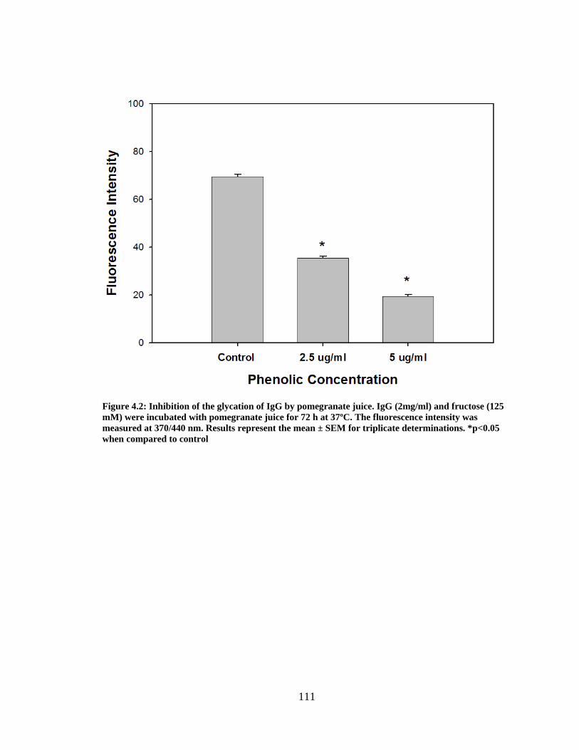
111
Figure 4.2: Inhibition of the glycation of IgG by pomegranate juice. IgG (2mg/ml) and fructose (125 mM) were incubated with pomegranate juice for 72 h at 37ºC. The fluorescence intensity was measured at 370/440 nm. Results represent the mean ± SEM for triplicate determinations. *p<0.05 when compared to control
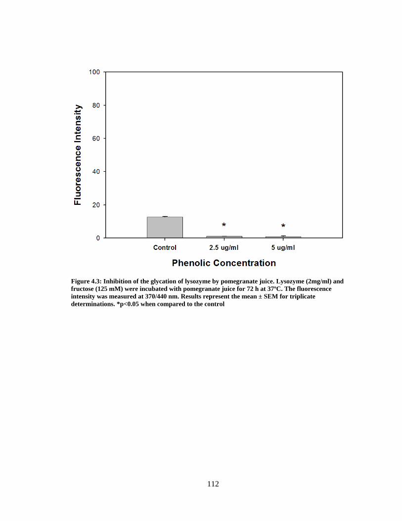
112
Figure 4.3: Inhibition of the glycation of lysozyme by pomegranate juice. Lysozyme (2mg/ml) and fructose (125 mM) were incubated with pomegranate juice for 72 h at 37ºC. The fluorescence intensity was measured at 370/440 nm. Results represent the mean ± SEM for triplicate determinations. *p<0.05 when compared to the control
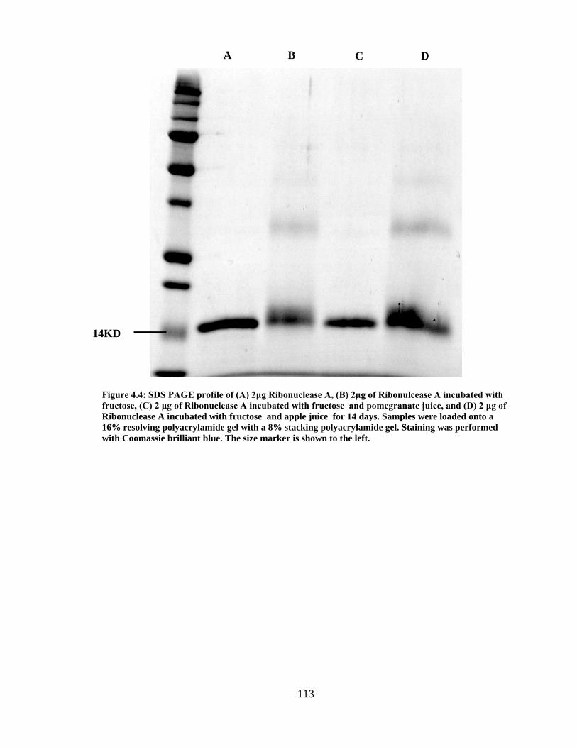
113
Figure 4.4: SDS PAGE profile of (A) 2μg Ribonuclease A, (B) 2μg of Ribonulcease A incubated with fructose, (C) 2 μg of Ribonuclease A incubated with fructose and pomegranate juice, and (D) 2 μg of Ribonuclease A incubated with fructose and apple juice for 14 days. Samples were loaded onto a 16% resolving polyacrylamide gel with a 8% stacking polyacrylamide gel. Staining was performed with Coomassie brilliant blue. The size marker is shown to the left.
14KD
D C B A
114
CHAPTER 5
CONCLUSIONS AND RECOMMENDATIONS
There are many FDA approved drugs designed to lower blood glucose levels; however no
drug currently exists to combat diabetic complications stemming from protein glycation.
Non-enzymatic protein glycation involves the binding of a protein amine group to the
carbonyl group of a reducing sugar. Resultant Schiff base compounds are usually
removed by various receptors; however in proteins that are not readily recycled,
irreversible AGEs adducts form.
Within the past decade there has been a large paradigm shift to increase the
consumption of phytochemicals (antioxidants) in the human diet. Rich in phytochemicals,
pomegranate has been extolled for its many medicinal qualities. In the current study
pomegranate juice, ellagic acid and punicalagin (major phenolics found in pomegranate),
extracts of pomegranate membrane and peel all demonstrated robust inhibitory activity of
albumin glycation mediated by fructose when compared to commonly consumed fruit
and fruit juices. Similar results were observed in other model proteins (gelatin, IgG,
ribonuclease A) demonstrating that pomegranate polyphenolics are non-specific
inhibitors of protein glycation.
To continue this research, the effect of pomegranate on the formation of AGES
using in vivo models of diabetes should be performed. One possible experimental system
is the streptozotocin induced diabetic rats and determining the rate of AGE formation in
tail collagen. To expand this project further, studies can be performed to determine
115
whether pomegranate juice or whole extracts of pomegranate fruit decrease the
concentration of glycated albumin in diabetic human subjects. Finally, chemical analogs
of punicalagin or ellagic acid could be synthesized in an attempt to find even more potent
anti-AGE compounds.
116
CHAPTER 6
APPENDIX
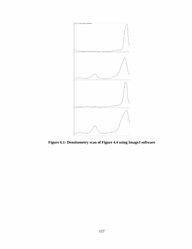
117
Figure 6.1: Densitometry scan of Figure 4.4 using ImageJ software