Punica granatum -Catenin in rat model - SciELO · Revista Brasileira de Farmacognosia 27 (2017)...
9
Revista Brasileira de Farmacognosia 27 (2017) 627–635 ww w.elsevier.com/locate/bjp Original Article Punica granatum suppresses colon cancer through downregulation of Wnt/-Catenin in rat model Hanaa H. Ahmed a , Hanan S. El-Abhar b , Elsayed Abdul Khalik Hassanin c , Noha F. Abdelkader b , Mohamed B. Shalaby c,∗ a Department of Hormones, Medical Research Division, National Research Centre, Cairo, Egypt b Department of Pharmacology and Toxicology, Faculty of Pharmacy, Cairo University, Cairo, Egypt c National Nutrition Institute, General Organization for Teaching Hospitals and Institutes, Ministry of Health, Cairo, Egypt a r t i c l e i n f o Article history: Received 17 March 2017 Accepted 17 May 2017 Available online 15 August 2017 Keywords: Colon cancer Pomegranate Wnt/-Catenin Inflammation Proliferation Apoptosis a b s t r a c t This study aims to elucidate the beneficial effect of Punica granatum L., Lythraceae (pomegranate) peel extract in the management of colon cancer induced intrarectally with N-methylnitrosourea. Adult male Sprague-Dawley rats were administered N-methylnitrosourea (2 mg in 0.5 ml water/rat) intrarectally three times/week for five weeks to induce colorectal cancer, followed by treatment with either 5- fluorouracil (12.5 mg/kg, i.p.) or Punica peel extract (2.25 or 4.5 g/kg, p.o.). Developed tumor elevated plasma TGF-, and Bcl2, serum epidermal growth factor, carcinoembryonic antigen, colon cancer specific antigens, and matrix metalloproteinase-7. Besides, immune-histochemical studies revealed an increase in COX-2, cyclin D1 and survivin content, as well as upregulation of the expression of colonic -Catenin, K-ras and C-myc genes. These results were further supported by the histological findings. Punica peel extract-treated rats, particularly those treated with a high dose, exhibited a marked reduction in the aforementioned parameters and improved the histological organization of the colon tissue. These alter- ations were consistent with those mediated through 5-fluorouracil. The present study encourages the use of P. granatum L. against colon cancer. Because Punica peel extract promotes apoptosis, mitigates inflammation and suppresses tumor cell proliferation in vivo, the potential mechanism underlying these activities might depend on the inhibition of the Wnt/-Catenin signaling pathway. © 2017 Published by Elsevier Editora Ltda. on behalf of Sociedade Brasileira de Farmacognosia. This is an open access article under the CC BY-NC-ND license (http://creativecommons.org/licenses/by-nc-nd/ 4.0/). Introduction Colorectal cancer (CRC) is the leading cause of cancer-related mortality worldwide and is the third most commonly diagnosed cancer in men and the second most commonly diagnosed can- cer in women in terms of incidence (Zhao et al., 2014). The onset and progression of CRC involves unregulated epithelial cell pro- liferation reflecting accumulated genetic mutations (Zhao et al., 2014). Recent evidence has shown that the prolonged survival of genetically unstable colorectal epithelial cells, eventually leading to malignant transformation is accompanied by the progressive suppression of apoptosis (Zhao et al., 2014). The majority of sporadic forms of CRC harbor genetic alter- ations in key elements of the Wnt/-Catenin signaling cascade, particularly in Adenomatous polyposis coli (APC) and -Catenin, ∗ Corresponding author. E-mail: [email protected] (M.B. Shalaby). thereby increasing the transcriptional activity of the latter (Kundu et al., 2006). -Catenin target genes play an ultimate role in tissue homeostasis, and the initiation and progression of CRC through the regulation of various cellular processes, including pro- liferation, stem cell fate, survival, differentiation, migration and angiogenesis (Srimuangwong et al., 2012). Particularly, the genes involved in proliferation and migration were overexpressed in CRC (Srimuangwong et al., 2012). Interestingly, the ingestion of a phytochemical-rich diet, includ- ing fruits and vegetables, has been associated with a decreased risk of CRC incidence (Sharma et al., 2010). Among foods, small fruits and berries have attracted much attention, and the associ- ation between their bioactive components and cancer prevention has become the focus of keen scientific interest. The fruit of Punica granatum L., Lythraceae (pomegranate), possesses many medicinal properties, reflecting anti-oxidant and anti-inflammatory potentials (Adhami et al., 2009). The compounds present in P. granatum, which have been largely inves- tigated for cancer preventive properties, include polyphenols, http://dx.doi.org/10.1016/j.bjp.2017.05.010 0102-695X/© 2017 Published by Elsevier Editora Ltda. on behalf of Sociedade Brasileira de Farmacognosia. This is an open access article under the CC BY-NC-ND license (http://creativecommons.org/licenses/by-nc-nd/4.0/).
Transcript of Punica granatum -Catenin in rat model - SciELO · Revista Brasileira de Farmacognosia 27 (2017)...

O
PW
HMa
b
c
a
ARAA
KCPWIPA
I
mccal2gts
ap
0(
Revista Brasileira de Farmacognosia 27 (2017) 627–635
ww w.elsev ier .com/ locate /b jp
riginal Article
unica granatum suppresses colon cancer through downregulation ofnt/�-Catenin in rat model
anaa H. Ahmeda, Hanan S. El-Abharb, Elsayed Abdul Khalik Hassaninc, Noha F. Abdelkaderb,ohamed B. Shalabyc,∗
Department of Hormones, Medical Research Division, National Research Centre, Cairo, EgyptDepartment of Pharmacology and Toxicology, Faculty of Pharmacy, Cairo University, Cairo, EgyptNational Nutrition Institute, General Organization for Teaching Hospitals and Institutes, Ministry of Health, Cairo, Egypt
r t i c l e i n f o
rticle history:eceived 17 March 2017ccepted 17 May 2017vailable online 15 August 2017
eywords:olon canceromegranatent/�-Catenin
nflammationroliferationpoptosis
a b s t r a c t
This study aims to elucidate the beneficial effect of Punica granatum L., Lythraceae (pomegranate) peelextract in the management of colon cancer induced intrarectally with N-methylnitrosourea. Adult maleSprague-Dawley rats were administered N-methylnitrosourea (2 mg in 0.5 ml water/rat) intrarectallythree times/week for five weeks to induce colorectal cancer, followed by treatment with either 5-fluorouracil (12.5 mg/kg, i.p.) or Punica peel extract (2.25 or 4.5 g/kg, p.o.). Developed tumor elevatedplasma TGF-�, and Bcl2, serum epidermal growth factor, carcinoembryonic antigen, colon cancer specificantigens, and matrix metalloproteinase-7. Besides, immune-histochemical studies revealed an increasein COX-2, cyclin D1 and survivin content, as well as upregulation of the expression of colonic �-Catenin,K-ras and C-myc genes. These results were further supported by the histological findings. Punica peelextract-treated rats, particularly those treated with a high dose, exhibited a marked reduction in theaforementioned parameters and improved the histological organization of the colon tissue. These alter-ations were consistent with those mediated through 5-fluorouracil. The present study encourages the
use of P. granatum L. against colon cancer. Because Punica peel extract promotes apoptosis, mitigatesinflammation and suppresses tumor cell proliferation in vivo, the potential mechanism underlying theseactivities might depend on the inhibition of the Wnt/�-Catenin signaling pathway.© 2017 Published by Elsevier Editora Ltda. on behalf of Sociedade Brasileira de Farmacognosia. This isan open access article under the CC BY-NC-ND license (http://creativecommons.org/licenses/by-nc-nd/
ntroduction
Colorectal cancer (CRC) is the leading cause of cancer-relatedortality worldwide and is the third most commonly diagnosed
ancer in men and the second most commonly diagnosed can-er in women in terms of incidence (Zhao et al., 2014). The onsetnd progression of CRC involves unregulated epithelial cell pro-iferation reflecting accumulated genetic mutations (Zhao et al.,014). Recent evidence has shown that the prolonged survival ofenetically unstable colorectal epithelial cells, eventually leadingo malignant transformation is accompanied by the progressiveuppression of apoptosis (Zhao et al., 2014).
The majority of sporadic forms of CRC harbor genetic alter-tions in key elements of the Wnt/�-Catenin signaling cascade,articularly in Adenomatous polyposis coli (APC) and �-Catenin,
∗ Corresponding author.E-mail: [email protected] (M.B. Shalaby).
http://dx.doi.org/10.1016/j.bjp.2017.05.010102-695X/© 2017 Published by Elsevier Editora Ltda. on behalf of Sociedade Brasileirahttp://creativecommons.org/licenses/by-nc-nd/4.0/).
4.0/).
thereby increasing the transcriptional activity of the latter (Kunduet al., 2006). �-Catenin target genes play an ultimate role intissue homeostasis, and the initiation and progression of CRCthrough the regulation of various cellular processes, including pro-liferation, stem cell fate, survival, differentiation, migration andangiogenesis (Srimuangwong et al., 2012). Particularly, the genesinvolved in proliferation and migration were overexpressed in CRC(Srimuangwong et al., 2012).
Interestingly, the ingestion of a phytochemical-rich diet, includ-ing fruits and vegetables, has been associated with a decreasedrisk of CRC incidence (Sharma et al., 2010). Among foods, smallfruits and berries have attracted much attention, and the associ-ation between their bioactive components and cancer preventionhas become the focus of keen scientific interest.
The fruit of Punica granatum L., Lythraceae (pomegranate),
possesses many medicinal properties, reflecting anti-oxidantand anti-inflammatory potentials (Adhami et al., 2009). Thecompounds present in P. granatum, which have been largely inves-tigated for cancer preventive properties, include polyphenols,de Farmacognosia. This is an open access article under the CC BY-NC-ND license

6 a de F
p3cmeotadsc(mefb(eMadat
M
H
pposm
P
iblfioeR
C
pp
D
n(sc5mTfmao
28 H.H. Ahmed et al. / Revista Brasileir
articularly ellagitannins (ET), punicalagins, flavonoids and the-glucosides/3,5-diglucosides of the anthocyanins delphinidins,yanidins and pelargonidins (Middha et al., 2013). Ellagitannins areetabolized into two active compounds, viz., ellagic acid (Sharma
t al., 2010) and urolithin A (UA), generated through the actionf gut microbiota on ET (Sharma et al., 2010). Urolithins suppresshe proliferation of colon cancer cells, stimulate cell cycle arrest,meliorate key cellular processes associated with colon cancerevelopment, such as mitogen-activated protein kinase (MAPK)ignaling (González-Sarrías et al., 2009); they also reduced theolonic mucosa inflammatory progression in a rat colitis modelLarrosa et al., 2009). Furthermore, rutin (a flavonol glycoside) pro-
otes apoptosis and cell cycle arrest in human colon cancer (Vijayt al., 2016). Noteworthy, the peel, which is the non-edible part ofruits, contains larger quantities of these polyphenols than the edi-le parts, accompanied by higher cancer anti-proliferative potentialOrgil et al., 2014). A recent study showed that P. granatum peelxtract (PPE) reduces cell proliferation and induces apoptosis inCF-7 human breast cancer cells via anti-oxidant and apoptotic
ctivities (Shirode et al., 2014). Accordingly, the present study waselineated to explore the underlying mechanism (s) in favor of thentitumor activity of P. granatum against colon cancer induced inhe experimental animals.
aterials and methods
erbal extract
Punica granatum L., Lythraceae, peel extract (PPE) was sup-lied by United Group Pharma Co. (Bader city, Cairo, Egypt). Testlants were authenticated by Prof. Ibrahim El-Garf, Departmentf Botany, Faculty of Science, Cairo University, Egypt. Voucherpecimen (number 20170402M) was kept in the herbarium of Phar-acognosy Department, Faculty of Pharmacy, Cairo University.
reparation of Punica granatum peel extract (PPE)
Four kilograms of peel were separated from P. granatum fruitsn August 2014 (5–6 kg). The peels were cut into small pieces andlended with 4 l of methanol (70%) using an electric blender, fol-
owed by incubation for 10–12 h. The extract was filtered throughlter paper and the solvent was evaporated using a rotary evap-rator (Büchi Labortechnik AG, Flawil, Switzerland). The resultingxtract was dehydrated in an oven at 50 ◦C for 24 h (El-Toumy andauwald, 2002).
hemicals
Gallic acid, protocatechuic acid, catechin, rutin, ellagic acid,uanicalagin were obtained from Sigma–Aldrich Chemical Com-any (St. Louis, USA).
etermination of polyphenols by HPLC-DAD
The HPLC system was an Agilent 1100 equipped with a quater-ary pump, online degasser, autosampler and diode-array detectorDAD). Data collection and analyses were performed using Chem-tation software. Chromatographic separations of the extracts werearried out on a Zorbax C18 column (250 × 4.6 mm, particle size
�m, Agilent) using water/acetic acid (98:2, v/v) (Solvent A) andethanol (Solvent B) as the mobile phases at a flow rate of 1 ml/min.
he elution program used was as follows: 5% B for 5 min, 5–70% B
or 25 min, and 70–5%B for 10 min. The column temperature wasaintained at 35 ◦C and the detection was monitored at 254, 280,nd 360 nm. UV spectra of the components were taken continu-usly between 200 and 400 nm throughout the elution in order
armacognosia 27 (2017) 627–635
to determine component identity and peak purity. The injectionvolume for standards and samples was 10 �l.
Animals and ethics statement
Adult male Wistar rats, weighing 150–170 g, were obtainedfrom the Animal Facility Breeding Colony of the National ResearchCenter, Cairo, Egypt. The animals were acclimatized for one weekin a specific pathogen-free barrier area at constant temperature(25 ± 1 ◦C), humidity (55%), and a 12 h light/dark cycle. Rats werehoused with a standard laboratory diet recommended by the Amer-ican Institute of Nutrition (Reeves et al., 1993) and had free accessto food and water and their body weight was assessed daily. Theanimals were managed according to the Guide for the Care andUse of Laboratory Animals published by the US National Institutesof Health (NIH Publication No. 85-23, revised 1996) and the studyprotocol was approved by the Ethical Committee for Animal Exper-imentation at the Faculty of Pharmacy, Cairo University (PermitNumber: PT 664).
Experimental design
Forty adult male Sprague-Dawley rats, weighing 150–170 g,were randomly allocated into five groups (8 rats/each group); ani-mals of the first group received vehicle (1 ml DMSO (5%), p.o.) andserved as the normal control group. Rats in the other four groupswere rectally administered N-MNU (2 mg in 0.5 ml water/rat) threetimes/week for five weeks to induce CRC (Ahmed et al., 2013) amodel that was reported to mimic histopathologically human colontumors (Narisawa and Fukaura, 2003). CRC animals were dividedinto group II, which received the vehicle and served as the CRCuntreated group, while the remaining three groups were treatedthroughout the four months experimental period with the follow-ing treatments. In group III, rats were treated with 5-fluorouracil(5-FU; 12.5 mg/kg, i.p. [equivalent to 2 mg/kg for humans (Shinet al., 2010)]) on days 1, 3 and 5, with the cycle repeated everyfour weeks (Watson et al., 1998). Animals in groups IV and V weretreated daily with PPE for four months at two dose levels (2.25 and4.5 g/kg in DMSO (5%), p.o.). The selected doses of PPE; viz., 4.5 g/kgb.wt [equivalent to 0.729 g/kg for humans (Shin et al., 2010)] andits half dose 2.25 g/kg b.wt [equivalent to 0.365 g/kg for humans(Shin et al., 2010)] were administered daily for 6 months. After anovernight fast, the final body weights were measured and the ratswere euthanized using CO2. Blood was collected through cardiacpuncture, and the blood samples were divided into two aliquots forthe separation of plasma and serum. Subsequently, the plasma andserum samples were acquired through centrifugation at 2555 × gfor 30 min at 4 ◦C. The colon tissues were rapidly excised, cleanedand washed in ice-cold saline, blotted dry and equally divided intotwo longitudinal portions. The first portion was preserved in forma-lin saline for histological and immunohistochemical examination,respectively, while the second portion was collected in liquid nitro-gen and stored at - 80 ◦C for subsequent biochemical and moleculargenetic analyses.
Biochemical measurements
Plasma transforming growth factor-beta (TGF-�), Bcl2 levels,and serum levels of epidermal growth factor (EGF), colon cancer-
specific antigen (CCSA-4), carcino-embryonic antigen (CEA), andmatrix metalloproteinase-7 (MMP-7) were assayed using commer-cial ELISA kits (Glory Science Co., Ltd, Del Rio, TX, USA) accordingto the manufacturer’s instructions.
a de F
Sˇ
I
uGmeitsDiioe
R
wuOT1
H.H. Ahmed et al. / Revista Brasileir
emi-quantitative real-time PCR (sqRT-PCR) detection of-Catenin, K-ras and C-myc gene expressions
solation of total RNATotal RNA was extracted from the colon tissue of rats
sing TRIzol®
reagent (Cat#15596-026, Invitrogen, Darmstadt,ermany) according to the manufacturer’s instructions withinor modifications. The tissue samples (50 mg) were homog-
nized in 1 ml of TRIzol®
reagent and the RNA was dissolvedn diethylpyrocarbonate (DEPC)-treated water. Total RNA wasreated with 1 U of RQ1 RNAse-free DNAse (Invitrogen, Darm-tadt, Germany) to digest DNA residues and re-suspended inEPC-treated water. The purity of total RNA was assessed accord-
ng to the 260/280 nm ratio (between 1.8 and 2.1), and thentegrity was assessed through ethidium bromide staining analysisf 28S and 18S bands after formaldehyde-containing agarose gellectrophoresis.
everse transcription (RT) reactionThe complete poly(A)+ RNA, isolated from each colon tissue,
as reverse transcribed into cDNA in a total volume of 20 �l
sing the Revert AidTM First Strand cDNA Synthesis Kit (MBI,pelstrasse, Germany) according to manufacturer’s instructions.he RT reaction was performed at 25 ◦C for 10 min, followed byh at 42 ◦C, and completed with a denaturation step at 99 ◦C for
0 5 10
mAU
0
200
400
600
800
DAD1 B, Sig=254,16 Ref=off (TRA\NOV00514
Gal
lic
Pro
toca
tech
uic
Rut
inE
llagi
c
Cat
each
in
0 5 10
mAU
0
200
400
600
800
1000
1200
1400
1600
DAD1 B, Sig=254,16 Ref=off (TRA\NOV00513
Gal
lic
prot
ocat
echu
icC
atea
chin
Rut
inE
llagi
c
A
B
Fig. 1. HPLC chromatogram of standards (A) and
armacognosia 27 (2017) 627–635 629
5 min. Subsequently, the reaction tubes containing RT preparationswere flash-cooled in an ice chamber until further use for DNAamplification through sqRT-PCR.
Semi-quantitative real-time-polymerase chain reaction
An iQ5-BIO-RAD Cycler (Cepheid, CA, USA) was used to deter-mine the cDNA copy number. PCR reactions were set up in 25 �lreaction mixtures containing 12.5 �l 1 × SYBR
®Premix ExTaqTM
(Takara, Biotech Co. Ltd., Saint-Germain-en-Laye, France), with0.5 �l of sense primers (0.2 �M), 0.5 �l of antisense primer(0.2 �M), 6.5 �l of distilled water, and 5 �l of cDNA template. Thesequences of the primers are described in Box 1. At the end ofeach sqRT-PCR a melting curve analysis was performed at 95 ◦C toassess the quality of the used primers. The relative quantificationof the target genes was determined through the �CT method using�-actin as a reference gene.
Immuno-histochemical and histological examination
After the colon tissues were fixed in formalin saline for 24 h,
the specimens were processed for paraffin embedding and twosets of 4 �m sections were prepared. In the first set, the sectionswere prepared for immune-histochemical examination. The sec-tions were collected onto glass-positive slides and were fixed inmi15 20
.D)
puan
ical
agin
mi15 20
.D)
Pua
nica
lagi
n
pomegranate peel extract (B) at 254 nm.

630 H.H. Ahmed et al. / Revista Brasileira de F
Box 1: Primer sequences used for sqRT-PCRGene Primer sequence (5′–3′) Sequence references
�-Catenin F: CAAT GGG TCA TAT CAC AGA TTCTTR: TCT CTT TTC TTC ACC ACA ACA TTT
Austinat et al. (2008)
K-ras F: AGT ACG ACC CTA CGA TAG AGGACT CCTR: CAA TCT GTA CTG TCG GAT CTCTCT CAC C
Fuentes-Calvo et al. (2010)
C-myc F: TGA CGA GAC CTT CGT GAA GAR: ATT GAT GTT ATT TAC ACT TAAGGG T
Tao et al. (2002)
�-Actin F: CCC CAT CGA GCA CGG TAT TG Eshak et al. (2010)
aaiNid5us
F(g
R: ATG GCG GGG GTG TTG AAG GTC
65 ◦C oven for 1 h. Subsequently, the slides were deparaffinizednd the samples were blocked for endogenous peroxidase activ-ty after immersing the slides in 3% hydrogen peroxide for 10 min.ext, the sections were washed with Tris buffered saline, and sim-
larly treated according to the immune-histochemical procedure
escribed above. The Power-StainTM-1.0 Poly HRP DAB Kit (Cat#4-0017, Genemed Biotechnologies, San Francisco, CA, USA) wassed to visualize any antigen-antibody reaction on the tissues. Thelides were subsequently incubated with rabbit primary polyclonalig. 2. Effect of PPE and 5-FU on plasma/serum levels of TGF-�, Bcl2, EGF, CEA, CCSA-4 ann = 8). Statistical analysis was carried out using one way ANOVA followed by Tukey’s multroups (p < 0.05).
armacognosia 27 (2017) 627–635
antibody cyclooxygenase-2 (COX-2; Cat# RB-9072-R7, Thermo-scientific, Waltham, MA, USA), cyclin D1 (Cat#RB-9041-R7, Ther-moscientific, Waltham, MA, USA) or survivin (Cat# RB-9245-R7,Thermoscientific, Waltham, MA, USA) overnight at 4 ◦C in a humid-ity chamber. Henceforward, poly-horse-radish peroxidase enzymeconjugate was applied for 20 min, and 3,3′-diaminobenzidine chro-mogen was prepared and applied for 2 min. Subsequently, theslides were rinsed, counterstained with Mayer’s hematoxylin, fol-lowed by cover-slipping as the final step prior to examining theslides under the light microscope. Image J Software (NIH, versionv1.45e, Bethesda, MD, USA) was calibrated for image analysis. In thesecond set, the sections were collected onto glass slides, deparaf-finized and stained with hematoxylin and eosin (H&E) for lightmicroscopic examination (Nikon Microscope SE, Nippon KogakuKK, Tokyo, Japan) at 40× and 64× magnifications of the histologicalchanges.
Statistical analysis
The study results were analyzed using GraphPad Prism 5
(GraphPad Software, Inc, La Jolla, CA, USA). The results areexpressed as the means ± SD; a probability level of less than0.05 was accepted as statistically significant. The results fromeach experimental group were compared using one-way ANOVAd MMP-7 in N-MNU-induced CRC in rats. The data are presented as the means ± SDiple comparison test. As compared with negative (*), tumor (#) and 5-FU (@) treated

H.H. Ahmed et al. / Revista Brasileira de Farmacognosia 27 (2017) 627–635 631
Table 1Body weight.
Groups Normal group Tumor group 5-FU PPE2.25 PPE4.5
Initial body weight 171.4 ± 4.0 181.1 ± 3.3 175.3 ± 2.3 173.5 ± 2.4 173.8 ± 2.5Final body weight 199.0 ± 6.5 150.8 ± 4.1a 184.5 ± 4.8b 178.6 ± 4.2b 181.1 ± 6.2b
V lloweM
ait
R
A
eFa09pU
E
sdndg
Fm5
alues are means ± SD. Statistical analysis was carried out using one way ANOVA foNU (b) control groups (p < 0.05). PPE: P. granatum peel extract.
nalysis of variance followed by Tukey’s post hoc test. Differencesn mean values among the groups were tested using Tukey’sest.
esults
nalysis of the extract by HPLC
HPLC chromatograms have affirm six marker componentsxistent in ethanol extract of P. granatum peel as shown inig. 1. These phenolic components have been identified as galliccid (Rt: 7.4 min; 1.366 mg/ml), protocatechuic acid (Rt: 8.8 min;.047 mg/ml), cateachin (Rt: 9.2 min; 0.377 mg/ml), rutin (Rt:.9 min; 0.136 mg/ml), ellagic acid (Rt:10.7 min; 4.643 mg/ml), andunicalagin (Rt:13.2 min, 1.910 mg/ml) by their retention time andV absorbance of purified standards.
ffect of different treatments on body weight and food intake
The initial body weight was not significantly different among thetudied groups. However, the final body weight was significantly
ecreased in the N-MNU group (tumor group) compared with theegative control group (p < 0.05). Treatment with 5-FU and the twooses of PPE significantly increased the body weight vs the N-MNUroup (p < 0.05) (Table 1).ig. 3. Effect of PPE and 5-FU on (A) �-Catenin, (B) K-ras and (C) C-myc genes expresseans ± SD (n = 8). Statistical analysis was carried out using one way ANOVA followed b
-FU (@) treated groups (p < 0.05).
d by Tukey’s multiple comparison test. As compared with control (a) and tumor/N-
Effect of PPE and 5-FU on plasma/serum levels of TGF-ˇ, Bcl2, EGF,CEA, CCSA-4 and MMP-7 in N-MNU-induced CRC in rats
As illustrated in Fig. 2, N-MNU-induced CRC resulted in a 1.5-fold elevation of plasma (A) TGF-� and (B) Bcl2, as well as serum(C) EGF, (D) CEA, (E) CCSA-4 and (F) MMP-7 compared with thenegative control group. In addition, treatment with PPE revertedsuch increments in a dose-dependent manner and the high doseeffects were consistent with those of 5-FU.
Effect of PPE and 5-FU on colon tissue mRNA levels of ˇ-Catenin,K-ras, and C-myc genes in N-MNU-induced colon cancer in rats
N-MNU markedly up-regulated the expression of �-Catenin, K-ras, and C-myc genes in colon tissues at the mRNA level almost 4-,2.8-, and 3.2-folds, respectively, versus the negative control group(Fig. 3). Treatment with 5-FU amended these alterations in the geneexpression levels 71.85%, 58.95%, and 64.66%, respectively, versusthe N-MNU group. Similarly, PPE significantly downregulated theexpression level of the tested genes in a dose-dependent mannerrelative to the N-MNU group.
Effect of PPE and 5-FU on immune-histochemical parameters in
N-MNU-induced colon cancer in ratsAs shown in Fig. 4, immune-histochemical staining of the colontissue in (A) negative control rats using a COX-2 antibody exhibited
ion level in N-MNU-induced colon cancer in rats. The data are presented as they Tukey’s multiple comparison test. As compared with negative (*), tumor (#) and

6 a de F
attiratcprF(rir
Fi
32 H.H. Ahmed et al. / Revista Brasileir
mild intracellular positive reaction, while (B) N-MNU and (C) 5-FUreated rats revealed a severe intracellular positive reaction. In con-rast, PPE significantly alleviated the aforementioned disruptionsn a dose dependent manner. (D) A moderate intracellular positiveeaction was observed following treatment at a low dose, while (E)
mild intracellular positive reaction was shown in the high dose-reated group. Immuno-histochemical examination of cyclin D1 inolon tissue of (F) negative control rats revealed mild intracellularositive reaction, whereas (G) a pronounced intracellular positiveeaction was observed in the cancer group. Treatment with (H) 5-U showed moderate intracellular positive reaction, while either
I) low or (J) high doses of PPE revealed mild intracellular positiveeactions. Similarly, immuno-histochemical reactions of survivinn (K) negative control rats revealed a mild intracellular positiveeaction, whereas (L) a distinct intracellular positive reaction wasig. 4. Effect of PPE and 5-FU on the immune-histochemical staining of rat colon tissue unduced colon cancer in rats.
armacognosia 27 (2017) 627–635
observed in the colon tissue of the cancer group. Managementwith (M) 5-FU or (O) a high dose of PPE showed mild intracellularpositive reactions, while (N) the low dose of PPE showed moderateintracellular positive reactions.
Effect of PPE and 5-FU on histopathological alterations inN-MNU-induced colon cancer in rats
As depicted in Fig. 5, photomicrographs of the (A) negativecontrol section of colon tissue showed the normal histologi-cal features of the mucosa, submucosa and muscularis layers.
However, the section (B) of N-MNU untreated group showsdysplasia and anaplasia in the epithelial cells lining the glan-dular structure and its (C) magnification shows the mitoticactivity of the nuclei (black arrows) and hyperchromasiasing antibodies against (A–E) COX-2, (F–J) cyclin-D1 and (K–O) survivin in N-MNU-
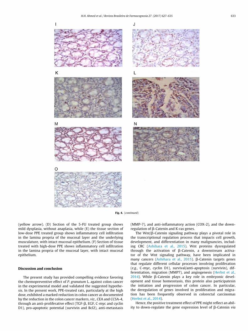
H.H. Ahmed et al. / Revista Brasileira de Farmacognosia 27 (2017) 627–635 633
(cont
(mlimtie
D
tisdbtD
Fig. 4.
yellow arrow). (D) Section of the 5-FU treated group showsild dysplasia, without anaplasia, while (E) the tissue section of
ow-dose PPE treated group shows inflammatory cell infiltrationn the lamina propria of the mucosal layer and the underlying
usculature, with intact mucosal epithelium. (F) Section of tissuereated with high-dose PPE shows inflammatory cell infiltrationn the lamina propria of the mucosal layer, with intact mucosalpithelium.
iscussion and conclusion
The present study has provided compelling evidence favoringhe chemopreventive effect of P. granatum L. against colon cancern the experimental model and validated the suggested hypothe-is. In the present work, PPE-treated rats, particularly at the high
ose, exhibited a marked reduction in colon cancer as documentedy the reduction in the colon cancer markers, viz., CEA and CCSA-4,hrough an anti-proliferative effect (TGF-�, EGF, C-myc and cyclin1), pro-apoptotic potential (survivin and Bcl2), anti-metastasisinued)
(MMP-7), and anti-inflammatory action (COX-2), and the down-regulation of �-Catenin and K-ras genes.
The Wnt/�-Catenin signaling pathway plays a pivotal role inthe transcriptional regulation process that impacts cell growth,development, and differentiation in many malignancies, includ-ing CRC (Ashihara et al., 2015). Wnt proteins dysregulatedthrough the activation of �-Catenin, a downstream activa-tor of the Wnt signaling pathway, have been implicated inmany cancers (Ashihara et al., 2015). �-Catenin targets genesthat regulate different cellular processes involving proliferation(e.g., C-myc, cyclin D1), survival/anti-apoptosis (survivin), dif-ferentiation, migration (MMP7), and angiogenesis (Herbst et al.,2014). While �-Catenin plays a key role in embryonic devel-opment and tissue homeostasis, this protein also participatesinthe initiation and progression of colon cancer. In particular,the deregulation of genes involved in proliferation and migra-
tion has been frequently observed in colorectal carcinomas(Herbst et al., 2014).Hence, the positive treatment effect of PPE might reflect an abil-ity to down-regulate the gene expression level of �-Catenin via

634 H.H. Ahmed et al. / Revista Brasileira de Farmacognosia 27 (2017) 627–635
Fig. 5. Representative photomicrographs of colon tissue with or without N-MNU-induced CRC. (A) Normal control section showing the normal histological features of themucosa, submucosa and muscularis layers, while (B) N-MNU untreated section shows dysplasia and anaplasia in the epithelial cells lining the glandular structure and its (C)magnification shows the mitotic activity of the nuclei (black arrows) and hyperchromasia (yellow arrow). (D) 5-FU treated section shows mild dysplasia, without anaplasia,w in them in the
tceWpabRaettocs(trPcctdpH
hile (E) section treated with low-dose PPE shows inflammatory cell infiltration
ucosal epithelium. (F) High-dose PPE section shows inflammatory cell infiltration
he attenuation of the Wnt signaling pathway in hepatocellulararcinoma (Bhatia et al., 2013). PPE active constituents, such asllagic acid (EA), have also been demonstrated to modulate thent signaling pathway through the inhibition of casein kinase, a
ositive regulator of the Wnt signaling pathway; EA also functionss a modulator of the interaction between �-Catenin and mem-ers of the �-Catenin destruction complex (Sharma et al., 2010).utin has antitumor effect through inducing G2/M cell cycle arrestnd promoting apoptosis and decreasing BCL2 expression (Chent al., 2013). Moreover, PPE accumulates in the G2/M phase ofhe cell cycle associated with the significant down-regulation ofhe C-myc gene (Adhami et al., 2009). Furthermore, puanicalagin,ne of the active anti-cancer components of PPE, inhibits humanolon cancer growth associated with the inhibition of cyclin D1 andurvivin expression through the Wnt/�-Catenin signaling cascadeTang et al., 2016). The suppression of �-Catenin translocation andhe subsequent expression of the target genes through PPE wereeflected in the histological examination of colon tissues throughPE-mediated protection against N-MNU-induced colon adenocar-inoma. In addition, PPE mediated apoptosis by enhancing Bcl2leavage, an effect presumably resulting from the action of this
reatment on the oncogenic Notch and Wnt pathways and theirownstream targets, viz., �-Catenin, C-myc, cyclin D1, cyclin B1,ERK, MMP-7, MMP-9 and EGF (Tao et al., 2002; Middha et al., 2013;erbst et al., 2014). Treatment with PPE extract also decreasedlamina propria of the mucosal layer and the underlying musculature, with intact lamina propria of the mucosal layer, with intact mucosal epithelium.
C-myc and COX-2, which are regulated through �-Catenin (Patelet al., 2008). Moreover, EA down-regulates the expression levels ofK-ras (González-Sarrías et al., 2009).
In the present study, the gene expression levels of �-Catenin,K-ras and C-myc in the colon tissues of the 5-FU treated ratsin the cancer group were down-regulated in compared with theuntreated cancer group. In addition, 5-FU substantially abatedthe levels of CEA, CCSA-4, TGF-�, EGF, cyclin D1, MMP-7, COX-2, Bcl2 and survivin. The activity of 5-FU primarily dependson intracellular delivery to the active metabolite, 5-fluoro-2′-deoxyuridine-5′-monophosphate, which inhibits DNA synthesisthrough the formation of a stable complex with thymidylate syn-thase (TS) in the presence of folates, followed by the initiation ofcell-cycle arrest or cell death (Kikuchi et al., 2009). TS decreasedBcl-2 expression (Longley et al., 2003), confirming the results ofthe present and previous studies. 5-FU triggers apoptosis in DN-HIF-transfected A549 cells via the reduction of sicyclin D1 (cyclinD1-specific interference RNA) and the downregulation of C-mycmRNA expression, phosphorylated C-myc in human colon cancerKM12C cells and survivin mRNA expression (Wen et al., 2010).Moreover, 5-FU significantly downregulated the expression of �-
Catenin protein and suppressed the Wnt canonical pathway (Refaatet al., 2015).In summary, this study demonstrated the antitumor activity ofP. granatum peel extract against colon cancer progression indicated

a de F
bwiamtiTt
E
Ptttl
Clp
Romo
A
lattyt
C
A
KIU
R
A
A
A
A
B
C
H.H. Ahmed et al. / Revista Brasileir
y decreasing colon cancer markers, viz., CEA and CCSA-4. PPE,hich is rich in multiple bioactive natural constituents, mediated
ts effect possibly by its anti-proliferative effect (TGF-�, EGF, C-mycnd cyclin D1), pro-apoptotic potential (survivin and Bcl2), anti-etastasis (MMP-7), and anti-inflammatory action (COX-2), and
he down-regulation of �-Catenin and K-ras genes. These effectsnvolve the modulation of the Wnt/�-Catenin signaling pathway.he study, hence, nominates the use of PPE as an additive on therapyo be studied in clinical trials.
thical disclosures
rotection of human and animal subjects. The authors declarehat the procedures followed were in accordance with the regula-ions of the relevant clinical research ethics committee and withhose of the Code of Ethics of the World Medical Association (Dec-aration of Helsinki).
onfidentiality of data. The authors declare that they have fol-owed the protocols of their work center on the publication ofatient data.
ight to privacy and informed consent. The authors havebtained the written informed consent of the patients or subjectsentioned in the article. The corresponding author is in possession
f this document.
uthors contributions
MBS (PhD student) contributed in collecting and running theaboratory work. HHA supervised the laboratory work. HHA, HSEnd NFA contributed in writing the manuscript. HHA and HSE con-ributed in designing the study, critical analysis of data, supervisedhe laboratory work. EAKH contributed to molecular and HPLC anal-sis. All the authors have read the final manuscript and approvedhe submission.
onflicts of interest
The authors declare no conflicts of interest.
cknowledgments
The authors express sincere appreciation to Prof. Adel Bakeerholoussy, Faculty of Veterinary Medicine, Cairo University; Prof.
brahim El-Garf, Department of Botany, Faculty of Science, Caironiversity and United Group Pharma Co. (Bader city, Cairo, Egypt).
eferences
dhami, V.M., Khan, N., Mukhtar, H., 2009. Cancer chemoprevention bypomegranate: laboratory and clinical evidence. Nutr. Cancer 61, 811–815.
hmed, H.H., Abdel-Rahman, M., Salem, F.E.H., Shalby, A.B., Lokman, M.S., 2013.Antitumor efficacy of Boswella serrata extract in management of colon cancerinduced in experimental animal. Int. J. Pharm. Pharmaceut. Sci. 5, 379–389.
shihara, E., Takada, T., Maekawa, T., 2015. Targeting the canonical Wnt/�-Cateninpathway in hematological malignancies. Cancer Sci. 106, 665–671.
ustinat, M., Dunsch, R., Wittekind, C., Tannapfel, A., Gebhardt, R., Gau-nitz, F., 2008. Correlation between beta-Catenin mutations and expressionof Wnt-signaling target genes in hepatocellular carcinoma. Mol. Cancer,http://dx.doi.org/10.1186/1476-4598-7-21.
hatia, D., Thoppil, R.J., Mandal, A., Samtani, K.A., Darvesh, A.S., Bishayee, A.,2013. Pomegranate bioactive constituents suppress cell proliferation and induceapoptosis in an experimental model of hepatocellular carcinoma: role ofWnt/�-Catenin signaling pathway. Evid Based Complement. Alternat. Med.,
http://dx.doi.org/10.1155/2013/371813.hen, H., Miao, Q., Geng, M., Liu, J., Hu, Y., Tian, L., Pan, J., Yang, Y., 2013.Anti-tumor effect of rutin on human neuroblastoma cell lines throughinducing G2/M cell cycle arrest and promoting apoptosis. Sci. World J.,http://dx.doi.org/10.1155/2013/269165.
armacognosia 27 (2017) 627–635 635
El-Toumy, S.A., Rauwald, H.W., 2002. Two ellagitannins from Punica granatum heart-wood. Phytochemistry 61, 971–974.
Eshak, M.G., Ghaly, I.S., Khalil, W.K.B., Farag, I.M., Ghanem, K.Z., 2010. Genetic alter-ations induced by toxic effect of thermally oxidized oil and protective role oftomatoes and carrots in mice. J. Am. Sci. 6, 175–188.
Fuentes-Calvo, I., Blázquez-Medela, A.M., Santos, E., López-Novoa, J.M., Martínez-Salgado, C., 2010. Analysis of k-ras nuclear expression in fibroblasts andmesangial cells. PLoS One 14, e8703.
González-Sarrías, A., Espín, J.C., Tomás-Barberán, F.A., García-Conesa, M.T., 2009.Gene expression, cell cycle arrest and MAPK signalling regulation in Caco-2 cellsexposed to ellagic acid and its metabolites, urolithins. Mol. Nutr. Food Res. 53,686–688.
Herbst, A., Jurinovic, V., Krebs, S., Thieme, S.E., Blum, H., Göke, B., Kolligs, F.T.,2014. Comprehensive analysis of �-Catenin target genes in colorectal carci-noma cell lines with deregulated Wnt/�-Catenin signaling. BMC Genomics,http://dx.doi.org/10.1186/1471-2164-15-74.
Kikuchi, M., Mikami, T., Sato, T., Tokuyama, W., Araki, K., Watanabe, M., Saigenji,K., Okayasu, I., 2009. High Ki67, Bax, and thymidylate synthase expressionwell correlates with response to chemoradiation therapy in locally advancedrectal cancers: proposal of a logistic model for prediction. Br. J. Cancer 101,116–123.
Kundu, J.K., Choi, K.Y., Surh, Y.J., 2006. beta-Catenin-mediated signaling: a novelmolecular target for chemoprevention with anti-inflammatory substances.Biochim. Biophys. Acta 1765, 14–24.
Larrosa, M., Yanéz-Gascón, M.J., Selma, M.V., González-Sarrías, A., Toti, S., Cerón,J.J., Tomás-Barberán, F., Dolara, P., Espín, J.C., 2009. Effect of a low dose ofdietary resveratrol on colon microbiota, inflammation and tissue damage in aDSS-induced colitis rat model. J. Agric. Food Chem. 57, 2211–2220.
Longley, D.B., Latif, T., Boyer, J., Allen, W.L., Maxwell, P.J., Johnston, P.G., 2003. Theinteraction of thymidylate synthase expression with p53-regulated signalingpathways in tumor cells. Semin. Oncol. 30, 3–9.
Middha, S.K., Usha, T., Pande, V., 2013. A review on antihyperglycemic and antihep-atoprotective activity of eco-friendly Punica granatum peel waste. Evid BasedComplement. Alternat. Med., http://dx.doi.org/10.1155/2013/656172.
Narisawa, T., Fukaura, Y., 2003. Prevention by intrarectal 5-aminosalicylic acid ofN-methylnitrosourea-induced colon cancer in F344 rats. Dis. Colon Rectum 46,900–903.
Orgil, O., Schwartz, E., Baruch, L., Matityahu, I., Mahajna, J., Amir, R., 2014. The antiox-idative and anti-proliferative potential of non-edible organs of the pomegranatefruit and tree. LWT-Food Sci. Technol. 58, 571–577.
Patel, R., Ingle, A., Maru, G.B., 2008. Polymeric black tea polyphenols inhibit1,2-dimethylhydrazine induced colorectal carcinogenesis by inhibiting cellproliferation via Wnt/beta-Catenin pathway. Toxicol. Appl. Pharmacol. 227,136–146.
Reeves, P.G., Nielsen, F.H., Fahey, G.C., 1993. AIN-93 purified diets for laboratoryrodents: final report of the American Institute of 365 Nutrition ad hoc writ-ing committee on the reformulation of the AIN-76A rodent diet. J. Nutr. 123,1939–1951.
Refaat, B., El-Shemi, A.G., Kensara, O.A., Mohamed, A.M., Idris, S., Ahmad, J., Khojah,A., 2015. Vitamin D3 enhances the tumouricidal effects of 5-fluorouracil throughmultipathway mechanisms in azoxymethane rat model of colon cancer. J. Exp.Clin. Cancer Res. 34, 71.
Sharma, M., Li, L., Celver, J., Killian, C., Kovoor, A., Seeram, N.P., 2010. Effects of fruitellagitannin extracts, ellagic acid, and their colonic metabolite, urolithin A, onWnt signaling. J. Agric. Food Chem. 58, 3965–3969.
Shin, J.W., Seol, I.C., Son, C.G., 2010. Interpretation of animal dose and human equiv-alent dose for drug development. J. Korean Oriental Med. 31, 1–7.
Shirode, A.B., Kovvuru, P., Chittur, S.V., Henning, S.M., Heber, D., Reliene, R., 2014.Antiproliferative effects of pomegranate extract in MCF-7 breast cancer cells areassociated with reduced DNA repair gene expression and induction of doublestrand breaks. Mol. Carcinog. 53, 458–470.
Srimuangwong, K., Tocharus, C., Tocharus, J., Suksamrarn, A., Chintana, P.Y.,2012. Effects of hexahydrocurcumin in combination with 5-fluorouracil ondimethylhydrazine-induced colon cancer in rats. World J. Gastroenterol. 18,6951–6959.
Tang, J.M., Min, J., Li, B.S., Hong, S.S., Liu, C., Hu, M., Li, Y., Yang, J., Hong, L., 2016.Therapeutic effects of punicalagin against ovarian carcinoma cells in associationwith �-Catenin signaling inhibition. Int. J. Gynecol. Cancer 26, 1557–1563.
Tao, L., Kramer, P.M., Wang, W., Yang, S., Lubet, R.A., Steele, V.E., Pereira, M.A., 2002.Altered expression of c-myc, p16 and p27 in rat colon tumors and its rever-sal by short-term treatment with chemopreventive agents. Carcinogenesis 23,1447–1454.
Vijay, M., Sivagami, G., Thayalan, K., Nalini, N., 2016. Radiosensitizing potential ofrutin against human colon adenocarcinoma HT-29 cells. Bratisl. Lek. Listy. 117,171–178.
Watson, S.A., Michael, D., Justin, T.A., Grimes, S., Morris, T.M., Robinson, G., Clarke,P.A., Hardcastle, J.D., 1998. Pre-clinical evaluation of the gastrimmune immuno-gen alone and in combination with 5-fluorouracil/leucovorin in a rat colorectalcancer model. Int. J. Cancer 75, 873–877.
Wen, W., Ding, J., Sun, W., Wu, K., Ning, B., Gong, W., He, G., Huang, S., Ding, X.,Yin, P., Chen, L., Liu, Q., Xie, W., Wang, H., 2010. Suppression of cyclin D1 by
hypoxia-inducible factor-1 via direct mechanism inhibits the proliferation and5-fluorouracil-induced apoptosis of A549 cells. Cancer Res. 70, 2010–2019.Zhao, Y., Miao, G., Li, Y., Isaji, T., Gu, J., Li, J., Qi, R., 2014. Microrna 130b suppressesmigration and invasion of colorectal cancer cells through downregulation ofintegrin �1. PLoS One 9, e87938.