Ptosis surgery
23
Ptosis Management Jagdish Dukre
-
Upload
jagdish-dukre -
Category
Health & Medicine
-
view
115 -
download
1
Transcript of Ptosis surgery

Ptosis Management
Jagdish Dukre

TREATMENT Non-surgical – Rehabilitative crutch glassesSurgical - Definitive Treatment
Decision making
When to operate Which procedure
concern is cosmetic any age concern is amblyopia early surgerysquint has to be operated firstblepharophimosis, telecanthus, epicanthus operated first
Depends on levator function + associated anomaly
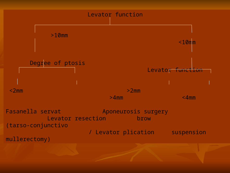
Levator function
>10mm <10mm
Degree of ptosis Levator function
<2mm >2mm >4mm <4mm
Fasanella servat Aponeurosis surgery Levator resection brow (tarso-conjunctivo / Levator plication suspension mullerectomy)

MODIFIED FASANELLA – SERVAT- Involves tarso-conjunctivo-mullerectomy - Best results upto 2mm congenital ptosis with minimum 10mm levator action - 2mm tarsectomy performed for every 1mm ptosis- Used for mild neurogenic or myogenic ptosis & residual ptosis following levator resection procedure - ANESTHESIA – Local or General
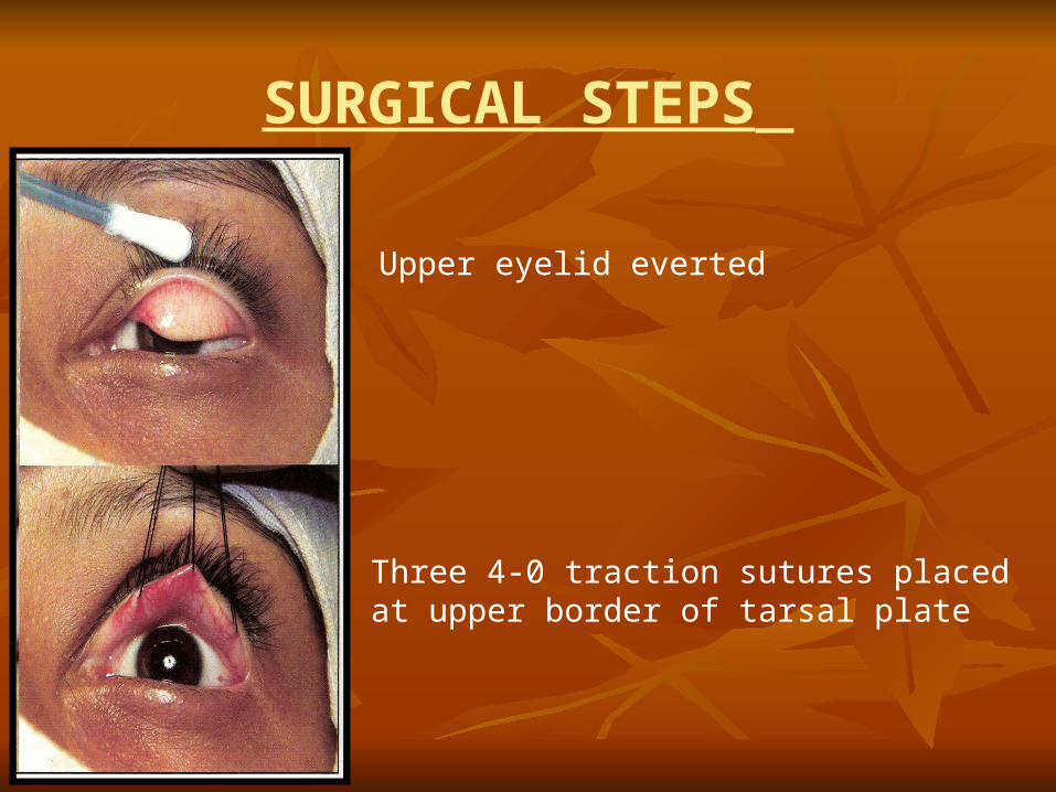
SURGICAL STEPS
Upper eyelid everted
Three 4-0 traction sutures placed at upper border of tarsal plate

Required amount of tarsal resection marked over everted tarsus staying parallel to eyelid margin.
Tarsus marked along line of excision with monopolar cautery.
Full thickness excision performed along entire extend.

Skin suture placed with 6-0 catgut at medial end of tarsal excision & needle brought out on conjunctival side to close tarsal wound.
Tarsal wound closed with continuous 6-0 catgut suture.
Suture end finally exteriorized on to eyelid skin at lateral end for final knot.
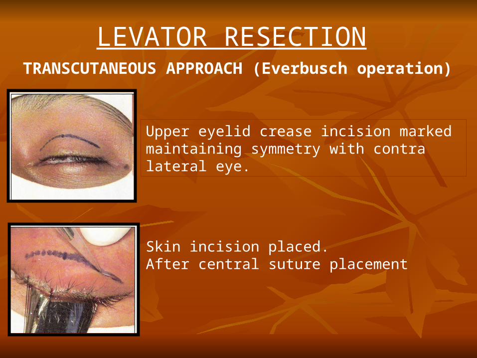
LEVATOR RESECTION
TRANSCUTANEOUS APPROACH (Everbusch operation)
Upper eyelid crease incision marked maintaining symmetry with contra lateral eye.
Skin incision placed.After central suture placement
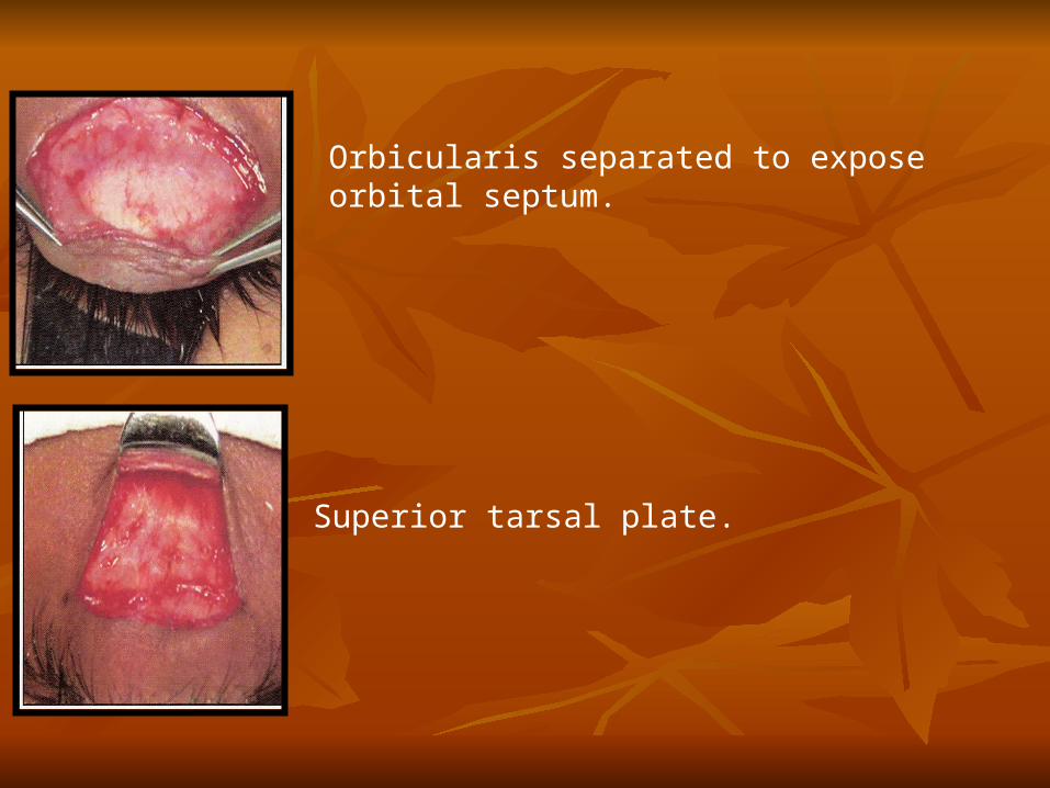
Orbicularis separated to expose orbital septum.
Superior tarsal plate.
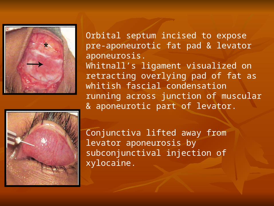
Orbital septum incised to expose pre-aponeurotic fat pad & levator aponeurosis.Whitnall’s ligament visualized on retracting overlying pad of fat as whitish fascial condensation running across junction of muscular & aponeurotic part of levator.
Conjunctiva lifted away from levator aponeurosis by subconjunctival injection of xylocaine.
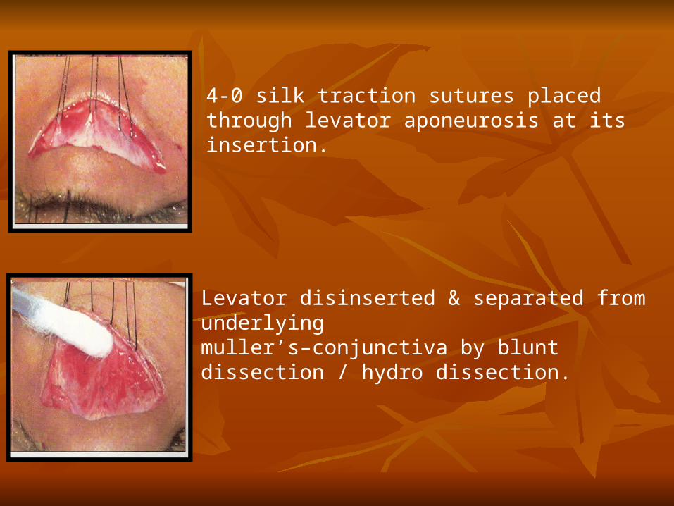
4-0 silk traction sutures placed through levator aponeurosis at its insertion.
Levator disinserted & separated from underlying muller’s–conjunctiva by blunt dissection / hydro dissection.

Medial & Lateral horn’s cut if necessary.
Three cardinal sutures passed from tarsus through levator muscle at desired height.Sutures are tightened with temporary knot’s to achieve predetermined lid height & natural contour.Placement of upper eyelid sutures during surgery based on levator action
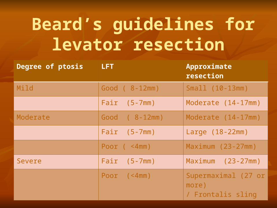
Beard’s guidelines for levator resection
Degree of ptosis LFT Approximate resection
Mild Good ( 8-12mm) Small (10-13mm)
Fair (5-7mm) Moderate (14-17mm)
Moderate Good ( 8-12mm) Moderate (14-17mm)
Fair (5-7mm) Large (18-22mm)
Poor ( <4mm) Maximum (23-27mm)
Severe Fair (5-7mm) Maximum (23-27mm)
Poor (<4mm) Supermaximal (27 or more)/ Frontalis sling
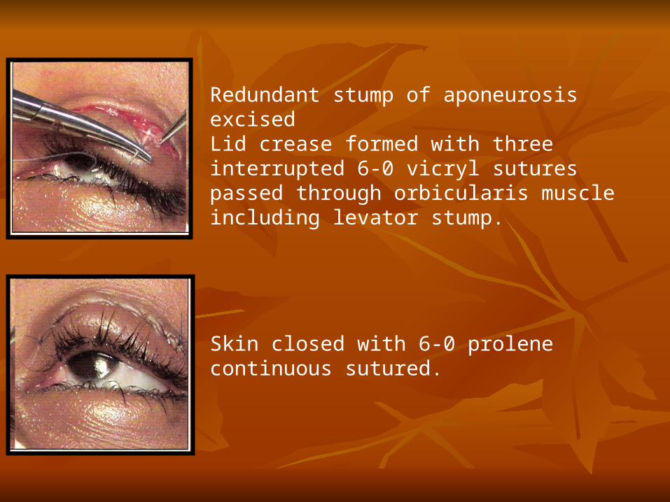
Redundant stump of aponeurosis excisedLid crease formed with three interrupted 6-0 vicryl sutures passed through orbicularis muscle including levator stump.
Skin closed with 6-0 prolene continuous sutured.

TARSOFRONTALIS SLING Ideal procedure for bilateral moderate to severe ptosis with poor
levator action.Unilaterally in rare situations – Following levator excision for marcus– Gunn phenomenon - Severe unilateral ptosis with poor levator action.
Performed with- 1. Non absorbable synthetic materials – 3-0 ethibond suture, 3-0 prolene suture, silicon material. 2. Autogenous fascia lata/Temporalis fascia - Best sling material.Performed under local / general anaesthesia
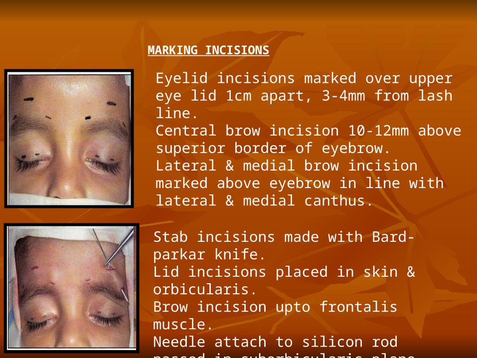
Eyelid incisions marked over upper eye lid 1cm apart, 3-4mm from lash line.Central brow incision 10-12mm above superior border of eyebrow.Lateral & medial brow incision marked above eyebrow in line with lateral & medial canthus.
MARKING INCISIONS
Stab incisions made with Bard-parkar knife.Lid incisions placed in skin & orbicularis.Brow incision upto frontalis muscle. Needle attach to silicon rod passed in suborbicularis plane from central brow incision to lateral brow incison.
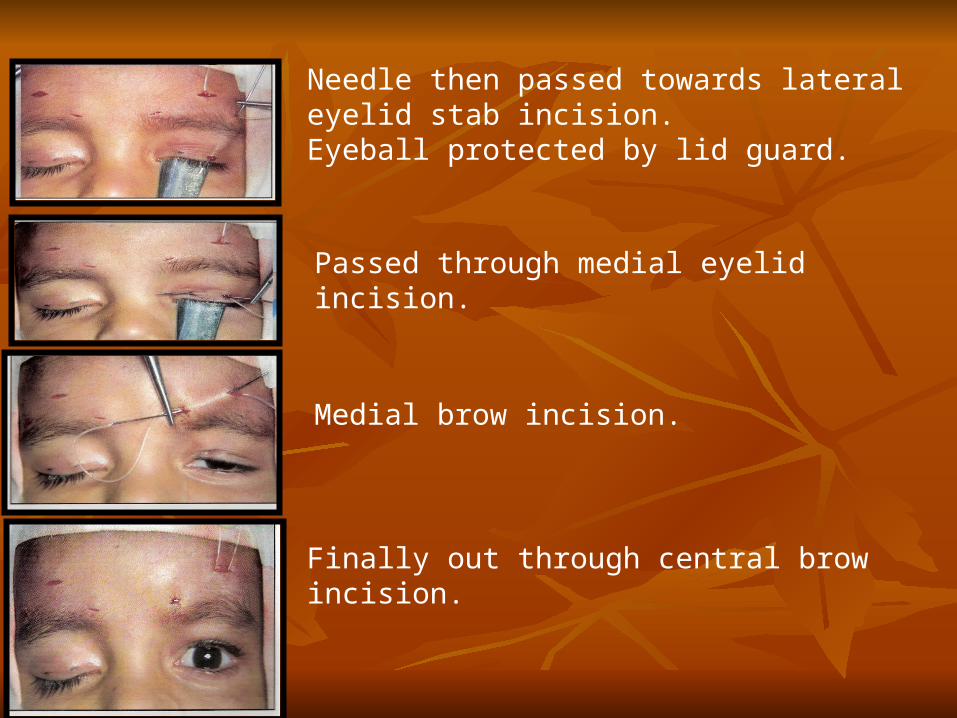
Needle then passed towards lateral eyelid stab incision.Eyeball protected by lid guard.
Passed through medial eyelid incision.
Medial brow incision.
Finally out through central brow incision.
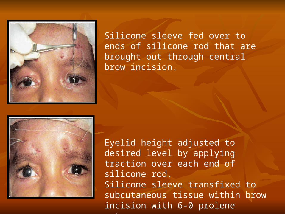
Silicone sleeve fed over to ends of silicone rod that are brought out through central brow incision.
Eyelid height adjusted to desired level by applying traction over each end of silicone rod.Silicone sleeve transfixed to subcutaneous tissue within brow incision with 6-0 prolene sutures.
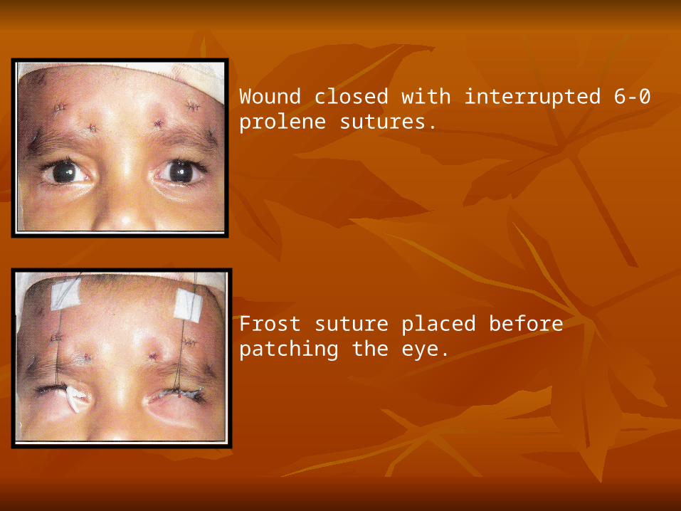
Wound closed with interrupted 6-0 prolene sutures.
Frost suture placed before patching the eye.

COMPLICATIONS OF PTOSIS SURGERY1. Under correction – Most frequent after congenital ptosis surgery• May improve as edema subsides• Persistent under correction – requires repeat surgery after 4-6
months. • Obviously under corrected eyelid - corrected within a week or
two immediately after surgery .
2. Over correction – Following surgery for acquired ptosis Rare in congenital ptosis Treatment - Various methodsDownward traction over eyelid with forceps (requires L.A)Surgical correction

3. Localized lid contour abnormalities
Treatment – Loosening cardinal suture for localized peaking Further small levator resection for localized flattening
4. Lid Crease Abnormalities – Absence of crease improper position overhanging skin Treatment – may require re-formation of eyelid crease at desired level
5. Conjunctival Prolapse – occur if forniceal attachment of levator disturbed during post dissection Improves without treatment May require fornix formation suture Excision of excess conjunctiva in some cases

6. Lagophthalmos - d/t - improper dissection of LPS aponeurosis from surrounding structures. - Overresection of LPS. - Tt- Massage Tarsotomy Levator recession
7. Exposure Keratitis - d/t overcorrection, lagophthalmos, decreased tear production or poor Bell’s phenomenon. - Treatment – Artificial tear substitute Punctal occlusion Tarsorrhaphy
8. Recurrence following surgery

THANK YOU…


![Index [link.springer.com]978-0-387-92855-5/1.pdf · Bell’s palsy acquired myogenic ptosis, 95 congenital myogenic ptosis, 79 involutional ptosis, 74 patient selection, 156 prior](https://static.fdocuments.in/doc/165x107/5e0ec41cf5a39e518c0f1033/index-link-978-0-387-92855-51pdf-bellas-palsy-acquired-myogenic-ptosis.jpg)


![MS And DO Ophthalmology Question Bank - Yolakumarsaurabh.yolasite.com/resources/Question Bank.pdfMS And DO Ophthalmology Question Bank ... surgery in a case of simple ptosis. [1986]](https://static.fdocuments.in/doc/165x107/5ac8dc2e7f8b9a42358cbaaa/ms-and-do-ophthalmology-question-bank-bankpdfms-and-do-ophthalmology-question.jpg)