prva verzija- amf-r is a valid target for cancer treatment
23
1 A Role for KAI1 in Promotion of Cell Proliferation and Mammary Gland Hyperplasia by the Gp78 Ubiquitin Ligase Bharat Joshi, Lei Li and Ivan R. Nabi * Department of Cellular and Physiological Sciences, Life Sciences Institute, University of British Columbia, Vancouver, BC, Canada V6T 1Z3 *Corresponding author: Department of Cellular and Physiological Sciences Life Sciences Institute University of British Columbia 2350 Health Sciences Mall Vancouver, BC V6T 1Z3 Canada (604) 822-7000 [email protected] Running title : Gp78 overexpression induces hyperplasia Key words : gp78/AMFR, transgenic, mammary gland, KAI1, hyperplasia http://www.jbc.org/cgi/doi/10.1074/jbc.M109.074344 The latest version is at JBC Papers in Press. Published on January 20, 2010 as Manuscript M109.074344 Copyright 2010 by The American Society for Biochemistry and Molecular Biology, Inc. by guest on January 30, 2018 http://www.jbc.org/ Downloaded from
-
Upload
nguyenliem -
Category
Documents
-
view
216 -
download
0
Transcript of prva verzija- amf-r is a valid target for cancer treatment

1
A Role for KAI1 in Promotion of Cell Proliferation and Mammary Gland Hyperplasia
by the Gp78 Ubiquitin Ligase
Bharat Joshi, Lei Li and Ivan R. Nabi*
Department of Cellular and Physiological Sciences, Life Sciences Institute, University of British Columbia, Vancouver, BC, Canada V6T 1Z3 *Corresponding author: Department of Cellular and Physiological Sciences Life Sciences Institute University of British Columbia 2350 Health Sciences Mall Vancouver, BC V6T 1Z3 Canada (604) 822-7000 [email protected] Running title: Gp78 overexpression induces hyperplasia Key words: gp78/AMFR, transgenic, mammary gland, KAI1, hyperplasia
http://www.jbc.org/cgi/doi/10.1074/jbc.M109.074344The latest version is at JBC Papers in Press. Published on January 20, 2010 as Manuscript M109.074344
Copyright 2010 by The American Society for Biochemistry and Molecular Biology, Inc.
by guest on January 30, 2018http://w
ww
.jbc.org/D
ownloaded from

2
SUMMARY Expression of gp78, an E3 ubiquitin ligase
in endoplasmic reticulum-associated degradation (ERAD), is associated with tumor malignancy. To study gp78 overexpression in mammary gland development and tumorigenicity, we generated MMTV-LTR driven gp78 transgenic mice. Embryos carrying the gp78 transgene cassette were implanted in FVB surrogate mothers and two founders with high copy integration showed elevated gp78 expression at both transcript and protein levels at virgin-stage and twelve days gestation. Transgenic mammary glands showed increased ductal branching, dense alveolar lobule formation and secondary terminal end bud development. BrdU staining showed increased proliferation in hyperplastic ductal regions at virgin-stage and twelve days gestation compared to wild-type mice. Reduced expression of the metastasis suppressor KAI1, a gp78 ERAD substrate, demonstrates that gp78 ubiquitin ligase activity is increased in MMTV-gp78 mammary gland. Similarly, metastatic MDA-435 cells exhibit increased gp78 expression, decreased KAI1 expression and elevated proliferation compared to non-metastatic MCF7 cells whose proliferation was enhanced upon knockdown of KAI1. Importantly, stable gp78 knockdown HEK293 cells showed increased KAI1 expression and reduced proliferation that was rescued upon KAI1 knockdown demonstrating that gp78 regulation of cell proliferation is mediated by KAI1. Mammary tumorigenesis was not observed in repeatedly pregnant MMTV-LTR-gp78 transgenic mice over a period of 18 months post-birth. Elevated gp78 ubiquitin ligase activity is therefore not sufficient for mammary tumorigenesis. However, the hyperplastic phenotype observed in mammary glands of MMTV-gp78 transgenic mice identifies a novel role for gp78 expression in enhancing mammary epithelial cell proliferation and non-tumorigenic ductal outgrowth.
INTRODUCTION Gp78, also called autocrine motility factor
receptor (AMFR), is an E3 ubiquitin ligase implicated in ER-associated degradation (ERAD) that also has a dual function as a cell surface
cytokine receptor for autocrine motility factor/phosphoglucose (1). Gp78 overexpression is closely linked to tumor malignancy and human cancer (2) and gp78 was identified as one of the 189 most mutated genes in breast and colon cancers (3). Gp78 expression correlates with aggressive tumor biology and poor outcome for malignancies of the lung, tongue, esophagus, stomach, colon, rectum, liver, breast, thymus and skin (2). Notably, in bladder, colorectal, gastric, skin and esophageal cancers (4-8), gp78 is either not expressed or expressed at significantly reduced levels in adjacent normal tissue. However, while overexpression of gp78 in tumors is well-documented, it has yet to be determined whether gp78 overexpression contributes to tumor progression.
Substrates of gp78 E3 ubiquitin ligase activity include CD3-delta, the T cell receptor, ApoB lipoprotein, HMG CoA reductase, cystic fibrosis transmembrane conductance regulator (CFTR), and the metastasis suppressor KAI1 (9-13). KAI1 (CD82) is a tetraspanin whose expression is lost in advanced stages of many human malignancies; however, the mechanism of action of this suppressor remains unknown (14,15). The recent identification of KAI1 as a gp78 ERAD substrate functionally links gp78 ubiquitin ligase activity in ERAD to its established role in metastasis (16).
In human breast cancer, ~25% of 370 primary invasive breast carcinomas showed membranous and cytoplasmic gp78 labelling although gp78 positivity did not correlate with prognosis (17). In order to further evaluate the role of gp78 in breast cancer progression, we generated a transgenic MMTV-gp78 mouse model overexpressing gp78 in the mammary gland. Analysis of gp78 transgenic mammary glands showed that gp78 expression induces a hyperplastic phenotype, increased ductal branching and dense alveolar lobule formation as well as down-regulation of the KAI1 metastasis suppressor. Using a knockdown approach in vitro, KAI1 was found to mediate gp78 regulation of cell proliferation defining a functional relationship between this E3 ubiquitin ligase and its substrate in cell proliferation.
by guest on January 30, 2018http://w
ww
.jbc.org/D
ownloaded from

3
EXPERIMENTAL PROCEDURES Antibodies, reagents and animals
Rat anti-gp78 3F3A monoclonal antibody was as previously described (18) and mouse anti-gp78 (ab54787-100) was purchased from Abcam. Rabbit monoclonal anti-Ki67 (Clon-SP6) was from Medicorp, rabbit anti-KAI1 (sc-1087) and anti-cyclin-D1 were from Santa Cruz, and mouse anti-β-actin and HRP conjugated anti-rabbit or anti-mouse secondary antibodies were from Sigma. Glucocorticoid analogue dexamethasone and MTT reagent (Thiazolyl Blue Tetrazolium Bromide) were from Sigma. DIG probe labeling, probing kit and BrdU were from Roche Diagnostics. Animals were maintained at the British Columbia Cancer Agency (BCCA) facility following guidelines and protocols of the University of British Columbia Animal Care Committee. All the animals in both wild-type and transgenic groups were between 8-10 weeks of age and a minimum of four animals from several litters was used for the assessment. Plasmids and siRNAs
MMTV-LTR plasmid (19-21) was obtained from the H. Lee Moffitt mouse model core facility. Mouse gp78 cDNA (22) was amplified using the following set of primers (5’GAA TTC ATG CCG CTG CTC TTC CTC GAG CGC3’ and 5’GAA TTC CTA GGT TGT CCG TTG CCT CTG AAG3’). Amplified cDNA was TA-cloned into pCRII (Invitrogen) and subsequently sub-cloned into the MMTV-LTR plasmid at the EcoRI site in the third exon of the rabbit β-globin gene that ends with BGH-polyA sequence. Final plasmid was sequenced to verify ligation junctions and cDNA integrity.
pcDNA6.2-GW/+EmGFP-miR was obtained from Invitrogen. Non-specific, non-targeting control (D-001810-10-05) as well as human CD82 (KAI1) (L-003901-00) specific siRNAs pools were purchased from Dharmacon.
Cell lines and transfection
MCF7 and MDA-435 cells were obtained from ATCC and maintained in RPMI medium supplemented with 10% FBS, 1% penicillin/streptomycin and 1% L-glutamate as described (17). HEK293 cells were maintained in DMEM medium supplemented with 10% FBS, 1%
penicillin/streptomycin and 1% L-glutamate. MMTV-LTR-gp78 or pCDNA were transiently transfected into MCF7 cells and cultured for 24 to 48 hours in the absence or presence of dexamethasone (1 mg/ml final concentration) prior to harvesting.
In order to generate gp78 knockdown stable cell lines, pre-miRNA sequences for gp78 and non-targeting control miRNAs were cloned into pcDNA6.2-GW/+EmGFP-miR (Invitrogen, USA). The microRNA nucleotide sequence targeting human gp78 (GenBank Accession number: NM_001144.4) corresponds to amino acids 293-300 relative to the initiating methionine codon. HEK293 cells were transfected with either control or gp78 miRNA vectors and selected for about two weeks against 5µg/ml blasticidin. GFP positive clones were sorted by FACS and gp78 downregulation was confirmed by western blotting using 3F3A antibody.
Control and KAI1 specific siRNA smart pools were transiently transfected in to MCF7 or HEK293 stable cell lines using Lipofectamine 2000 (Invitrogen) following the protocol described before (23). KAI1 knockdown was assessed by western blotting.
DNA preparation and microinjection
All experiments involving mice were performed following the guidelines and regulations of the University of British Columbia’s Animal Care Committee. MMTV-LTR-gp78 plasmid was digested with NruI-AatII and trimmed off the residual backbone leaving the MMTV-LTR promoter, gp78 cDNA embedded in to β-globin exons/intron and BGH-polyA tail. The released cassette was extracted from agarose gel and purified for microinjection using GeneClean (MP Biomedicals, LLC). The DNA was re-suspended to a final concentration of 2.5 ng/µl in TE buffer (10 mM Tris-HCL pH 7.5, 1mM EDTA). Transgenic lines were generated by DNA microinjection of super-ovulated 4 week old FVB/Ntac zygotes at the BC Cancer Agency transgenic facility. Surviving zygotes were transferred to pseudo pregnant ICR females that gave birth to pups 20 days post-implantation. Tail snips of pups were collected two weeks after birth and used for genomic DNA extraction.
by guest on January 30, 2018http://w
ww
.jbc.org/D
ownloaded from

4
Genotyping by PCR and Southern blotting Genomic DNA was extracted from mouse
tails following standard protocols. Briefly, tails were digested overnight at 55°C in buffer (10mM Tris-HCl, pH 8.0, 50mM NaCl, 25mm EDTA, pH 8.0 and 0.5% SDS) containing 0.5mg/ml proteinase K. Digested proteins and debris were separated from DNA using 6M saturated NaCl and centrifugation. Subsequently, ethanol-precipitated DNA from the supernatant was re-suspended in 50µl sterile TE buffer. Purified DNA (1µl) was PCRed using a forward primer designed from the MMTV-LTR promoter (5’TTG CCC AAC CTT GCG GTT CCC AG 3’) and a reverse primer from gp78 cDNA (5’GCT GAG GCC CGT GTA GGT GCG3’). Genomic DNA obtained from the wild type FVB/N mouse was used as a negative control and the MMTV-LTR-gp78 plasmid was used as a positive control. The 1.5 kb PCR product was separated on 1% agarose gels. Genotyping or verification of copy number integration of MMTV-LTR-gp78-BGH-polyA cassette was confirmed by southern blotting. Briefly 10 µg of genomic DNA isolated from mouse tails was restriction digested overnight with high-concentrate KpnI (NEB). Restriction digested DNAs were phenol extracted, ethanol precipitated and re-suspended in sterile RNase/DNase free PCR water (Invitrogen). KpnI digested genomic DNA fragments were separated on 0.8% agarose gel, depurinated, denatured, neutralized and capillary blotted overnight in 10X SSC onto Zeta-probe charged nylon membrane following the manufacturer’s protocol (BioRad), and then UV cross linked (UV Stratalinker 2400, Stratagene).
DIG labelled probe was prepared and optimized using the PCR DIG probe synthesis kit (Roche). Briefly, PCR was done using the following set of primers (5’CCT CTG CTA ACC ATG TTC ATG3’, 5’GCT GAG GCC CGT GTA GGT GCG3’) and MMTV-LTR-gp78 plasmid as a template to generate a ~550bp product. The probe was stored at -20°C until required.
Pre-hybridization, hybridization and immune-probing of membrane were performed using manufacturer’s protocol supplied with DIG hybridization kit (Roche). The membrane was exposed to autographic film and signal was developed.
Tissue harvest Mice were killed by CO2 asphyxiation.
Age and estrous cycle matched, superovulated female mice were injected with 100µg/g body weight BrdU in PBS (Sigma) 2 hours prior to sacrifice. The right 3rd thoracic and 4th inguinal mammary glands were fixed in 10% formalin for 48-72 hours and processed for paraffin embedding. Tissue embedding and sections (5 µm) were prepared by UBC Hospital Histopathology Core or Wax-IT Histology (Vancouver, Canada) and stained with hematoxylin and eosin. The left 3rd and 4th glands were whole mounted, fixed overnight in Carnoy’s fixative (60% absolute alcohol, 30% chloroform and 10% glacial acetic acid), and defatted through 3 changes of acetone. Specimens were rehydrated and stained overnight in 0.2% carmine, 0.5% aluminum potassium sulfate. Slides were dehydrated to absolute ethanol, cleared in xylene, permanently mounted (CC/Mount, Sigma), and imaged using dissection microscope. RT-PCR
Total RNA was isolated from MCF7 cells or mouse mammary gland (pubertal virgin (8 weeks old) or 12 day mid-gestation wild-type or transgenic) using Trizol (Invitrogen) following the protocol supplied with reagent. Integrity of the total RNA was assessed on formaldehyde gel prior to using them. Reverse transcription and semi-quantitative cDNA synthesis was carried out using Superscript double stranded cDNA synthesis kit (Invitrogen) following the supplied protocol and the following set of primers: (5’CCT CTG CTA ACC ATG TTC ATG3’, 5’GCT GAG GCC CGT GTA GGT GCG3’). Lysate preparation and western blotting
For western blotting, cells were grown to 80% confluency, washed in cold PBS and cell lysates separated on SDS-PAGE, transferred to nitrocellulose membranes and western blotted for desired proteins as described previously (17). For tissues, approximately 10 mg of wild-type or transgenic mammary glands were manually homogenized in 1X SDS-PAGE loading buffer, boiled for five minutes and centrifuged at 14,000 rpm for ten minutes at 4° C. Supernatants were collected and equal protein amounts were loaded on 10% acrylamide gels; proteins were separated,
by guest on January 30, 2018http://w
ww
.jbc.org/D
ownloaded from

5
transferred to nitrocellulose membrane and western blotted for target proteins (gp78, KAI1 and β-Actin).
Immunohistochemistry
Paraffin sections were rehydrated with PBS and processed using one of the following protocols. Sections to be stained with anti-gp78 antibody (3F3A), anti-KAI1 (Santa Cruz) or anti-Ki67 (Medicorp) were rinsed in dH2O and subjected to antigen retrieval in Citrate Buffer (4.5 mL of 0.1M Citric Acid; 20.5 mL of 0.1M sodium citrate; fill up to 250 ml with dH2O; pH 5.6) for about 30 minutes at 90-98°C (24,25). Slides were cooled for 20 minutes, rinsed 3 times PBS, and incubated for 30 minutes with 0.3% H2O2 and subsequently 30 minutes with DAKO protein-block reagent. Sections were incubated at 4°C overnight in primary antibody (3F3A 1/20) prepared in DAKO dilution buffer. Next day slides were subjected to several washes with PBS-Tween-20 (0.1%) and stained with secondary anti-rat HRP (Vector) antibody (1/100 in DAKO dilution buffer) for 1 hour at room-temperature; subsequently washed 3 times with PBS-Tween-20 on shaker. Slides were incubated with strepavidin-peroxidase conjugate DAKO LSAB + HRP (K0690, Vector) washed and stained with Nova-Red (SK-4800, Vector). Slides were counter-stained with Haematoxylin (Gill 1) for 20 seconds, washed, Xylene cleaned and mounted. For BrdU injected mice, mammary glands were isolated and paraffin embedded, and sections of 5µm thickness were used for BrdU staining. 5-bromo-2’-deoxy-uridine labelling and detection kit II from Roche Diagnostics Corporation was used for detecting BrdU labelled proliferating cells. The slides were counterstained with hematoxylin and the data represent the mean value of total cell nuclei versus BrdU positive cell nuclei from three microscopic fields (10X) in tissue slides from three different animals. All slides were imaged using 10X, 20X and 63X Plan-Apochromat objectives of a Zeiss Axioskop microscope.
MTT assay
Cell proliferation assays were performed as described previously (23). Briefly, 10,000 cells were seeded on 96 well plates for 48-72 hours, prior to adding MTT reagent diluted 1:10 from 5
mg/ml stock in RPMI (MCF7; MDA-435) or DMEM (HEK293) medium after which the cells were allowed to grow in a CO2 incubator in the dark for 4-5 hours. Medium was decanted, precipitates of metabolite (Formazan) dissolved in 200µl DMSO and absorbance measured at 570nm. Background absorbance in the presence of DMSO was subtracted, normalized and subjected to statistical analysis. For siRNA knockdown experiments, cells were transiently transfected with control and target specific siRNAs in 96 well plates and incubated for 48 hours prior to performing the MTT assay. RESULTS Preparation of the MMTV-LTR-gp78 plasmid and in vitro validation
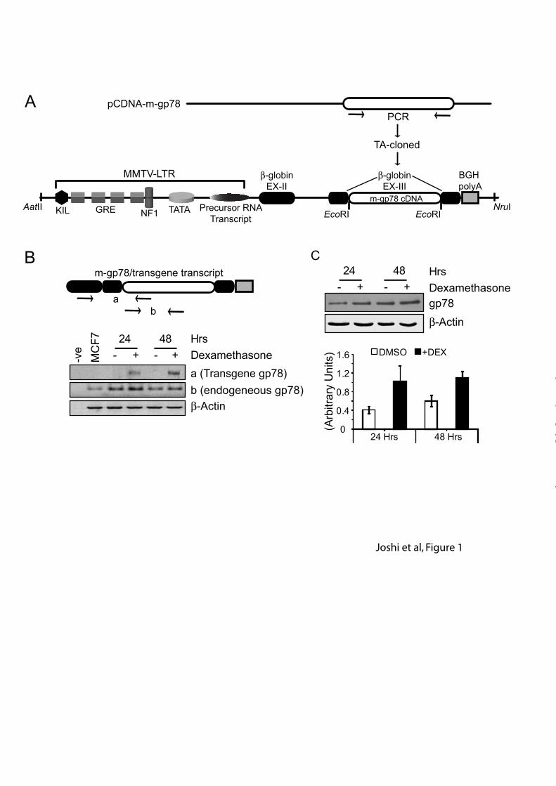
In order to overexpress gp78 in the mammary gland, we placed gp78 cDNA downstream of the MMTV-LTR promoter gene at the EcoRI site in exon3 of the rabbit β-globin gene followed by the BGH polyA sequence that facilitates termination of transcription (Figure 1 A). The MMTV-LTR-gp78 plasmid was validated in vitro prior to generating transgenic animals. The MMTV-LTR promoter carries several glucocorticoid (GC) response elements (GRE) that can also be turned on by the GC analogue dexamethasone (Figure 1A). MMTV-LTR-gp78 was transiently transfected into breast carcinoma MCF7 cells that express GC receptors and internalize dexamethasone, and treated with DMSO or 1 mg/ml dexamethasone for 24 to 48 hours. Cells were harvested for RNA as well as protein samples and subjected to RT-PCR or western blot analysis. As seen in Figure 1B, the presence of chimeric transcript is selectively observed in dexamethasone treated cells in both 24 and 48 hours samples and is absent in control DMSO treated samples. Total transcript levels are elevated in dexamethasone treated cells relative to controls. Similarly, when assessed for gp78 protein level by western blotting with the 3F3A anti-gp78 antibody (18), dexamethasone induced cells showed elevated gp78 protein levels (Figure 1C). These results suggest that in expressing cells, the MMTV-LTR-gp78 plasmid is responsive to the GC analogue dexamethasone and that gp78 overexpression is induced in a controlled fashion.
by guest on January 30, 2018http://w
ww
.jbc.org/D
ownloaded from

6
Generating gp78 overexpressing transgenic mouse
To generate transgenic mice, the cassette of 5.5kb size containing MMTV-LTR promoter-gp78 cDNA with β-globin exon-intron and BGH polyA was released using AatII-NruI restriction digestion (Figure 2A). The purified target fragment was microinjected into super-ovulated 4 week old FVB/Ntac zygotes and transferred to pseudo-pregnant ICR females. 20 day post-implantation females gave birth to pups and at two weeks of age, tail snips were collected and subjected to genomic DNA extraction and genotyping. For assessing complete cassette integration, we chose two different areas (a,b) for PCR genotyping (Figure 2B Schematic). Primer set (a) identified seven founders (of 40 mice tested; 18% efficiency) containing transgenic DNA (Figure 2B, left panel). Of these, six founders had intact cassette integration when assessed with the other set (b) of primers (Figure 2B, right panel) while one appeared to have lost part of the DNA during recombination.
Although, MMTV-LTR promoter is driven by GC, some reports have described an impact of copy number integration on protein expression (26). We therefore assessed which of the six positive founders had an intermediate to high copy number of transgene cassettes integrated using a PCR-generated DIG-labelled primer corresponding to primer set (c) (Figure 2B) that was optimized and found to run heavier on gel compared to non-labelled probe (Figure 2C, left panel). In order to check head-to-tail multi-copy integration of cassette, the unique KpnI site in the cassette was used for restriction digestion (Figure 2C, Schematic) of genomic DNAs of six founders and FVB as control. Founder lines 236RF (lane 1) and 235RLF (lane 2) carried higher copy numbers of the transgene cassette compared to other founder lines (lanes 3-6) (Figure 2C, right panels). Negative control FVB did not show the presence of any band whereas positive control MMTV-LTR-gp78 cassette showed hybridization to a band of approximately 5.3 kb.
Transgene gp78 overexpression and its impact on KAI1 expression
The 235RLF founder line was chosen for further analysis. In order to assess transgene expression, total RNA was isolated from age and
estrous cycle matched, superovulated pubertal virgin (8 weeks old) or mid-gestation pregnant (12 days) wild type or transgenic mammary glands. Using primer sets for regions ‘A’ and ‘B’ (Figure 1B Schematic), semi-quantitative RT-PCR was performed on total cDNA generated from extracted RNAs. This facilitated identification of the expression of transgene gp78 (primer set A) as well as total gp78 (primer set B) in both wild type and transgenic mouse. Transgene gp78 was expressed only in the 235RLF founder line and, as expected, was absent in wild-type virgin or mid-gestation samples. This was further confirmed when total transcript level assessed at both virgin and mid-gestation time points showed elevated levels of message in 235RLF compared to wild-type mice (Figure 3A). Gp78 western blotting of lysates prepared from the 2nd right mammary gland showed about twofold increase at both virgin and 12 day mid-gestation time points in the 235RLF founder line compared to wild-type mice (Figure 3B). This was further verified by immunohistochemistry of mammary gland frozen sections with the anti-gp78 3F3A monoclonal antibody to examine transgene expression within mammary glands of wild-type and transgenic mice (Figure 3C). Elevated levels of gp78 were observed in mammary glands of pubertal virgin or mid-gestation pregnant 235RLF transgenic mice compared to age-matched wild-type mice. A recent report identified the metastatic suppressor protein KAI1 as a potential gp78 substrate for ubiquitylation and degradation (16). As can be seen in Figure 4A (left panel), KAI1 levels decrease upon overexpression of gp78 at pubertal virgin stage and are almost abolished at the 12 day mid-gestation time point in the 235RLF transgenic line whereas KAI1 expression levels remain unaltered in wild-type mice. This result was further confirmed by immunohistochemistry (Figure 4B) where KAI1 expression showed a similar reduction in transgenic mammary tissue. Reduced expression of KAI1 provides functional validation of increased gp78 ubiquitin ligase activity in mammary tissue of the MMTV-LTR-gp78 transgenic mouse. Gp78 overexpression increases cell proliferation and induces non-classical hyperplasia
by guest on January 30, 2018http://w
ww
.jbc.org/D
ownloaded from

7
H&E staining of mammary glands from both wild type and gp78 transgenic mice showed hyperplasia in ducts and lobules. As shown in Figure 5A, wild type mammary gland, at both virgin and 12 day mid-gestation time points, showed smooth and nearly single layered ductal or alveolar epithelium (top panels). Gp78 transgenic mammary gland, at both virgin and 12 day mid-gestation time points, presented multi-layered, diffuse hyperplasia in both ducts and alveolar structures as well as luminal protrusions (bottom panels). Interestingly, mid-gestation alveolar lobules in gp78 transgenic mammary gland also presented numerous vacuoles in addition to the hyperplastic phenotype.
This observation was further confirmed when the 3rd and 4th mammary glands were carnoy fixed and whole mounted, showing hyperplasia of individual ducts. Age matched wild type mammary glands from virgin or 12 day mid-gestation mice show smooth normal duct development whereas gp78 transgenic whole mounts (Figure 5B, bottom panels) show ductal hyperplasia (i.e., an increase in the number of small ducts) and/or proliferation of ductal epithelium intralumenally (i.e., an increase in the number of epithelial cell layers within a duct as shown in Figure 5A bottom panel) resulting in a dense network of ducts at both virgin and 12 day mid-gestation time points that is clearly evident in the zoomed image of pubertal virgin mammary glands (Fig. 5B, boxes).
To assess proliferative activities in hyperplastic regions of the transgenic mammary gland, mice were injected with BrdU two hours prior to asphyxiation. Immunohistological staining for BrdU positive cells showed increased levels of BrdU incorporation in ductal and alveolar epithelia of gp78 transgenic mammary glands compared to wild type (Figure 6A). Quantification showed a ~3-5-fold increase in BrdU positive cells at pubertal virgin and 12 day mid-gestational time points, respectively (Figure 6B). This was further confirmed by staining with Ki-67, a proliferative marker that showed markedly increased number of positive cells in ducts and alveolar of gp78 transgenic mammary gland compared to wild type (Figure 6C). However, there was no significant increase in expression of the cell cycle marker cyclin D1 at either stage (data not shown). Therefore, overexpression of gp78 induces
mammary gland hyperplasia resulting in increased duct number and network density. However, we did not observe tumor formation in repeatedly pregnant MMTV-LTR-gp78 transgenic mice over a period of about 18 months post birth.
KAI1 mediates gp78 regulation of cell proliferation
Metastatic MDA-435 cells exhibit elevated gp78 expression levels relative to non-metastatic MCF7 breast carcinoma cells (17). Interestingly, MDA-435 cells showed reduced KAI1 expression and significantly increased cell proliferation relative to MCF7 (Figure 7A,B). siRNA mediated knockdown of KAI1 in MCF7 cells resulted in a significant increase in proliferation compared to control siRNA transfected MCF7 cells (Figure 7C,D) defining a role for KAI1 in the regulation of cell proliferation of these breast tumor cells.
To better define the role of gp78 as a regulator of cell proliferation, we established stable gp78 knockdown HEK293 cell lines using a miRNA based strategy. Scrambled miRNA and specific gp78 targeted miRNA plasmids were transfected into HEK293 cells and stable lines generated. By western blot, gp78 miRNA lines but not scrambled miRNA lines, showed reduced expression of gp78. Stable gp78 knockdown cells showed elevated levels of KAI1 as well as reduced proliferation relative to the scrambled miRNA line (Figure 8A,B). Interestingly, when these stable cell lines were subjected to transient KAI1 knockdown by siRNA, only the gp78 knockdown line showed statistically significant elevation in cell proliferation (Figure 8C,D). Gp78 therefore promotes cell proliferation via degradation of KAI1 defining a mechanistic link between this E3 ubiquitin ligase and its substrate in controlling cell proliferation both in vitro and in vivo in mammary glands of the MMTV-LTR-gp78 transgenic mice.
by guest on January 30, 2018http://w
ww
.jbc.org/D
ownloaded from

8
DISCUSSION The ability of gp78 overexpression to
induce a hyperplastic phenotype upon overexpression in mammary tissue defines a novel potential role for this protein in early stages of cellular transformation. Hyperplastic morphological patterns in mouse show alveolar outgrowth with lobulo-alveolar morphology whereas the lesser known non-classical hyperplasia is associated with growth of small ducts and many tertiary branches, that represent non-tumorigenic ductal outgrowths (27). The gp78 transgenic mammary gland whole mount shows this non-classical preneoplastic hyperplasia that was further supported by observations of multi-layered epithelium in ductal and alveolar structures. In the MMTV-p53 (172Arg to Leu, ‘gain of function’ mutant) transgenic, intra-luminal epithelial proliferation results in focal hyperplasia that progresses through DCIS to invasive cancer (27,28). However, gp78 transgenics, repeatedly pregnant and observed for >18 months post birth, did not form tumors. This suggests that the gp78 induced phenotype corresponds to non-tumorigenic ductal outgrowth involving development of small ducts and plenty of tertiary branches (27).
In human breast cancer, gp78 expression correlates significantly with pAkt (17) and the gp78 ligand, AMF/PGI, has been shown to induce pAkt expression in various cellular models (17,29) functionally linking AMF/PGI signalling via gp78 to pAkt in human breast cancer. Autocrine AMF/PGI signaling has long been associated with tumor cell motility and metastasis (30) and AMF/PGI has also been shown to have growth stimulatory and anti-apoptotic activity via pAkt (29,31). It is therefore possible that increased gp78 cell surface cytokine receptor function enables AMF/PGI growth stimulatory activity promoting mammary cell proliferation. However, we did not observe increases in levels of either AMF or pAkt in gp78 overexpressing mammary tissue (data not shown) suggesting that gp78 overexpression alone is not sufficient to promote AMF/PGI pAkt signaling.
Gp78 is a major E3 ubiquitin ligase in ERAD (1,32). The reduction of KAI1 levels in gp78 overexpressing mammary gland confirms enhanced gp78 ubiquitin ligase activity in these mammary epithelial cells and provides in vivo
confirmation of the previously described inverse relationship between gp78 and KAI1 expression in sarcoma (16). KAI1 has been characterized as a metastasis suppressor, however its mechanism of action remains unknown (14,33). Downregulation of KAI1 expression has been widely reported in tumor promotion and distant metastases in various cancers supporting its proposed anti-metastatic function. Reduced or abrogated expression of KAI1 is linked to elevated tumor cell migration, invasion and proliferation in various carcinomas and tumors (34-36). In pancreatic cancer cells, KAI1 overexpression inhibits cell proliferation, colony formation and abrogates anchorage-independent growth through cell cycle arrest (37). KAI1 physically interacts with the endothelial cell surface protein, DARC (also known as gp-Fy), and promotes inhibition of cell proliferation in vascular endothelial cells (38). While gp78 overexpression is likely associated with reduced expression of other proteins, these reports are consistent with the association between reduction of KAI1 and increased proliferation in hyperplastic regions of mammary glands in gp78 overexpressing transgenic mice. A similar relationship between gp78 overexpression, reduced KAI1 expression and increased cell proliferation was also observed in breast carcinoma cells in vitro and, importantly, upon gp78 knockdown in HEK293 cells. The ability to rescue the reduced proliferation of gp78 knockdown cells, but not gp78 expressing cells, by reducing KAI1 levels with siRNA provides direct evidence that gp78-mediated degradation of KAI1 regulates cell proliferation.
Gp78 overexpression in mammary gland is therefore associated with induction of hyperplasia defining a novel role for this E3 ubiquitin ligase in early stages of cellular transformation and potentially promoting enhanced susceptibility to tumorigenesis induced by other agents. These studies further identify KAI1 as a critical mediator of gp78 regulation of cell proliferation. Reduced KAI1 expression in hyperplastic gp78-overexpressing tissue provides novel insight into KAI1 function in tumor progression. Reduced expression of KAI1 due to gp78 overexpression may lead to preneoplastic hyperplasia as well as predispose these cells to metastasis upon subsequent transformation and tumor formation.
by guest on January 30, 2018http://w
ww
.jbc.org/D
ownloaded from

9
ACKNOWLEDGEMENTS This study was supported by a grant from
the Canadian Institutes of Health Research (CIHR MOP-64333). Lei Li is the recipient of a UBC Four Year Doctoral Fellowship.
by guest on January 30, 2018http://w
ww
.jbc.org/D
ownloaded from

10
REFERENCES 1. Fairbank, M., St-Pierre, P., and Nabi, I. (2009) Molecular Biosystems 5(8), 793-801 2. Chiu, C. G., St-Pierre, P., Nabi, I. R., and Wiseman, S. M. (2008) Expert Rev Anticancer Ther
8(2), 207-217 3. Sjoblom, T., Jones, S., Wood, L. D., Parsons, D. W., Lin, J., Barber, T. D., Mandelker, D., Leary,
R. J., Ptak, J., Silliman, N., Szabo, S., Buckhaults, P., Farrell, C., Meeh, P., Markowitz, S. D., Willis, J., Dawson, D., Willson, J. K., Gazdar, A. F., Hartigan, J., Wu, L., Liu, C., Parmigiani, G., Park, B. H., Bachman, K. E., Papadopoulos, N., Vogelstein, B., Kinzler, K. W., and Velculescu, V. E. (2006) Science 314(5797), 268-274
4. Hirono, Y., Fushida, S., Yonemura, Y., Yamamoto, H., Watanabe, H., and Raz, A. (1996) Br. J. Cancer 74, 2003-2007
5. Nakamori, S., Watanabe, H., Kameyama, M., Imaoka, S., Furukawa, H., Ishikawa, O., Sasaki, Y., Kabuto, T., and Raz, A. (1994) Cancer 74, 1855-1862
6. Otto, T., Birchmeier, W., Schmidt, U., Hinke, A., Schipper, J., Rübben, H., and Raz, A. (1994) Cancer Res. 54, 3120-3123
7. Maruyama, K., Watanabe, H., Hitoshi, S., Takayama, T., Gofuku, J., Yano, H., Inoue, M., Tamura, S., Raz, A., and Monden, M. (1995) Int. J. Cancer 64, 316-321
8. Nagai, Y., Ishikawa, O., Miyachi, Y., and Watanabe, H. (1996) Dermatology 192, 8-11 9. Fang, S., Ferrone, M., Yang, C., Jensen, J. P., Tiwari, S., and Weissman, A. M. (2001) Proc.
Natl. Acad. Sci. USA 98(25), 14422-14427 10. Liang, J.-s., Kim, T., Fang, S., Yamaguchi, J., Weissman, A. M., Fisher, E. A., and Ginsberg, H.
N. (2003) J. Biol. Chem. 278(26), 23984-23988 11. Song, B. L., Sever, N., and DeBose-Boyd, R. A. (2005) Mol. Cell 19(6), 829-840 12. Zhong, X., Shen, Y., Ballar, P., Apostolou, A., Agami, R., and Fang, S. (2004) J. Biol. Chem.
279(44), 45676-45684 13. Morito, D., Hirao, K., Oda, Y., Hosokawa, N., Tokunaga, F., Cyr, D. M., Tanaka, K., Iwai, K.,
and Nagata, a. K. (2008) Mol. Biol. Cell 19(4), 1328-1336 14. Jackson, P., Marreiros, A., and Russell, P. J. (2005) Int J Biochem Cell Biol 37(3), 530-534 15. Sridhar, S. C., and Miranti, C. K. (2006) Oncogene 25(16), 2367-2378 16. Tsai, Y. C., Mendoza, A., Mariano, J. M., Zhou, M., Kostova, Z., Chen, B., Veenstra, T., Hewitt,
S. M., Helman, L. J., Khanna, C., and Weissman, A. M. (2007) Nat Med 13(12), 1504-1509 17. Kojic, L. D., Joshi, B., Lajoie, P., Le, P. U., Leung, S., Cox, M. E., Turbin, D. A., Wiseman, S.
A., and Nabi, I. R. (2007) J. Biol. Chem. 282(40), 29305-29313 18. Nabi, I. R., Watanabe, H., and Raz, A. (1990) Cancer Res. 50, 409-414 19. Matsui, Y., Halter, S. A., Holt, J. T., Hogan, B. L., and Coffey, R. J. (1990) Cell 61(6), 1147-
1155 20. Guy, C. T., Cardiff, R. D., and Muller, W. J. (1992) Mol. Cell Biol. 12(3), 954-961 21. Guy, C. T., Webster, M. A., Schaller, M., Parsons, T. J., Cardiff, R. D., and Muller, W. J. (1992)
Proc. Natl. Acad. Sci. USA 89(22), 10578-10582 22. Registre, M., Goetz, J. G., St Pierre, P., Pang, H., Lagace, M., Bouvier, M., Le, P. U., and Nabi, I.
R. (2004) Biochem. Biophys. Res. Comm. 320(4), 1316-1322 23. Joshi, B., Strugnell, S. S., Goetz, J. G., Kojic, L. D., Cox, M. E., Griffith, O. L., Chan, S. K.,
Jones, S. J., Leung, S. P., Masoudi, H., Leung, S., Wiseman, S. M., and Nabi, I. R. (2008) Cancer Res. 68(20), 8210-8220
24. Janssen, P. J., Brinkmann, A. O., Boersma, W. J., and Van der Kwast, T. H. (1994) J Histochem Cytochem 42(8), 1169-1175
25. Shi, S. R., Key, M. E., and Kalra, K. L. (1991) J Histochem Cytochem 39(6), 741-748 26. van Rossum, A., van Bragt, M., Schuuring-Scholtes, E., van der Ploeg, J., van Krieken, J., Kluin,
P., and Schuuring, E. (2006) BMC Cancer 6(1), 58
by guest on January 30, 2018http://w
ww
.jbc.org/D
ownloaded from
11
27. Medina, D. (2000) J Mammary Gland Biol Neoplasia 5(4), 393-407 28. Li, B., Murphy, K. L., Laucirica, R., Kittrell, F., Medina, D., and Rosen, J. M. (1998) Oncogene
16(8), 997-1007 29. Tsutsumi, S., Hogan, V., Nabi, I. R., and Raz, A. (2003) Cancer Res. 63(1), 242-249 30. Liotta, L. A., Mandler, R., Murano, G., Katz, D. A., Gordon, R. K., Chiang, P. K., and Schiffman,
E. (1986) Proc. Natl. Acad. Sci. USA 83, 3302-3306 31. Silletti, S., and Raz, A. (1993) Biochem. Biophys. Res. Comm. 194, 446-457 32. Fang, S., Lorick, K. L., Jensen, J. P., and Weissman, A. M. (2003) Semin. Cancer Biol. 13(1), 5-
14 33. Miranti, C. K. (2009) Cell Signal 21(2), 196-211 34. Ruseva, Z., Geiger, P. X., Hutzler, P., Kotzsch, M., Luber, B., Schmitt, M., Gross, E., and
Reuning, U. (2009) Exp. Cell Res. 315(10), 1759-1771 35. Choi, U. J., Jee, B. K., Lim, Y., and Lee, K. H. (2009) Cell Biochem Funct 27(1), 40-47 36. Christgen, M., Bruchhardt, H., Ballmaier, M., Krech, T., Langer, F., Kreipe, H., and Lehmann, U.
(2008) Int. J. Cancer 123(10), 2239-2246 37. Guo, X. Z., Xu, J. H., Liu, M. P., Kleeff, J., Ho, C. K., Ren, L. N., Li, H. Y., Koninger, J., Cui, Z.
M., Wang, D., Wu, C. Y., Zhao, J. J., and Friess, H. (2005) Oncol. Rep. 14(1), 59-63 38. Bandyopadhyay, S., Zhan, R., Chaudhuri, A., Watabe, M., Pai, S. K., Hirota, S., Hosobe, S.,
Tsukada, T., Miura, K., Takano, Y., Saito, K., Pauza, M. E., Hayashi, S., Wang, Y., Mohinta, S., Mashimo, T., Iiizumi, M., Furuta, E., and Watabe, K. (2006) Nat Med 12(8), 933-938
by guest on January 30, 2018http://w
ww
.jbc.org/D
ownloaded from

12
FIGURE LEGENDS Figure 1: In vitro validation of the MMTV-LTR-gp78 plasmid. (A) Schematic diagram showing the design of the MMTV-LTR gp78 construct. Mouse gp78 was PCR amplified, TA cloned and placed at the EcoRI restriction site in β-globin exon-III of the MMTV-LTR promoter shown with all elements. GRE represents glucocorticoid response elements. (B) Schematic presents gp78 transgene transcript with whole mouse gp78 fused to β-globin exons 2,3 and BGH polyA sequence. ‘a’ and ‘b’ labelled on schematic of cassette, flanked by arrows (representing primer binding sites), show area used for PCR amplification. Bottom panel shows RT-PCR where gp78 transgene transcript is present only in dexamethasone treated samples, amplified as ‘a’, and corresponds to the elevated level of total gp78 transcript, amplified as ‘b’, at both 24 and 48 hours. Negative controls show no product and control β-actin transcript levels remain unaltered. (C) Western blots probed with 3F3A anti-gp78 rat IgM (1:1000) show elevation in gp78 expression upon dexamethasone treatment at both 24 and 48 hour time points. Graph represents quantification of three individual experiments showing about 2-3 fold increase in gp78 expression levels relative to β-actin loading control (n=3; ±SEM; *p<0.05, **p<0.001) . Figure 2. Gp78 overexpressing transgenic mice. (A) The MMTV-LTR gp78 cassette (~5.3kb) was released from plasmid using a combination of AatII-NruI restriction enzymes and the backbone removed (~2.6kb). Cassette DNA was gel purified, dissolved in TE at 2.5ng/µl and injected into super-ovulating 4 week old FVB/Ntac mice. (B) Schematic showing released cassette with area flanked by arrows marked ‘a’,’b’ and ‘c’ that were used for PCR genotyping. Tail-genomic DNA of the obtained seven founders was assessed by PCR for transgene presence (bottom panels) and only six had complete integration of the cassette (right panel compared to left panel). Negative controls show no product whereas a positive control using plasmid as template DNA showed amplified product of the correct size. (C) Schematic shows the unique restriction site ‘KpnI’ used for copy number integration of the gp78 transgene cassette. Digestion produces ~5.3kb repeat fragments if integration of the cassette is head-to-tail. Left panel shows PCR optimization to prepare DIG labelled PCR-Probe encompassing area designated as ‘c’ in (B) and * shows selected condition. Labeled probe runs slower compared to non-labelled product on agarose gel. Right panels show Southern blot genotyping where ~10µg of tail-genomic DNA from six founders was digested with KpnI and separated on 0.7% agarose gel along with controls (FVB genomic and MMTV-LTR-gp78 DNAs), capillary transferred on Zeta-probe (BioRad) nylon membrane and subjected to hybridization and development. Top arrow indicates genomic DNA-integrated Kpn1-released cassette plus 5’ or 3’ genomic DNA extension while bottom arrow indicates actual cassette fragment released with Kpn1 providing a measure of copy number of integrated cassettes. Founder lines 236RF and 235RLF show elevated levels of Kpn1 restriction product at the position corresponding to the positive MMTV-LTR-gp78 DNA that was absent in negative control, FVB genomic DNA, and weakly present in rest of the founder lines. Figure 3. Gp78 overexpression in mammary gland of mouse MMTV-gp78 transgenics. (A) RT-PCR analysis of mammary glands from age and estrous cycle matched wild-type and gp78 transgenic (235RLF) mice. Total RNA prepared from pubertal virgin or 12d mid-gestation mammary glands was subjected to cDNA synthesis and assessed for transgene and endogenous transcript levels. Gp78 transgene transcript is present only in 235RLF transgenic mice (top panel). Elevated levels of total gp78 message are observed in 235RLF mammary glands compared to wild-type. β-actin transcript levels are unaltered. Quantification of total gp78 transcript normalized to β-actin is presented as a bar graph (n=3; ±SEM). (B) 10 mg of tissue from mammary glands of wild-type and gp78 transgenic mice was homogenized in 1X SDS PAGE loading buffer, boiled for 5 minutes, centrifuged and 15µl lysates were loaded on 10% gel and subjected to western blotting. Gp78 expression levels probed with anti-gp78 MAb or anti-gp78 3F3A rat IgM are increased in both pubertal virgin as well as 12d mid-gestation mammary glands of 235RLF transgenic mouse compared to age-matched wild-type. Loading control β-actin shows equal loading.
by guest on January 30, 2018http://w
ww
.jbc.org/D
ownloaded from

13
Quantification of three individual experiments showed 2-3 fold elevation in expression level of gp78 in the transgenic line probed with 3F3A (n=3; ±SEM). (C) Immunohistochemistry analysis of mouse mammary gland. Panels show 60X magnification of wild-type as well as gp78 transgenic mouse mammary glands from pubertal virgin and 12 day mid-gestation time points stained for gp78 with 3F3A rat-IgM antibody. Increased gp78 staining of ducts and alveolar structures in transgenic glands is visible at both time points. Scale bars: 100µm (*p<0.05, **p<0.001). Figure 4. Reduced KAI1 expression in mammary glands of MMTV-gp78 transgenic mice. (A) Age matched mammary gland lysates from wild type or 235RLF transgenic mice from virgin or 12 day mid-gestation time points were separated on SDS-PAGE, western blotted and probed for KAI1. KAI1 expression levels are significantly reduced in gp78 transgenic 235RLF mice at both time points relative to wild type. Graph presents densitometric quantification (n=3; ±SEM). (B) Immunohistochemistry for KAI1 on mammary glands obtained from wild type and gp78 transgenic mouse is shown at 60X magnification. Gp78 transgenic 235RLF shows reduced level of KAI1 in ducts of transgenic mammary glands at both virgin and 12 day mid-gestation time points compared to age-matched wild type controls. Scale bars: 100 µm (**p<0.001). Figure 5. Gp78 overexpression induces hyperplasia and increases ductal number and network branching in mouse mammary glands. (A) H&E-stained sections (10X and 40X) show mammary gland histology of age-matched wild type and gp78 235RLF transgenic mice. Wild type mammary glands show predominantly adipose tissue and simple epithelia in ductal or alveolar structures whereas gp78 transgenic mammary glands show ductal alveolar hyperplasia appearing as a multi-layered epithelia protruding into the lumen, with increased presence of vacuoles. Scale bars: 100 µm (**p<0.001). (B) Mammary ductal structure in carmine alum-stained whole mounts of age and estrous cycle matched wild type and transgenic mouse mammary glands shows hyperplastic, differentiated and densely formed networks of tubules in gp78 transgenic 235RLF line compared to wild type at both virgin and 12 day mid-gestation time points. Boxed area shows zoomed region. Similarly, 12 day mid-gestation whole mounts show significantly increased branching and alveolar development in gp78 transgenic mouse. Scale bars: 400 µm. Figure 6. Increased cell proliferation in gp78 overexpressing mammary glands. (A) Mammary glands of BrdU-injected age-matched wild type and gp78 235RLF transgenic mice were isolated, paraffin embedded and 5µm sections stained for BrdU to detect proliferating cells. (B) Bar graph shows quantification (mean±SEM) of three randomly chosen fields from mammary glands of three individual transgenic or wild-type mice scored for number of nuclei presenting BrdU incorporation relative to total nuclei. Significantly higher numbers of BrdU incorporated nuclei are visible in ducts and alveolar structures of 235RLF gp78 transgenic mice at pubertal virgin and 12 day mid-gestation time points compared to wild type mice. (C) Ductal and alveolar epithelium of mammary glands of age matched gp78 transgenic or wild type mice were stained with the proliferation marker Ki67. Increased Ki67 staining is present in ductal epithelia and alveolae of transgenic mammary glands compared to wild type. Scale bars: A, C, 100 µm. Figure 7. KAI1 regulates cell proliferation in breast carcinoma cells. (A) Non-metastatic MCF7 and metastatic MDA-435 breast carcinoma cell lines were western blotted for gp78 (3F3A rat IgM), KAI1 and β-actin. Quantification of gp78 and KAI1 relative to β-actin is presented as a bar graph (n=3; ±SEM). (B) Cell proliferation of MCF7 and MDA-435 cells was determined using an MTT assay (±SEM; n=3; *p< 0.05). (C) MCF7 cells were transfected with KAI1 specific and control siRNA and KAI1 knockdown validated by western blot of KAI1 and β-actin. Quantification of KAI1 relative to β-actin is presented as a bar graph (n=3; ±SEM). (D) MTT assay was performed on control and KAI1 siRNA transfected MCF7 cells (±SEM; n=3; *p<0.05).
by guest on January 30, 2018http://w
ww
.jbc.org/D
ownloaded from

14
Figure 8. KAI1 mediates gp78 regulation of cell proliferation. (A) HEK293 stable cell lines expressing control (Scr=Scrambled) and gp78 targeting miRNA were western blotted for gp78 (3F3A rat IgM), KAI1 and β-actin. Quantification of gp78 and KAI1 levels relative to β-actin is presented as a bar graph (n=3; ±SEM). Gp78 knockdown results in increased KAI1 expression level but has no effect on β-actin expression levels. (B) Using the MTT assay, the stable gp78 knockdown cell line shows significantly reduced proliferation compared to control cell line (±SEM; n=3; *p< 0.05). (C) HEK293 stable cell lines expressing control (Scr=Scrambled) and gp78 targeting miRNA were transfected with control or KAI1-specific siRNA and probed by western blot for KAI1 and β-actin. (D) MTT assay was performed on HEK293 stable cell lines expressing control (Scr=Scrambled) or gp78 targeting miRNA transfected with either control or KAI1-specific siRNA. KAI1 siRNA increased cell proliferation only in gp78 miRNA stable HEK293 knockdown cells (±SEM; n=3; *p< 0.05).
by guest on January 30, 2018http://w
ww
.jbc.org/D
ownloaded from
MMTV-LTR
GRE TATANF1KIL Precursor RNATranscript
β-globinEX-II
β-globinEX-III
m-gp78 cDNA
BGH polyA
AatII NruIEcoRIEcoRI
PCR
TA-cloned
pCDNA-m-gp78A
B C
ab
m-gp78/transgene transcript
Dexamethasone24
- + - +48 Hrs
-ve
MC
F7
a (Transgene gp78)b (endogeneous gp78)β-Actin
gp78
β-Actin
Dexamethasone24
- + - +48 Hrs
0
0.4
0.8
1.2
1.6 DMSO +DEX
24 Hrs 48 Hrs
(Arb
itrar
y U
nits
)
Joshi et al, Figure 1
by guest on January 30, 2018http://w
ww
.jbc.org/D
ownloaded from

kb85
32
AatII-NruI (5.3kb)MMTV-gp78 cassette
Backbone (2.6kb)
Purified
Injected
a bAatII NruI
A
B
C
Primers(0.2-1mM)
c
+ - DIGPCR (c)
234L
F23
5RLF
236R
F
230F
219R
MFv
b
234R
F
+ve
235R
LF
236R
F
+ve
KpnI KpnI KpnI KpnI KpnI KpnI
MMTV-LTRgp78 Cassette
0.7% Agarose Gel (KpnI) Southern blot
~5.3kb
1 2 3 4 5 6 7 8 9 10
230_
234R
234L
235R
L23
6R21
9R21
9_F
219_
M21
9RL
Fvb
+ve
-ve
b1 2 3 4 5 6 7 8 9 10
230_
234R
234L
235R
L23
6R21
9R21
9_F
219_
M21
9RL
Fvb
+ve
-ve
a
*
Joshi et al., Figure 2
by guest on January 30, 2018http://w
ww
.jbc.org/D
ownloaded from

Wild
-type
235R
LF (t
g)
Virgin 12 day mid gestation
gp78 (3F3A MAb)
Wild
-type
Wild
-type
235R
LF
235R
LF
gp78 (3F3A MAb)
β-Actin
Virgin 12d. gest.
Wild
-type
Wild
-type
235R
LF
235R
LF
Virgin 12d. gest.
gp78 transcript
β-Actin transcript
A B
C
0.00.20.40.60.81.01.21.4 Wt Tg
**
**
(Arb
itrar
y U
nit)
3F
3AVirgin 12d mid
gestation
gp78 transgene gp78 (MAb)
Wt Tg
0.00.20.40.60.81.01.21.4
(Arb
itrar
y U
nit)
To
tal g
p78
tran
scrip
t
Virgin 12d midgestation
***
Joshi et al., Figure 3
by guest on January 30, 2018http://w
ww
.jbc.org/D
ownloaded from

Wild
-type
Wild
-type
235R
LF
235R
LF
Virgin 12d. gest.
KAI1
β-Actin
Virgin 12 day mid gestation
KAI1
A
B
Wild
-type
235R
LF (t
g)
0.0
0.2
0.4
0.6
0.8
1.0 TgWt
(Arb
itrar
y U
nit)
KA
I1
Virgin 12d. mid-gest.
**
**
Joshi et al., Figure 4
by guest on January 30, 2018http://w
ww
.jbc.org/D
ownloaded from

A
Virgin 12d.mid-gestation
Wild
-type
235R
LF (t
g)
10X 40X 10X 40X
BWT
Wild
-type
235R
LF (t
g)
Virg
in
12d.
mid
-ges
tatio
n
10X 10X
Joshi et al., Figure 5
TG-235
by guest on January 30, 2018http://w
ww
.jbc.org/D
ownloaded from

A VirginW
ild-ty
pe23
5RLF
(tg)
B
C
0
10
20
30
40TgWt
(% B
rdU
pos
itive
cel
ls)
Virgin
*
0
10
20
30
40
12d.mid-gest.
*
Wild
-type
12d.mid-gestation
Wild
-type
235R
LF (t
g)
Virgin 12d.mid-gestation
Joshi et al., Figure 6
by guest on January 30, 2018http://w
ww
.jbc.org/D
ownloaded from

gp78
KAI1
β-Actin
MCF7MDA-435
0.00.20.40.60.81.01.2
MCF7 MDA-435
MTT
-ass
ay(A
rbitr
ary
Uni
ts) *
KAI1β-Actin
Ctrl.
KAI1
siRNA
0.0
0.2
0.4
0.6
0.8
1.0
KAI1CtrlsiRNA
MTT
-ass
ay(A
rbitr
ary
Uni
ts)
*
A C
Joshi et al, Figure 7
0.00.20.40.60.81.01.2
(Arb
itrar
y U
nits
)
KAI1Ctrl siRNA
B D
0.0
0.4
0.8
1.2
1.6
2.0
(Arb
itrar
y U
nits
)
MCF7
MDA-435MCF7
MDA-435
KAI1gp78
**
*
MCF7
MCF7
by guest on January 30, 2018http://w
ww
.jbc.org/D
ownloaded from

gp78
KAI1
β-Actin
Scr-miRNA
gp78-miRNA
0.0
0.1
0.2
0.3
0.4
0.5
0.6
Scr. gp78miRNA
MTT
-ass
ay(A
rbitr
ary
Uni
ts)
0.00.10.20.30.40.50.60.7
*
*
Scr. miRNA
gp78 miRNA
MTT
-ass
ay(A
rbitr
ary
Uni
ts)
Ctrl. Ctrl.KAI1 KAI1
siRNA
Scr. miRNA gp78 miRNA
Ctrl.
Ctrl.
KAI1KAI1
siRNA
KAI1
β-Actin
*
A C
Joshi et al, Figure 8
0.00.20.40.60.81.01.21.41.61.8 Scr. miRNA
gp78 miRNA
Ctrl.
Ctrl.
KAI1KAI1siRNA:
(Arb
itrar
y U
nits
)
B D
0.0
0.4
0.8
1.2
1.6
Scr. gp78 Scr. gp78
(Arb
itrar
y U
nits
)
KAI1gp78
* *
*
*
by guest on January 30, 2018http://w
ww
.jbc.org/D
ownloaded from

Bharat Joshi, Lei Li and Ivan Robert Nabithe Gp78 ubiquitin ligase
A role for KAI1 in promotion of cell proliferation and mammary gland hyperplasia by
published online January 20, 2010J. Biol. Chem.
10.1074/jbc.M109.074344Access the most updated version of this article at doi:
Alerts:
When a correction for this article is posted•
When this article is cited•
to choose from all of JBC's e-mail alertsClick here
by guest on January 30, 2018http://w
ww
.jbc.org/D
ownloaded from