Pre‐contamination of new gypsum wallboard with potentially...
20
General rights Copyright and moral rights for the publications made accessible in the public portal are retained by the authors and/or other copyright owners and it is a condition of accessing publications that users recognise and abide by the legal requirements associated with these rights. • Users may download and print one copy of any publication from the public portal for the purpose of private study or research. • You may not further distribute the material or use it for any profit-making activity or commercial gain • You may freely distribute the URL identifying the publication in the public portal If you believe that this document breaches copyright please contact us providing details, and we will remove access to the work immediately and investigate your claim. Downloaded from orbit.dtu.dk on: May 22, 2018 Pre-contamination of new gypsum wallboard with potentially harmful fungal species Andersen, Birgitte; Dosen, Ina; Lewinska, Anna Malgorzata; Nielsen, Kristian Fog Published in: Indoor Air Link to article, DOI: 10.1111/ina.12298 Publication date: 2017 Document Version Peer reviewed version Link back to DTU Orbit Citation (APA): Andersen, B., Dosen, I., Lewinska, A. M., & Nielsen, K. F. (2017). Pre-contamination of new gypsum wallboard with potentially harmful fungal species. Indoor Air, 27(1), 6-12. DOI: 10.1111/ina.12298
Transcript of Pre‐contamination of new gypsum wallboard with potentially...
General rights Copyright and moral rights for the publications made accessible in the public portal are retained by the authors and/or other copyright owners and it is a condition of accessing publications that users recognise and abide by the legal requirements associated with these rights.
• Users may download and print one copy of any publication from the public portal for the purpose of private study or research. • You may not further distribute the material or use it for any profit-making activity or commercial gain • You may freely distribute the URL identifying the publication in the public portal
If you believe that this document breaches copyright please contact us providing details, and we will remove access to the work immediately and investigate your claim.
Downloaded from orbit.dtu.dk on: May 22, 2018
Pre-contamination of new gypsum wallboard with potentially harmful fungal species
Andersen, Birgitte; Dosen, Ina; Lewinska, Anna Malgorzata; Nielsen, Kristian Fog
Published in:Indoor Air
Link to article, DOI:10.1111/ina.12298
Publication date:2017
Document VersionPeer reviewed version
Link back to DTU Orbit
Citation (APA):Andersen, B., Dosen, I., Lewinska, A. M., & Nielsen, K. F. (2017). Pre-contamination of new gypsum wallboardwith potentially harmful fungal species. Indoor Air, 27(1), 6-12. DOI: 10.1111/ina.12298
Acc
epte
d A
rtic
le
This article has been accepted for publication and undergone full peer review but has not
been through the copyediting, typesetting, pagination and proofreading process, which may
lead to differences between this version and the Version of Record. Please cite this article as
doi: 10.1111/ina.12298
This article is protected by copyright. All rights reserved.
Received Date : 19-Oct-2015
Revised Date : 08-Jan-2016
Accepted Date : 04-Mar-2016
Article type : Original Article
Pre-contamination of new gypsum wallboard with potentially harmful fungal species
Birgitte Andersen*, Ina Dosen, Anna Malgorzata Lewinska, Kristian Fog Nielsen
Department of Systems Biology, Søltofts Plads, Technical University of Denmark, DK-2800 Kgs.
Lyngby, Denmark
*Corresponding author:
Phone: (+45) 4525 2726;
Fax: (+45) 4588 4922;
E-mail: [email protected] (B. Andersen)
Running title: Pre-contamination of gypsum wallboard
Acc
epte
d A
rtic
le
This article is protected by copyright. All rights reserved.
Keywords: Drywall, Chaetomium, Neosartorya, Stachybotrys, surface disinfection, recycled
paper/cardboard
Practical implications
This study shows that wet gypsum wallboard, regardless of type or brand, is prone to fungal growth
because fungi are already incorporated into the material during production. This study demonstrates
the importance of securing buildings constructed with high amounts of gypsum wallboard against
water-damage and high humidity. It also shows that industry has a major task ahead in ensuring that
fungal spores are not built into their products and that a hazard analysis and critical control points
approach, as used in the food industry, would seem to be a good strategy to ensure minimal fungal
contamination of new building materials.
ABSTRACT
Gypsum wallboard is a popular building material, but is also very frequently overgrown by
Stachybotrys chartarum after severe and/or undetected water damage. The purpose of this study
was to determine if Stachybotrys and other fungi frequently isolated from wet gypsum wallboard are
already present in the panels directly from the factory. Surface disinfected gypsum discs were
wetted with sterile water, sealed and incubated for 70 days. The results showed that Neosartorya
hiratsukae (≡ Aspergillus hiratsukae) was the most dominant fungus on the gypsum wallboard
followed by Chaetomium globosum and Stachybotrys chartarum. Our results suggest that these
three fungal species are already embedded in the materials, presumably in the paper/carton layer
surrounding the gypsum core, before the panels reach the retailers/building site.
Acc
epte
d A
rtic
le
This article is protected by copyright. All rights reserved.
1. Introduction
Gypsum wallboard (drywall or plasterboard) is used extensively in both new builds and renovation
projects throughout the world because it is cheap, easy to work with and fire resistant. It is,
however, also prone to fungal growth if exposed to high humidity or water ingress. Growth of the
filamentous fungus Stachybotrys chartarum is particularly associated with wet gypsum wallboard
worldwide (Flannigan and Miller, 2011) and research has shown that in Danish water damaged
buildings S. chartarum occurs on 25 % of the gypsum wallboard samples, compared to only 4 % in
other samples examined (Andersen et al., 2011). Other filamentous fungal genera, such as
Penicillium, Chaetomium and Ulocladium, have also been associated with wet gypsum wallboard
(Gravesen et al., 1999; Flannigan and Miller, 2011), but these associations are less significant
(Andersen et al., 2011).
Many filamentous indoor fungi can produce species specific bioactive metabolites during
growth. Chaetomium globosum produces chaetoglobosins and Penicillium chrysogenum produces
PR-toxin, roquefortines and penicillins, whereas S. chartarum is known for its production of
macrocyclic trichothecenes and atranones (Nielsen et al., 1998; Samson et al., 2010). Some of these
metabolites (roquefortine A, chaetoglobosin A and roridin E (a macrocyclic trichothecene) (Polizzi et
al., 2009)) as well as fungal cell wall components (β-glucans (Rand and Miller, 2011)) have been
detected in indoor air and fungal growth in buildings is therefore problematic, as some of these
compounds may have a negative impact on the health and well-being of the occupants (Nikulin et
al., 1997; Mussalo-Rauhamaa et al., 2001; Carey et al., 2012; Rosenblum Lichtenstein et al., 2015).
Especially S. chartarum is of concern, because it is also able to produce hemolytical proteins (Nayak
et al., 2013) and macrocyclic trichothecenes that have previously been associated with of a number
of animal and human health problems, e.g. leukopenia in horses, sheep and rabbits (Drobotko, 1944;
Forgacs et al., 1958; Harrach, 1983; Jarvis et al., 1986) and pulmonary hemorrhage and
hemosiderosis in infants in Cleveland (Jarvis et al., 1998; Etzel, 2007).
Acc
epte
d A
rtic
le
This article is protected by copyright. All rights reserved.
S. chartarum can be found on mouldy cellulose rich materials, like hay, plant debris,
enriched soil and paper (Ellis, 1971; Domsch et al., 2007), but is rarely detected in air (Baxter et al.,
2005; Viegas et al., 2014), because the spores are borne in sticky slime heads (Samson et al., 2010).
Detection of viable spores in air samples is usually only possible when large areas (>m2) covered with
Stachybotrys growth are disturbed and sampled close-up (Dill et al., 1997; Tiffany and Bader, 2000).
C. globosum can be found in similar cellulose rich habitats as S. chartarum (Domsch et al. 2007) and
is equally difficult to get airborne since its spores are borne in ascomata formed in cracks and
cavities (von Arx et al., 1986). In nature, both species are thought to have beetles, ants and mites as
dispersal vectors (von Arx et al., 1986; McGinnis, 2007).
Common belief has it that fungal growth in damp/wet buildings happens because fungal
spores from outside drift indoors and randomly start growing on any available material. This may be
true for genera, such as Aspergillus, Cladosporium and Penicillium, which are readily airborne and
much more common in the environment (Samson et al., 2010). In the case of Chaetomium and
Stachybotrys on gypsum wallboard, however, the lack of viable airborne spores in the outer
environment and the overrepresentation of Stachybotrys on wet gypsum wallboard point to another
source than outside air/environment as the origin of contamination. Price and Ahearn (1999) found
Chaetomium and Stachybotrys in their untreated gypsum samples and noticed in a passing remark:
“These species were presumably part of the inherent bioburden on the gypsum wallboard following
manufacturing and storage.” The purpose of this study is therefore to determine if Stachybotrys and
other indoor fungi are already present in unused and undamaged gypsum wallboard.

Acc
epte
d A
rtic
le
This article is protected by copyright. All rights reserved.
2. Materials and Methods
2.1. Building materials, treatment and incubation
Thirteen different panels of gypsum wallboard (900/1200 × 2400 × 13 mm) were bought in four
different do it yourself (DIY) shops around Copenhagen over a period of six months. Two different
brands and three different types of gypsum wallboard were used in the studies (see Table 1). Each
panel was divided into four sections (900/1200 × 600 mm) and three circular discs (70 mm in diam.)
were cut from each section (twelve discs in total per panel). Each set of 12 discs was surface
disinfected for 30-45 sec. by submerging one disc at a time in 1000 ml of 96 % ethanol and gently
rubbing both surfaces. Three additional sets of twelve discs from panel # 9 were also surface
disinfected in Rodalon (according to the directions for use), household bleach (according to the
directions for use) or sterile water following the same procedure as for ethanol.
After surface disinfection, the discs were placed in a rack and allowed to air dry to constant
weight, usually overnight, in a sterile laminar air flow (LAF) bench (23-25 °C and 30-35 % RH). Each
disc was placed aseptically into an empty, sterile Petri dish (plastic, 90 mm in diam.) and weighed.
Sterile water was added to the Petri dish to reach approximate 23 % water content (w/w of the
constant weight) and weighed again. Each Petri dish was then sealed with parafilm and incubated at
room temperature (22-23 °C) for 70 days. Discs of the first 9 planes were evaluated qualitatively for
fungal growth every other week (presence or absence of each genus on a disc), through the lid
without disturbing the disc, using a stereo microscope. Fungal colonies were identified to genus level
based on their morphology. Discs of the last four planes were also evaluated on day 3 and 7. After 70
days the Petri dishes were weighed again, opened and representative colonies isolated for species
identification using morphology and metabolite profiling according to Andersen and Nissen (2000),
Andersen et al. (2002; 2003), Samson et al. (2007), Samson et al. (2010) and Wang et al. (2016).

Acc
epte
d A
rtic
le
This article is protected by copyright. All rights reserved.
3. Results
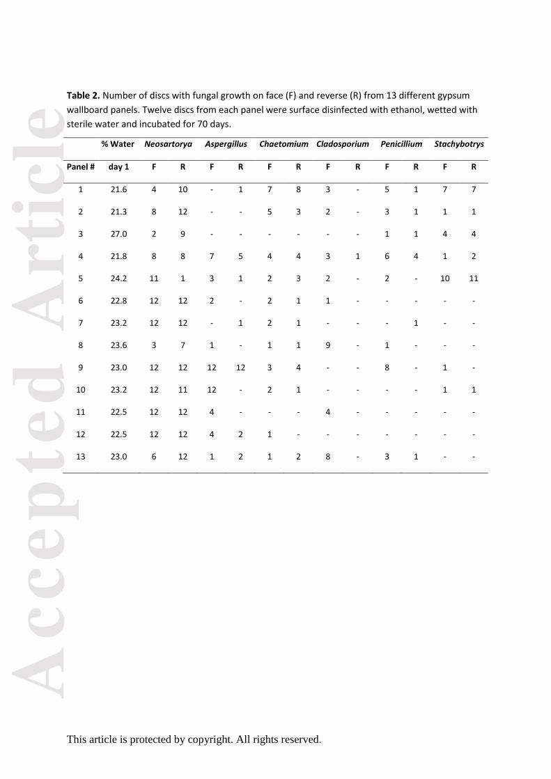
Analysis of the thirteen different planes of gypsum wallboard showed that there was fungal growth
of one or more fungal species, on either the face, the reverse or on both, on all tested planes (Table
2). Plane 9 (moisture resistant, brand B) had the highest total fungal count (64 fungal counts on 12
discs), whilst plane 3 (fire resistant, brand B) had the lowest (21 fungal counts on 12 discs). The
analyses also showed that there were no major differences between brands or between same types
of plane purchased from different DIY outlets. During the 70 days each experiment lasted fungal
growth and dissemination were followed and recorded using a stereo microscope. Only six genera,
Neosartorya, Aspergillus, Chaetomium, Penicillium, Cladosporium and Stachybotrys, reoccurred on
three or more of the thirteen panels (Table 2). Alternaria, Botrytis, Phoma and Ulocladium were
found on only one panel and one or two discs, while Harzia, Paecilomyces and Pochonia, were found
only once.
3.1. Fungal identification
At day 70 the seals of the Petri dishes were broken and representative colonies were isolated and
identified to species. The most abundant fungal species found on gypsum wallboard was
Neosartorya hiratsukae. This fungus was found on all thirteen panels (100 %) and on most discs;
both face and reverse. It appeared first on the reverse of the discs, but within days it was also visible
on the face. The fungus had covered the whole disc with small (100-230 µm in diam.) white
ascomata (cleistothecia) (Fig. 1A/D) in 3-7 days after first appearance. Only on panel 5 was N.
hiratsukae more abundant on the face than on the reverse (Table 2). The most abundant Aspergillus
turned out to be A. hiratsukae, which is the asexual state of N. hiratsukae (i.e. the same organism as
N. hiratsukae), and was found around the edge of the discs and constituted most of the recorded
“Aspergillus” in Table 2. C. globosum was the second most abundant species, found on eleven panels
Acc
epte
d A
rtic
le
This article is protected by copyright. All rights reserved.
(85 %) and was equally common on both face and reverse. It took ca. 14 days after onset for the
fungus to cover the whole surface of the disc with large (300-500 µm in diam.) dark ascomata
(perithecia) with green curly hair (Fig. 1B/E). Both P. chrysogenum and Cladosporium cladosporioides
showed the same pattern by occurring mostly on the face of nine (69 %) and eight (62 %) panels,
respectively, and only as discrete, slow growing colonies that stopped expanding after 14-21 days
(Fig. 1B). S. chartarum (both chemo types) was found on seven (54 %) of the thirteen panels and
would cover the whole surface of the disc within 7 days after onset with black clusters of slime
heads (Fig. 1C/F). Alternaria infectoria, Aspergillus versicolor and Cladosporium sphaerospermum
were found on only one panel and on one or two discs, while Aspergillus fumigatus, Chaetomium
elatum, Penicillium polonicum were found only once.
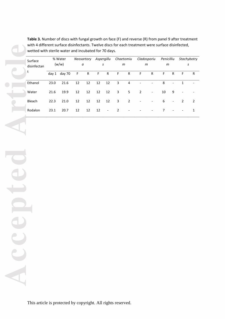
3.2. Surface disinfection
After the emergence of N. hiratsukae ascomata on the first 5 panels, it was speculated whether the
ethanol was triggering the growth of this fungus or if it could be present in the ethanol. Therefore
two other surface disinfectants, household bleach and Rodalon, and autoclaved water were also
used on subsamples of panel 9. Analysis of the three subsamples showed similar results compared to
ethanol disinfection (Table 3). With water and bleach the ascomata of N. hiratsukae appeared just as
rapidly (14 days) as they did with ethanol, whereas the Rodalon treatment delayed the ascomata
production by ca. 7 days. The subsample washed in autoclaved water did not show any additional
fungal growth and neither bleach nor Rodalon hampered the growth of S. chartarum. Table 3 also
shows that the water content of the discs decreased slightly from 22.5 % to 20.8 % on average
during the 70 day long incubation period despite several layers of parafilm around the edge of the
Petri dishes.
Acc
epte
d A
rtic
le
This article is protected by copyright. All rights reserved.
3.3. Onset of fungal growth
The last four panels (panels 10-13) were also examined on day 3 and day 7 to see how quickly the
fungi could germinate, grow and sporulate. Figure 2 shows the onset of sporulation for each genus
and the end time where no new fungal colonies appear. The first undifferentiated hyphal growth
was evident on day 3 (graph not shown) and conidial heads of A. hiratsukae were visible after only 7
days of incubation, whereas N. hiratsukae ascomata started to appear after 14 days. Also C.
cladosporioides and P. chrysogenum were visible after 14 days with conidiophores and the first
chains of conidia. The first slime heads of S. chartarum were visible after 21 days, while C. globosum
ascomata were visible after 28 days (panels 6-9, result not shown). No new colonies appeared after
42-45 days.
4. Discussion
The results of this study show that gypsum wallboard is already contaminated with fungal spores
before the panels reach their end-users, since the same fungal species, Neosartorya hiratsukae (≡
Aspergillus hiratsukae), Chaetomium globosum and Stachybotrys chartarum, were found repeatedly
in the paper/cardboard of all surface disinfected samples irrespectively of type, brand or outlet.
N. hiratsukae, which is an uncommon fungus in the environment, has to our knowledge
never been reported on gypsum wallboard before. It has previously been isolated from soil, fruit
juice and indoor air (Samson et al., 2007). N. hiratsukae is also reported to be pathogenic to humans
(Guarro et al., 2002) and to produce avenaciolide (Samson et al., 2007). One reason for occupants
and surveyors not to realize growth of this fungus could be that the small white ascomata are evenly
distributed across the white paper surface of the gypsum wallboard and therefore not readily visible
to the naked eye (Fig. 1A) and difficult to see without a slanted light source on the stereo
microscope. The pale green anamorphic state of the fungus, A. hiratsukae, is produced only
Acc
epte
d A
rtic
le
This article is protected by copyright. All rights reserved.
sparingly on gypsum wallboard. During the 70 day incubation period it was noted how the ascomata
slowly disintegrated and released vast numbers of ascospores. This suggests that N. hiratsukae
spores (both live and dead) and micro-particles from the ascomata can easily become airborne and
constitute a health risk in buildings with water damaged gypsum wallboard without the knowledge
of the occupants.
A limited number of research studies have been published on the occurrence of C.
globosum in water damaged buildings and even fewer have reported C. globosum on paper and
gypsum wallboard (Price and Ahearn, 1999; Jerusik, 2010; Flannigan and Miller, 2011). One reason
for not detecting C. globosum, even though it is very conspicuous on gypsum wallboard when
mature (Fig. 1B), may be its long lag phase on both gypsum and laboratory media. Furthermore,
samples from water damaged buildings often contain other fungi (e.g. Penicillium spp.) that grow
much faster on laboratory media and thereby obscure any growth of C. globosum.
The association between S. chartarum and gypsum wallboard, on the other hand, is well
documented and it was therefore expected that S. chartarum would be the dominant fungus. Still,
more than 50 % of our panels were contaminated with S. chartarum, which is more than some
studies report (Gravesen et al., 1999; Andersen et al., 2011), but less than the 60-77 % Flannigan and
Miller (2011) found in their study. If our results are representative for a panel (1 out of 12 discs is
contaminated with one viable S. chartarum spore) it would suggest that a whole panel would be
contaminated with 40-60 viable spores depending on the size of the panel (ca. 20 spores/m2). This
corresponds well to our field observations in buildings with severe water-damage. There we have
seen simultaneous outgrowth of numerous discrete Stachybotrys colonies on the same gypsum
panel indicating that Stachybotrys spores are distributed throughout the material. On our most
contaminated sample, panel 5, where 10 discs were contaminated, the same estimation would give
approximately 200 viable S. chartarum spores per m2. This combined with the rapid growth rate on
Acc
epte
d A
rtic
le
This article is protected by copyright. All rights reserved.
gypsum wallboard (covering a disc in less than a week) might explain why S. chartarum can
dominate a water damaged building so quickly.
Most gypsum wallboard manufacturers advertise that they use recycled materials in their
production. Recycled paper/cardboard is often collected in big bails at the recycling centre and
stored under less than optimal conditions (Jerusik, 2010) where bails are exposed to rain, soil and
insects, which results in high microbial loads (Betz and Cerny, 1999). N. hiratsukae, as well as C.
globosum and S. chartarum, can survive at temperatures around 40 °C (Samson et al., 2007; Domsch
et al., 2007) and even if a small percentage of the fungal spores survive pulping and drying, there
could be invisible fungal growth on the gypsum wallboard after as little as 7 days after major water-
damage. However, since it is not known where in the process the different fungi enter or what their
contamination and survival rates are, further research into the specific production methods of the
raw materials is needed. One approach to safer gypsum wallboard is for the manufacturers to use
hazard analysis and critical control points (HACCP), which is a system used by the food industry for
decades to ensure food safety, but other types of industry are increasingly using HACCP as a
competitive parameter.
Ongoing chemical analyses of the discs at our department will show which fungal
metabolites (e.g. avenaciolide, chaetoglobosins, atranones and satratoxins) these inbuilt fungal
contaminants are able to produce during their growth on the gypsum wallboards and if it is
consistent with previous findings on Stachybotrys spp. (Nielsen et al., 1998). Also a rapid detection
method using DNA sequencing directly on the paper/cardboard surface is being developed at our
department.
Acc
epte
d A
rtic
le
This article is protected by copyright. All rights reserved.
5. Conclusions
The results of this work show that gypsum wallboard is contaminated with Neosartorya,
Chaetomium and Stachybotrys and suggest that the spores of these fungi are embedded in the
paper/cardboard surrounding the gypsum core. Even though the manufacturers do not market their
gypsum wallboard as “sterile” or “fungal free”, most consumers trust that there are no potentially
harmful fungi in their building materials. Since growth of these fungi can result in large quantities of
micro-particles and bioactive compounds, which may be released into the indoor air after water
damage, there is a need for manufacturers to undertake a stricter quality control of their raw
materials and finished products. However, even the best efforts of the manufacturers would be
rendered futile if proper/correct shipping, handling and storage are not equally strictly controlled.
During storage, construction and occupancy, gypsum wallboard must be kept dry, clean and
undamaged in order to provide safe and healthy buildings.
Acknowledgements
The authors would like to thank the VILLUM Foundation for financial support for this work, Peter
Meinke, DTU Systems biology, for cutting all the gypsum wallboard discs and Jens C. Frisvad, DTU
Systems biology, for confirming the identity of Neosartorya hiratsukae/Aspergillus hiratsukae.
References
Andersen, B. and Nissen, A.T. (2000) Evaluation of media for detection of Stachybotrys and
Chaetomium species. International Biodeterioration and Biodegradation, 46, 111-116.
Andersen, B., Frisvad, J.C., Søndergaard, I., Rasmussen, I.S. and Larsen, L.S. (2011) Associations
between fungal species and water damaged building materials. Applied and Environmental
Microbiology, 77, 4180-4188.

Acc
epte
d A
rtic
le
This article is protected by copyright. All rights reserved.
Andersen, B., Nielsen, K.F. and Jarvis, B.B. (2002) Characterization of Stachybotrys from water-
damaged buildings based on morphology, growth and metabolite production, Mycologia 94,
392-403.
Andersen, B., Nielsen, K.F., Thrane, U., Szaro, T., Taylor, J.W. and Jarvis, B.B. (2003) Molecular and
phenotypic descriptions of Stachybotrys chlorohalonata sp. nov. and two chemotypes of
Stachybotrys chartarum found in water-damaged buildings, Mycologia, 95, 1227-1238.
Baxter, D.M., Perkins, J.L., McGhee, C.R. and Seltzer, J.M. (2005) A Regional Comparison of Mold
Spore Concentrations Outdoors and Inside “Clean” and “Mold Contaminated” Southern
California Buildings, Journal of Occupational and Environmental Hygiene 2, 8–18.
Betz, M and Cerny, G. (1999) Mikrobiologische Untersuchungen an Hygienpapieren mit und ohne
Zusatz von Altpapier Papier, 53, 376-383.
Carey, S.A., Plopper, C.G., Hyde, D.M., Islam, Z., Pestka, J.J. and Harkema, J.R. (2012) Satratoxin-G
from black mold Stachybotrys chartarum induces rhinitis and apoptosis of olfactory sensory
neurons in the nasal airways of rhesus monkeys, Toxicologic Pathology, 40, 887-898.
Dill, I., Trautmann C. and Szewzyk, R. (1997) Massenentwicklung von Stachybotrys chartarum auf
kompostierbaren Pflanztöpften aus Altpapier, Mycoses, 40, 110-114.
Domsch, K.H., Gams, W. and Anderson, T.H. (2007) Compendium of soil fungi, Germany, Eching,
IHW-Verlag, Ed. 2 pp 4–672.
Drobotko, V.G. (1944) Stachybotryotoxicosis. A new disease of horses and humans, American Review
of Soviet Medicine, Vol 2, 238-242.
Ellis, M.B. (1971) Dematiaceous Hyphomycetes, Kew, Surrey, UK, Commonwealth Mycological
Institute, pp 5-608.
Acc
epte
d A
rtic
le
This article is protected by copyright. All rights reserved.
Etzel, R.A. (2007) Indoor and outdoor air pollution: Tobacco smoke, moulds and diseases in infants
and children, Int. J. Hyg. Environ. Health, 210, 611–616.
Flannigan, B. and Miller, J.D. (2011) Microbial growth in indoor environments. In: B. Flannigan, R.A.
Samson and J. David Miller (eds) Microorganisms in Home and Indoor Work Environments.
Diversity, health impacts, investigation and control, Boca Raton, FL, CRC Press, Ed. 2, pp 57-
107.
Forgacs, J., Carll, W. T., Herring, A. S. and Hinshaw, W.R. (1958) Toxicity of Stachybotrys atra for
animals, Trans New York Acad Sci, 20, 787-808.
Gravesen, S., Nielsen, P.A., Iversen, R. and Nielsen, K.F. (1999) Microfungal contamination of damp
buildings – examples of risk constructions and risk materials, Environ Health Perspect,
107,505-508.
Guarro, J., Kallas, E.G., Godoy, P., Karenina, A., Gené, J., Stchigel, A. and Colombo, A.L. (2002)
Cerebral aspergillosis caused by Neosartorya hiratsukae, Brazil, Emerging Infectious Diseases,
8, 989-991.
Harrach, B., Bata, A., Bajmocy, E. and Benko, M. (1983) Isolation of satratoxins from the bedding
straw of a sheep flock with fatal stachybotryotoxicosis, Applied and Environmental
Microbiology, 45, 1419-1422.
Jarvis, B.B., Lee, Y.W., Comezoglu, S.N. and Yatawara, C.S. (1986) Trichothecenes produced by
Stachybotrys-atra from Eastern-Europe, Applied and Environmental Microbiology, 51, 915-
918.
Jarvis, B.B., Sorenson, W.G., Hintikka, E.L., Nikulin, M., Zhou, Y.H., Jiang, J., Wang, S.G., Hinkley, S.,
Etzel, R.A. and Dearborn, D. (1998) Study of toxin production by isolates of Stachybotrys
Acc
epte
d A
rtic
le
This article is protected by copyright. All rights reserved.
chartarum and Memnoniella echinata isolated during a study of pulmonary hemosiderosis in
infants. Applied And Environmental Microbiology, 64, 3620-3625.
Jerusik, R.J. (2010) Fungi and paper manufacture, Fungal Biology Reviews, 24, 68-72.
McGinnis, M.R. (2007) Indoor mould development and dispersal, Medical Mycology, 45, 1-9.
Mussalo-Rauhamaa, H., Nikulin, M., Koukila-Kahkola, P., Hintikka, E.L., Malmberg, M. and Haahtela,
T. (2010) Health Effects of Residents Exposed to Stachybotrys in Water-damaged Houses in
Finland, Indoor and Built Environment, 19, 476-485
Nayak, A.P., Green, B.J. and Beezhold, D.H. (2013) Fungal hemolysins, Medical Mycology, 51, 1–16.
Nielsen, K.F., Hansen, M.Ø., Larsen, T.O. and Thrane, U. (1998) Production of trichothecene
mycotoxins on water damaged gypsum boards in Danish buildings, International
Biodeterioration & Biodegradation, 42, 1-7.
Nikulin, N., Reijula, K., Jarvis, B.B. and Hintikka E.-L. (1997) Experimental lung mycotoxicosis induced
by Stachybotrys atra, Int. J. Experim. Pathol. 77:213-218.
Price, D.L. and Ahearn, D.G. (1999) Sanitation of Wallboard Colonized with Stachybotrys chartarum,
Current Microbiology, 39, 21–26.
Polizzi, V., Delmulle, B., Adams, A., Moretti, A., Susca, A., Picco, A.M., Rosseel, Y., Kindt, R., Van
Bocxlaer, J., De Kimpe, N., Van Peteghem, C., De Saeger, S. (2009) JEM spotlight: Fungi
mycotoxins and microbial volatile organic compounds in mouldy interiors from water-
damaged buildings, Journal of Environmental Monitoring, 11, 1849-1858.
Rand, T.G. and Miller, J.D. (2011) Analysis for toxins and inflammatory compounds. In: B. Flannigan,
R.A. Samson and J. David Miller (eds) Microorganisms in Home and Indoor Work
Environments. Diversity, health impacts, investigation and control, Boca Raton, FL, CRC Press,
Ed. 2, pp 291-306.
Acc
epte
d A
rtic
le
This article is protected by copyright. All rights reserved.
Rosenblum Lichtenstein, J.H., Hsu, Y.-H., Gavin, I.M., Donaghey, T.C., Molina, R.M., Thompson, K.J.,
Chi, C.-L., Gillis, B.S. and Brain, J.D. (2015) Environmental mold and mycotoxin exposures elicit
specific cytokine and chemokine responses, PLoS ONE, 10, e0126926.
Samson, R.A., Hong, S., Peterson, S.W., Frisvad, J.C. and Varga, J. (2007) Polyphasic taxonomy of
Aspergillus section Fumigati and its teleomorph Neosartorya, Stud. Mycol., 59, 147-207.
Samson, R.A., Houbraken, J., Thrane, U., Frisvad, J.C. and Andersen, B. (2010) Food and Indoor Fungi.
Utrecht, the Netherlands, CBS-KNAW-Fungal Biodiversity Centre, pp. 1-398.
Tiffany, J.A. and Bader, H.A. (2000) Detection of Stachybotrys chartarum: The effectiveness of
culturable-air sampling and other methods, Journal of Environmental Health, 62, 9-11.
Viegas, S., Sabino, R., Viegas, C., Faria, T., Quintal Gomes, A. and Seco, A. (2014) Fungal
contamination in two Portuguese wastewater treatment plants. Journal of Toxicology and
Environmental Health, 77, 90-102.
von Arx, J.A., Guarro, J. and Figueras, M.J. (1986) The Ascomycete Genus Chaetomium, Nova
Hedwigia, Heft 84, 1-162.
Wang, X.W., Lombard, L., Groenewald, J.Z., Videira, S.I.R., Samson, R.A., Liu, X.Z. and Crous P.W.
(2016) Phylogenetic reassessment of the Chaetomium globosum species complex, Persoonia,
36, 83-133.

Acc
epte
d A
rtic
le
This article is protected by copyright. All rights reserved.
Figure legends
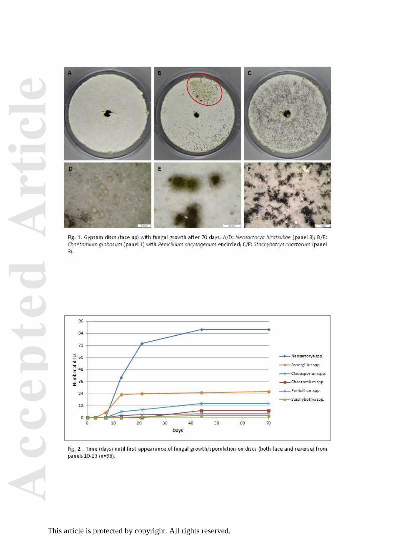
Fig. 1. Gypsum discs (face up) with fungal growth after 70 days. A/D: Neosartorya hiratsukae (panel
3); B/E: Chaetomium globosum (panel 1) with Penicillium chrysogenum encircled; C/F:
Stachybotrys chartarum (panel 3).
Fig. 2. Time (days) until first appearance of fungal growth/sporulation on discs (both face and
reverse) from panels 10-13 (n=96).
Table 1. Type and origin (anonymised) of the gypsum wallboard panels used in this study.
Panel # Type of wallboard Brand Outlet Purchase date
1 Fire resistant A DTU* 14-01-2015
2 Fire resistant A DIY-1 20-01-2015
3 Fire resistant B DIY-2 17-03-2015
4 Moisture resistant B DIY-2 17-03-2015
5 Fire resistant-spacer A DIY-1 23-03-2015
6 Fire resistant A DIY-3 30-04-2015
7 Moisture resistant A DIY-3 30-04-2015
8 Fire resistant B DIY-4 30-04-2015
9 Moisture resistant B DIY-4 30-04-2015
10 Fire resistant A DIY-1 13-07-2015
11 Regular A DIY-1 13-07-2015
12 Fire resistant-spacer A DIY-1 13-07-2015
13 Fire resistant B DIY-2 13-07-2015
* Clean, unused surplus panel from a building site at the Technical University of Denmark.
Acc
epte
d A
rtic
le
This article is protected by copyright. All rights reserved.
Table 2. Number of discs with fungal growth on face (F) and reverse (R) from 13 different gypsum
wallboard panels. Twelve discs from each panel were surface disinfected with ethanol, wetted with
sterile water and incubated for 70 days.
% Water Neosartorya Aspergillus Chaetomium Cladosporium Penicillium Stachybotrys
Panel # day 1 F R F R F R F R F R F R
1 21.6 4 10 - 1 7 8 3 - 5 1 7 7
2 21.3 8 12 - - 5 3 2 - 3 1 1 1
3 27.0 2 9 - - - - - - 1 1 4 4
4 21.8 8 8 7 5 4 4 3 1 6 4 1 2
5 24.2 11 1 3 1 2 3 2 - 2 - 10 11
6 22.8 12 12 2 - 2 1 1 - - - - -
7 23.2 12 12 - 1 2 1 - - - 1 - -
8 23.6 3 7 1 - 1 1 9 - 1 - - -
9 23.0 12 12 12 12 3 4 - - 8 - 1 -
10 23.2 12 11 12 - 2 1 - - - - 1 1
11 22.5 12 12 4 - - - 4 - - - - -
12 22.5 12 12 4 2 1 - - - - - - -
13 23.0 6 12 1 2 1 2 8 - 3 1 - -
Acc
epte
d A
rtic
le
This article is protected by copyright. All rights reserved.
Table 3. Number of discs with fungal growth on face (F) and reverse (R) from panel 9 after treatment
with 4 different surface disinfectants. Twelve discs for each treatment were surface disinfected,
wetted with sterile water and incubated for 70 days.
Surface
disinfectan
t
% Water
(w/w)
Neosartory
a
Aspergillu
s
Chaetomiu
m
Cladosporiu
m
Penicilliu
m
Stachybotry
s
day 1 day 70 F R F R F R F R F R F R
Ethanol 23.0 21.6 12 12 12 12 3 4 - - 8 - 1 -
Water 21.6 19.9 12 12 12 12 3 5 2 - 10 9 - -
Bleach 22.3 21.0 12 12 12 12 3 2 - - 6 - 2 2
Rodalon 23.1 20.7 12 12 12 - 2 - - - 7 - - 1
Acc
epte
d A
rtic
le
This article is protected by copyright. All rights reserved.