Practical micro
35
Fig. 1.1: Simple laboratory autoclave(moist heat) Temp. 121°C (due to double atmospheric pressure) Time : required: 20 min. Used for: surgical inst ,gauze,cotton,&culture media that are destroyed by heat. It is an efficient method of sterilization due to penetration power of steam &high temp. Sterilization How to test efficacy of autoclave? By 1. chemical method :by cultivation of bacillus sterothermophilus( that can survive up to 120 c and beyond that it die) then it is cultivated to test efficacy. 2. biological method :by chemical indicator on a tape that changes its colour into black or dark purple if the autoclave is efficient.
-
Upload
hayam-mosad -
Category
Documents
-
view
255 -
download
1
Transcript of Practical micro

Fig. 1.1: Simple laboratory
autoclave(moist heat) Temp. 121°C (due to double atmospheric pressure)
Time : required: 20 min.
Used for: surgical inst ,gauze,cotton,&culture media that
are destroyed by heat.
It is an efficient method of sterilization due to penetration
power of steam &high temp.
Sterilization
How to test efficacy of autoclave?
By 1. chemical method :by cultivation of bacillus sterothermophilus( that can survive up to 120 c and beyond that it die) then it is cultivated to test efficacy.
2. biological method :by chemical indicator on a tape that changes its colour into black or dark purple if the autoclave is efficient.

Fig. 1.2: Hot air oven(dry heat).
Temp. 160°C Time required: 1 hour
Used for: metal & glass – ware
equipment &powder and oil.

With discs have varying
diameter & varying pore size
• Suitable for large volume of
fluid.
• and any biological fluids
contain proteins that would
coagulate if sterilized by
autoclave as serum, vaccines
and media used for cultivation
of viruses

N.B.
Bacterial loop : ( enoculating needle )
Sterilized by red heat
PLASTIC SYRINGE:
By ethylene oxide gas or gamma rays.

Fig. 2.1: Blood agar opaque red in color
Type:
Enriched & indicator medium
Differentiate bet bact. By their hemolytic
action on red cells (complete , partial, no
hemolysis) .it is not sterilized by autoclave
The sterile blood is added to sterile agar at
temp. of 55 and poured in sterile plates
Fig. 2.2: Blood agar showing:
-Alpha heaemolysis (partial & greenish)
-Beta haemolysis (complete )
Media

Fig. 2.3: MacConkey’s medium
Reddish transparent medium
Fig. 2.4: MacConkey’s medium showing:
-Lactose fermenter → rose pink colonies
-Non Lactose fermenter → pale yellow colonies
Type: indicator (differential media)
indicator: neutral red
sugar content : lactose
Use: to differ. Between lactose , non lactose frementers and for isolation of enteric
media
Sterilized by autoclave

Suitable for: growth of Nesisseriameningitides &haemophilussterilize by : autoclavetype: enriched media
Sterilize by: heating in inspissator (oven) at
80c for 2h for 3 successive days to kill
bacterial spores

Growh after 6-8 weeks
Type: selective media
inhibitory substance by: malachite green that inhibit all bacterial flora exept
mycobacterium tuberculosis.
Sterilize by: heating in inspissator (oven) at 80c for2 hours for 3 successive days
Fig. 2.8: TCBS media
-Green, Transparent medium
- It is used for isolation of Vibrio cholera

Used for: cultivation of anerobicbact.Suitable for growth of clostridia & other anaerobes.sterilize by : autoclave
Used for: cultivation of anaerobic bact.Indicator: methylene blue

Fig. 3.1: Gram-positive bacilli
(violet in colour)
Bacterial Identification
Fig. 3.2: Gram – negative bacilli
(Red in colour)

Fig. 3.8: Catalase test
-Positive test shows
gas production
-Give positive: with
staphylococci
Fig. 3.11: Oxidase test
Positive test gives deep
purple color
Give positive: with
Neisseria , vibrio
&pseudomonas
Fig. 3.10: Urease test
Positve test gives Pink color
Give positive: with proteus
Indicator: phenol red

Fig. 3.14: Antibiotic sensitivity test
Use: To choose the most effective drug used for ttt.
Type: Disc diffusion method
The drug show the largest zone of inhibition is the most effective
هتيجى ف االمتحان مرقمه ومين اكتر دواء يستخدم كعالج
SENSITIVE (LARGEST ZONE OF INHIBITION)
Non sensitive (resistant)

Fig. 1.1: Staph.
Aureus on blood
agar it produce Beta
haemolysis
Fig. 1.2: Staph
aureus on nutrient
agar it produce
golden yellow
endopigment
Staphylococci
Systemic bacteriology
Or bunches or gape like

Fig. 1.6: Coagulase test Staph. Aureus gives positive coagulase
test
Fig. 1.5: Catalase test
all Staphylococci are
catalase positive

strepcoocci
Fig. 2.2: Growth of
Strept viridians on
blood agar showing
partial or alpha
heaemolysis
Fig. 2.1: Growth of
Strept pyogenes on
blood agar showing
complete or Beta
haemolysis
Fig. 2.3: Strept. In
culture gram stain.
(Gram positve cocci
arranged in chain)

Fig. 2.8: Optochin
sensitivity test
strept. Pneum is
sensitive
strept. Viridance is
resistant
Fig. 2.9: Bacitracin
sensitivty test
strept. Pyogens is
sensitive
strept. Agalactiae is
resistant
Fig. 2.4: Sterpt. In
pus Gram positive
cocci

pneumococci
Fig. 2.7:Pneumococci
, Quelling reaction
(capsule swelling with specific anti-sera)
Fig. 2.6: Pneumococciin tissue, gram stain gram positive capsulated diplococci

Added to 50 – 100 ml fluid medium
Use of Blood culture
for diagnosis of acute bacterial endocarditis
Disease diagnosed by bl. Culture:subacute bacterial endocarditis(streptococcus viridans) “ sanguis, salivarius” + post streptococcal disease , brucellosis ,typhoid fever and puerperal sepsis.
5 – 10 ml blood
Blood culture
Use:
1. For diagnosis of rheumatic
fever
2. detection of post
streptococcal infection.
3. Acute glomerulonephiritis
Type of test:
Neutralization (in vitro)
The presence of antitoxin in
sera neutralizes the hemolytic
effect of toxin on addition of
red blood cells.
Diagnostic test: >200 todd
unit
3. Toxin-Antitoxin Neutralization Test
Antistreptolysin test O (ASO)
Fig. 2. 11: Antistreptolysin O Titer
(ASO)
1/25 1/50 1/100 1/200 1/400 1/800 control
Titer 1/400

Neisseria menngitides
Fig. 3.2: Neisseria in
cluture Gram negative
cocci
Fig. 3.3: Pathogenic
neisseria in pus, gram
stain. (Intra &
exteracellular Gram
negative diplococci)
Fig. 3.4: Oxidase test
All neisseria is oxidase positive
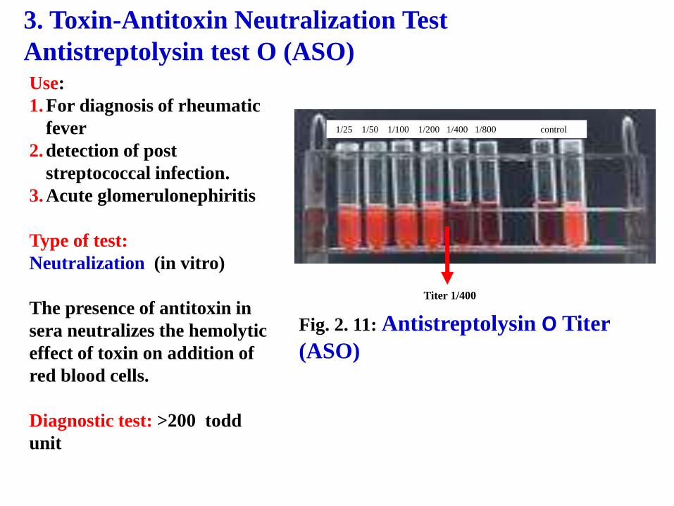
Neisseria gonorrhea
• The media used for cultivation is Thayer martin media which is chocolate blood agar + antibiotics (vancomycin for gm positvebacteria +nystatin for fungi +cholesyin for gm negative bacteria )
• Antibiotics are put because it is separated from vagina or urethera.

CORYNEBACTERIUM GROUP
Media: loffler’s serum agar.
Stain: gram & methylene blue
Test: Elek’s test
Fig. 4.3: Coryn.
Diphtheria in culture
methylen blue stain
Fig. 4.4: Coryn. Diphtheria
(Gram positive bacilli
have Chinese letter
arrangement)

Fig. 4.6: Elek’s test Positive test
Test strain
Precipitation band
Negative test
Artiserum
in the strip.
Type of reaction: precipitation test (Ag- Ab reaction)
use: Detection of toxiogenic strain of C . Diphtheriae
it is a double immunodiffusion test diffusion of the organism toxin with the antitoxin forming precipitation band or line

Mycobacterium
Fig. 5.3: Myc. TB in Sputum,
Z.N stain
(few thin pink bacilli with blue
background)
Fig. 5.1: Selective
media
for Myc. T.B.
Fig. 5.2: Culture of Myc.
TB on L.J. media
- Grow after 6-8 week

Fig. 5.5: Tuberculin test It involves intradermal injection of Purified Protein Derivative (PPD)
type of test: Delayed type hypersenstivity used for Diagnosis of T.B.
Type of reaction: antigen-anibody reaction
+ve: give area of induration about 9 mm Time: 48 – 72 hours after injection
Media: lowensten jensen media stain: ziehl neelsen stain

Protaus
Fig. 8.4: Proteus culture
on Nut. Agar (swarming
growth)
Fig. 8.5: Proteus in
culture, gram stain
(gram negative bacilli
proteus showing
pleomorphism )
Fig. 8.6: Urease
test (proteus is
urease test passivit

+ve -ve
Fig. 8.9: Oxidase test(pseudomonas & Vibrio
are ) oxidase psoitveFig. 8.8:
Pseudomonas
culture on Nut.
Agar (produce
greenish blue
exopigment
Fig. 8.7:
Pseudomonas in
culture, gram stain(gram negative bacilli)
Pseudomanas

Spore forming gram-positive bacilli(Bacillus & clostridium group)
Fig. 9.4 Gram stained film of
clost. Tetani in culture -
Gram-positive long bacillus
with terminal plugging spore
(dram-stick) appearance
Fig. 9.6 Robertson’s cooked-meat broth medium glutathione is released after boiling (reducing agent)

Fig. 11.2 Fontana stained film showing commensally spirochetes
Type of stain:
Special stain
Fig. 11.3 Dark ground illumination microscopy of Treponema pallidum.
نادرا اما تنزل
Fig. 11.4:Indirect Immunofluorescence for spirochetes. It is called indirect as we put antibody then we put antiantibodywith fluorescent die. To identify treponema pallidum
Spirochaetes

Reading of the results:
- No hemolysis means a
positive reaction
i.e the complement is bound
to the antigen-antibody
complex.
- Hemolysis means a
negative reaction
Fig. C-2: Complement fixation test
wasserman test
wasserman test
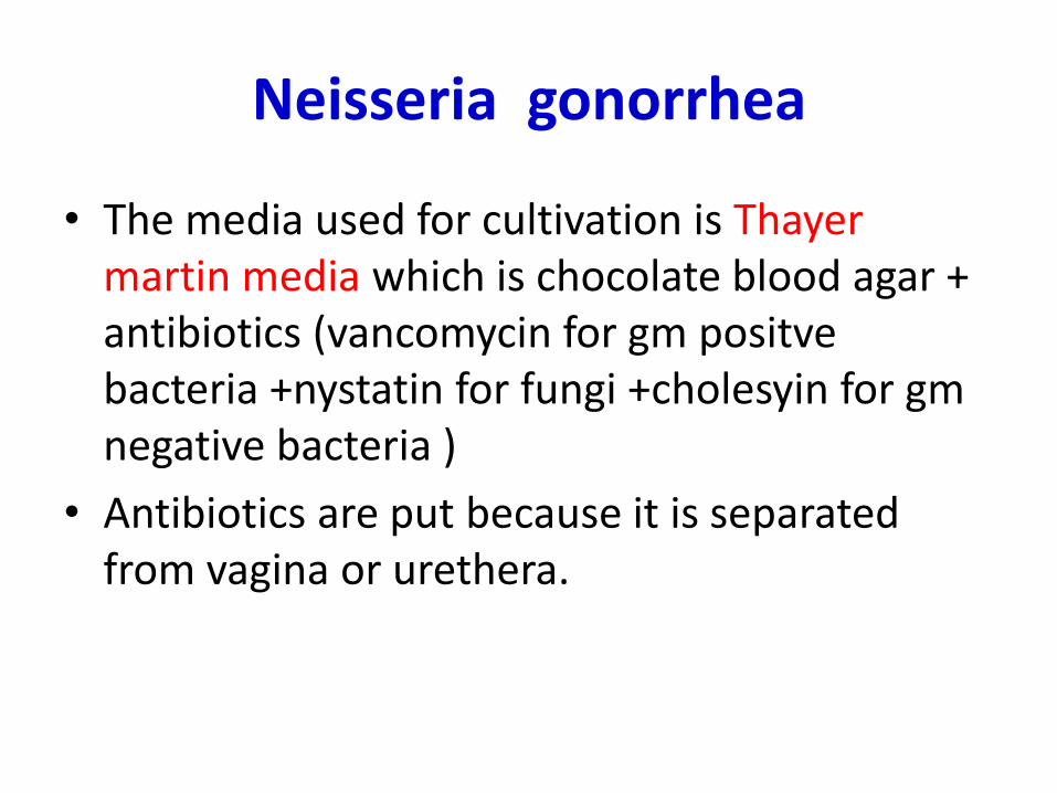
MYCOLOGY
Mention type of test to identify the candida organism : Germ tube
Mention 2 type for commensals of candida: vagina & alimentary tract & mouth
Fig. 12.3 Germ tube test
for pathogenic strain of
C. albicans
Fig. 12.1 Gram’s
stained films of
Candida albicans
Fig. 12.2 Candida albicans culture on sabouraud’s dextrose agar

Fig. 1.4: CPE (cytopathic effect of Herpes virus)
Showing: Enlarged, aggregated and ballooned
cells causing multi- nucleated giant cells
Cell culture for vesicular lesion : herpes giant
cell virus
VIROLOGY

E. Immunofluorecence
Fig. E-1: Direct immunofluorecence
for detection of specific antigen
immunology
Direct immunofluorecence
- A specific antibody labeled with a fluorescent molecule (fluorescein or rhodamine) is added to the unknown antigen in the specimen.
- It is used to detect the presence of an antigen on a cell or tissue.

-It is used for detection of antigen, antibodies.-ELISA is based on the measurement of an enzymatic reaction associated with immune complexes as detected by color development.
F. Enzyme Linked Immunosorbent Assay (ELISA)
Fig. F-1: Plate of ELISA test

Immunologic reactions
1. Antistreptolysin o titer
2. Elek’s test
3. Direct immunoflourescence test
4. Elisa plate test
5. Tuberculin skin test