
Positioning of autoimmune TCR-Ob.2F3 and TCR-Ob.3D1 on …Positioning of autoimmune TCR-Ob.2F3 and...
6
Positioning of autoimmune TCR-Ob.2F3 and TCR-Ob.3D1 on the MBP85–99/HLA-DR2 complex Zenichiro Kato †‡§¶ , Joel N. H. Stern † , Hironori K. Nakamura § , Kazuo Kuwata § , Naomi Kondo ‡§¶ , and Jack L. Strominger † † Department of Molecular and Cellular Biology, Harvard University, Cambridge, MA 02138; and ‡ Department of Pediatrics, Graduate School of Medicine, § Center for Emerging Infectious Diseases, and ¶ Center for Advanced Drug Research, Gifu University, 1-1 Yanagido, Gifu 5010-1194, Japan Contributed by Jack L. Strominger, August 11, 2008 (sent for review July 8, 2008) Since the first determination of structure of the HLA-A2 complex, >200 MHC/peptide structures have been recorded, whereas the available T cell receptor (TCR)/peptide/MHC complex structures now are <20. Among these structures, only six are TCR/peptide/ MHC Class II (MHCII) structures. The most recent of these structures, obtained by using TCR-Ob.1A12 from a multiple sclerosis patient and the MBP85–99/HLA-DR2 complex, was very unusual in that the TCR was located near the N-terminal end of the peptide-binding cleft of the MHCII protein and had an orthogonal angle on the peptide/MHC complex. The unusual structure suggested the pos- sibility of a disturbance of its signaling capability that could be related to autoimmunity. Here, homology modeling and a new simulation method developed for TCR/peptide/MHC docking have been used to examine the positioning of the complex of two additional TCRs obtained from the same patient (TCR-Ob.2F3 or TCR-Ob.3D1 with MBP85–99/HLA-DR2). The structures obtained by this simulation are compatible with available data on peptide specificity of the TCR epitope. All three TCRs from patient Ob including that from the previously determined crystal structure show a counterclockwise rotation. Two of them are located near the N terminus of the peptide-binding cleft, whereas the third is near the center. These data are compatible with the hypothesis that the rotation of the TCRs may alter the downstream signaling. human leukocyte antigen multiple sclerosis myelin basic protein structural docking signaling T he elucidation of structures of protein complexes is an arduous procedure particularly when the complexes are very large. The production of protein by recombinant techniques and subsequent crystallization of the complexes followed by x-ray diffraction analysis is a standard method. Structures can also be determined by NMR but that technique is presently limited to only relatively small proteins or complexes not 40 –50 kDa. The third method, simulation of structures by homology modeling, has improved greatly in recent years. However, this technique is usable only when an appropriate template structure is available. Crystallization and structure determination by x-ray diffrac- tion of a MHC-encoded Class I (MHCI) protein/peptide com- plex was first accomplished in 1987 and MHCII in 1993 (1, 2). Since then, 200 structures of such complexes have been recorded (3). The chain of the TCRs that recognize these complexes were first cloned in 1984 (4, 5), and the first structure of a TCR chain was published in 1995 (6). Approximately 40 complete TCR structures including both and chains are available now (3, 7). Similarly, the first TCR/peptide/MHC complex structures were published in 1996 (8, 9) but the number of such complex structures available now is 20 (3). Among these, only six are TCR/peptide/MHCII structures. The most recent of these structures is very unusual in that the TCR was located near the N-terminal end of the peptide-binding cleft of the MHCII protein and its orthogonal angle on the MHCII/ peptide was 84° as compared with a diagonal angle of 40 –53° for the other five structures (10). Also, it was rotated counterclock- wise on the MHC molecule relative to the other structures. This unusual structure suggested the possibility of a disturbance of its signaling capability that could be related to autoimmunity because this TCR, termed Ob.1A12 had been obtained from an autoreactive clone derived from a patient with multiple sclerosis (10). In fact, eight clones were obtained from this patient, two of which represent unique isolates (TCR-Ob.1A12 and TCR- Ob.3D1) and six of which have identical sequences, TCR-Ob.2F3 being an example (11). In this article, we have used homology modeling of TCR structures on the appropriate templates and a new simulation method developed for TCR/peptide/MHC docking to examine the structures of the TCR/peptide/MHCII complexes of TCR- Ob.2F3 and TCR-Ob.3D1 in complex with the same MHC/ peptide, namely HLA-DR2 (DRB1*1501/DRA) binding the myelin basic protein peptide epitope MBP85–99. MBP85–99 has previously been identified as the autoreactive peptide epitope in humans (12). Results and Discussion The AutoDock procedure was originally developed for docking studies of small chemicals to their receptors, for example the docking of a substrate to an enzyme (13). It makes use of charge and hydrophobicity calculations for both the receptor and the ligand (see Materials and Methods). By using this method in the present context, the peptide in question was first docked to the appropriate MHC molecule and separately to the TCR protein. The confor- mation of the peptide used in the docking in each case was taken from the crystal structure of the MBP85–99/HLA-DR2 (DRB1*1501, DRA) complex (10). After TCR/peptide and pep- tide/MHC structures were simulated, the two structures were merged by using the conformation of the peptide as the basis for merging. To validate the procedure, the technique was carried out by using two known TCR/peptide/MHC structures, that of the HLA-DR1 (DRB1*0101/DRA)/hemagglutinin (HA) 306–318 molecule in complex with the HLA-DR1-restricted HA306–318- specific TCR-HA1.7 (PDB ID code 1FYT) and then of the HLA-DR2/MBP85–99 molecule in complex with TCR-Ob.1A12 (PDB ID code 1YMM). Simulated Structure of the TCR-HA1.7/HA306 –318/HLA-DR1 Complex and the TCR-Ob.1A12/MBP85–99/HLA-DR2 Complex. The docking of HA306–318 to HLA-DR1 was simulated 10 times. Six of the 10 simulations showed exactly the same conformation inside the peptide-binding groove with energy equal to 33.8 kcal/mol (Fig. 1A). Four of the 10 showed different conformations binding outside of the groove with higher energies of 5.59, 5.59, Author contributions: Z.K., J.N.H.S., K.K., N.K., and J.L.S. designed research; Z.K. and H.K.N. performed research; Z.K. and H.K.N. analyzed data; and Z.K., J.N.H.S., and J.L.S. wrote the paper. The authors declare no conflict of interest. To whom correspondence may be addressed. E-mail: [email protected] or [email protected]. © 2008 by The National Academy of Sciences of the USA www.pnas.orgcgidoi10.1073pnas.0807338105 PNAS October 7, 2008 vol. 105 no. 40 15523–15528 IMMUNOLOGY Downloaded by guest on May 10, 2020
Transcript of Positioning of autoimmune TCR-Ob.2F3 and TCR-Ob.3D1 on …Positioning of autoimmune TCR-Ob.2F3 and...
Positioning of autoimmune TCR-Ob.2F3and TCR-Ob.3D1 on the MBP85–99/HLA-DR2 complexZenichiro Kato†‡§¶�, Joel N. H. Stern†, Hironori K. Nakamura§, Kazuo Kuwata§, Naomi Kondo‡§¶,and Jack L. Strominger†�
†Department of Molecular and Cellular Biology, Harvard University, Cambridge, MA 02138; and ‡Department of Pediatrics, Graduate School of Medicine,§Center for Emerging Infectious Diseases, and ¶Center for Advanced Drug Research, Gifu University, 1-1 Yanagido, Gifu 5010-1194, Japan
Contributed by Jack L. Strominger, August 11, 2008 (sent for review July 8, 2008)
Since the first determination of structure of the HLA-A2 complex,>200 MHC/peptide structures have been recorded, whereas theavailable T cell receptor (TCR)/peptide/MHC complex structuresnow are <20. Among these structures, only six are TCR/peptide/MHC Class II (MHCII) structures. The most recent of these structures,obtained by using TCR-Ob.1A12 from a multiple sclerosis patientand the MBP85–99/HLA-DR2 complex, was very unusual in that theTCR was located near the N-terminal end of the peptide-bindingcleft of the MHCII protein and had an orthogonal angle on thepeptide/MHC complex. The unusual structure suggested the pos-sibility of a disturbance of its signaling capability that could berelated to autoimmunity. Here, homology modeling and a newsimulation method developed for TCR/peptide/MHC docking havebeen used to examine the positioning of the complex of twoadditional TCRs obtained from the same patient (TCR-Ob.2F3 orTCR-Ob.3D1 with MBP85–99/HLA-DR2). The structures obtained bythis simulation are compatible with available data on peptidespecificity of the TCR epitope. All three TCRs from patient Obincluding that from the previously determined crystal structureshow a counterclockwise rotation. Two of them are located nearthe N terminus of the peptide-binding cleft, whereas the third isnear the center. These data are compatible with the hypothesisthat the rotation of the TCRs may alter the downstream signaling.
human leukocyte antigen � multiple sclerosis � myelin basic protein �structural docking � signaling
The elucidation of structures of protein complexes is anarduous procedure particularly when the complexes are very
large. The production of protein by recombinant techniques andsubsequent crystallization of the complexes followed by x-raydiffraction analysis is a standard method. Structures can also bedetermined by NMR but that technique is presently limited toonly relatively small proteins or complexes not �40–50 kDa. Thethird method, simulation of structures by homology modeling,has improved greatly in recent years. However, this technique isusable only when an appropriate template structure is available.
Crystallization and structure determination by x-ray diffrac-tion of a MHC-encoded Class I (MHCI) protein/peptide com-plex was first accomplished in 1987 and MHCII in 1993 (1, 2).Since then, �200 structures of such complexes have beenrecorded (3). The � chain of the TCRs that recognize thesecomplexes were first cloned in 1984 (4, 5), and the first structureof a TCR chain was published in 1995 (6). Approximately 40complete TCR structures including both � and � chains areavailable now (3, 7). Similarly, the first TCR/peptide/MHCcomplex structures were published in 1996 (8, 9) but the numberof such complex structures available now is �20 (3). Amongthese, only six are TCR/peptide/MHCII structures. The mostrecent of these structures is very unusual in that the TCR waslocated near the N-terminal end of the peptide-binding cleft ofthe MHCII protein and its orthogonal angle on the MHCII/peptide was 84° as compared with a diagonal angle of 40–53° forthe other five structures (10). Also, it was rotated counterclock-wise on the MHC molecule relative to the other structures. This
unusual structure suggested the possibility of a disturbance of itssignaling capability that could be related to autoimmunitybecause this TCR, termed Ob.1A12 had been obtained from anautoreactive clone derived from a patient with multiple sclerosis(10). In fact, eight clones were obtained from this patient, twoof which represent unique isolates (TCR-Ob.1A12 and TCR-Ob.3D1) and six of which have identical sequences, TCR-Ob.2F3being an example (11).
In this article, we have used homology modeling of TCRstructures on the appropriate templates and a new simulationmethod developed for TCR/peptide/MHC docking to examinethe structures of the TCR/peptide/MHCII complexes of TCR-Ob.2F3 and TCR-Ob.3D1 in complex with the same MHC/peptide, namely HLA-DR2 (DRB1*1501/DRA) binding themyelin basic protein peptide epitope MBP85–99. MBP85–99 haspreviously been identified as the autoreactive peptide epitope inhumans (12).
Results and DiscussionThe AutoDock procedure was originally developed for dockingstudies of small chemicals to their receptors, for example thedocking of a substrate to an enzyme (13). It makes use of charge andhydrophobicity calculations for both the receptor and the ligand(see Materials and Methods). By using this method in the presentcontext, the peptide in question was first docked to the appropriateMHC molecule and separately to the TCR protein. The confor-mation of the peptide used in the docking in each case was takenfrom the crystal structure of the MBP85–99/HLA-DR2(DRB1*1501, DRA) complex (10). After TCR/peptide and pep-tide/MHC structures were simulated, the two structures weremerged by using the conformation of the peptide as the basis formerging. To validate the procedure, the technique was carried outby using two known TCR/peptide/MHC structures, that of theHLA-DR1 (DRB1*0101/DRA)/hemagglutinin (HA) 306–318molecule in complex with the HLA-DR1-restricted HA306–318-specific TCR-HA1.7 (PDB ID code 1FYT) and then of theHLA-DR2/MBP85–99 molecule in complex with TCR-Ob.1A12(PDB ID code 1YMM).
Simulated Structure of the TCR-HA1.7/HA306–318/HLA-DR1 Complexand the TCR-Ob.1A12/MBP85–99/HLA-DR2 Complex. The docking ofHA306–318 to HLA-DR1 was simulated 10 times. Six of the 10simulations showed exactly the same conformation inside thepeptide-binding groove with energy equal to �33.8 kcal/mol(Fig. 1A). Four of the 10 showed different conformations bindingoutside of the groove with higher energies of �5.59, �5.59,
Author contributions: Z.K., J.N.H.S., K.K., N.K., and J.L.S. designed research; Z.K. and H.K.N.performed research; Z.K. and H.K.N. analyzed data; and Z.K., J.N.H.S., and J.L.S. wrote thepaper.
The authors declare no conflict of interest.
�To whom correspondence may be addressed. E-mail: [email protected] [email protected].
© 2008 by The National Academy of Sciences of the USA
www.pnas.org�cgi�doi�10.1073�pnas.0807338105 PNAS � October 7, 2008 � vol. 105 � no. 40 � 15523–15528
IMM
UN
OLO
GY
Dow
nloa
ded
by g
uest
on
May
10,
202
0
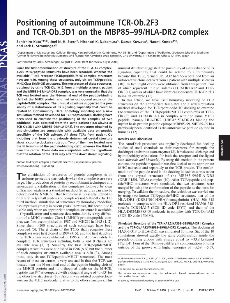
�5.68 and �8.73 kcal/mol (Fig. 1B). One of the clusteredconformations inside the groove with the lowest energy wasselected as representative.
Similarly, HA306–318 docking to TCR-HA1.7 was simulated10 times. Nine of the 10 simulations had almost the sameconformation with energy equal to �5.59 kcal/mol five times andenergy �5.67 four times (Fig. 1C). One of the 10 simulationsyielded a different conformation with a higher energy of �5.02kcal/mol (Fig. 1D). Again, one of the five clustered conforma-tions with the lowest energy was selected as representative.
Next, the TCR-HA1.7/HA306–318 simulated complex wasmerged with the HA306–318/HLA-DR1 simulated complex togive the TCR-HA1.7/HA306–318/HLA-DR1 complex by usingthe structure of the peptide as the basis for merging (a relatedprocedure was used in a docking study of the dimeric maltose-binding complex involving maltose-binding protein and aspar-
tate receptor, although in this case the octapeptide used wasfrom a functional region of the maltose-binding protein) (14).
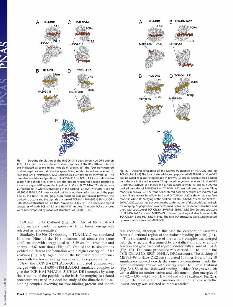
This simulated structure of the ternary complex was mergedwith the structure determined by crystallization and x-ray dif-fraction and gave excellent reproducibility with a rmsd of 1.64 Å(Fig. 1E). The same procedure was carried out to obtain theTCR-Ob.1A12/MBP85–99/HLA-DR2 structure. The docking ofMBP85–99 to HLA-DR2 was simulated 10 times. Four of the 10simulations showed exactly the same conformations inside thepeptide-binding groove with energy equal to �26.8 kcal/mol(Fig. 2A). Six of the 10 showed binding outside of the groove eachwith a different conformation and with much higher energies of�3.02, �4.95, �4.95, �5.54, �5.64 and �5.91 kcal/mol (Fig. 2B).One of the clustered conformations inside the groove with thelowest energy was selected as representative.
Fig. 1. Docking simulation of the HA306–318 peptide on HLA-DR1 and onTCR-HA1.7. (A) The six clustered docked peptides of HA306–318 on HLA-DR1are indicated as space filling models in brown. (B) The four nonclustereddocked peptides are indicated as space filling models in yellow. In A and B,HLA-DR1 (DRB1*0101/DRA) (20) is shown as a surface model in white. (C) Thenine clustered docked peptides of HA306–318 on TCR-HA1.7 are indicated asspace filling models in brown. (D) The one nonclustered docked peptide isshown as a space-filling model in yellow. In C and D, TCR-HA1.7 is shown as asurface model in white. (E) Merging of the docked TCR-HA1.7/HA306–318 andHA306–318/HLA-DR1 was carried out by using the conformation of the pep-tide as the basis for merging. Superposition was performed between thedocked structure and the crystal structure of TCR-HA1.7/HA306–318/HLA-DR1(20). Docked structure of TCR-HA1.7 in cyan, HA306–318 in brown, and crystalstructures of both TCR-HA1.7 and HLA-DR1 in blue. The two TCR structureswere superimposed by means of structures of HA306–318.
Fig. 2. Docking simulation of the MBP85–99 peptide on HLA-DR2 and onTCR-Ob.1A12. (A) The four clustered docked peptides of MBP85–99 on HLA-DR2are indicated as space filling models in brown. (B) The six nonclustered dockedpeptides are indicated as space filling models in yellow. In A and B, HLA-DR2(DRB1*1501/DRA) (10) is shown as a surface model in white. (C) The six clustereddocked peptides of MBP85–99 on TCR-Ob.1A12 are indicated as space fillingmodels in brown. (D) The four nonclustered docked peptides are indicated asspace filling models in yellow. In C and D, TCR-Ob.1A12 is shown as a surfacemodel in white. (E) Merging of the docked TCR-Ob.1A12/MBP85–99 and MBP85–99/HLA-DR2wascarriedoutbyusingtheconformationof thepeptideas thebasisfor merging. Superposition was performed between the docked structure andthe crystal structure of TCR-Ob.1A12/MBP85–99/HLA-DR2 (10). Docked structureof TCR-Ob.1A12 in cyan, MBP85–99 in brown, and crystal structures of bothTCR-Ob.1A12 and HLA-DR2 in blue. The two TCR structures were superimposedby means of structures of MBP85–99.
15524 � www.pnas.org�cgi�doi�10.1073�pnas.0807338105 Kato et al.
Dow
nloa
ded
by g
uest
on
May
10,
202
0
Similarly, MBP85–99 docking to TCR-Ob.1A12 was simulated 10times. Six of the 10 simulations had almost the same conformationwith energy equal to �4.78 five times and energy �4.65 once (Fig.2C). Four of the 10 simulations each yielded a different confor-mation with a similar energy of �4.50, �5.08, �5.32, and �5.32kcal/mol (Fig. 2D). Again, one of the six clustered conformationswith the lowest energy was selected as representative.
The TCR-Ob.1A12/MBP85–99 simulated complex was alsomerged with the MBP85–99/HLA-DR2 simulated complex as
described above to give the TCR-Ob1A12/MBP85–99/HLA-DR2 complex. Merging of the simulated structure with thestructure determined by x-ray crystallography gave excellentreproducibility with a rmsd of 1.54 Å (Fig. 2E). Thus, by usingtwo known TCR/peptide/MHC complexes, these data provideda validation for the docking procedure used.
Notably, the energies of docking of the two peptides to theirrespective MHC proteins (�34 and �27 kcal/mol) were much lowerthan the energies of their docking to their respective TCR (�5.6
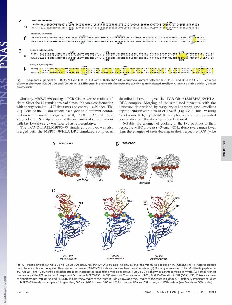
Fig. 3. Sequence alignment of TCR-Ob.2F3 and TCR-Ob.3D1 with TCR-Ob.1A12. (A) Sequence alignment between TCR-Ob.2F3 and TCR-Ob.1A12. (B) Sequencealignment between TCR-Ob.3D1 and TCR-Ob.1A12. Differences in amino acids between the two clones are indicated in yellow. *, identical amino acids; �, similaramino acids.
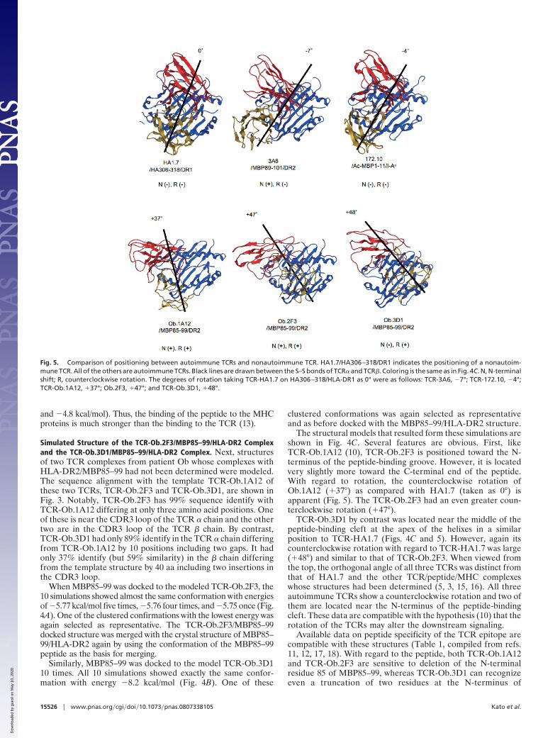
Fig. 4. Positioning of TCR-Ob.2F3 and TCR-Ob.3D1 on MBP85–99/HLA-DR2. (A) Docking simulation of the MBP85–99 peptide on TCR-Ob.2F3. The 10 clustered dockedpeptides are indicated as space filling models in brown. TCR-Ob.2F3 is shown as a surface model in white. (B) Docking simulation of the MBP85–99 peptide onTCR-Ob.3D1. The 10 clustered docked peptides are indicated as space filling models in brown. TCR-Ob.3D1 is shown as a surface model in white. (C) Comparison ofpositioning of the TCRs obtained from patient Ob. on the MBP85–99/HLA-DR2 structure. The structures of TCRs, MBP85–99 and HLA-DR2 (DRB1*1501/DRA) are shownas ribbon models. MBP85–99 and HLA-DR2 in blue, the � chains of the three TCRs in yellow, and the � chains of the three TCRs in red. Functionally important residuesof MBP85–99 are shown as space filling models, E85 and N86 in green, V88 and K93 in orange, H90 and F91 in red, and I95 in yellow (see Results and Discussion).
Kato et al. PNAS � October 7, 2008 � vol. 105 � no. 40 � 15525
IMM
UN
OLO
GY
Dow
nloa
ded
by g
uest
on
May
10,
202
0
and �4.8 kcal/mol). Thus, the binding of the peptide to the MHCproteins is much stronger than the binding to the TCR (13).
Simulated Structure of the TCR-Ob.2F3/MBP85–99/HLA-DR2 Complexand the TCR-Ob.3D1/MBP85–99/HLA-DR2 Complex. Next, structuresof two TCR complexes from patient Ob whose complexes withHLA-DR2/MBP85–99 had not been determined were modeled.The sequence alignment with the template TCR-Ob.1A12 ofthese two TCRs, TCR-Ob.2F3 and TCR-Ob.3D1, are shown inFig. 3. Notably, TCR-Ob.2F3 has 99% sequence identify withTCR-Ob.1A12 differing at only three amino acid positions. Oneof these is near the CDR3 loop of the TCR � chain and the othertwo are in the CDR3 loop of the TCR � chain. By contrast,TCR-Ob.3D1 had only 89% identify in the TCR � chain differingfrom TCR-Ob.1A12 by 10 positions including two gaps. It hadonly 37% identify (but 59% similarity) in the � chain differingfrom the template structure by 40 aa including two insertions inthe CDR3 loop.
When MBP85–99 was docked to the modeled TCR-Ob.2F3, the10 simulations showed almost the same conformation with energiesof �5.77 kcal/mol five times, �5.76 four times, and �5.75 once (Fig.4A). One of the clustered confirmations with the lowest energy wasagain selected as representative. The TCR-Ob.2F3/MBP85–99docked structure was merged with the crystal structure of MBP85–99/HLA-DR2 again by using the conformation of the MBP85–99peptide as the basis for merging.
Similarly, MBP85–99 was docked to the model TCR-Ob.3D110 times. All 10 simulations showed exactly the same confor-mation with energy �8.2 kcal/mol (Fig. 4B). One of these
clustered conformations was again selected as representativeand as before docked with the MBP85–99/HLA-DR2 structure.
The structural models that resulted form these simulations areshown in Fig. 4C. Several features are obvious. First, likeTCR-Ob.1A12 (10), TCR-Ob.2F3 is positioned toward the N-terminus of the peptide-binding groove. However, it is locatedvery slightly more toward the C-terminal end of the peptide.With regard to rotation, the counterclockwise rotation ofOb.1A12 (�37°) as compared with HA1.7 (taken as 0°) isapparent (Fig. 5). The TCR-Ob.2F3 had an even greater coun-terclockwise rotation (�47°).
TCR-Ob.3D1 by contrast was located near the middle of thepeptide-binding cleft at the apex of the helixes in a similarposition to TCR-HA1.7 (Figs. 4C and 5). However, again itscounterclockwise rotation with regard to TCR-HA1.7 was large(�48°) and similar to that of TCR-Ob.2F3. When viewed fromthe top, the orthogonal angle of all three TCRs was distinct fromthat of HA1.7 and the other TCR/peptide/MHC complexeswhose structures had been determined (5, 3, 15, 16). All threeautoimmune TCRs show a counterclockwise rotation and two ofthem are located near the N-terminus of the peptide-bindingcleft. These data are compatible with the hypothesis (10) that therotation of the TCRs may alter the downstream signaling.
Available data on peptide specificity of the TCR epitope arecompatible with these structures (Table 1, compiled from refs.11, 12, 17, 18). With regard to the peptide, both TCR-Ob.1A12and TCR-Ob.2F3 are sensitive to deletion of the N-terminalresidue 85 of MBP85–99, whereas TCR-Ob.3D1 can recognizeeven a truncation of two residues at the N-terminus of
Fig. 5. Comparison of positioning between autoimmune TCRs and nonautoimmune TCR. HA1.7/HA306–318/DR1 indicates the positioning of a nonautoim-mune TCR. All of the others are autoimmune TCRs. Black lines are drawn between the S–S bonds of TCR� and TCR�. Coloring is the same as in Fig. 4C. N, N-terminalshift; R, counterclockwise rotation. The degrees of rotation taking TCR-HA1.7 on HA306–318/HLA-DR1 as 0° were as follows: TCR-3A6, �7°; TCR-172.10, �4°;TCR-Ob.1A12, �37°; Ob.2F3, �47°; and TCR-Ob.3D1, �48°.
15526 � www.pnas.org�cgi�doi�10.1073�pnas.0807338105 Kato et al.
Dow
nloa
ded
by g
uest
on
May
10,
202
0
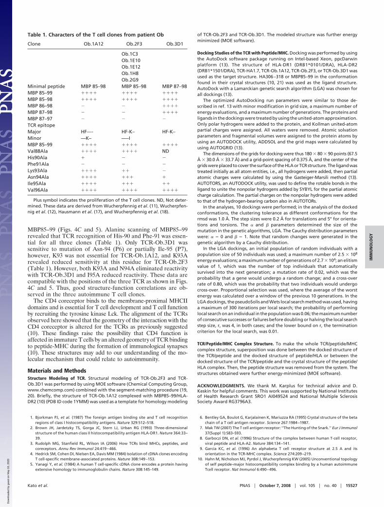
MBP85–99 (Figs. 4C and 5). Alanine scanning of MBP85–99revealed that TCR recognition of His-90 and Phe-91 was essen-tial for all three clones (Table 1). Only TCR-Ob.3D1 wassensitive to mutation of Asn-94 (P6) or partially Ile-95 (P7),however, K93 was not essential for TCR-Ob.1A12, and K93Arevealed reduced sensitivity at this residue for TCR-Ob.2F3(Table 1). However, both K93A and N94A eliminated reactivitywith TCR-Ob.3D1 and I95A reduced reactivity. These data arecompatible with the positions of the three TCR as shown in Figs.4C and 5. Thus, good structure-function correlations are ob-served in the three autoimmune T cell clones.
The CD4 coreceptor binds to the membrane-proximal MHCIIdomains and is essential for T cell development and T cell functionby recruiting the tyrosine kinase Lck. The alignment of the TCRsobserved here showed that the geometry of the interaction with theCD4 coreceptor is altered for the TCRs as previously suggested(10). These findings raise the possibility that CD4 function isaffected in immature T cells by an altered geometry of TCR bindingto peptide-MHC during the formation of immunological synapses(10). These structures may add to our understanding of the mo-lecular mechanism that could relate to autoimmunity.
Materials and MethodsStructure Modeling of TCR. Structural modeling of TCR-Ob.2F3 and TCR-Ob.3D1 was performed by using MOE software (Chemical Computing Group,www.chemcomp.com) combined with the segment-matching procedure (19,20). Briefly, the structure of TCR-Ob.1A12 complexed with MBP85–99/HLA-DR2 (10) (PDB ID code 1YMM) was used as a template for homology modeling
of TCR-Ob.2F3 and TCR-Ob.3D1. The modeled structure was further energyminimized (MOE software).
Docking Studies of the TCR with Peptide/MHC. Docking was performed by usingthe AutoDock software package running on Intel-based Xeon, ppcDarwinplatform (13). The structure of HLA-DR1 (DRB1*0101/DRA), HLA-DR2(DRB1*1501/DRA), TCR-HA1.7, TCR-Ob.1A12, TCR-Ob.2F3, or TCR-Ob.3D1 wasused as the target structure. HA306–318 or MBP85–99 in the conformationfound in their crystal structures (10, 21) was used as the ligand structure.AutoDock with a Lamarckian genetic search algorithm (LGA) was chosen forall dockings (13).
The optimized AutoDocking run parameters were similar to those de-scribed in ref. 13 with minor modification in grid size, a maximum number ofenergy evaluations, and a maximum number of generations. The proteins andligands in the dockings were treated by using the united-atom approximation.Only polar hydrogens were added to the protein, and Kollman united-atompartial charges were assigned. All waters were removed. Atomic solvationparameters and fragmental volumes were assigned to the protein atoms byusing an AUTODOCK utility, ADDSOL and the grid maps were calculated byusing AUTOGRID (13).
The dimensions of the grids for docking were thus 180 � 80 � 90 points (67.5Å � 30.0 Å � 33.7 Å) and a grid-point spacing of 0.375 Å, and the center of thegrids were placed to cover the surface of the HLA or TCR structure. The ligand wastreated initially as all atom entities, i.e., all hydrogens were added, then partialatomic charges were calculated by using the Gasteiger-Marsili method (13).AUTOTORS, an AUTODOCK utility, was used to define the rotable bonds in theligand to unite the nonpolar hydrogens added by SYBYL for the partial atomiccharge calculation. The partial charges on the nonpolar hydrogens were addedto that of the hydrogen-bearing carbon also in AUTOTORs.
In the analyses, 10 dockings were performed; in the analysis of the dockedconformations, the clustering tolerance as different conformations for thermsd was 1.0 Å. The step sizes were 0.2 Å for translations and 5° for orienta-tions and torsions. The � and � parameters determined the size of themutation in the genetic algorithms, LGA. The Cauchy distribution parameterswere: � � 0 and � � 1. Note that random changes were generated in thegenetic algorithm by a Cauchy distribution.
In the LGA dockings, an initial population of random individuals with apopulation size of 50 individuals was used; a maximum number of 2.5 � 108
energy evaluations; a maximum number of generations of 2.7 � 104; an elitismvalue of 1, which was the number of top individuals that automaticallysurvived into the next generation; a mutation rate of 0.02, which was theprobability that a gene would undergo a random change; and a cross-overrate of 0.80, which was the probability that two individuals would undergocross-over. Proportional selection was used, where the average of the worstenergy was calculated over a window of the previous 10 generations. In theLGA dockings, the pseudoSolis and Wets local search method was used, havinga maximum of 300 iterations per local search; the probability of performinglocal search on an individual in the population was 0.06; the maximum numberof consecutive successes or failures before doubling or halving the local searchstep size, r, was 4, in both cases; and the lower bound on r, the terminationcriterion for the local search, was 0.01.
TCR/Peptide/MHC Complex Structure. To make the whole TCR/peptide/MHCcomplex structure, superposition was done between the docked structure ofthe TCR/peptide and the docked structure of peptide/HLA or between thedocked structure of the TCR/peptide and the crystal structure of the peptide/HLA complex. Then, the peptide structure was removed from the system. Thestructures obtained were further energy-minimized (MOE software).
ACKNOWLEDGMENTS. We thank M. Karplus for technical advice and D.Keskin for helpful comments. This work was supported by National Institutesof Health Research Grant 5RO1 AI049524 and National Multiple SclerosisSociety Award RG3796A3.
1. Bjorkman PJ, et al. (1987) The foreign antigen binding site and T cell recognitionregions of class I histocompatibility antigens. Nature 329:512–518.
2. Brown JH, Jardetzky TS, Gorga JC, Stern LJ, Urban RG (1993) Three-dimensionalstructure of the human class II histocompatibility antigen HLA-DR1. Nature 364:33–39.
3. Rudolph MG, Stanfield RL, Wilson IA (2006) How TCRs bind MHCs, peptides, andcoreceptors. Annu Rev Immunol 24:419–466.
4. Hedrick SM, Cohen DI, Nielsen EA, Davis MM (1984) Isolation of cDNA clones encodingT cell-specific membrane-associated proteins. Nature 308:149–153.
5. Yanagi Y, et al. (1984) A human T cell-specific cDNA clone encodes a protein havingextensive homology to immunoglobulin chains. Nature 308:145–149.
6. Bentley GA, Boulot G, Karjalainen K, Mariuzza RA (1995) Crystal structure of the betachain of a T cell antigen receptor. Science 267:1984–1987.
7. Mak TW (2007) The T cell antigen receptor: ‘‘The Hunting of the Snark.’’ Eur J Immunol37(Suppl 1):S83–S93.
8. Garboczi DN, et al. (1996) Structure of the complex between human T-cell receptor,viral peptide and HLA-A2. Nature 384:134–141.
9. Garcia KC, et al. (1996) An alphabeta T cell receptor structure at 2.5 A and itsorientation in the TCR-MHC complex. Science 274:209–219.
10. Hahn M, Nicholson MJ, Pyrdol J, Wucherpfennig KW (2005) Unconventional topologyof self peptide–major histocompatibility complex binding by a human autoimmuneTcell receptor. Nat Immunol 6:490–496.
Table 1. Characters of the T cell clones from patient Ob
Clone Ob.1A12 Ob.2F3 Ob.3D1
Ob.1C3Ob.1E10Ob.1E12Ob.1H8Ob.2G9
Minimal peptide MBP 85–98 MBP 85–98 MBP 87–98MBP 85–99 ���� ���� ����
MBP 85–98 ���� ���� ����
MBP 86–98 � � ����
MBP 87–98 � � ����
MBP 87–97 � � �
TCR epitopeMajor HF—- HF-K– HF-K–Minor —K– —–IMBP 85–99 ���� ���� ����
Val88Ala ���� ���� NDHis90Ala � � �
Phe91Ala � � �
Lys93Ala ���� �� �
Asn94Ala ���� ��� �
Ile95Ala ���� ��� ��
Val96Ala ���� ���� ����
Plus symbol indicates the proliferation of the T cell clones. ND, Not deter-mined. These data are derived from Wucherpfennig et al. (11), Wucherpfen-nig et al. (12), Hausmann et al. (17), and Wucherpfennig et al. (18).
Kato et al. PNAS � October 7, 2008 � vol. 105 � no. 40 � 15527
IMM
UN
OLO
GY
Dow
nloa
ded
by g
uest
on
May
10,
202
0
11. Wucherpfennig KW, et al. (1994) Clonal expansion and persistence of human T cellsspecific for an immunodominant myelin basic protein peptide. J Immunol 152:5581–5592.
12. Wucherpfennig KW, et al. (1994) Structural requirements for binding of an immuno-dominant myelin basic protein peptide to DR2 isotypes and for its recognition byhuman T cell clones. J Exp Med 179:279–290.
13. Morris GM, Goodsell DS, Huey R, Olson AJ (1996) Distributed automated docking offlexible ligands to proteins: Parallel applications of AutoDock 2.4. J Comput Aided MolDes 10:293–304.
14. Stoddard BL, Koshland DE, Jr (1992) Prediction of the structure of a receptor-proteincomplex using a binary docking method. Nature 358:774–776.
15. Maynard J, et al. (2005) Structure of an autoimmune T cell receptor complexed with classII peptide- MHC: Insights into MHC bias and antigen specificity. Immunity 22:81–92.
16. Li Y, et al. (2005) Structure of a human autoimmune TCR bound to a myelin basicprotein self-peptide and a multiple sclerosis-associated MHC class II molecule. EMBO J24:2968–2979.
17. Hausmann S, Martin M, Gauthier L, Wucherpfennig KW (1999) Structural features ofautoreactive TCR that determine the degree of degeneracy in peptide recognition.J Immunol 162:338–344.
18. Wucherpfennig KW, Hafler DA, Strominger JL (1995) Structure of human T-cell recep-tors specific for an immunodominant myelin basic protein peptide: Positioning of T-cellreceptors on HLA-DR2/peptide complexes. Proc Natl Acad Sci USA 92:8896–8900.
19. Fechteler T, Dengler U, Schomberg D (1995) Prediction of Protein Three-DimensionalStructures in Insertion and Deletion Regions: A Procedure for Searching Databases ofRepresentative Protein Fragments Using Geometric Scoring Criteria. J Mol Biol253:114–131.
20. Levitt M (1992) Accurate Modeling of Protein Conformation by Automatic SegmentMatching. J Mol Biol 226:507–533.
21. Hennecke J, Carfi A, Wiley DC (2000) Structure of a covalently stabilized complex of ahuman �� T-cell receptor, influenza HA peptide and MHCII molecule, HLA-DR1. EMBOJ 19:5611–5624.
15528 � www.pnas.org�cgi�doi�10.1073�pnas.0807338105 Kato et al.
Dow
nloa
ded
by g
uest
on
May
10,
202
0