since 1972 Japanese Society of Oral Implantology 1 2 (Sat ...
Volume 2, Issue 4, December 2014
www.poseido.net www.poseido.info An official publication of the POSEIDO Academic network
in partnership with:
ABROSS (Academia Brasileira de Osseointegração)
AFI (Association Française d’Implantologie)
PSI (Polish Society of Implantology)
RADI (Russian Association of Dental Implantology)
SEI (Sociedad Española de Implantes)
SENAME Association (South European, North African,
Middle Eastern) SICOI (Societa Italiana di
Chirurgia Orale ed Implantologia)
SIOCH (Sociedad de Implantología Oral de Chile) SSA (Shanghai Stomatology
Association) TAO (Taiwan Academy of
Osseointegration) USP (Ukrainian Society of
Periodontologists)
ISSN 2307-5295, Published by the POSEIDO Organization & Foundation under a Creative Commons Attribution-NonCommercial-NoDerivatives 4.0 International
(CC BY-NC-ND 4.0) License
POSEIDO Journal Periodontology, Oral Surgery, Esthetic & Implant Dentistry Open
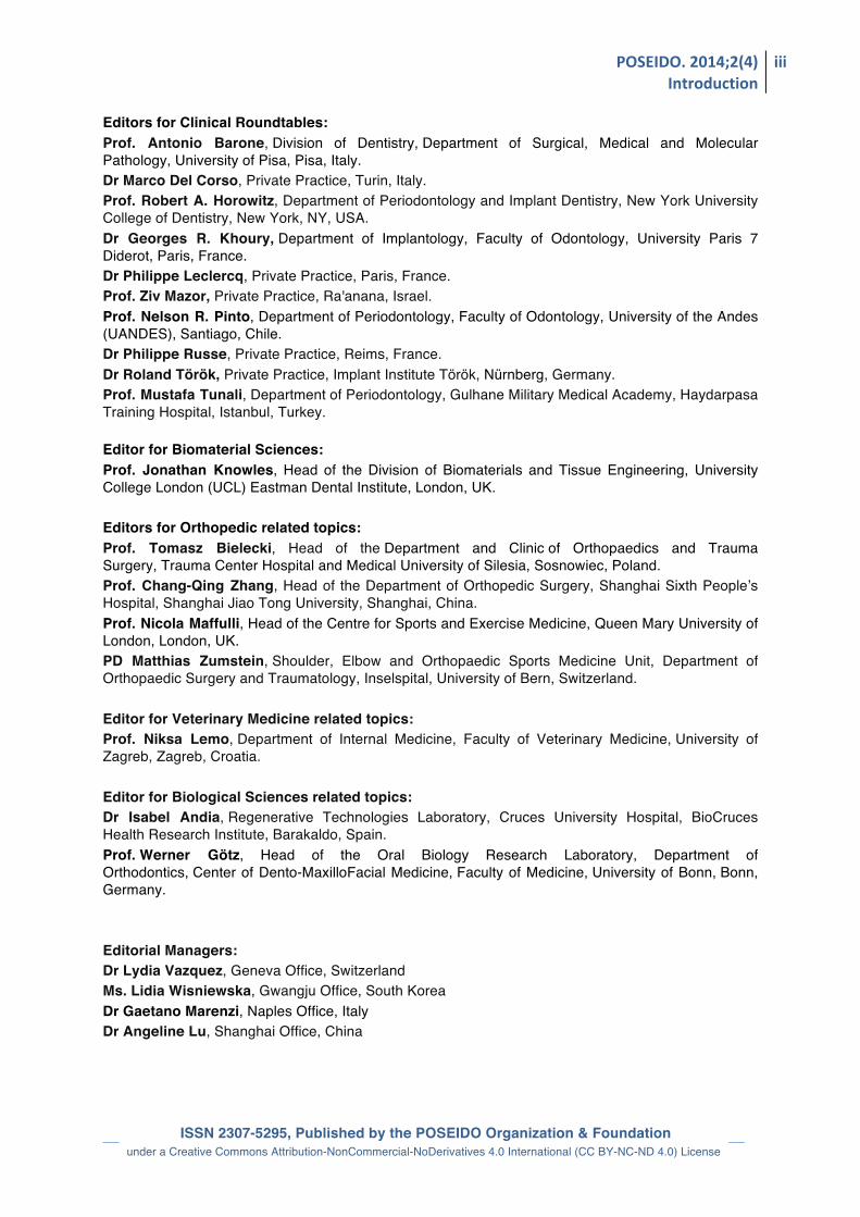
Table of Contents POSEIDO. 2014;2(4):225-61. Special Theme: Clinical round table about the implant-supported rehabilitation of the resorbed posterior mandible (Part 2)
Editorial The development of special theme issues and the POSEIDO Forum Civitatis: OASIS, PACT, ISAIAS, APOLLO, CRONOS, or a new approach to transdisciplinarity David M. Dohan Ehrenfest, Lidia M. Wisniewska, Nelson R. Pinto, Marsel Z. Mirgazizov, Chang-Qing Zhang, Adriano Piattelli Research articles: rehabilitation of the resorbed posterior mandible Interpositional graft associated with alveolar osteotomy for posterior mandible ridge augmentation (sandwich osteotomy) using hydroxyapatite or autogenous bone: histological evaluation Karen Bechara, Alexandre M. Dottore, Paulo Y. Kawakami, Alessandra Cassoni, Gabriela Giro, Jose Augusto Rodrigues, Leandro Chambrone, Adriano Piattelli, Giovanna Iezzi, Jamil Awad Shibli Comparative evaluation between one-piece implants of zirconia or titanium placed in posterior mandible: 6 months follow-up Ricardo R. Vecchiatti, Andre L.O. Campos, Welington F. Morais, Jose A. Rodrigues, Alessandra Cassoni, Jamil Awad Shibli Research article Accuracy of guided osteotomy using dental implant treatment-planning software in combination with an optical scan of a dental cast Bruce J. Kowalski, Marc Manos, Michael Levi, Joe “Ambrose” D’Ambrosia

POSEIDO. 2014;2(4) Introduction
ii
ISSN 2307-5295, Published by the POSEIDO Organization & Foundation
under a Creative Commons Attribution-NonCommercial-NoDerivatives 4.0 International (CC BY-NC-ND 4.0) License
Editorial Board Editor-in-Chief: Prof. David M. Dohan Ehrenfest, Head of the LoB5 unit, Research Center for Biomineralization Disorders, School of Dentistry Chonnam National University, Gwangju, South Korea. Senior Editors: Prof. Jean-Pierre Bernard, Head of the Department of Stomatology, Oral Surgery, Implantology and Dental and Maxillofacial Radiology and Vice-Dean, School of Dental Medicine, University of Geneva, Geneva, Switzerland. Prof. Gilberto Sammartino, Head of the Department of Oral Surgery, Faculty of Medicine, University Federico II, Naples, Italy. Prof. Jamil Awad Shibli, Head of Oral Implantology Clinic, Department of Periodontology and Oral Implantology, Dental Research Division, University of Guarulhos, Guarulhos, Sao Paulo, Brazil. Prof. Hom-Lay Wang, Endowed Collegiate Professor of Periodontics, Director of Graduate Periodontics, Department of Periodontics and Oral Medicine, School of Dentistry, University of Michigan, Ann Arbor, MI, USA. Prof. De-Rong Zou, Head of the Department of Stomatology, Shanghai Sixth People’s Hospital, Shanghai Jiao Tong University, Shanghai, China. Associate Editors: Prof. Camile S. Farah, Head of the Oral Oncology Research Program, School of Dentistry, University of Queensland, Brisbane, Australia. Prof. Erhan Firatli, Head of the Department of Periodontology, School of Dentistry, Istanbul University, Istanbul, Turkey. Prof. Arthur B. Novaes Jr., Head of the Department of Periodontology, School of Dentistry of Ribeirao Preto, University of Sao Paulo, Brazil. Prof. Adriano Piattelli, Professor of Oral Pathology and Medicine, School of Dentistry, University of Chieti-Pescara, Chieti, Italy. Prof. Georgios E. Romanos, Associate Dean for Clinical Affairs, School of Dental Medicine, Stony Brook University, Stony Brook, NY, USA. Prof. Dong-Seok Sohn, Head of the Department of Oral and Maxillofacial Surgery, School of Medicine, Catholic University of Daegu, Daegu, South Korea. Prof. Tiziano Testori, Istituto Ortopedico Galeazzi, Department of Implantology and Oral rehabilitation, School of Dentistry, University of Milan, Italy. Prof. Laurence J. Walsh, Head of the School of Dentistry, University of Queensland, Brisbane, Australia.
Volume 2, Issue 4, December 2014
POSEIDO. 2014;2(4) Introduction
iii
ISSN 2307-5295, Published by the POSEIDO Organization & Foundation
under a Creative Commons Attribution-NonCommercial-NoDerivatives 4.0 International (CC BY-NC-ND 4.0) License
Editors for Clinical Roundtables: Prof. Antonio Barone, Division of Dentistry, Department of Surgical, Medical and Molecular Pathology, University of Pisa, Pisa, Italy. Dr Marco Del Corso, Private Practice, Turin, Italy. Prof. Robert A. Horowitz, Department of Periodontology and Implant Dentistry, New York University College of Dentistry, New York, NY, USA. Dr Georges R. Khoury, Department of Implantology, Faculty of Odontology, University Paris 7 Diderot, Paris, France. Dr Philippe Leclercq, Private Practice, Paris, France. Prof. Ziv Mazor, Private Practice, Ra'anana, Israel. Prof. Nelson R. Pinto, Department of Periodontology, Faculty of Odontology, University of the Andes (UANDES), Santiago, Chile. Dr Philippe Russe, Private Practice, Reims, France. Dr Roland Török, Private Practice, Implant Institute Török, Nürnberg, Germany. Prof. Mustafa Tunali, Department of Periodontology, Gulhane Military Medical Academy, Haydarpasa Training Hospital, Istanbul, Turkey. Editor for Biomaterial Sciences: Prof. Jonathan Knowles, Head of the Division of Biomaterials and Tissue Engineering, University College London (UCL) Eastman Dental Institute, London, UK. Editors for Orthopedic related topics: Prof. Tomasz Bielecki, Head of the Department and Clinic of Orthopaedics and Trauma Surgery, Trauma Center Hospital and Medical University of Silesia, Sosnowiec, Poland. Prof. Chang-Qing Zhang, Head of the Department of Orthopedic Surgery, Shanghai Sixth People’s Hospital, Shanghai Jiao Tong University, Shanghai, China. Prof. Nicola Maffulli, Head of the Centre for Sports and Exercise Medicine, Queen Mary University of London, London, UK. PD Matthias Zumstein, Shoulder, Elbow and Orthopaedic Sports Medicine Unit, Department of Orthopaedic Surgery and Traumatology, Inselspital, University of Bern, Switzerland. Editor for Veterinary Medicine related topics: Prof. Niksa Lemo, Department of Internal Medicine, Faculty of Veterinary Medicine, University of Zagreb, Zagreb, Croatia. Editor for Biological Sciences related topics: Dr Isabel Andia, Regenerative Technologies Laboratory, Cruces University Hospital, BioCruces Health Research Institute, Barakaldo, Spain. Prof. Werner Götz, Head of the Oral Biology Research Laboratory, Department of Orthodontics, Center of Dento-MaxilloFacial Medicine, Faculty of Medicine, University of Bonn, Bonn, Germany. Editorial Managers: Dr Lydia Vazquez, Geneva Office, Switzerland Ms. Lidia Wisniewska, Gwangju Office, South Korea Dr Gaetano Marenzi, Naples Office, Italy Dr Angeline Lu, Shanghai Office, China

POSEIDO. 2014;2(4) Introduction
iv
ISSN 2307-5295, Published by the POSEIDO Organization & Foundation
under a Creative Commons Attribution-NonCommercial-NoDerivatives 4.0 International (CC BY-NC-ND 4.0) License
Volume 2, Issue 4, December 2014
Aims and Scope of the POSEIDO Journal The POSEIDO journal focuses on all aspects of the interconnected clinical and
research fields of periodontal sciences, oral and cranio-maxillofacial surgery and medicine, esthetic and restorative dentistry, with a particular interest in implant dentistry, and related research.
Most publications are connected to the dental and maxillofacial field, but some are also from orthopedics, material sciences or other scientific disciplines interconnected with the POSEID research topics (e.g. bone implantable materials, bone regenerative medicine strategies), in order to promote transversal translational research.
POSEIDO is organized as an info journal (international forum), and is therefore publishing a significant quantity of editorial material, as a basis of information, debate and discussion for our community. This editorial material takes particularly the form of clinical case letters and research letters.
The objective of this strong editorial section is to create links between international research teams, to organize our international research community and to develop a neutral international platform for the publication of debates and consensus conferences in the fast-growing and evolving fields of the POSEID disciplines.
The journal is also publishing a classical content with full-length articles (original articles and reviews), following a strict double peer-review process. The journal is particularly interested in original research articles and clinical studies about new techniques, biomaterials and biotechnologies with direct clinical applications in the interconnected fields of periodontology, oral surgery, esthetic and implant dentistry. Review articles are also welcome if they make the clear synthesis of debated topics.
Detailed guidelines for authors can be found on http://www.poseido.info
POSEIDO. 2014;2(4) Introduction
v
ISSN 2307-5295, Published by the POSEIDO Organization & Foundation
under a Creative Commons Attribution-NonCommercial-NoDerivatives 4.0 International (CC BY-NC-ND 4.0) License
The POSEIDO Journal is the official publication of the POSEIDO Academic network, in partnership with:
Main partners: ABROSS (Academia Brasileira de Osseointegração), Brazil
APPO (Asociacion Peruana de Periodoncia y Oseointegracion), Peru AFI (Association Française d'Implantologie), France
International Piezosurgery Academy, International PSI (Polish Society of Implantology), Poland
RADI (Russian Association of Dental Implantology), Russia SEI (Sociedad Española de Implantes), Spain
SENAME Association (South European, North African, Middle Eastern, Implantology & Modern Dentistry Association), International
SICOI (Societa Italiana di Chirurgia Orale ed Implantologia), Italy SIOCH (Sociedad de Implantología Oral de Chile), Chile
SSA (Shanghai Stomatology Association), China Swiss International Academy for Osseointegration and Maxillofacial Research
Foundation, Switzerland TAO (Taiwan Academy of Osseointegration), Taiwan
UASOM (Union des Associations Scientifiques Odontologiques Marocaines), Morocco USP (Ukrainian Society of Periodontologists), Ukraine
Partners of the POSEIDO Network: Academy of Non Transfusional HEmo-Components (ANTHEC), Italy Association Tunisienne Odontologique de Recherches et d'Etudes en Chirurgie et Douleur (ATORECD), Tunisia Fundación para el Estudio y Desarrollo de la Implantología, Cirugía Oral y Maxilofacial (FEDICOM), Spain Fund of Development of High Stomatologic Technologies of Russia (Biocompatible materials and implants), Russia Lebanese Society of Oral Surgery, Lebanon Mongolian Association of Periodontology, Mongolia Mongolian Dental Association, Mongolia Moroccan Society of Oral Medicine and Oral Surgery, Morocco Sociedad Española de Cirugía Bucal (SECIB), Spain Sociedad Española de Prótesis Estomatológica y Estética (SEPES), Spain Société d'Implantologie Orale et de Prothese Appliquee (SIOPA), France Société Internationale de Formation et de Recherche en Implantologie Orale (SIFRIO), France Société Tunisienne d'Implantologie et de Dentisterie Esthétique (STIDE), Tunisia Stomatology for all / International Dental Review Magazine, Russia Titanium Club, International
POSEIDO. 2014;2(4) Introduction
vi
ISSN 2307-5295, Published by the POSEIDO Organization & Foundation
under a Creative Commons Attribution-NonCommercial-NoDerivatives 4.0 International (CC BY-NC-ND 4.0) License
This issue of the POSEIDO Journal is supported by a grant from the National Research Foundation of Korea (NRF) funded by the Korean government-MEST (No. 2011-0030121) and by the LoB5 Foundation for Research, France.
Table of Contents POSEIDO. 2014;2(4):225-61. Special Theme: Clinical round table about the implant-supported rehabilitation of the resorbed posterior mandible (Part 2) Editorial The development of special theme issues and the POSEIDO Forum Civitatis: OASIS, PACT, ISAIAS, APOLLO, CRONOS, or a new approach to transdisciplinarity David M. Dohan Ehrenfest, Lidia M. Wisniewska, Nelson R. Pinto, Marsel Z. Mirgazizov, Chang-Qing Zhang, and Adriano Piattelli Research articles: rehabilitation of the resorbed posterior mandible Interpositional graft associated with alveolar osteotomy for posterior mandible ridge augmentation (sandwich osteotomy) using hydroxyapatite or autogenous bone: histological evaluation Karen Bechara, Alexandre M. Dottore, Paulo Y. Kawakami, Alessandra Cassoni, Gabriela Giro, Jose Augusto Rodrigues, Leandro Chambrone, Adriano Piattelli, Giovanna Iezzi, and Jamil Awad Shibli Comparative evaluation between one-piece implants of zirconia or titanium placed in posterior mandible: 6 months follow-up Ricardo R. Vecchiatti, Andre L.O. Campos, Welington F. Morais, Jose A. Rodrigues, Alessandra Cassoni, and Jamil Awad Shibli Research article Accuracy of guided osteotomy using dental implant treatment-planning software in combination with an optical scan of a dental cast Bruce J. Kowalski, Marc Manos, Michael Levi, and Joe “Ambrose” D’Ambrosia
225-31 233-9 241-51 253-61
POSEIDO. 2014;2(4) Special theme issues and Forum Civitatis
225
ISSN 2307-5295, Published by the POSEIDO Organization & Foundation
under a Creative Commons Attribution-NonCommercial-NoDerivatives 4.0 International (CC BY-NC-ND 4.0) License
Editorial The development of special theme issues and the POSEIDO Forum Civitatis: OASIS, PACT, ISAIAS, APOLLO, CRONOS, or a new approach to transdisciplinarity David M. Dohan Ehrenfest,1,* Lidia M. Wisniewska,1,2,3 Nelson R. Pinto,4,5 Marsel Z. Mirgazizov,6 Chang-Qing Zhang,7 and Adriano Piattelli.8 1 LoB5 research unit, School of Dentistry & Research Center for Biomineralization Disorders, Chonnam National University, Gwangju, South Korea 2 Department of Didactics and School Organization, Faculty of Education Sciences, University of Granada, Granada, Spain 3 Department of International Relations, Paris Sorbonne University, Paris, France 4 Graduate School of Periodontics and Implant Dentistry, University of the Andes (UANDES), Santiago, Chile 5 Department of Oral Health Sciences, Katholieke Universiteit Leuven (KUL) & Periodontology, University Hospitals Leuven, Leuven, Belgium 6 Federal Fund of Development of High Stomatologic Technologies of Russia (Biocompatible materials and implants), Moscow, Russia 7 Department of Orthopedic Surgery, Shanghai Sixth People’s Hospital, Shanghai Jiao Tong University, Shanghai, China 8 Department of Medical, Oral and Biotechnological Sciences, University of Chieti-Pescara, Chieti, Italy *Corresponding author: David M. Dohan Ehrenfest, [email protected] Submitted on October 17th, 2014; accepted after minor corrections on October 27th, 2014.
The POSEIDO (Periodontology, Oral Surgery, Esthetic & Implant Dentistry Open)
journal is now finishing its second year of successful existence as a new concept of inter-academic collaborative publishing platform [1]. The journey of the POSEIDO Community is only beginning, and many efforts are still needed to reach our vision and objectives, but these 2 first years allowed use to build the general skeleton of the POSEIDO environment, to illustrate the expected functioning of this platform, and also to launch the development of many working groups and collaborative projects, particularly a first series of Forum termed Forum Civitatis (in Latin, the “Forum of the City” literally, i.e. a platform of discussion and cooperation for our Community of Experts). On all these aspects, we can already gather a few observations and comments from these 2 years of activity.
1. Special themes, but each issue opened to all submissions As it was initially stated, the first POSEIDO journal is not a mass publication journal,
but a specialized theme journal [1]. One important characteristic of POSEIDO is the wish to create a platform of discussion and cooperation, to promote real international debates and sometimes consensus in the POSEID (Periodontology, Oral Surgery, Esthetic & Implant Dentistry) disciplines [2]. To reach this objective, a special theme is allocated to each issue of the journal, in order to regroup articles on a specific aspect of clinical or basic research and try to develop joint discussion and consensus articles.
This concept was designed for educational purposes, as the international scientific literature is growing extensively and data are more and more difficult to sort and interpret for the general readership, even for specialists, what leaves most publications with a very
226 Editorial: Dohan Ehrenfest DM, et al. (2014)
ISSN 2307-5295, Published by the POSEIDO Organization & Foundation
under a Creative Commons Attribution-NonCommercial-NoDerivatives 4.0 International (CC BY-NC-ND 4.0) License
limited real impact. By gathering articles around a theme and try to reach a consensus or at least a debate around this theme, the journal becomes more reader-friendly, and therefore more interesting and useful for our wide readership. We shall never forget that a scientific publication is first of all designed to serve a Community - in the case of POSEIDO, it is particularly a wide Community of oral and maxillofacial clinicians and basic researchers. The biggest success for a journal nowadays, it is to interest its readers and, in short, to be read extensively and kept preciously as a real source of knowledge. The special themes are therefore an interesting method to reach these educational and clear communication objectives.
The development of special themes in each issue is, however, only a complementary strategy, and shall not be considered as a mandatory style of the Journal. This notion of special theme was sometimes understood as a limitation for submission. Many authors hesitated to submit their articles on different topics than the special themes, believing that they would not fit the profile of the journal. In reality, the journal is opened to all submissions, whatever the topic. If the Editors try to regroup the items in thematic issues, this is not a mandatory characteristic and independent articles can be published without limitations as long as they can interest our readership and bring interesting information. Moreover, authors and research group members of the POSEIDO network are welcome to submit proposals of theme issues and suggestions of publications. The board of the journal will review all proposals carefully and openly, and motivated initiatives will always be appreciated. The functioning of POSEIDO is very flexible and adjusted to merits and timely initiatives. All authors are welcome to participate to this effort, and they will only be considered based on their work and scientific skills, with a special expectation on creativity and innovation.
In summary, if you have something interesting to publish, please do not hesitate to submit to the POSEIDO Journal, whatever the upcoming special themes.
2. The Forum Civitatis, a platform for transdisciplinarity The concept of Special Theme was completed by another method of gathering of thematic knowledge, with a particular emphasis on transdisciplinarity and translational research. The knowledge that interests the POSEIDO diverse readership cannot be limited to clinical techniques or evaluation of materials in the oral field, as many aspects of the daily practice are in fact interconnected with other disciplines, particularly medical sciences (plastic surgery, orthopedics, oncology, etc.), engineering or education. This transdisciplinarity and translational approach are often needed to understand correctly some key parameters of the oral disciplines, but no integrated platform of cooperation and debate existed previously. This is why the notion of Special Theme was completed by the concept of Forum Civitatis.
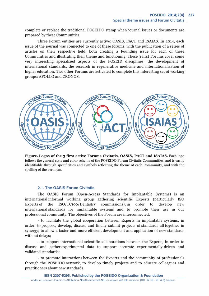
The Forum Civitatis - or Forum of the City, our Community of Colleagues - is regrouping several platforms of exchange and debates, under the form of wide communities or smaller working groups for Experts, with the idea to develop new ideas and concepts in various aspects of the POSEID disciplines and to promote it through education and research. These forums are clearly identifiable through their own logo, following the general style and color scheme of the POSEIDO Forum Civitatis Communities (Figure). Each logo presents clear specificities and symbols reflecting the theme of each Community and the spelling of the acronym is always indicated. These icons were designed to be easily identifiable and to
POSEIDO. 2014;2(4) Special theme issues and Forum Civitatis
227
ISSN 2307-5295, Published by the POSEIDO Organization & Foundation
under a Creative Commons Attribution-NonCommercial-NoDerivatives 4.0 International (CC BY-NC-ND 4.0) License
complete or replace the traditional POSEIDO stamp when journal issues or documents are prepared by these Communities.
Three Forum entities are currently active: OASIS, PACT and ISAIAS. In 2014, each issue of the journal was connected to one of these forums, with the publication of a series of articles on their respective field, both creating a Founding issue for each of these Communities and illustrating their theme and functioning. These 3 first Forums cover some very interesting specialized aspects of the POSEID disciplines: the development of international standards, the research in regenerative medicine and internationalization of higher education. Two other Forums are activated to complete this interesting set of working groups: APOLLO and CRONOS.
Figure. Logos of the 3 first active Forums Civitatis, OASIS, PACT and ISAIAS. Each logo follows the general style and color scheme of the POSEIDO Forum Civitatis Communities, and is easily identifiable through specificities and symbols reflecting the theme of each Community, and with the spelling of the acronym.
2.1. The OASIS Forum Civitatis
The OASIS Forum (Open-Access Standards for Implantable Systems) is an international informal working group gathering scientific Experts (particularly ISO Experts of the ISO/TC106/Dentistry commissions), in order to develop new international standards for implantable systems and to promote their use in our professional community. The objectives of the Forum are interconnected:
- to facilitate the global cooperation between Experts in implantable systems, in order: to propose, develop, discuss and finally submit projects of standards all together in synergy; to allow a faster and more efficient development and application of new standards without delays;
- to support international scientific collaborations between the Experts, in order to discuss and gather experimental data to support accurate experimentally-driven and validated standards;
- to promote interactions between the Experts and the community of professionals through the POSEIDO network, to develop timely projects and to educate colleagues and practitioners about new standards.
228 Editorial: Dohan Ehrenfest DM, et al. (2014)
ISSN 2307-5295, Published by the POSEIDO Organization & Foundation
under a Creative Commons Attribution-NonCommercial-NoDerivatives 4.0 International (CC BY-NC-ND 4.0) License
The OASIS Forum has been designed and funded by the POSEIDO Foundation in order to remain completely independent from the commercial pressures and lobbying. The resources of the Foundation can be allocated by the Board of Directors in the form of research grants for the development of outstanding projects of standards.
The first issue of 2014 (Volume 2, Issue 1) was the first OASIS issue of the POSEIDO Journal and focused on a first open-access standard for Implant Surface Identification (ISI)[3]. This massive work was published in a series of articles prepared by an international panel of Experts, and is a good example of extensive data the OASIS Consortium is able to gather for the POSEIDO readership.
2.2. The PACT Forum Civitatis
The PACT Forum (Platelet & Advanced Cell Therapies) is an international research and education scientific community in the field of tissue engineering and regenerative medicine, interested in various forms of cell therapies, particularly stem cell research and platelet concentrates for surgical or infiltrative use (Leukocyte- and Platelet-Rich Plasma L-PRP and Leukocyte- and Platelet-Rich Fibrin L-PRF)[4].
The concept of this community is to promote international debates and translational and transversal research, i.e. from the basic science research to the clinical applications (translational) and between the many disciplines involved in this wide field of research (transversal). Transdisciplinarity - as a research strategy crossing discipline boundaries to create a holistic approach - is the key to reach common consensus and terminology in the many disciplines involved in this wide field of regenerative medicine, and is an absolute necessity for an efficient and safe development of these technologies.
The PACT Forum has been designed and funded by the POSEIDO Foundation in order to remain completely independent from the commercial pressures and lobbying, which constitute the major threat damaging the credibility of this scientific field.
The second issue of 2014 (Volume 2, Issue 2) was the first PACT issue of the POSEIDO Journal [4]. It reviewed the current endeavor in the field and gathered several major articles on the topic, particularly concerning the diversity of cells observable in a platelet concentrate and the impact of centrifuge quality and protocol on the cell content and biological signature of L-PRF (Leukocyte- and Platelet-Rich Fibrin) clots and membranes [5]. This issue is a good illustration of the PACT philosophy and a major scientific milestone prepared by this Community. 2.3. The ISAIAS Forum Civitatis
The ISAIAS Forum (Intercultural Sensitivity Academic Index & Advanced Standards) is an international informal working group about the internationalization of higher education and research in general, and its impact in dentistry in particular. This group is developing new concepts, methods and instruments of observation and development of the internationalization of an academic environment (University, campus, laboratory) and its impact on the perceptions and behaviors of all academic stakeholders (particularly students, teachers and researchers). This is a major instrument to improve international cooperation.
The third issue of 2014 (Volume 2, Issue 3) was the first ISAIAS issue of the POSEIDO Journal [6]. It described the general philosophy of international cooperation promoted by the POSEIDO Consortium, and the integration of the ISAIAS program as an interface to optimize and monitor the cooperation and internationalization of higher
POSEIDO. 2014;2(4) Special theme issues and Forum Civitatis
229
ISSN 2307-5295, Published by the POSEIDO Organization & Foundation
under a Creative Commons Attribution-NonCommercial-NoDerivatives 4.0 International (CC BY-NC-ND 4.0) License
education and research within the network. This issue illustrates also the kind of research this working group is implementing among the members of the network, as a powerful instrument of dialogue and preparation for deeper cooperation.
2.4. The APOLLO Forum Civitatis The APOLLO Forum (Advanced Plastic & Orthopedic Literature & Logic Open
Forum) is an international research and education scientific community in the field of plastic and orthopedic surgery and related research, designed to promote a transdisciplinary approach, debate and literature between medical fields and the POSEID sciences.
Plastic and orthopedic sciences are strongly connected with the POSEID disciplines, as these fields are sharing common interests in implantable biomaterials, surgical techniques and regenerative strategies and materials. For example, platelet concentrates (Leukocyte- and Platelet-Rich Plasma L-PRP and Leukocyte- and Platelet-Rich Fibrin L-PRF)[7] used in oral and maxillofacial surgery to improve soft and hard tissue healing, are also widely used as injection in sports medicine [8] and orthopedics [9] or as wound dressing for skin ulcer and wound healing in plastic surgery [10]. The experience gathered in one field is very precious for the understanding and improvement in other fields. Another example can be found in implant surface and bone biomaterials, where similar materials are used in implant dentistry and in orthopedics, while both disciplines are rarely sharing the same research perception and interests [11].
Despite this obvious transdisciplinarity among these fields, the cooperation between the domains remains indirect in most cases; the literature produced in one domain has in general a limited impact on others. As it can be observed in regenerative medicine and for implantable materials, there cannot be any significant advance without a better integration of the shared knowledge of these fields.
The objective of this open forum is, as the acronym spells it, to develop an integrated platform of literature and logic in plastic, orthopedic and dental sciences. The boundaries of this forum are in fact wider than plastic and orthopedic sciences, and the APOLLO Forum can cover most interconnected medical aspects in general. This original path requires to support interdisciplinary discussions, research and publications, to slowly create a common integrated publication body shared by Experts from these different fields.
2.5. The CRONOS Forum Civitatis The CRONOS Forum (Cancer Research, Oncology & Novel Oncological Systems) is an
international informal working group in the field of cancer research and oncology, with a particular interest in a transdisciplinary approach of cancer understanding and treatment, and in a more holistic conception of the oncological paradigms. Cancer research is an important field in the oral and maxillofacial area: cancers located in this area are among the most difficult to treat and their diagnosis is often connected to the observations of the oral specialists. Moreover, oral pathologies can influence negatively the outcomes of cancer treatments. Finally, the cancers and their treatments have always a significant impact on the oral health, both in terms of quality of life (discomfort, difficulties of swallowing and eating) and general health (e.g. development of mycosis or oral infections impacting the general health). Oral health is an important parameter and marker in oncology.
The CRONOS Forum does not focus strictly on oral cancers and oral aspects of cancer treatments. However, the objective of this group is not to publish extensively on cancer (what
230 Editorial: Dohan Ehrenfest DM, et al. (2014)
ISSN 2307-5295, Published by the POSEIDO Organization & Foundation
under a Creative Commons Attribution-NonCommercial-NoDerivatives 4.0 International (CC BY-NC-ND 4.0) License
would be outside of the theme of the POSEIDO Journal). CRONOS is first designed to promote a transdisciplinary cooperation on this topic and a more holistic approach of oncological research. Through this obvious need of transdisciplinarity, CRONOS is opened to discuss new paradigms in oncology and to offer original perspectives in the field. Finally, the CRONOS Forum has been designed and funded by the POSEIDO Foundation in order to remain completely independent from the commercial pressures and lobbying.
3. Perspectives On many aspects, the POSEIDO journal is an original and non-conventional
publication. POSEIDO was designed since its creation as an open-access self-managed cooperative platform for discussion and publication, based on international and transdisciplinary cooperation. Special themes and specialized Forums are strong elements of this strategy and identity.
The POSEIDO journal is particularly interested in transdisciplinarity and translational research. It can be observed in the board of the journal, which is including scholars from other non-dental disciplines such as material sciences, orthopedics, veterinary sciences, biological sciences, plastic surgery and oncology. Transdisciplinarity is very often the key for the successful development of new treatments and technologies, as it allows to combine the Expertise from different disciplines and to offer fresh perspectives to problems encountered in a field. In order to promote these cooperation and dialogues between different fields of medicine and research and to support this transdisciplinarity, the combination of special themes and specialized community Forums is our innovative path.
Finally, like many terms used in the POSEIDO environment, the term “Forum Civitatis” was selected carefully with its strong Ancient Latin meaning of a Community gathering around the Forum, a neutral place to exchange and talk. In the same way, it is important that all members of the POSEIDO Consortium contribute with honesty and efficiency to the development of the POSEIDO Common House, to make from this concept a major success. In this journey, specialized themes or forums are only instruments of cooperation, and the sincere motivation to join and participate to this Community remains the heart and motor of the development of this project worldwide. All members shall always remember that POSEIDO is an open platform and that all initiatives and proposals are welcome. Disclosure of interests
The authors have no conflict of interest to report. Acknowledgements
This issue was partially supported by a grant from the National Research Foundation of Korea (NRF) funded by the Korean government-MEST (No. 2011-0030121) and by the LoB5 Foundation for Research, France.
References [1] Dohan Ehrenfest DM, Sammartino G, Bernard JP. The Periodontology, Oral Surgery, Esthetic and Implant Dentistry Organization (POSEIDO) and Open Journal: an international academic and scientific community for a new approach of open-access publishing. POSEIDO. 2013;1(1):1-5.
POSEIDO. 2014;2(4) Special theme issues and Forum Civitatis
231
ISSN 2307-5295, Published by the POSEIDO Organization & Foundation
under a Creative Commons Attribution-NonCommercial-NoDerivatives 4.0 International (CC BY-NC-ND 4.0) License
[2] Dohan Ehrenfest DM, Sammartino G, Shibli JA, Wang HL, Zou DR, Bernard JP. Guidelines for the publication of articles related to platelet concentrates (Platelet-Rich Plasma - PRP, or Platelet-Rich Fibrin - PRF): the international classification of the POSEIDO. POSEIDO. 2013;1(1):17-27. [3] Dohan Ehrenfest DM, Del Corso M, Kang BS, Leclercq P, Mazor Z, Horowitz RA, Russe P, Oh HK, Zou DR, Shibli JA, Wang HL, Bernard JP, Sammartino G. Identification card and codification of the chemical and morphological characteristics of 62 dental implant surfaces. Part 1: description of the Implant Surface Identification Standard (ISIS) codification system. POSEIDO. 2014;2(1):7-22. [4] Sammartino G, Del Corso M, Wisniewska LM, Bielecki T, Andia I, Pinto NR, Zhang CQ, Zou DR, Dohan Ehrenfest DM. The PACT (Platelet & Advanced Cell Therapies) Forum: fostering translational research, transdisciplinarity and international collaboration in tissue engineering and regenerative medicine. POSEIDO. 2014;2(2):105-15. [5] Pinto NR, Pereda A, Jiménez P, Del Corso M, Kang BS, Wang HL, Quirynen M, Dohan Ehrenfest DM. The impact of the centrifuge characteristics and centrifugation protocols on the cells, growth factors and fibrin architecture of a Leukocyte- and Platelet-Rich Fibrin (L-PRF) clot and membrane. Part 2: macroscopic, photonic microscopy and Scanning Electron Microscopy analysis of 4 kinds of L-PRF clots and membranes. POSEIDO. 2014;2(2):141-54. [6] Dohan Ehrenfest DM, Wisniewska LM, Shibli JA, Mirgazizov MZ, Zou DR, Pinto NR, Bernard JP, Sammartino G, Wang HL. Developing a global scientific Community through cooperation and partnership: reinventing the intercultural interface, or the ISAIAS Prophecy in Internationalization of Higher Education and Research. POSEIDO. 2014;2(3):167-77. [7] Dohan Ehrenfest DM, Rasmusson L, Albrektsson T. Classification of platelet concentrates: from pure platelet-rich plasma (P-PRP) to leucocyte- and platelet-rich fibrin (L-PRF). Trends Biotechnol. 2009;27(3):158-67. [8] Mishra A, Harmon K, Woodall J, Vieira A. Sports medicine applications of platelet rich plasma. Curr Pharm Biotechnol. 2012;13(7):1185-95. [9] Yuan T, Guo SC, Han P, Zhang CQ, Zeng BF. Applications of leukocyte- and platelet-rich plasma (L-PRP) in trauma surgery. Curr Pharm Biotechnol. 2012;13(7):1173-84. [10] Cieslik-Bielecka A, Choukroun J, Odin G, Dohan Ehrenfest DM. L-PRP/L-PRF in esthetic plastic surgery, regenerative medicine of the skin and chronic wounds. Curr Pharm Biotechnol. 2012;13(7):1266-77. [11] Dohan Ehrenfest DM, Coelho PG, Kang BS, Sul YT, Albrektsson T. Classification of osseointegrated implant surfaces: materials, chemistry and topography. Trends Biotechnol. 2010;28(4):198-206. This article can be cited as: Dohan Ehrenfest DM, Wisniewska LM, Pinto NR, Mirgazizov MZ, Zhang CQ, Piattelli A. The development of special theme issues and the POSEIDO Forum Civitatis: OASIS, PACT, ISAIAS, APOLLO, CRONOS, or a new approach to transdisciplinarity. POSEIDO. 2014;2(4):225-31.
ISSN 2307-5295, Published by the POSEIDO Organization & Foundation
under a Creative Commons Attribution-NonCommercial-NoDerivatives 4.0 International (CC BY-NC-ND 4.0) License
POSEIDO. 2014;2(4) Sandwich osteotomy material in posterior mandible
233
ISSN 2307-5295, Published by the POSEIDO Organization & Foundation
under a Creative Commons Attribution-NonCommercial-NoDerivatives 4.0 International (CC BY-NC-ND 4.0) License
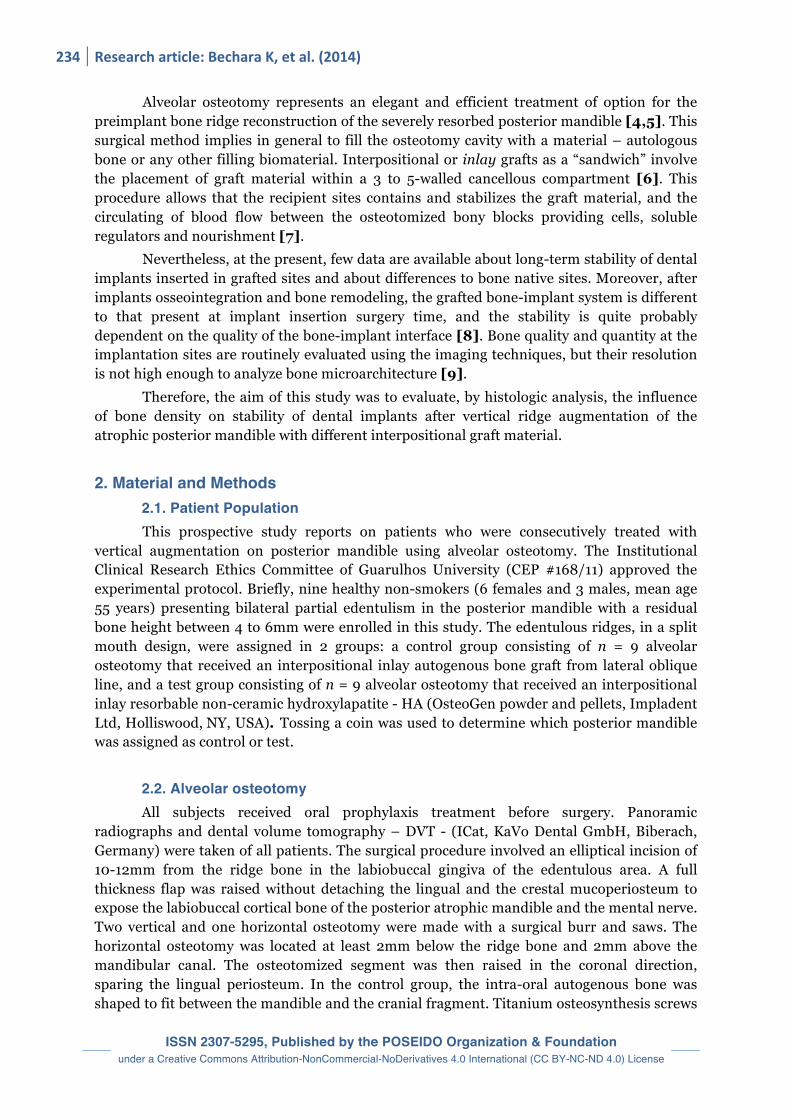
Research article Interpositional graft associated with alveolar osteotomy for posterior mandible ridge augmentation (sandwich osteotomy) using hydroxyapatite or autogenous bone: histological evaluation Karen Bechara,1 Alexandre M. Dottore,1 Paulo Y. Kawakami,1 Alessandra Cassoni,2 Gabriela Giro,1 Jose Augusto Rodrigues,2 Leandro Chambrone,1 Adriano Piattelli,3 Giovanna Iezzi,3 and Jamil Awad Shibli.1,* 1 Department of Periodontology and Oral Implantology, Dental Research Division, University of Guarulhos - UnG, São Paulo, Brazil 2 Department of Restorative Dentistry, Dental Research Division, University of Guarulhos - UnG, São Paulo, Brazil 3 Department of Medical, Oral and Biotechnological Sciences, University of Chieti-Pescara, Chieti, Italy *Corresponding author: Jamil Awad Shibli, [email protected] Submitted on April 10th, 2014; accepted after minor corrections on April 25th, 2014.
Abstract Background and objectives. The influence of interpositional graft material on bone behavior associated with alveolar osteotomy for posterior mandible ridge augmentation (osteotomy with sandwich technique) is not fully understood. Therefore, this study evaluated, histologically, the impact of 2-inlay grafts material in posterior mandible. Materials and Methods. Alveolar augmentation osteotomies were performed bilaterally in 9 partially edentulous mandibular patients in a split-mouth design. The alveolar segmental osteotomies were assigned in 2 groups: test group, interpositional hydroxyapatite (HA), and control group, interpositional intra-oral autogenous bone graft. After 6 months of healing, a bone core was retrieved from each side for histological evaluation before implant placement. Results. Ground sections depicted more newly-formed bone for autogenous group (p<0.05) and more residual-grafted material in the HA group (p<0.05). Discussion and conclusions. The results of this split mouth design suggest that both intra-oral autogenous bone and HA as an interpositional graft material to vertically augment posterior atrophic mandibles could be used. Keywords. Alveolar bone grafting, bone substitutes, dental implants, hydroxyapatites, osteotomy.
1. Introduction The resorption of the alveolar process may preclude implant placement, mainly in the
atrophic posterior mandible. Reconstruction of the alveolar process with bone augmentation prior to implant placement will facilitate the latter, but the result is influenced by the quality and quantity of the regenerated bone [1-3].
234 Research article: Bechara K, et al. (2014)
ISSN 2307-5295, Published by the POSEIDO Organization & Foundation
under a Creative Commons Attribution-NonCommercial-NoDerivatives 4.0 International (CC BY-NC-ND 4.0) License
Alveolar osteotomy represents an elegant and efficient treatment of option for the preimplant bone ridge reconstruction of the severely resorbed posterior mandible [4,5]. This surgical method implies in general to fill the osteotomy cavity with a material – autologous bone or any other filling biomaterial. Interpositional or inlay grafts as a “sandwich” involve the placement of graft material within a 3 to 5-walled cancellous compartment [6]. This procedure allows that the recipient sites contains and stabilizes the graft material, and the circulating of blood flow between the osteotomized bony blocks providing cells, soluble regulators and nourishment [7].
Nevertheless, at the present, few data are available about long-term stability of dental implants inserted in grafted sites and about differences to bone native sites. Moreover, after implants osseointegration and bone remodeling, the grafted bone-implant system is different to that present at implant insertion surgery time, and the stability is quite probably dependent on the quality of the bone-implant interface [8]. Bone quality and quantity at the implantation sites are routinely evaluated using the imaging techniques, but their resolution is not high enough to analyze bone microarchitecture [9].
Therefore, the aim of this study was to evaluate, by histologic analysis, the influence of bone density on stability of dental implants after vertical ridge augmentation of the atrophic posterior mandible with different interpositional graft material.
2. Material and Methods 2.1. Patient Population
This prospective study reports on patients who were consecutively treated with vertical augmentation on posterior mandible using alveolar osteotomy. The Institutional Clinical Research Ethics Committee of Guarulhos University (CEP #168/11) approved the experimental protocol. Briefly, nine healthy non-smokers (6 females and 3 males, mean age 55 years) presenting bilateral partial edentulism in the posterior mandible with a residual bone height between 4 to 6mm were enrolled in this study. The edentulous ridges, in a split mouth design, were assigned in 2 groups: a control group consisting of n = 9 alveolar osteotomy that received an interpositional inlay autogenous bone graft from lateral oblique line, and a test group consisting of n = 9 alveolar osteotomy that received an interpositional inlay resorbable non-ceramic hydroxylapatite - HA (OsteoGen powder and pellets, Impladent Ltd, Holliswood, NY, USA). Tossing a coin was used to determine which posterior mandible was assigned as control or test.
2.2. Alveolar osteotomy All subjects received oral prophylaxis treatment before surgery. Panoramic
radiographs and dental volume tomography – DVT - (ICat, KaVo Dental GmbH, Biberach, Germany) were taken of all patients. The surgical procedure involved an elliptical incision of 10-12mm from the ridge bone in the labiobuccal gingiva of the edentulous area. A full thickness flap was raised without detaching the lingual and the crestal mucoperiosteum to expose the labiobuccal cortical bone of the posterior atrophic mandible and the mental nerve. Two vertical and one horizontal osteotomy were made with a surgical burr and saws. The horizontal osteotomy was located at least 2mm below the ridge bone and 2mm above the mandibular canal. The osteotomized segment was then raised in the coronal direction, sparing the lingual periosteum. In the control group, the intra-oral autogenous bone was shaped to fit between the mandible and the cranial fragment. Titanium osteosynthesis screws

POSEIDO. 2014;2(4) Sandwich osteotomy material in posterior mandible
235
ISSN 2307-5295, Published by the POSEIDO Organization & Foundation
under a Creative Commons Attribution-NonCommercial-NoDerivatives 4.0 International (CC BY-NC-ND 4.0) License
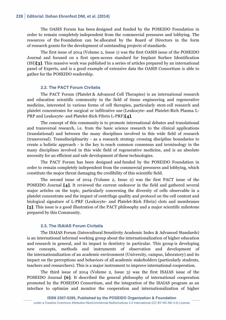
and plates were used to obtain stability. In the test group, the titanium plates were place before the hydroxyapatite (HA) to create an enough space. Follow, a mix of HA powder and pellets was added between the osteotomized bony. Gaps in the vertical osteotomies were filled with particulated autogenous (control group) or HA (test group)(Figure 1).
Primary wound closure was achieved with horizontal mattress sutures alternated with interrupted sutures to ensure a submerged healing procedure in segmented alveolar bone.
Postoperative care consisted of a 0.12% chlorhexidine mouth-rinse twice a day for 14 days without mechanical cleaning at the surgical areas. Anti-inflammatory medication (dexamethasone, 4 mg), was administered once analgesia (paracetamol, 750 mg) and antibiotic regimen with amoxicillin were prescribed. No removable prosthesis was allowed for 6 months. Figure 1. Clinical view of the posterior mandible fixed with miniplates and grafted with A) autogenous bone and B) hydroxylapatite.
2.3. Implant placement and bone biopsies Six months after augmentation, under local anesthesia, titanium miniplates and
screws were removed. Knife-edge ridges were flatted to allow a thickness of at least 4mm. Bone cores were harvested using a 2.0 x 10 mm– diameter trephine bur under sterile saline solution irrigation. The bone cores were retrieved at a minimum distance of 5 mm from the nearest teeth; the dimension of the bone cores was almost 2 x 6 mm. Screw-parallel shaped implants (Implacil, De Bortolli, São Paulo, Brazil; and 3i Biomet Implants, Palm Beach Gardens, FL, USA) with sandblasted acid-etched surface, 4.1 mm diameter and 7 to 11mm length were then inserted. Implants were positioned at the bone crest level.
2.4. Histologic analysis A total of 18 bone cores were retrieved. The specimens were fixed by immediate
immersion at 10% buffered formalin and processed (Precise 1 Automated System, Assing, Rome, Italy) to obtain thin ground sections. The specimens were dehydrated in an ascending series of alcohol rinses and embedded in a glycolmethacrylate resin (Technovit 7200 VLC, Kulzer, Wehrheim, Germany). After polymerization, the specimens were sectioned
236 Research article: Bechara K, et al. (2014)
ISSN 2307-5295, Published by the POSEIDO Organization & Foundation
under a Creative Commons Attribution-NonCommercial-NoDerivatives 4.0 International (CC BY-NC-ND 4.0) License
longitudinally along the major axis of the implants with a high-precision diamond disc at about 150 µm and ground down to about 30 µm. One to two slides were obtained for each bone biopsy.
The slides were stained with basic fuchsin and toluidine blue. The slides were observed under a light microscope. Histomorphometry of newly formed bone, remaining particles and/or non-vital bone and marrow space were carried out on the whole sample at low magnification (25x). These evaluations were performed using a light microscope connected to a high-resolution video camera and interfaced to a monitor and personal computer. This optical system was associated with a digitizing pad and a histometry software package with image-capture functionalities (Image-Pro Plus 4.5, Media Cybernetics Inc., Immagini & Computer Snc, Milan, Italy).
2.5. Statistical analysis The mean and standard deviation of histometric variables were calculated for each
site and then for each group. Wilcoxon test was used to calculate the differences between groups. The unit of analysis was the patient and the level of significance was 0.05.
3. Results 3.1. Clinical Assessment and vertical bone gain None of the patients presented complications following implant placement. All
mandibular sites showed optimal bone graft integration without signs of inflammation. The vertical bone gain was 6.5±1.6mm and 7.0±1.12mm for autogenous and HA groups respectively (p>0.05).
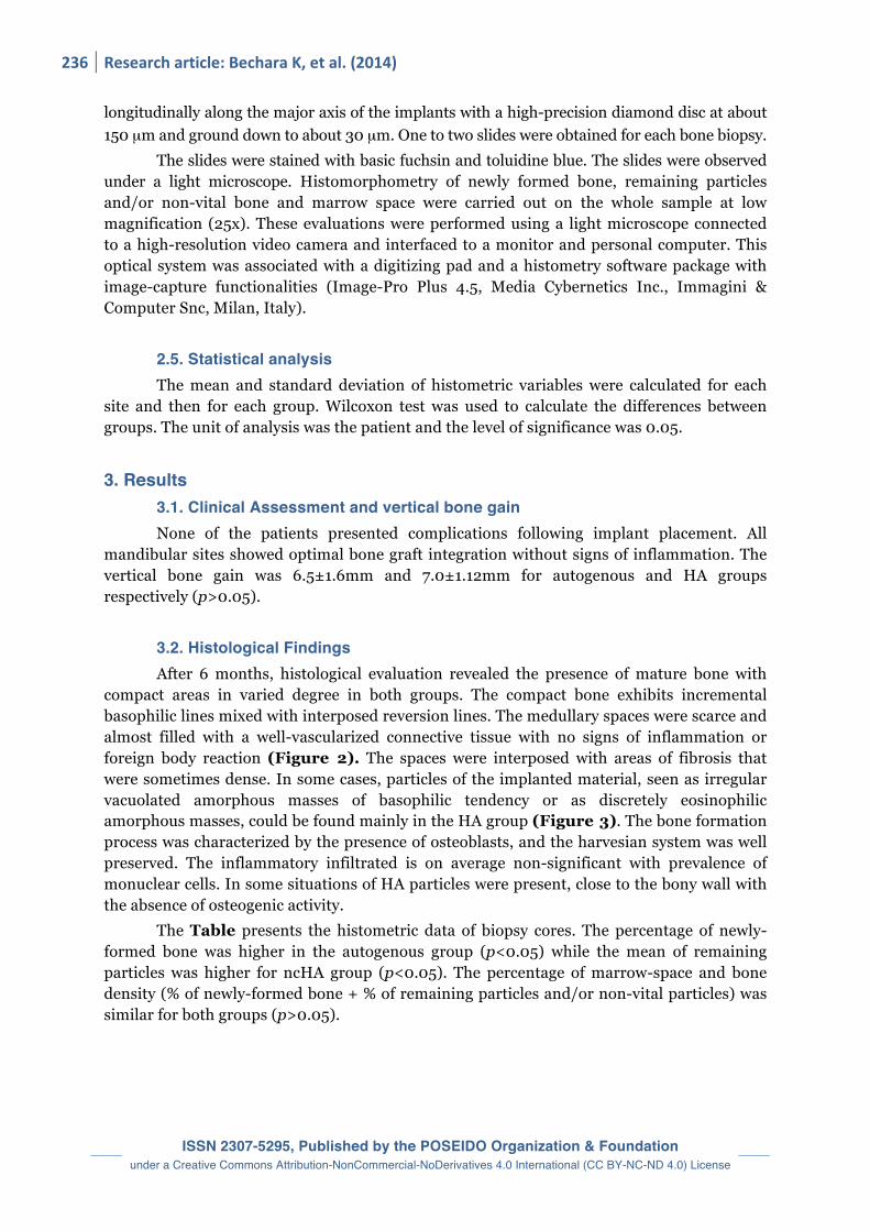
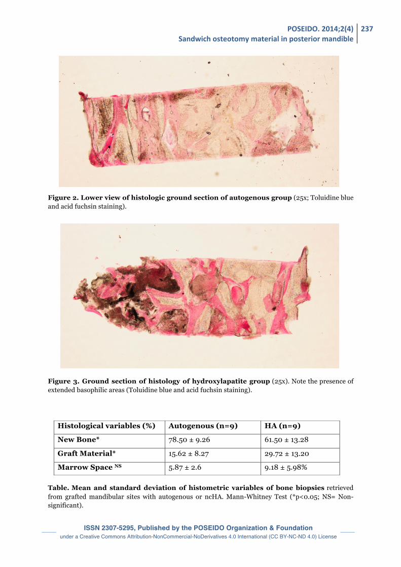
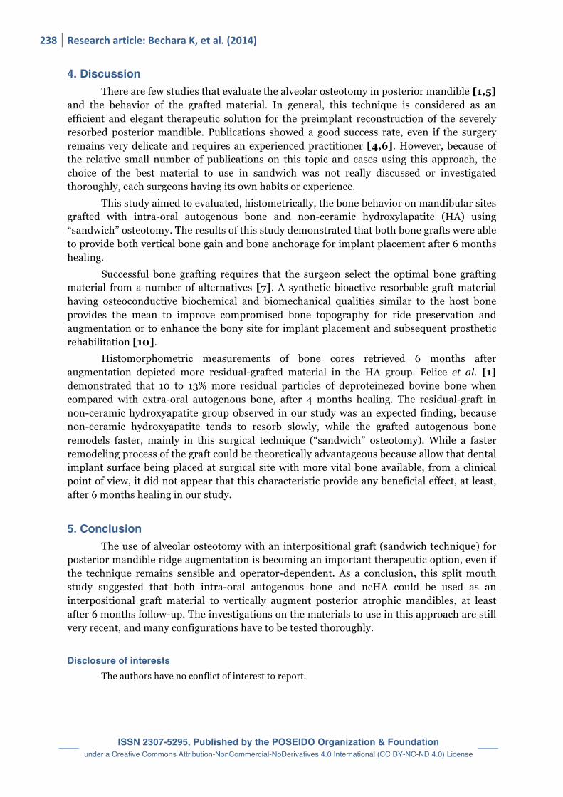
3.2. Histological Findings After 6 months, histological evaluation revealed the presence of mature bone with compact areas in varied degree in both groups. The compact bone exhibits incremental basophilic lines mixed with interposed reversion lines. The medullary spaces were scarce and almost filled with a well-vascularized connective tissue with no signs of inflammation or foreign body reaction (Figure 2). The spaces were interposed with areas of fibrosis that were sometimes dense. In some cases, particles of the implanted material, seen as irregular vacuolated amorphous masses of basophilic tendency or as discretely eosinophilic amorphous masses, could be found mainly in the HA group (Figure 3). The bone formation process was characterized by the presence of osteoblasts, and the harvesian system was well preserved. The inflammatory infiltrated is on average non-significant with prevalence of monuclear cells. In some situations of HA particles were present, close to the bony wall with the absence of osteogenic activity. The Table presents the histometric data of biopsy cores. The percentage of newly-formed bone was higher in the autogenous group (p<0.05) while the mean of remaining particles was higher for ncHA group (p<0.05). The percentage of marrow-space and bone density (% of newly-formed bone + % of remaining particles and/or non-vital particles) was similar for both groups (p>0.05).
POSEIDO. 2014;2(4) Sandwich osteotomy material in posterior mandible
237
ISSN 2307-5295, Published by the POSEIDO Organization & Foundation
under a Creative Commons Attribution-NonCommercial-NoDerivatives 4.0 International (CC BY-NC-ND 4.0) License
Figure 2. Lower view of histologic ground section of autogenous group (25x; Toluidine blue and acid fuchsin staining).
Figure 3. Ground section of histology of hydroxylapatite group (25x). Note the presence of extended basophilic areas (Toluidine blue and acid fuchsin staining).
Table. Mean and standard deviation of histometric variables of bone biopsies retrieved from grafted mandibular sites with autogenous or ncHA. Mann-Whitney Test (*p<0.05; NS= Non-significant).
Histological variables (%) Autogenous (n=9) HA (n=9)
New Bone* 78.50 ± 9.26 61.50 ± 13.28
Graft Material* 15.62 ± 8.27 29.72 ± 13.20
Marrow Space NS 5.87 ± 2.6 9.18 ± 5.98%
238 Research article: Bechara K, et al. (2014)
ISSN 2307-5295, Published by the POSEIDO Organization & Foundation
under a Creative Commons Attribution-NonCommercial-NoDerivatives 4.0 International (CC BY-NC-ND 4.0) License
4. Discussion There are few studies that evaluate the alveolar osteotomy in posterior mandible [1,5]
and the behavior of the grafted material. In general, this technique is considered as an efficient and elegant therapeutic solution for the preimplant reconstruction of the severely resorbed posterior mandible. Publications showed a good success rate, even if the surgery remains very delicate and requires an experienced practitioner [4,6]. However, because of the relative small number of publications on this topic and cases using this approach, the choice of the best material to use in sandwich was not really discussed or investigated thoroughly, each surgeons having its own habits or experience.
This study aimed to evaluated, histometrically, the bone behavior on mandibular sites grafted with intra-oral autogenous bone and non-ceramic hydroxylapatite (HA) using “sandwich” osteotomy. The results of this study demonstrated that both bone grafts were able to provide both vertical bone gain and bone anchorage for implant placement after 6 months healing.
Successful bone grafting requires that the surgeon select the optimal bone grafting material from a number of alternatives [7]. A synthetic bioactive resorbable graft material having osteoconductive biochemical and biomechanical qualities similar to the host bone provides the mean to improve compromised bone topography for ride preservation and augmentation or to enhance the bony site for implant placement and subsequent prosthetic rehabilitation [10].
Histomorphometric measurements of bone cores retrieved 6 months after augmentation depicted more residual-grafted material in the HA group. Felice et al. [1] demonstrated that 10 to 13% more residual particles of deproteinezed bovine bone when compared with extra-oral autogenous bone, after 4 months healing. The residual-graft in non-ceramic hydroxyapatite group observed in our study was an expected finding, because non-ceramic hydroxyapatite tends to resorb slowly, while the grafted autogenous bone remodels faster, mainly in this surgical technique (“sandwich” osteotomy). While a faster remodeling process of the graft could be theoretically advantageous because allow that dental implant surface being placed at surgical site with more vital bone available, from a clinical point of view, it did not appear that this characteristic provide any beneficial effect, at least, after 6 months healing in our study.
5. Conclusion The use of alveolar osteotomy with an interpositional graft (sandwich technique) for posterior mandible ridge augmentation is becoming an important therapeutic option, even if the technique remains sensible and operator-dependent. As a conclusion, this split mouth study suggested that both intra-oral autogenous bone and ncHA could be used as an interpositional graft material to vertically augment posterior atrophic mandibles, at least after 6 months follow-up. The investigations on the materials to use in this approach are still very recent, and many configurations have to be tested thoroughly. Disclosure of interests
The authors have no conflict of interest to report.
POSEIDO. 2014;2(4) Sandwich osteotomy material in posterior mandible
239
ISSN 2307-5295, Published by the POSEIDO Organization & Foundation
under a Creative Commons Attribution-NonCommercial-NoDerivatives 4.0 International (CC BY-NC-ND 4.0) License
Acknowledgements The authors want to thank Intra-Lock Brazil (São Paulo, Brazil) and Impladent Ltd
(Holliswood, NY, USA), for providing the hydroxyapatite; Implacil de Bortolli Implants (São Paulo, Brazil), for providing the dental implants. Author Contributions JAS was in charge of the elaboration of the study proposal and the financial support of the study, and he participated to the elaboration of the manuscript and the treatment planning of each case. JAR, AC, LC and GG were in charge of the statistical analysis and the financial support for the study. KB, AD, PYK were in charge of the treatment planning of each case, the implant placement surgery and they participated to the elaboration of the manuscript. AP, GI and GG were in charge of histological procedures. JAR and AC also participated to the elaboration of the study design and proposal. References [1] Felice P, Marchetti C, Iezzi G, Piattelli A, Worthington H, Pellegrino G, Esposito M. Vertical ridge augmentation of the atrophic posterior mandible with interpositional bloc grafts: bone from the iliac crest vs. bovine anorganic bone. Clinical and histological results up to one year after loading from a randomized-controlled clinical trial. Clin Oral Implants Res. 2009;20(12):1386-93. [2] Block MS, Haggerty CJ. Interpositional osteotomy for posterior mandible ridge augmentation. J Oral Maxillofac Surg. 2009;67(11 Suppl):31-9. [3] Sammartino G, Bernard JP. A clinical round table about the treatment of the severely resorbed posterior mandible. Part 1: challenges, endeavor and perspectives. POSEIDO. 2013;1(2):65-7. [4] Bormann KH, Suarez-Cunqueiro MM, von See C, Kokemuller H, Schumann P, Gellrich NC. Sandwich osteotomy for vertical and transversal augmentation of the posterior mandible. Int J Oral Maxillofac Surg. 2010;39(6):554-60. [5] Mazor Z, Lorean A. Preimplant reconstruction of the severely resorbed posterior mandible using the Sandwich technique with piezosurgical osteotomy and Leukocyte- and Platelet-Rich Fibrin (L-PRF): a 5-year follow-up with histological controls. POSEIDO. 2013;1(2):117-24. [6] Lopez-Cedrun JL. Implant rehabilitation of the edentulous posterior atrophic mandible: the sandwich osteotomy revisited. Int J Oral Maxillofac Implants. 2011;26(1):195-202. [7] Smiler D, Soltan M. The bone-grafting decision tree: a systematic methodology for achieving new bone. Implant Dent. 2006;15(2):122-8. [8] Scarano A, Degidi M, Iezzi G, Petrone G, Piattelli A. Correlation between implant stability quotient and bone-implant contact: a retrospective histological and histomorphometrical study of seven titanium implants retrieved from humans. Clin Implant Dent Relat Res. 2006;8(4):218-22. [9] Roze J, Babu S, Saffarzadeh A, Gayet-Delacroix M, Hoornaert A, Layrolle P. Correlating implant stability to bone structure. Clin Oral Implants Res. 2009;20(10):1140-5. [10] Valen M, Ganz SD. A synthetic bioactive resorbable graft for predictable implant reconstruction: part one. J Oral Implantol. 2002;28(4):167-77. This article can be cited as: Bechara K, Dottore AM, Kawakami PY, Cassoni A, Giro G, Rodrigues JA, Chambrone L, Piattelli A, Giovanna Iezzi G, Shibli JA. Interpositional graft associated with alveolar osteotomy for posterior mandible ridge augmentation (sandwich osteotomy) using hydroxyapatite or autogenous bone: histological evaluation. POSEIDO. 2014;2(4):233-9.

ISSN 2307-5295, Published by the POSEIDO Organization & Foundation
under a Creative Commons Attribution-NonCommercial-NoDerivatives 4.0 International (CC BY-NC-ND 4.0) License
POSEIDO. 2014;2(4) Zirconia implants in the posterior mandible
241
ISSN 2307-5295, Published by the POSEIDO Organization & Foundation
under a Creative Commons Attribution-NonCommercial-NoDerivatives 4.0 International (CC BY-NC-ND 4.0) License
Research article Comparative evaluation between one-piece implants of zirconia or titanium placed in posterior mandible: 6 months follow-up Ricardo R. Vecchiatti,1 Andre L.O. Campos,1 Welington F. Morais,1 Jose A. Rodrigues,2 Alessandra Cassoni,1 and Jamil Awad Shibli.1,* 1 Department of Periodontology and Oral Implantology, Dental Research Division, University of Guarulhos - UnG, São Paulo, Brazil 2 Department of Restorative Dentistry, Dental Research Division, University of Guarulhos - UnG, São Paulo, Brazil *Corresponding author: Jamil Awad Shibli, [email protected] Submitted on July 10th, 2014; accepted after minor corrections on July 28th, 2014.
Abstract Background and objectives. At present, only few studies with zirconia implants have been developed, but there are no conclusive results. This prospective study evaluated one-piece zirconia implants placed in posterior mandible assessing implant survival rate, implant success and marginal bone remodeling. Materials and Methods. 43 one-piece implants were placed in 2 groups: zirconia implants (Zi, n=21 implants) and sandblasted acid-etched titanium surface (SAE, n=22 implants). These implants were inserted in partially edentulous mandible of 15 patients in a split-mouth design. At 6-months loading follow-up, clinical and radiographic parameters were assessed. Mann-Whitney statistical analysis was performed to compare groups (α=0.05). Success criteria included absence of pain, sensitivity, suppuration, implant mobility; absence of continuous peri-implant radiolucency; distance between the implant shoulder and the first visible bone contact (DIB) <1.5 mm. Results. After a 6 months loading time, the overall implant survival rate was 94.59%, with 3 implant losses (2 Zi and 1 SAE). Among the surviving implants (34 out of 37), all fulfill the success criteria; therefore, the implant success was 94.59%. The mean distance between the implant shoulder and the first visible bone contact (DIB) for Zi and SAE implants were 0.34 ± 0.95 mm and 0.43 ± 0.85 mm, respectively (p>0.05). Discussion and Conclusion. Within the limits of this study, one-piece implants made of Zi or SAE seem to represent a safe and successful procedure for implant-supported restoration, after 180 ± 60 days follow-up. Keywords. Biomedical and dental materials, bone remodeling, ceramics, dental implants, zirconia.
1. Introduction The implant-supported restorations have been used as an alternative to prosthesis
[1]. The material of choice for dental implants is the commercially pure titanium. Due to its biocompatible, this material has been extensively used, showing high success rates during the years due to osseointegration [2-4].
242 Research article: Vecchiatti RR, et al. (2014)
ISSN 2307-5295, Published by the POSEIDO Organization & Foundation
under a Creative Commons Attribution-NonCommercial-NoDerivatives 4.0 International (CC BY-NC-ND 4.0) License
Osseointegration has been defined as a direct, functional linkage with the implant surface and the surrounding bone. With the intention to evaluate osseointegration of dental implants, many methods had been developed. An established method is the measurement of the Bone Implant Contact (BIC) by histomorphometric analysis, in animal studies, as a direct contact between bone and implant surface without any connective tissue interposition [5].
The ceramic zirconia material was introduced as an option to metallic abutments implants [6,7] because of its white color. However, the peri-implant mucosa color and biotype also influences the implant esthetics [8]. The development of zirconia material as an option to implant abutments [9,10] had also represented an important goal to achieve esthetic result associated with function. Dental implants made with zirconia had been evaluated [11]. These implants present an excellent resistance to corrosion and wear, good biocompatibility, high bending strength and fracture toughness [12], and low bacterial adhesion [13].
Zirconia implants with modified surfaces have showed osseointegration similar to titanium implants [14,15]. Degidi et al. (2006) have stated that occurs a lower inflammatory infiltrate with much lower extension at zirconia healing caps than at titanium healing caps [16]. In addition, animal studies with histological observations have stated similar capacity of zirconia implants for osseointegration as titanium implants [17-20].
One-piece implants have several clinical advantages. The surgery is minimally invasive, restorative procedures are simple, and abutment screw loosening cannot happen. Furthermore, the amount of crestal bone resorption may be minimized since there is no microgap or micromovement between an implant and an abutment [21,22].
Reports of excellent implant stability, esthetics, and patient satisfaction in the short-term perspective have been published with regard to this specific implant, although some reports have demonstrated lower survival rates related to the implant design [22,23].
This prospective study evaluated one-piece zirconia implants placed in posterior mandible assessing implant survival rate, implant success and marginal remodeling bone.
2. Materials and methods The Ethics Committee for Human Clinical Trials at Guarulhos University approved
the study protocol, which was explained to each subject, and all patients signed informed consent.
2.1. Selection of the subjects
Fifteen partially edentulous subjects (four males and eleven females, mean age of 40.58 ± 10.68 years) in posterior mandible were included in this study. These patients fulfilled the following inclusion criteria: patients had to meet pre-established inclusion parameters (i) at least 2 lower posterior mandibular teeth missing, (ii) reasonable to good oral and general health, (iii) not be pregnant or breath feeding; (iv) no history of irradiation on head and neck, (v) adequate amount of bone height for placement of implants with a minimum length of 8.5mm in a prosthetic optimal position, (vi) implant site free from acute infection.
Exclusion criteria included (i) previous bone augmentation in the implant site, (ii) moderate to severe chronic periodontitis (i.e., suppuration, bleeding on probing in more than 30% of the subgingival sites or any site with probing depth ≥ 5mm), (iii) not controlled diabetes or any systemic condition that could affect the bone healing.
POSEIDO. 2014;2(4) Zirconia implants in the posterior mandible
243
ISSN 2307-5295, Published by the POSEIDO Organization & Foundation
under a Creative Commons Attribution-NonCommercial-NoDerivatives 4.0 International (CC BY-NC-ND 4.0) License
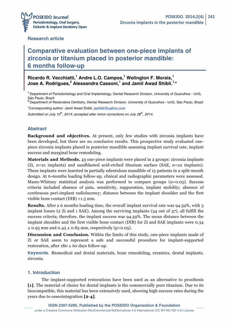
2.2. Special manufactured of one-piece implants One-piece screw-shaped implants (4.1 mm in diameter and 8.5 to 10 mm length; AS
Technology Titanium-FIX, São José dos Campos, SP, Brazil) were especially manufactured with titanium (SAE) or yttrium-stabilized tetragonal zirconia polycrystal Y-TZP (Zi). The grade-4 titanium implants were additionally blasted with 100 µm Al3O2 particles. After sandblasting, the specimens were ultrasonically cleaned with an alkaline solution, washed in distilled water and pickled with HNO3 (Figure 1). Figure 1. Lateral view of the one-piece titanium and zirconia implants. The samples were first checked for chemical composition with XPS/ESCA (X-Ray Photoelectron Spectroscopy/Electron Spectroscopy for Chemical Analysis), and no significant pollution was detected [24,25]. The topographies at the microscale were then visualized using routine Scanning Electron Microscopy (SEM) control. At the nanoscale, the SEM confirmed that both surface types were nanosmooth, following the current definition. The sole difference between these 2 tested implant types was therefore the specific material (zirconia and titanium) as well as the microtopography [26]. The implants were characterized by a Confocal White Light Microscope (Leica Scan DCM 3D - Leica Microsystems Ltd, Switzerland) with an objective magnification 50X, to measure one-piece dental implant surface topography.
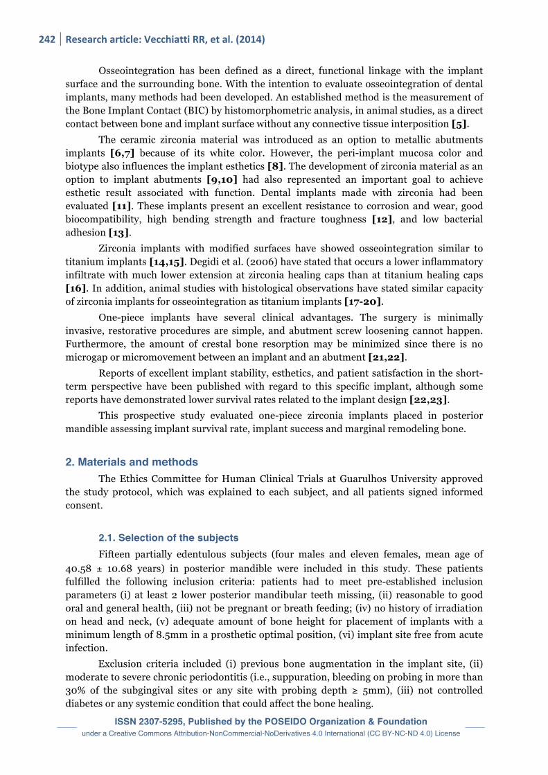
Titanium and zirconia groups were evaluated and the surface roughness was calculated. Surface roughness (Ra) length of 254.64 µm (768 X 56 pixels) was recorded (Figure 2).

244 Research article: Vecchiatti RR, et al. (2014)
ISSN 2307-5295, Published by the POSEIDO Organization & Foundation
under a Creative Commons Attribution-NonCommercial-NoDerivatives 4.0 International (CC BY-NC-ND 4.0) License
Figure 2. Representative profile of Surface Roughness for SAE titanium surface (A) and zirconia surface (B).
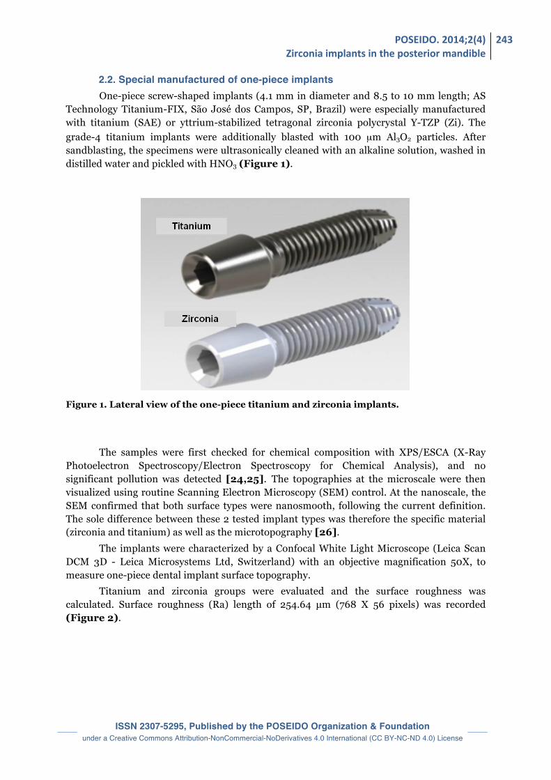
2.3. Implant placement One-piece screw-shaped implants for both groups were placed in posterior mandible
(Figure 3). The preparation of implant sites was carried out with twist drills of increasing diameter (2.0, 2.8, 3.15 and 3.35 mm) to place 4.1 mm diameter implants, under constant irrigation.
Care was taken to assess the position of the mental foramen. Implants sites were marked using a surgical template. The templates were based on the diagnostic waxing with perforations on the longitudinal axis, on the premolar and molar regions, according to ideal position of final implant supported restorations. Interrupted sutures to ensure a non-submerged healing procedure in dental implants were done.
POSEIDO. 2014;2(4) Zirconia implants in the posterior mandible
245
ISSN 2307-5295, Published by the POSEIDO Organization & Foundation
under a Creative Commons Attribution-NonCommercial-NoDerivatives 4.0 International (CC BY-NC-ND 4.0) License
Figure 3. Clinical view of titanium and zirconia implants being inserted in posterior mandible.
2.4. Post-operative treatment All patients received oral antibiotics (Clindamicyn, 900mg each day) for 7 days
Postoperative pain was controlled by administering 100 mg Nimesulide every 12 hours for 5 days. Detailed oral hygiene instructions were provided, with mouth-rinses with 0.12% chlorhexidine administered for 7 days. Suture removal was performed at 7 days. After surgery, the patients were instructed to avoid brushing and any trauma to surgical site. A cold and soft diet was recommended for the first day, and a soft diet for the first week.
2.5. Restorative procedure Following four months of implant healing, an impression was taken utilizing a silicon
putty polyvinylsiloxane (Contrast – VOCO) directly on the implants. Laboratory templates were made and a master cast was fabricated.
The implant-supported restoration made with ceramic (IPS D’Sign - Ivoclar Vivadent) was placed direct on the implant. These restorations were fixed with a resin cement (Variolink II - Ivoclar Vivadent). All centric and lateral contacts were assessed by an articulating paper and adjusted if necessary.
2.6. Clinical and radiographic evaluation For each implant, the following clinical parameters [27] were investigated, after 6
months of functional loading, as the presence or absence of: 1.) pain/sensitivity, 2.) suppuration/exudation and 3.) implant mobility. The number 3 was tested manually using the handles of two dental mirrors.
246 Research article: Vecchiatti RR, et al. (2014)
ISSN 2307-5295, Published by the POSEIDO Organization & Foundation
under a Creative Commons Attribution-NonCommercial-NoDerivatives 4.0 International (CC BY-NC-ND 4.0) License
Moreover, intraoral periapical radiographs were taken for each implant, at the baseline (immediately after implant insertion) and at the 6 months of occlusal loading. Radiographs were taken using a Rinn alignment system with a rigid film-object-X-ray source coupled to a beam-aiming device in order to achieve reproducible exposure geometry. The distance between the first angle of the implant body and the first visible bone contact, was measured in mm by an appropriate software (ImageTool - Texas University).
Crestal bone level changes at 6 months were registered as modifications in the distance from the implant shoulder to the bone level on the mesial and distal implant side. To correct dimensional distortion, the apparent dimension of each implant was measured on the radiograph and then compared with the actual implant length.
2.7. Implant Survival and Implant Success criteria The evaluation of implant survival and implant success was performed according to
the following clinical and radiographic parameters [28]. Implants were basically divided into two categories: “survived” and “failed” implants. An implant was classified as a “survived implant” when it was still in function at the end of the study, after 6 months of functional loading. To achieve implant success, the following clinical and radiographic success criteria should be fulfilled as absence of: pain or sensitivity upon function, suppuration or exudation, clinically detectable implant mobility, continuous peri-implant radiolucency, and DIB < 1.5 mm after 6 months of functional loading. Implant losses were categorized as failures; implants presenting pain upon function, suppuration or clinical mobility were removed, and were all failure categories. The conditions for which implant removal could be indicated included failure of osseointegration or infection, recurrent peri-implantitis, or implant loss due to mechanical overload.
3. Results At the time of evaluation, of the 15 patients, 13 had only returned. After a 6 months
loading time, the overall implant survival rate was 95% (titanium implants) and 89.47% (zirconia implants), with 3 out of 39 implant losses by mobility without infection (2 Zi and 1 SAE). Among the surviving implants (34 out of 37), all fulfill the success criteria; therefore, the implant success was 94.59%. Two zirconia abutments at single-tooth implant-supported restoration fractured at final insertion, showing no influence on the osseointegration.
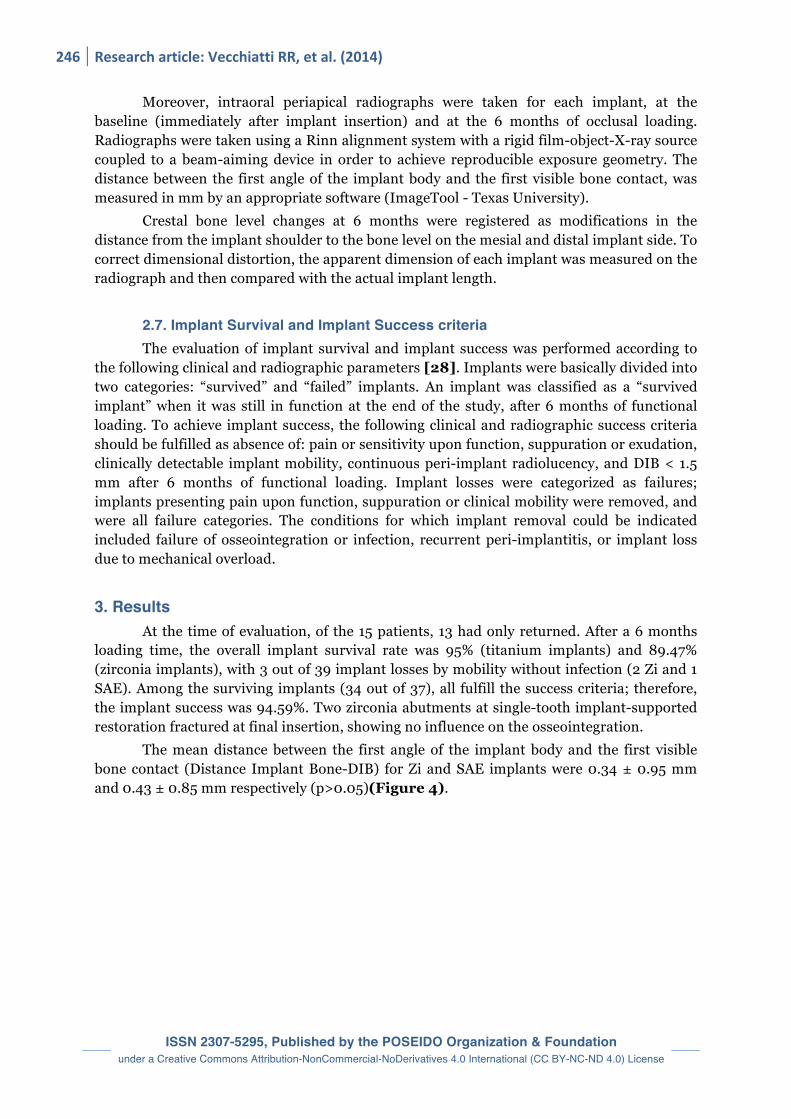
The mean distance between the first angle of the implant body and the first visible bone contact (Distance Implant Bone-DIB) for Zi and SAE implants were 0.34 ± 0.95 mm and 0.43 ± 0.85 mm respectively (p>0.05)(Figure 4).
POSEIDO. 2014;2(4) Zirconia implants in the posterior mandible
247
ISSN 2307-5295, Published by the POSEIDO Organization & Foundation
under a Creative Commons Attribution-NonCommercial-NoDerivatives 4.0 International (CC BY-NC-ND 4.0) License
Figure 4. Mean and standard deviation (mm) of remodeling crestal bone of evaluated group.
4. Discussion The present study showed a 94.59% success rate for all implants placed in the
posterior mandible. Specifically, zirconia implants presented an 89.47% success rate while one-piece titanium implants showed 95% under the same loading conditions. These data agree with previous studies that evaluated the success rate of zirconia implants placed in human jaws [29]. In that study, the authors achieved a success rate of 92.7% in zirconia implants without surface topography preparation, in the same condition as performed in our study. In a later study, Oliva et. al., (2007) reported a higher success rate ranged around 98%, however, this zirconia surface was previously coated, data that range very close to the titanium implants evaluated in our study [30]. These data could suggest that the implant surface topography should increase the bone healing around the peri-implant environment, and consequently increase the success rate.
When comparing the results with animals, Akagawa et. al. (1998) evaluating seven monkeys and twenty-eight non-treated zirconia implants, reported success index ranged between 54% and 71% after twelve months of follow-up [31].
Several studies have demonstrated bone integration of threaded zirconia implants under both unloaded and loaded conditions [32]. Akagawa et al. (1998) showed, in monkeys, the long-term stability of partially stabilized zirconia implants placed with different loading designs in a one-stage procedure [31]. Direct bone apposition to the implant was generally seen. Fractures of the implants did not appear, confirming the favorable mechanical properties of zirconia.
A review of the literature indicates that sensitivity to titanium is rare, two reports showed possible hypersensitive reactions to titanium. Oliva et al. (2010) describes a full-mouth oral rehabilitation of a titanium allergic patient with zirconia implants [33] hence, the zirconia implants provide the possibility of a metal-free treatment option to patients who request this [34-36].
248 Research article: Vecchiatti RR, et al. (2014)
ISSN 2307-5295, Published by the POSEIDO Organization & Foundation
under a Creative Commons Attribution-NonCommercial-NoDerivatives 4.0 International (CC BY-NC-ND 4.0) License
Kohal et al. (1997) compared custom-made titanium and zirconia implants used to support metal crowns in the superior jaw of six monkeys [37]. The two types of implants were sandblasted, and the titanium was also acid- etched. All implants maintained stability and no mechanical problems were reported. Histologic examinations confirmed no differences in the bone tissue response to the titanium and zirconia implants. In a systematic review, Wenz et al. (2008) have related that the osseointegration of yttria–tetragonal zirconia polycrystal implants (Y-TZP) were similar to titanium implants; modifications of their surfaces have the potential to improve initial bone healing and resistance to removal torque; low temperature degradation might affect the behavior of Y-TZP [38]. That suggested the higher adhesion capacity developed by cells on zirconia could be related to the earlier and abundant production of the adhesion protein fibronectin than titanium. Although, the more precocious and high production of organized structure for adhesion observed on zirconia, associated with the low toxicity exerted by this material, concur to explain the higher growth rate of cells incubated on it when compared with fibroblasts growing in contact with FE ceramic [12]. Newly formed bone was observed in close contact with zirconia ceramic surfaces in a rabbit study that reported a bone-to-implant contact of 68.4% [39]. Kohal et al. (2004) compared airborne-particle-abraded zirconia implants with acid-etched titanium implants in monkeys; after 9 months of healing and 5 months of loading, no significant difference in bone-to-implant contact between the two groups could be found [32]. Kohal et al. (2009) reported that the mean mineralized bone-to-implant contact for the Ti machined group was 39.4%, 46.6% for the ZrO2 machined group, 55.2% for the TiUnite group and 59.4% for the ZrO2 modified group [15]. That investigation supports those findings that the surface modifications for the titanium, and also for the zirconia implants resulted in higher BIC compared with the machined surfaces. Because there was no significant difference between the TiUnite surface and the modified zirconia surface, it can be concluded that zirconia surface is as biocompatible and osteoconductive as the modified titanium surface [15]. Sennerby et al. (2005) reported a strong bone tissue response to surface-modified zirconia implants after 6 weeks of healing in rabbit bone [20]. The modified zirconia implants showed a resistance to torque forces similar to that of oxidized implants and a four to fivefold increase compared with machined zirconia implants. The findings suggest that surface-modified zirconia implants can reach firm stability in bone. The research of Koch et al. (2010) showed that all implants (titanium and zirconia) had areas of tight BIC [11]. The trabecular architecture of bone around the zirconia, coated zirconia and titanium implants was classified as lamellar bone due to the circular apposition of bone lamellae around canals of Havers. All zirconia and titanium implants showed few signs of natural bone remodeling, apposition of osteoid and osteoblasts or lacunae of osteoclasts. Osteoblasts surrounding the bone trabeculae showed functional and mature bone architecture nearby the zirconia and titanium implants. Morphologically, the zirconia as well as the titanium implants were tightly osseointegrated. They showed that zirconia implants are capable of establishing BIC rates similar to what is known from the osseointegration behavior of roughened titanium implants with the same surface modification and roughness. Despite many positive experiences, the use of zirconia implants remains debated and quite scarce in daily practice. There are still many uncertainties on the kind of zirconia to use and in which configuration, in order to have the best biomechanical behavior and long-term clinical results. The posterior mandible remains one of the most delicate area to rehabilitate
POSEIDO. 2014;2(4) Zirconia implants in the posterior mandible
249
ISSN 2307-5295, Published by the POSEIDO Organization & Foundation
under a Creative Commons Attribution-NonCommercial-NoDerivatives 4.0 International (CC BY-NC-ND 4.0) License
with such implant, as the biomechanical conditions are in theory the most unfavorable. This first study brings therefore a very interesting highlight on the opportunities and threats of this therapeutic option.
5. Conclusion Within the limits of this study, one-piece implants made of Zi or SAE seem to
represent a safe and successful procedure for implant-supported restoration in the posterior mandible, after 180 ± 60 days follow-up. A long-term follow-up of these patients (and of a larger group of patients) is needed to give a better feedback on the biomechanical and clinical potential of these implants in the posterior mandible, and to bring further this preliminary research. Disclosure of interests The authors have no conflict of interest to report. Acknowledgements The authors would like to thank Titanium Fix, São José dos Campos, Brazil for supplying the zirconia and titanium implants. Author Contributions JAS was in charge of the elaboration of the study proposal and the financial support of the study, and he participated to the elaboration of the manuscript and the treatment planning of each case. JAR and AC were in charge of the statistical analysis, the implant surface characterization and the financial support for the study. RRV, ALOC and WFM were in charge of the treatment planning of each case, the implant placement surgery and they participated to the elaboration of the manuscript. JAR and AC also participated to the elaboration of the study design and proposal. References [1] Al-Nawas B, Kammerer PW, Morbach T, Ladwein C, Wegener J, Wagner W. Ten-year retrospective follow-up study of the TiOblast dental implant. Clin Implant Dent Relat Res. 2012;14(1):127-34. [2] Branemark PI, Hansson BO, Adell R, Breine U, Lindstrom J, Hallen O, Ohman A. Osseointegrated implants in the treatment of the edentulous jaw. Experience from a 10-year period. Scand J Plast Reconstr Surg Suppl. 1977;16:1-132. [3] Adell R, Eriksson B, Lekholm U, Branemark PI, Jemt T. Long-term follow-up study of osseointegrated implants in the treatment of totally edentulous jaws. Int J Oral Maxillofac Implants. 1990;5(4):347-59. [4] Orsini E, Giavaresi G, Trire A, Ottani V, Salgarello S. Dental implant thread pitch and its influence on the osseointegration process: an in vivo comparison study. Int J Oral Maxillofac Implants. 2012;27(2):383-92. [5] Albrektsson T, Branemark PI, Hansson HA, Lindstrom J. Osseointegrated titanium implants. Requirements for ensuring a long-lasting, direct bone-to-implant anchorage in man. Acta Orthop Scand. 1981;52(2):155-70. [6] Bressan E, Paniz G, Lops D, Corazza B, Romeo E, Favero G. Influence of abutment material on the gingival color of implant-supported all-ceramic restorations: a prospective multicenter study. Clin Oral Implants Res. 2011;22(6):631-7. [7] van Brakel R, Noordmans HJ, Frenken J, de Roode R, de Wit GC, Cune MS. The effect of zirconia and titanium implant abutments on light reflection of the supporting soft tissues. Clin Oral Implants Res. 2011;22(10):1172-8. [8] Jung RE, Sailer I, Hammerle CH, Attin T, Schmidlin P. In vitro color changes of soft tissues caused by restorative materials. Int J Periodontics Restorative Dent. 2007;27(3):251-7.
250 Research article: Vecchiatti RR, et al. (2014)
ISSN 2307-5295, Published by the POSEIDO Organization & Foundation
under a Creative Commons Attribution-NonCommercial-NoDerivatives 4.0 International (CC BY-NC-ND 4.0) License
[9] Sailer I, Zembic A, Jung RE, Siegenthaler D, Holderegger C, Hammerle CH. Randomized controlled clinical trial of customized zirconia and titanium implant abutments for canine and posterior single-tooth implant reconstructions: preliminary results at 1 year of function. Clin Oral Implants Res. 2009;20(3):219-25. [10] Zembic A, Sailer I, Jung RE, Hammerle CH. Randomized-controlled clinical trial of customized zirconia and titanium implant abutments for single-tooth implants in canine and posterior regions: 3-year results. Clin Oral Implants Res. 2009;20(8):802-8. [11] Koch FP, Weng D, Kramer S, Biesterfeld S, Jahn-Eimermacher A, Wagner W. Osseointegration of one-piece zirconia implants compared with a titanium implant of identical design: a histomorphometric study in the dog. Clin Oral Implants Res. 2010;21(3):350-6. [12] Rocchietta I, Fontana F, Addis A, Schupbach P, Simion M. Surface-modified zirconia implants: tissue response in rabbits. Clin Oral Implants Res. 2009;20(8):844-50. [13] Scarano A, Piattelli M, Caputi S, Favero GA, Piattelli A. Bacterial adhesion on commercially pure titanium and zirconium oxide disks: an in vivo human study. J Periodontol. 2004;75(2):292-6. [14] Depprich R, Zipprich H, Ommerborn M, Naujoks C, Wiesmann HP, Kiattavorncharoen S, Lauer HC, Meyer U, Kubler NR, Handschel J. Osseointegration of zirconia implants compared with titanium: an in vivo study. Head Face Med. 2008;4:30. [15] Kohal RJ, Wolkewitz M, Hinze M, Han JS, Bachle M, Butz F. Biomechanical and histological behavior of zirconia implants: an experiment in the rat. Clin Oral Implants Res. 2009;20(4):333-9. [16] Degidi M, Artese L, Scarano A, Perrotti V, Gehrke P, Piattelli A. Inflammatory infiltrate, microvessel density, nitric oxide synthase expression, vascular endothelial growth factor expression, and proliferative activity in peri-implant soft tissues around titanium and zirconium oxide healing caps. J Periodontol. 2006;77(1):73-80. [17] Josset Y, Oum'Hamed Z, Zarrinpour A, Lorenzato M, Adnet JJ, Laurent-Maquin D. In vitro reactions of human osteoblasts in culture with zirconia and alumina ceramics. J Biomed Mater Res. 1999;47(4):481-93. [18] Hoffmann O, Angelov N, Gallez F, Jung RE, Weber FE. The zirconia implant-bone interface: a preliminary histologic evaluation in rabbits. Int J Oral Maxillofac Implants. 2008;23(4):691-5. [19] Schultze-Mosgau S, Schliephake H, Radespiel-Troger M, Neukam FW. Osseointegration of endodontic endosseous cones: zirconium oxide vs titanium. Oral Surg Oral Med Oral Pathol Oral Radiol Endod. 2000;89(1):91-8. [20] Sennerby L, Dasmah A, Larsson B, Iverhed M. Bone tissue responses to surface-modified zirconia implants: A histomorphometric and removal torque study in the rabbit. Clin Implant Dent Relat Res. 2005;7 Suppl 1:S13-20. [21] Oh TJ, Yoon J, Misch CE, Wang HL. The causes of early implant bone loss: myth or science? J Periodontol. 2002;73(3):322-33. [22] Ostman PO, Hellman M, Albrektsson T, Sennerby L. Direct loading of Nobel Direct and Nobel Perfect one-piece implants: a 1-year prospective clinical and radiographic study. Clin Oral Implants Res. 2007;18(4):409-18. [23] Swart LC, van Niekerk DJ. Simplifying the implant treatment for an unrestorable premolar with a one-piece implant: a clinical report. J Prosthet Dent. 2008;100(2):81-5. [24] Dohan Ehrenfest DM, Kang BS, Sammartino G, Shibli JA, Wang HL, Zou DR, Bernard JP. Guidelines for the publication of articles related to implant surfaces and design from the POSEIDO: a standard for surface characterization. POSEIDO. 2013;1(1):7-15. [25] Dohan Ehrenfest DM, Del Corso M, Kang BS, Leclercq P, Mazor Z, Horowitz RA, Russe P, Oh HK, Zou DR, Shibli JA, Wang HL, Bernard JP, Sammartino G. Identification card and codification of the chemical and morphological characteristics of 62 dental implant surfaces. Part 1: description of the Implant Surface Identification Standard (ISIS) codification system. POSEIDO. 2014;2(1):7-22. [26] Miranda PV, Rodrigues JA, Blay A, Shibli JA, Cassoni A. Surface alterations of zirconia and titanium substrates after Er,Cr:YSGG irradiation. Lasers Med Sci. 2015;30(1):43-8. [27] Mangano F, Mangano C, Ricci M, Sammons RL, Shibli JA, Piattelli A. Single-tooth Morse taper connection implants placed in fresh extraction sockets of the anterior maxilla: an aesthetic evaluation. Clin Oral Implants Res. 2012;23(11):1302-7. [28] Albrektsson T, Zarb GA. Determinants of correct clinical reporting. Int J Prosthodont. 1998;11(5):517-21. [29] Oliva J, Oliva X, Oliva JD. Five-year success rate of 831 consecutively placed Zirconia dental implants in humans: a comparison of three different rough surfaces. Int J Oral Maxillofac Implants. 2010;25(2):336-44. [30] Oliva J, Oliva X, Oliva JD. One-year follow-up of first consecutive 100 zirconia dental implants in humans: a comparison of 2 different rough surfaces. Int J Oral Maxillofac Implants. 2007;22(3):430-5.
POSEIDO. 2014;2(4) Zirconia implants in the posterior mandible
251
ISSN 2307-5295, Published by the POSEIDO Organization & Foundation
under a Creative Commons Attribution-NonCommercial-NoDerivatives 4.0 International (CC BY-NC-ND 4.0) License
[31] Akagawa Y, Hosokawa R, Sato Y, Kamayama K. Comparison between freestanding and tooth-connected partially stabilized zirconia implants after two years' function in monkeys: a clinical and histologic study. J Prosthet Dent. 1998;80(5):551-8. [32] Kohal RJ, Weng D, Bachle M, Strub JR. Loaded custom-made zirconia and titanium implants show similar osseointegration: an animal experiment. J Periodontol. 2004;75(9):1262-8. [33] Oliva X, Oliva J, Oliva JD. Full-mouth oral rehabilitation in a titanium allergy patient using zirconium oxide dental implants and zirconium oxide restorations. A case report from an ongoing clinical study. Eur J Esthet Dent. 2010;5(2):190-203. [34] Peters MS, Schroeter AL, van Hale HM, Broadbent JC. Pacemaker contact sensitivity. Contact Dermatitis. 1984;11(4):214-8. [35] Lalor PA, Revell PA, Gray AB, Wright S, Railton GT, Freeman MA. Sensitivity to titanium. A cause of implant failure? J Bone Joint Surg Br. 1991;73(1):25-8. [36] Sicilia A, Cuesta S, Coma G, Arregui I, Guisasola C, Ruiz E, Maestro A. Titanium allergy in dental implant patients: a clinical study on 1500 consecutive patients. Clin Oral Implants Res. 2008;19(8):823-35. [37] Kohal RJ, Hurzeler MB, Mota LF, Klaus G, Caffesse RG, Strub JR. Custom-made root analogue titanium implants placed into extraction sockets. An experimental study in monkeys. Clin Oral Implants Res. 1997;8(5):386-92. [38] Wenz HJ, Bartsch J, Wolfart S, Kern M. Osseointegration and clinical success of zirconia dental implants: a systematic review. Int J Prosthodont. 2008;21(1):27-36. [39] Scarano A, Di Carlo F, Quaranta M, Piattelli A. Bone response to zirconia ceramic implants: an experimental study in rabbits. J Oral Implantol. 2003;29(1):8-12. This article can be cited as: Vecchiatti RR, Campos ALO, Welington F. Morais WF, Rodrigues JA, Cassoni A, Shibli JA. Comparative evaluation between one-piece implants of zirconia or titanium placed in posterior mandible: 6 months follow-up. POSEIDO. 2014;2(4):241-51.

ISSN 2307-5295, Published by the POSEIDO Organization & Foundation
under a Creative Commons Attribution-NonCommercial-NoDerivatives 4.0 International (CC BY-NC-ND 4.0) License

POSEIDO. 2014;2(4) Accuracy of software-‐guided implant osteotomy
253
ISSN 2307-5295, Published by the POSEIDO Organization & Foundation
under a Creative Commons Attribution-NonCommercial-NoDerivatives 4.0 International (CC BY-NC-ND 4.0) License
Research article Accuracy of guided osteotomy using dental implant treatment-planning software in combination with an optical scan of a dental cast Bruce J. Kowalski,1,* Marc Manos,2 Michael Levi,3 and Joe “Ambrose” D’Ambrosia.1 1 ROE Dental Laboratory, Inc., Cleveland, OH, USA 2 University of Illinois at Chicago College of Dentistry, Chicago, IL, USA 3 Indiana University School of Dentistry, Indianapolis, IN, USA *Corresponding author: B.J. Kowalski, [email protected] Submitted on July 17th, 2014; accepted after minor corrections on August 7th, 2014.
Abstract Background and objectives. The advent of the mainstream use of diagnostic cone beam computed tomography (CBCT) and software-driven stereolithography (STL) for rapid prototyping of three-dimensional (3D) object models has achieved new levels of precision in dental implant treatment planning. The objective of this study was to assess the accuracy of using such software as a mode of capture to reproduce the optical scanning data to be used in STL surgical-guide fabrication. Materials and Methods. We conducted an in vitro “case series” comprising 5 stone casts (made from impressions of partially edentulous patients) in which arbitrary holes were drilled to simulate implant osteotomies. These casts with the holes were optically scanned, and the scan data imported into an implant treatment-planning software package that was used to fabricate drill guides via rapid prototyping on a 3D printer. These guides were then used to drill a second smaller hole within each corresponding simulated osteotomy, and the discrepancies between the center points of superimposed holes were assessed by light microscopy and metrology software. Results. The discrepancies at the implant entrance center points ranged from 0.09 to 0.23 mm; calculated angular discrepancies at the depth of the osteotomy relative to entrance-point centering were all < 1 mm (maximum = 0.73 mm) for a 16-mm implant depth. Discussion and conclusions. These findings suggest a level of accuracy for the software, optical scan-generated data, and STL modeling that would yield clinically acceptable results. Keywords. Cone beam computed tomography, dental implants, osteotomy, software, three-dimensional printing.
1. Introduction The combination of treatment planning software with optical scanning is an
interesting evolution of the implant placement planning. The precision of these techniques, both at the surgical [1] and prosthetic steps [2], needs always to be investigated, to validate any new approach and clinical application. This pilot study was designed to identify the accuracy of making a guided osteotomy by using a treatment planning software program in
254 Research article: Kowalski BJ, et al. (2014)
ISSN 2307-5295, Published by the POSEIDO Organization & Foundation
under a Creative Commons Attribution-NonCommercial-NoDerivatives 4.0 International (CC BY-NC-ND 4.0) License
combination with an optical scan of a dental cast in the process of three-dimensional (3D) rapid prototyping of a surgical guide via stereolithography (STL).
A number of recent systematic reviews have analyzed studies that used cone beam computed tomography (CBCT) systems and, in some cases, optical tracking devices [1], with respect to achievement of high implant survival rates comparable to those of conventionally placed implants (91-100%)[1], in addition to a 96.6% overall mean survival rate over 1 year [3]. An additional review by Neugebauer et al. [4] suggested that intraoperative optical tracking systems offer greater accuracy than surgical guide systems. Jung et al. pointed out the need to consider the radiation exposure required for CBCT [3].
Van der Zel [5] recently reviewed a combined scanning protocol that incorporates optical scanning of casts, guides and occlusal registrations that are geometrically matched to CBCT data, resulting in a higher level of accuracy in an STL-produced surgical guide than via use of CBCT data alone. The author noted that such combined technology affords greater control over not only implant placement, but over prosthesis design as well [5]. So, while there appears to be some movement in the direction of incorporating data from optical scans into the 3D treatment-planning process with the goal of improving accuracy of implant placement, data are scarce in regard to optical scanning as a supplementary modality in implant treatment planning.
The current study was designed to evaluate a laboratory method of obtaining a more precise assessment of the accuracy of execution of an osteotomy, based on a data set obtained with an optical scan, and verified by data from measurements obtained from stereolithographic (STL) surgical guides that were produced for experimental osteotomies using the treatment-planning software.
2. Materials and Methods Impressions were made from 5 partially edentulous patients and used to produce
dental stone casts. In each edentulous area, an arbitrary hole approximately 5 mm in diameter and 3 mm in depth was drilled, for a total of 9 experimental osteotomy sites. The casts were then optically scanned using the 3Shape D800 scanner (3Shape A/S, Copenhagen, Denmark). The scan files where then imported into the Blue Sky Plan program (version 2.19) Blue Sky Bio, LLC, Grayslake, IL, USA), and osteotomies were planned in the software to be exactly in the centers of the arbitrary osteotomies in the casts.
Drill guides were then printed using an Objet model 250 3D printer (Stratasys Ltd., Edina, MN, USA) to fabricate the guides from Med610 biocompatible resin (Stratasys Ltd., Edina, MN, USA), in an STL-based rapid prototyping technique. Titanium sleeves (iDent, Ft. Lauderdale, FL, USA) were inserted in the printed guides. These guides were then used to drill a second set of osteotomies of smaller diameter, one within each larger arbitrary osteotomy. At completion, each edentulous site had a larger arbitrary osteotomy, and a smaller osteotomy within it that was created using an optical scan-generated STL-printed guide.
The casts were then mounted on a paralleling table, and each site was aligned under a digital microscope camera in such a way that the long axes of the holes were in line with the optical axis of the microscope.
The images were analyzed with an edge-detecting metrology (field-of-view) software (ZView DMP2000, ZView Inc., Huntington Beach, CA, USA). The observed alignments of the center points of the arbitrary and the guided holes were reproduced in this software, and the
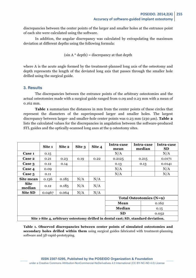
POSEIDO. 2014;2(4) Accuracy of software-‐guided implant osteotomy
255
ISSN 2307-5295, Published by the POSEIDO Organization & Foundation
under a Creative Commons Attribution-NonCommercial-NoDerivatives 4.0 International (CC BY-NC-ND 4.0) License
discrepancies between the center points of the larger and smaller holes at the entrance point of each site were calculated using the software.
In addition, the angular discrepancy was calculated by extrapolating the maximum deviation at different depths using the following formula:
(sin A * depth) = discrepancy at that depth where A is the acute angle formed by the treatment-planned long axis of the osteotomy and depth represents the length of the deviated long axis that passes through the smaller hole drilled using the surgical guide.
3. Results The discrepancies between the entrance points of the arbitrary osteotomies and the
actual osteotomies made with a surgical guide ranged from 0.09 and 0.23 mm with a mean of 0.162 mm.
Table 1 summarizes the distances in mm from the center points of these circles that represent the diameters of the superimposed larger and smaller holes. The largest discrepancy between larger- and smaller-hole center points was 0.23 mm (230 µm). Table 2 lists the calculated values for the discrepancies in angulation between the software-produced STL guides and the optically-scanned long axes at the 9 osteotomy sites.
Table 1. Observed discrepancies between center points of simulated osteotomies and secondary holes drilled within them using surgical guides fabricated with treatment-planning software and 3D rapid-prototyping.
Site 1 Site 2 Site 3 Site 4 Intra-case mean
Intra-case median
Intra-case SD
Case 1 0.15 N/A N/A Case 2 0.21 0.23 0.19 0.22 0.2125 0.215 0.0171 Case 3 0.12 0.14 0.13 0.13 0.0141 Case 4 0.09 N/A N/A Case 5 0.11 N/A N/A
Site mean 0.136 0.185 N/A N/A
Site median 0.12 0.185 N/A N/A
Site SD 0.0467 0.064 N/A N/A Total Osteotomies (N=9)
Mean 0.162 Median 0.15
SD 0.052 Site 1-Site 4, arbitrary osteotomy drilled in dental cast; SD, standard deviation.
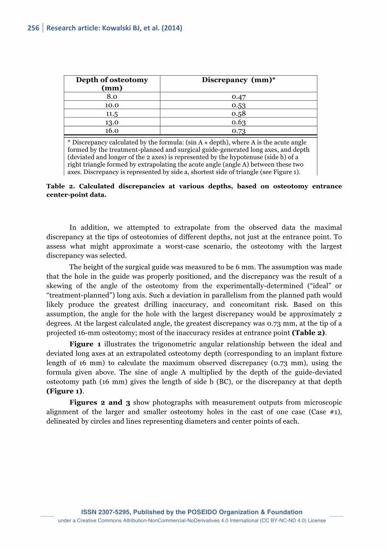
256 Research article: Kowalski BJ, et al. (2014)
ISSN 2307-5295, Published by the POSEIDO Organization & Foundation
under a Creative Commons Attribution-NonCommercial-NoDerivatives 4.0 International (CC BY-NC-ND 4.0) License
Table 2. Calculated discrepancies at various depths, based on osteotomy entrance center-point data.
In addition, we attempted to extrapolate from the observed data the maximal discrepancy at the tips of osteotomies of different depths, not just at the entrance point. To assess what might approximate a worst-case scenario, the osteotomy with the largest discrepancy was selected.
The height of the surgical guide was measured to be 6 mm. The assumption was made that the hole in the guide was properly positioned, and the discrepancy was the result of a skewing of the angle of the osteotomy from the experimentally-determined (“ideal” or “treatment-planned”) long axis. Such a deviation in parallelism from the planned path would likely produce the greatest drilling inaccuracy, and concomitant risk. Based on this assumption, the angle for the hole with the largest discrepancy would be approximately 2 degrees. At the largest calculated angle, the greatest discrepancy was 0.73 mm, at the tip of a projected 16-mm osteotomy; most of the inaccuracy resides at entrance point (Table 2).
Figure 1 illustrates the trigonometric angular relationship between the ideal and deviated long axes at an extrapolated osteotomy depth (corresponding to an implant fixture length of 16 mm) to calculate the maximum observed discrepancy (0.73 mm), using the formula given above. The sine of angle A multiplied by the depth of the guide-deviated osteotomy path (16 mm) gives the length of side b (BC), or the discrepancy at that depth (Figure 1).
Figures 2 and 3 show photographs with measurement outputs from microscopic alignment of the larger and smaller osteotomy holes in the cast of one case (Case #1), delineated by circles and lines representing diameters and center points of each.
Depth of osteotomy (mm)
Discrepancy (mm)*
8.0 0.47 10.0 0.53 11.5 0.58 13.0 0.63 16.0 0.73
* Discrepancy calculated by the formula: (sin A × depth), where A is the acute angle formed by the treatment-planned and surgical guide-generated long axes, and depth (deviated and longer of the 2 axes) is represented by the hypotenuse (side h) of a right triangle formed by extrapolating the acute angle (angle A) between these two axes. Discrepancy is represented by side a, shortest side of triangle (see Figure 1).
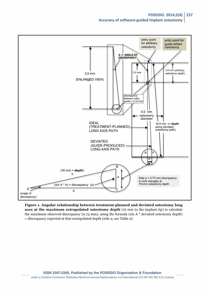
POSEIDO. 2014;2(4) Accuracy of software-‐guided implant osteotomy
257
ISSN 2307-5295, Published by the POSEIDO Organization & Foundation
under a Creative Commons Attribution-NonCommercial-NoDerivatives 4.0 International (CC BY-NC-ND 4.0) License
Figure 1. Angular relationship between treatment-planned and deviated osteotomy long axes at the maximum extrapolated osteotomy depth (16 mm to the implant tip) to calculate the maximum observed discrepancy (0.73 mm), using the formula (sin A * deviated osteotomy depth) = discrepancy expected at that extrapolated depth (side a; see Table 2).
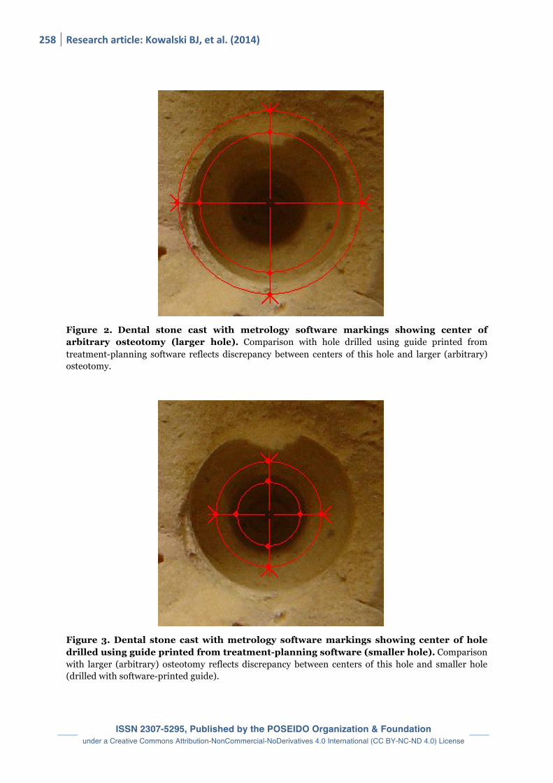
258 Research article: Kowalski BJ, et al. (2014)
ISSN 2307-5295, Published by the POSEIDO Organization & Foundation
under a Creative Commons Attribution-NonCommercial-NoDerivatives 4.0 International (CC BY-NC-ND 4.0) License
Figure 2. Dental stone cast with metrology software markings showing center of arbitrary osteotomy (larger hole). Comparison with hole drilled using guide printed from treatment-planning software reflects discrepancy between centers of this hole and larger (arbitrary) osteotomy.
Figure 3. Dental stone cast with metrology software markings showing center of hole drilled using guide printed from treatment-planning software (smaller hole). Comparison with larger (arbitrary) osteotomy reflects discrepancy between centers of this hole and smaller hole (drilled with software-printed guide).
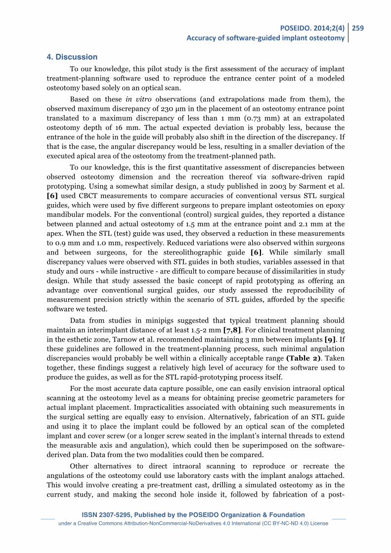
POSEIDO. 2014;2(4) Accuracy of software-‐guided implant osteotomy
259
ISSN 2307-5295, Published by the POSEIDO Organization & Foundation
under a Creative Commons Attribution-NonCommercial-NoDerivatives 4.0 International (CC BY-NC-ND 4.0) License
4. Discussion To our knowledge, this pilot study is the first assessment of the accuracy of implant
treatment-planning software used to reproduce the entrance center point of a modeled osteotomy based solely on an optical scan.
Based on these in vitro observations (and extrapolations made from them), the observed maximum discrepancy of 230 µm in the placement of an osteotomy entrance point translated to a maximum discrepancy of less than 1 mm (0.73 mm) at an extrapolated osteotomy depth of 16 mm. The actual expected deviation is probably less, because the entrance of the hole in the guide will probably also shift in the direction of the discrepancy. If that is the case, the angular discrepancy would be less, resulting in a smaller deviation of the executed apical area of the osteotomy from the treatment-planned path.
To our knowledge, this is the first quantitative assessment of discrepancies between observed osteotomy dimension and the recreation thereof via software-driven rapid prototyping. Using a somewhat similar design, a study published in 2003 by Sarment et al. [6] used CBCT measurements to compare accuracies of conventional versus STL surgical guides, which were used by five different surgeons to prepare implant osteotomies on epoxy mandibular models. For the conventional (control) surgical guides, they reported a distance between planned and actual osteotomy of 1.5 mm at the entrance point and 2.1 mm at the apex. When the STL (test) guide was used, they observed a reduction in these measurements to 0.9 mm and 1.0 mm, respectively. Reduced variations were also observed within surgeons and between surgeons, for the stereolithographic guide [6]. While similarly small discrepancy values were observed with STL guides in both studies, variables assessed in that study and ours - while instructive - are difficult to compare because of dissimilarities in study design. While that study assessed the basic concept of rapid prototyping as offering an advantage over conventional surgical guides, our study assessed the reproducibility of measurement precision strictly within the scenario of STL guides, afforded by the specific software we tested.
Data from studies in minipigs suggested that typical treatment planning should maintain an interimplant distance of at least 1.5-2 mm [7,8]. For clinical treatment planning in the esthetic zone, Tarnow et al. recommended maintaining 3 mm between implants [9]. If these guidelines are followed in the treatment-planning process, such minimal angulation discrepancies would probably be well within a clinically acceptable range (Table 2). Taken together, these findings suggest a relatively high level of accuracy for the software used to produce the guides, as well as for the STL rapid-prototyping process itself.
For the most accurate data capture possible, one can easily envision intraoral optical scanning at the osteotomy level as a means for obtaining precise geometric parameters for actual implant placement. Impracticalities associated with obtaining such measurements in the surgical setting are equally easy to envision. Alternatively, fabrication of an STL guide and using it to place the implant could be followed by an optical scan of the completed implant and cover screw (or a longer screw seated in the implant’s internal threads to extend the measurable axis and angulation), which could then be superimposed on the software-derived plan. Data from the two modalities could then be compared.
Other alternatives to direct intraoral scanning to reproduce or recreate the angulations of the osteotomy could use laboratory casts with the implant analogs attached. This would involve creating a pre-treatment cast, drilling a simulated osteotomy as in the current study, and making the second hole inside it, followed by fabrication of a post-
260 Research article: Kowalski BJ, et al. (2014)
ISSN 2307-5295, Published by the POSEIDO Organization & Foundation
under a Creative Commons Attribution-NonCommercial-NoDerivatives 4.0 International (CC BY-NC-ND 4.0) License
placement cast of the implant with its analog attached. A laboratory scanner could then be used to superimpose the inserted implant-analog position with the original optically scanned cast, to assess the accuracy captured by the treatment-planning software.
Based on such hypotheses, some reproducible modality to compare clinically the predicted angulation with actual positioning of the implant is a viable next step. The clinical implications of these findings will need to be assessed by using this technique as guidance for the actual placement of implants - importantly - in combination with CBCT scanning, in situations presenting a variety of clinical and/or anatomic variables arising from assessing both modalities together.
5. Conclusion The maximum discrepancy between cast intra-osteotomy center points at the implant
entrance was 0.23 mm (230 µm), suggesting that the secondary hole drilled with the software-produced surgical guide was accurate. Calculated discrepancies at the implant tip relative to the entrance-point centering were all less than 1 mm, even at a maximum implant length of 16 mm and at an angle of 2 degrees.
Using an optical scan of a cast to generate data for importation to treatment-planning software is a highly accurate technique for creating a surgical guide. This method could be used in conjunction with CBCT to produce clinically acceptable results in regard to alignment of the osteotomy entrance point. Combining CBCT with optical scan data in subsequent studies will be essential to verify optimized fit of the surgical guide, and any consequently observed clinical improvement in drilling precision.
The primary limitation of this pilot study is that only the entrance point of the osteotomy was evaluated, and that measurements of discrepancies at the implant tip - and thus, the implant axis angulations - were obtained by extrapolation. Such discrepancy values might be tested by optical scanning of experimental osteotomies drilled at greater depths, corresponding to actual implant fixture lengths. Further studies are necessary to evaluate the accuracy of implant axis angulation relative to osteotomy depth, with the objective of developing a safe and standardized clinical protocol for actual implant placement using this approach. Disclosure of interests
The authors have no conflict of interest to report. Acknowledgements
The authors want to thank Scott A. Saunders, DDS, ELS, CMPP for professional medical writing services provided in preparation of this article. References [1] Schneider D, Marquardt P, Zwahlen M, Jung RE. A systematic review on the accuracy and the clinical outcome of computer-guided template-based implant dentistry. Clin Oral Implants Res. 2009;20 Suppl 4:73-86. [2] Del Corso M, Aba G, Vazquez L, Dargaud J, Dohan Ehrenfest DM. Optical three-dimensional scanning acquisition of the position of osseointegrated implants: an in vitro study to determine method accuracy and operational feasibility. Clin Implant Dent Relat Res. 2009;11(3):214-21.
POSEIDO. 2014;2(4) Accuracy of software-‐guided implant osteotomy
261
ISSN 2307-5295, Published by the POSEIDO Organization & Foundation
under a Creative Commons Attribution-NonCommercial-NoDerivatives 4.0 International (CC BY-NC-ND 4.0) License
[3] Jung RE, Schneider D, Ganeles J, Wismeijer D, Zwahlen M, Hammerle CH, Tahmaseb A. Computer technology applications in surgical implant dentistry: a systematic review. Int J Oral Maxillofac Implants. 2009;24 Suppl:92-109. [4] Neugebauer J, Stachulla G, Ritter L, Dreiseidler T, Mischkowski RA, Keeve E, Zoller JE. Computer-aided manufacturing technologies for guided implant placement. Expert Rev Med Devices. 2010;7(1):113-29. [5] van der Zel JM. Implant planning and placement using optical scanning and cone beam CT technology. J Prosthodont. 2008;17(6):476-81. [6] Sarment DP, Sukovic P, Clinthorne N. Accuracy of implant placement with a stereolithographic surgical guide. Int J Oral Maxillofac Implants. 2003;18(4):571-7. [7] Elian N, Bloom M, Dard M, Cho SC, Trushkowsky RD, Tarnow D. Effect of interimplant distance (2 and 3 mm) on the height of interimplant bone crest: a histomorphometric evaluation. J Periodontol. 2011;82(12):1749-56. [8] Elian N, Bloom M, Dard M, Cho SC, Trushkowsky RD, Tarnow D. Radiological and micro-computed tomography analysis of the bone at dental implants inserted 2, 3 and 4 mm apart in a minipig model with platform switching incorporated. Clin Oral Implants Res. 2014;25(2):e22-9. [9] Tarnow DP, Cho SC, Wallace SS. The effect of inter-implant distance on the height of inter-implant bone crest. J Periodontol. 2000;71(4):546-9. This article can be cited as: Kowalski BJ, Manos M, Levi M, D’Ambrosia J. Accuracy of guided osteotomy using dental implant treatment-planning software in combination with an optical scan of a dental cast. POSEIDO. 2014;2(4):253-61.
ISSN 2307-5295, Published by the POSEIDO Organization & Foundation
under a Creative Commons Attribution-NonCommercial-NoDerivatives 4.0 International (CC BY-NC-ND 4.0) License