
ORAL - FOR.org · EJOI ORAL IMPLANTOLOGY EUROPEAN JOURNAL OF Official publication of the British...
148
EJ OI ORAL IMPLANTOLOGY EUROPEAN JOURNAL OF Official publication of the British Society of Oral Implantology (BSOI), the Italian Society of Oral Surgery and Implantology (SICOI), the Danish Society for Oral Implantology (DSOI), the German Association of Oral Implantology (DGI), the Spanish Society of Implantology (SEI), the British Academy of Implant & Restorative Dentistry (BAIRD), and the Advanced Dental Implant Research & Education Center (AIC) A FOR consensus conference on Prosthetic Protocols in Implant-Based Oral Rehabilitations University of Pennsylvania, Philadelphia, USA November 30th to December 1st, 2016 VOLUME 10 / SUPPLEMENT 1 AUTUMN 2017
Transcript of ORAL - FOR.org · EJOI ORAL IMPLANTOLOGY EUROPEAN JOURNAL OF Official publication of the British...

EJOI
ORAL IMPLANTOLOGYEUROPEAN JOURNAL OF
Official publication of the British Society of Oral Implantology (BSOI),the Italian Society of Oral Surgery and Implantology (SICOI),
the Danish Society for Oral Implantology (DSOI),the German Association of Oral Implantology (DGI),
the Spanish Society of Implantology (SEI),the British Academy of Implant & Restorative Dentistry (BAIRD),
and the Advanced Dental Implant Research & Education Center (AIC)
A FOR consensus conference onProsthetic Protocols in Implant-Based Oral RehabilitationsUniversity of Pennsylvania, Philadelphia, USANovember 30th to December 1st, 2016
VOLUME 10 / SUPPLEMENT 1 AUTUMN 2017

S2 n
Eur J Oral Implantol 2017;10(Suppl1):S2
EDITORIAL
Editorial
This supplemental issue of EJOI is dedicated to the Foundation for Oral Rehabilitation (FOR) consen-sus conference, ‘Prosthetic Protocols in Implant-Based Oral Rehabilitations’, which was held on the 30th November to 1st December 2016 at the Uni-versity of Pennsylvania, Philadelphia, USA. Scientific associations and other organisations using EJOI as their official publication are welcome to publish the outcome of their consensus conferences or working groups in the journal.
It is the policy of EJOI that these publications will not be peer reviewed as they are normally. Conse-quently, readers are encouraged to critically evaluate the findings presented, as they would with all scien-tific publications. Guidance on how to develop criti-cal skills for research, analysis and the evaluation of scientific publications (an important mission of EJOI) can be found in the ‘educational articles’1-4 and on the EQUATOR (Enhancing the QUAlity and Trans-parency Of health Research) website (http://www.equatornetwork.org/). The EQUATOR Network is aimed at helping authors properly report their health research studies. After selecting the ‘Resource Cen-tre’, please click on the ‘Library for health research
reporting’ and you will access a comprehensive list of reporting guidelines, organised by study type. More specifically, to evaluate systematic reviews please go to the PRISMA transparency guidelines (http://www.prisma-statement.org/).
The results of consensus conferences or work-ing groups can be interpreted differently, depending on people’s perspectives and circumstances. Please consider the conclusions presented carefully. They are the opinions of the review authors, and are not necessarily shared by EJOI editors.
We would like to thank all contributors to this supplement for their efforts.
Marco Esposito, Reinhilde Jacobs and Michele Nieri
1. Worthington HV, Esposito M, Nieri M, Glenny AM. What is a systematic review? Eur J Oral Implantol 2008;1:235–238.
2. Glenny AM, Nieri M, Worthington H, Espostio M. The importance of the study design: from the case report to the randomised controlled clinical trial. Eur J Oral Implantol 2008;1:317–321.
3. Nieri M, Glenny AM, Worthington H, Esposito M. How to interpret meta-analyses of randomised clinical trials. Eur J Oral Implantol 2009;2:61–66.
4. Glenny AM, Worthington HV, Esposito M, Nieri M. What are clinical guidelines? Eur J Oral Implantol 2009;2:145–148.

n 3
Eur J Oral Implantol 2017;10(Suppl1):3
GUEST EDITORIAL
Guest Editorial
The methods by which single crowns and prostheses are designed and fabricated for implant-based treat-ments have changed over the years. Recently, new innovative materials and techniques have been introduced, along with related scientific evidence. Therefore, this consensus conference was focused on the prosthodontic aspects of such implant-based rehabilitations.
At a time when an over-abundance of information is always readily available through internet-based outlets, discerning sound scientific evidence from questionable and biased data has become increas-ingly challenging. Systematic literature reviews with meta analyses, where appropriate, are at the pinna-cle of the “quality-of-scientific-evidence” pyramid and have, therefore, become invaluable tools in the assessment of clinical data and the decision-making process in the practice of dentistry.
The Foundation for Oral Rehabilitation (FOR) is partnering with academicians and leading universi-ties around the world to provide such assessments on a variety of highly relevant topics and consolidate the outcomes in consensus statements.
A group of 10 international experts was selected, based on their expertise and publications related to specific aspects of prosthodontic treatment. Each participant was tasked with completing a systematic and comprehensive review of the literature and syn-thesizing it into the form of a literature review. Each
paper was submitted and reviewed by the panel of experts prior to the actual conference meeting. Then, at the meeting, each participant presented a synopsis of their conclusions, followed by time for discussion and critique by all the attendees. After the face-to-face meeting, final papers were submitted and the consensus text was developed for inclusion in this special supplement of the journal.
The conference took place at the University of Pennsylvania, School of Dental Medicine, in Phila-delphia, Pennsylvania, for two days. It was a privi-lege for both of us to serve as co-chairs of this FOR Consensus Conference and have the opportunity to interact with this distinguished group of interna-tional experts. We also want to express our appre-ciation to Dr Daniel van Steenberghe for his invalu-able service by providing a written record of the key findings of the conference and helping to develop the consensus text. In addition, we wish to thank Dr Friedrich Neukam, chairman of the FOR Board of Trustees, who provided oversight and input during the conference.
We are pleased to submit the outcomes of this conference as another ongoing service of the Foun-dation for Oral Rehabilitation to benefit the profes-sion and enhance knowledge regarding the prostho-dontic treatments available to the public we all serve.
Charles Goodacre and Markus Blatz
Charles J. Goodacre, DDS, MSDDistinguished ProfessorLoma Linda University School of DentistryLoma Linda, California, USA 92350
Markus B. Blatz, DMD, PhDProfessor and Chair, Depart-ment of Preventive and Restorative SciencesUniversity of Pennsylvania School of Dental Medicine, Philadelphia, USA

4 n
Eur J Oral Implantol 2017;10( Suppl1):4
IMPRINT
European Journal of Oral Implantology
The European Journal of Oral Implantology is published quarterly by Quintessence Pub lishing Co. Ltd, Quintessence House, Grafton Road, New Malden, Surrey KT3 3AB, UK. Court domic-ile and place of performance: London, England.The European Journal of Oral Implantology is listed in MEDLINE, Science Citation Index Expan-ded and Journal Citation Reports/Science Edition.
Copyright © 2017 by Quintessence Publishing Co. Ltd. All rights reserved.
No part of this journal may be reproduced in any material form (including photocopy-ing or storing it in any medium by elect-ronic means and whether transiently or in-cidentally to some other use of this journal), without the written permission of the publisher except in accordance with the provisions of the Copyright, Designs and Patents Act 1988 or under the terms of a licence issued by The Copy-right Licensing Agency Ltd, Saffron House, 6-10 Kirby Street, London EC1N 8TS, UK. Application for the copyright owner’s written permission to reproduce any part of this journal should be
Subscription informationContact your nearest Quintessence office:
Quintessence Publishing Co. Ltd,Quintessence House, Grafton Road,New Malden, Surrey KT3 3AB, UK.Tel: +44 (0)20 8949 6087Fax: +44 (0)20 8336 1484Email: [email protected]
Quintessenz Verlags-GmbHIfenpfad 2–4, D-12107 Berlin, GermanyTel: +49-30-761 80-5Fax: +49-30-761 80-68-0Email: [email protected]
Quintessence Publishing Co. Inc4350 Chandler Drive,Hanover Park, Illinois 60133, USATel: (630) 736-3600Fax: (630) 736-3633Email: [email protected]
Subscription rates (includes online version, http://ejoi.quintessenz.de)
Europe: Surface mail Air mailIndividual €148 / £126 £146Institutional €320 / £272 £292Student* €78 / £66 £86Single issue €38 / £32 £37 North America and rest of world:Individual $170; Institutional $305; Student* $90
* Student verification must accompany order.
Subscriptions may begin at any time. Please allow 6 weeks for any change of address notification to be processed. Claims for missing journals will be serviced only within 6 months of publication date. Otherwise, single copy price will be charged on missing issues.
Postmaster: Send address changes toQuintessence Publishing Co. Ltd, Quintessence House, Grafton Road, New Malden, Surrey KT3 3AB, UK, orQuintessenz Verlags-GmbH, Ifenpfad 2–4, D-12107, Berlin, Germany
Manuscript submission information: Go to www.manuscriptmanager.com/ejoi to submit online. For more information, see the Guideli-nes for Authors page in this issue.
Impact factor 2016 3.567ISSN 1756-2406 (Print)ISSN 1756-2414 (Online)
addressed to the publisher. The publisher assu-mes no responsibility for unsolicited manuscripts. All opinions are those of the authors.
Advertising Policy: All advertising appearing in the European Journal of Oral Implantology must be approved by the Editors/ Editorial Board. The publication of an advert is not to be contrued as an endorsement of approval by the journal or its publisher.
Permission to photocopy items solely for in-ternal or personal use and for the internal and personal use of specific clients is granted by Quintessence Publishing Co. Ltd.
Publisher: Dr. h. c. H.-W. Haase Publishing Director: Johannes W. WoltersEditorial Coordinator: Natalie WardSubscription Managers: Angela Köthe: Germany, Austria, SwitzerlandAndrew Johnson: All other countriesAdvertising: Sue Newbury and Sabina BeganovicLayout/Production: Ina SteinbrückPrinted in Germany
Editors
Marco Esposito, Arcore, Italy (Editor-in-Chief)Reinhilde Jacobs, Leuven, BelgiumMichele Nieri, Florence, Italy
Official publication of the British Society of Oral Implantology (BSOI), the Italian Society of Oral Surgery and Implantology (SICOI), the Danish Society for Oral Implantology (DSOI),the German Association of Oral Implantology(DGI),the Spanish Society of Implantology (SEI), British Academy of Implant & Restorative Dentistry (BAIRD),and the Advanced Dental Implant Research & Education Center (AIC)
Editorial Board
Carlos Aparicio, SpainKarl Bishop, UKIan Brook, UKJason Buglass, UKGioacchino Cannizzaro, ItalyVittorio Checchi, Italy Matteo Chiapasco, ItalyNoel Claffey, IrelandStJohn Crean, UKRubén Davó, SpainNikolaos Donos, UK Pietro Felice, ItalyAnne-Marie Glenny, UKRonnie Goené, The NetherlandsKlaus Gotfredsen, DenmarkStefano Gracis, ItalyTommaso Grandi, Italy Kerstin Grondahl, SwedenUeli Grunder, SwitzerlandM Gabriella Grusovin, Italy Dominic Hassall, UKFederico Hernández-Alfaro, SpainSøren Hillerup, DenmarkFlemming Isidor, DenmarkReinhilde Jacobs, Belgium
Torsten Jemt, SwedenSøren Jepsen, GermanyRonald Jung, SwitzerlandMatthias Kern, GermanyFouad Khoury, GermanyYe Lin, ChinaHassan Maghaireh, UK Chantal Malevez, BelgiumPaulo Maló, PortugalMauro Merli, ItalyKen Nicholson, UKAlan Payne, New ZealandRoberto Pistilli, Italy Bjarni E Pjetursson, IcelandStefan Renvert, SwedenMariano Sanz, SpainPaul Stone, UKHendrik Terheyden, GermanyTiziano Testori, ItalyGeorg Watzek, AustriaDieter Weingart, GermanyHelen V Worthington, UKGiovanni Zucchelli, Italy Otto Zuhr, Germany

n S5
Eur J Oral Implantol 2017;10(Suppl1):S5–S6
CONTENTS
European Journal of Oral ImplantologySupplement 1, Autumn 2017
EDITORIALMarco Esposito, Reinhilde Jacobs, Michele Nieri S2
GUEST EDITORIALCharles Goodacre, Markus Blatz S3
CONSENSUS STATEMENTSFoundation for Oral Rehabilitation (FOR) Consensus Text on “Prosthetic Protocols in Implant-Based Oral Rehabilitations” S7
REVIEWSFixed vs removable complete arch implant prostheses: A literature review of prosthodontic outcomesCharles Goodacre, Brian Goodacre S13
Clinical outcomes of full arch fixed implant-supported zirconia prostheses: A systematic reviewAvinash Bidra, Patchanee Rungruanganunt, Marissa Gauthier S35
Impact of prosthetic material on mid- and long-term outcome of dental implants supporting single crowns and fixed partial dentures: A systematic review and meta-analysisSamir Abou-Ayash, Malin Strasding, Gerta Rücker, Wael Att S47
Influence of implant abutment fabrication method on clinical outcomes: a systematic reviewLong Long, Hatem Alqarni and Radi Masri S67
Immediate loading of zygomatic implants: A systematic review of implant survival, prosthesis survival and potential complicationsFrank J. Tuminelli, Leora R Walter, Jay Neugarten, Edmond Bedrossian S79
Prosthetic protocols in implant-based oral rehabilitations: A systematic review on the clinical outcome of monolithic all-ceramic single- and multi-unit prosthesesFrank A. Spitznagel, Sebastian D. Horvath, Petra C. Gierthmuehlen S89
Accuracy of digital implant impressions with intraoral scanners. A systematic reviewVygandas Rutkūnas, Agnė Gečiauskaitė, Darius Jegelevičius, Mantas Vaitiekūnas S101
Contents
Contents

CONTENTSS6 n
Eur J Oral Implantol 2017;10(Suppl1):S5–S6
Misfit of implant prostheses and its impact on clinical outcomes. Definition, assessment and a systematic review of the literatureJoannis Katsoulis, Takuro Takeichi, Ana Sol Gaviria, Lukas Peter, Konstantinos Katsoulis S121
Performance of CAD/CAM monolithic ceramic Implant-supported restorations bonded to titanium inserts: A systematic reviewJulian Conejo, Toyoaki Kobayashi, Evantia Anadioti, Markus B. Blatz S139
Imprint S4

n 7
Eur J Oral Implantol 2017;10(Suppl1):7–11
CONSENSUS STATEMENTS
Foundation for Oral Rehabilitation (FOR) Consensus Text on “Prosthetic Protocols in Implant-Based Oral Rehabilitations”
n Methodology used for establishing the consensus text
The Foundation for Oral Rehabilitation (FOR) assem-bled a group of nine international experts to exam-ine specific aspects of prosthodontic treatment. Each participant completed a systematic and comprehen-sive literature review and their resulting papers were submitted and reviewed by each participant prior to the conference. During the meeting, each par-ticipant presented their conclusions with time for discussion and critique by all the attendees. After all the experts had submitted the final papers, the most pertinent findings were synthesized into this consensus text.
n Background to the conference
Edentulism, both complete and partial, have benefit-ted from osseointegrated endosseous implants that retain or support a prosthesis. After the pioneering epoch of the 1970s, high long-term survival and suc-cess rates have been universally reported. Implant-based oral rehabilitations have thus often become the number one treatment choice for partial and complete edentulism.
Over the past two decades, new developments were geared towards increased success/survival rates of implants and prostheses, less-invasive approaches, versatility of treatment options, as well as improved patient satisfaction and quality of life.
Since the scientific literature has well documented high implant success/survival rates, the present con-sensus conference focused on the prosthodontic aspects of implant-based oral rehabilitation.
n Prosthodontic outcomes for fixed vs removable complete arch implant prosthesis1
Based on the results of clinical studies that evalu-ated both fixed and removable complete arch pros-theses in the same study, it was determined that both types of prostheses are associated with high implant survival rates. However, both were affected by post-placement maintenance that could be called “normal wear and tear” and also by prosthetic “complications” that were judged to be unexpected events requiring additional treatment. Implant over-dentures were associated with more maintenance needs/complications than fixed prostheses. In addi-tion, the amount of post-placement residual ridge resorption was greater with implant overdentures. The level of patient satisfaction was high with both types of prostheses, but implant overdentures were determined to be more cost-effective.
n Clinical outcomes of full arch implant-supported zirconia prostheses2
Fixed complete dentures (formerly called “full arch fixed implant-supported prostheses”) have seen impressive material innovations, one being zirconia, which has the highest fracture toughness of all of the ceramic materials used in oral health care. There are several types and designs of zirconia prostheses. A few examples include: zirconia that is monolithic, zirconia that is veneered (conventional, minimal or gingival) or a zirconia framework with individually cemented crowns. Additionally, the zirconia prosthe-sis may be a one-piece design, or segmented with

Consensus statements 8 n
Eur J Oral Implantol 2017;10(Suppl1):7–11
fixed complete dentures and restorations on zirconia implants (two studies) excluded from the review.
For the meta-analysis of implant survival rates of single crowns, the results of two study cohorts of veneered base metal alloys, 11 of veneered precious alloys, 13 of veneered zirconia, five of veneered alu-mina, four of lithium-disilicate (monolithic or par-tially veneered) and one that used a resin matrix ceramic, were included.
The choice of prosthetic material seems to have no influence on implant or prosthetic survival rates in fixed restorations. Subgroup analyses for the pros-thetic complication rates also revealed no statistically significant differences for screw loosening, abutment fractures, or chipping between any of the groups. The incidence rate for decementation in one study was significantly higher for the resin matrix ceramic group relative to all other groups (P < 0.0001).
The meta-analysis of all-ceramic vs metal-based fixed partial dentures included one cohort study of all-ceramic prostheses, two of metal-based pros-theses with facial resin veneering, and eight of metal-based prostheses veneered with ceramics.
For the survival rate of both implants and pros-theses, no differences were observed among the dif-ferent materials. The incidence rates of screw loos-ening and abutment fractures were similar. On the other hand, the incidence rate for chipping was sig-nificantly higher in the metal-composite resin group when compared with the metal-ceramic and the all-ceramic groups.
n Influence of implant abutment fabrication method on clinical outcomes4
Materials used to fabricate abutments and their man-ufacturing processes are important to clinical success. This paper investigated the literature relative to the effect of CAD/CAM manufactured abutments on the treatment outcome and on the peri-implant tissues.
The review investigated 24 studies on CAD/CAM manufactured abutments, of which 11 were com-parative, to assess factors like survival and success rates, white and pink aesthetic scores and bone loss.
CAD/CAM abutments have good survival and success rates and provide comparable, if not better,
multiple fixed prostheses. The present review was focused on one-piece zirconia fixed complete den-tures with and without veneered porcelain.
Twelve studies meeting the selection criteria were identified (three prospective and nine retro-spective) involving a total of 223 patients with 285 fixed complete dentures and up to 8 years follow-up. The number of implants supporting the one-piece zirconia fixed complete dentures ranged from 3 to 15 implants with an average of 4 implants. Of the 285 prostheses, four frameworks fractured, two in one treatment centre and one each in two other centres. Limited vertical prosthetic space could be a risk factor since it was associated with reported fractures.
Minor prosthetic complications that did not require prosthesis replacement were reported for 46 out of 285 prostheses. Veneered porcelain fracture occurred in 42 prostheses. These minor complications were sig-nificantly lower than what is reported in the literature for metal-acrylic resin fixed complete dentures.
Chipping of veneered porcelain did not require a remake of any prostheses. Chairside polishing and adjustment or occasional laboratory fabricated por-celain veneer sufficed in the majority of patients. Chipping of veneered gingival porcelain was not reported in any of the studies.
Based on available data, monolithic zirconia with gingival colouring (“gingival staining”), or zirconia with veneered porcelain limited to the gingival area, offers promising results for fixed complete dentures. Since the complications occurred with various types of zirconia, the properties and manufacturing pro-cess of zirconia are relevant factors. None of the studies reported adverse effects on implants, oppos-ing natural dentition, hard and soft tissues, temporo-mandibular joints or patient dissatisfaction due to the use of zirconia fixed complete dentures on single or double jaw rehabilitations.
n Impact of prosthetic material on mid- and long-term outcome of implants supporting single crowns and fixed dental prostheses3
The impact of the type of prosthetic material on im-plant survival was reviewed with implant-supported

Consensus statements n 9
Eur J Oral Implantol 2017;10(Suppl1):7–11
clinical outcomes than conventional abutments. One study reported a better aesthetic outcome at 1 year and another reported less soft tissue recession at 2 years compared with conventional abutments. However, available studies comparing CAD/CAM and conventional abutments are few and the major-ity are limited to the short-term.
n Prosthesis survival and complication with immediate loading of zygomatic implants5
Zygomatic implants offer an alternative treatment option for patients with severely resorbed maxillae. The overall survival rate after 12 to 72 months is 96 to 100% for the zygomatic implants. This per-centage range applies to zygomatic implants where two implants were placed bilaterally and also when there was a single zygomatic implant bilaterally with splinting to conventional anterior endosseous implants. The studies indicated a favourable anterior-posterior spread was achievable by both designs.
There were 17 studies that reported on con-ventional anterior implants splinted to zygomatic implants, with a survival rate of the conventional implants ranging from 95 to 100%. However, five of these studies reported anterior implant failures along with zygomatic implant failure. The survival of prostheses relates to the number and position of the zygomatic implants. When the prosthetic design used one zygomatic implant bilaterally with anterior endosseous implants, the loss of one zygomatic im-plant resulted in the loss of part of the prostheses, necessitating remake or modification.
Prosthetic complications were identified in numerous papers and included loosening and frac-ture of prosthetic screws, with fracture of abutment screws reported in two studies. There were also reports of metal framework fracture and ceramic fracture from the underlying metal substructure. One paper reported excessive wear of the restora-tive tooth material.
Inflammatory reactions in the maxillary sinus were reported in 12 papers, with incidences ranging from less than 1.0 % to over 20%. Multiple authors report a reduction in sinusitis, with the extra-sinus (external) approach. One paper demonstrated that 15 to 20%
of patients had inflammatory reactions, as noted on radiographic examination, but the patients were asymptomatic. One article suggested use of the buc-cal fat pad to potentially reduce intraoral mucositis. However, most studies did not apply this approach.
n Clinical outcome of monolithic ceramic implant supported single and multi-unit prostheses6
A systematic review on the clinical outcome of mon-olithic ceramic implant supported single and multi-unit prostheses identified three studies included in the review.
Two articles reported on monolithic lithium disilicate implant-supported single crowns (SC) and revealed a survival rate of 97.8 and 100% after 28 to 31 months. One study investigated implant-supported mono-lithic zirconia SCs and fixed partial dentures (FPD) and showed a survival rate of 100% after 5 years. The use of zirconia induces minimal wear to opposing struc-tures, especially after adaptation and polishing of the occlusal surfaces. The risk of fracture and chipping was significantly reduced in monolithic restorations. No study on the clinical performance of monolithic resin matrix ceramic restorations could be identified.
Clinical studies on the long-term outcome of implant-supported monolithic all-ceramic single- and multi-unit restorations are lacking.
n Digital vs conventional implant impressions7
The literature review on digital vs conventional implant impressions identified one in vivo and 15 in vitro studies. The majority of the studies (n = 12) evaluated accuracy of digital implant impressions (DII) by superimposing images to refer-ence models and reported mean errors ranging from 6 to 337 µm.
Results from three recent in vitro studies directly comparing the accuracy of DII and conventional impression techniques reported similar results for single and multiple implants.
Factors such as the type of scanner, angula-tion and number of implants, distance in between

Consensus statements 10 n
Eur J Oral Implantol 2017;10(Suppl1):7–11
implants, geometry of scan bodies and scanning techniques that potentially affect the accuracy, were not sufficiently investigated.
High deviations of up to 328 µm were reported by studies investigating accuracy of milled models produced from DII. Further studies are needed to evaluate accuracy of 3D printing techniques to fab-ricate master models for implant-supported single crowns and fixed partial dentures. Also, data is lack-ing on IOS accuracy for digital interocclusal records.
Since intraoral scanning is more challenging than in-vitro scanning of a model, more in vivo studies are needed to define clinical indications for different types of IOS. However, the in vivo evaluation of accuracy is limited by the possibilities to obtain true reference values under clinical conditions.
n Misfit of implant prostheses and its impact on clinical outcomes8
Ten articles met the inclusion criteria: five on humans and five on animals, relating to the misfit of implant prostheses.
It was concluded that the available literature does not provide sufficient evidence on the effect of misfit at the prosthesis-implant interface on clinical out-comes of screw-retained implant prostheses. Mar-ginal gaps and static strains due to screw tightening were not found to have negative effects on initial osseointegration or peri-implant bone stability over time. Based on two clinical studies, the risk for tech-nical screw-related complications was slightly higher.
While the degree of tolerable misfit remains a matter of debate, the present data do not imply that clinicians should neglect good fit.
n Clinical performance of CAD/CAM monolithic ceramic implant-supported restorations bonded to titanium inserts9
Current trends and the more frequent application of chairside digital dentistry suggest the clinical appli-cation CAD/CAM monolithic implant-supported ceramic restorations. Many of these systems, espe-cially the ones applied chairside, require that these
all-ceramic crowns be bonded to titanium (Ti) inserts with composite resins after adequate pre-treatment of the bonding interfaces. This systematic review of the literature revealed there is currently no clinical evidence on CAD/CAM monolithic implant-sup-ported ceramic restorations that are bonded to Ti-inserts. However, several laboratory studies on select aspects of Ti-inserts and similar prosthetic designs are available.
These studies indicate that Ti-inserts improve the overall fracture strength of ceramic abutments and crowns, protect the implant connection from wear, and offer a better marginal fit when compared to all-ceramic abutments. However, to recommend this prosthetic design for routine use in clinical practice, independent clinical trials that document its long-term performance are necessary.
n Recommendations of the group of experts
The following statements reflect the opinions of the individuals participating in the consensus confer-ence, to the best of their knowledge and experience:• The systematic reviews assessed the clinical evi-
dence on a variety of relevant aspects of modern implant prosthetics. It should be cautioned, how-ever, that absence of scientific evidence in the fast-evolving field of implant-based rehabilita-tion does not necessarily imply that a treatment modality is ineffective.
• The choice of a fixed or removable complete arch prosthesis varies according to patient preference.
• Definitions of professional maintenance, com-plication and/or failure are missing. Professional prosthetic maintenance implies compensating for the predictable wear of prosthetic components. A prosthetic complication is an unanticipated event that affects the prosthesis and requires inter-vention or not, but without replacement of the prosthesis. Failure requires removal and remake of prosthesis or change of treatment. Future research should distinguish between these two possible consequences to avoid inflated compli-cation and failure rates.
• The number of prosthetic maintenance issues associated with adjusting and replacing the

Consensus statements n 11
Eur J Oral Implantol 2017;10(Suppl1):7–11
retentive device of overdentures indicates the need for longer-lasting retentive devices. One of the members of the group even suggested it would be beneficial to have a retentive device that could be changed by patients, while rec-ognising this process does not and should not replace regular professional care, but could be helpful for those patients who do not have easy access to regular professional care.
• The current scientific evidence does not favour a specific material for single crowns and multi-unit fixed partial dentures.
• The material selection is rather based on the clini-cian’s preference and the aesthetic and functional needs of the patient.
• Although the clinical evidence is currently weak, monolithic all-ceramic implant-supported single crowns and multiunit fixed partial dentures are reliable based on short-term observation.
• Laboratory studies demonstrate that bonded tita-nium inserts provide several mechanical advan-tages for all-ceramic abutments and crowns, such as protecting the implant-abutment connection. There is, however, no clinical data currently avail-able to support their routine use.
• Clinical experience favours intraoral digital scan-ning but evidence is lacking. Clinical factors affecting the accuracy of modern intraoral scan-ners, as well as accuracy of resulting models and restorations, should be further investigated.
• Current data supports the use of zygomatic implants to support a fixed dental prosthesis. The implants should preferably emerge on the crest of the ridge, as opposed to the palate, to facilitate a more anatomical prosthesis.
• CAD/CAM abutments should be used when possible. Not only do they provide restorations that possess excellent overall survival and suc-cess rates, they can be fabricated with proper contours for optimized aesthetic outcomes. They also allow for excellent fit and control of finish line position to facilitate cement removal.
• One-piece zirconia fixed complete dentures have promising outcomes in edentulous patients. The zirconia can be veneered at the gingiva or be monolithic, with only gingival colouring (“gin-gival staining”) to reduce prosthetic complications associated with veneered porcelain fracture.
• Clinical evidence on the effect of misfit of screw-retained implant prostheses is missing for biological factors and weak for technical complications. These findings do not imply that misfit of prosthetic implant components is without consequences. Therefore, the expert panel encourages clinicians to continue aiming for the best fit possible.
n References
1. Goodacre CJ, Goodacre BJ. Fixed versus removable com-plete arch implant prostheses: A literature review of pros-thodontic outcomes.
2. Bidra AS, Rungruanganunt P, Gauthier MF. Clinical out-comes of full arch implant-supported zirconia prostheses: A systematic review.
3. Abou-Ayash S, Strasding M, Rücker G, Att W. Impact of prosthetic material on mid- and long-term outcome of implants supporting single crowns and fixed dental pros-theses: A systematic review and meta-analysis.
4. Long L, Alqarni H, Masri R. Influence of Implant Abutment Fabrication Method on Clinical Outcomes: A Systematic Review.
5. Tuminelli FJ, Walter LR, Neugarten J, Bedrossian E. Immediate loading of zygomatic implants: A systematic review of implant survival, prosthesis survival and potential complications.
6. Spitznagel FA, Horvath SD, Gierthmuehlen PC. Prosthetic protocols in implant-based oral rehabilitations: A systematic review of monolithic all-ceramic single- and multi-unit res-torations.
7. Rutkunas V, Geciauskaite A, Jegelevičius D, Vaitiekünas M. Accuracy of digital implant impressions with intraoral scan-ners. A systematic review.
8. Katsoulis J, Takeichi T, Gaviria AS, Peter L, Katsoulis K. Misfit of implant prostheses and its impact on clinical outcomes. Defini-tion, assessment and a systematic review of the literature.
9. Conejo J, Kobayashi T, Anadioti E, Blatz MB. Clinical perfor-mance of CAD/CAM monolithic ceramic implant-supported restorations bonded to titanium inserts: A systematic review.
n Co-chairs:
Dr. Markus Blatz (USA) & Dr. Charles Goodacre (USA)
n Experts:
Dr. Wael Att (DE) Dr. Avinash Bidra (USA) Dr. Markus Blatz (USA)Dr. Petra Gierthmuehlen (DE) Dr. Charles Goodacre (USA) Dr. Joannis Katsoulis (CH) Dr. Radi Masri (USA) Dr. Vygandas Rutkunas (LT) Dr. Frank Tuminelli (USA)


n 13
Eur J Oral Implantol 2017;10(Suppl1):13–34
REVIEW
Charles J Goodacre, DDS, MSDDistinguished ProfessorLoma Linda University School of DentistryLoma Linda, California, USA
Brian J Goodacre, DDSAdvanced Education Student in Prosthodontics and Im-plant DentistryLoma Linda University School of DentistryLoma Linda, California, USA
Correspondence to:Professor Charles J Goodacre DDS MSDLoma Linda University School of DentistryLoma Linda, California, USA 92350Email: [email protected]
Charles Goodacre, Brian Goodacre
Fixed vs removable complete arch implant prostheses: A literature review of prosthodontic outcomes
Key words bone changes, cost-effectiveness, masticatory performance, patient satisfaction, prosthesis complications, prosthesis survival, quality of life
Aim: To compare implant fixed complete dentures with implant overdentures relative to prosthodon-tic outcomes.Material and methods: An electronic Medline (PubMed) with MeSH terms, and Cochrane library search was performed, focusing on studies that included implant fixed complete dentures and im-plant overdentures in the same study, with the results based on studies that included both types of prostheses.Results: The following six categories of comparative studies were identified in the literature: 1) Im-plant and prosthesis survival; 2) Prosthesis maintenance/complications; 3) Bone changes; 4) Patient satisfaction and quality of life; 5) Cost-effectiveness; and 6) Masticatory performance. It was deter-mined that both the fixed and removable treatments were associated with high implant survival rates. However, both types of prostheses were impacted by the need for post-placement mechanical maintenance or prosthetic complications. More maintenance/complications occurred with implant overdentures than with fixed complete dentures. Residual ridge resorption was greater with implant overdentures. Patient satisfaction was high with each prosthesis, with three studies revealing higher satisfaction with fixed complete dentures and five studies finding no difference. All but one study on cost-effectiveness indicated implant overdentures were more cost-effective. Based on two studies, it appears the masticatory performance of implant fixed complete dentures and implant overdentures is comparable.Conclusions: Multiple factors must be considered when determining whether an implant-fixed com-plete denture or implant overdentures are best suited for patients with completely edentulous jaws.
Conflict-of-interest statement: The authors declare they have no conflicts of interest.
n Introduction
Prior to the introduction of osseointegrated implants, complete dentures served as the primary means of replacing the entire dentition. However, multiple studies determined they lacked reten-tion and patients experienced movement of their mandibular dentures1-3. Patients were dissatisfied
with their dentures4 and their attitude affected the perception of comfort, speech, and the ability to chew5. Difficulties while eating certain foods were documented2,3,6-8 and some patients experienced discomfort or pain when chewing or biting8,9. While altered taste sensation was reported to be a short-term effect in one study of complete dentures7, another study8 reported that complete denture

Goodacre and Goodacre Fixed vs removable complete arch implant prostheses14 n
Eur J Oral Implantol 2017;10(Suppl1):13–34
Therefore, the purpose of this literature review was to compare implant-fixed complete dentures (IFCD) with implant overdentures (IOD), based on the comparisons that have been studied in the dental literature.
n Materials and methods
Electronic searches of MEDLINE (PubMed) along with MeSH terms and the Cochrane Central Register of Controlled Trials search were conducted up to and including September 2016. The following key-words were used in the search: implant overdenture, implant fixed complete denture, implant supported prosthesis, implant retained prosthesis, fixed-detach-able prosthesis, fixed-detachable implant prosthesis, fixed-detachable implant denture, hybrid prosthe-sis, hybrid denture, all-on-four prosthesis, fixed vs removable implant prostheses, fixed prosthesis vs implant overdenture, implants overdentures and fixed complete dentures, implant overdentures and fixed implant dentures, implant overdenture and all-on-four dentures, implant overdentures and hybrid implant prostheses, implant overdentures and fixed-detachable prostheses, implant overdentures and fixed-detachable implant prostheses, implant over-dentures and hybrid implant prostheses, implant overdentures vs fixed prostheses, implant retained/supported prostheses, survival of implant prostheses, dental implant survival, implant failure, implant complications, maintenance of dental implants, and complications with complete arch prostheses.
As part of the discussions during the consensus conference, a distinction was made between what could be called “normal wear and tear” prosthe-sis maintenance and prosthesis “complications”, judged to be unexpected events requiring additional treatment.
After reviewing the citations using the differ-ent search terms, a decision was made to focus the detailed review on only those studies that compared implant-fixed complete dentures with implant over-dentures in the same publication, many of which also included conventional complete dentures. As a result of all the citation reviews, the following categories of comparative studies were identified where both IFCDs and IODs were evaluated in the same study:
patients exhibited the lowest scores for taste and texture perception. Additionally, complete denture patients have been known to reduce their social con-tact due to embarrassment as a result of wearing dentures6.
Another important factor is the residual ridge resorption that occurs from wearing complete den-tures, this being particularly reflected as mandibular superior surface resorption10. There is a decrease in the maximum bite force compared with dentate patients11-13 and the masticatory performance (abil-ity to comminute food) is one-quarter to one-seventh that of individuals with natural dentitions11,14-16. Complete denture patients have a lower intake of nutrient-rich foods such as vegetables17,18, dietary fibre18,19-21, carrots18,21, fruits17,18, and salads21 with biochemical analyses of blood samples show-ing that complete denture patients have lower levels of the nutrients found in vegetables and fruits21,22.
These limitations of complete dentures were first counteracted through the use of complete arch fixed prostheses attached to multiple mandibular implants. The Glossary of Prosthodontic Terms23 uses the term “fixed complete denture”, but other names have been used in the literature for this type of prosthesis, such as hybrid denture, hybrid prosthesis and fixed-detachable prosthesis. In this review, the term “im-plant fixed complete denture” is used to describe a complete arch prosthesis that is attached to implants and cannot be removed by the patient, and “implant overdenture” is used to describe a complete arch im-plant prosthesis that the patient can remove.
Following the successful use of multiple implants in conjunction with fixed complete dentures, the im-plant treatment protocol was expanded to include implant overdentures. Many positive outcomes emerged from the use of these two complete arch im-plant prostheses compared with complete dentures, including bone preservation24, greater comfort25 and improved masticatory performance26, as well as enhanced patient satisfaction and quality of life27. However, complications can arise with both types of prostheses and it is important to understand what can occur so complications can be avoided, or at least minimised. In addition, it is important to understand how these two types of complete arch prostheses compare with each other as an aid in treatment plan-ning for completely edentulous patients.

Goodacre and Goodacre Fixed vs removable complete arch implant prostheses n 15
Eur J Oral Implantol 2017;10(Suppl1):13–34
1. Implant and prosthesis survival;2. Prosthesis maintenance/complications;3. Bone changes;4. Patient satisfaction and quality of life;5. Cost-effectiveness;6. Masticatory performance.
In addition to the focused reviews, a limited number of systematic reviews and individual clinical studies were included that provided data related to either implant overdentures or implant-fixed complete dentures, but not both. A synopsis of these stud-ies is presented in the introduction to each of the above categories as background information before reviewing the studies that specifically compared both IFCDs and IODs in the same study.
n Implant and prosthesis survival: Background information related to implant and prosthesis survival rates
Mandibular implant fixed complete denture systematic review
A 2016 systematic review by Moraschini et al28 included 19 studies. The cumulative implant sur-vival rate associated with prostheses supported by four implants (all-on-4) was 96.3% after a mean follow-up time of 40 months and the rate for pros-theses supported by three implants was 95.5% at 32 months. Prosthesis survival rates ranged from 93.7% to 100%, with an overall CSR of 98.6%.
Maxillary implant overdenture systematic reviews
A 2010 systematic review of maxillary implant over-dentures was published by Slot et al29 based on 31 studies after a mean follow-up of at least 1 year. The authors identified an implant survival rate of 98.2% per year with six implants and bars. With four implants and bars the implant survival rate was 96.3%, and with four individual implants and ball abutments the implant survival rate was 95.2%. Prosthesis survival was calculated to be 97.4% per year with six or more implants and 96.5% with four or fewer implants and bar anchorage. The authors were unable to calculate prosthesis survival with four
or fewer implants and ball anchorage because only one of the included studies presented the overden-ture survival rate for this design. The authors con-cluded that six implants connected by a bar was the most successful treatment regarding both implant and prosthesis survival.
A similar 2014 systematic review by Raghoebar et al30 included 24 studies after a mean observation time of at least 1 year. The meta-analysis identified an implant survival rate of 98.1% and an overden-ture survival rate of 99.5% per year when six or more implants were splinted with bars. When four or fewer implants were splinted with bars, the im-plant survival rate was 97.0% and the overdenture survival was 96.9% per year. When four or fewer implants were not splinted, the implant survival rate was 88.9% and the prosthesis survival was 98.8% per year. The authors concluded there were high implant and prosthesis survival rates with four or more splinted implants, but there was an increased risk of implant loss when four or fewer non-splinted implants were used.
Systematic review and meta-analysis of post-loading implant loss
A 2016 systematic review by Kern et al31 included 54 studies, with an estimated 5-year implant survival rate of 97.9% in the maxilla and 98.9% in the mandi-ble. Implant-fixed complete dentures had significantly lower implant loss rates than implant overdentures.
Systematic reviews of implant survival with all-on-4 fixed complete dentures
A systematic review by Patzelt et al32 included 4,804 implants. Of the 74 failed implants, 37 were ax-ially placed and 37 were tilted. Seventy-four per-cent of the failed implants occurred within the first 12 months of surgical placement, 12% between 12 and 24 months, 3% within the 24 to 36-month time period, while 11% failed after 36 months. In their systematic review, Menini et al33 evaluated 778 tilted and 845 upright implants following 1 year of function. The cumulative implant survival rate was 97.97%. No significant difference was found between the failure rates of tilted implants (2.19%) and upright implants (1.89%).

Goodacre and Goodacre Fixed vs removable complete arch implant prostheses16 n
Eur J Oral Implantol 2017;10(Suppl1):13–34
n Implant survival/success rates when both IFCDs and IODs were included in the same study
Maxillary and mandibular implant survival rates
Mangano et al34 reported the results of a prospective study where completely edentulous patients were restored with 60 fixed complete dentures retained by eight implants and 93 overdentures supported by four implants and bars. The overall implant survival rate was 98.23%, with a maxillary survival rate of 97.25% and a mandibular survival rate was 99.05%.
Studies reporting only mandibular implant survival/success data
The following data compared fixed complete den-tures and implant overdentures for the mandibular arch only:1. Five-year cumulative implant survival rate of
100% with fixed complete dentures retained by six implants and 97.4% with implant over-dentures attached to four implants connected by bars35.
2. There was a 100% successful implant integra-tion after 5 years for fixed complete dentures attached to four to six implants and 95% for implant overdentures supported by two implants and a bar36.
3. An implant success rate of 90.1% for fixed com-plete dentures retained by four to six implants and 92.6% for implant overdentures supported by two to three implants and a bar37.
Studies reporting both maxillary and mandibular implant survival/success data
1. The 1-year implant survival rate was 100% for fixed complete dentures retained by three implants, the same as for overdentures with two ball abutments. The prosthesis survival rate was also 100% for both the fixed complete dentures and overdentures38.
2. The 10-year cumulative implant success rate for eight-implant fixed complete dentures in the maxilla was 92.1%, and 96.2% for eight-implant prostheses in the mandible. For maxillary implant
overdentures, the 10-year rate was 92.2% for six-implant milled bars and 86.9% for four-implant Dolder bars. For mandibular implant overdentures, there was a 93.9% success rate for four-implant Dolder bars and 93.7% for two-implant ball abutments39.
n Prosthesis survival/success rates when both IFCDs and IODs were included in the same study
1. The 1-year prosthesis survival rate was 100% for fixed complete dentures retained by three implants and the rate was also 100% for over-dentures retained by two-ball abutments38.
2. A prospective randomized clinical trial calcu-lated the 36-month survival of maxillary bar-supported implant overdentures and mandibular fixed complete dentures, both placed on five to six implants. The cumulative prosthesis survival for fixed complete dentures was 96.1%. With the overdentures, the prosthesis survival rate was 95.2% for bar-supported overdentures, 90.5% for bar-retained and mucosal-supported designs, and 87.0% for cap-retained overdentures40.
3. The 10-year cumulative prosthesis survival rate for eight-implant fixed complete dentures was 96.4% in the maxilla and 100% in the mandi-ble. The maxillary overdenture prosthesis survival rate was 94.7% for six implant milled bars and 87.5% for four-implant Dolder bars. The man-dibular overdenture prosthesis survival rate was 97.7% for four-implant Dolder bars and 98.8% for two-implant ball abutments39.
n Prosthesis maintenance/complications
Implant fixed complete denture systematic review
In 2011, Bozini et al41 included 19 studies in a sys-tematic review and meta-analysis of prosthodon-tic complication rates associated with IFCDs after a follow-up time of at least 5 years. Estimated cumala-tive rates were calculated for observations periods of 5, 10, and 15 years. Almost 70% of the prostheses presented with some form of resin tooth fracture after 15 years, with almost half exhibiting material

Goodacre and Goodacre Fixed vs removable complete arch implant prostheses n 17
Eur J Oral Implantol 2017;10(Suppl1):13–34
wear (resin tooth wear). The 15-year cumulative complication rate for abutment screw loosening was 13.4%, while for abutment screw fracture it was 6.3%. The prosthetic screw-loosening rate was calculated to be 15.0% after 15 years and the pros-thetic screw fracture rate was 11.7%. The rate for framework fracture was 8.8%. Aesthetic deficiencies were reported to be 9% at 15 years.
A 2012 systematic review by Papaspyridakos et al42 included seven studies that examined the inci-dence and types of complications associated with implant-fixed complete dentures. They evaluated a total of 281 prostheses after a mean follow-up time of 9.5 years and recorded 653 complication events. After 5 and 10 years, the likelihood of hav-ing a complication was 70.7% and 91.4%, respect-ively. The most common prosthesis-related mech-anical complication was chipping/fracturing/wear of the resin teeth, with a frequency of 33.3% at 5 years and 66.6% at 10 years. The most frequent implant-related mechanical complication was abut-ment/occlusal screw loosening, with a 10-year rate of 20.8%. The authors concluded that complications would continue to occur over time and while these may not lead to failure, the amount of maintenance needs to be considered.
Implant overdenture systematic reviews
A 2010 systematic review by Çehreli et al43 included 49 articles and found similar frequencies of compli-cations and maintenance requirements for overden-tures placed in both jaws, in the maxilla alone, or mandible alone. Bars-clips were the most commonly used retentive mechanism in the included studies, with several studies that included ball abutments and a few with magnets. Matrix-patrix maintenance con-stituted the most common requirement after 5 years, with negligible differences between the different retentive mechanisms. The authors concluded that prosthetic maintenance requirements were compar-able for both maxillary and mandibular overdentures regardless of the attachment system. The frequency of fractures, relines and remakes of overdentures were similar during the review time period.
A 2010 systematic review completed by Andreio-telli et al44 included 18 studies relating to overden-ture maintenance/complications. The most common
prosthetic maintenance issues were associated with the attachment system, regardless of the attachment system used, and included loss of retention requiring repair and/or replacement of the attachment com-ponents. The authors indicated there was a higher incidence of mechanical problems associated with maxillary overdentures compared with mandibular overdentures, especially with maxillary overden-tures that did not have palatal coverage. Regarding a comparison of different retentive mechanisms, the authors indicated “an objective assessment of the preferred retention system” was not possible due to different prosthetic procedures and small sample sizes.
n Prosthesis complications when both IFCDs and IODs were included in the same study
Systematic review
Berglundh et al45 performed a systematic review of multiple types of implant restorations, including 15 overdentures and 14 fixed complete dentures. Maintenance/complications associated with supra-structures were about 4 to 10 times higher with overdentures than with fixed restorations. The num-ber of incidences per patient over a 5-year period was 1.56 for overdentures, compared with 0.19 inci-dences/patient for fixed complete dentures.
Individual studies
Several articles identified the types of prosthesis maintenance/complications that occurred with fixed complete dentures and overdentures. Table 1 presents implant overdenture data from six of the nine studies summarized below. These six studies either provided data related to specific prosthesis maintenance/complications, or calculations could be made by the authors of this paper. For the Tin-sley et al46 study, the table reports the percentage of complications that occurred on just one occa-sion. However, Tinsley et al46 also reported com-plications that occurred twice, and three or more times. Table 2 provides the same information for implant-fixed complete denture maintenance/com-plications.

Goodacre and Goodacre Fixed vs removable complete arch implant prostheses18 n
Eur J Oral Implantol 2017;10(Suppl1):13–34
Hemmings et al37 completed a 5-year prospec-tive clinical study involving 50 edentulous man-dibles, with 25 overdentures (23 bar-clip and two magnet prostheses) and 25 fixed complete dentures (cast metal with acrylic resin and denture teeth). The overdentures required more adjustments than the fixed prostheses during the first year, but over
the 5-year follow-up time, fixed complete dentures required more maintenance. The average number of recalls per year for the fixed prostheses was 2.27, whereas the overdentures recall rate was 1.57. Five patients in each group noticed that their oppos-ing denture was loose following placement of the mandibular prosthesis and therefore required a
Table 1 Implant overdenture complications.
Study Authors Walton et al48.
Watson et al49.
Walton et al50.
Tinsley et al46.
De Kok et al38.
Katsoulis et al50.
Follow-up Time 30 Months 5 Years 22 Months 4-6 Years 1 Year 2 years
Type (#) of Overdentures in study
Hader (50), Ball (8),
Dolder (7), Misc. (6)
Dolder (20) Bar/Clip (17), Individual(3)
Non-Splint-ed*(27)
One-time complications
Ball (10) Dolder(16) Milled Tita-nium (12)
Clip Loosening/Loss 31.4% 25.0% 55.0%
Clip Fracture 8.8% 12.5%
Loss of Retention 55.0% 75.0% 58.3%
Overdenture Reline 27.0% 10.0% 22.0% 10.0% 12.5% 16.7%
Overdenture Remake/Rebase 35.0% 33.0%
Overdenture Repair 4.0% 4.0%
Overdenture Fracture
Denture Modification 50.0%
Contour Adjustment 50.7% 50.0%
Occlusal Adjustment 14.7% 11.0% 37.5% 16.7%
Other Adjustments 9.3% 35.0%
Sore Spot 6.3% 33.3%
Discoloration of Acrylic Resin 25.0%
Fractured Denture Tooth 7.3% 4.6% 68.8%
Fractured Acrylic Resin 5.8% 25.0% 6.3% 8.3%
Fractured Framework/bar 5.1% 25.0%
Other Repairs 14.6% 7.0%
Screw Loosening 35.0% 32.0% 1.5%
Gold Screw Loosening 30.0%
Screw Fracture
Retentive Abutment Loosen-ing
25.3% 10.0%
Opposing Prosthesis Reline and/or (Remake)
15%(33%) 20.0%
Opposing Prosthesis main-tenance
30.0% 4.0%
Peri-implant inflammation/Hyperplasia
55.0% 68.8% 8.3%
New Abutment with higher tissue height needed
10.0%
*Study did not specify exact type of attachment system

Goodacre and Goodacre Fixed vs removable complete arch implant prostheses n 19
Eur J Oral Implantol 2017;10(Suppl1):13–34
reline. Complications for fixed prostheses included abutment or gold-screw fracture and loosening, acrylic-resin component failure, and peri-implant inflammation or hyperplasia. Complications with overdentures included abutment screw loosening, clip fracture, clip loosening, magnet-keeper loosen-ing/fracture, overdenture reline, reline/remake of
opposing prosthesis, and peri-implant inflamma-tion/hyperplasia. The most common maintenance requirement with overdentures was clip loosening, and the most common complication was clip frac-ture. There were 11 remakes required with the fixed prostheses, but only three with the overdentures. Relines were required in 32% (8 out of 25) of the
Table 2 Implant fixed complete denture complications.
Study Authors Walton et al48. Watson et al36. Walton et al49. Tinsley et al46. De Kok et al38. Katsoulis et al50.
Follow-up Time 30 Months 5 Years 22 Months 4-6 Years 1 Year 2 years
Type (#) of Fixed Prostheses in study
FCD(79), IFPD(29), and SC(12) com-bined data
FCD(20) FCD(49), IFPD(38), and SC(88) com-bined data
FCD(21) One-time complica-
tions
FCD(10) FCD(13)
Denture Tooth Fracture 18.0% 10.0% 30.0% 61.5%
Acrylic Resin Fracture 14.4% 10.0% 47.0% 38.5%
Gold Screw Fracture 27.0%
Abutment screw or Coping fracture
10.0%
Porcelain Fracture 7.2%
Framework Fracture 6.3% 4.7%
Abutment Fracture 10.0% 9.5%
Other Fracture 17.1%
Abutment Loosening 10.0%
Screw Loosening 18.4% 10.0% 27.0% 10.0%
Gold Screw Loosening 5.0%
Reseal Screw Access Opening 31.0% 24.0%
Reline 23.1%
Remake FCD 7.7%
Remake FCD due to tooth wear
9.5%
Remake FCD due to Hyper-plasia
9.5%
Adjust Contour 43.9% 14.0%
Denture Tooth Wear 7.7%
Discoloration of Acrylic Resin 7.7%
Other Repair 22.0%
Occlusal Adjustment 22.4% 14.0% 38.5%
Other Adjustment 15.3% 27.0%
Clean Implant Prosthesis 18.0%
Sore Spot 7.7%
Hyperplasia of Soft Tissue 35.0%
Opposing Prosthesis Main-tenance
25.0%
Opposing Denture Reline 31.0% 20.0%
Opposing Denture Remake 25.0%
Fixed Complete Denture (FCD), Implant Fixed Partial Denture (IFPD), Single Crown (SC)
Goodacre and Goodacre Fixed vs removable complete arch implant prostheses20 n
Eur J Oral Implantol 2017;10(Suppl1):13–34
overdentures over the 5-year period. This study was the only one that determined that fixed complete dentures required more repairs than overdentures.
Tinsley et al46 compared 21 patients with man-dibular fixed complete dentures and 27 patients with mandibular overdentures. They separated mainten-ance/complications into those that occurred once, twice, and three or more times. Fixed maintenance/complications occurring only once included reseal-ing the access channel (24%), remakes (24%), repair of opposing denture (13%), reline of opposing den-ture (31%), and remake of opposing denture (25%). Maintenance/complications occurring twice included reline of opposing denture (13%), and remake of opposing denture (13%). Those occurring three or more times included a need to reseal the access open-ing (19%), and repair of opposing denture (6%).
Removable prosthesis maintenance/complications occurring once included overdenture repair (4%), overdenture remake (33%), overdenture reline (22%), repair of opposing denture (4%), reline of opposing denture (15%), and remake of opposing denture (33%). Those occurring twice included remakes of the overdenture (15%), overdenture reline (4%), repair of opposing denture (4%), reline of opposing denture (4%), and remake of opposing denture (19%). Issues related to maintenance/complications occurring three or more times included repairs of overdenture (4%), overdenture reline (4%), repair of opposing den-ture (4%), reline of opposing denture (11%), and remake of opposing denture (4%). Maintenance for both groups was higher than expected and patients required more appointments in the removable group both during the first year and beyond.
Walton and MacEntee47 published the results of a retrospective study comparing the follow-up care required with 12 fixed complete dentures and 20 im-plant overdentures. The incidence of repairs was sig-nificantly higher with removable prostheses (78% of the repairs occurred with the removable prostheses).
In 1994, Walton and MacEntee48 published the results of a second retrospective study of implant-fixed complete dentures and implant overdentures based on records obtained from six general den-tists and eight prosthodontists. The study evaluated maintenance associated with 156 patients after they had been wearing their prostheses for a mean time of 30 months. Complications for the fixed prostheses
included gold screw fracture (27%), denture tooth fracture (18%), acrylic resin fracture (14.4%), abut-ment screw or coping fracture (10%), porcelain fracture (7.2%), framework fracture (6.3%), with other fractures making up 17%. Maintenance/com-plications for the removable prostheses included lost or loose retentive clips (31.4%), reline (27.0%), fractured denture clip (8.8%), fractured denture tooth (7.3%), fractured acrylic resin (5.8%), frac-tured framework (5.1%), and other complication (14.6%). Patients expressed more satisfaction with their IFCDs than those who had IODs, except for the ability to clean where the removable prostheses were preferred. The implant overdentures required three times as many adjustments and twice as many repairs per prosthesis as the fixed prostheses.
A third study by Walton and MacEntee49, pub-lished in 1997, was a prospective study of data obtained from eight private prosthodontic practices after an average post-placement time of 22 months. The study evaluated the number of adjustments, repairs, time, and the costs involved with maintain-ing 69 implant prostheses. The most common adjust-ments for the fixed prostheses included retightening of screws (27%), cleaning the prosthesis (18%), contour changes (14%), occlusion (14%), and other adjustments (27%). The most common adjustments for removable prostheses included contour changes (50%), tightening of abutment screws (32%), occlu-sion (11%), and other adjustments (7%). Fixed pros-theses repairs included fractured restorative material (47%), resealing screw access openings (31%), and other repairs (22%). Removable prostheses repairs included replacement of retentive components (55%), other adjustments (35%), and relines (10%). On average, each removable prosthesis required 4 times as many adjustments and around twice as many repairs as the fixed prostheses. Most of the adjustments and repairs were required within the first year of service with removable prostheses and needed almost 3 times as much time as the fixed prostheses.
Watson et al36 studied prosthetic maintenance/complications in 40 patients with 20 fixed complete dentures and 20 overdentures in the mandible, after a follow-up time of 5 years.
Fixed prosthesis complications included frac-tured abutments (10%), screw loosening (10%),

Goodacre and Goodacre Fixed vs removable complete arch implant prostheses n 21
Eur J Oral Implantol 2017;10(Suppl1):13–34
gold-alloy screw loosening (5%), acrylic-resin crack-ing (10%), fractured teeth (10%), soft tissue hyper-plasia (35%), and opposing dentures needing main-tenance (25%). Overdenture complications included the need to have a new abutment placed to improve cuff height and tissue health (10%), screw loosening (35%), gold-alloy screw loosening (30%), perfor-ation or fracture of the base around an abutment requiring repair (25%), remaking or rebasing (35%), some sort of denture modification required (50%), fracture or looseness of the sleeve in the overdenture (25%), loss of retention (55%), soft tissue hyperpla-sia (55%), and opposing denture needed mainten-ance (30%). The mean number of maintenance visits (adjusted to avoid bias of non-attendance) was 16.3 for overdentures and 10.6 for the fixed prostheses.
In a 5-year prospective clinical study Makkonen et al35 compared 13 mandibular 4-implant fixed com-plete dentures with 20 mandibular four-implant over-dentures retained by Dolder bars. Fixed prosthesis complications included one loose screw, one frame-work fracture, one fixed prosthesis fracture, one infec-tion/severe mucositis, and one bone loss greater than 1.0 mm/year. Overdenture complications included one bar fracture, a metal corrosion, and one clip frac-ture. Overall, very few complications and repairs were reported during the 5-year follow-up.
Katsoulis et al50 evaluated 28 overdentures (16 with gold bars and 12 with titanium milled bars) and 12 fixed complete dentures, all of which were maxil-lary prostheses. Fixed prosthesis maintenance/com-plications included acrylic resin denture base fracture (38.5%), tooth fracture (61.5%), new denture or redesign (7.7%), sore spots (7.7%), relining (23.1%), occlusal corrections by remounting (38.5%), exces-sive wear (7.7%), mucosal hyperplasia (0%), and dis-coloration of acrylic resin (7.7%). Gold overdenture complications included attachment fracture (12.5%), attachment loss of retention (75%), fracture of bar (25%), acrylic resin denture base fracture (6.3%), denture tooth fracture (68.8%), sore spots (6.3%), relining (12.5%), occlusal corrections by remount-ing (37.5%), excessive tooth wear 0%, hyperpla-sia (68.8%), and discoloration of acrylic resin (0%). Titanium overdenture maintenance/complications included attachment fracture (0%), attachment loss of retention (58.3%), bar fracture (0%), acrylic resin denture base fracture (8.3%), denture tooth fracture
(0%), sore spots (33.3%), relining (16.7%), occlusal corrections and remounting (16.7%), excessive wear (0%), mucosal hyperplasia (8.3%), and discoloration of acrylic resin (25%). Fixed prostheses had a slightly lower number of maintenance visits (0.98 annual events) than titanium milled bar overdentures (1.36) and gold bar overdentures (1.24) but the difference was not significant. More adjustments were required during the first year with removable prostheses.
De Kok et al38 evaluated 10 implant fixed com-plete dentures using three implants and 10 implant overdentures using two implants and ball abutments after a follow-up time of 1 year. There were 66 total maintenance/complications issues encountered dur-ing the year. The following percentage incidence data were provided: 55 of the 66 were prosthesis adjustments (83%), opposing arches denture reline (6.05%), denture tooth fracture (4.55%), ball attach-ment loosening (3.03%), prosthetic screw loosening (1.52%), and mandibular overdenture reline (1.52%).
n Speech adaptation
A study by Walton et al47 determined that five out of 29 patients (17.2%) noted mild to moderate speech difficulties, four of which were associated with over-dentures and one with a fixed prosthesis.
Jacobs et al51 evaluated speech function in 138 edentulous patients wearing fixed or removable prostheses. Patients were divided into the follow-ing four groups: 1) maxillary denture opposing mandibular fixed complete denture; 2) maxillary fixed complete denture opposing mandibular nat-ural dentition, 3) maxillary denture opposing man-dibular two-implant overdenture, and 4) fixed complete denture in both jaws. The control group included patients with natural dentition in both jaws. Overall results showed that 84% of the im-plant prosthesis patients made one or more pronun-ciation errors, which were significantly higher than in the control group where 52% made one or more errors. Patients had the most difficulty with the pronunciation of “s” and “z” sounds and/or “t” and “d” sounds than in the control group. Speech differences were more pronounced with fixed com-plete dentures on maxillary implants since they had more difficulty with pronouncing the “s” and “z” sounds. Subjects with implant-fixed prostheses
Goodacre and Goodacre Fixed vs removable complete arch implant prostheses22 n
Eur J Oral Implantol 2017;10(Suppl1):13–34
opposing implant-fixed prostheses as well as max-illary dentures opposing implant-fixed prostheses experienced more problems with the pronunciation of “t” and “d” sounds.
Zitzmann and Marinello52 evaluated patients restored with maxillary implant-fixed prostheses and maxillary implant-supported overdentures. Patients restored with maxillary implant-supported overden-tures reported higher general speech ratings than the fixed group. One fixed patient experienced speech issues related to space between the maxillary pros-thesis and the soft tissue allowing air to escape during speech. The authors stressed the importance of being aware that fixed prostheses in the resorbed maxilla can have a negative effect on speech.
In a prospective study Van Lierde et al53 evalu-ated the effect of 20 “all-on-4” fixed prostheses on articulation and speech. There were 11 maxillary prostheses and nine mandibular prostheses. Fifty-three percent of the patients mentioned problems with speech 7.3 months after prosthesis placement, although all were determined to have intelligible speech. Patients experienced two speech adapta-tion periods, with the first one occurring when the provisional prosthesis was placed and the other one when the final prosthesis was placed. Patients had most difficulty with “s” sounds.
n Background information related to Residual Ridge Resorption (RRR)
A study of mandibular posterior RRR associated with implant overdentures was completed by de Jong et al54, with the authors concluding there was more posterior bone resorption associated with two implants splinted with a round bar than with four implants splinted with a round bar. The change in posterior bone height over a 10-year period was determined to be 1.44 mm for the two-implant group and 0.74 mm for the four-implant group.
In comparing the two implant bar-clip retentive mechanism group with that of complete dentures after 5 years, Kordatzis et al55 recorded an aver-age mandibular residual ridge height reduction of 1.63 mm for the conventional complete denture group and 0.69 mm for the overdenture group. Female study participants had greater resorption than male participants.
Jacobs et al56 compared the amount of bone resorption associated with maxillary complete den-tures when they were opposed by either a mandibular complete denture, a mandibular implant overdenture with two implants, or a mandibular implant fixed complete denture attached to four to six implants. The maxillary bone resorption was more pronounced with the mandibular complete denture than the man-dibular overdenture. The ridge resorption associated with the maxillary complete denture was greater with the fixed complete denture group than the overden-ture group, but not significantly different.
n RRR when both IFCDs and IODs were included in the same study
Jacobs et al57 measured the mandibular posterior residual ridge resorption in 30 participants with man-dibular overdentures (two implants connected by a bar) and compared that with 25 participants with im-plant fixed complete dentures attached to four to six implants. They also included 85 individuals with man-dibular conventional complete dentures where no implants were present as controls. There was minimal resorption in the fixed prosthesis group, with more in the complete denture group and overdenture groups. The overdenture group had more resorption than the complete denture group in participants who were edentulous for less than 10 years, whereas there was no difference in the resorption between the overden-ture and complete denture groups when individuals had been edentulous for more than 10 years.
Wright et al58 compared the mandibular posterior RRR associated with 23 implant-fixed complete den-tures attached to five or six implants and 21 implant overdentures attached to two implants splinted with a bar. The average decrease in the Posterior Area Index (PAI) of the overdenture group was 0.053, whereas the implant fixed complete denture group had an average bone gain of 0.046. In both groups, these changes represented an overall change in area of approximately 20 mm2. When averaged over the residual ridge crest length, the PAI values represented about 0.5 mm loss for the overdenture group over a mean time period of 5 years, whereas there was about a 0.5 mm gain for the fixed complete denture group over a mean time period of 3 years.

Goodacre and Goodacre Fixed vs removable complete arch implant prostheses n 23
Eur J Oral Implantol 2017;10(Suppl1):13–34
n Patient satisfaction and quality of life
Background information
Multiple studies have compared complete arch im-plant prostheses with conventional complete den-tures where no implants were used. They identified improved patient satisfaction, positive psychological benefits, and improved quality of life25,59,60, when dental implants were used.
Implant fixed complete dentures (IFCDs) and conventional complete dentures (CDs) compared
Cibirka et al25 indicated that IFCD patients expe-rienced significantly improved comfort, function, speech, aesthetics, self-image, dental health, and improved quality of life when mandibular CDs were replaced with IFCDs. Blomberg and Lindquist59 determined that implant patients had more confi-dence and self-esteem, with improved social inter-actions, compared with CD patients. In addition, Hoogstraten and Lamers61 found wellbeing to be substantially better after implant-based treatment with both physical and social aspects being enhanced compared with complete dentures.
Implant overdentures and conventional complete dentures compared with use of vestibuloplasty
Raghoebar et al62 compared 32 patients with im-plant overdentures with 28 patients who received a surgical vestibuloplasty to enhance the mandibular residual ridge prior to receiving new dentures. A third group of 30 patients received new CDs without pre-prosthetic surgery. After 5 years, complaints about the mandibular prosthesis were significantly lower in the implant group than the other two groups. The favourable 1-year results for the preprosthetic vestibuloplasty decreased after 5 years and became comparable to the complete denture group.
Number of implants
When six maxillary implants splinted with bars were compared with four implants also splinted with bars in a 1-year randomised controlled trial63, there were
no differences in patient satisfaction. However, both groups experienced significantly greater satisfaction with their maxillary overdentures compared with their pre-treatment maxillary complete denture.
Type of retentive mechanism
Using mandibular implant overdentures, Timmerman et al64 compared two implants and ball attachments with two implants and a single bar and four implants with three bars. The participants completed a ques-tionnaire about satisfaction and it was determined the retention and stability decreased significantly in the two-implant ball attachment group over time whereas the other two groups remained at the same level.
Naert et al65 completed a 5-year prospective randomized clinical trial that compared patient sat-isfaction with two-implant mandibular overdentures made with either a bar, two individual ball attach-ments, or two individual magnets. After 5 years, there was similar general satisfaction, phonetics, and aesthetics recorded for all the groups, but the magnet group scored significantly lower relative to prosthesis stability and chewing comfort.
Walton et al66 compared one midline ball attach-ment with two laterally positioned ball attachments. Similar satisfaction was reported, with both groups having increased satisfaction compared with their baseline satisfaction with complete dentures. Pros-thodontic maintenance was similar for both groups.
Quality of life
Using a self-administered Oral Health Impact Pro-file (OHIP) in a randomised controlled clinical trial, Awad et al27 concluded that patients who received implant overdentures experienced greater improve-ment in their perceived oral health than those who received complete dentures. In addition, Heydecke et al67 determined there was significantly better Oral Health-Related Quality of Life with overdentures than complete dentures.
Beikler and Flemming68 published a review from the European Association for Osseointegration (EAO) indicating that mandibular implant overden-tures using two or four implants improved the Oral Health-Related Quality of Life compared with con-ventional complete dentures.

Goodacre and Goodacre Fixed vs removable complete arch implant prostheses24 n
Eur J Oral Implantol 2017;10(Suppl1):13–34
Implant overdenture design variations
Wismeijer et al69 treated 110 patients with man-dibular implant overdentures using the follow-ing three treatment methods: two implants and ball attachments; two implants and a bar attach-ment; and four implants with a triple bar attach-ment. A self-administered questionnaire was used to assess patient perception before they received their implant therapy and then at 16 months after treatment. No significant differences were found between the three treatment strategies, with an overdenture retained by two ball abutments judged to be sufficient.
n Patient satisfaction and quality of life when both IFCDs and IODs were included in the same study
Effect of receiving the desired treatment
Allen et al6 compared edentulous participants who requested and received implants to stabilise a com-plete fixed or removable prosthesis, another group who requested implant prostheses, but received con-ventional dentures, and a third group who requested and received conventional dentures. The study par-ticipants who received their preferred treatment reported much greater satisfaction than those who did not receive their requested treatment. As a result, the authors indicated conventional complete den-tures could be an effective means of treating eden-tulous patients when their expectations are encom-passed by that treatment. The outcomes of patients who requested complete dentures supported this perception, since their satisfaction improved and they indicated being edentulous had little psycho-social impact upon their quality of life. Allen and McMillan70 published a study showing that those who requested and received implant prostheses indi-cated chewing significantly improved, whereas those who had requested implant prostheses, but received complete dentures, reported no change or deteriora-tion following treatment.
Studies reporting limited or no difference between IFCDs and IODs
Using a within-subject cross-over clinical study design, de Grandmont et al71 used a psychometric assessment to measure the perception of 15 patients who had worn either a fixed or removable complete arch prosthesis and were then switched to the other prosthesis design. After an adaptation period of at least two months, each patient completed a ques-tionnaire three different times that assessed his or her perceptions of the first prosthesis. They had pre-viously rated their existing complete dentures three times prior to treatment. The factors rated were general satisfaction, speaking ability, aesthetics, and ability to chew various foods. Higher scores were reported for the implant prostheses than for their complete dentures, but there were no significant dif-ferences between the two implant prostheses except for mastication of certain foods. The participants rated the fixed complete dentures as being better for chewing carrots, apples and sausages.
On the basis of 86 participants, Oh et al72 compared implant overdentures (27 participants), implant-fixed complete dentures (29 participants), and complete dentures (30 participants) relative to patient satisfaction and oral health-related quality of life (OHRQoL). After receiving their new pros-theses, the patient satisfaction and OHRQoL were greater for both implant treatments compared with the complete dentures, but there were no significant differences between the two implant groups.
Zitzmann and Marinello52 compared 10 patients who received a maxillary fixed complete denture with 10 patients treated with implant overdentures on the basis of comfort, retention, function, aesthet-ics and appearance, taste, speech, and self-esteem. The participants completed a questionnaire after their first consultation and again 6 months after wearing their prosthesis. Both designs produced sig-nificant improvements in all the above factors.
A retrospective study by Zani et al73 of 15 partici-pants with implant overdentures and 15 with fixed complete dentures was completed at least 2 years following prosthesis placement. There were no sig-nificant differences in patient satisfaction or any sig-nificant differences in the condition of the prostheses as evaluated by the single prosthodontist examiner.

Goodacre and Goodacre Fixed vs removable complete arch implant prostheses n 25
Eur J Oral Implantol 2017;10(Suppl1):13–34
De Kok et al38 treated 10 participants with an implant overdenture attached to two ball abutments and 10 individuals with fixed complete dentures attached to three implants. Participants responded to questionnaires before treatment and after 6 and 12 months following implant placement. There were no significant differences between the two groups, except for the ease of cleaning the mandibular denture, which was rated higher in the overden-ture group. Regarding Oral Health-Related Quality of Life, there also were no significant differences between the groups but marginally better scores were reported for the fixed group relative to physical pain, physical disability, and functional limitations.
Studies reporting better results with IFCDs
Preciado et al74 evaluated Oral Health-Related Qual-ity of Life with implant overdentures and implant-fixed complete dentures using both a short ques-tionnaire and the OHIP-20sp questionnaire. For the overdentures, four implants were used in the maxilla and two to four in the mandible. For the fixed pros-theses, four to six implants were used in both arches. Participants with the fixed prostheses had the great-est quality of life based on oral pain and chewing dif-ficulty, although the differences were not significant. The authors concluded that fixed prostheses were the preferred option related to pain and chewing compared with implant overdentures.
Castillo-Oyagüe et al75 also used the short OHIP-20sp questionnaire to examine the Oral Health-Related Quality of Life of 38 participants with implant overdentures, 37 with fixed complete dentures, and a control group of 38 individuals with complete dentures. The participants with im-plant overdentures recorded lower functional and global satisfaction then those with fixed prostheses. The overdenture participants exhibited significantly lower quality of life related to physical pain. Based on this, the authors concluded that implant overden-tures were the least predicable option.
Brennan et al76 determined the Oral Health-Related Quality of Life of 37 participants with fixed complete dentures (nine maxillary and 28 man-dibular prostheses) and 25 participants with implant overdentures (22 maxillary and three mandibular). Those with implant overdentures had significantly
lower overall satisfaction and lower satisfaction with their ability to chew, as well as the aesthetics of their prostheses. Individuals with a fixed prosthesis reported better OHRQol, with the significant dif-ferences occurring in the domains of psychological discomfort and psychological disability.
Reasons for patients selecting IFCDs and IODs
A study by de Grandmont et al71 reported the results of asking 15 participants to choose their preferred prosthesis after previously wearing either a fixed or removable complete arch prosthesis and then being switched to the other prosthesis design. Eight of the 15 participants chose the fixed prosthesis and seven selected the removable prosthesis. While both groups reported significantly better mastica-tion with the fixed prosthesis, those who preferred the removable prosthesis indicated ease of cleaning as the most important factor affecting their deci-sion, followed by the aesthetic result and prosthesis stability. Participants who preferred the removable overdenture stated that is was as comfortable as the fixed complete denture, but was more aesthetic and easier to clean. Another participant reported feeling uncomfortable about not being able to remove the prosthesis. Those individuals who selected the fixed prosthesis indicated it could be cleaned in public whereas the removable one could not. It was also stated that while cleaning was more difficult with the fixed prosthesis, it was not a major problem. There were multiple comments from those who selected the fixed prosthesis about the removable prosthesis not being as stable. There was even a comment about being able to chew gum with the fixed prosthesis.
Heydecke et al67 compared maxillary fixed com-plete dentures with implant overdentures in the same patients. After placing four to six implants and waiting for 6 months, two maxillary prostheses were fabricated for each patient by the same prosthodon-tist. After wearing their first prosthesis for 2 months, it was removed and the other one placed. After wearing each prosthesis, the participants rated their general satisfaction, comfort, ability to speak, sta-bility, aesthetics, hygiene ease, occlusion, and their ability to chew seven types of food. In addition, they

Goodacre and Goodacre Fixed vs removable complete arch implant prostheses26 n
Eur J Oral Implantol 2017;10(Suppl1):13–34
were asked to choose their preferred prosthesis. The implant overdentures received significantly higher rat-ings for general satisfaction, ability to speak, and ease of cleaning than the fixed prostheses. No differences were recorded for comfort, stability, aesthetics, occlu-sion, and ability to chew the foods. Nine of the 13 participants chose the removable prosthesis. The fac-tors that caused selection of an implant overdenture were speaking ability, oral hygiene ease, aesthetics, and general satisfaction. Those who chose the fixed prosthesis indicated their choice was based on com-fort, ability to speak, stability, and general satisfaction.
n Cost, time and cost-effectiveness: Background information
Implant overdentures and complete dentures compared
Zitzmann et al77 compared the cost-effectiveness of an implant overdenture with four implants and bars, an overdenture with two implants and ball attach-ments, and a conventional complete denture group, with 20 participants in each group. The cost over 3 years was 9,100 Swiss Francs for the two-implant group and 19,800 Swiss Francs for the four implant group. Over the estimated 10-year period, the costs were reduced to 3,800 Swiss Francs for two implants and 7,100 Swiss Francs for four implants per qual-ity-adjusted prosthesis year. The authors concluded that implant treatment becomes cost-effective over the 10-year time horizon with the two individual implants and ball attachments being the treatment of choice, assuming the patient is willing to pay at least 3,800 Swiss Francs per quality-adjusted pros-thesis year gained.
Heydecke et al78 compared the cost-effective-ness of two-implant mandibular overdentures with conventional mandibular dentures in 65 to 75-year-old edentulous participants. Thirty of the participants received conventional dentures and 30 received a two-implant overdenture with opposing maxillary complete denture. After 1 year, the average cost of the IOD was $1600 Canadian dollars more than the CD. However, the annual cost in Canadian dollars was $399 for the complete denture and $625 for the implant overdenture, using an average patient life expectancy of 17.9 years for someone aged 65.
Beikler and Flemmig68 published a review that evaluated the economics of implant-supported pros-theses. The review, based on two studies, indicated that implant overdentures using two or four implants were cost-effective compared with conventional complete dentures when individuals were willing to pay the increased cost.
Cost-effectiveness of implant overdenture designs
Stoker et al79 compared the 8-year follow-up costs of four implants and a triple bar, two-implants and a single bar, and two individual implants with ball attachments. The initial treatment costs comprised 71% to 78% of the total costs and were significantly higher in the four-implant-bar group compared with the other two groups. The mean total cost was €3,410 for the two individual implants, €3,563 for the two-implant-single bar group, and €4,548 for the four-implant-three-bar group. The ball attach-ment group needed to visit the prosthodontist more often between scheduled follow-up appointments to have the retentive mechanism re-activated. The authors concluded that two-implants connected by a single bar “might be the first treatment of choice” since it had high cost-effectiveness along with “ef-ficacy and proven stability for a long-term period”.
Slot et al80 compared bar overdentures on four implants in the anterior maxilla (three bars connect-ing the four implants and a posteriorly cantilevered bar on each side) with overdentures supported by six implants (four anterior implants and two pos-terior implants with connecting bars and distal bar cantilevers) in a one-year randomised controlled trial. The authors concluded that both the four and six-implant overdentures provided comparable treatment outcomes but the results favoured the four-implant bar overdenture on the basis of cost-effectiveness.
Listl et al81 performed an economic evaluation based on a literature review of available evidence comparing four and six implants overdentures in the maxilla. They only found one article by Slot et al80, reviewed above. The Slot et al study formed the basis for an extrapolated 10-year timeline where the authors concluded that bar-retained maxillary overdentures with six implants provide better patient

Goodacre and Goodacre Fixed vs removable complete arch implant prostheses n 27
Eur J Oral Implantol 2017;10(Suppl1):13–34
satisfaction than four-implant bar overdentures, but at considerably higher treatment cost.
n Cost-effectiveness when both IFCDs and IODs were included in the same study
A study of implant treatment costs in Canada by MacEntee and Walton82 reported that implant over-dentures with two implants were 7 times the cost of a conventional denture. They also indicated a fixed complete denture attached to five implants was 17 times more expensive than conventional com-plete dentures.
Tinsley et al83 performed a 6-year prospective clinical trial that compared 21 participants with five-implant fixed complete dentures and 27 participants with an implant overdenture using two or three implants. The time required to complete treatment was comparable with a mean of eight visits for the fixed prosthesis and seven visits for the overdenture. The implant overdenture cost less than half the price of the fixed complete denture based on both time and implant component costs. However, the implant overdenture participants required more post-place-ment appointments during the first year and beyond.
Over a time period of 10 years, Attard et al84 compared the cost of implant overdentures (two-implants used) and implant fixed complete dentures (five implants used) in patients treated during the early “pioneer” years with other patients also fol-lowed for 10 years, but treated subsequently to those treated during the pioneer years. The authors indicated there was a learning curve to the treatment with both the fixed prosthesis and overdenture pio-neer groups requiring more maintenance during the “learning curve” years. The patients treated after the pioneer group had a 62% improved mainten-ance cost. The authors indicated the fixed complete denture design was more expensive to fabricate and maintain over the 10 years of observation, with the implant overdenture being the less expensive approach.
Palmqvist et al85 compared the time and costs associated with the placement of three mandibular implants in 17 participants who had been followed for 1 to 2 years. Eleven of these patients received a fixed complete denture on their three implants and
seven received an implant overdenture with bars on their three implants with bilateral distal extensions and four clips. The mean number of clinical hours was 1 hour higher in the overdenture group, but five more laboratory hours were required with the fixed group. Based on using an equal number of implants, the authors indicated the fixed prosthesis could be provided for about the same cost as an overdenture.
A literature review by Vogel et al86 evaluated the health economic implications and cost-effectiveness of dental implants and included 14 studies in their review based on the assessment of these studies using the Drummond checklist. The literature indi-cated that both implant-fixed complete dentures and implant overdentures were associated with high levels of patient satisfaction and Oral Health-Related Quality of Life compared with conventional com-plete dentures, even though the cost was greater. Also, the authors concluded that implant overden-tures are likely to be a cost-effective option com-pared with conventional complete dentures. How-ever, the review did not provide conclusions that directly related to a comparison of fixed complete dentures and implant overdentures but did review Attard’s results84, cited above, that determined over-dentures were more cost-effective than fixed com-plete dentures.
n Masticatory performance: Background information
Masticatory performance with implant-fixed complete dentures (IFCDs)
Haraldson and Carlsson87 evaluated the oral func-tion of 19 IFCD patients with an average of five implants and determined that all but one patient indicated they could chew as they wished. Similarly, Lundqvist88 studied 21 consecutive patients with maxillary implant-fixed complete dentures and indi-cated there was a progressive increase in chewing efficiency and clenching force along with a subjec-tive improvement in oral function.
Patients with implant-fixed complete dentures reported fewer problems with chewing, speaking, swallowing, kissing, laughing, and moving their tongue than patients with complete dentures89.

Goodacre and Goodacre Fixed vs removable complete arch implant prostheses28 n
Eur J Oral Implantol 2017;10(Suppl1):13–34
In addition, implant overdentures were found to enhance the oral function of prostheses90 and patients were able to chew foods they previously avoided91.
After receiving IFCDs, Blomberg and Lindquist59 determined that 17 of 26 participants did not have to be cautious with what they ate or drank, whereas only two of the 26 complete denture patients said they had no challenges related to eating or drink-ing. Carlsson and Lindquist92 completed a 10-year longitudinal study of 23 patients who received man-dibular IFCDs and concluded that those who were dissatisfied with complete dentures experienced a “rapid and dramatic improvement” in their masti-catory ability and that some patients subsequently requested that a maxillary fixed prosthesis be placed. The benefits of the maxillary prosthesis were deter-mined to be mainly psychological since the func-tional tests only produced minor improvements. Masticatory performance was compared by Men-donça et al93 between a group of 21 complete den-ture participants, 16 individuals with fixed complete dentures, and 15 subjects with natural dentitions. After receiving mandibular fixed prostheses, the masticatory performance improved to a level that reached 61% of that recorded in those with natural dentitions. The individuals with complete dentures also improved their masticatory performance, but only by 31%.
Masticatory cycle comparisons
Jemt et al94 tested the mandibular movements of 16 edentulous patients before and after receiv-ing mandibular IFCDs and determined there was a wide variation in the number of chewing strokes required to reach the swallowing threshold (range of eight to 26 strokes). During mandibular open-ing, there was a significant increase in displacement and also in velocity after placement of the implant prostheses. The authors stated that rehabilitation with a mandibular implant-fixed complete den-ture produced a chewing pattern more like that of dentate patients previously tested by the authors. Lindquist and Carlsson95 found there was a sig-nificant decrease in the chewing time from a little over 40 s, after new or optimised dentures were tested, to about 20 s after placement of implant
fixed complete dentures. The number of chewing strokes decreased from a mean of 50 after the new or optimised dentures were placed, to a mean of 28 when using the implant prostheses.
Masticatory performance with implant overdentures a comparison of retentive mechanisms
The masticatory function of three different implant overdenture retentive mechanisms (bar-clip, ball abutments, and magnet) attached to two implants was investigated by van Kampen et al96 and com-pared with the function in complete dentures with-out the attachments present. All three attachments improved masticatory function compared with no attachments. There was slightly better masticatory performance with the bar-clip and ball abutments compared with the magnets.
Comparison of overdentures with complete dentures
Haraldson et al91 evaluated oral function before and after treatment with implant overdentures. When nine individuals wore complete dentures, they all avoided certain foods such as meats, nuts, and apples, but after receiving implant overdentures, only three of the nine patients indicated they now had to avoid certain foods.
Bakke et al97 determined that patients felt their chewing and biting ability improved after implant placement, with all of them being able to chew hard and tough foods as determined by the ability to reach the threshold of swallowing and then swallow the food during a 2 min time period. Before implant treat-ment, only half the patients were able to pass this test. Geertman et al98 also determined that partici-pants with IODs produced significantly better food comminution than those with complete dentures who required 1.5 to 3.6 times more chewing strokes than the implant patients to achieve the same reduction in particle size. In contrast, Garrett et al99 recorded no significant advantage for the implant overdenture group compared with the complete denture group, a finding the the authors related to the patient popula-tion because the complete denture patients were able to comminute food exceptionally well.

Goodacre and Goodacre Fixed vs removable complete arch implant prostheses n 29
Eur J Oral Implantol 2017;10(Suppl1):13–34
Kimoto and Garrett100 compared participants who received new complete dentures with those who received implant overdentures. The overden-ture participants with a low mandibular residual ridge height (≤ 21 mm) had improved masticatory performance compared with the complete denture group with low ridge height. However, this improved masticatory performance with implant overdentures was not recorded for participants with moderate or high residual ridge heights. The authors concluded that patients with advanced residual ridge resorption were more likely to have improved masticatory per-formance with an implant overdenture than those with more bone height, and that clinicians should consider the amount of resorption before recom-mending implant overdenture treatment for patients who want to improve their chewing. Pera et al101 also compared the masticatory efficiency of complete denture participants with severe mandibular residual ridge resorption before and after they received two implants and an overdenture. Both the masticatory efficiency and degree of satisfaction improved in the 12 participants after treatment, but there was no correlation between the degree of satisfaction and the masticatory efficiency/oral function factors, a finding the authors attributed to the complexity of factors associated with patient satisfaction.
Boven et al102 systematically reviewed the lit-erature related to masticatory performance and included studies that assessed function before and at least 1 year after treatment. Fifty-three articles met the inclusion criteria. The study concluded that treating complete denture participants by providing implant overdentures increased chewing efficiency.
Fontijn-Tekamp et al103 administered chewing tests to patients with natural dentitions, complete dentures, implant overdentures, and patients with dentures overlaying the roots of natural teeth, but not attached to the roots by a mechanical attach-ment. The particle reduction of the test food was significantly better in the implant overdenture group than in the complete denture group with low man-dibles (height between 9 and 15 mm) but not with the high mandible complete denture group (heights of 16 mm or more) or the group with overdentures on tooth roots. The chewing efficiency was best for the natural dentition group.
Masticatory cycle comparisons
Bakke et al97 determined the chewing cycle duration generally decreased after IOD therapy both in those patients who were completely satisfied and those who were not. Jemt and Stablad104 studied man-dibular movements and determined the mean num-ber of chewing strokes was 33 with a mandibular complete denture, and 24 after implant overdenture placement. The chewing rhythm increased signifi-cantly and there was a reduction in the duration of the chewing cycle. Pera et al101 compared 12 complete denture participants with severe man-dibular residual ridge resorption before and after they received two implants and an overdenture. The chewing cycles increased in width by 92% and in height by 45% after implant treatment.
n Masticatory performance when both IFCDs and IODs were included in the same study
Fueki et al105 performed a systematic review of masticatory performance that included both implant fixed complete dentures and implant overdentures but they were not directly compared with each other but with complete dentures. Fixed complete den-tures were compared with conventional removable partial dentures and also with complete dentures. Implant overdentures were compared with complete dentures. The authors concluded that implant-fixed complete dentures provided significantly better masticatory performance than mandibular com-plete dentures when individuals were not satisfied with their complete dentures. Also, mandibular im-plant overdentures opposed by a complete denture improved the masticatory performance compared with complete dentures in both arches when the patients had persistent functional problems with their mandibular complete denture and had severe mandibular residual ridge resorption, but not in those with less resorption.
Pjetursson106 provided commentary on the Fueki study105 and discussed the findings that the implant overdentures only improved masticatory performance in those individuals with severe resorption. He indi-cated this was a surprising result since two other stud-ies compared implant overdentures with previously

Goodacre and Goodacre Fixed vs removable complete arch implant prostheses30 n
Eur J Oral Implantol 2017;10(Suppl1):13–34
worn complete dentures and reported significant masticatory improvement with implant overdentures. Two randomised controlled trials also concluded that implant overdentures improved masticatory perfor-mance compared with complete dentures.
In 1994 Feine et al107 compared the mastica-tory function of 15 participants who were divided into two groups. Eight subjects first received a fixed complete denture and seven received an implant overdenture. After a two-month adaptation period, masticatory function was measured by mandibular movement and jaw muscle electromyographic activ-ity recordings, as well as the chewing of standard-ised test foods. The authors concluded that long-bar overdentures “appear to be no less efficient than the fixed prosthesis” and they also stated that was contrary to what was expected.
Müller et al108 studied chewing efficiency with both fixed complete dentures and implant over-dentures. There were two control groups, one with complete dentures and the other with natural denti-tions. The chewing efficiency of those with implant overdentures and implant-fixed complete dentures was better than the group with complete dentures, but not as good as the dentate group, with no sig-nificant difference between the two implant groups. It was interesting to note that participants from the fixed complete denture group who had experienced chipping of the veneer material or framework frac-ture exhibited significantly lower chewing efficiency and maximum bite force, causing the authors to con-clude that a fracture experience may limit the func-tional benefit of fixed prostheses.
n Conclusions
1. Studies reported high implant and prosthesis sur-vival rates for traditional fixed complete dentures and overdentures. With “all-on-4” fixed complete dentures, implant and prosthesis survival rates were also high, with comparable implant survival rates reported for both upright and tilted implants.
2. A substantial number of prosthetic mainten-ance/complication issues have been reported with both implant fixed complete dentures and implant overdentures. More maintenance/complications were encountered with implant
overdentures than with implant-fixed complete dentures.
3. With fixed complete dentures, resin tooth wear was a common sequela from usage, with one systematic review indicating 70% of prostheses presented with some form of wear and another review reporting this was the most common mechanical maintenance requirement with a 33% frequency at 5 years and 66% at 10 years. Other less frequently reported issues included screw-related and aesthetic complications.
4. With “all-on-4” fixed complete dentures, the major prosthetic complication was fracture of the all-resin conversion prosthesis.
5. With implant overdentures, the most common prosthetic maintenance requirement involved the retentive mechanism that frequently needed either an adjustment or replacement.
6. Only one study reported very few maintenance requirements or complications with fixed pros-theses and overdentures, whereas all but one of the other studies reported more occurrences with overdentures than with fixed prostheses.
7. While the number of studies was limited, more residual ridge resorption was reported over time with implant overdentures than with fixed com-plete dentures.
8. Patients who received the specific treatment they desired had greater satisfaction.
9. Patient satisfaction was high with both types of prostheses.
10. Multiple studies reported limited or no difference in patient satisfaction between implant fixed complete dentures and implant overdentures, but three studies noted improved patient satis-faction with fixed complete dentures.
11. In two studies patients were permitted to wear both a fixed prosthesis and overdenture and then were asked to select their preferred pros-thesis. In one of these two studies, an equivalent number of patients chose each type of pros-thesis whereas most of the patients in another study preferred the overdenture. Reasons for selecting the fixed prosthesis included better mastication, increased stability, and greater comfort. Reasons for selecting the overdenture included ease of cleaning, aesthetics, and gen-eral satisfaction. In was interesting to note that
Goodacre and Goodacre Fixed vs removable complete arch implant prostheses n 31
Eur J Oral Implantol 2017;10(Suppl1):13–34
the patients in one study who preferred over-dentures stated it was because of their “abil-ity to speak” compared with fixed prostheses, while those who chose the fixed prosthesis in that study also indicated one of their reasons was the “ability to speak.”
12. Multiple studies reported that implant over-dentures were more cost-effective than implant fixed complete dentures. However, one study indicated that fixed prostheses retained by three implants could be provided at about the same cost as implant overdentures that also used three implants.
13. When the masticatory performance of implant overdentures and implant-fixed complete den-tures were individually compared with complete dentures, both types of prostheses improved the masticatory performance. However, both types of prostheses were only compared with each other in two studies and it was not possible to definitively determine which type of prosthesis was superior to the other.
n References
1. Meijer HJA, Raghoebar GM, van’t Hof MA, Geertman ME, van Oort RP. Implant-retained mandibular overdentures compared with complete dentures; a 5-years’ follow-up study of clinical aspects and patient satisfaction. Clin Oral Implants Res 1999;10: 238–244.
2. Kiyak HA, Beach BH, Worthington P, Taylor T, Bolender C, Evans J. The psychological impact of osseointegrated dental implants. Int J Oral Maxillofac Implants 1990;5: 61–69.
3. Geertman ME, Slagter AP, van’t Hof M, van Waas MA, Kalk W. Masticatory performance and chewing experience with implant-retained mandibular overdentures. J Oral Rehabil 1999;26: 7–13.
4. van Waas MA. Determinants of dissatisfaction with den-tures: A multiple regression analysis. J Prosthet Dent 1990; 64: 569–572.
5. Bergman B, Carlsson GE. Review of 54 complete denture wearers patients’ opinions 1 year after treatment. Acta Odontol Scand 1972;30: 399–414.
6. Allen PF, McMillan AS, Walshaw D. A patient-based assess-ment of implant-stabilized and conventional complete dentures. J Prosthet Dent 2001;85: 141–147.
7. Hartsook EI. Food selection, dietary adequacy, and related dental problems of patients with dental prostheses. J Pros-thet Dent 1974;32: 32–40.
8. Chauncey HW, Muench ME, Kapur KK, Wayler AH. The effect of the loss of teeth on diet and nutrition. Int Dent J 1984;34 : 98–104.
9. Fontijn-Tekamp FA, Slagter AP, van’t Hof MA, Kalk W, Jansen JA. Pain and instability during biting with mandibu-lar implant-retained overdentures. Clin Oral Implants Res 2001;12: 46–51.
10. Tallgren A. The continuing reduction of the residual alveolar ridges in complete denture wearers: A mixed-longitudinal study covering 25 years. J Prosthet Dent 1972;27: 120–132.
11. Slagter AP, Bosman F, van der Glas HW, van der Bilt A. Human jaw-elevator muscle activity and food comminution in the dentate and edentulous state. Arch Oral Biol 1993;38: 195–205.
12. Haraldson T, Karlsson U, Carlsson GE. Bite force and oral function in complete denture wearers. J Oral Rehabil 1979; 6: 41–48.
13. Fontijn-Tekamp FA, Slagter AP, van der Bilt A, van’t Hof MA, Witter DJ, Kalk W, Jansen JA. Biting and chewing in overdentures, full dentures, and natural dentitions. J Dent Res 2000;79: 1519–1524.
14. Kapur KK, Soman SD. Masticatory performance and efficiency in denture wearers. J Prosthet Dent 1964;14: 687–694.
15. Heath MR. The effect of maximum biting force and bone loss upon masticatory function and dietary selection of the elderly. Biting Force and Bone Loss. Int Dent J 1982;32: 345–356.
16. Slagter AP, Olthoff LW, Steen WH, Bosman F. Comminution of food by complete-denture wearers. J Dent Res 1992;71: 380–386.
17. Halling A, Bengtsson C, Lenner RA. Diet in relation to number of remaining teeth in a population of middle-aged women in Gothenburg, Sweden. Swed Dent J 1988;12: 39–45.
18. Joshipura KJ, Willett WC, Douglass CW. The impact of edentulousness on food and nutrient intake. J Am Dent Assoc 1996;127: 459–467.
19. Moynihan PJ, Snow S, Jepson NJA, Butler TJ. Intake of non-starch polysaccharide (dietary fibre) in edentulous and dentate persons: an observational study. Br Dent J 1994;177: 243–247.
20. Krall E, Hayes C, Garcia R. How dentition status and mas-ticatory function affect nutrient intake. J Am Dent Assoc 1998;129: 1261–1269.
21. Nowjack-Raymer RE, Sheiham A. Association of eden-tulism and diet and nutrition in US adults. J Dent Res 2003;82:123–126.
22. Sheiham A, Steele JG, Marcenes W, Lowe C, Finch S, Bates CJ, Prentice A, Walls AW. The relationship among dental status, nutrient intake, and nutritional status in older people. J Dent Res 2001;80: 408–413.
23. The Glossary of Prosthodontic Terms. ed.8. J Prosthet Dent 2005;94: 10–92.
24. Adell R, Lekholm U, Rockler B, Brånemark PI. A 15-year study of osseointegrated implants in the treatment of the edentulous jaw. Int J Oral Surg 1981;10: 387–416.
25. Cibirka RM, Razzoog M, Lang BR. Critical evaluation of patient responses to dental implant therapy. J Prosthet Dent 1997;78: 574–81.
26. Carlsson GE, Lindquist LW. Ten-year longitudinal study of masticatory function in edentulous patients treated with fixed complete dentures on osseointegrated implants. Int J Prosthodont 1994;7: 448–53.
27. Awad MA, Locker D, Korner-Bitensky N, Feine JS. Measur-ing the effect of intra-oral implant rehabilitation on health-related quality of life in a randomized controlled clinical trial. J Dent Res 2000;79: 1659–1663.
28. Moraschini V, Velloso G, Luz D, Cavalcante DM, dos Santos Porto Barboza E. Fixed rehabilitation of edentulous mandibles using 2 to four-implants: A systematic review. Imp Dent 2016;25: 435–444.
29. Slot W, Raghoebar GM, Vissink A, Huddleston Slater JJ, Meijer HJ. A systematic review of implant-supported maxil-lary overdentures after a mean observation period of at least 1 year. J Clin Periodontol 2010;37: 98–110.
30. Raghoebar GM, Meijer HJ, Slot W, Slater JJ, Vissink A. A systematic review of implant-supported overdentures in the edentulous maxilla, compared to the mandible: how many implants? Eur J Oral Implantol 2014;7 Suppl 2:S191–201.

Goodacre and Goodacre Fixed vs removable complete arch implant prostheses32 n
Eur J Oral Implantol 2017;10(Suppl1):13–34
31. Kern J-S, Kern T, Wolfart S, Heussen N. A systematic review and meta-analysis of removable and fixed implant-support-ed prostheses in edentulous jaws: post-loading implant loss. Clin Oral Implants Res 2016;27: 174–195.
32. Patzelt SB, Bahat O, Reynolds MA, Strub JR. The all-on-four treatment concept: a systematic review. Clin Implant Dent Relat Res 2014;16: 836–855.
33. Menini M, Signori A, Tealdo T, Bevilacqua M, Pera F, Ravera G, Pera P. Tilted implants in the immediate loading of the maxilla: a systematic review. J Dent Res 2012;91: 821–827.
34. Mangano C. Mangano F, Shibli JA, Tettamanti L, Figli-uzzi M, d’Avila S, Sammons RL, Piattelli A. Prospective evaluation of 2,549 Morse taper connection implants: 1- to 6-year data. J Periodontol 2011;82: 52–61.
35. Makkonen TA, Holmberg S, Niemi L, Olsson C, Tammisalo T, Peltola J. A 5-year prospective clinical study of Astra Tech dental implants supporting fixed bridges or overdentures in the edentulous mandible. Clin Oral Impl Res 1997;8: 469–475.
36. Watson RM, Davis DM. Follow up and maintenance of im-plant supported prostheses: a comparison of 20 complete mandibular overdentures and 20 complete mandibular fixed cantilever prostheses. Br Dent J 1996;181: 321–327.
37. Hemmings KW, Schmitt A, Zarb GA. Complications and maintenance requirements for fixed prostheses and over-dentures in the edentulous mandible: a 5-year report. Int J Oral Maxillofac Implants 1994;9: 191–196.
38. De Kok IJ, Chang K-H, Lu T-S, Cooper LF. Comparison of three-implant-supported fixed dentures and two-implant-retained overdentures in the edentulous mandible: a pilot study of treatment efficacy and patient satisfaction. Int J Oral Maxillofac Implants 2011;26: 415–426.
39. Ferrigno N, Laureti M, Fanali S, Grippaudo G. A long-term follow-up study of non-submerged ITI implants in the treatment of totally edentulous jaws. Part I: Ten-year life table analysis of a prospective multicenter study of 1286 implants. Clin Oral Implants Res 2002;13: 260–273.
40. Rodriquez AM, Orenstein IH, Morris HF, Ochi S. Survival of various implant-supported prosthesis designs following 36 months of clinical function. Ann Periodontol 2000;5: 101–108.
41. Bozini T, Petridis H, Tzanas K, Garefis P. A meta-analysis of prosthodontic complication rates of implant-supported fixed dental prostheses in edentulous patients after an observation period of at least 5 years. Int J Oral Maxillofac Implants 2011;26: 304–318.
42. Papaspyridakos P, Chen C-J, Chuang S-K, Weber H-P, Gallucci GO. A systematic review of biologic and technical complications with fixed implant rehabilitations for eden-tulous patients. Int J Oral Maxillofac Implants 2012;27: 102–110.
43. Çehreli MC, Karasoy D, Kökat AM, Akça K, Eckert SE. Systematic review of prosthetic maintenance requirements of implant-supported overdentures. Int J Oral Maxillofac Implants 2010;25: 163–180.
44. Andreiotelli M, Att W, Strub JR. Prosthodontic complica-tions with implant overdentures: a systematic literature review. Int J Prosthodont 2010;23: 195–203.
45. Berglundh T, Persson K, Klinge B. A systematic review of the incidence of biological and technical complications in implant dentistry reported in prospective longitudinal stud-ies of at least 5 years. J Clin Periodontol 2002;29: 197–212.
46. Tinsley D, Watson CJ, Russell JL. A comparison of hydroxy-lapatite coated implant retained fixed and removable pros-theses over 4 to 6 years. Clin Oral Implants Res 2001;12: 159–166.
47. Walton JN, MacEntee MI. A retrospective study on the maintenance and repair of implant-supported prostheses. Int J Prosthodont 1993;6: 451–455.
48. Walton JN, MacEntee MI. Problems with prostheses on implants: a retrospective study. J Prosthet Dent 1994;71: 283–288.
49. Walton JN, MacEntee MI. A prospective study on the maintenance of implant prostheses in private practice. Int J Prosthodont 1997;10: 453–458.
50. Katsoulis J, Brunner A, Mericske-Stern R. Maintenance of implant-supported maxillary prostheses: A 2-year con-trolled clinical trial. Int J Oral Maxillofac Implants 2011;26: 648–656.
51. Jacobs R, Manders E, Van Looy C, Lembrechts D, Naert I, van Steenberghe D. Evaluation of speech in patients reha-bilitated with various oral implant-supported prostheses. Clin Oral Impl Res 2001;12: 167–173.
52. Zitzmann NU, Marinello CP. Treatment outcomes of fixed or removable implant-supported prostheses in the edentu-lous maxilla. Part I: Patients’ assessments. J Prosthet Dent 2000;83: 424–433.
53. Van Lierde KM, Browaeys H, Corthals P, Matthys C, Muss-che P, Van Kerckhove E, De Bruyn H. Impact of fixed im-plant prosthetics using the “all-on-four” treatment concept on speech intelligibility, articulation and oromyofunctional behaviour. Int J Oral Maxillofac Surg 2012;41: 1150–1557.
54. de Jong MH, Wright PS, Meijer HJ, Tymstra N. Posterior mandibular residual ridge resorption in patients with over-dentures supported by two or four endosseous implants in a 10-year prospective comparative study. Int J Oral Maxil-lofac Implants 2010;25: 1168–1174.
55. Kordatzis K, Wright PS, Meijer HJ. Posterior mandibular residual ridge resorption in patients with conventional dentures and implant overdentures. Int J Oral Maxillofac Implants 2003;18: 447–452.
56. Jacobs R, van Steenberghe D, Nys M, Naert I. Maxillary bone resorption in patients with mandibular implant-supported overdentures or fixed prostheses. J Prosthet Dent 1993;70: 135–140.
57. Jacobs R, Schotte A, van Steenberghe D, Quirynen M, Naert I. Posterior jaw bone resorption in osseointegrated implant-supported overdentures. Clin Oral Implants Res 1992;3: 63–70.
58. Wright PS, Glantz PO, Randow K, Watson RM. The effects of fixed and removable implant-stabilised prostheses on posterior mandibular residual ridge resorption. Clin Oral Implants Res 2002;13: 169–174.
59. Blomberg S, Lindquist LW. Psychological reactions to eden-tulousness and treatment with jawbone-anchored bridges. Acta Psychiatr Scand 1983;68: 251–62.
60. Grogono AL, Lancaster DM, Finger IM. Dental implants: A survey of patients’ attitudes. J Prosthet Dent 1989;62: 573–576.
61. Hoogstraten J, Lamers LM. Patient satisfaction after inser-tion of an osseointegrated implant bridge. J Oral Rehabil 1987;14: 481–487.
62. Raghoebar GM, Meijer HJ, Stegenga B, van’t Hof MA, van Oort RP, Vissink A. Effectiveness of three treatment modalities for the edentulous mandible. Clin Oral Impl Res 2000;11: 195–201.
63. Slot W, Raghoebar GM, Vissink A, Meijer HJ. Maxillary overdentures supported by four or six implants in the an-terior region: 1-year results from a randomized controlled trial. J Clin Periodontol 2013;40: 303–310.
64. Timmerman R, Stoker GT, Wismeijer D, Oosterveld P, Vermeeren JI, van Waas MA. An eight-year follow-up to a randomized clinical trial of participant satisfaction with three types of mandibular implant-retained overdentures. J Dent Res 2004;83: 630–633.
65. Naert I, Gizani S, Vuylsteke M, van Steenberghe D. A 5-year prospective randomized clinical trial on the influence

Goodacre and Goodacre Fixed vs removable complete arch implant prostheses n 33
Eur J Oral Implantol 2017;10(Suppl1):13–34
of splinted and unsplinted oral implants retaining a man-dibular overdenture: prosthetic aspects and patient satisfac-tion. J Oral Rehabil 1999;26:195-202.
66. Walton JN, Glick N, MacEntee MI. A randomized clinical trial comparing patient satisfaction and prosthetic outcomes with mandibular overdentures retained by one or two implants. Int J Prosthodont 2009;22: 331–339.
67. Heydecke G, Boudrias P, Awad MA, De Albuquerque RF, Lund JP, Feine JS. Within-subject comparisons of maxillary fixed and removable implant prostheses: Patient satisfaction and choice of prosthesis. Clin Oral Implants Res 2003;14: 125–130.
68. Beikler T, Flemming TF. EAO consensus conference: eco-nomic evaluation of implant-supported prostheses. Clin Oral Implants Res 2015;26: 57–63.
69. Wismeijer D, van Waas MA, Vermeeren JI, Mulder J, Kalk W. Patient satisfaction with implant-supported mandibular over-dentures. Int J Oral Maxillofac Surg 1997;26: 263–267.
70. Allen F, McMillan A. Food selection and perceptions of chewing ability following provision of implant and conven-tional prostheses in complete denture wearers. Clin Oral Implants Res 2002;13: 320–326.
71. de Grandmont P, Feine JS, Taché R, Boudrias P, Dono-hue WB, Tanguay R, Lund JP. Within-subject comparisons of implant-supported mandibular prostheses: psychometric evaluation. J Dent Res 1994;73: 1096–1104.
72. Oh SH, Kim Y, Park JY, Jung YJ, Kim SK, Park SY. Com-parison of fixed implant-supported prostheses, removable implant-supported prostheses, and complete dentures: patient satisfaction and oral health-related quality of life. Clin Oral Implants Res 2016;27:e31–37.
73. Zani SR, Rivaldo EG, Frasca LC, Caye LF. Oral health impact profile and prosthetic condition in edentulous patients reha-bilitated with implant-supported overdentures and fixed prostheses. J Oral Sci 2009;51: 535–543.
74. Preciado A, Del Río J, Lynch CD, Castillo-Oyagüe R. A new, short, specific questionnaire (QoLIP-10) for evaluating the oral health-related quality of life of implant-retained overdenture and hybrid prosthesis wearers. J Dent 2013;41: 753–763.
75. Castillo-Oyagüe R, Suárez-García MJ, Perea C, Río JD, Lynch CD, Gonzalo E, Torres-Lagares D, Preciado A. Valida-tion of a new, specific, complete, and short OHRQoL scale (QoLFAST-10) for wearers of implant overdentures and fixed-detachable hybrid prostheses. J Dent 2016;49: 22–32.
76. Brennan M, Houston F, O’Sullivan M, O’Connell B. Patient satisfaction and oral health-related quality of life outcomes of implant overdentures and fixed complete dentures. Int J Oral Maxillofac Implants 2010;25: 791–800.
77. Zitzmann NU, Marinello CP, Sendi P. A cost-effectiveness ana-lysis of implant overdentures. J Dent Res 2006;85: 717–721.
78. Heydecke G, Penrod JR, Takanashi Y, Lund JP, Feine JS, Thomason JM. Cost-effectiveness of mandibular two-implant overdentures and conventional dentures in the edentulous elderly. J Dent Res 2005;84: 794–799.
79. Stoker GT, Wismeijer D, van Waas MA. An eight-year fol-low-up to a randomized clinical trial of aftercare and cost-analysis with three types of mandibular implant-retained overdentures. J Dent Res 2007;86: 276–280.
80. Slot W, Raghoebar GM, Cune MS, Vissink A, Meijer HJ. Maxillary overdentures supported by four or six implants in the anterior region: 5-year results from a randomized controlled trial. J Clin Periodontol 2016; 43: 1180–1187.
81. Listl S, Fischer L, Giannakopoulos NN. An economic evaluation of maxillary implant overdentures based on six vs four implants. BMC Oral Health 2014;14:105. Doi: 10.1186/1472-6831–14–105.
82. MacEntee MI, Walton JN. The economics of com-plete dentures and implant-related services: A framework for analysis and preliminary outcomes. J Prosthet Dent 1998;79: 24–30.
83. Tinsley D, Watson CJ, Russell JL. A comparison of hydroxy-lapatite coated implant retained fixed and removable man-dibular prostheses over 4 to 6 years. Clin Oral Implants Res 2001;12: 159–166.
84. Attard NJ, Zarb GA, Laporte A. Long-term treatment costs associated with implant-supported mandibular pros-theses in edentulous patients. Int J Prosthodont 2005;18: 117–123.
85. Palmqvist S, Owall B, Schou S. A prospective randomized clinical study comparing implant-supported fixed pros-theses and overdentures in the edentulous mandible: prosthodontic production time and costs. Int J Prosthodont 2004;17: 231–235.
86. Vogel R, Smith-Palmer J, Valentine W. Evaluating the health economic implications and cost-effectiveness of dental implants: a literature review. Int J Oral Maxillofac Implants 2013;28: 343–356.
87. Haraldson T, Carlsson GE. Bite force and oral function in patients with osseointegrated oral implants. Scand J Dent Res 1977;85: 200–208.
88. Lundqvist S. Speech and other oral functions. Clinical and experimental studies with special reference to maxillary rehabilitation on osseointegrated implants. Swed Dent J Suppl 1993;91: 1–39.
89. Harle TJ, Anderson JD. Patient satisfaction with implant-supported prostheses. Int J Prosthodont 1993;6: 153–162.
90. Wismeijer D, Van Waas MA, Vermeeren JI, Mulder J, Kalk W. Patient satisfaction with implant-supported man-dibular overdentures. A comparison of three treatment strategies with ITI-dental implants. Int J Oral Maxillofac Surg 1997;26: 263–267.
91. Haraldson T, Jemt T, Stalblad PA, Lekholm U. Oral function in subjects with overdentures supported by osseointegrated implants. Scand J Dent Res 1988;96: 235–242.
92. Carlsson GE, Lindquist LW. Ten-year longitudinal study of masticatory function in edentulous patients treated with fixed complete dentures on osseointegrated implants. Int J Prosthodont 1994;7: 448–453.
93. Mendonça DB, Prado MM, Mendes FA, Borges Tde F, Mendonça G, do Prado CJ, Neves FD. Comparison of masticatory function between subjects with three types of dentition. Int J Prosthodont 2009;22: 399–404.
94. Jemt T, Lindquist L, Hedegard B. Changes in chewing pat-terns of patients with complete dentures after placement of osseointegrated implants in the mandible. J Prosthet Dent 1985 ;53 : 578–583.
95. Lindquist LW, Carlsson GE. Long-term effects on chew-ing with mandibular fixed prostheses on osseointegrated implants. Acta Odontol Scand 1985;43: 39–45.
96. van Kampen FM, van der Bilt A, Cune MS, Fontijn-Tekamp FA, Bosman F. Masticatory function with implant-supported overdentures. J Dent Res 2004;83: 708–711.
97. Bakke M, Holm B, Gotfredsen K. Masticatory function and patient satisfaction with implant-supported mandibular overdentures: A prospective 5-year study. Int J Prosthodont 2002;15: 575–581.
98. Geertman ME, Slagter AP, van Waas MA, Kalk W. Com-minution of food with mandibular implant-retained over-dentures. J Dent Res 1994;73: 1858–1864.
99. Garrett NR, Kapur KK, Hamada MO, Roumanas ED, Freymiller E, Han T, Diener RM, Levin S, Chen T. A rand-omized clinical trial comparing the efficacy of mandibular implant-supported overdenture and conventional dentures in diabetic patients. Part II. Comparisons of masticatory performance. J Prosthet Dent 1998;79: 632–640.
100. Kimoto K, Garrett NR. Effect of mandibular ridge height on masticatory performance with mandibular conventional and implant-assisted overdentures. Int J Orla Maxillofac Implants 2003;18: 523–530.

Goodacre and Goodacre Fixed vs removable complete arch implant prostheses34 n
Eur J Oral Implantol 2017;10(Suppl1):13–34
101. Pera P, Bucca C, Borro P, Bernocco C, De LA, Carossa S. Influence of mastication on gastric emptying. J Dent Res 2002;81: 179–181.
102. Boven GC, Raghoebar GM, Vissink A, Meijer HJ. Improving masticatory performance, bite force, nutritional state and patient’s satisfaction with implant overdentures: a systematic review of the literature. J Oral Rehabil 2015;42: 220–233.
103. Fontijn-Tekamp FA, Slagter AP, van der Bilt A, van’t Hof MA, Witter DJ, Kalk W, Jansen JA. Biting and chew-ing in overdentures, full dentures, and natural dentitions. J Dent Res 2000;79: 1519–1524.
104. Jemt T, Stablad PA. The effect of chewing movements on changing mandibular complete dentures to osseointegrated overdentures. J Prosthet Dent 1986;55: 357–361.
105. Fueki K, Kimoto K, Ogawa T, Garrett NR. Effect of implant-supported or retained dentures on masticatory performance: a systematic review. J Prosthet Dent 2007;98: 470–477.
106. Pjetursson BE. Implant supported dentures and masticatory performance. Evid Based Dent 2008;9:22. Doi:10.1038/sj.ebd.6400568.
107. Feine JS, Maskawi K, de Grandmont P, Donohue WB, Tan-guay R, Lund JP. Within-subject comparisons of implant-supported mandibular prostheses: evaluation of mastica-tory function. J Dent Res 1994;73: 1646–1656.
108. Müller K, Morais J, Feine J. Nutritional and anthropometric analysis of edentulous patients wearing implant overdentures or conventional dentures. Braz Dent J 2008;19:145–150.

n 35
Eur J Oral Implantol 2017;10(Suppl1):35–45
REVIEW
Avinash S Bidra, BDS, MS, FACP Clinical Associate Professor and Program Director, Post-Graduate Prosthodontics, Department of Reconstruc-tive Sciences, University of Connecticut Health Center, Farmington, Connecticut, USA
Patchanee Rungruan-ganunt, DDS, MSD, FACP Clinical Associate Professor, Department of Reconstruc-tive Sciences, University of Connecticut Health Center, Farmington, Connecticut, USA
Marissa F Gauthier, MLISResearch and Instructional Services Librarian, LM Stowe Library, University of Con-necticut Health Center, Farm-ington, Connecticut, USA
Correspondence to:Dr Avinash S Bidra University of Connecticut Health Center263 Farmington Avenue, L7041Farmington, CT 06030Email: [email protected]: 860-679-1370
Avinash Bidra, Patchanee Rungruanganunt, Marissa Gauthier
Clinical outcomes of full arch fixed implant-supported zirconia prostheses: A systematic review
Key words edentulism, fixed prosthesis, full arch, implant prosthesis, porcelain, zirconia
Aim: The primary aim of this systematic review was to study the clinical outcomes of one-piece fixed complete dentures (complete arch fixed implant-supported prostheses) made of zirconia for edentulous patients. The secondary aim was to compare the clinical outcomes of monolithic zirconia vs zirconia veneered with porcelain (conventional, minimal or gingival) for fixed complete dentures. Materials and methods: Two investigators conducted an independent electronic search of the litera-ture, using PubMed and Scopus search engines from January 1, 2000, to August 31, 2016. After application of pre-determined inclusion and exclusion criteria, the final list of articles was reviewed to meet the aims of this review.Results: A total of 12 observational studies were identified that satisfied the inclusion criteria of this systematic review. Short-term results from a combined 223 patients with 285 one-piece zirconia fixed complete dentures showed a mean failure rate of 1.4% due to the fracture of four prostheses. Pros-thetic complications occurred in 46 prostheses (16.1%). Out of these, 42 prostheses (14.7%) had minor complications exclusive to fracture of veneered porcelain.Conclusions: Current evidence indicates that zirconia fixed complete dentures have a very low failure rate in the short term, but have a substantial rate of minor complications related to chip-ping of veneered porcelain. Use of monolithic zirconia with only gingival stains, or zirconia that is veneered only at the gingiva may offer promising results, but will need to be validated by future long-term studies.
Conflict-of-interest statement: All authors report no conflict of interest.
n Introduction
Fixed complete dentures, also known as com-plete/full arch fixed implant-supported prostheses, are an excellent treatment option for edentulous patients1-12. A gamut of prosthetic materials and designs has been described in the literature13. They can be differentiated using four main parameters: 1. Mode of retention (screw-retained, cement-
retained or a combination where a single milled bar has individual crowns cemented over it);
2. Framework design (one-piece, segmented or a combination);
3. Prosthetic material blend (metal-acrylic resin, metal-composite resin, metal-ceramic, mono-lithic zirconia or zirconia-ceramic)
4. Use of gingival material (denture base acrylic resin, gingival composite resin, gingival porcelain or gingival staining).
Metal-acrylic resin remains a popular choice for fixed complete dentures because of its long track record

Bidra et al Clinical outcomes of full arch fixed implant-supported zirconia prostheses36 n
Eur J Oral Implantol 2017;10(Suppl1):35–45
good-quality zirconia blanks, carefully designing the framework digitally (including digital cut-back) to offer optimal support for veneered porcelain. Additionally, newer research related to adoption of slower heating and cooling rates during the firing of porcelain has shown to be beneficial in redu-cing stress and eventual fracture of veneered por-celain21,24. Low temperature degradation has been noted to be an issue with zirconia during in vitro studies, but the clinical evidence for this issue is lacking6,21,25,26.
Another emerging solution to entirely eliminate fracture of veneered porcelain is monolithic zirco-nia, with the addition of stains (tooth coloured or gingiva coloured) and internal coloration for aesthetics. However, monolithic zirconia presents a unique set of challenges. Denry and Kelly21 dis-cussed challenges that emerge from the produc-tion of shaded zirconia, long-term chemical stabil-ity and the tribological behaviour of the material. They recommended that every step of the fabri-cation process, including blank fabrication, green machining, sintering process, and surface treat-ments (chemical, thermal, or mechanical), have to be carefully controlled to achieve expected mech-anical and chemical properties21. Prudent manu-facturers who ensure these processes are carefully controlled have now started to provide warranties ranging from 5 to 7 years on their zirconia restor-ations, including fixed complete dentures, which is significantly reassuring to both clinicians and patients18-20. There is one systematic review that was recently published on “monolithic zirconia” fixed complete dentures27. However, of the nine articles included in this systematic review, four articles were single-case reports with short-term follow-up. Furthermore, only one clinical study9 in this systematic review truly reported on “mono-lithic” zirconia, while the rest had some amount of veneered porcelain.
Therefore, the purpose of this systematic review was to analyse the clinical outcomes of zirconia fixed complete dentures for edentulous patients. The sec-ondary objective was to compare the clinical out-comes of monolithic zirconia vs zirconia veneered with porcelain (conventional, minimal or gingival) for fixed complete dentures.
in the literature, simplicity, reduced cost, easier reparability and clinicians’ comfort level with this material over the years14. However, the high rate of prosthetic complications related to this material combination is also well known to clinicians. A long-term prospective cohort study of 24 metal-acrylic resin fixed complete dentures found that each prosthesis required resin maintenance work five to six times over a 10-year period14. Other such studies exist in the literature15,16. This main-tenance represents a significant inconvenience and financial investment for both the practitioner and the patient. Additionally, some patients with distal cantilevers, limited prosthetic space or par-afunctional habits have even higher complication rates15,16. Traditional alternatives to metal-acrylic resin fixed complete dentures are metal-composite resin or metal-ceramic, both of which are expen-sive, laborious to fabricate, difficult to repair, and technique sensitive, which precludes their use for a wide variety of patients13.
Zirconia is an emerging material for fixed com-plete dentures that has numerous purported advan-tages and disadvantages (Table 1). Zirconia has been used in dentistry for almost 15 years for vary-ing indications, with a primary focus on improving aesthetics, due to its natural colour being white, as it is a crystalline oxide of zirconium21. Zirconia used in dentistry is yttria-stabilised tetragonal zirconia polycrystal (Y-TZP) and has a high fracture tough-ness ranging from 5 to 10 MPa·m1/2 and flexural strength ranging from 900 to 1400 MPa22. This is regarded as the highest of all dental ceramics presently available21. However, the primary clinical issue related to use of zirconia for fixed dental pros-theses is the substantially high rate of veneered porcelain fracture (“porcelain chipping”) reported in the scientific literature, ranging from 15% to 54%8,23. The framework/core fracture itself is reported to be less than 1%23. Purported reasons for veneered porcelain fracture include intrin-sic mechanical properties related to the brand of zirconia itself, framework design and support for veneered porcelain and laboratory handling pro-cedures6,8,10.
Improved understanding of clinical perfor-mance and complications have led these issues to be successfully addressed by carefully selecting

Bidra et al Clinical outcomes of full arch fixed implant-supported zirconia prostheses n 37
Eur J Oral Implantol 2017;10(Suppl1):35–45
n Materials and methods
Two investigators conducted an independent elec-tronic search of the literature, using PubMed and Scopus search engines. The specific search terms, search string, and limits are presented in Table 2. The population, intervention, comparison, outcome (PICO) questions of this systematic review were: for edentulous patients, do one-piece fixed com-plete dentures made of zirconia have good clinical outcomes?; and does monolithic zirconia compared with zirconia veneered with porcelain (conventional, minimal or gingival) or other prosthetic materials have superior clinical outcomes over each other)? The period searched was from January 1, 2000, to August 31, 2016. The only search limits applied to the electronic search were the English language, search period and clinical studies.
The predetermined inclusion criteria were: 1) English language article in a peer-reviewed jour-nal; 2) any clinical study published between Janu-ary 1, 2000 and August 31, 2016; and 3) any clinical study on implant-supported zirconia fixed complete dentures with a minimum of five patients.
The predetermined exclusion criteria were: 1) articles that did not pertain to items described in the inclusion criteria; 2) articles that did not pertain to the objectives of the systematic review; 3) articles that that did not clearly describe zirconia fixed complete dentures; 4) articles that described data on multiple piece fixed zirconia prosthesis; 5) review articles or technique articles without asso-ciated clinical study and data; 6) case reports or case series with fewer than 5 patients; 7) patients or data being repeated in other included articles; and 8) art-icle description that would not allow extraction of qualitative or quantitative data related to objectives of the study.
The electronic search process using the PubMed and Scopus search engines was systematically con-ducted in three stages according to the PRISMA (Preferred Reporting Items for Systematic Reviews and Meta-analyses) format28. In the filtering pro-cess this procedure helped to remove duplicate arti-cles, as well as ensuring a systematic search pro-cess. In stage 1, the two investigators independently screened all relevant titles of the electronic search, and any disagreement was resolved by discussion.
Table 1 Advantages and disadvantages of zirconia for use in fixed complete denture treatment of edentulous patients.
Advantages Disadvantages
1 Good dental and gingival aesthetics1,6,8 Inability to repair framework fractures
2 Superior strength and rigidity6 Difficulty in adjusting and polishing21
3 Excellent wear compatibility6,8,17 Heavier than metal-resin or metal-ceramic pros-theses.
4 Fabrication requires CAD/CAM providing superior fit of the prosthesis
Low tolerance for minor inaccuracies in impression and can result in fracture of the prosthesis at the time of insertion.
5 Reduced laboratory cost due to less laborious nature of fabrication
High rate of chipping/fracture of veneering porce-lain2
6 Provision of warranty by dental laboratories and manufacturers against fracture18-20
Empirical reporting of occasional clicking sounds in double arch situations may be objectionable to some patients
7 Digital files can be stored permanently for fabrica-tion of future prosthesis if necessary9
Minimal long-term scientific data on clinical out-comes
8 Can be used in monolithic form with stains or with veneered porcelain (conventional, minimal or gingival)
9 Allows fabrication and testing of prototype pros-thesis in PMMA for patient approval and for future contingency use1,6,9
10 Reduced staining compared to acrylic resin
11 Good biocompatibility8
12 Reduced plaque accumulation and favourable soft tissue response8
Bidra et al Clinical outcomes of full arch fixed implant-supported zirconia prostheses38 n
Eur J Oral Implantol 2017;10(Suppl1):35–45
In situations where the application of the exclusion criteria was unclear, the controversial article was included for consideration in the abstract stage. In stage 2, the investigators independently analysed the abstracts of all selected titles, and disagreements were resolved by discussion. In situations of uncer-tainty, the abstract was advanced for the full-text stage. After the application of the exclusion criteria, the definitive list of articles was screened at stage 3 by the investigators to extract qualitative and quanti-tative data. Authors of three clinical studies2,3,6 were contacted by email for further clarification of data, and all the authors responded and successfully clari-fied missing data. A supplemental electronic search for articles from the Cochrane database, along with a hand search of references of all included articles, was conducted using systematic methods. Addition-ally, articles that had a lag time before appearing on the PubMed search engine were also screened for the three stages, as part of the supplemental search. Data from all included studies were then tabulated, analysed, and compared to satisfy the objectives of the systematic review. In this systematic review, the authors defined failure as fracture of any part of a zirconia prosthesis that required removal and remake of a new prosthesis or alteration of treatment. Pros-thetic complication was defined as an unanticipated event that affected the prosthesis and required an intervention or none, but without replacement and remake of new zirconia prosthesis.
n Results
The initial electronic search using the specific search terms from PubMed (2278) and Scopus search engine (871, after removal of duplicates from Pub-Med) resulted in a combined total of 3149 titles, out of which 35 abstracts were applicable to the study. Reviewing the abstracts resulted in 21 full-text articles being appropriate for further review. Incorporating a supplemental electronic search pro-cess resulted in 249 additional titles from PubMed and Scopus. Systematic examination eventually resulted in the inclusion of 12 full text articles, all of which reported data on zirconia fixed complete dentures for edentulous patients (Fig 1). These 12 studies were included for qualitative data extraction and analysis (Table 3).
All 12 articles were observational in nature (three prospective and nine retrospective studies). All of them were published in the past 5 years, with eight of them being published in the past 2 years. Seven studies were conducted in a university set-ting and five were conducted in a private practice. The total number of implants per arch to support the one-piece zirconia prosthesis ranged from as low as three implants to as high as 15 implants, with a majority of studies using at least four implants per arch. Only one study reported on an entirely monolithic zirconia fixed prosthesis, where the gingival region was not veneered with porcelain, but simply characterised with gingival stains9. The
Table 2 Description of the terms and search process used in the PubMed search engine.
Search Query Results
#1 (“Zirconium”[Mesh] OR “Zirconium oxide”[Supplementary concept] OR zirconium[tiab] OR zirconia[tiab] OR “zirconium oxide”[tiab] OR “fixed zirconia”[tiab] OR “monolithic zirconia”[tiab] OR “minimally veneered zirconia”[tiab] OR “full arch zirconia”[tiab] OR “full-arch zirconia”[tiab] OR “full arch monolithic zirconia”[tiab] OR “full-arch mono-lithic zirconia”[tiab] OR “full arch prosthesis”[tiab] OR “full-arch prosthesis”[tiab] OR “full arch restoration”[tiab] OR “full-arch restoration”[tiab] OR “all-ceramic zirconia”[tiab] OR “all ceramic zirconia”[tiab] OR “complete arch zirconia”[tiab] OR “complete arch mono-lithic zirconia”[tiab] OR “full contour zirconia”[tiab] OR “solid zirconia”[tiab] OR “zirconia framework”[tiab] OR “zirconium-oxide fixed dental prosthesis”[tiab] OR “zirconium oxide fixed dental prosthesis”[tiab] OR “zirconia hybrid prosthesis”[tiab] OR “CAD CAM zirconia”[tiab] OR “CADCAM zirconia”[tiab] OR “CAD/CAM zirconia”[tiab]) AND (implant[tiab] OR “dental implant”[tiab] OR “Dental Implants”[Mesh] OR implant prosthe*[tiab] OR “Dental Prosthesis, Implant-Supported”[Mesh] OR “Dental Prosthesis”[Mesh] OR dental prosthe*[tiab] OR “full arch restorations”[tiab] OR “full arch restoration”[tiab])
2541
#2 #1 + English 2436
#3 2000-2017 2278

Bidra et al Clinical outcomes of full arch fixed implant-supported zirconia prostheses n 39
Eur J Oral Implantol 2017;10(Suppl1):35–45
remaining 11 studies all had zirconia veneered with porcelain (conventional, minimal or gingival). Out of these, six studies reported conventional veneer-ing of porcelain; five studies reported “minimal” veneering of porcelain that was restricted to the facial surfaces of anterior teeth and/or the gingival region only. Zirconia from various manufacturers were used across the 12 studies, with four studies using Nobel Procera Zirconia (Nobel Biocare, Yorba Linda, CA, USA), four studies using Prettau Zirconia ((Zirkonzahn, South Tyrol, Italy) and the remaining four studies each using zirconia from unique manu-facturers (Table 3). The opposing jaw characteristics were all heterogeneous, but 10 studies reported that at least a few patients in their study had fixed complete dentures made of various materials (zir-conia, metal-resin, metal-ceramic). One study9 reported that the opposing maxilla was restricted to a complete denture, while another study7 did not report on the opposing jaw characteristics. A total of nine studies reported that some sort of cantilever extension was present in the prostheses, while two studies4,10 did not clarify on the presence of canti-levers, but it is likely that at least some of the pros-theses in these two studies had distal cantilevers, given the number of implants per arch reported in these two studies4,10.
A total of 223 patients with 285 one-piece zirco-nia fixed complete dentures were included in these 12 studies (Table 4). The sample size ranged from nine to 40 patients per study, which was the same for the number of zirconia prostheses per study. The reported range of follow-up of patients varied from as low as 0.1 years (2 months) to 8 years. Most stud-ies had a follow-up of 1 year, but six studies (96 prostheses) reported up to 3 years, five studies (55 prostheses) reported up to 4 years, three studies reported up to 5 years (42 prostheses) and only one study (nine prostheses) reported up to 8 years. This finding was anticipated at the start of the systematic review given that zirconia fixed complete dentures are a relatively new treatment. Most studies did not specify how many patients were followed up at each time interval. In total, eight studies reported on zir-conia fixed complete dentures being used for single arch and/or double arch rehabilitation, four studies were exclusively for single arch rehabilitation and one study did not report this data.
The pooled data from 285 zirconia fixed com-plete dentures showed a total of four reported failures due to fracture of the prosthesis (mean failure 1.4%). The four fractures occurred in vari-ous types of zirconia2,7,9. A total of 46 prostheses (16.1%) had prosthetic complications and 42 of these (14.7%) had complications exclusive to chip-ping of veneered porcelain. The four prostheses with complications other than chipping of veneered porcelain included two fractured abutments9 and two loose abutments9,12. One study also reported on six chippings of denture teeth in the opposing jaw9, which was not counted towards prosthetic complications in this systematic review. Two stud-ies reported on complications related to the use of primary and secondary components, which again did not count towards prosthetic complications
Fig 1 Flowchart of the systematic search process.
Stage 1: Electronic search from PubMed (2278) and Scorpus (871) resulted in a combined 3149 titles
Stage 2: Independent analysis of abstracts of the 35 selected articles by the investigations
Stage 3: Independent analysis of full text of the 21 selected abstracts by the investigations
Application of exclusion criteria and discussion led to
exclusion of 3114 titles
Application of exclusion criteria and discussion led to
exclusion of 14 abtracts
Application of exclusion criteria and discussion led to exclusion
of 10 full texts articles
Stage 4: 11 full text articles analysed Supplemental
search led to addition of 1 full text articles from
a total of 249 titlesFinal number of articles included
for reporting: 12

Bidra et al Clinical outcomes of full arch fixed implant-supported zirconia prostheses40 n
Eur J Oral Implantol 2017;10(Suppl1):35–45
Tabl
e 3
Des
crip
tive
data
fro
m t
he 1
2 in
clud
ed s
tudi
es t
hat
repo
rted
on
one-
piec
e zi
rcon
ia fi
xed
com
plet
e de
ntur
es.
Stud
y N
ame
Year
Nat
ure
of
the
stud
ySe
ttin
g of
th
e st
udy
Com
posi
tion
of
mat
eria
l of
the
fixe
d co
mpl
ete
dent
ure
Opp
osin
g ja
w c
hara
cter
isti
csZi
rcon
ia M
anuf
actu
rer
Info
rmat
ion
Stud
y su
ppor
t/
Con
flic
t of
int
eres
t re
port
ed
Roj
as-V
iz-
caya
120
16R
etro
spec
t-iv
e st
udy
Priv
ate
prac
tice
PFZ
(ven
eerin
g w
as
rest
ricte
d to
gin
giva
)Zi
rcon
ia fi
xed
com
plet
e de
ntur
esZi
rcon
ia:
Pret
tau
Zirc
onia
, Zi
rkon
zahn
; V
enee
ring
porc
elai
n: I
CE
Zirk
on C
eram
-ic
s, Z
irkon
zahn
Aut
hor
repo
rted
no
confl
ict
of in
tere
st
Tart
aglia
et
al2
2016
Ret
rosp
ect-
ive
stud
yPr
ivat
e pr
actic
ePF
Z Fi
xed
com
plet
e de
ntur
es m
ade
of
zirc
onia
or
met
al-r
esin
, na
tura
l tee
thZi
rcon
ia:
Zirit
e, K
eram
o Sp
a; V
enee
ring
porc
elai
n: C
ZR,
Nor
itake
Aut
hors
rep
orte
d no
co
nflic
t of
inte
rest
Sann
ino
et
al3
2016
Ret
rosp
ect-
ive
stud
yU
nive
rsity
PFZ
Fixe
d co
mpl
ete
dent
ures
mad
e of
zi
rcon
ia o
r m
etal
-res
in,
rest
ored
na
tura
l tee
th,
rem
ovab
le p
artia
l de
ntur
e, c
ompl
ete
dent
ure
Zirc
onia
: Pr
ocer
a; N
obel
Bio
care
; V
enee
ring
porc
elai
n: C
arab
ien
Zirc
onia
, K
urar
ay,
Nor
itake
Den
tal
Aut
hors
rep
orte
d no
co
nflic
t of
inte
rest
Car
ames
et
al4
2015
Ret
rosp
ect-
ive
stud
yU
nive
rsity
PFZ
(ven
eerin
g w
as
rest
ricte
d to
fac
ial
surf
aces
of
teet
h an
d gi
ngiv
a)
Fixe
d co
mpl
ete
dent
ures
mad
e of
zirc
onia
, co
mpl
ete
dent
ures
, re
stor
ed n
atur
al t
eeth
Zirc
onia
: Pr
etta
u Zi
rcon
ia,
Zirk
onza
hn;
Ven
eerin
g po
rcel
ain:
IC
E Zi
rkon
Cer
am-
ics,
Zirk
onza
hn
Aut
hors
rep
orte
d no
co
nflic
t of
inte
rest
Ven
ezia
et
al5
2015
Ret
rosp
ect-
ive
stud
yTw
o pr
ivat
e pr
actic
es
PFZ
(ven
eerin
g re
stric
ted
to in
cisa
l and
fac
ial
aspe
cts
of a
nter
ior
teet
h)
Fixe
d co
mpl
ete
dent
ures
mad
e of
zi
rcon
ia,
natu
ral t
eeth
Zirc
onia
: Sa
gem
ax Z
r; S
agem
ax B
ioce
r-am
ics
Inc)
; V
enee
ring
porc
elai
n: E
-max
C
eram
; Iv
ocla
r V
ivad
ent
AG
Not
rep
orte
d
Mos
covi
tch6
2015
Pros
pect
ive
stud
ySi
ngle
Pr
ivat
e pr
actic
e
PFZ
(ven
eerin
g w
as
rest
ricte
d to
fac
ial
surf
aces
of
teet
h an
d gi
ngiv
a);
Fixe
d co
mpl
ete
dent
ures
mad
e of
zi
rcon
ia,
natu
ral t
eeth
Zirc
onia
: Pr
etta
u Zi
rcon
ia,
Zirk
onza
hn;
Ven
eerin
g po
rcel
ain:
VIT
A V
M9,
VIT
A
Zahn
fabr
ik
Aut
hor
repo
rted
no
confl
ict
of in
tere
st
Wor
ni e
t al
720
15R
etro
spec
t-iv
e st
udy
Uni
vers
ityPF
ZN
RZi
rcon
ia:
Proc
era;
Nob
el B
ioca
re;
Ven
eerin
g po
rcel
ain:
Not
spe
cifie
dN
ot r
epor
ted
Pozz
i et
al8
2015
Ret
rosp
ect-
ive
stud
yU
nive
rsity
PFZ
Fixe
d co
mpl
ete
dent
ures
, c
ompl
ete
dent
ure,
nat
ural
tee
thZi
rcon
ia:
Proc
era;
Nob
el B
ioca
re;
Ven
eerin
g po
rcel
ain:
CZR
, N
orita
keN
ot r
epor
ted
Lim
mer
et
al9
2014
Pros
pect
ive
stud
y U
nive
rsity
Mon
olith
ic z
ircon
ia
pros
thes
es w
ith g
ingi
val
stai
ning
Com
plet
e de
ntur
eZi
rcon
ia:
Pret
tau
Zirc
onia
, Zi
rkon
zahn
; V
enee
ring
porc
elai
n: N
one
(onl
y gi
n-gi
val s
tain
s w
ere
used
)
Den
tspl
y (M
olnd
al,
Swed
en)
& Z
irkon
zahn
(G
ais,
Ita
ly)
prov
ided
te
st m
ater
ials
.
Lars
son
et
al10
2013
Pros
pect
ive
stud
y U
nive
rsity
PFZ
(cem
ent
reta
ined
)Fi
xed
com
plet
e de
ntur
es (
met
al-
cera
mic
), r
esto
red
natu
ral t
eeth
, re
mov
able
par
tial d
entu
re
Zirc
onia
: C
erco
n, D
eguD
ent;
Ven
eerin
g po
rcel
ain:
Cer
con
cera
m S
, D
eguD
ent
Aut
hors
rep
orte
d no
co
nflic
t of
inte
rest
Papa
spyr
ida-
kos
et a
l11
2013
Ret
rosp
ect-
ive
stud
yU
nive
rsity
PFZ
Fixe
d co
mpl
ete
dent
ures
mad
e of
zi
rcon
ia o
r m
etal
-cer
amic
), r
esto
red
natu
ral t
eeth
, re
mov
able
par
tial
dent
ure,
com
plet
e de
ntur
e, o
ver-
dent
ure
Zirc
onia
: Pr
ocer
a; N
obel
Bio
care
; V
enee
ring
porc
elai
n: N
ot s
peci
fied
Aut
hors
rep
orte
d no
co
nflic
t of
inte
rest
Oliv
a et
al1
220
12R
etro
spec
t-iv
e st
udy
Priv
ate
prac
tice
PFZ
(ven
eerin
g w
as
rest
ricte
d to
fac
ial s
ur-
face
s of
tee
th)
Fixe
d co
mpl
ete
dent
ures
mad
e of
zi
rcon
ia,
natu
ral t
eeth
Zirc
onia
: (C
eraC
row
n sy
stem
, O
ral I
ce-
berg
) V
enee
ring
porc
elai
n: N
ot s
peci
fied
Aut
hors
rep
orte
d no
co
nflic
t of
inte
rest
PFZ:
Por
cela
in f
used
/fire
d to
zirc
onia
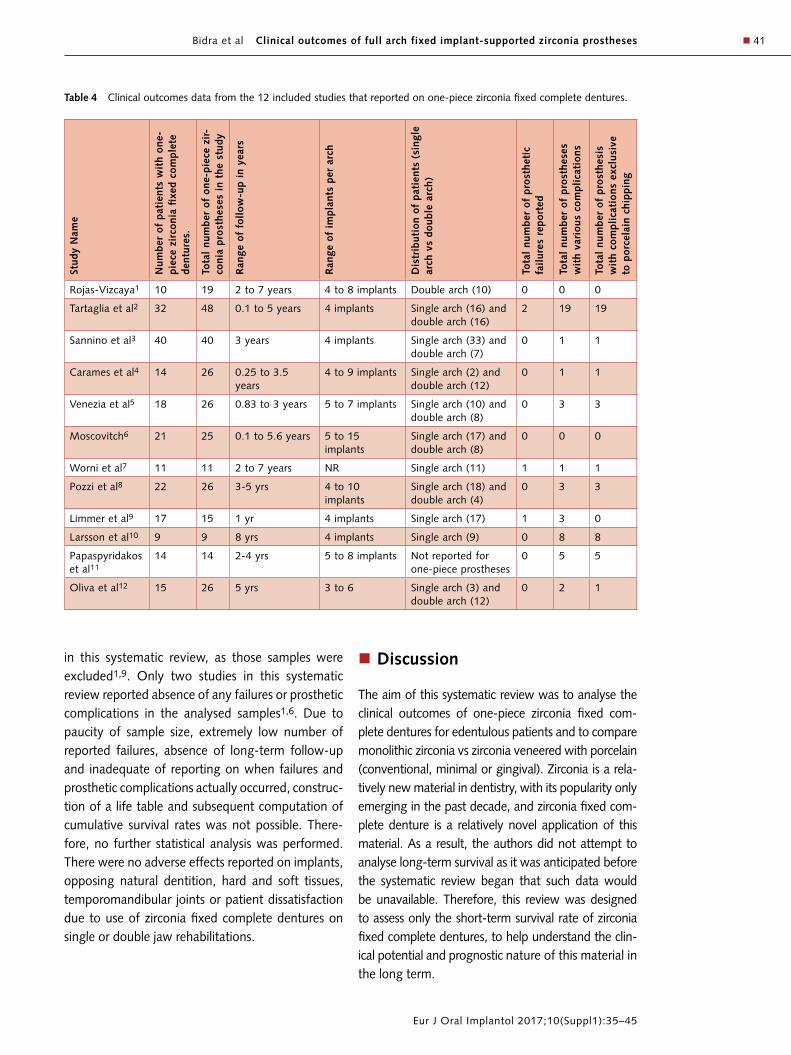
Bidra et al Clinical outcomes of full arch fixed implant-supported zirconia prostheses n 41
Eur J Oral Implantol 2017;10(Suppl1):35–45
Table 4 Clinical outcomes data from the 12 included studies that reported on one-piece zirconia fixed complete dentures.
Stud
y N
ame
Num
ber
of p
atie
nts
wit
h on
e-pi
ece
zirc
onia
fixe
d co
mpl
ete
dent
ures
.
Tota
l nu
mbe
r of
one
-pie
ce z
ir-
coni
a pr
osth
eses
in
the
stud
y
Ran
ge o
f fo
llow
-up
in y
ears
Ran
ge o
f im
plan
ts p
er a
rch
Dis
trib
utio
n of
pat
ient
s (s
ingl
e ar
ch v
s do
uble
arc
h)
Tota
l nu
mbe
r of
pro
sthe
tic
fail
ures
rep
orte
d
Tota
l nu
mbe
r of
pro
sthe
ses
wit
h va
riou
s co
mpl
icat
ions
Tota
l nu
mbe
r of
pro
sthe
sis
wit
h co
mpl
icat
ions
exc
lusi
ve
to p
orce
lain
chi
ppin
g
Rojas-Vizcaya1 10 19 2 to 7 years 4 to 8 implants Double arch (10) 0 0 0
Tartaglia et al2 32 48 0.1 to 5 years 4 implants Single arch (16) and double arch (16)
2 19 19
Sannino et al3 40 40 3 years 4 implants Single arch (33) and double arch (7)
0 1 1
Carames et al4 14 26 0.25 to 3.5 years
4 to 9 implants Single arch (2) and double arch (12)
0 1 1
Venezia et al5 18 26 0.83 to 3 years 5 to 7 implants Single arch (10) and double arch (8)
0 3 3
Moscovitch6 21 25 0.1 to 5.6 years 5 to 15 implants
Single arch (17) and double arch (8)
0 0 0
Worni et al7 11 11 2 to 7 years NR Single arch (11) 1 1 1
Pozzi et al8 22 26 3-5 yrs 4 to 10 implants
Single arch (18) and double arch (4)
0 3 3
Limmer et al9 17 15 1 yr 4 implants Single arch (17) 1 3 0
Larsson et al10 9 9 8 yrs 4 implants Single arch (9) 0 8 8
Papaspyridakos et al11
14 14 2-4 yrs 5 to 8 implants Not reported for one-piece prostheses
0 5 5
Oliva et al12 15 26 5 yrs 3 to 6 Single arch (3) and double arch (12)
0 2 1
in this systematic review, as those samples were excluded1,9. Only two studies in this systematic review reported absence of any failures or prosthetic complications in the analysed samples1,6. Due to paucity of sample size, extremely low number of reported failures, absence of long-term follow-up and inadequate of reporting on when failures and prosthetic complications actually occurred, construc-tion of a life table and subsequent computation of cumulative survival rates was not possible. There-fore, no further statistical analysis was performed. There were no adverse effects reported on implants, opposing natural dentition, hard and soft tissues, temporomandibular joints or patient dissatisfaction due to use of zirconia fixed complete dentures on single or double jaw rehabilitations.
n Discussion
The aim of this systematic review was to analyse the clinical outcomes of one-piece zirconia fixed com-plete dentures for edentulous patients and to compare monolithic zirconia vs zirconia veneered with porcelain (conventional, minimal or gingival). Zirconia is a rela-tively new material in dentistry, with its popularity only emerging in the past decade, and zirconia fixed com-plete denture is a relatively novel application of this material. As a result, the authors did not attempt to analyse long-term survival as it was anticipated before the systematic review began that such data would be unavailable. Therefore, this review was designed to assess only the short-term survival rate of zirconia fixed complete dentures, to help understand the clin-ical potential and prognostic nature of this material in the long term.

Bidra et al Clinical outcomes of full arch fixed implant-supported zirconia prostheses42 n
Eur J Oral Implantol 2017;10(Suppl1):35–45
It is remarkable that only four out of 285 zirco-nia fixed complete dentures (1.4%) exhibited fail-ures (fractures requiring prosthesis remake) in this systematic review. The four failures were restricted to three studies2,7,9. Tartaglia et al2 reported two fractures that required a remake of the prostheses. In this study, the authors clarified that the fit of the frameworks were passive and the connectors were designed with a minimum dimension of 10 mm2, with no more than a 2 mm space for veneered por-celain. This indicates that these fixed complete den-tures were all made as “dentition-only” replace-ments without any gingival prosthesis13. Despite proper precautions, two prostheses fractured and 19 prostheses had chipping of veneered porcelain indicating that this may either be an issue with intrinsic properties of the chosen zirconia itself, or perhaps that zirconia fixed complete dentures may require increased prosthetic space for increased thickness of zirconia for sufficient strength. This can be attained by appropriate bone reduction and incorporating gingival prosthesis for proper aesthet-ics and tooth proportions. There was no mention of the use of an occlusal device or the parafunctional habits of these patients, although the authors clari-fied that they used a slower heating and cooling protocol during the firing of porcelain. Worni et al7 reported one prosthesis fracture on a 12-unit fixed dental prosthesis on a patient who exhibited brux-ism (among other framework fractures in partially edentulous arches). The fractured prosthesis was a “dentition-only” replacement without any gin-gival prosthesis, once again suggesting that zirco-nia fixed complete dentures may require increased prosthetic space for increased thickness of zirconia. Limmer et al9 reported one fracture of a monolithic zirconia prosthesis (with staining used for gingival aspect) 6 months after insertion. The authors clari-fied explicitly that the fracture occurred vertically through the entire body of the prosthesis imme-diately distal to the terminal abutment, resulting in the loss of the distal cantilever segment on the affected side. Authors reported that inadequate prosthetic space, cantilever length or intrinsic prop-erties of the zirconia may have caused the fracture. In summary, an analysis of the four fractures from all three studies indicated that reduced prosthetic space may be a risk factor for fracture of zirconia
fixed complete dentures and warrants careful case selection or appropriate bone reduction for optimal prosthetic space.
Commonly reported complications of zirconia-based restorations typically involve fracturing or delamination of the veneered porcelain. This study is no different. A total of 42 out of 285 prostheses (14.7%) from nine studies, reported chipping of veneered porcelain. Although this number is lower than the fracture of veneered resin/denture teeth typically seen in metal-resin prostheses, this number is substantial because of the expensive and complex nature of porcelain repair procedures in the dental laboratory. It is remarkable that none of the chipping of veneered porcelain required a remake or replace-ment of the prosthesis across all studies. One study even reported that many patients were unaware of the fractures of the veneered porcelain at follow-up and concluded that the importance of fractures of the veneered porcelain should not be overstated10. Use of an occlusal device (night guard) was reported to have aided in risk reduction for fracture of veneered porcelain, by multiple authors3,4, 9,11,12. Chairside polishing and adjustment was performed for most porcelain chippings, and in some situations a lami-nate was fabricated.
Except for one recent study by Tartaglia et al2, which reported an abnormally high rate of fracture of veneered porcelain, most studies had a minimal number of fractures (range 1 to 8).
However, it is important to note that compared with newer studies, the older studies or those with a longer follow-up period had higher numbers of veneered porcelain fracture, indicating that the older studies may not have adopted slower heating and cooling rates during the firing of porcelain, which is regarded as important to minimise veneered por-celain fracture21,24. It is also interesting to note that zirconia prostheses that had conventional veneer-ing and minimal veneering both reported fracture of veneered porcelain, indicating that minimal veneer-ing may not help reduce the risk of porcelain chip-ping, but may help to reduce a patient’s dissatisfac-tion once the fracture has occurred. Additionally, chipping of veneered gingival porcelain was not reported as a complication in any of the studies, perhaps because this is located in a non-functional and non-load bearing area. In summary, an analysis

Bidra et al Clinical outcomes of full arch fixed implant-supported zirconia prostheses n 43
Eur J Oral Implantol 2017;10(Suppl1):35–45
of the 42 porcelain chippings from nine studies indi-cates that fracture of veneered porcelain may be a minor complication in zirconia fixed complete den-tures and clinicians and patients are less likely to have the prosthesis remade or repaired in the laboratory. Furthermore, use of monolithic zirconia with gin-gival stains, or zirconia with only veneered gingival porcelain, may aid in risk reduction for failures and complications with the material, but further studies are needed.
Despite the lack of long-term clinical evidence, the popularity of zirconia fixed complete dentures is likely to continue growing, as many dental man-ufacturers and dental laboratories in the United States now offer a warranty against any prosthetic complications. The warranty period varies between laboratories/manufacturers, provided there is suf-ficient prosthetic space to allow for adequate zir-conia thickness18-20. The warranty concept is not only based on the fidelity of the material, but also due to the reduced cost of fabrication due to CAD/CAM technology and reduced labour. The clinician’s labour and time in removing fractured zirconia pros-thesis and replacement with an interim (prototype) resin prosthesis is minimal, due to the screw-retained nature of the prosthesis. Thus, the warranty reas-sures the patient and clinician of indemnification of a fractured prosthesis, and having a digital file perma-nently stored allows fabrication of a new monolithic zirconia prosthesis, to mimic the original prosthe-sis. Additionally, the patient can continue to wear the prototype prosthesis (milled from the same scan using resin-based materials) during the fabrication of a new prosthesis.
Although this systematic review satisfied most PRISMA guidelines, there were some limitations to this review: 1) some aspects of the results section were not applicable or amenable to the PRISMA checklist; 2) due to the nature of the topic and PICO questions posed in this systematic review, the authors did not find significant quantitative data for construction of a life table and subsequent computation of cumulative survival rates was not possible. Therefore, no further statistical analysis was performed; 3) only one study identified in this systematic review compared the clinical outcomes of zirconia fixed complete dentures with another material (metal-resin)2. This study concluded that
prosthetic material did not influence the compli-cation risk. However, both prostheses were not equally divided in this study, and further bias was introduced against zirconia because authors stated that one of the criteria for patients to be provided with zirconia prostheses was occurrence of fractures in the interim prosthesis. Additionally, the zirconia prostheses were all “dentition-only” replacement without any gingival prosthesis, implying reduced prosthetic space; 4) there were no studies identified that compared monolithic zirconia fixed prostheses with zirconia fixed prostheses with veneered porce-lain (conventional, minimal or gingival). In fact, only one study in this systematic review was identified that had truly monolithic zirconia fixed prostheses (with gingival staining)9. Many studies claimed to use “monolithic zirconia”, but study descriptions clearly indicated that some level of porcelain was veneered to the zirconia. Future studies should adhere to the definition of monolithic and make this aspect clearer to the reader to allow a fair assess-ment and comparison of prostheses. Finally, we did not include studies or samples within the included studies that described zirconia fixed complete den-tures which were not one-piece by design1,9,11. We deemed that segmented fixed dental prostheses (multiple units of crown and bridge) or fixed com-plete dentures with primary and secondary com-ponents like a veneer or crown cemented over the access opening, have different biomechanical and prosthetic considerations, impeding a fair compari-son with the typical one-piece prostheses.
Despite an exhaustive search process, it is possi-ble that the authors failed to identify some articles in the search process29, as in most systematic reviews. Grey literature (information that falls outside the mainstream of published journal and monograph literature) was not considered here because articles of this type are usually non-peer reviewed, with a potential for biased information or information that is restricted for use30. Additionally, published tri-als tend to be larger and show an overall greater treatment effect than grey trials31. However, it is unknown whether incorporation of these omitted articles would change the conclusions of this sys-tematic review.
Unanswered questions from this systematic review that will hopefully be answered by future

Bidra et al Clinical outcomes of full arch fixed implant-supported zirconia prostheses44 n
Eur J Oral Implantol 2017;10(Suppl1):35–45
investigators include differences in clinical out-comes of zirconia fixed complete dentures between: 1) single arch vs double arch prostheses; 2) distal cantilever vs non-cantilever prostheses; 3) maxilla vs mandible; 4) monolithic vs veneered zirconia pros-theses; 5) various types of veneering porcelain (con-ventional vs minimal vs gingival); 6) implant level vs abutment level prostheses; 7) indirect zirconia inter-face (bonded to metal cylinders) vs direct zirconia interface with abutments; and 8) different thickness of zirconia for different prosthetic space require-ments based on prosthetic designs.
n Conclusions
Within the limitations of this systematic review, the following conclusions were drawn: 1. One-piece zirconia fixed complete dentures have
a very low failure rate in the short term, but have a substantial rate of chipping of veneered por-celain. Reduced prosthetic space was associated with all fractures.
2. Chipping of veneered porcelain did not require the remake of any prostheses, but only required chairside polishing and adjustment or, in some cases, a porcelain laminate veneer was fabricated, indicating that this generally may be regarded as a minor complication.
3. Chipping of veneered gingival porcelain was not reported as a complication in any of the samples in the included studies.
4. Use of monolithic zirconia with gingival stains, or zirconia with only veneered gingival porcelain, may offer promising results for fixed complete dentures, but will need to be validated by future long-term studies.
5. There were no adverse effects reported on implants, hard and soft tissues, temporoman-dibular joints or patient dissatisfaction due to use of zirconia fixed complete dentures on single or double jaw rehabilitations.
6. Most of the studies evaluated were short-term and there is a need for long-term data to provide more definitive conclusions.
n References
1. Rojas Vizcaya F. Retrospective 2- to 7-Year Follow-Up Study of 20 Double Full-Arch Implant-Supported Monolithic Zirco-nia Fixed Prostheses: Measurements and Recommendations for Optimal Design. J Prosthodont. 2016. doi: 10.1111/jopr.12528. [Epub ahead of print]
2. Tartaglia GM, Maiorana C, Gallo M, Codari M, Sforza C. Implant-Supported Immediately Loaded Full-Arch Rehabili-tations: Comparison of Resin and Zirconia Clinical Outcomes in a 5-Year Retrospective Follow-Up Study. Implant Dent 2016;25: 74–82.
3. Sannino G, Barlattani A. Straight Versus Angulated Abut-ments on Tilted Implants in Immediate Fixed Rehabilitation of the Edentulous Mandible: A 3-Year Retrospective Com-parative Study. Int J Prosthodont 2016;29: 219–226.
4. Carames J, Tovar Suinaga L, Yu YC, Pérez A, Kang M. Clin-ical Advantages and Limitations of Monolithic Zirconia Res-torations Full Arch Implant Supported Reconstruction: Case Series. Int J Dent 2015;2015:392496.
5. Venezia P, Torsello F, Cavalcanti R, D’Amato S. Retrospective analysis of 26 complete-arch implant-supported monolithic zirconia prostheses with feldspathic porcelain veneering limited to the facial surface. J Prosthet Dent 2015;114: 506–512.
6. Moscovitch M. Consecutive case series of monolithic and minimally veneered zirconia restorations on teeth and implants: up to 68 months. Int J Periodontics Restorative Dent 2015 35: 315–323.
7. Worni A, Kolgeci L, Rentsch-Kollar A, Katsoulis J, Mericske-Stern R. Zirconia-Based Screw-Retained Prostheses Sup-ported by Implants: A Retrospective Study on Technical Complications and Failures. Clin Implant Dent Relat Res 2015;17: 1073–1081.
8. Pozzi A, Holst S, Fabbri G, Tallarico M. Clinical reliability of CAD/CAM cross-arch zirconia bridges on immediately load-ed implants placed with computer-assisted/template-guided surgery: a retrospective study with a follow-up between 3 and 5 years. Clin Implant Dent Relat Res 2015;17:e86–96.
9. Limmer B, Sanders AE, Reside G, Cooper LF. Complications and patient-centered outcomes with an implant-supported monolithic zirconia fixed dental prosthesis: 1 year results. J Prosthodont 2014;23: 267–275.
10. Larsson C, Vult von Steyern P. Implant-supported full-arch zirconia-based mandibular fixed dental prostheses. Eight-year results from a clinical pilot study. Acta Odontol Scand 2013;71: 1118–1122.
11. Papaspyridakos P, Lal K. Computer-assisted design/comput-er-assisted manufacturing zirconia implant fixed complete prostheses: clinical results and technical complications up to 4 years of function. Clin Oral Implants Res 2013;24: 659–665.
12. Oliva J, Oliva X, Oliva JD. All-on-three delayed implant loading concept for the completely edentulous maxilla and mandible: a retrospective 5-year follow-up study. Int J Oral Maxillofac Implants 2012;27: 1584–1592.
13. Bidra AS. Three-dimensional esthetic analysis in treatment planning for implant-supported fixed prosthesis in the eden-tulous maxilla: review of the esthetics literature. J Esthet Restor Dent 2011;23: 219–236.
14. Purcell BA, McGlumphy EA, Holloway JA, Beck FM. Pros-thetic complications in mandibular metal-resin implant-fixed complete dental prostheses: a 5- to 9-year analysis. Int J Oral Maxillofac Implants 2008;23: 847–857.
15. Fischer K, Stenberg T. Prospective 10-year cohort study based on a randomized, controlled trial (RCT) on implant-supported full-arch maxillary prostheses. Part II: prosthetic outcomes and maintenance. Clin Implant Dent Related Res 2013;15: 498–508.
Bidra et al Clinical outcomes of full arch fixed implant-supported zirconia prostheses n 45
Eur J Oral Implantol 2017;10(Suppl1):35–45
16. Priest G, Smith J, Wilson MG. Implant survival and prosthetic complications of mandibular metal-acrylic resin implant complete fixed dental prostheses. J Prosthet Dent 2014;111: 466–475.
17. Cardelli P, Manobianco FP, Serafini N, Murmura G, Beuer F. Full-Arch, Implant-Supported Monolithic Zirconia Rehabili-tations: Pilot Clinical Evaluation of Wear Against Natural or Composite Teeth. J Prosthodont 2016;25: 629–633.
18. Glidewell Dental Laboratories. Policies and Warranties. Available at: http://glidewelldental.com/contact-us/poli-cies-and-warranties/ Accessed on September 14, 2016.
19. Tischler Dental Laboratory. Prettau Bridge Warranty. Avail-able at: https://www.tischlerdentallab.com/clinical-info/warranty. Accessed on September 14, 2016.
20. Crystal Zirconia. Crystal Lifetime Warranty. Available at: http://www.crystalzirconia.com/index.php/patient-infor-mation/26. Accessed on September 14, 2016.
21. Denry I, Kelly JR. Emerging ceramic-based materials for den-tistry. J Dent Res 2014;93: 1235–1242.
22. Miyazaki T, Nakamura T, Matsumura H, Ban S, Kobayashi T. Current status of zirconia restoration. J Prosthodont Res 2013;57: 236–261.
23. Heintze SD, Rousson V. Survival of zirconia- and metal-supported fixed dental prostheses: a systematic review. Int J Prosthodont 2010;23: 493–502.
24. Benetti P, Kelly JR, Sanchez M, Della Bona A. Influence of thermal gradients on stress state of veneered restorations. Dent Mater 2014;30: 554–563.
25. Alghazzawi TF, Lemons J, Liu PR, Essig ME, Bartolucci AA, Janowski GM. Influence of low-temperature environmental exposure on the mechanical properties and structural stabil-ity of dental zirconia. J Prosthodont 2012;21: 363–369.
26. Flinn BD, deGroot DA, Mancl LA, Raigrodski AJ. Accelerated aging characteristics of three yttria-stabilized tetragonal zir-conia polycrystalline dental materials. J Prosthet Dent 2012; 108:223–230.
27. Abdulmajeed AA, Lim KG, Närhi TO, Cooper LF. Complete-arch implant-supported monolithic zirconia fixed dental prostheses: A systematic review. J Prosthet Dent 2016;115:672 –677.
28. Moher D, Liberati A, Tetzlaff J: PRISMA Group. Preferred Reporting Items for Systematic Reviews and Meta-Analyses: The PRISMA Statement. J Clin Epidemiol 2009;62: 1006–1012.
29. Savoie I, Helmer D, Green CJ, et al: Beyond Medline: redu-cing bias through extended systematic review search. Int J Technol Assess Health Care 2003;19: 168–178.
30. Auger CP (ed): Information Sources in Grey Literature (ed 2). London, Bowker-Saur, 1989.
31. Hopewell S, McDonald S, Clarke M, Egger M. Grey literature in meta-analyses of randomized trials of health care inter-ventions. Cochrane Database Syst Rev 2007;(2):MR000010.


n 47
Eur J Oral Implantol 2017;10(Suppl1):47–65
REVIEW
Samir Abou-AyashDepartment of Prosthodon-tics, School of Dentistry, Medical Center – University of Freiburg, Freiburg, Ger-many; Faculty of Medicine, Medical Center – Univer-sity of Freiburg, Freiburg, Germany
Malin Strasding Department of Prosthodon-tics, School of Dentistry, Medical Center – University of Freiburg, Freiburg, Ger-many; Faculty of Medicine, Medical Center – Univer-sity of Freiburg, Freiburg, Germany
Gerta Rücker Institute for Medical Bio-metry and Statistics, Medical Center – University of Freiburg, Freiburg, Germany; Faculty of Medicine, Medi-cal Center – University of Freiburg, Freiburg, Germany
Wael Att Department of Prosthodon-tics, School of Dentistry, Medical Center - University of Freiburg, Hugstetter Str. 55, 79106 Freiburg, Ger-many; Faculty of Medicine, Medical Center – Univer-sity of Freiburg, Freiburg, Germany
Correspondence to: Prof Dr Wael AttDepartment of Prosthodon-tics, School of Dentistry, Medical Center - Univer-sity of Freiburg, Hugstetter Str. 55, 79106 Freiburg, GermanyEmail: [email protected]: +4976127049250, Tel: +4976127047380
Samir Abou-Ayash, Malin Strasding, Gerta Rücker, Wael Att
Impact of prosthetic material on mid- and long-term outcome of dental implants supporting single crowns and fixed partial dentures: A systematic review and meta-analysis
Key words fixed partial dentures, implants, material selection, meta-analysis, single crowns, systematic review
Aim: The impact of prosthetic material selection on implant survival is not clear. The current criteria for choosing a prosthetic material seem to be based on clinician preferences. This systematic review aims to evaluate the impact of restorative materials on the mid- and long-term survival of implants supporting single crowns and fixed partial dentures.Materials and methods: Hand and MEDLINE searches were performed to identify relevant literature for single crowns (SC) and fixed partial dentures (FPD). Further inclusion criteria were a mean follow-up period of at least 3 years, the inclusion of at least 10 patients in a relevant study cohort, and a clear description of prosthesis type and prosthetic material.Results: A total of 63 studies for the SC group and 11 studies for the FPD group were included. Full arch restorations were not included. The materials utilised in the SC group were metal-ceramic (pre-cious and non-precious), lithium-disilicate, veneered zirconia, veneered alumina, and nanoceramics. The materials used in the FPD group were metal-ceramic (precious), veneered titanium, metal-resin (precious), and veneered zirconia. No significant impact on the prosthetic material relating to mid- or long-term implant survival was identified. Furthermore, there were no statistically significant differ-ences between the survival rates of the dental prostheses made from different materials (SC and FPD group). Single crowns made of nanoceramics showed a higher risk for decementation relative to other materials (0.80, 95% CI [0.67; 0.89]; P < 0.0001), whereas metal-resin FPDs showed a higher risk for chipping (0.36, 95% CI [0.23; 0.52]; P = 0.0072).Conclusion: The current evidence suggests that prosthetic material selection has no influence on mid- and long-term survival of implants restored with single crowns and fixed partial dentures. Simi-larly, the prosthetic material seems to have no significant impact on prosthetic survival rates. Further research is required to provide more evidence regarding the impact of the prosthetic material on long-term outcome.
Conflict-of-interest statement: The authors declare that they have no conflict of interest.
n Introduction
The use of oral endosseous implants for the replace-ment of missing teeth has become a routine clin-ical procedure1. Today, oral implants are being used
to treat various clinical situations, in particular par-tially and completely edentulous jaws. For implant-supported dental rehabilitations, several prosthetic solutions and materials are available2. In fact, clin-icians have a wide variety of materials available for

Abou-Ayash et al Impact of prosthetic material on mid- and long-term outcome of dental implants48 n
Eur J Oral Implantol 2017;10(Suppl1):47–65
limited to, restoration type, retention mechanism, and implant length. For example, the choice of the prosthetic material is an additional factor to be con-sidered in combination with the aforementioned variables, as it might play a role in determining long-term outcome8.
While many studies have reported on implants and factors affecting their survival, little knowledge is available about the impact of the prosthetic material. For example, it is not clear whether an all-ceramic crown would lead to an improved outcome for the implant relative to a metal-ceramic crown. Also, the influence of the restorative material on the clinical outcome of the implant-borne prosthetic rehabilita-tion is unclear. So far, studies addressing the outcome of implant rehabilitations made of different materials primarily compared two materials. Moreover, varying study designs make it difficult to assess the outcome and impact of a specific material. An evaluation of the impact of the prosthetic material on implant survival would enhance clinical knowledge and provide clin-icians with guidelines regarding material selection.
Therefore, the aim of this systematic literature review and meta-analysis is to examine whether a correlation exists between the type of restorative material and the clinical outcome of implants, as well as the associated prosthetic rehabilitations.
n Materials and methods
n Study protocol
The study protocol was set in accordance with the PRISMA (Preferred Reporting Items for System-atic Reviews and Meta-Analyses) statement9. The focused leading question was set according to the PICO model for clinical questions. This model aids the discovery of clinically relevant evidence in lit-erature by dividing the leading question into four subcategories (population, intervention, compari-son, and outcome). The four criteria for this study were as follows:• Population: Partially edentulous patients receiv-
ing fixed implant-supported restorations;• Intervention: Prosthodontic rehabilitation by
means of implant-supported single crowns (SCs) or fixed partial dentures (FPDs);
prosthetic solutions. With the progress of CAD/CAM techniques and improvements to the aesthetic char-acteristics of contemporary materials, new possibili-ties for both fixed and removable rehabilitations are continuously being introduced. For example, high-strength ceramics can be used today as a framework material for veneered restorations, as well as for a final monolithic restoration. With such a wide spec-trum of available materials and restorative options, clinicians are often confused about selecting the ideal prosthetic material, which facilitates ideal aesthetics, biocompatibility, and long-term stability. Apparently, material selection for the definitive prosthesis seems to depend not only on mechanical properties and anatomic and patient-related factors, but also on a clinician’s individual preferences3.
The influence of many factors on the long-term outcome and on technical or biological complication rates of implant-borne fixed partial dentures (FPDs) is well described in scientific literature. For example, retention mechanisms seem to have an influence on technical and biological complication rates4,5. On the one hand, screw-retained single crowns and fixed partial dentures seem to have a higher risk for technical complications than cement-retained single crowns or fixed partial prostheses. On the other hand, when all fixed restorations (regardless of the restoration type) are compared, significantly fewer complications were observed with screw retention4. This demonstrates that factors such as the retention mechanism can have different effects on various res-toration types.
Another potential factor influencing the long term-outcome in partially edentulous patients is the length of the utilised implants. A recently published systematic review comparing short vs standard-length implants found that there were no signifi-cant differences in marginal bone loss, complication, or prosthesis failure rates. However, implants with lengths of less than 8 mm presented a higher risk of implant failure6. Another 5-year follow-up of a ran-domised controlled trial showed similar survival and success rates, and no statistically significant compli-cation rates in partially edentulous cases, when 6 mm implants were compared with 10 mm implants7.
There appear to be several factors influencing the long-term survival and success rates of implant-borne dental restorations, including, but not necessarily

Abou-Ayash et al Impact of prosthetic material on mid- and long-term outcome of dental implants n 49
Eur J Oral Implantol 2017;10(Suppl1):47–65
• Comparison: Performance of various prosthetic materials used for each type of restoration;
• Outcome: Implant and prosthetic survival, as well as technical complications related to the re-storative material and the type of prosthesis.
After analysing the different points of this model, the resulting PICO question was: “Does the choice of restorative material influence the mid- and long-term outcome of implants and/or fixed partial den-tures?”
n Definitions
Prior to the systematic search, several terms were defined: “Implant survival” was defined as implants that were still in situ at the point of observation. Implant conditions such as surrounding bone, soft tissue, or signs of inflammation were not parameters considered in the evaluation of survival. “Prosthetic survival” was defined as prostheses that were still in situ, even if repairs of any kind were necessary (e.g. renewal of the veneering material). “Techni-cal complications” were subdivided into four dis-tinct categories: abutment fracture, chipping, screw loosening, and decementation of the superstructure.
n Inclusion and exclusion criteria
For the systematic literature search, the following inclusion and exclusion criteria were compiled:
Inclusion criteria
• Human clinical studies (randomised controlled trials, controlled trials, prospective studies, retro-spective studies, case series);
• Fixed implant-supported prostheses and single crowns;
• Titanium implants;• Partially edentulous patients;• Documentation of prosthetic material;• Documentation of restoration type;• Number of patients/ study arm or cohort ≥ 10;• Mean follow-up period ≥ 3 years;• Publication in English.
Exclusion criteria
• In vitro or animal studies;• Removable partial denture;• Ceramic implants;• Edentulous patients;• Insufficient documentation of prosthetic ma-
terial;• No documentation of restoration type;• Fewer than 10 patients in relevant study arm/
cohort;• Mean follow-up period less than 3 years;• Publications not written in English;• Combined tooth-implant-supported restor-
ations;
n Search strategy and study selection
For the initial electronic search in the MEDLINE library (via PubMed), the types of dental restorations were divided into two different groups: single crowns (SC group) and fixed partial dentures (FPD group). For each group, a separate initial search of litera-ture without any filters was performed using distinct key words (Table 1). Furthermore, reference lists of review articles with similar topics were systemati-cally screened, and potentially relevant articles were added to the results of the electronic search. After elimination of duplicates, the titles of the remaining articles were checked for adequacy, according to the inclusion criteria. Irrelevant titles (e.g. in vitro stud-ies) were excluded. When the relevance of studies was uncertain according to the title, the studies were included for abstract screening. When the relevance of the studies remained unclear after reading the
Table 1 Key words for initial database search.
Group Search Terms Initial hits
SC (“crown” OR “crowns” OR “partially edentulous” OR “single Crown” OR “single Crowns”) AND (“implant” OR “implants” OR “dental implant” OR “dental implants”)
3411
FPD (“fixed partial prostheses” OR “fixed partial prosthesis” OR “partially edentulous” OR “im-plant bridge”) AND (“implant” OR “implants” OR “dental implants“)
1147

Abou-Ayash et al Impact of prosthetic material on mid- and long-term outcome of dental implants50 n
Eur J Oral Implantol 2017;10(Suppl1):47–65
abstract, they were included for full text screening, resulting in a selection of possibly eligible full texts. After reviewing the full texts, irrelevant articles were excluded, and data of the remaining articles was extracted whenever possible. Study selection and data extraction were performed for each group inde-pendently by two reviewers (SA and MS). After-wards, every disagreement regarding the inclusion of specific articles was solved by discussion. Tables 2a and 2b illustrate the study selection process for each group. For data extraction, identical study forms for each group were designed, including the following parameters: authors, title, year of publication, study design, study period, number of patients, number of implants in the study, implant system, implant lengths, implant diameters, implant material, abut-ment material, timing of implant placement, loading protocol, number of implants per prosthesis, number of prostheses in total, jaw, number of cantilevers, length of cantilever, type of retention, bone aug-mentation, soft tissue augmentation, CAD/CAM workflow, mean radiographic bone loss, implant loss before loading, prosthetic survival rate, screw loos-ening, decementation, chipping, prosthesis fracture, framework fracture, abutment fracture, peri-implan-titis, mucositis, implant fracture, and superstructure material.
n Risk of bias within the studies
For quality assessment, six quality categories were implemented to evaluate the included studies10: “fair” for retrospective studies, “average” for pro-spective case studies, “good” for prospective stud-ies with historical control, “better” for prospective studies with concurrent controls, “best” for double-blind randomised controlled trials, and “unknown” for studies not fitting one of the other five criteria. Due to the high degree of heterogeneity observed in study design and in results of the different studies that were considered, a decision was made to include all studies that were rated at least “fair”.
n Risk of bias across the studies
In most of the studies, primary outcomes differed from our leading question. The choice of prosthetic material was often considered as a marginal note. Therefore, a potential risk of bias might be intro-duced via extracting and evaluating data from stud-ies that do not refer to the influence of the prosthetic material.
Tables 2a and 2b Study selection process for single crowns (SCs) and fixed partial dentures (FPDs).
Results of initial search after elimination of
duplicates (n = 3411)
Title screening (n = 3411)
Abstract screening (n = 568)
Full text screening (n = 271)
Data extraction (n = 83)
Studies included for meta analysis (n = 62)
Studies excluded (n = 2843)
Studies excluded (n = 297)
Studies excluded (n = 188)
Studies excluded (n = 21)
Results of initial search after elimination of
duplicates (n = 1147)
Title screening (n = 1147)
Abstract screening (n = 284)
Full text screening (n = 138)
Data extraction (n = 18)
Studies included for meta analysis (n = 11)
Studies excluded (n = 863)
Studies excluded (n = 146)
Studies excluded (n = 120)
Studies excluded (n = 7)

Abou-Ayash et al Impact of prosthetic material on mid- and long-term outcome of dental implants n 51
Eur J Oral Implantol 2017;10(Suppl1):47–65
n Statistical analysis
For evidence synthesis using meta-analysis, the open source statistical environment R (version 3.2.0)11 with the R packages “meta”12 and “metafor”13 was used. The random effects model was used through-out. The outcomes were the proportion of surviv-ing implants, the prosthetic survival proportion, the proportion of screw-loosening events by number of implants, the proportion of abutment fractures by the number of implants, the proportion of chipping by the number of prostheses, and the proportion of decementation by the number of prostheses. Pro-portions were pooled using the logit transformation. In addition, loss of implants was measured as an incidence rate per average follow-up time. For all outcomes, the impact of material was analysed using meta-regression13. The generally poor reporting in the primary studies made it impossible to adjust for potential intrasubject correlation due to the varying unit of analysis (patients, implants, prostheses).
n Synthesis of results
Most of the included studies did not compare mul-tiple restorative materials directly, but described, for example, different augmentation techniques.
Data were extracted whenever the restorative material was mentioned and the study met the inclu-sion criteria. For simplification, not all of the columns of the study form had to be completed, if certain information was not provided in a given study (e.g. implant lengths). Whenever implant or prosthetic survival rates were not reported, studies were not included for data extraction, but taken into consid-eration for strengthening or weakening results of the meta-analysis. If multiple study arms or cohorts were identified in the same study, data from each group was recorded separately. This resulted in a higher number of study populations than indicated by the number of included studies.
The primary outcome of the meta-analysis was to evaluate the implant and prosthetic survival rates as functions of the restorative material and restor-ation type. Therefore, restorations were divided into metal-based and all-ceramic restorations, and secondly, whenever possible, divided into groups according to the exact restoration material (e.g. glass
ceramics or ceramic-veneered precious alloy). Hence, several studies did not describe the exact restoration material, but only whether an all-ceramic or metal-based restoration was used, such that the number of included studies was higher for the comparison between metal-based and all-ceramic materials than for the comparison of the exact materials used.
Furthermore, to be able to compare the results of the included studies, despite the variable follow-up periods, the implant loss rate per 10 implant years was calculated. This rate describes the risk of an im-plant loss regarding one single implant for a period of 10 years, or the risk of an implant loss of two implants over 5 years. Additionally, the incidence rates for screw loosening, decementation of super-structures, chipping, and abutment fractures were calculated.
n Results
n Literature search
As previously described, an initial literature search via MEDLINE and a manual search were performed for each group separately (Table 1). 3411 studies were included for the SC group, while 1147 stud-ies were included for FPDs. After title-, abstract-, and full text screening, 83 studies remained in the SC group, and 18 remained in the FPD group for data extraction. During data extraction, 21 further studies from the SC group and seven from the FPD group were excluded, resulting in a final number of included studies of 62 for the SC group and 11 for the FPD group (Tables 2a and 2b). Reasons for exclu-sion during data extraction are listed in Tables 3a and 3b. The results for the two groups are henceforth described separately.
n Study characteristics
Most of the included studies were prospective stud-ies, but RCTs, retrospective studies and case series were also included. The type of study and qual-ity assessment of each study is shown in Tables 4a and 4b. The majority of the studies had an observa-tion period between 3 and 10 years. Studies with longer observation periods were scarce. In the SC

Abou-Ayash et al Impact of prosthetic material on mid- and long-term outcome of dental implants52 n
Eur J Oral Implantol 2017;10(Suppl1):47–65
group, 76 study populations across 62 studies and in the FPD group, 15 populations across 11 studies were investigated. Various implant types with differ-ent surface modifications were placed in these stud-ies. Several types of prosthetic materials were used in the studies (all types of metal-ceramic, metal-resin and all-ceramic materials). Screw-retained, as well as cemented restorations were analysed. Differences in time of implant placement, loading protocols, augmentation procedures, number of implants per prosthesis, abutment materials, implant lengths, im-plant diameters, number of cantilevers, and lengths of cantilevers were not taken into consideration for the meta-analysis.
n Single crowns
All-ceramic vs metal-based SCsFor the analysis of implant survival, the results of 24 study cohorts of the all-ceramic group and 53 of the metal-based group were used for the meta-analysis (Tables 5 and 6). The implant survival rate in
the all-ceramic group (included studies n = 23) was 0.97 (95% CI [0.95; 0.98]) with a mean observation period of 5.4 years, and 0.96 (95% CI [0.95; 0.97]) among metal-based SCs with a mean observation period of 5.6 years. This difference was not statistic-ally significant (P = 0.1724). The meta-regression-analysis showed no impact on the different observa-tion periods for the two groups (P = 0.5976). The calculated implant loss rate per 10 implant years was 0.06 (95% CI [0.04; 0.08]) for the all-ceramic and 0.07 (95% CI [0.05; 0.10]) for the metal-based group. This difference was not statistically significant (P = 0.3737).
Prosthetic survival rates were 0.95 (95% CI [0.94; 0.97]) in the all-ceramic (included studies n =19) and 0.97 (95% CI [0.96; 0.98] in the metal-based group (n = 27). The difference was not significant (P = 0.0872). Subgroup analyses for the incidence rates of screw loosening, decementation, abutment fractures and chipping revealed no statistical differ-ences between the two groups.
n Exact restorative materials
For the meta-analysis of implant survival rates, the results of two study cohorts of veneered non-pre-cious alloys, 11 of veneered precious alloys, 10 of veneered zirconia, five of veneered alumina, four of glass ceramics, and one for nanoceramics, were included. The veneering material was ceramic. For the analysis of the prosthetic survival rates, three study cohorts of veneered precious alloys, 12 of veneered zirconia, four of veneered alumina, four of glass ceramics, and one of nanoceramics, were included. For veneered non-precious alloys, no
Table 3a Reasons for study exclusion (SC group).
Study Reason for exclusion
Bergenblock et al. 201214 Pooled results
Bianchi et al. 200415 Pooled results
Branzen et al. 201516 Pooled results
Dierens et al. 201617 Pooled results
Ekfeldt et al. 201118 Pooled results
Grassi et al. 201519 Zirconia implants
Hälg et al. 200820 Pooled results
Hosseini et al. 201321 Pooled results
Lops et al 201222 Pooled results
Mangano et al. 201023 Pooled results
Mangano et al. 201524 Pooled results
Moberg et al. 199925 Pooled results
Özkan et al. 201126 Pooled results
Özkan et al. 200727 Pooled results
Passos et al. 201628 Pooled results
Spies et al. 201529 Zirconia implants
Spies et al. 201530 Zirconia implants
Tartaglia et al. 201531 Pooled results
Thoma DS et al. 201532 Different target variables
Vigolo et al. 200033 Pooled results
Walton 201534 Pooled results
Table 3b Reasons for study exclusion (FPD-group).
Study Reason for exclusion
Astrand et al. 200435 No implant/ prosthetic survival rates reported
Becktor et al. 200736 No implant/ prosthetic survival rates reported
Bornstein et al 201037 Pooled results
Felice et al. 201438 No implant/ prosthetic survival rates reported
Maló P et al 201339 Pooled results
Nissan J et al. 201140 Unclear statistics
Ortorp A et al. 199941 Pooled results

Abou-Ayash et al Impact of prosthetic material on mid- and long-term outcome of dental implants n 53
Eur J Oral Implantol 2017;10(Suppl1):47–65
Table 4a Quality assessment of included studies (SC group).
Authors Type of study
Quality assessment
Balshi TJ et al.42 Prospective good
Berberi AN et al.43 Prospective good
Bilhan H et al.44 Retrospective fair
Bonde MJ et al.45 Retrospective fair
Buser D et al.46 Prospective good
Calandriello R & Tomatis M47 Prospective good
Cannizzaro G et al.48 RCT best
Cannizzaro G et al.49 RCT best
De Bruyn H et al.50 Prospective better
Cosyn J et al.51 Prospective good
Covani U et al.52 Prospective good
Covani U et al.53 Prospective good
Davis DM54 Prospective good
Donati M et al.55 Prospective good
Enkling N et al.56 RCT best
Fenner N et al.57 Prospective better
Glauser R et al.58 Prospective good
Gotfredsen K et al.59 Prospective better
Guarnieri R et al.60 Prospective good
Ioannidis A et al.61 RCT best
Jemt T62 Retrospective fair
Jemt T & Pettersson P63 Retrospective fair
Khraisat A et al.64 Prospective good
Kolgeci L et al.65 Prospective good
Lai HC et al.66 Retrospective fair
Lang LA et al.67 Prospective good
Lops D et al.68 Prospective better
Mangano C et al.69 Retrospective fair
Mangano FG et al.70 Prospective good
Table 4b Quality assessment of included studies (FPD-group).
Authors Type of study Quality assessment
Barnea E et al.100 Retrospective fair
Brägger U et al.101 Prospective good
Francetti L et al.102 Prospective good
Göthberg C et al.103 RCT best
Jemt T et al.104 Retrospective fair
Kreissl ME et al.105 Prospective good
Mangano F et al.106 Prospective good
Pozzi A et al.107 Prospective better
Romeo E et al.108 Prospective good
Romeo E et al.109 Prospective good
Romeo E et al.7 RCT best
Authors Type of study
Quality assessment
Mangano FG et al.71 Retrospective fair
Mericske-Stern R et al.72 Retrospective fair
Montoya-Salazar V. et al.73 RCT best
Nejatidanesh et al.74 Retrospective fair
Palmer RM75 Prospective good
Perelli M. et al.76 Prospective good
Pieri F et al.77 Prospective better
Proussaefs P & Lozada J78 Prospective good
Rinke S et al.79 Retrospective fair
Rinke S et al.80 Retrospective fair
Roccuzzo M et al.81 Prospective good
Romanos GE et al.82 RCT best
Romeo E et al.83 Prospective good
Rossi F et al.84 Prospective good
Rossi F et al.85 RCT best
Schepke U86 Prospective good
Schropp L et al.87 RCT best
Schropp L et al.88 RCT best
Sorrentino R et al.89 Retrospective fair
Turkyilmaz I et al.90 Prospective better
Vanioglu AB et al.91 Prospective better
Vigolo P et al.92 Prospective better
Visser A et al.93 Prospective good
Weber HP et al.94 Prospective good
Worni A et al.95 Prospective good
Zarone F et al.96 Prospective good
Zembic A et al.97 RCT best
Zembic A et al.98 Prospective good
Zhao X et al.99 Retrospective fair
details regarding prosthetic survival rates could be found. Implant and prosthetic survival rates, as well as the 95% CI, can be seen in Table 7. The differences in implant and prosthetic survival rates were not statistically significant (implant survival: P = 0.4061; prosthetic survival: P = 0.8580). The meta-regression-analysis showed no impact of the different observation periods on the compari-son between any of the groups (implant survival: P = 0.2120; prosthetic survival: P = 0.9622). The calculated implant loss rate per 10 implant years showed no statistically significant differences among all groups (P = 0.6502).

Abou-Ayash et al Impact of prosthetic material on mid- and long-term outcome of dental implants54 n
Eur J Oral Implantol 2017;10(Suppl1):47–65
Tabl
e 5
Inc
lude
d st
udie
s in
the
all-
cera
mic
sin
gle
crow
n gr
oup.
Stud
ySt
udy
peri
od
(yea
rs)
Pati
ents
(n)
Impl
ants
(n)
Pros
-th
eses
(n
)
Supe
rstr
uctu
re
mat
eria
lA
but-
men
t m
ater
ial
Impl
ant
surv
ival
(%
)
Pros
thet
ic
surv
ival
(%
)
Abu
tmen
t fr
actu
re
(%)
Chi
p-pi
ng
(%)
Scre
w
loos
enig
(%
)D
ecem
enta
-ti
on (
%)
Berb
eri A
N e
t al
.43
320
2020
Ven
eere
d Zi
rcon
iaTi
100
Bond
e M
J et
al.4
5 10
4246
46A
ll ce
ram
ic (
non
defin
ed)
Ti93
.593
.5
Buse
r D
et
al.4
6 3
2020
20V
enee
red
Zirc
onia
Ti10
010
0
Cal
andr
iello
R &
To
mat
is M
475
3340
40V
enee
red
Alu
min
aTi
9510
00
00
0
De
Bruy
n H
et
al.5
05
5555
55V
enee
red
Alu
min
aTi
94.6
De
Bruy
n H
et
al.5
05
5858
58V
enee
red
Alu
min
aTi
98.3
Fenn
er N
et
al.5
77.
213
1313
All
cera
mic
(no
n de
fined
)A
l10
010
00
00
0
Gla
user
R e
t al
.58
4.1
1836
36G
lass
cer
amic
sZr
100
97.2
08.
35.
60
Gua
rnie
ri R
et
al.6
05
2121
21A
ll ce
ram
ic (
non
defin
ed)
Zr95
.20
00
4.8
Jem
t T6
210
1518
18A
ll ce
ram
ic (
non
defin
ed)
Ti10
00
016
.70
Kol
geci
L e
t al
.65
3.3
6712
012
0V
enee
red
Zirc
onia
Zr98
.395
.80
Lops
D e
t al
.68
537
3737
Ven
eere
d Zi
rcon
iaZr
100
100
010
.82.
70
Nej
atid
anes
h et
al.7
44.
912
223
223
2V
enee
red
Zirc
onia
Ti99
.297
.51.
6
Pier
i F e
t al
.77
529
2929
Ven
eere
d Zi
rcon
iaTi
/Zr
100
100
07
03.
5
Rin
ke S
et
al.7
96.
527
4242
Ven
eere
d Zi
rcon
iaZr
100
97.6
2,3
11.9
4.7
9.5
Sche
pke
U e
t al
.86
1.1
5050
Nan
o C
eram
icZr
100
9480
Sorr
entin
o R
et
al.8
96
4881
81V
enee
red
Alu
min
aTi
/Al
97.6
95.1
0.9
00
Van
iogl
u A
B et
al.9
15
611
11G
lass
cer
amic
sZr
100
100
00
9.1
0
Van
iogl
u A
B et
al.9
15
612
12G
lass
cer
amic
sTi
100
100
00
00
Vis
ser
A e
t al
.93
593
9292
Ven
eere
d Zi
rcon
iaTi
96.7
880
1.1
2.2
0
Wor
ni A
et
al.9
55
7070
Ven
eere
d Zi
rcon
iaZr
98.6
94.2
Zaro
ne F
et
al.9
64
5858
Ven
eere
d A
lum
ina
Al/
Ti10
098
.3
Zem
bic
A e
t al
.97
5.6
1818
18A
ll ce
ram
ic (
non
defin
ed)
Zr88
.910
00
00
0
Zem
bic
A e
t al
.98
11.3
1623
23G
lass
cer
amic
sZr
100
100
9.7
6.5

Abou-Ayash et al Impact of prosthetic material on mid- and long-term outcome of dental implants n 55
Eur J Oral Implantol 2017;10(Suppl1):47–65
Tabl
e 6
(co
nt. n
ext
2 pa
ges)
Incl
uded
stu
dies
in t
he m
etal
-cer
amic
sin
gle
crow
n gr
oup.
Stud
ySt
udy
peri
od
(yea
rs)
Pati
ents
(n)
Impl
ants
(n)
Pros
-th
eses
(n
)
Supe
rstr
uctu
re m
a-te
rial
Abu
tmen
tm
ater
ial
Impl
ant
surv
ival
(%
)
Pros
thet
ic
surv
ival
(%
)A
butm
ent
frac
ture
(%
)C
hipp
ing
(%)
Scre
w
loos
enig
(%
)
Dec
e-m
enta
-ti
on (
%)
Bals
hi T
J et
al.4
25.
514
016
416
4M
etal
-Cer
amic
(p
reci
ous)
-95
.7
Bilh
an H
et
al.4
43
8012
212
2M
etal
-Cer
amic
(n
on-d
efine
d)-
91.8
Can
nizz
aro
G e
t al
.48
430
2929
Met
al-C
eram
ic
(non
-defi
ned)
-96
.7
Can
nizz
aro
G e
t al
.48
430
3131
Met
al-C
eram
ic
(non
-defi
ned)
-96
.8
Can
nizz
aro
G e
t al
.48
430
2929
Met
al-C
eram
ic
(non
defi
ned)
-96
,7
Can
nizz
aro
G e
t al
.49
320
5252
Met
al-C
eram
ic
(non
-defi
ned)
-10
010
00
3.8
1.9
0
Can
nizz
aro
G e
t al
.49
320
5656
Met
al-C
eram
ic
(non
-defi
ned)
-10
010
00
3.6
00
Cos
yn J
et
al.5
13
2525
25M
etal
-Cer
amic
(n
on-d
efine
d)-
9610
00
00
4
Cov
ani U
et
al.5
210
9115
915
9M
etal
-Cer
amic
(n
on-d
efine
d)-
91.8
100
01.
35.
70
Cov
ani U
et
al.5
35
4747
47M
etal
-Cer
amic
(n
on-d
efine
d)Ti
95.7
Dav
is D
M54
520
2323
Met
al-C
eram
ic
(non
-defi
ned)
-87
1318
.252
.2
Don
ati M
et
al.5
512
3135
35M
etal
-Cer
amic
(n
on-d
efine
d)Ti
90.9
05.
711
.4
Enkl
ing
N e
t al
.56
325
2525
Met
al-C
eram
ic
(non
-defi
ned)
Ti10
0
Enkl
ing
N e
t al
.56
325
2525
Met
al-C
eram
ic
(non
-defi
ned)
Ti10
0
Fenn
er N
et
al.5
77.
215
1515
Met
al-C
eram
ic
(non
-defi
ned)
Ti10
010
00
13.3
00
Got
fred
sen
K e
t al
.59
1010
1010
Met
al-C
eram
ic
(non
-defi
ned)
Ti10
090
1510
10
Got
fred
sen
K e
t al
.59
1010
1010
Met
al-C
eram
ic
(non
-defi
ned)
Ti10
090
1510
10
Ioan
nidi
s A
et
al.6
13
1515
15M
etal
-Cer
amic
(n
on-d
efine
d)-
100
05.
95.
90

Abou-Ayash et al Impact of prosthetic material on mid- and long-term outcome of dental implants56 n
Eur J Oral Implantol 2017;10(Suppl1):47–65
Stud
ySt
udy
peri
od
(yea
rs)
Pati
ents
(n)
Impl
ants
(n)
Pros
-th
eses
(n
)
Supe
rstr
uctu
re m
a-te
rial
Abu
tmen
tm
ater
ial
Impl
ant
surv
ival
(%
)
Pros
thet
ic
surv
ival
(%
)A
butm
ent
frac
ture
(%
)C
hipp
ing
(%)
Scre
w
loos
enig
(%
)
Dec
e-m
enta
-ti
on (
%)
Ioan
nidi
s A
et
al.6
13
1717
17M
etal
-Cer
amic
(n
on-d
efine
d)-
100
05.
90
0
Jem
t &
Pet
ters
son
633
4969
69M
etal
-Cer
amic
(n
on-d
efine
d)-
98.5
44.9
Jem
t T
6210
1315
15M
etal
-Cer
amic
(n
on-d
efine
d)Ti
100
100
00
200
Khr
aisa
t A
et
al.
643
1212
12M
etal
-Cer
amic
(n
on-d
efine
d)-
100
Khr
aisa
t A
et
al.
643
1212
12M
etal
-Cer
amic
(n
on-d
efine
d)-
100
Lai H
C e
t al
.66
1016
823
123
1M
etal
-Cer
amic
(n
on-d
efine
d)-
98.3
95.2
0.4
2.6
5.6
3.9
Lang
LA
et
al.6
75
1010
10M
etal
-Cer
amic
(n
on-d
efine
d)-
100
Lops
D e
t al
.68
544
4444
Met
al-C
eram
ic
(non
-defi
ned)
Ti10
010
00
6.8
2.3
0
Man
gano
C e
t al
.69
320
2020
Met
al-C
eram
ic
(non
-defi
ned)
-10
010
0
Man
gano
FG
et
al.7
05.
619
121
221
2M
etal
-Cer
amic
(n
on-d
efine
d)-
98.5
100
00.
90.
50
Man
gano
FG
et
al.7
110
482
482
Met
al-C
eram
ic
(non
-defi
ned)
-99
.299
.20
00.
620
Mer
icsk
e-St
ern
R e
t al
.72
4.3
7210
910
9M
etal
-Cer
amic
(p
reci
ous)
Ti97
.395
.84.
21.
426
.41.
4
Mon
toya
-Sal
azar
V.
et a
l.73
318
1818
Met
al-C
eram
ic
(non
-pre
ciou
s)Ti
94.4
Mon
toya
-Sal
azar
V.
et a
l.73
318
1818
Met
al-C
eram
ic
(non
-pre
ciou
s)Ti
100
Palm
er R
M 7
55
1414
14M
etal
-Cer
amic
(n
on-d
efine
d)-
100
100
07.
10
7.1
Pere
lli M
. et
al.
765
8711
011
0M
etal
-Cer
amic
(n
on-d
efine
d)-
9093
.10
0.9
1.8
Prou
ssae
fs P
& L
ozad
a J
783
1010
10M
etal
-Cer
amic
(n
on-d
efine
d)-
100
100
Rin
ke S
et
al.8
06.
865
112
112
Met
al-C
eram
ic
(pre
ciou
s)-
100
97.3
08.
93.
614
.3

Abou-Ayash et al Impact of prosthetic material on mid- and long-term outcome of dental implants n 57
Eur J Oral Implantol 2017;10(Suppl1):47–65
Stud
ySt
udy
peri
od
(yea
rs)
Pati
ents
(n)
Impl
ants
(n)
Pros
-th
eses
(n
)
Supe
rstr
uctu
re m
a-te
rial
Abu
tmen
tm
ater
ial
Impl
ant
surv
ival
(%
)
Pros
thet
ic
surv
ival
(%
)A
butm
ent
frac
ture
(%
)C
hipp
ing
(%)
Scre
w
loos
enig
(%
)
Dec
e-m
enta
-ti
on (
%)
Roc
cuzz
o M
et
al.8
110
3036
36M
etal
-Cer
amic
(p
reci
ous)
-10
0
Rom
anos
GE
et a
l.82
12.1
1036
36M
etal
-Cer
amic
(n
on-d
efine
d)-
100
100
Rom
anos
GE
et a
l.82
12.4
1036
36M
etal
-Cer
amic
(n
on-d
efine
d)-
100
100
Rom
eo E
et
al.
837
103
176
176
Met
al-C
eram
ic
(pre
ciou
s)-
96.8
Ros
si F
et
al.8
45
4560
60M
etal
-Cer
amic
(p
reci
ous)
Ti86
.7
Ros
si F
et
al.8
45
4560
60M
etal
-Cer
amic
(p
reci
ous)
Ti96
.7
Ros
si F
et
al.8
55
3540
40M
etal
-Cer
amic
(n
on-d
efine
d)Ti
95
Schr
opp
L et
al.
875
1818
18M
etal
-Cer
amic
(n
on-d
efine
d)-
88.9
88.9
00
0
Schr
opp
L et
al.
875
1616
16M
etal
-Cer
amic
(n
on-d
efine
d)-
93.7
93.7
00
0
Schr
opp
L et
al.
8810
1818
18M
etal
-Cer
amic
(n
on-d
efine
d)-
90.9
Schr
opp
L et
al.
8810
4037
37M
etal
-Cer
amic
(n
on-d
efine
d)-
97.6
Turk
yilm
az I
et
al.
904
2934
34M
etal
-Cer
amic
(p
reci
ous)
Ti94
.4
Turk
yilm
az I
et
al.
904
1023
23M
etal
-Cer
amic
(p
reci
ous)
Ti95
.7
Vig
olo
P et
al.9
24
2040
40M
etal
-Cer
amic
(p
reci
ous)
Au/
Ti10
010
00
00
0
Web
er H
P et
al.
943
152
152
Met
al-C
eram
ic
(non
-defi
ned)
-10
010
00
00
0
Zem
bic
A e
t al
. 97
5.6
1810
10M
etal
-Cer
amic
(n
on-d
efine
d)Ti
9010
00
300
0
Zhao
X e
t al
.99
6.2
4545
45M
etal
-Cer
amic
(p
reci
ous)
Ti10
010
00
4.4
00

Abou-Ayash et al Impact of prosthetic material on mid- and long-term outcome of dental implants58 n
Eur J Oral Implantol 2017;10(Suppl1):47–65
Subgroup analyses for the prosthetic complica-tion rates showed no statistically significant differ-ences for screw loosening, abutment fractures, or chipping between any of the groups. The incidence rate for decementation was significantly higher for the nanoceramic group relative to all of the other groups (P < 0.0001). Incidence rates and 95% CI can be seen in Table 7.
n Fixed Partial Dentures
All-ceramic vs metal-based FPDs
For the analysis of implant survival, the results from one study cohort of the all-ceramic group, two of the metal-based FPDs with resin facings, and eight of metal-based FPDs veneered with ceramics were included in the meta-analysis (Table 8). The sur-vival rate of the all-ceramic group was 0.96 (95% CI [0.89; 0.99]) with a mean observation time of 3 years, 0.97 (95% CI [0.94; 0.99]) in the group of the metal-based FPDs with resin facings with a mean observation time of 5 years, and 0.96 (95% CI [0.94; 0.98]) in the ceramic-veneered group with a mean observation period of 5.9 years. The differ-ences were not statistically significant (P = 0.835). The calculated implant loss rate per 10 implant years was 0.12 (95% CI [0.04; 0.38]) for the all-ceramic, 0.05 (95% CI [0.03; 0.11]) for the metal-based group with resin facings, and 0.06 (95% CI [0.03; 0.15]) for the ceramic-veneered group. The differ-ences were not statistically significant (P = 0.4840).
Prosthetic survival rates were 0.99 (95% CI [0.82; 1.00]) for the all-ceramic, 0.99 (95% CI [0.93; 1.00]) for the metal-based group with resin facings,
Table 7 Results for exact restorative materials (SC group).
Material Number of studies included
Implant survival rates + 95% CI
Implant loss per 10 implant years
Number of studies included
Prosthetic survival rates + 95% CI
Decementation + 95% CI
Veneered metal-ceramics (non-precious)
2 0.96 [0.81; 0.99] 0.15 [0.03; 0.73] - - -
Veneered metal-ceramics (precious)
11 0.96 [0.93; 0.98] 0.07 [0.04; 0.13] 4 0.97 [0.94; 0.98] 0.03 [0.01; 0.15]
Veneered zirconia 10 0.98 [0.97; 0.99] 0.04 [0.02; 0.07] 9 0.96 [0.93; 0.97] 0.04 [0.02; 0.09]
Veneered alumina 5 0.97 [0.93; 0.98] 0.07 [0.03; 0.13] 3 0.96 [0.92; 0.98] 0.01 [0.00; 0.06]
Glass ceramics 4 0.97 [0.87; 0.99] 0.06 [0.01; 0.31] 4 0.97 [0.91; 0.99] 0.03 [0.01; 0.13]
Nano Ceramics 1 0.99 [0.86; 1.00] 0.09 [0.01; 1.45] 1 0.94 [0.83; 0.98] 0.80 [0.67; 0.89]
and 0.96 (95% CI [0.91; 0.98]) for the ceramic-veneered group. The difference was also not signifi-cant (P = 0.3695).
Subgroup analyses for the incidence rates of screw loosening (P = 0.0641) and abutment fractures (P = 1) revealed no statistical differences among the three materials. Data concerning prosthesis fracture and decementation was only available for the metal-ceramic group. The incidence rate for chipping was significantly higher (P = 0.0072) in the metal-resin group (0.36; 95% CI [0.23; 0.52]), compared with the metal-ceramic (0.09; 95% CI [0.02; 0.31]) and the all-ceramic group (0.08; 95% CI [0.03; 0.22]).
n Types of restorative materials
For the meta-analysis of implant survival rates, the results of three study cohorts from ceramic-veneered precious alloy FPDs group, two from ceramic-veneered and titanium based FPDs group, two from precious alloy-resin FPDs, and one from veneered zirconia FPDs, were included. For the analysis of the prosthetic survival rates, two study cohorts from the ceramic-veneered precious alloy FPDs group, two from precious alloy-resin FPDs, and one from veneered zirconia FPDs, were included. For the ceramic-veneered titanium group, no details regard-ing the prosthetic survival rates could be found. Im-plant and prosthetic survival rates, as well as the 95% CI, are given in Table 9. The differences in implant and prosthetic survival rates were not statistically significant (implant survival: P = 0.8249; prosthetic survival: P = 0.9486). The meta-regression-analy-sis showed no impact of the different observation periods for any of the groups (implant survival:

Abou-Ayash et al Impact of prosthetic material on mid- and long-term outcome of dental implants n 59
Eur J Oral Implantol 2017;10(Suppl1):47–65
Tabl
e 8
Inc
lude
d st
udie
s in
the
FPD
gro
up.
Stud
ySt
udy
peri
od
(yea
rs)
Pati
ents
(n
)Im
plan
ts
(n)
Pros
-th
eses
(n
)
Supe
rstr
uctu
re m
ater
ial
Abu
tmen
t m
ater
ial
Impl
ant
surv
ival
(%
)
Pros
thet
ic
surv
ival
(%
)A
butm
ent
frac
ture
(%
)
Chi
ppin
g (%
)Sc
rew
lo
osen
ig (
%)
Dec
emen
-ta
tion
(%
)
Barn
ea E
et
al.1
004.
8629
2929
Met
al-C
eram
ic (
non-
defin
ed)
-93
.10
100
Barn
ea E
et
al.1
004.
8629
2929
Met
al-C
eram
ic (
non-
defin
ed)
-89
.60
100
Bräg
ger
U e
t al
.101
1029
6933
Met
al-C
eram
ic (
non-
defin
ed)
-93
.9
Fran
cett
i L e
t al
.102
639
18M
etal
-Cer
amic
(pr
ecio
us)
Ti10
010
0
Göt
hber
g C
et
al.1
033
2678
26Ti
tani
um-C
eram
ic
Ti94
.87
Göt
hber
g C
et
al.1
033
2472
24Ti
tani
um-C
eram
ic
Ti97
.22
Jem
t &
Lek
holm
104
510
136
Met
al-R
esin
-97
.210
044
.44
36.1
1
Jem
t &
Lek
holm
104
515
858
Met
al R
esin
-97
.210
029
.31
8.62
Kre
issl
ME
et a
l.105
515
966
Met
al-C
eram
ic (
prec
ious
)Ti
6.06
3.03
Man
gano
F e
t al
.106
1063
624
2M
etal
-Cer
amic
(no
n-de
fined
)-
98.4
02.
81.
6
Pozz
i A e
t al
.107
327
8137
Ven
eere
d Zi
rcon
iaTi
/Zi
96.3
100
08.
10
Rom
eo E
et
al.1
087
295
137
Met
al-C
eram
ic (
prec
ious
)Ti
96.1
2.92
2.19
3.65
Rom
eo E
et
al.1
098.
245
105
45M
etal
-Cer
amic
(pr
ecio
us)
Ti/A
u10
010
048
.89
6.6
Rom
eo E
et
al.7
59
9M
etal
-Cer
amic
(no
n-de
fined
)Ti
900
11.1
133
.33
Rom
eo E
et
al.7
59
9M
etal
-Cer
amic
(no
n-de
fined
)Ti
100
011
.11
22.2
2
P = 0.5270; prosthetic survival: P = 0.6558). The calculated implant loss rate per 10 implant years showed no statistically significant differences among the groups (P = 0,1534).
Subgroup analyses for the incidence rates of screw loosening (P = 0.0641) showed no statistic-ally significant differences between the used mater-ials. Data regarding prosthesis fracture, abutment fracture, and decementation was only available for the metal-ceramic group. The incidence rate for chipping was significantly higher (P = 0.0176) in the metal-resin group (0.36; 95% CI [0.23; 0.52]), com-pared to the metal-ceramic (0.11; 95% CI [0.01; 0.56]) and the veneered zirconia group (0.08; 95% CI [0.03; 0.22]).
n Discussion
n Summary of evidence
The aim of this systematic review and meta-analysis was to analyse whether the choice of prosthetic ma-terial for fixed implant-supported restorations has an impact on the survival rates of oral endosseous implants. Furthermore, additional analyses for dental prosthesis survival rates and the incidence rates of prosthetic complications were performed.Current data regarding the influence of prosthetic materials on implant survival is unsatisfactory. To our knowledge, there is not a single study investigat-ing the impact of the prosthetic material on implant survival. There are several reviews dealing with im-plant and prosthetic survival rates for all kinds of fixed prostheses, but each of these posed a different scientific question, e.g. restoration types or implant lengths, while none of them addressed the prosthetic material during data collection7,110-116. Therefore, the evidence for the influence of the prosthetic ma-terial has to be evaluated as nonexistent. Further-more, even the abutment material as well as the abutment shape might play a role in implant survival. The most commonly used abutment material in the present study was titanium, but zirconia, alumina or gold alloy abutments were also used for the different types of restoration.
Even more interesting than the implant or pros-thetic survival rates, are the associated success rates.
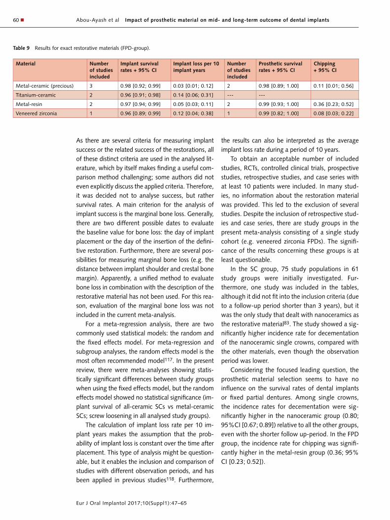
Abou-Ayash et al Impact of prosthetic material on mid- and long-term outcome of dental implants60 n
Eur J Oral Implantol 2017;10(Suppl1):47–65
As there are several criteria for measuring implant success or the related success of the restorations, all of these distinct criteria are used in the analysed lit-erature, which by itself makes finding a useful com-parison method challenging; some authors did not even explicitly discuss the applied criteria. Therefore, it was decided not to analyse success, but rather survival rates. A main criterion for the analysis of implant success is the marginal bone loss. Generally, there are two different possible dates to evaluate the baseline value for bone loss: the day of implant placement or the day of the insertion of the defini-tive restoration. Furthermore, there are several pos-sibilities for measuring marginal bone loss (e.g. the distance between implant shoulder and crestal bone margin). Apparently, a unified method to evaluate bone loss in combination with the description of the restorative material has not been used. For this rea-son, evaluation of the marginal bone loss was not included in the current meta-analysis.
For a meta-regression analysis, there are two commonly used statistical models: the random and the fixed effects model. For meta-regression and subgroup analyses, the random effects model is the most often recommended model117. In the present review, there were meta-analyses showing statis-tically significant differences between study groups when using the fixed effects model, but the random effects model showed no statistical significance (im-plant survival of all-ceramic SCs vs metal-ceramic SCs; screw loosening in all analysed study groups).
The calculation of implant loss rate per 10 im-plant years makes the assumption that the prob-ability of implant loss is constant over the time after placement. This type of analysis might be question-able, but it enables the inclusion and comparison of studies with different observation periods, and has been applied in previous studies118. Furthermore,
the results can also be interpreted as the average implant loss rate during a period of 10 years.
To obtain an acceptable number of included studies, RCTs, controlled clinical trials, prospective studies, retrospective studies, and case series with at least 10 patients were included. In many stud-ies, no information about the restoration material was provided. This led to the exclusion of several studies. Despite the inclusion of retrospective stud-ies and case series, there are study groups in the present meta-analysis consisting of a single study cohort (e.g. veneered zirconia FPDs). The signifi-cance of the results concerning these groups is at least questionable.
In the SC group, 75 study populations in 61 study groups were initially investigated. Fur-thermore, one study was included in the tables, although it did not fit into the inclusion criteria (due to a follow-up period shorter than 3 years), but it was the only study that dealt with nanoceramics as the restorative material83. The study showed a sig-nificantly higher incidence rate for decementation of the nanoceramic single crowns, compared with the other materials, even though the observation period was lower.
Considering the focused leading question, the prosthetic material selection seems to have no influence on the survival rates of dental implants or fixed partial dentures. Among single crowns, the incidence rates for decementation were sig-nificantly higher in the nanoceramic group (0.80; 95%CI [0.67; 0.89]) relative to all the other groups, even with the shorter follow up-period. In the FPD group, the incidence rate for chipping was signifi-cantly higher in the metal-resin group (0.36; 95% CI [0.23; 0.52]).
Table 9 Results for exact restorative materials (FPD-group).
Material Number of studies included
Implant survival rates + 95% CI
Implant loss per 10 implant years
Number of studies included
Prosthetic survival rates + 95% CI
Chipping + 95% CI
Metal-ceramic (precious) 3 0.98 [0.92; 0.99] 0.03 [0.01; 0.12] 2 0.98 [0.89; 1.00] 0.11 [0.01; 0.56]
Titanium-ceramic 2 0.96 [0.91; 0.98] 0.14 [0.06; 0.31] --- ---
Metal-resin 2 0.97 [0.94; 0.99] 0.05 [0.03; 0.11] 2 0.99 [0.93; 1.00] 0.36 [0.23; 0.52]
Veneered zirconia 1 0.96 [0.89; 0.99] 0.12 [0.04; 0.38] 1 0.99 [0.82; 1.00] 0.08 [0.03; 0.22]

Abou-Ayash et al Impact of prosthetic material on mid- and long-term outcome of dental implants n 61
Eur J Oral Implantol 2017;10(Suppl1):47–65
n Limitations
The results of this meta-analysis must be regarded to consider the following limitations:
Data was mostly extracted from non-compara-tive studies. This was done because there were no studies comparing the influence of various restora-tive materials on implant survival. Especially due to the fact that the use of implants is a relatively new in dentistry, it is challenging to find comparative studies for every aspect. Many factors that have been con-sidered to be more important have been investigated in randomised studies with different study cohorts.
It is worth mentioning that the primary questions of each of the included RCTs and prospective stud-ies did not regard the choice of restorative material. Therefore, the level of evidence might be lower than assumed by regarding the quality assessment of the included studies. The scarcity of RCTs led to a lack of high-quality studies that could be included in this meta-analysis.
As the restorative material was not the key point of the included studies, heterogeneity among them was high. Furthermore, in many studies it was not clearly stated whether the baseline was the time of implant placement or the time of prosthesis delivery. Therefore, the mean calculated observation in the study groups could be different from reality.
The glass ceramic restorations of the SC group comprised different types of glass ceramic mater-ials. In two studies the single crowns were made of leucite ceramics and in the two other studies, the crowns were made of lithium-disilicate ceram-ics58,91,98. Although the material properties are dif-ferent, we decided not to separate them into distinct groups as the crowns were all fabricated in a mono-lithic way. This seemed to have no influence on our outcome measures.
Further factors with a possible influence on sur-vival, such as implant type, implant material, im-plant diameter, implant length, abutment material, location of implants, soft- or hard tissue grafting, biological complications, time of implant placement, loading protocols, type of retention, existing canti-lever, or the number of implants per prosthesis, were not taken into consideration. Many of these factors are known to have an influence on implant survival and/or on the prosthetic outcome. However, due to
the aforementioned heterogeneity, it was not pos-sible to include these factors in the present meta-analysis.
Full-arch implant-supported prostheses were not included in the study. The heterogeneity of variables within this type of restoration was too large for the analysis performed here (e.g. the number of implants per prosthesis). Further research on the influence of the prosthetic materials, especially on this type of prosthesis, would be interesting, as there are no remaining teeth in full arch prostheses. Therefore, bite forces are directly transferred to the bone by the implant and the prosthetic restoration, without any buffering by teeth or the periodontal ligaments, and the impact of the restorative material on implant survival might be significant in this group. But for a representative analysis, a closer selection of inclu-sion criteria must be performed than was required for this study.
n Conclusions
A wide range of literature is available to analyse the impact of prosthetic material selection on implant and prosthetic survival rates in fixed dental restor-ations. However, most of the studies investigate single crowns rather than fixed partial dentures. Despite the limitations discussed above, the follow-ing conclusions can be made:• The choice of prosthetic material seems to have
no influence on implant survival rates in fixed restorations;
• The prosthetic material seems to have no influ-ence on prosthetic survival rates of fixed implant-borne restorations;
• Nanoceramic SCs seem to have a higher risk of decementation relative to other materials, and;
• Metal-resin FPDs seem to have higher risk of chipping relative to other materials.
For future research, controlled clinical trials are essential to minimise the heterogeneity of literature concerning material selection, thereby enhancing our knowledge about the influence of restorative material on implant survival.

Abou-Ayash et al Impact of prosthetic material on mid- and long-term outcome of dental implants62 n
Eur J Oral Implantol 2017;10(Suppl1):47–65
n References
1. Moraschini V, Poubel LA, Ferreira VF, Barboza Edos S. Evaluation of survival and success rates of dental implants reported in longitudinal studies with a follow-up period of at least 10 years: a systematic review. Int J Oral Maxillofac Surg 2015;44: 377–388.
2. Bornstein MM, Halbritter S, Harnisch H, Weber HP, Buser D. A retrospective analysis of patients referred for implant placement to a speciality clinic: Indications, surgical procedures and early failures. Int J Oral Maxillofac Implants 2008;23: 1109–1116.
3. Dhingra K. Oral rehabilitation considerations for partially edentulous periodontal patients. J Prosthodont 2012;21: 494–513.
4. Millen C, Brägger U, Wittneben JG. Influence of prosthesis type and retention mechanism on complications with fixed implant-supported prostheses: a systematic review apply-ing multivariate analyses. Int J Oral Maxillofac Implants 2015;30: 110–124.
5. Ma S, Fenton A. Screw- versus cement-retained implant prostheses: a systematic review of prosthodontic mainten-ance and complications. Int J Prosthodont 2015;28: 127–145.
6. Lemos CA, Ferro-Alves ML, Okamoto R, Mendonça MR, Pellizzer EP. Short dental implants versus standard dental implants placed in the posterior jaws: A systematic review and meta-analysis. J Dent 2016;47: 8–17.
7. Romeo E, Storelli S, Casano G, Scanferla M, Botticelli D. Six-mm versus 10-mm long implants in the rehabilitation of posterior edentulous jaws: a 5-year follow-up of a randomised controlled trial. Eur J Oral Implantol 2014;7: 371–381.
8. Gallucci GO, Avrampou M, Taylor JC, Elpers J, Thalji G, Cooper LF. Maxillary Implant-Supported Fixed Prosthesis: A Survey of Reviews and Key Variables for Treatment Plan-ning. Int J Oral Maxillofac Implants 2016;Suppl: 192–207.
9. Moher D, Liberati A, Tetzlaff J, Altman DG, PRISMA Group. Preferred reporting items for systematic reviews and meta-analyses: the PRISMA statement. J Clin Epidemiol 2009;62: 1006–1012.
10. Proskin HM, Jeffcoat RL, Catlin A, Campbell J Jeffcoat MK. A meta-analytic approach to determine the state of science on implant dentistry. Int J Oral Maxillofac Implants 2007;22: 11–18.
11. R Core Team. A language and environment for statistical computing. R Foundation for Statistical Computing 2015; Vienna Austria. http://www.R-project.org/
12. Schwarzer G. General Package for Meta-Analysis. R pack-age version 4.3-2 2015; URL http://CRAN.R-project.org/package=meta
13. Viechtbauer W. Conducting meta-analyses in R with the metafor package. Journal of Statistical Software 2010;36: 1–48.
14. Bergenblock S, Andersson B, Fürst B, Jemt T. Long-term follow-up of CeraOne™ single-implant restorations: an 18-year follow-up study based on a prospective patient cohort. Clin Implant Relat Res 2012;14: 471–479.
15. Bianchi AE, Sanfilppo F. Single-tooth replacement by imme-diate implant and connective tissue graft: a 1-9-year clinical evaluation. Clin Oral Implants Res 2004;15: 269–277.
16. Branzén M, Eliasson A, Arnrup K, Bazargani F. Implant-Supported Single Crowns Replacing Congenitally Missing Maxillary Lateral Incisors: A 5-Year Follow-Up. Clin Implant Dent Relat Res 2015;17: 1134–1140.
17. Dierens M, De Bruyn H, Kisch J, Nilner K, Cosyn J, Van-deweghe S. Prosthetic Survival and Complication Rate of Single Implant Treatment in the Periodontally Healthy Patient after 16 to 22 Years of Follow-Up. Clin Implant Dent Relat Res 2016;18: 117–128.
18. Ekfeldt A, Fürst B, Carlsson GE. Zirconia abutments for single-tooth implant restorations: a retrospective and clin-ical follow-up study. Clin Oral Implants Res 2011;22: 1308–1314.
19. Grassi FR, Capogreco M, Consonni D, Bilardi D, Buti J, Kale-maj Z. Immediate Occlusal Loading of One-Piece Zirconia Implants: Five-Year Radiographic and Clinical Evaluation. Int J Oral Maxillofac Implants 2015;30: 671–680.
20. Hälg GA, Schmid J, Hämmerle CH. Bone level changes at implants supporting crowns or fixed partial dentures with or without cantilevers. Clin Oral Implants Res 2008;19: 983–990.
21. Hosseini M, Worsaae N, Schiødt M, Gotfredsen K. A 3-year prospective study of implant-supported, single-tooth restorations of all-ceramic and metal-ceramic mater-ials in patients with tooth agenesis. Clin Oral Implants Res 2013;24: 1078–1087.
22. Lops D, Bressan E, Pisoni G, Cea N, Corazza B, Romeo E. Short implants in partially edentulous maxillae and mandi-bles: a 10 to 20 years retrospective evaluation. Int J Dent 2012; doi: 10.1155/2012/351793. Epub 2012 Jul 9.
23. Mangano C, Mangano F, Piattelli A, Iezzi G, Mangano A, La Colla L. Prospective clinical evaluation of 307 single-tooth morse taper-connection implants: a multicenter study. Int J Oral Maxillofac Implants 2010;25: 394–400.
24. Mangano C, Iaculli F, Piattelli A, Mangano F. Fixed restor-ations supported by Morse-taper connection implants: a retrospective clinical study with 10-20 years of follow-up. Clin Oral Implants Res 2015;26: 1229–1236.
25. Moberg LE, Köndell PA, Kullman L, Heimdahl A, Gynther GW. Evaluation of single-tooth restorations on ITI dental implants. A prospective study of 29 patients. Clin Oral Implants Res 1999;10: 45–53.
26. Özkan Y, Akoğlu B, Kulak-Özkan Y. Five-year treatment outcomes with four types of implants in the posterior maxilla and mandible in partially edentulous patients: a retrospective study. Int J Oral Maxillofac Implants 2011;26: 639–647.
27. Özkan Y, Özcan M, Akoğlu B, Ucankale M, Kulak-Özkan Y. Three-year treatment outcomes with three brands of implants placed in the posterior maxilla and mandible of partially edentulous patients. J Prosthet Dent 2007: 78–84.
28. Passos SP, Linke B, Larjava H, French D. Performance of zirconia abutments for implant-supported single-tooth crowns in esthetic areas: a retrospective study up to 12-year follow-up. Clin Oral Implants Res 2016;27: 47–54.
29. Spies BC, Balmer M, Patzelt SB, Vach K, Kohal RJ. Clinical and Patient-reported Outcomes of a Zirconia Oral Implant: Three-year Results of a Prospective Cohort Investigation. J Dent Res 2015;94: 1385–1391.
30. Spies BC, Stampf S, Kohal RJ. Evaluation of Zirconia-Based All-Ceramic Single Crowns and Fixed Dental Prosthesis on Zirconia Implants: 5-Year Results of a Prospective Cohort Study. Clin Implant Dent Relat Res 2015;17: 1014–1028.
31. Tartaglia GM, Sidoti E, Sforza C. Seven-year prospective clinical study on zirconia-based single crowns and fixed dental prostheses. Clin Oral Investig 2015;19: 1137–1145.
32. Thoma DS, Brandenberg F, Fehmer V, Knechtle N, Häm-merle CH, Sailer I. The Esthetic Effect of Veneered Zirconia Abutments for Single-Tooth Implant Reconstructions: A Randomized Controlled Clinical Trial. Clin Implant Dent Relat Res 2015;doi:10.1111/cid.12388. [Epub ahead of print].
33. Vigolo P, Givani A. Clinical evaluation of single-tooth mini-implant restorations: a five-year retrospective study. J Prosthet Dent 2000;84: 50–54.
34. Walton TR. An Up-to-15-Year Comparison of the Survival and Complication Burden of Three-Unit Tooth-Supported Fixed Dental Prostheses and Implant-Supported Single Crowns. Int J Oral Maxillofac Implants 2015;30: 851–861.
Abou-Ayash et al Impact of prosthetic material on mid- and long-term outcome of dental implants n 63
Eur J Oral Implantol 2017;10(Suppl1):47–65
35. Astrand P, Engquist B, Anzén B, Bergendal T, Hallman M, Karlsson U, Kvint S, Lysell L, Rundcranz T. A three-year follow-up report of a comparative study of ITI Dental Implants and Brånemark System implants in the treatment of the partially edentulous maxilla. Clin Implant Dent Relat Res 2004;6: 130–141.
36. Becktor JP, Isaksson S, Sennerby L. Endosseous implants and bone augmentation in the partially dentate maxilla: an analysis of 17 patients with a follow-up of 29 to 101 months. Int J Oral Maxillofac Implants 2007;22: 603–608.
37. Bornstein MM, Wittneben JG, Brägger U, Buser D. Early loading at 21 days of non-submerged titanium implants with a chemically modified sandblasted and acid-etched surface: 3-year results of a prospective study in the poster-ior mandible. J Periodontol 2010;81: 809–818.
38. Felice P, Cannizzaro G, Barausse C, Pistilli R, Esposito M. Short implants versus longer implants in vertically augmented pos-terior mandibles: a randomised controlled trial with 5-year after loading follow-up. Eur J Oral Implantol 2014;7: 359–369.
39. Maló P, de Araujo Nobre M, Lopes A. The prognosis of partial implant-supported fixed dental prostheses with cantilevers. A 5-year retrospective cohort study. Eur J Oral Implantol 2013;6: 51–59.
40. Nissan J, Narobai D, Gross O, Ghelfan O, Chaushu G. Long-term outcome of cemented versus screw-retained implant-supported partial restorations. Int J Oral Maxillofac Implants 2011;26: 1102–1107.
41. Ortorp A, Jemt T. Clinical experiences of implant-supported prostheses with laser-welded titanium frameworks in the partially edentulous jaw: a 5-year follow-up study. Clin Im-plant Dent Relat Res 1999;1: 84–91.
42. Balshi TJ, Wolfinger GJ, Wulc D, Balshi SF A prospective analysis of immediate provisionalization of single implants. J Prosthodont 2011;20: 10–15.
43. Berberi AN, Noujeim ZN, Kanj WH, Merawi RJ, Sala-meh ZH. Immediate Placement and Loading of Maxillary Single-Tooth Implants: A 3 Year Prospective Study of Mar-ginal Bone Level. J Contemp Dent Pract 2014;15: 202–208.
44. Bilhan H, Mumcu E, Geçkili O, Atalay B. The evaluation of the success of immediately placed single implants: a retro-spective clinical study. Implant Dent 2011;20: 215–225.
45. Bonde MJ, Stokholm R, Schou S, Isidor F. Patient satisfac-tion and aesthetic outcome of implant-supported single-tooth replacements performed by dental students. Eur J Oral Implantol 2013;6: 387–395.
46. Buser D, Wittneben J, Bornstein MM, Grütter L, Chappuis V, Belser UC. Stability of Contour Augmentation and Esthetic Outcomes of Implant-Supported Single Crowns in the Esthetic Zone: 3-Year Results of a Prospective Study With Early Implant Placement Postextraction. J Periodontol 2011;82: 342–349.
47. Calandriello R, Tomatis M. Immediate occlusal loading of single lower molars using Brånemark System® Wide Platform TiUnite™ implants: a 5-year follow-up report of a prospective clinical multicenter study. Clin Implant Dent Relat Res 2011;13: 311–318.
48. Cannizzaro G, Felice P, Leone M, Ferri V, Viola P, Esposito M. Immediate versus early loading of 6.5 mm-long flapless-placed single implants: a 4-year after loading report of a split-mouth randomised controlled trial. Eur J Oral Implantol 2012;2: 111–121.
49. Cannizzaro G, Leone M, Consolo U, Ferri V, Esposito M. Immediate functional loading of implants placed with flapless surgery versus conventional implants in partially edentulous patients: a 3-year randomized controlled clinical trial. Int J Oral Maxillofac Implants 2008;23: 867–875.
50. De Bruyn H, Raes F, Cooper LF, Reside G, Carriga JS, Tar-rida LG, Wiltfang J, Kern M. Three-years clinical outcome of immediate provisionalization of single OsseospeedTM implants in extraction sockets and healed ridges. Clin Oral Implants Res 2013;24: 217–223.
51. Cosyn J, Eghbali A, De Bruyn H, Collys K, Cleymaet R, De Rouck T. Immediate single-tooth implants in the anterior maxilla: 3-year results of a case series on hard and soft tissue response and aesthetics. J Clin Periodontol 2011;38: 746–753.
52. Covani U, Chiappe G, Bosco M, Orlando B, Quaranta A, Barone A. A 10-year evaluation of implants placed in fresh extraction sockets: a prospective cohort study. J Periodon-tol. 2012;83: 1226–1234.
53. Covani U, Canullo L, Toti P, Alfonsi F, Barone A. Tissue stabil-ity of implants placed in fresh extraction sockets: a 5-year prospective single-cohort study. J Periodontol 2014;85: e323–332.
54. Davis DM, Watson RM, Packer ME. Single tooth crowns supported on hydroxyapatite coated endosseous dental implants: a prospective 5-year study on twenty subjects. Int Dent J 2004;65: 201–205.
55. Donati M, Ekestubbe A, Lindhe J, Wennström JL. Implant-supported single-tooth restorations. A 12-year prospective study. Clin Oral Implants Res 2016;27: 1207–1211.
56. Enkling N, Jöhren P, Katsoulis J, Bayer S, Jervøe-Storm PM, Mericske-Stern R, Jepsen S. Influence of platform switching on bone-level alterations: a three-year randomized clinical trial. J Dent Res 2013;92:139S–145S.
57. Fenner N, Hämmerle CH, Sailer I, Jung RE. Long-term clinical, technical, and esthetic outcomes of all-ceramic vs. titanium abutments on implant supporting single-tooth reconstructions after at least 5 years. Clin Oral Implants Res 2016;27: 716–723.
58. Glauser R, Sailer I, Wohlwend A, Studer S, Schibli M, Schärer P. Experimental Zirconia Abutments for Implant-Supported Single-Tooth Restorations in Esthetically Demanding Regions: 4-Year Results of a Prospective Clin-ical Study. Int J Prosthodont 2004;17: 285–290.
59. Gottfredsen K. A 10-year prospective study of single tooth implants placed in the anterior maxilla. Clin Implant Dent Relat Res 2012:14: 80–87.
60. Guarnieri R, Ceccherini A, Grande M. Single-Tooth Replace-ment in the Anterior Maxilla by Means of Immediate Im-plantation and Early Loading: Clinical and Aesthetic Results at 5 Years. Clin Implant Dent Relat Res 2015;17: 314–326.
61. Ioannidis A, Gallucci GO, Jung RE, Borzangy S, Häm-merle CH, Benic GI. Titanium-zirconium narrow-diameter versus titanium regular-diameter implants for anterior and premolar single crowns: 3-year results of a randomized controlled clinical study. J Clin Periodontol 2015;42: 1060–1070.
62. Jemt T. Cemented CeraOne® and Porcelain Fused to TiAdapt™ Abutment Single-Implant Crown Restorations: A 10-Year Comparative Follow-Up Study. Clin Implant Dent Relat Res 2009;11: 303–310.
63. Jemt T, Pettersson P. A 3-year follow-up study on single implant treatment. J Dent 1993;21: 203–208.
64. Khraisat A, Zembic A, Jung RE, Hammerle CH. Marginal bone levels and soft tissue conditions around single-tooth implants with a scalloped neck design: results of a prospective 3-year study. Int J Oral Maxillofac Implants 2013;550–555.
65. Kolgeci L, Mericske E, Worni A, Walker P, Katsoulis J, Mericske-Stern R. Technical Complications and Failures of Zirconia-Based Prostheses Supported by Implants Followed Up to 7 Years: A Case Series. Int J Prosthodont 2014;27: 544–552.
66. Lai HC, Si MS, Zhuang LF, Shen H, Liu YL, Wismeijer D. Long-term outcomes of short dental implants supporting sin-gle crowns in posterior region: a clinical retrospective study of 5-10 years. Clin Oral Implants Res 2013;24: 230–237.
67. Lang LA, Turkyilmaz I, Edgin WA, Verrett R, Garcia LT. Immediate restoration of single tapered implants with non-occluding provisional crowns: a 5-year clinical prospective study. Clin Implant Dent Relat Res 2014;16: 248–258.

Abou-Ayash et al Impact of prosthetic material on mid- and long-term outcome of dental implants64 n
Eur J Oral Implantol 2017;10(Suppl1):47–65
68. Lops D, Bressan E, Chipasco M, Rossi A, Romeo E. Zirconia and titanium implant abutments for single-tooth implant prostheses after 5 years of function in posterior regions. Int J Oral Maxillofac Implants 2013;281–287.
69. Mangano C, Levrini L, Mangano A, Mangano F, Macchi A, Caprioglio A. Esthetic evaluation of implants placed after orthodontic treatment in patients with congenitally missing lateral incisors. J Esthet Resto Dent 2014;26: 61–71.
70. Mangano FG, Shibli JA, Sammons RL, Iaculli F, Piattelli A, Mangano C. Short (8-mm) locking-taper implants support-ing single crowns in posterior region: a prospective clinical study with 1-to 10-years of follow-up. Clin Oral Implants Res 2014;25: 933–940.
71. Mangano F, Macchi A, Caprioglio A, Sammons RL, Piat-telli A, Mangano C. Survival and complication rates of fixed restorations supported by locking-taper implants: a prospective study with 1 to 10 years of follow-up. J Pros-thodont 2014;26: 434–444.
72. Mericske-Stern R, Grütter L, Rösch R, Mericske E. Clin-ical evaluation and prosthetic complications of single tooth replacements by non-submerged implants. Clin Oral Implants Res 2001;12: 309–318.
73. Montoya-Salazar V, Castillo-Oyagüe R, Torres-Sánchez C, Lynch CD, Gutiérrez-Pérez JL, Torres-Lagares D. Outcome of single immediate implants placed in post-extraction infected and non-infected sites, restored with cemented crowns: a 3-year prospective study. J Dent 2014;645–652.
74. Nejatidanesh F, Moradpoor H, Savabi O. Clinical outcomes of zirconia-based implant- and tooth-supported single crowns. Clin Oral Investig 2016; 20: 169–178.
75. Palmer RM, Palmer PJ, Smith BJ. A 5-year prospective study of Astra single tooth implants. Clin Oral Implants Res 2000;11: 179–182.
76. Perelli M, Abundo R, Corrente G, Saccone C. Short (5 and 7 mm long) porous implants in the posterior atrophic max-illa: a 5-year report of a prospective single-cohort study. Eur J Oral Implantol 2012;5: 265–272.
77. Pieri F, Aldini NN, Marchetti C, Corinaldesi G. Esthetic out-come and tissue stability of maxillary anterior single-tooth implants following reconstruction with mandibular block grafts: a 5-year prospective study. Int J Oral Maxillofac Implants 2013; 28: 270 – 280.
78. Proussaefs P, Lozada J. Immediate loading of hydroxyapatite-coated implants in the maxillary premolar area: three-year results of a pilot study. J Prosthet Dent 2004;91: 228–233.
79. Rinke S, Lattke A, Eickholz P, Kramer K, Ziebolz D. Practice-based clinical evaluation of zirconia abutments for anterior single-tooth restorations. Quintessence Int 2015;46: 19–29
80. Rinke S, Roediger M, Eickholz P, Lange K, Ziebolz D. Tech-nical and biological complications of single-molar implant restorations. Clin Oral Implants Res 2015; 26: 1024–1030.
81. Roccuzzo M, Gaudioso L, Bunino M, Dalmasso P. Long-term stability of soft tissues following alveolar ridge pres-ervation: 10-year results of a prospective study around nonsubmerged implants. Int J Periodontics Restorative Dent 2014;34: 795–804.
82. Romanos GE, Aydin E, Locher K, Nentwig GH. Immediate vs. delayed loading in the posterior mandible: a split-mouth study with up to 15 years of follow-up. Clin Oral Implants Res 2016;e74–79.
83. Romeo E, Chiapasco M, Ghisolfi M, Vogel G. Long-term clin-ical effectiveness of oral implants in the treatment of partial edentulism. Seven-year life table analysis of a prospective study with ITI dental implants system used for single-tooth restorations. Clin Oral Implants Res 2002;13: 133–143.
84. Rossi F, Lang NP, Ricci E, Ferraioli L, Marchetti C, Botticelli D. Early loading of 6-mm-short implants with a moderately rough surface supporting single crowns--a prospective 5-year cohort study. Clin Oral Implant Res 2015; 26: 471–477.
85. Rossi F, Botticelli D, Cesaretti G, De Santis E, Storelli S, Lang NP. Use of short implants (6 mm) in a single-tooth replacement: a 5-year follow-up prospective randomized controlled multicenter clinical study. Clin Oral Implant Res 2016;27: 458–464.
86. Schepke U, Meijer HJ, Vermeulen KM, Raghoebar GM, Cune MS. Clinical Bonding of Resin Nano Ceramic Res-torations to Zirconia Abutments: A Case Series within a Randomized Clinical Trial. Clin Implant Dental Relat Res 2015;18: 984–982.
87. Schropp L, Isidor F. Clinical outcome and patient satisfaction following full-flap elevation for early and delayed place-ment of single-tooth implants: a 5-year randomized study. Int J Oral Maxillofac Implants 2008;23: 733–743.
88. Schropp L, Wenzel A, Stavropoulos A. Early, delayed, or late single implant placement: 10-year results from a randomized controlled clinical trial. Clin Oral Implants Res 2014;25: 1359–1365.
89. Sorrentino R, Galasso L, Tetè S, De Simone G, Zarone F. Clinical evaluation of 209 all-ceramic single crowns cemented on natural and implant-supported abutments with different luting agents: a 6-year retrospective study. Clin Implant Dent Relat Res 2012;14: 184–197.
90. Turkyilmaz I, Avci M, Kuran S, Ozbek EN. A 4-year prospec-tive clinical and radiological study of maxillary dental implants supporting single-tooth crowns using early and delayed load-ing protocols. Clin Oral Implants Res 2007;9: 222–227.
91. Vanlioglu BA, Özkan Y, Evren B, Özkan YK. Experimental custom-made zirconia abutments for narrow implants in esthetically demanding regions: a 5-year follow-up. Int J Oral Maxillofac Implants 2012;27: 1239–1242.
92. Vigolo P, Givani A, Majzoub Z, Cordioli G. A 4-year prospec-tive study to assess peri-implant hard and soft tissues adja-cent to titanium versus gold-alloy abutments in cemented single implant crowns. J Prosthodont 2006;15: 250–256.
93. Visser A, Raghoebar GM, Meijer HJ, Meijndert L, Vissink A. Care and aftercare related to implant-retained dental crowns in the maxillary aesthetic region: a 5-year prospec-tive randomized clinical trial. Clin Implant Dent Relat Res 2011;13: 157–167.
94. Weber HP, Kim DM, Ng MW, Hwang JW, Fiorellini JP. Peri-implant soft-tissue health surrounding cement- and screw-retained implant restorations: a multi-center, 3-year pro-spective study. Clin Oral Implants Res 2006;17: 375–379.
95. Worni A, Kolgeci L, Rentsch-Kollar A, Katsoulis J, Mericske-Stern R. Zirconia-Based Screw-Retained Prostheses Sup-ported by Implants: A Retrospective Study on Technical Complications and Failures. Clin Implant Dent Relat Res 2015;17: 1073–1081.
96. Zarone F, Sorrentino R, Vaccaro F, Russo S, De Simone G. Retrospective clinical evaluation of 86 Procera AllCeram an-terior single crowns on natural and implant-supported abut-ments. Clin Implant Dent Relat Res 2005; 7 Suppl: 95–103.
97. Zembic A, Bösch A, Jung RE, Hämmerle CH, Sailer I. Five-year results of a randomized controlled clinical trial comparing zirconia and titanium abutments supporting single-implant crowns in canine and posterior regions. Clin Oral Implants Res 2013;24: 284–390.
98. Zembic A, Philipp AO, Hämmerle CH, Wohlwend A, Sailer I. Eleven-Year Follow-Up of a Prospective Study of Zirconia Implant Abutments Supporting Single All-Ceramic Crowns in Anterior and Premolar Regions. Clin Implant Dent Relat Res. 2015;17Suppl 2;e417–426.
99. Zhao X, Qiao SC, Shi JY, Uemura N, Arai K, Lai HC. Evalu-ation of the clinical and aesthetic outcomes of Straumann®
Standard Plus implants supported single crowns placed in non-augmented healed sites in the anterior maxilla: a 5-8 years retrospective study. Clin Oral Implants Res 2016;27: 106–112.
Abou-Ayash et al Impact of prosthetic material on mid- and long-term outcome of dental implants n 65
Eur J Oral Implantol 2017;10(Suppl1):47–65
100. Barnea E, Tal H, Nissan J, Tarrasch R, Peleg M, Kolerman R. The Use of Tilted Implant for Posterior Atrophic Maxilla. Clin Implant Dent Relat Res 2016;18: 788–800.
101. Brägger U, Karoussis I, Persson R, Pjetursson B, Salvi G, Lang N. Technical and biological complications/failures with single crowns and fixed partial dentures on implants: a 10-year prospective cohort study. Clin Oral Implants Res 2005;16: 326–334.
102. Francetti L, Azzola F, Corbella S, Taschieri S, Del Fabbro M. Evaluation of clinical outcomes and bone loss around tita-nium implants with oxidized surface: six-year follow-up results from a prospective case series study. Clin Implant Dent Relat Res 2014;16: 81–88.
103. Göthberg C, André U, Gröndahl K, Thomsen P, Slotte C. Bone Response and Soft Tissue Changes Around Implants With/Without Abutments Supporting Fixed Partial Den-tures: Results From a 3-Year, Prospective, Randomized, Controlled Study. Clin Implant Dent Relat Res 2016;18: 309–322.
104. Jemt T, Lekholm U. Oral implant treatment in posterior partially edentulous jaws: a 5-year follow-up report. Int J Oral Maxillofac Implants 1993;8: 635–640.
105. Kreissl ME, Gerds T, Muche R, Heydecke G, Strub JR. Technical complications of implant-supported fixed partial dentures in partially edentulous cases after an average observation period of 5 years. Clin Oral Implants Res 2007 18: 720–276.
106. Mangano F, Macchi A, Caprioglio A, Sammons RL, Piat-telli A, Mangano C. Survival and complication rates of fixed restorations supported by locking-taper implants: a prospective study with 1 to 10 years of follow-up. J Pros-thodont 2014;23: 434–444.
107. Pozzi A, Sannino G, Barlattani A. Minimally invasive treat-ment of the atrophic posterior maxilla: a proof-of-concept prospective study with a follow-up of between 36 and 54 months. J Prosthet Dent 2012; 108:286 –297.
108. Romeo E, Lops D, Margutti E, Ghisolfi M, Chiapasco M, Vogel G. Long-term survival and success of oral implants in the treatment of full and partial arches: a 7-year prospec-tive study with the ITI dental implant system. Int J Oral Maxillofac Implants 2004; 19: 247–259.
109. Romeo E, Tomasi C, Finini I, Casentini P, Lops D. Implant-supported fixed cantilever prosthesis in partially edentulous jaws: a cohort prospective study. Clin Oral Implants Res 2009; 20: 1278–1285.
110. Albrektsson T, Donos N. Implant survival and complica-tions. The Third EAO consensus conference 2012. Clin Oral Implants Res 2012;23 Suppl6: 63–65.
111. Jung RE, Pjetursson BE, Glauser R, Zembic A, Zwahlen M, Lang NP. A systematic review of the 5-year survival and complication rates of implant-supported single crowns. Clin. Oral Impl Res 19,2008;119–130.
112. Hjalmarsson L, Gheisarifar M, Jemt T. A systematic review of survival of single implants as presented in longitudinal studies with a follow-up of at least 10 years. Eur J Oral Implantol. 2016;9: 155–62.
113. Jung RE, Zembic A, Pjetursson BE. Zwahlen M, Thoma DS. Systematic review of the survival rate and the incidence of biological, technical, and aesthetic complications of single crowns on implants reported in longitudinal studies with a mean follow-up of 5 years. Clin. Oral Implants Res 2012;Suppl6: 2–21.
114. Bryant SR, MacDonald-Jankowski D, Kim K. Does the type of implant prosthesis affect outcomes for the completely edentulous arch? Int J Oral Maxillofac Implants. 2007; 22 Suppl: 117–139.
115. Crespi R, Capparè P, Gastaldi G, Gherlone EF. Immediate occlusal loading of full-arch rehabilitations: screw-retained versus cement-retained prosthesis. An 8-year clinical evalu-ation. Int J Oral Maxillofac Implants; 29: 1406–1411.
116. Maló P, de Araújo Nobre M, Lopes A, Francischone C, Rigolizzo M. “All-on-4” Immediate-Function Concept for Completely Edentulous Maxillae: A Clinical Report on the Medium (3 Years) and Long-Term (5 Years) Outcomes. Clin Implant Dent Relat Res 2012;14 Suppl1:e139–150.
117. Thompson SG, Higgins JPT. How should meta-regression analyses be undertaken and interpreted? Stat Med 2002; 21: 1559–1573.
118. Kern JS, Kern T, Wolfart S, Heussen N. A systematic review and meta-analysis of removable and fixed implant sup-ported prostheses in edentulous jaws: post-loading implant loss. Clin Oral Impl Res 2016;27: 174–195.

n 67
Eur J Oral Implantol 2017;10(Suppl1):67–77
REVIEW
Long LongGraduate Student, Johns Hopkins University Bloomberg School of Public Health, Baltimore, Maryland, USA
Hatem AlqarniProsthodontics Resident, Loma Linda University, California, USA Radi MasriAssociate Professor, University of Maryland, Baltimore, Maryland, USA
Correspondence to:Radi Masri, DDS, MS, PhDAssociate Professor, University of Maryland Baltimore, School of Dentistry, 650 W Baltimore St, Office: 6253, Baltimore, MD 21201. Tel: 410-706-8133. Email: [email protected].
Long Long, Hatem Alqarni and Radi Masri
Influence of implant abutment fabrication method on clinical outcomes: a systematic review
Key words CAD/CAM, clinical outcome, conventional abutment, implant abutment, systematic review
Aim: The aim of this systematic review was to evaluate and synthesise the existing evidence on the effect of the prosthetic implant abutment design and fabrication process on mechanical, biological and aesthetic clinical outcomes.Materials and methods: Two electronic databases (PubMed and Embase) were searched in August 2016 to identify clinical studies evaluating the clinical outcomes of CAD/CAM abutments. The stud-ies were screened and two reviewers used the full text to extract data independently. A qualitative synthesis was performed on the extracted data and summary tables were prepared. Due to hetero-geneity in the studies included, no meta-analysis was performed.Results: Twenty-four studies were included in this review. Of these, 13 studies focused solely on CAD/CAM abutment and did not include a control group, or a comparison with conventional implant abutments. Eleven studies compared clinical outcomes of CAD/CAM abutments with conventional abutments. There were only three clinical trials and the majority of the studies were observational or case series studies. The most commonly reported clinical outcomes measured were soft tissue volume and aesthetic scores, survival and success rates, and marginal bone levels. Conclusion: The results of the review demonstrate that CAD/CAM abutments had overall good survival and success rates and that they provide comparable, if not better, clinical outcomes when compared with conventional abutments. However, existing evidence is weak as few randomised control trials were conducted and follow-up periods were, in general, short.
Conflict-of-interest and funding statement: The authors report no financial or other relationships that might lead to a conflict of interest. This systematic review was conducted as a part of the 2016 Foundation of Oral Rehabilitation Consensus Conference on “Prosthetic Protocols in Implant-based Oral Rehabilitation”. The authors received no funding to conduct the systematic review, but received reimbursement to participate in the Consensus Conference.
n Introduction
The restoration of dental implants requires the use of carefully designed prosthetic abutments to retain the final restoration. The abutment must be made of a biocompatible material that can endure the harsh oral environment. It must possess adequate
mechanical and physical properties to withstand occlusal forces and to distribute the load favourably along the supporting implant1. As such, the long-term success of implant abutments heavily depends on abutment material properties.
Prosthetic abutments can be fabricated using various materials, including titanium, gold and

Long et al Abutment Type and Clinical Outcomes68 n
Eur J Oral Implantol 2017;10(Suppl1):67–77
of abutment materials on clinical outcomes, none investigated CAD/CAM abutments and how they compared with conventional abutments. Therefore, the objective of this review was to analyse the lit-erature relating to the use of CAD/CAM prosthetic implant abutments to address the following PICO question14: In subjects treated with fixed implant-supported restorations, what is the effect of using CAD/CAM abutments, compared with conventional abutments, on mechanical, biological and aesthetic treatment outcomes.
n Materials and methods
This systematic review was conducted following the Preferred Reporting Items for Systematic Reviews and Meta-Analysis (PRISMA) guidelines14.
n Eligibility criteria
Clinical studies examining humans of any age, race/ethnicity, or gender were eligible for this review. Studies reporting on the clinical outcomes of CAD/CAM abutments in implant-supported restorations were included. Also included were studies compar-ing CAD/CAM abutments with conventional abut-ments. Clinical outcomes were broadly categorised as mechanical, biological and aesthetic outcomes. Conventional abutments were defined as those fab-ricated using non-digital methods, including stock abutments, prefabricated abutments and custom-ised abutments. Case reports with fewer than five cases, conference abstracts, non-systematic reviews, letters, opinion articles and animal studies were excluded.
n Source and search methods
Two different electronic databases were searched using search strategies designed for each database – P ubMed and Embase. There were no language or date restrictions in the electronic search. The litera-ture search was first performed on 3 March, 2016, and then updated on 21 August, 2016. The follow-ing is a brief description of the strategy used in the PubMed search: The MeSH term “computer-aided design” was combined with MeSh terms: “dental
ceramics. These materials exhibit different degrees of biocompatibility and harmony with soft tissues2. Titanium has been traditionally preferred because of its strength, biocompatibility, reliability, and machinability3-5. Zirconia prosthetic abutments are increasingly used as they tend to evoke better colour response of peri-implant mucosa and superior aes-thetic outcomes compared with titanium abutments, as measured by the Pink Esthetic Score (PES), espe-cially in subjects with thin mucosal phenotype6,7.
The long-term success of implant abutments is not only dependent on material choice, but it is also dependent on design and manufacturing process as these may significantly influence abutment ma-terial properties. For example, physical and mechan-ical properties of zirconia are heavily influenced by preparation technique and the design of prosthetic components8-10. Indeed, the design and geometry of the prosthetic abutment significantly affects stress distribution in implant-retained restorations, which, in turn, impacts clinical outcomes1.
A successful prosthetic implant abutment must also support the peri-implant mucosa. This will allow for the fabrication of a restoration endowed with optimum contours and a proper emergence pro-file. This will ultimately result in a restoration that is functional, aesthetic and cleansable. The design and manufacturing process also influences abutment contours and finish, which are factors directly influ-encing aesthetic outcomes.
In addition, abutment design and manufactur-ing process have critical biological consequences. For example, the accumulating body of evidence suggests that the use of stock prosthetic abutments for cement-retained restorations should be avoided, as they may complicate cement removal11-13. Thus, abutment design and contours may affect the ability of clinicians to remove excess cement. This further highlights the importance of abutment design and fabrication technique in affecting the clinical out-comes of implant restorations.
With the recent explosion of digital technology and the increased use of computer-aided design and computer-aided manufacture (CAD/CAM) processes in the fabrication of implant prosthetic abutments, it is important to assess if the design and fabrication method affects clinical treatment outcomes. While previous systematic reviews focused on the effect

Long et al Abutment Type and Clinical Outcomes n 69
Eur J Oral Implantol 2017;10(Suppl1):67–77
implants”, “dental implantation”, “dental abut-ments”, “dental implant-abutment design”, “den-tal implantation, endosseous/methods”, or “dental prosthesis, implant-supported”. In addition, the text words “CAD”, “CAM”, “customized abutment*”, “computer-generated”, “computer-assisted manu-facture*”, “computer-assisted designed manufac-tured”, “Computer-assisted design*”, “computer-aided manufacture*”, “Computer-aided design*”, “computer assisted”, “cad-cam”, “CAD/CAM”, “sirona”, “procera”, “e4d”, or “atlantis abutment*” were combined with the text words: “abutment*”, “dental prosthesis implant-supported”, “implant abutment*”, “implant dentistry”, “implant-sup-ported abutment*”, “single implant*”, or “dental implant*”. The filter for excluding animal studies was also applied. The complete search strategy for both databases is described in the appendices – (appen-dix 1: PubMed; appendix 2: Embase). In addition to searching electronic databases, a manual search was conducted to identify additional relevant literature. The National Institutes of Health Research Portfolio Online Reporting Tools (RePort) and search engines, such as “Google” and “Google Scholar”, were also used to search for grey literature, unpublished stud-ies and ongoing clinical trials. Only manuscripts with full text were included in this review.
n Selection of studies
Two authors independently screened the titles and abstracts from the electronic and manual searches. Abstracts were initially classified as rele-vant, maybe relevant or not relevant. For abstracts labelled relevant or maybe relevant, full-text cop-ies of the manuscripts were obtained. Two authors independently assessed each article and determined whether to include, exclude or designate each study as a “maybe”. After the end of independent review, agreement between the reviewers was documented and conflicts were resolved through discussion or consultation with the third author.
n Data collection
Two authors independently extracted the data. The following study characteristics were extracted: study design, CAD/CAM abutment system and material
used, conventional abutment type and material used, clinical outcomes, loss to follow-up, and fol-low-up period.
To assess the risk of bias for clinical trials, the Cochrane Risk of Bias tool was used15. To evaluate the reporting quality of observational studies, the Strengthening the Reporting of Observational Stud-ies in Epidemiology (STROBE) checklist16 was used. For case series studies, the Case Reporting (CARE) checklist17 was used. All of the 21 STROBE domains and 14 CARE topics were assessed. Risk of bias assessments for observational studies and case series studies were not conducted due to the heterogene-ous quality of reporting in these studies.
n Synthesis of results
The manuscripts were divided into studies that only investigated CAD/CAM abutments, and studies that compared CAD/CAM abutments with conven-tional abutments. A “summary of findings” table was produced describing the commonly reported outcomes from these studies. Because of the extreme clinical and methodological heterogeneity among these studies, a meta-analysis of the results was not feasible.
n Results
The study selection process is illustrated in Figure 1. During the title and abstract screening process, stud-ies were excluded if they did not examine humans or did not examine clinical outcomes of CAD/CAM abutments in fixed implant-supported restorations. Reasons for exclusion during the full-text review included: limited reporting such as abstracts, letters, opinion articles; treatment irrelevant for the pre-sent review: implant-supported facial reconstruc-tion; patient population irrelevant for the present review: edentulous patients. A total of 24 studies were included in this review (Fig 1).
Thirteen studies used CAD/CAM abutment only and did not utilise conventional abutments. The study design, type of CAD/CAM abutments used, and main study outcomes of these studies are listed in Table 1. Among these studies, six were descrip-tive studies (case series), one was a cross-sectional
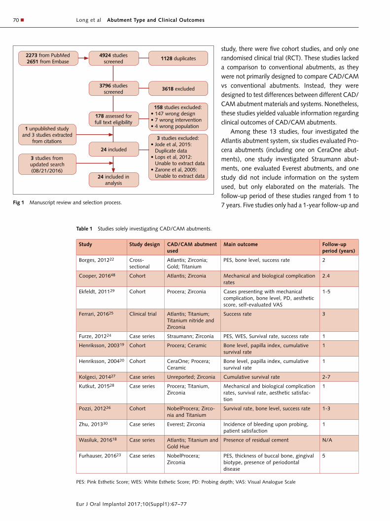
Long et al Abutment Type and Clinical Outcomes70 n
Eur J Oral Implantol 2017;10(Suppl1):67–77
study, there were five cohort studies, and only one randomised clinical trial (RCT). These studies lacked a comparison to conventional abutments, as they were not primarily designed to compare CAD/CAM vs conventional abutments. Instead, they were designed to test differences between different CAD/CAM abutment materials and systems. Nonetheless, these studies yielded valuable information regarding clinical outcomes of CAD/CAM abutments.
Among these 13 studies, four investigated the Atlantis abutment system, six studies evaluated Pro-cera abutments (including one on CeraOne abut-ments), one study investigated Straumann abut-ments, one evaluated Everest abutments, and one study did not include information on the system used, but only elaborated on the materials. The follow-up period of these studies ranged from 1 to 7 years. Five studies only had a 1-year follow-up and Fig 1 Manuscript review and selection process.
Table 1 Studies solely investigating CAD/CAM abutments.
Study Study design CAD/CAM abutment used
Main outcome Follow-up period (years)
Borges, 201222 Cross- sectional
Atlantis; Zirconia; Gold; Titanium
PES, bone level, success rate 2
Cooper, 201648 Cohort Atlantis; Zirconia Mechanical and biological complication rates
2.4
Ekfeldt, 201129 Cohort Procera; Zirconia Cases presenting with mechanical complication, bone level, PD, aesthetic score, self-evaluated VAS
1-5
Ferrari, 201625 Clinical trial Atlantis; Titanium; Titanium nitride and Zirconia
Success rate 3
Furze, 201224 Case series Straumann; Zirconia PES, WES, Survival rate, success rate 1
Henriksson, 200319 Cohort Procera; Ceramic Bone level, papilla index, cumulative survival rate
1
Henriksson, 200420 Cohort CeraOne; Procera; Ceramic
Bone level, papilla index, cumulative survival rate
1
Kolgeci, 201427 Case series Unreported; Zirconia Cumulative survival rate 2-7
Kutkut, 201528 Case series Procera; Titanium, Zirconia
Mechanical and biological complication rates, survival rate, aesthetic satisfac-tion
1
Pozzi, 201226 Cohort NobelProcera; Zirco-nia and Titanium
Survival rate, bone level, success rate 1-3
Zhu, 201330 Case series Everest; Zirconia Incidence of bleeding upon probing, patient satisfaction
1
Wasiluk, 201618 Case series Atlantis; Titanium and Gold Hue
Presence of residual cement N/A
Furhauser, 201623 Case series NobelProcera; Zirconia
PES, thickness of buccal bone, gingival biotype, presence of periodontal disease
5
PES: Pink Esthetic Score; WES: White Esthetic Score; PD: Probing depth; VAS: Visual Analogue Scale
2273 from PubMed2651 from Embase
1128 duplicates4924 studies
screened
24 included in analysis
3618 excluded
158 studies excluded:• 147 wrong design• 7 wrong intervention• 4 wrong population
3 studies excluded:• Jode et al, 2015:
Duplicate data• Lops et al, 2012:
Un able to extract data• Zarone et al, 2005:
Unable to extract data
1 unpublished study and 3 studies extracted
from citations
3 studies from updated search (08/21/2016)
3796 studies screened
24 included
178 assessed for full text eligibility

Long et al Abutment Type and Clinical Outcomes n 71
Eur J Oral Implantol 2017;10(Suppl1):67–77
seven studies had a follow-up of more than 1 year (Table 1). There was one study18 with no follow-up, because the outcome (presence of residual cement) was measured immediately after the restoration was delivered.
Of the 13 studies solely focussing on CAD/CAM abutments, five reported on changes in the papilla index of tissues surrounding the CAD/CAM abut-ment, including reporting on soft tissue volume19,20, soft tissue response using a special four-point scale21, PES22-24, and White Esthetic Score24. Four of the 13 studies investigated the success rate of CAD/CAM abutments22,24-26, which was defined, in the major-ity of studies as the proportion of restorations that remained in situ without any modification. Six out of 13 studies investigated the survival rate of CAD/CAM abutments19,20,24,26-28, which was defined, in the majority of studies, by the proportion of res-torations that remained clinically acceptable in situ, even if they received modifications25. However, each study had its own predefined criteria for success and survival assessment. Only three of the 13 studies
reported patient-centred outcomes26,28,29, includ-ing aesthetic and functional satisfaction. The success and survival rates of the various CAD/CAM abut-ments at 1 year of follow-up based on the system and materials used are presented in Tables 2 and 3 respectively.
For the only clinical trial25 investigating clin-ical outcomes of CAD/CAM abutments, risk of bias assessment was performed according to the Cochrane Risk of Bias Assessment Tool15. The clinical trial had a low risk of selection bias because a com-puter-generated random list and allocation conceal-ment were performed. However, it might be subject to information bias because of the subjective nature of the outcomes measured.
The reporting quality of observational studies was evaluated according to the STROBE checklist16. All of the six observational studies (cross-sectional and cohort) included exhibited good reporting quality in the title, abstract and introduction sec-tions. However, half of the studies did not clearly articulate the study design and eligibility criteria,
Table 3 Success and survival rates of CAD/CAM abutments based on abutment material.
Outcomes CAD/CAM abutment
Outcome at 1-year follow-up
Number of abutments
Number of studies
Comments
Success rate Zirconia See comments 41 322,24,25 82.2% success rate reported at 3-year time point in study25. Others reported 100% success rate.
Titanium 100% 78 222,25 100% success rate was also reported at 2-year or 3-year time points.
Ceramic 100% 24 119
Survival rate Zirconia 97.4%–100% 91 224,26 7-year cumulative survival rate was reported as 96.4% in study27.
Titanium 95.2% 42 126
Ceramic 100% 9 120
Table 2 Success and survival rates of CAD/CAM abutments based on the system used.
Parameter CAD/CAM abutment
Outcome at 1-year follow-up
Number of abutments
Number of studies
Comments
Success rate Atlantis See comments 109 222,25 82.2% to 100% success rate report-ed at 2-year or 3-year time point
Procera See comments 91 126 91.9% cumulative success rate reported at 3-year time point
Straumann 100% 10 124
Survival rate Straumann 100% 10 124
Procera 96.3%–100% 164 419,20,26,28 3 implants failed in study26
CeraOne 100% 10 120

Long et al Abutment Type and Clinical Outcomes72 n
Eur J Oral Implantol 2017;10(Suppl1):67–77
and only one study included matched subjects. Five of the six studies suffered from missing follow-up data, and none of the studies addressed the missing data or performed sensitivity analysis16. None of the studies discussed external validity of the pre-sented results and only two studies reported the source of funding.
The reporting quality of case-series studies was evaluated according to the CARE checklist17. Among the six case series studies, five studies indicated the study type in the title. However, none of these stud-ies included “case report” or “case series” in the key words. All studies exhibited good reporting quality in their abstracts, introductions, interventions, out-comes and discussions. None of the studies included a timeline that indicated specific dates and times in a table, figure or graphic. Only half of the stud-ies described the specific reasons and conditions of
patients who received implant treatment. Half of the studies reported on the evaluation of patients before treatment. Two of the six case-series studies reported patient perspective of the experience28,30. Two18,28
did not report on the informed consent process and whether it was obtained from patients, and only two studies included an acknowledgements section.
n Systematic review of studies comparing CAD/CAM abutments with conventional abutments
Eleven studies compared clinical outcomes of CAD/CAM abutments with conventional abutments. The study design, type of CAD/CAM abutment, type of conventional abutment, and main study outcomes are summarised in Table 4. Among these 11 studies, three were descriptive studies (case series), one was
Table 4 Studies investigating CAD/CAM vs conventional abutments.
Study Study Design
CAD/CAM Abutment Conventional Abutment Main Outcome Follow up Period (years)
System, material
Number Type, material Number
Borges, 201439
Cohort Atlantis, Zir-conia, Gold Titanium
25 Custom, Metal 12 PES, bone level, survival rate 1
Choquet, 200140
Cross- sectional
Procera, N/A
11 Customised, Titanium and Ceramic;
Standard, N/A (Cer-aOne)
16 PES, bone level, PD, soft tissue height Minimum of 0.5
Dierens, 201349
Case series CeraOne, N/A
N/A Customised, N/A N/A PES, WES, bone level, PD, soft tissue height
16-22
Joda, 201431 Case series Straumann, Titanium
3 Prefabricated, Titanium 3 Time, cost, WES N/A
Joda, 201532 Cohort Straumann Titanium
20 Prefabricated, Titanium 20 Time, cost, CMA N/A
Joda, 201533 Cohort Straumann, Titanium
20 Prefabricated, Titanium 20 Fitting and adjustment time N/A
Korsch, 201537
Cohort Atlantis, N/A
96 Prefabricated, N/A 312 Loosening rate, survival rate 2
Lops, 201534
Cohort Atlantis, Zirconia
36 Stock, Zirconia and Titanium
36 Success rate, survival rate, soft tissue recession
2
Schepke, 201535
Case series Atlantis, Zirconia
25 Stock, Zirconia 25 Survival rate, success rate 1
Borzangy, 201538
RCT N/A, Zirco-nia
15 Prefabricated, Titanium 15 PES, WES, success rate, plaque score, bleeding score, PD, bone change
1
Schepke, 201636
RCT Atlantis, Zirconia
25 Stock (ZirDesign), Zirconia
25 Bone change, plaque index, calculus formation, bleeding score, PD, soft tis-sue recession, patients’ satisfaction
1
RCT: Randomised Clinical Trial; PES: Pink Esthetic Score; WES: white Esthetic Score; PD: Probing depth; CMA: clinical productivity rate/cost minimization; N/A: not reported.

Long et al Abutment Type and Clinical Outcomes n 73
Eur J Oral Implantol 2017;10(Suppl1):67–77
a cross-sectional study, five were cohort studies, and two were RCTs.
Three studies31-33 comparing CAD/CAM and prefabricated Titanium abutments (Straumann) used the same patient cohort, but each reported on dif-ferent outcomes. Two studies compared the survival rate and success rate of Atlantis zirconia abutments to stock zirconia abutments34,35, but the study design and follow-up period were different between the studies. The follow-up time of these 11 studies ranged from six months to 22 years (Table 4). There were three studies31-33 with no follow-up because the outcome measured did not require one.
Of the 11 studies, only one36 reported patient-centred outcomes. Three studies31-33 reported cost-effective outcomes, such as comparative time and economical analysis. In Table 5 a summary of the systematic review is presented when all CAD/CAM abutments are grouped in one category, regardless of the system and material used, and the same for conventional abutments. The table was produced for outcomes after 1 year of follow-up. However, two of the 11 studies reviewed reported outcomes after a 2-year follow-up. Overall, the studies reported no significant differences in the majority of outcomes between CAD/CAM and conventional abutments after 1 year of follow-up. However, when studies investigating the 2-year follow ups were consid-ered; one study34 reported a significantly better soft tissue reaction (less recession) around CAD/CAM abutments (n = 36) compared with conventionally
fabricated abutments (n = 36). In another study37, although the survival rate was not significantly differ-ent between CAD/CAM (n = 96) and prefabricated abutments (n = 312), CAD/CAM abutments with single crown restorations suffered from significantly less crown loosening than restorations retained by conventionally fabricated abutments over a 2-year follow-up.
The Risk of Bias assessment of the two clinical trials included36,38 revealed they were vulnerable to selection bias, as both provided incomplete descrip-tions of the randomisation process. They were also subject to some information bias, as allocation con-cealment was not performed. Detailed risk of bias assessment for each study is summarised in Table 6.
All six observational studies (cross-sectional and cohort studies) exhibited good reporting quality in title, abstract, and introduction sections. Half of the studies clearly presented the study design. Of the six studies, five described the eligibility criteria, but no study was a matched study. Two studies39,40 had missing follow-up data, but they did not address the missing data or perform sensitivity analysis. In one study, subgroup analysis was performed39. No study discussed the external validity of the study results, and only two studies32,33 reported the source of funding.
Reporting quality for the three case series studies was also assessed. Two studies indicated the study type in the title, but none of the studies included “case report” or “case series” in the key words.
Table 5 Outcomes of CAD/CAM vs conventional abutments.
Outcomes 1-year results without significant difference
1-year results with significant difference (CAD/CAM showing
better performance)
Comments
No. of total abutments
No. of studies
No. of total abutments
No. of studies
Success rate 79 235,38 0 0
Survival rate 87 235,39 0 0 Study37 reported less crown loosen-ing on CAD/CAM abutment at 2-year follow-up time.
PES 29 138 37 139
WES 29 138 0 0
Crest bone level
116 336,38,39 0 0
Soft tissue recession
50 136 0 0 Study34 reported less soft tissue reces-sion from CAD/CAM abutments at 2-year follow-up time.

Long et al Abutment Type and Clinical Outcomes74 n
Eur J Oral Implantol 2017;10(Suppl1):67–77
All studies had a good report quality in abstracts, introductions, interventions, outcomes and discus-sions. However, no study created a timeline that indicated specific dates and times in a table, figure or graphic. Two studies described the specific rea-sons and conditions of patients who received implant treatment, and two studies reported on how subjects were evaluated before treatment. None of the stud-ies reported on patient perspective of experience, and one study31 did not report on the informed con-sent procedure from patients. Two studies included an acknowledgements section.
n Discussion
The 24 studies included in this review represent research conducted on the clinical outcomes of CAD/CAM abutments used in implant-supported restorations compared with conventional abutments. These studies demonstrated substantial heterogene-ity regarding the systems and materials of abutments examined, specific clinical outcomes observed, as well as the design of studies and measurement tech-niques employed.
The results of studies that solely examined CAD/CAM abutments showed an overall high survival rate at the 1- and 3-year follow-ups (Table 1). For studies comparing CAD/CAM abutments with conventional abutments, there was no significant difference in most of the clinical outcomes at 1-year follow up. However, one study39 showed better interproximal
papilla PES in Atlantis CAD/CAM zirconia and gold titanium abutments compared with custom metal abutments. In addition, two studies34,37 reported higher survival rate and less tissue recession when Atlantis CAD/CAM abutments were compared to conventional abutments after 2 years of follow-up. Among the 24 included clinical studies, there were only three RCTs25,36,38. Two compared CAD/CAM abutments with conventional stock abutments36,38. However, both studies used a different material of stock abutments in the control group. Therefore, data from the two trials could not be combined for further analysis. The results for plaque index, bleed-ing score, probing depth, and change in marginal bone levels were reported as not significantly dif-ferent in both of these RCTs. More RCTs with long-term follow-ups are needed to determine if the use of CAD/CAM abutments results in improved clinical outcomes when compared with conventional abut-ments. If RCTs are not feasible, cohort studies with adequate sample size and long follow-up periods are encouraged.
Reporting quality assessment for observational and descriptive studies revealed that about 40% of the studies did not describe the patient popula-tion in detail; none discussed the impact of missing data; more than half omitted to report the funding sources of the research; and less than 20% stated patient-reported outcomes. Detailed population description, including specific chief complaints and other relevant medical history, could help research-ers and readers evaluate the external validity of the studies41. Missing data could have a great impact on the outcomes of observational studies, especially when the sample size is small42. Funding sources and the role of the funders could have potential conflict of interest and may lead to bias in report-ing the results43. Therefore, it is recommended that researchers report selection criteria and character-istics in detail, conduct qualitative or quantitative analysis taking in account missing data, and report and acknowledge funding sources.
Some outcomes evaluated in the reviewed man-uscripts were not completely objective, such as aes-thetic evaluation of restorations. To test aesthetic outcomes, several standards were adopted, and in different interproximal sites relative to the restor-ation. Thus, it is recommended that researchers try
Table 6 Risk of bias in clinical trials comparing outcomes of CAD/CAM and conventional abutments.
Schepke, 201636
Borzangy, 201538
Random sequence generation (Selection bias)
? ?
Allocation concealment (Selec-tion bias)
? +
Blinding of participants, person-nel (Information bias)
? ?
Incomplete outcome data (In-formation bias)
+ ?
Selective outcome reporting (Information bias)
? +
Other sources of bias ? +
+: low risk of bias; -: high risk of bias; ?: unclear risk of bias.

Long et al Abutment Type and Clinical Outcomes n 75
Eur J Oral Implantol 2017;10(Suppl1):67–77
to provide a detailed description of the methods of assessment and sources of data, including specifying the system, materials, measurement methods and specific sites tested and assessed. To enhance the quality and transparency of research, as well as the searchable characteristic, accurate reporting of lit-erature is a critical consideration. It is recommended that researchers consult EQUATOR-network.org44 for appropriate reporting guidelines to report their studies.
When risk of bias assessment is considered, none of the RCTs discussed blinding of subjects. In studies reporting objective clinical outcomes, such as bone level reduction through measurements on radiographs, subject blinding may have little impact on the outcomes. However, it may have significant influence on subjective outcomes, such as patient-reported outcomes45. However, it is important to note that the literature reviewed showed little emphasis on patient-reported outcomes. Patient-reported outcomes provide important information regarding treatment alternatives, and inform the research process46,47. Research on CAD/CAM abut-ments would benefit greatly from studies that are patient-outcome centred.
This systematic review is subject to some limita-tions. Firstly, unpublished results uncovered using the NIH RePORT and grey literature searches were not included, as full texts were not publically available. Secondly, an overall quality assessment of included studies was not conducted, however, reporting quality assessment for observational and descrip-tive studies and risk of bias assessment for clinical trials was conducted. These assessment processes are among several considerations contributing to a study’s overall quality and are in line with Cochrane guidelines43.
n Conclusions
The results of the review suggest that CAD/CAM abutments have overall good survival and success rate and provide comparable, if not better, clinical outcomes when compared with conventional abut-ments. However, available studies comparing CAD/CAM and conventional abutments are few and the majority of studies included are short term.
n References
1. Peixoto HE, Bordin D, Del Bel Cury AA, da Silva WJ, Faot F. The role of prosthetic abutment material on the stress distribution in a maxillary single implant-supported fixed prosthesis. Mater Sci Eng C Mater Biol Appl 2016;65: 90–96.
2. Abrahamsson I, Berglundh T, Glantz PO, Lindhe J. The mucosal attachment at different abutments. An experimen-tal study in dogs. J Clin Periodontol 1998;25: 721–727.
3. Sailer I, Philipp A, Zembic A, Pjetursson BE, Hämmerle CH, Zwahlen M. A systematic review of the performance of cer-amic and metal implant abutments supporting fixed implant reconstructions. Clin Oral Implants Res 2009;20: 4–31.
4. Zembic A, Bosch A, Jung RE, Hämmerle CH, Sailer I. Five-year results of a randomized controlled clinical trial com-paring zirconia and titanium abutments supporting single-implant crowns in canine and posterior regions. Clin Oral Implants Res 2013;24: 384–390.
5. Zembic A, Sailer I, Jung RE, Hämmerle CH. Randomized-controlled clinical trial of customized zirconia and titanium implant abutments for single-tooth implants in canine and posterior regions: 3-year results. Clin Oral Implants Res 2009;20: 802–808.
6. Linkevicius T, Vaitelis J. The effect of zirconia or titanium as abutment material on soft peri-implant tissues: a systematic review and meta-analysis. Clin Oral Implants Res 2015;26: 139–147.
7. Park SE, Da Silva JD, Weber HP, Ishikawa-Nagai S. Optical phenomenon of peri-implant soft tissue. Part I. Spectro-photometric assessment of natural tooth gingiva and peri-implant mucosa. Clin Oral Implants Res 2007;18: 569–574.
8. Denry I. How and when does fabrication damage adversely affect the clinical performance of ceramic restorations? Dent Mater 2013;29: 85–96.
9. Ellingsen JE. Are the mechanical properties of zirconia abutments sufficient for clinical use? Int J Oral Maxillofac Implants 2012;27: 744–746.
10. Sailer I, Zembic A, Jung RE, Siegenthaler D, Holderegger C, Hämmerle CH. Randomized controlled clinical trial of custom-ized zirconia and titanium implant abutments for canine and posterior single-tooth implant reconstructions: preliminary results at 1 year of function. Clin Oral Implants Res 2009;20: 219–225.
11. Korsch M, Obst U, Walther W. Cement-associated peri-implantitis: a retrospective clinical observational study of fixed implant-supported restorations using a methacrylate cement. Clin Oral Implants Res 2014;25: 797–802.
12. Linkevicius T, Vindasiute E, Puisys A, Peciuliene V. The influence of margin location on the amount of undetected cement excess after delivery of cement-retained implant res-torations. Clin Oral Implants Res 2011;22: 1379–1384.
13. Wadhwani C, Rapoport D, La Rosa S, Hess T, Kretschmar S. Radiographic detection and characteristic patterns of resid-ual excess cement associated with cement-retained implant restorations: a clinical report. J Prosthet Dent 2012;107: 151–157.
14. Moher D, Liberati A, Tetzlaff J, Altman DG. Preferred report-ing items for systematic reviews and meta-analyses: the PRISMA statement. PLoS Med 2009;6:e1000097.
15. Higgins JP, Altman DG, Gotzsche PC, Juni P, Moher D, Oxman AD, Savovic J, Schulz KF, Weeks L, Sterne JA. The Cochrane Collaboration’s tool for assessing risk of bias in randomised trials. BMJ 2011;343:d5928.
16. von Elm E, Altman DG, Egger M, Pocock SJ, Gotzsche PC, Vandenbroucke JP. The Strengthening the Reporting of Observational Studies in Epidemiology (STROBE) statement: guidelines for reporting observational studies. J Clin Epide-miol 2008;61:344-349.

Long et al Abutment Type and Clinical Outcomes76 n
Eur J Oral Implantol 2017;10(Suppl1):67–77
17. Gagnier JJ, Kienle G, Altman DG, Moher D, Sox H, Riley D. The CARE guidelines: consensus-based clinical case report guideline development. J Clin Epidemiol 2014;67: 46–51.
18. Wasiluk G, Chomik E, Gehrke P, Pietruska M, Skurska A, Pietruski J. Incidence of undetected cement on CAD/CAM monolithic zirconia crowns and customized CAD/CAM implant abutments. A prospective case series. Clin Oral Implants Res 2016 [Epub ahead of print].
19. Henriksson K, Jemt T. Evaluation of custom-made procera ceramic abutments for single-implant tooth replacement: a prospective 1-year follow-up study. Int J Prosthodont 2003;16: 626–630.
20. Henriksson K, Jemt T. Measurements of soft tissue volume in association with single-implant restorations: a 1-year comparative study after abutment connection surgery. Clin Implant Dent Relat Res 2004;6: 181–189.
21. Jemt T. Regeneration of gingival papillae after single-implant treatment. Int J Periodontics Rest Dent 1997;17: 326–333.
22. Borges T, Lima T, Carvalho A, Carvalho V. Clinical Outcome of Inter-Proximal Papilla between a Tooth and a Single Im-plant Treated with CAD/CAM Abutments: a Cross-Sectional Study. J Oral Maxillofac Res 2012;3:e4.
23. Furhauser R, Mailath-Pokorny G, Haas R, Busenlechner D, Watzek G, Pommer B. Immediate Restoration of Immediate Implants in the Esthetic Zone of the Maxilla Via the Copy-Abutment Technique: 5-Year Follow-Up of Pink Esthetic Scores. Clin Implant Dent Relat Res 2016;19: 28–37.
24. Furze D, Byrne A, Donos N, Mardas N. Clinical and esthetic outcomes of single-tooth implants in the anterior max-illa. Quintessence international (Berlin, Germany: 1985) 2012;43: 127–134.
25. Ferrari M, Tricarico MG, Cagidiaco MC, Vichi A, Gher-lone EF, Zarone F, Sorrentino R. 3-Year Randomized Con-trolled Prospective Clinical Trial on Different CAD-CAM Implant Abutments. Clin Implant Dent Relat Res 2016;18: 1134–1141.
26. Pozzi A, Sannino G, Barlattani A. Minimally invasive treat-ment of the atrophic posterior maxilla: a proof-of-concept prospective study with a follow-up of between 36 and 54 months. J Prosthet Dent 2012;108: 286–297.
27. Kolgeci L, Mericske E, Worni A, Walker P, Katsoulis J, Mericske-Stern R. Technical complications and failures of zirconia-based prostheses supported by implants followed up to 7 years: a case series. Int J Prosthodont;27: 544–552.
28. Kutkut A, Abu-Hammad O, Mitchell R. Esthetic Consid-erations for Reconstructing Implant Emergence Profile Using Titanium and Zirconia Custom Implant Abutments: Fifty Case Series Report. J Oral implant 2015;41: 554–561.
29. Ekfeldt A, Furst B, Carlsson GE. Zirconia abutments for single-tooth implant restorations: a retrospective and clinical follow-up study. Clin Oral Implants Res 2011;22: 1308–1314.
30. Zhu GL, Wang RF, Chen HL. [Clinical study on base bonding CAD/CAM customized zirconium abutment]. Shanghai Kou Qiang Yi Xue 2013;22: 676–679.
31. Joda T, Brägger U. Complete digital workflow for the pro-duction of implant-supported single-unit monolithic crowns. Clin Oral Implants Res 2014;25: 1304–1306.
32. Joda T, Brägger U. Digital vs. conventional implant pros-thetic workflows: a cost/time analysis. Clin Oral Implants Res 2015;26: 1430–1435.
33. Joda T, Katsoulis J, Brägger U. Clinical Fitting and Adjust-ment Time for Implant-Supported Crowns Comparing Digi-tal and Conventional Workflows. Clin Implant Dent Relat Res 2016;18: 946–954.
34. Lops D, Bressan E, Parpaiola A, Sbricoli L, Cecchinato D, Romeo E. Soft tissues stability of CAD/CAM and stock abut-ments in anterior regions: 2-year prospective multicentric cohort study. Clin Oral Implants Res 2015;26: 1436–1442.
35. Schepke U, Meijer HJ, Vermeulen KM, Raghoebar GM, Cune MS. Clinical Bonding of Resin Nano Ceramic Restor-ations to Zirconia Abutments: A Case Series within a Rand-omized Clinical Trial. Clin Implant Dent Relat Res 2016;18: 984–992.
36. Schepke U, Meijer HJ, Kerdijk W, Raghoebar GM, Cune M. Stock Versus CAD/CAM Customized Zirconia Implant Abut-ments – Clinical and Patient-Based Outcomes in a Rand-omized Controlled Clinical Trial. Clin Implant Dent Relat Res 2017;19: 74–84.
37. Korsch M, Walther W. Prefabricated Versus Customized Abutments: A Retrospective Analysis of Loosening of Cement-Retained Fixed Implant-Supported Reconstructions. Int J Prosthodont 2015;28: 522–526.
38. Borzangy S. A Comparison of Zirconia CAD:CAM to Con-ventionally Fabricated Single Implant Restorations in the Esthetic Zone. Doctoral dissertation, Harvard School of Dental Medicine 2015.
39. Borges T, Lima T, Carvalho A, Dourado C, Carvalho V. The influence of customized abutments and custom metal abutments on the presence of the interproximal papilla at implants inserted in single-unit gaps: a 1-year prospective clinical study. Clin Oral Impl Res 2014;25: 1222–1227.
40. Choquet V, Hermans M, Adriaenssens P, Daelemans P, Tar-now DP, Malevez C. Clinical and radiographic evaluation of the papilla level adjacent to single-tooth dental implants. A retrospective study in the maxillary anterior region. J Peri-odontol 2001;72: 1364–1371.
41. Chou R. Using evidence in pain practice: Part II: Interpreting and applying systematic reviews and clinical practice guide-lines. Pain Med 2008;9: 531–541.
42. Li T, Hutfless S, Scharfstein DO, Daniels MJ, Hogan JW, Little RJ, Roy JA, Law AH, Dickersin K. Standards should be applied in the prevention and handling of missing data for patient-centered outcomes research: a systematic review and expert consensus. J Clin Epidemiol 2014;67: 15–32.
43. Higgins JP, Green S. Cochrane handbook for systematic reviews of interventions: Wiley Online Library; 2008.
44. http://www.equator-network.org/. Accessed on [08/21/2016].45. Li T, Vedula SS, Scherer R, Dickersin K. What comparative
effectiveness research is needed? A framework for using guidelines and systematic reviews to identify evidence gaps and research priorities. Ann Intern Med 2012;156: 367–377.
46. Ahmed S, Berzon RA, Revicki DA, Lenderking WR, Moin-pour CM, Basch E, Reeve BB, Wu AW. The use of patient-reported outcomes (PRO) within comparative effectiveness research: implications for clinical practice and health care policy. Med Care 2012;50: 1060–1070.
47. Snyder CF, Jensen RE, Segal JB, Wu AW. Patient-reported outcomes (PROs): putting the patient perspective in patient-centered outcomes research. Med Care 2013;51:S73–79.
48. Cooper LF, Stanford C, Feine J, McGuire M. Prospective assessment of CAD/CAM zirconia abutment and lithium disilicate crown restorations: 2.4 year results. J Prosthet Dent 2016;116: 33–39.
49. Dierens M, de Bruecker E, Vandeweghe S, Kisch J, de Bruyn H, Cosyn J. Alterations in soft tissue levels and aes-thetics over a 16-22 year period following single implant treatment in periodontally-healthy patients: a retrospective case series. J Clin Periodontol 2013;40: 311–318.

Long et al Abutment Type and Clinical Outcomes n 77
Eur J Oral Implantol 2017;10(Suppl1):67–77
n Appendix 1
n PubMed search strategy
Implant abutment key terms:
((dental implants[MeSH Terms]) OR den-tal implantation[MeSH Terms]) OR ((endosse-ous implant*[Text Word]) OR (((((((((((Dental Abutments[MeSH Terms]) OR Dental Implant-abut-ment design[MeSH Terms]) OR Dental implantation, endosseous/methods[MeSH Subheading]) OR Den-tal prosthesis, implant-supported[MeSH Terms]) OR abutment*[Text Word]) OR dental prosthesis implant-supported[Text Word]) OR implant abutment*[Text Word]) OR implant dentistry[Text Word]) OR implant-supported abutment*[Text Word]) OR single implant*[Text Word]) OR dental implant*[Text Word]))
CAD/CAM key terms:
(((((((((((((((((CAM[Text Word]) OR CAD[Text Word]) OR customized abutment*[Text Word]) OR com-puter-generated[Text Word]) OR computer-assisted manufactur*[Text Word]) OR ((computer-assisted designed[Text Word] AND manufactured[Text Word]))) OR Computer-assisted design*[Text Word]) OR computer-aided manufactur*[Text Word]) OR Computer-aided design*[Text Word]) OR computer assisted[Text Word]) OR cad-cam[Text Word]) OR CAD/CAM[Text Word]) OR Computer-aided design[MeSH Terms])) OR sirona) OR procera) OR e4d) OR atlantis abutment*
Not animal studies filter:
NOT (“animals”[MeSH Terms] NOT (“humans”[MeSH Terms] AND “animals”[MeSH Terms]))
n Appendix 2.
n Embase search strategy
Implant abutment key terms:
‘dental abutment’/exp OR ‘dental abutment’ OR ‘dental abutments’ OR ‘dental implant abutment design’ OR ‘dental implant-abutment design’ OR ‘tooth implant’/exp OR ‘dental implant’ OR ‘dental
implants’ OR ‘dental implants, single-tooth’ OR ‘im-plant, teeth’ OR ‘implant, tooth’ OR ‘implants, teeth’ OR ‘implants, tooth’ OR ‘teeth implant’ OR ‘teeth implants’ OR ‘tooth implant’ OR ‘tooth implants’ OR ‘tooth implantation’/exp OR ‘dental implantation’ OR ‘dental implantation, endosseous’ OR ‘dental implant-ation, endosseous, endodontic’ OR ‘dental implant-ation, subperiosteal’ OR ‘immediate dental implant loading’ OR ‘tooth implantation’ OR ‘tooth prosthe-sis’/exp OR ‘dental prostheses’ OR ‘dental prosthe-sis’ OR ‘dental prosthesis repair’ OR ‘dental prosthe-sis, implant-supported’ OR ‘prostheses, dental’ OR ‘prosthesis, dental’ OR ‘prosthesis, tooth’ OR ‘tooth prosthesis’ OR ‘tooth, artificial’ OR abutment*:ab,ti OR ‘dental implant’:ab,ti OR ‘dental implants’:ab,ti OR ‘dental prosthesis implant-supported’:ab,ti OR ‘endosseous implant’:ab,ti OR ‘endosseous implants’:ab,ti OR ‘implant abutment’:ab,ti OR ‘im-plant abutments’:ab,ti OR ‘implant dentistry’:ab,ti OR ‘implant-supported abutment’:ab,ti OR ‘implant-supported abutments’:ab,ti OR ‘single implant’:ab,ti OR ‘single implants’:ab,ti
CAD/CAM key terms:
‘computer aided design’/exp OR ‘computer aided design’ OR ‘computer assisted design’ OR ‘com-puter-aided design’ OR ‘design, computer assisted’ OR ‘cad/cam’:ab,ti OR cad:ab,ti OR ‘cad cam’:ab,ti OR cam:ab,ti OR ‘computer assisted’:ab,ti OR ‘computer-aided design’:ab,ti OR ‘computer-aided manufacture’:ab,ti OR ‘computer-aided manufacturing’:ab,ti OR ‘computer aided manufac-turing’ OR ‘computer-assisted design’:ab,ti OR ‘com-puter-assisted designed and manufactured’:ab,ti OR ‘computer-assisted manufacture’:ab,ti OR ‘com-puter-assisted manufacturing’:ab,ti OR ‘computer generated’:ab,ti OR ‘customized abutment’:ab,ti OR ‘customized abutments’:ab,ti OR ‘computer-aided design and manufacturing’:ab,ti OR ‘atlan-tis abutment’ OR ‘atlantis abutments’ OR ‘procera abutment’ OR ‘procera abutments’ OR ‘procera’ OR sirona:ab,ti OR e4d
Not animal studies filter:
NOT ‘animal’/exp NOT (‘animal’/exp AND ‘human’/exp)

n 79
Eur J Oral Implantol 2017;10(Suppl1):79–87
REVIEW
Frank J Tuminelli, DMD, FACP Diplomate, American Board of Prosthodontics, Director, Graduate Prosthodontics, Manhattan VAMC, New York, NY, USA
Leora R Walter, DDS Course Director Prostho-dontics, Woodhull Medical Center, Brooklyn, NY, USA
Jay Neugarten, DDS, MDDiplomate American Board of Oral and Maxillofacial Surgery, Private Practice, Associate Clinical Professor Weill – Cornell Medical CenterNew York, NY USA
Edmond Bedrossian, DDS, FACD, FACOMS Diplomate, American Board of Oral & Maxillofacial Surgery, Professor, University of the Pacific, Private Prac-tice, San Francisco, CA, USA
Correspondence to:Frank J Tuminelli, DMD FACP23 Bond Street,Great Neck, New York 11021USAE-mail: [email protected]
Frank J Tuminelli, Leora R Walter, Jay Neugarten, Edmond Bedrossian
Immediate loading of zygomatic implants: A systematic review of implant survival, prosthesis survival and potential complications
Key words immediate load dental implant, zygoma, zygomatic implant
Statement of problem: Zygomatic implants have been utilised for the treatment of the severely atrophic maxilla since 1998. However, few articles exist as to the success of zygomatic implants and immediate loading of its prosthesis. Aim: To systematically review the outcome of immediate loaded zygomatic implants. Materials and methods: An electronic PubMed search was performed to identify case reports, pro-spective and retrospective studies of immediately loaded zygomatic implants with a mean follow-up of 12 months. Assessment of the identified studies was performed using the Delphi method. Reviewers independently assessed the articles for inclusion, with a facilitator coordinating responses. A consensus was reached on the articles that were included.Results: The search provided 236 titles for immediately loaded zygomatic implants and resulted in 106 abstracts for analysis. Full-text analysis was performed on 67 articles, resulting in the inclusion of 38 articles for this systematic review.Conclusion: Based on the present systematic review, the authors report that immediately loading zygomatic implants for the restoration of the severely atrophic maxilla presents a viable alternative for treatment of the atrophic maxilla.
n Introduction
The maxillary atrophic edentulous patient may require multiple surgeries and bone augmentation to achieve a fixed result. The introduction of zygo-matic implants by PI Brånemark in 1988 enabled the utilisation of the facial skeleton as anchorage for oral rehabilitation. Ten years later, after proven clin-ical success, this implant was made available to the dental profession1.
This graft-less approach was initially intended for patients who presented with an atrophic maxilla, and for a variety of reasons could not undergo “trad-itional” sinus elevation, grafting, and implant place-ment. It was also indicated for those who preferred
to avoid multiple surgeries, sinus lifts and bone place-ment. The latter extended treatment times for heal-ing and subsequent implant placement. It also elimi-nated the need to employ a long-term transitional prosthesis prior to fabrication of the final prosthesis.
The initial Brånemark protocol called for the placement of two zygomatic implants bilaterally in the posterior maxilla, and additional root form implants in the anterior maxilla. All implants were splinted with a rigid prosthesis at the time of stage 1 surgery. Following the recommended healing phase of 6 months, a final fixed dental prosthesis was fabri-cated. This approach enjoyed a high surgical success rate of 94% and a prosthetic success rate of 96% after 5 years1.

Tuminelli et al Immediate loading of zygomatic implants80 n
Eur J Oral Implantol 2017;10(Suppl1):79–87
loaded zygomatic implants not attached to anterior implants.
n Exclusion criteria:
Non-English journals; non-peer reviewed jour-nals; articles prior to 1990; studies with fewer than 12 months’ follow-up after immediate loading; zygomatic implants not immediately loaded; ptery-goid implants; maxillofacial treatment; technique articles.
n Selection of studies:
Titles were initially screened by two reviewers (LW, FJT) for possible inclusion in this systematic review. Abstracts were then reviewed by four independent reviewers (LW, FJT, JN, EB) to assess their validity for inclusion. Any disagreements were resolved using the Delphi method, with LW and FJT acting as the facilitators.
n Results
n Study characteristics:
Titles were reviewed by LW and FJT. Of the 236 that were initially included, LW and FJT sent 106 abstracts to four independent reviewers (LW, FJT, JN, EB) to assess their validity for inclusion. Any disa-greements were resolved using the Delphi method, with LW and FJT acting as the facilitators. From those 106 abstracts, a consensus of 67 was chosen for full-text analysis. After analysing the complete articles, 38 met the inclusion criteria. The articles excluded the use of dental implants for facial plastic surgery or in maxillofacial rehabilitation as skeletal anchor-age, pterygoid implants, studies that had less than 12 months’ follow-up, or situations in which there was no immediate loading.
n Immediate load survival:
The success of implants and prostheses ranged from 96% to 100%.
Despite its success, a limitation of the early zygomatic procedure included the emergence of the abutment interface medial to the residual alveolar ridge, with encroachment on the hard palate. This resulted in patient complaints of tongue irritation and difficulty in maintaining routine daily hygiene. This surgical approach also necessitated traversing the maxillary sinus and, in 15 to 20% of subjects, a potential for abnormal radiological findings without clinical symptoms4.
As surgical procedures became more refined, the zygomatic implant was placed so that its emergence was through the alveolar ridge and within the tooth alveolar envelope. This results in a prosthesis that is anatomically closer to the normal position of the missing dentition, and allows for improved, aesthet-ics, function and hygiene.
Further developments led to the use of a purely zygoma approach, which places two implants in each zygoma and a full arch fixed prosthesis on four zygomatic implants (“Quad zygoma”). This favour-able anterior posterior distribution negates the need for anterior implants and satisfies the biomechanical requirements that would otherwise demand multiple implants. The emergence is on the alveolar ridge, thus mimicking the natural dentition26.
The purpose of this systematic review was to report on the outcome of immediately loaded zygomatic im-plant scenarios, the surgical and prosthetic success, and complications from 1990 until June 2016.
n Materials and methods
n Search strategy
An electronic PubMed search was performed from January 1990 until June 2016 searching for “zygoma implants,” “zygomatic implants,” “immediate load zygoma,” “quad zygoma implants,” “immedi-ate function zygoma implants,” and “zygomaticus implants.”
n Inclusion criteria
Case reports with at least 12 months’ follow-up after immediate loading; immediately loaded zygomatic implants attached to anterior implants; immediately

Tuminelli et al Immediate loading of zygomatic implants n 81
Eur J Oral Implantol 2017;10(Suppl1):79–87
n Complications:
Complications of immediately loaded zygomatic implants include: failure of the implant and/or prosthesis, fracture of the implant, screw loosening (abutment and prosthetic), soft tissue inflammation around the implant abutments, speech complica-tions, hygiene difficulties, chronic rhino-sinusitis.
In order to summarise the available information about immediately loaded zygomatic implants, all studies that met the inclusion criteria were utilised in this systematic review. This included randomised controlled studies, retrospective studies and case studies.
Brånemark reported on 81 patients with 132 zygomatic implants immediately loaded and con-nected to anterior endosseous implants, with a success rate of 97%1. In 2000, Higuchi reported on 86 patients with 162 zygomatic implants and 258 conventional implants. Ten patients had uni-lateral zygomatic implants all immediately loaded. His suggestions were that all implants needed to be anchored to at least two conventional anterior implants to control torsional forces; the palatal bone offered little to no support; the sinus needed to be disease free; and the procedure should be performed under general anaesthesia2.
Consistent with this, Davo et al (2007)3 reported on 36 immediate loaded zygomatic and conven-tional implants in 18 maxillas. The patients were fol-lowed for 29 months with an average follow-up of 14 months. All prostheses were inserted within 48 h of the surgical placement. No zygomatic implants were lost. The conventional implants had a 95.5% success rate. The only complication relating to the zygomatic implant was one case of sinusitis that was resolved with antibiotic therapy3. Davo published specifically on sinuses that same year and found that when sinuses are penetrated by zygomatic implants, the sinuses maintain their normal physiology. There are cases where radiologically the sinuses will appear compromised, but there are no clinical symptoms4.
In a prospective study by Aparicio et al5, a cohort (69 patients/69 prostheses) of patients with atrophic maxillas were restored with 435 implants (131 zygo-matic) and followed for a period of up to 5 years. Two conventional implants failed, whereas no fail-ures were seen with zygomatic implants. There were
three cases of sinusitis, which were resolved with antibiotic therapy. He reported screw loosening in nine out of 69 patients, one gold screw fracture, one out of 69 prosthesis fractures, and the fracture of four prosthetic teeth. Of interest was the prosthetic design: 57 FDPs were screw retained and 12 FDPs were cement retained. The screw-retained restor-ations were removed at all recall appointments and Periotest (Siemens AG, Bensheim, Germany) values were recorded. The Periotest values of zygomatic implants decreased over time indicating increased density of bone and higher levels of integration5.
A retrospective study by Bedrossian published the same year showed similar results6. Immediate function zygomatic implants had a success rate of 100% followed by a minimum of 12 months’ follow-up. This was attributed to the high initial stability of the zygomatic implants.
Duarte (2007)7 reported on the treatment of the severely atrophic maxilla with immediate load using the “Quad zygoma” approach and no an-terior endosseous implants. Twelve patients received a total of 48 zygoma implants loaded immediately with a rigid provisional. They were followed for 6 to 30 months. One zygomatic implant was lost. The surgery used in this case was a palatal approach. There were no other complications reported7. In 2008, Mozzati et al4 reported on 14 zygomatic implants and 34 endosseous anterior implants fol-lowed for 24 months. All zygomatic implants were either anchored to four or five anterior implants. There was a 100% survival rate of all implants and prostheses and the authors suggested that the use of anterior implants gave more predictable results8.
Davo (2008)9 reported on 42 patients (19 male and 23 female), with an average follow-up of 20.5 months. In total, 37 patients were completely edentulous and five presented with partial edentu-lism. A total of 81 zygomatic and 140 conventional implants were inserted. All prostheses were loaded within 48 h. 100% of the zygomatic implants, and 97% of the conventional implants survived. 100% of the prostheses were in place, with one reported case of sinusitis9. Davo et al4 also reported on a radiographic analysis of the maxillary sinus in 26 patients with 52 immediate loaded zygomatic implants (44 machined surface and 27 TiUnite) fol-lowed for 3 to 20 months. There was no evidence

Tuminelli et al Immediate loading of zygomatic implants82 n
Eur J Oral Implantol 2017;10(Suppl1):79–87
Table 1 Complications of immediately loaded zygomatic implants.
Author Success Rate Success, CI Prosthetic Complications Sinusitis
Aparicio C et al (2006)5
100% 99% Loosening of ZI gold screws (9/69 patients) Fracture of gold screw (1/69) Fracture prosthesis (1/69) Fracture anterior prosthetic teeth (4/69)
3/69 patients
Bedrossian E et al (2006)6
100% Not reported No fractures/loosening of screws Fracture of denture (2/14 patients)
Not reported
Duarte LR et al (2007)7
97.9% Not reported No prosthetic failures/complications No sinusitis
Davo R et al (2007)3 100% 95.60% No prosthetic failures/complications Not reported
Davó R et al (2008)4 Not reported Not reported Not reported 15 to 20% had radiological findings without clinical symptoms
Davó R et al (2008)9 100% 97% No prosthetic failures/complications 1/42 patients
Mozzati M et al (2008)8
100% 100% No prosthetic failures/complications No sinusitis
Maló P et al (2008)10 98.50% 100% Not reported Not reported
Balshi SF et al (2009)12 96.37% Not reported No prosthetic failures/complications Not reported
Aparicio C et al (2010)15
100% Not reported No prosthetic failures/complications No sinusitis
Chow J et al (2010)16 100% Not reported Not reported No sinusitis
Davo R et al (2010)39 100% Not reported No prosthetic failures complications No sinusitis
Kuabara MR et al (2010)17
100% Not reported No prosthetic failures/complications No sinusitis
Stiévenart M, Malevez C. (2010)13
96% Not reported Not reported 1/20 patients
Bedrossian E. (2010)36 2/74 ZI failed & were replaced with 100% success
Not reported Not reported 3/36 patients
Aparicio C et al (2010)14
100% 99.20% Fracture of 1 abutment screw Fracture anterior teeth (5/25 patients)
Not reported
Ferreira EJ et al (2010)11
Not reported Not reported Not reported Not reported
Cordero EB et al (2011)19
100% Not reported Not reported Not reported
Migliorança RM et al (2011)40
98.70% 99.30% No prosthetic failures/complications No sinusitis
Sartori EM et al (2012)18
100% 100% Fracture of prosthetic screw Loosening of prosthetic screw Loosening of abutment screw Wear of teeth
Not reported
Balshi TJ et al (2012)20 96.50% Not reported Not reported Not reported
Maló P et al (2012)21 100% 100% No prosthetic failures/complications 5/39 patients
Migliorança RM et al (2012)22
97.50% 95.90% Metal bar broken No sinusitis
Davó R, Pons O. (2013)26
100% Not reported Fracture of abutment screw 1/17 patients Fracture of prosthesis 2/17
2/17 patients
Davó R et al (2013)25 98.50% 94.90% Not reported Not reported
Farret MM et al (2013)27
100% 100% Not reported Not reported
Aparicio C et al (2014)28
Not reported Not reported Not reported Not reported
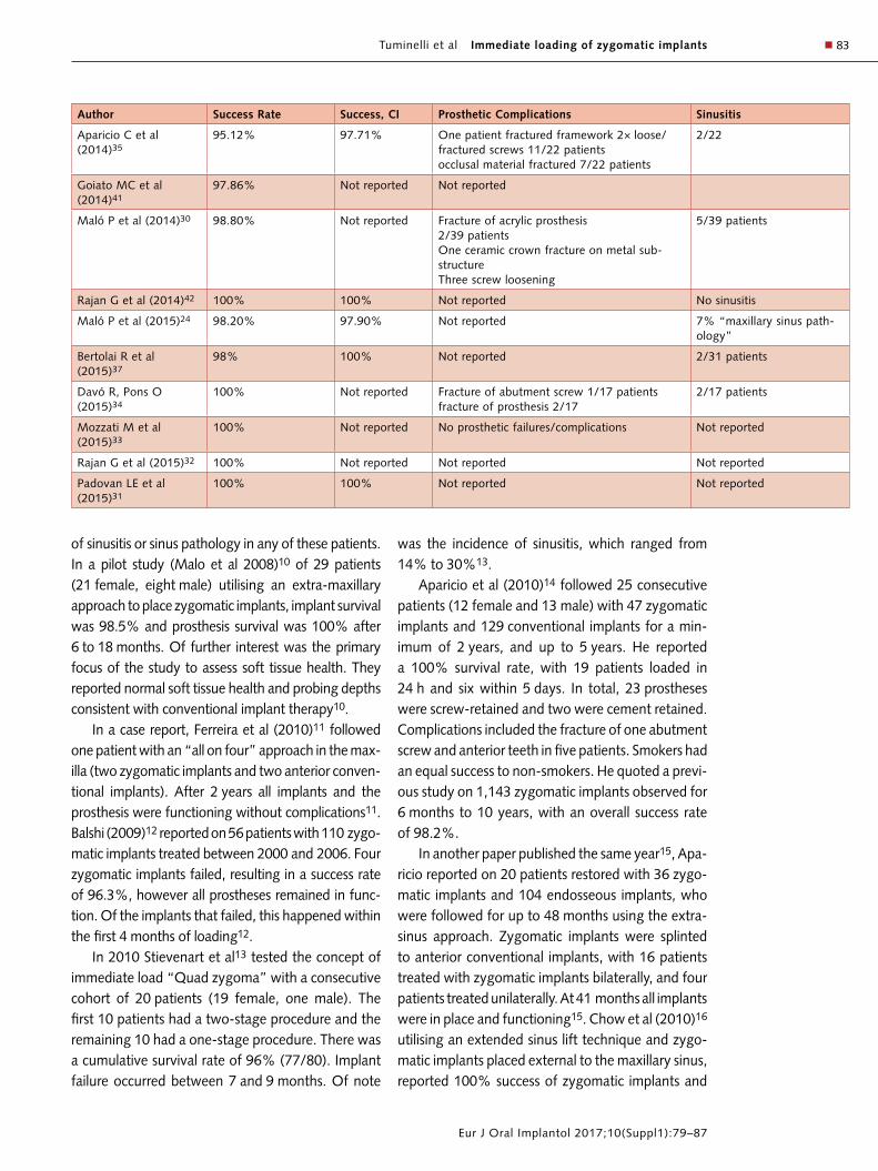
Tuminelli et al Immediate loading of zygomatic implants n 83
Eur J Oral Implantol 2017;10(Suppl1):79–87
Author Success Rate Success, CI Prosthetic Complications Sinusitis
Aparicio C et al (2014)35
95.12% 97.71% One patient fractured framework 2× loose/fractured screws 11/22 patients occlusal material fractured 7/22 patients
2/22
Goiato MC et al (2014)41
97.86% Not reported Not reported
Maló P et al (2014)30 98.80% Not reported Fracture of acrylic prosthesis 2/39 patients One ceramic crown fracture on metal sub-structure Three screw loosening
5/39 patients
Rajan G et al (2014)42 100% 100% Not reported No sinusitis
Maló P et al (2015)24 98.20% 97.90% Not reported 7% “maxillary sinus path-ology”
Bertolai R et al (2015)37
98% 100% Not reported 2/31 patients
Davó R, Pons O (2015)34
100% Not reported Fracture of abutment screw 1/17 patients fracture of prosthesis 2/17
2/17 patients
Mozzati M et al (2015)33
100% Not reported No prosthetic failures/complications Not reported
Rajan G et al (2015)32 100% Not reported Not reported Not reported
Padovan LE et al (2015)31
100% 100% Not reported Not reported
of sinusitis or sinus pathology in any of these patients. In a pilot study (Malo et al 2008)10 of 29 patients (21 female, eight male) utilising an extra-maxillary approach to place zygomatic implants, implant survival was 98.5% and prosthesis survival was 100% after 6 to 18 months. Of further interest was the primary focus of the study to assess soft tissue health. They reported normal soft tissue health and probing depths consistent with conventional implant therapy10.
In a case report, Ferreira et al (2010)11 followed one patient with an “all on four” approach in the max-illa (two zygomatic implants and two anterior conven-tional implants). After 2 years all implants and the prosthesis were functioning without complications11. Balshi (2009)12 reported on 56 patients with 110 zygo-matic implants treated between 2000 and 2006. Four zygomatic implants failed, resulting in a success rate of 96.3%, however all prostheses remained in func-tion. Of the implants that failed, this happened within the first 4 months of loading12.
In 2010 Stievenart et al13 tested the concept of immediate load “Quad zygoma” with a consecutive cohort of 20 patients (19 female, one male). The first 10 patients had a two-stage procedure and the remaining 10 had a one-stage procedure. There was a cumulative survival rate of 96% (77/80). Implant failure occurred between 7 and 9 months. Of note
was the incidence of sinusitis, which ranged from 14% to 30%13.
Aparicio et al (2010)14 followed 25 consecutive patients (12 female and 13 male) with 47 zygomatic implants and 129 conventional implants for a min-imum of 2 years, and up to 5 years. He reported a 100% survival rate, with 19 patients loaded in 24 h and six within 5 days. In total, 23 prostheses were screw-retained and two were cement retained. Complications included the fracture of one abutment screw and anterior teeth in five patients. Smokers had an equal success to non-smokers. He quoted a previ-ous study on 1,143 zygomatic implants observed for 6 months to 10 years, with an overall success rate of 98.2%.
In another paper published the same year15, Apa-ricio reported on 20 patients restored with 36 zygo-matic implants and 104 endosseous implants, who were followed for up to 48 months using the extra-sinus approach. Zygomatic implants were splinted to anterior conventional implants, with 16 patients treated with zygomatic implants bilaterally, and four patients treated unilaterally. At 41 months all implants were in place and functioning15. Chow et al (2010)16 utilising an extended sinus lift technique and zygo-matic implants placed external to the maxillary sinus, reported 100% success of zygomatic implants and

Tuminelli et al Immediate loading of zygomatic implants84 n
Eur J Oral Implantol 2017;10(Suppl1):79–87
prostheses, with no incidence of sinusitis. This was in 16 patients restored with 37 zygomatic implants followed for up to 24 months. He concluded that this approach could potentially reduce the incidence of sinusitis16. In a single case report with a 20-month follow-up, Kuabara found 100% success rate of his immediately loaded “Quad zygoma”, with no com-plications17.
In 2012, Sartori et al18 reported on patient satis-faction with immediate loaded prostheses on zygo-matic implants. Sixteen patients were followed from 2005 to 2009 and surveyed with a questionnaire. Half of the patients were completely satisfied with their prosthesis. The other 50% had complaints that fell into the categories of hygiene, aesthetics, phonetics and the ability to chew. All prostheses were in place. They reported fracture of a prosthetic screw, loosening of prosthetic and abutment screws and wear of the prosthetic teeth. Some of these patients’ concerns could be addressed or eliminated with the lateral approach to zygoma placement and the emergence of new materials for restora-tive options18. Using the Stella and extra-sinus tech-niques, Cor dero19 had a 100% success rate.
Balshi et al (2012)20 evaluated the bone to implant contact of 173 zygomatic implants in 77 patients, 62% of the patients were female. He reported that 35.9% with a variable of 11.7% of the implant had contact with bone. Males had an average of 16.5 mm and females 14.7 mm. Malo (2012)21 reported on a 3-year follow-up of 39 patients restored with a com-bination of zygomatic implants (92) and conventional implants (77), all immediately loaded. No implants were lost in the population that was followed, but about 10% of the patients were lost from the study. There were five cases of sinusitis but all patients reported sinus disease prior to surgery. There were no prosthetic failures noted21. Miglioranca (2012)22 immediately loaded zygomatic implants and with an 8-year follow up had a success rate of 97.5%. The conventional implants, on the other hand, had a suc-cess rate of 95.9% and the definitive prosthesis a 95.2% success. There was one prosthetic complica-tion of a metal bar fracture in one patient. There were no reports of screw loosening or fracture. All patients were free of sinus symptoms and disturbances22.
Chrcanovic and Bruno (2013)23 performed a literature search and reported on complications in
12 studies of zygomatic implants with immediate function. There were 70 cases of sinusitis, 15 cases of paresthesia, and 17 of oroantral fistulas. In addi-tion, there were 48 reports of soft tissue infection. Overall, the cumulative success rate of the zygo-matic implants was 96.7%23.
Malò (2013)24 reported on 352 completely eden-tulous patients who received 747 zygomatic implants in combination with 795 conventional implants, all immediately loaded. The surgical procedure was modified to have an extra maxillary approach. A total of four patients lost seven zygomatic implants, pro-ducing an overall success rate of 98.2%; 10 patients lost 17 conventional implants for a success rate of 96.7%. He also reported on 156 “mechanical com-plications”, of which one-third were in patients with a history of bruxing. Two out of the 17 prostheses fractured and one abutment screw fractured24. Davo25 presented data (2013) on 42 patients with a total of 81 zygomatic implants and 140 conventional implants followed for 5 years. The success rate for the zygomatic implant was 98.5% and the conven-tional implants 94.9% with all implants immediately loaded. All prostheses were in place25. In another study published the same year26, Davo looked at 3 years of prospective data of immediately loaded zygomatic implants. He had a 100% success rate of the zygomatic implants. In a questionnaire admin-istered regarding oral health related quality of life, Davo et al found that patients who had immediately loaded implants had an improved quality of life.
In a clinical case report, Farrett described using zygomatic implants in conjunction with conventional implants to support a fixed maxillary prosthesis27. After 8 years, he reported excellent results, with opti-mum tissue health. There was no mention of any prosthetic complications.
Aparacio et al (2014)28 described a zygomatic success code and established criteria/protocol for successful implementation of the immediate loaded zygomatic implant. The success code has four cri-teria that are used to determine if the final result is successful: zygomatic implant stability, sinus path-ology, soft tissue peri-implant tissue health, and prosthetic offset. Each is then graded 1 to 4, with specific criteria. He concluded that when compared with conventional grafting procedures, the zygo-matic approach had distinct advantages, reduced

Tuminelli et al Immediate loading of zygomatic implants n 85
Eur J Oral Implantol 2017;10(Suppl1):79–87
healing time, fewer surgical procedures, and expe-dited treatment time. One patient twice experienced a fractured framework. There were reports of screw loosening in 11 out of 22 patients and fracture of the occlusal material in seven out of 22 patients28.
Rajan (2014)29 followed two patients with gen-eralised periodontal disease and loss of all teeth that had full mouth rehabilitation with two zygomatic implants and four anterior implants immediately loaded and converted to final restorations after 6 months. All implants were in place and the most common complication was gingival inflammation, which was readily managed conservatively29.
Malo’s 5-year retrospective (2014)30 consisting of 39 patients, 92 zygomatic implants, and 77 con-ventional implants, had a 98.8% success rate for the zygomatic implants. He did not report on the success rate for conventional implants. Two of the 39 acrylic prostheses fractured, as well as one metal ceramic crown. He had a 100% success rate for the prostheses, concluding that immediately load-ing zygomatic implants alone or in conjunction with conventional implants was satisfactory30.
Padovan et al (2015)31 followed one patient for 55 months with an immediate loaded prosthesis with three zygomatic implants on one side, one zygo-matic on the contra-lateral side, and one anterior conventional implant – all splinted. In a case report by Rajan et al (2015)32 of a patient with an imme-diately loaded prosthesis supported by quad zygo-matic implants and followed for 3 years, a 100% success rate was reported for all implants. No pros-thetic complications were reported.
Mozzati (2015) reported on a new surgical pro-tocol for the insertion of zygomatic implants using an ultrasonic technique33. With 30 to 32 months of follow up, he had a 100% success rate for these implants and their associated prostheses. According to the authors this technique gives the surgeon better surgical visualisation in comparison to drilling proto-cols, better tissue management, and better healing.
In 2015, Davo et al, in a 5-year outcome of cross arch immediately loaded zygomatic implants using the quad zygoma approach in 14 patients (ori-ginal cohort of 17 patients), reported one abutment screw fracture and two prostheses fractures34. In the 14 patients there was 100% survival of the zygomatic implants and the prostheses. Half of the patients had
complications (screw loosening, abutment fracture, soft tissue inflammation etc) that were managed with routine post insertion care. The patients were asked about their overall satisfaction using the OHIP-14. The average score was 3.8, which is consistent with that of the overall population34.
All studies demonstrated excellent survival and success of the immediate loaded zygomatic implant. Complications were few, but were defined as cata-strophic when either an implant was lost or a pros-thesis was compromised or lost due to implant fail-ure. The most significant complication was implant failure and/or fracture.
n Discussion
The use of zygomatic implants for the edentulous maxilla has been well documented since Brånemark’s first report in 1988. The original protocol called for the placement of two zygomatic implants bilater-ally and two to four anterior endosseous implants splinted. This approach yielded a 94.9%25 to 100% success rate for endosseous implants and a 95.12%35 to 100% success rate for zygomatic implants. The prosthetic complications reported were screw-loos-ening, fracture of prosthetic and abutment screws, wear or loss of the prosthetic teeth and fracture of the prosthesis.
The original protocol had zygomatic implants traversing the maxillary sinus and engaging the palatal bone in the coronal aspect, providing there was sufficient volume and the zygoma in the apical aspect. The thought process was to achieve bi-cor-tical stabilisation. This, however, yielded prosthetic designs that had less than ideal access for hygiene purposes. One author reported that 50% of patients had concerns about speech, hygiene, phonetic and the ability to chew18. Malo et al reported that 44.3% of patients (156/352) had experienced mechanical complications, including prosthesis fracture, pros-thetic and abutment and prosthetic screw loosening/fracture24. Of note was that these were all in patients with a history of bruxing. There were also multiple reports of maxillary sinusitis5,9,13,21,24,26,30,34-37. Of note was a report of complications from 12 studies that included 70 cases of sinusitis, 15 cases of pares-thesia, and 17 oroantral fistulas23.

Tuminelli et al Immediate loading of zygomatic implants86 n
Eur J Oral Implantol 2017;10(Suppl1):79–87
With the evolution of the surgical technique and a large data pool of successful zygomatic implant placements, there was a shift in focus to move the implant to a more lateral and vertical position, negat-ing the need to traverse the maxillary sinus. One author has reported that this approach has zero inci-dences of sinusitis and 100% of implant/prosthetic success without complications16. The emergence of the zygoma was now closer to the residual alveolar ridge and in the “tooth alveolar” envelope.
Further surgical initiatives led to the use of four zygomatic implants, with two in each zygoma. The immediate load protocol was also employed in this approach. The reported success rate of the zygo-matic implants ranged from 96% to 100%34,35. The reported prosthetic complications were prosthetic and abutment screw loosening, fracture of abut-ment screws, fracture and wear of prosthetic tooth replacement and fracture of the prosthesis. The lat-eralised approach to the zygoma placement created a different soft tissue concern. The lack of attached tissue in the buccal aspect of the residual ridge, in specific clinical presentations resulted in mucosal irritation due to the movement of the tissue on the implant surface. To help resolve this problem, the buccal fat pad was used to wrap around the implant surface and ameliorate the issue38.
n Conclusions
Based on the present systematic review, the authors recommend immediately loading and splinting zygo-matic implants for the restoration of the severely atrophic maxilla with or without anterior conven-tional implants. The complication rates are relatively few, rarely catastrophic, and easily managed. Further randomised clinical trials should be conducted.
n References
1. Brånemark PI, Gröndahl K, Ohrnell LO, Nilsson P, Petru-son B, Svensson B, Engstrand P, Nannmark U. Zygoma fix-ture in the management of advanced atrophy of the maxilla: technique and long-term results. Scand J Plast Reconstr Surg Hand Surg 2004;38: 70–85.
2. Higuchi KW. The zygomaticus fixture: an alternative approach for implant anchorage in the posterior maxilla. Ann R Australas Coll Dent Surg 2000;15: 28–33.
3. Davo R, Malevez C, Rojas J. Immediate function in the atrophic maxilla using zygoma implants: a preliminary study. J Prosthet Dent 2007;97:S44–51.
4. Davo R, Malevez C, López-Orellana C, Pastor-Beviá F, Rojas J. Sinus reactions to immediately loaded zygoma implants: a clinical and radiological study. Eur J Oral Implan-tol 2008;1: 53–60.
5. Aparicio C, Ouazzani W, Garcia R, Arevalo X, Muela R, Fortes V. A prospective clinical study on titanium implants in the zygomatic arch for prosthetic rehabilitation of the atrophic edentulous maxilla with a follow-up of 6 months to 5 years. Clin Implant Dent Relat Res 2006;8: 114–122.
6. Bedrossian E, Rangert B, Stumpel L, Indresano T. Immediate function with the zygomatic implant: a graftless solution for the patient with mild to advanced atrophy of the maxilla. Int J Oral Maxillofac Implants 2006;21: 937–942.
7. Duarte LR, Filho HN, Francischone CE, Peredo LG, Bråne-mark PI. The establishment of a protocol for the total rehabilitation of atrophic maxillae employing four zygomatic fixtures in an immediate loading system–-a 30-month clin-ical and radiographic follow-up. Clin Implant Dent Relat Res 2007;9: 186–196.
8. Mozzati M, Monfrin SB, Pedretti G, Schierano G, Bassi F. Immediate loading of maxillary fixed prostheses retained by zygomatic and conventional implants: 24-month preliminary data for a series of clinical case reports. Int J Oral Maxillofac Implants 2008;23:308-314.
9. Davo R, Malevez C, Rojas J, Rodríguez J, Regolf J. Clinical outcome of 42 patients treated with 81 immediately loaded zygomatic implants: a 12- to 42-month retrospective study. Eur J Oral Implantol 2008;1: 141–150.
10. Malo P, Nobre Mde A, Lopes I. A new approach to re-habilitate the severely atrophic maxilla using extramaxillary anchored implants in immediate function: a pilot study. J Prosthet Dent 2008;100: 354–366.
11. Ferreira EJ, Kuabara MR, Gulinelli JL. “All-on-four” con-cept and immediate loading for simultaneous rehabilitation of the atrophic maxilla and mandible with conventional and zygomatic implants. Br J Oral Maxillofac Surg 2010;48: 218–220.
12. Balshi SF, Wolfinger GJ, Balshi TJ. A retrospective analysis of 110 zygomatic implants in a single-stage immediate loading protocol. Int J Oral Maxillofac Implants. 2009;24: 335–341.
13. Stiévenart M, Malevez C. Rehabilitation of totally atrophied maxilla by means of four zygomatic implants and fixed pros-thesis: a 6-40-month follow-up. Int J Oral Maxillofac Surg 2010;39: 358–363.
14. Aparicio C, Ouazzani W, Aparicio A, Fortes V, Muela R, Pascual A, Codesal M, Barluenga N, Franch M. Immediate/Early loading of zygomatic implants: clinical experiences after 2 to 5 years of follow-up. Clin Implant Dent Relat Res 2010;12:e77–82.
15. Aparicio C, Ouazzani W, Aparicio A, Fortes V, Muela R, Pascual A, Codesal M, Barluenga N, Manresa C, Franch M. Extrasinus zygomatic implants: three year experience from a new surgical approach for patients with pronounced buc-cal concavities in the edentulous maxilla. Clin Implant Dent Relat Res 2010;12: 55–61.
16. Chow J, Wat P, Hui E, Lee P, Li W. A new method to elimi-nate the risk of maxillary sinusitis with zygomatic implants. Int J Oral Maxillofac Implants 2010;25: 1233–1240.
17. Kuabara MR, Ferreira EJ, Gulinelli JL, Panzarini SR. Use of 4 immediately loaded zygomatic fixtures for retreatment of atrophic edentulous maxilla after complications of maxillary reconstruction. J Craniofac Surg 2010;21: 803–805.
18. Sartori EM, Padovan LE, de Mattias Sartori IA, Ribeiro PD Jr, Gomes de Souza Carvalho AC, Goiato MC. Evaluation of satisfaction of patients rehabilitated with zygomatic fixtures. J Oral Maxillofac Surg 2012;70: 314–319.

Tuminelli et al Immediate loading of zygomatic implants n 87
Eur J Oral Implantol 2017;10(Suppl1):79–87
19. Cordero EB, Benfatti CA, Bianchini MA, Bez LV, Stanley K, de Souza Magini R. The use of zygomatic implants for the rehabilitation of atrophic maxillas with 2 different tech-niques: Stella and Extrasinus. Oral Surg Oral Med Oral Pathol Oral Radiol Endod 2011;112:e49–53.
20. Balshi TJ, Wolfinger GJ, Shuscavage NJ, Balshi SF. Zygo-matic bone-to-implant contact in 77 patients with partially or completely edentulous maxillas. J Oral Maxillofac Surg 2012;70: 2065–2069.
21. Malo P, Nobre Md, Lopes A, Francischone C, Rigolizzo M. Three-year outcome of a retrospective cohort study on the rehabilitation of completely edentulous atrophic maxil-lae with immediately loaded extra-maxillary zygomatic implants. Eur J Oral Implantol 2012;5: 37–46.
22. Migliorança RM, Sotto-Maior BS, Senna PM, Francis-chone CE, Del Bel Cury AA. Immediate occlusal loading of extrasinus zygomatic implants: a prospective cohort study with a follow-up period of 8 years. Int J Oral Maxillofac Surg 2012;41: 1072–1076.
23. Chrcanovic BR, Albrektsson T, Wennerberg A. Survival and Complications of Zygomatic Implants: An Updated System-atic Review. J Oral Maxillofac Surg 2016;74: 1949–1964.
24. Malo P, de Araújo Nobre M, Lopes A, Ferro A, Moss S. Extramaxillary surgical technique: clinical outcome of 352 patients rehabilitated with 747 zygomatic implants with a follow-up between 6 months and 7 years. Clin Implant Dent Relat Res 2015;17:e153–162.
25. Davo R, Malevez C, Pons O. Immediately loaded zygomatic implants: a 5-year prospective study. Eur J Oral Implantol. 2013;6: 39–47.
26. Davo R, Pons O. Prostheses supported by four immediately loaded zygomatic implants: a 3-year prospective study. Eur J Oral Implantol 2013;6: 263–269.
27. Farret MM, Farret MM, Carlesso J, Carlesso O. Orthodontic treatment and implant-prosthetic rehabilitation of a partially edentulous patient. J Prosthodont 2013;22: 587–590.
28. Aparicio C, Manresa C, Francisco K, Claros P, Alández J, González-Martín O, Albrektsson T. Zygomatic implants: indications, techniques and outcomes, and the zygomatic success code. Periodontol 2000 2014;66: 41–58.
29. Rajan G, Natarajarathinam G, Kumar S, Parthasarathy H. Full mouth rehabilitation with zygomatic implants in patients with generalized aggressive periodontitis: 2 year follow-up of two cases. J Indian Soc Periodontol 2014;18: 107–111.
30. Malo P, Nobre Mde A, Lopes A, Ferro A, Moss S. Five-year outcome of a retrospective cohort study on the rehabilita-tion of completely edentulous atrophic maxillae with imme-diately loaded zygomatic implants placed extra-maxillary. Eur J Oral Implantol 2014;7: 267–281.
31. Padovan LE, Ribeiro-Júnior PD, de Mattias Sartori IA, Thomé G, Sartori EM, Uhlendorf J. Multiple zygomatic implants as an alternative for rehabilitation of the extremely atrophic maxilla: a case letter with 55 months of follow-up. J Oral Implantol. 2015;41: 97–100.
32. Rajan G, Mariappan S, Ramasubramanian H, Somasunda-ram S, Natarajarathinam G. Restoration of Atrophic Eden-tulous Maxilla of a Patient with Ectodermal Dysplasia Using Quadruple Zygomatic Implants: A Case Report. J Maxillofac Oral Surg. 2015;14: 848–852.
33. Mozzati M, Mortellaro C, Arata V, Gallesio G, Previgliano V. Rehabilitation with 4 zygomatic implants with a new sur-gical protocol using ultrasonic technique. J Craniofac Surg 2015;26: 722–728.
34. Davó R, Pons O. 5-year outcome of cross-arch prostheses supported by four immediately loaded zygomatic implants: A prospective case series. Eur J Oral Implantol 2015;8: 169–174.
35. Aparicio C, Manresa C, Francisco K, Ouazzani W, Claros P, Potau JM, Aparicio A. The long-term use of zygomatic implants: a 10-year clinical and radiographic report. Clin Im-plant Dent Relat Res 2014;16: 447–459.
36. Bedrossian E. Rehabilitation of the edentulous maxilla with the zygoma concept: a 7-year prospective study. Int J Oral Maxillofac Implants 2010;25: 1213–1221.
37. Bertolai R, Aversa A, Catelani C, Rossi A, Giannini D. Treat-ment of extreme maxillary atrophy with Zygoma implants. Minerva Stomatol. 2015;64: 253–264.
38. de Moraes EJ. The buccal fat pad flap: an option to pre-vent and treat complications regarding complex zygomatic implant surgery. Preliminary report. Int J Oral Maxillofac Implants 2012;27: 905–910.
39. Davo R, Pons O, Rojas J, Carpio E. Immediate function of four zygomatic implants: a 1-year report of a prospective study. Eur J Oral Implantol 2010;3: 323–334.
40. Migliorança RM, Coppedê A, Dias Rezende RC, de Mayo T. Restoration of the edentulous maxilla using extra-sinus zygomatic implants combined with anterior convention-al implants: a retrospective study. Int J Oral Maxillofac Implants 2011;26: 665–672.
41. Goiato MC, Pellizzer EP, Moreno A, Gennari-Filho H, dos Santos DM, Santiago JF Jr, dos Santos EG. Implants in the zygomatic bone for maxillary prosthetic rehabilitation: a systematic review. Int J Oral Maxillofac Surg 2014;43: 748–757.
42. Rajan G, Natarajarathinam G, Kumar S, Parthasarathy H. Full mouth rehabilitation with zygomatic implants in patients with generalized aggressive periodontitis: 2 year follow-up of two cases. J Indian Soc Periodontol 2014;18: 107–111.


n 89
Eur J Oral Implantol 2017;10(Suppl1):89–99
REVIEW
Frank SpitznagelDMD, Dr med dentAssistant Professor, Depart-ment of Prosthodontics, School of Dentistry, Heinrich-Heine-University, Düsseldorf, Germany
Sebastian HorvathDMD, Dr med dentAdjunct Assistant Professor, Department of Preventive and Restorative Sciences, University of Pennsylvania, Philadelphia, PA, USADentist in Private Practice, Jestetten, Germany
Both authors contributed equally to the manuscript
Petra GierthmuehlenDDS, PhD, Prof Dr med dentProfessor and Chair, Depart-ment of Prosthodontics, School of Dentistry, Heinrich-Heine-University, Düsseldorf, Germany
Correspondence to:Dr Frank A. Spitznagel, DMDDepartment of Prosthodon-tics, School of Dentistry, Heinrich-Heine-University Moorenstraße 5, 40225 Düsseldorf, GermanyTel: +49 211-81-04440Fax: +49 211-81-16280Email: [email protected]
Frank A. Spitznagel, Sebastian D. Horvath, Petra C. Gierthmuehlen
Prosthetic protocols in implant-based oral rehabilitations: A systematic review on the clinical outcome of monolithic all-ceramic single- and multi-unit prostheses
Key words dental implants, dental restoration failure, humans, implant-supported, single tooth, survival rate
Aim: The purpose of this systematic review was to assess the clinical performance of implant- supported monolithic all-ceramic single- and multi-unit restorations.Materials and methods: The electronic databases of MEDLINE via PubMed, the Cochrane Library (CENTRAL) and EMBASE were searched for clinical studies on monolithic all-ceramic single and multi-unit implant-supported fixed dental prostheses. Human studies with a mean follow-up of at least 2 years and published in English or German language peer-reviewed journals up until August 2016 were included. Two independent examiners conducted the literature search and review process.Results: The search resulted in 2510 titles and of these, 57 studies were selected for full-text evalu-ation. Three studies were included on the basis of the pre-determined criteria. Two articles reported on monolithic lithium disilicate implant-supported single crowns (SC) and revealed a survival rate of 97,8 and 100% after 3 years. One study investigated implant- supported monolithic zirconia SCs and fixed partial dentures (FPD) and showed a survival rate of 100% after 5 years. No studies could be identified on the clinical performance of monolithic resin matrix ceramic restorations. Clinical stud-ies are lacking on the long-term outcome of implant-supported monolithic all-ceramic single- and multi-unit restorations.Conclusions: Preliminary clinical data indicate high short-term survival for implant-supported mono-lithic lithium disilicate and zirconia single- and multi-unit restorations. Randomised clinical studies and observations with a longer duration are necessary to validate the broad application of this therapy.
Conflict-of-interest statement: The authors declare no conflict of interest.
n Introduction
Therapeutic concepts for the prosthetic rehabilita-tion of various types of edentulism have changed significantly over past decades due to the high sur-vival of dental implants reported in the literature. Implant-supported single crowns and fixed dental prostheses are recognised as a reliable treatment option for partial edentulism, with an implant sur-vival rate well above 90%1,2.
Clinicians face challenges with the choice of materials available today for implant prosthodon-tics. The survival rates of implant-supported metal-ceramic single crowns and FPDs are high; 96.3% for single crowns and 95.4% for FPDs after 5 years are reported1,2. However, technical problems, such as fractures of the veneering material, abutment or screw loosening and loss of retention of cemented res-torations, are described as major limitations for bilayer gold acrylic and porcelain veneered metal-based

Spitznagel et al Prosthetic protocols in implant-based oral rehabilitations90 n
Eur J Oral Implantol 2017;10(Suppl1):89–99
Neuss, Germany), glass ceramic in a resin interpen-etrating matrix (Vita Enamic, Vita Zahnfabrik), and zirconia-silica ceramic in a resin interpenetrating matrix (e.g. Shofu Block HC, Shofu, Kyoto, Japan)3.
Polycrystalline ceramics, such as alumina oxide ceramics (e.g. Procera Alumina, Nobel Biocare, Kloten, Switzerland), were first introduced in the mid-1990s. They were commonly applied for im-plant restorations, but became less important with the increased use of zirconia and lithium disilicate restorations3.
In the early 1990s yttrium oxide partially-sta-bilised tetragonal zirconia polycrystal (Y-TZP) was introduced to dentistry as a core material for all-ceramic restorations. Due to a transformation, toughening mechanism Y-TZP exhibits superior mechanical properties compared with other all-ceramic systems3. Zirconia ceramics have been used in dentistry as copings and frameworks for bilayered restorations with porcelain veneers, for implants, implant abutments, posts and cores, as well as for orthodontic brackets.
The introduction of computer-aided design and computer-aided manufacturing of all-ceramic res-torations provided new approaches for addressing restorative challenges in implant dentistry.
The high reliability of zirconia as abutment, as well as framework material for implant-borne crowns and fixed dental prostheses4, was confirmed in sev-eral clinical studies5,6. However, the clinical success of zirconia-based implant-supported restorations is limited by veneering porcelain fractures (chipping), exhibiting the most common technical complica-tion7-9. Attempts were made to reduce the incidence of chip fractures with zirconia-based restorations. Anatomical core design for adequate support for the veneering ceramic and slow cooling firing protocols for the veneer application were proposed in the den-tal literature10. However, it is well known that higher functional impact forces, impaired feedback from periodontal neural receptors, and rigidity of osseoin-tegrated implants put implant supported restorations at higher risk for porcelain fracture11.
To overcome the limitations of bilayer systems with a weak veneering layer, several systems such as resin matrix ceramics12, lithium disilicate13 and zir-conia ceramics14 are increasingly used in monolithic application. The advantages of monolithic vs bilayer
restorations. Moreover, poor gingival aesthetics has been reported with these metal-based restorations over short- and long-term observations1,2.
Thus, alternative prosthetic solutions evolved. Several all-ceramic systems were developed over past decades to meet increased clinician and patient demand for metal-free restorations3.
In the early 1990s the lost wax press technique was introduced to the dental market as an innova-tive processing method for all-ceramic restorations. A pressable leucite-reinforced glass-ceramic evolved (IPS Empress, Ivoclar Vivadent, Schaan, Liechtenstein) and further enhancements of this system led to the introduction of a lithium disilicate glass-ceramic sys-tem (IPS Empress II, Ivoclar Vivadent), which started in 1998, with a significantly increased strength. A consecutive pressable lithium disilicate glass-ceramic (IPS e.max Press, Ivoclar Vivadent) with improved physical properties and translucency through differ-ent firing processes was then launched, followed by a CAD/CAM version of this lithium disilicate glass-ceramic (IPS e.max CAD, Ivoclar Vivadent).
In 2013, IPS e.max CAD blocks for the chairside fabrication of implant crowns with pre-fabricated screw access holes and insertion grooves for the cor-responding titanium base were introduced. Hence, hybrid implant abutments, as well as full-contour hybrid implant abutment crowns, which are adhe-sively bonded to a titanium base (Ti Base, Dentsply Sirona, York, USA), are now available.
As the market share of lithium disilicate ceram-ics increased enormously over recent years, several manufacturers developed novel glass ceramic sys-tems. The zirconia-reinforced lithium silicate material (VITA SUPRINITY, Vita Zahnfabrik, Bad Säckingen, Germany; CELTRA, CELTRA DUO, Dentsply Sirona, York, USA), which was launched in 2013, is one example.
In addition, a novel material class – resin-matrix-ceramics – has been introduced for the CAD/CAM fabrication of fixed restorations. These resin matrix ceramics are composed of inorganic glasses, porce-lains or glass-ceramics that are clustered and embed-ded in a cross-linked resin matrix3. They reveal a modulus that simulates the modulus of dentine and are easier to CAD/CAM mill and to adjust. According to their inorganic composition they can be divided into resin nano ceramics (Lava Ultimate, 3M ESPE,
Spitznagel et al Prosthetic protocols in implant-based oral rehabilitations n 91
Eur J Oral Implantol 2017;10(Suppl1):89–99
restorations are well described in the dental litera-ture13. In vitro data evaluating the potential of mon-olithic resin matrix ceramic15, lithium disilicate16-18
and zirconia19 systems for the fabrication of implant-supported restorations are promising. Various short- and mid-term clinical reports on monolithic and min-imally veneered zirconia implant supported full-arch restorations have shown a favourable performance by these full-contour restorations20. However, the clinical performance of monolithic all-ceramic sys-tems for implant-supported single- and multi-unit restorations is currently not well described in the dental literature.
Therefore, it was the aim of this systematic review to analyse the clinical outcome of implant-supported monolithic all-ceramic single- and multi-unit restor-ations.
n Materials and methods
n Search strategy
The following databases for articles published until August 22nd, 2016, in the dental literature were searched: MEDLINE via PubMed, the Cochrane Central Register of Controlled Trials (CENTRAL) and EMBASE. Furthermore, an additional manual search was carried out for reference lists of all full-text pub-lications, as well as for selected recently published reviews relating to this topic (see “list of reviews”). Moreover, the websites of clinicaltrials.gov, the World Health Organization (WHO) and the Ger-man Register for Clinical Trials (DRKS – Deutsches Register Klinischer Studien) were checked.
The search was conducted according to Cochrane guidelines for systematic reviews. PICOS question were defined as follows:• P (population) compromised patients who
received one or more dental implants (titanium or ceramic);
• I (intervention) included monolithic single crowns (SC: cemented or screw-retained) or short im-plant supported fixed-dental prosthesis (FPD, 3-5 units);
• C (comparison) was not applicable in this review;• O (outcome and study design) was survival or
success rate;
• S (study type) compromised randomised con-trolled trials (RCT), clinical follow-up studies (prospective and retrospective studies) and case series.
Search terms:
In each database the following search combinations and terms were applied:• Population AND Intervention AND (Outcome
OR Study type)• Intervention AND (Outcome OR Study type) • Population: dental implant OR oral implant OR
bone screw* OR endosseous implant • Intervention: dental restoration OR dental crown
OR dental bridge OR cantilever OR restoration OR FPD OR fixed prosthesis; (dental prosthesis AND implant supported) OR (restoration AND implant supported); CAD CAM OR digital OR CEREC OR computer aided) OR (monolithic OR full contour)
• Outcome and study type: clinical evaluation OR RCT OR clinical performance OR failure OR clin-ical study OR clinical trial OR follow up study OR survival OR longevity OR success OR survival rate
The search strategy is displayed in Figure 1.
n Inclusion criteria:
As there were no randomised controlled clinical tri-als, this systematic review collected the data from prospective and retrospective cohort studies and case series. Inclusion and exclusion criteria were defined as followed: • Human trials• Language restriction to English and German • Peer-reviewed dental journals • Studies with a mean follow-up time of 2 years or
more in function • Case series with 10 or more patients
n Exclusion criteria:
• In vitro studies• Poster abstracts, interviews or protocols • Studies reporting on interfering systemic or local
factors

Spitznagel et al Prosthetic protocols in implant-based oral rehabilitations92 n
Eur J Oral Implantol 2017;10(Suppl1):89–99
• Studies with the same sample (most complete/most recent was considered)
• Studies not reporting in detail on the prostho-dontic components
• Studies not meeting the inclusion criteria
n Selection of studies:
Two authors (FS, SH) independently screened the titles from this extensive search, based on the inclu-sion and exclusion criteria. Any disagreement was dissolved by discussion with a third author (PG). Afterwards, abstracts of all relevant titles were cap-tured and examined for relevant studies. Based on the selection of abstracts, articles were then obtained for full-text analysis.
Full-text analysis was again performed inde-pendently by two readers (FS, SH) by screening “Material and methods”, “Results” and “Discus-sion”, and then double-checked. Any disagree-ment was solved by discussion within the group of authors.
Data extraction
From the included studies the following informa-tion was extracted: study, year of publication, study design, setting, type of restoration (SC, FPD), implant system, implant material, retention system, recon-struction material, number of restorations, number of failures, follow-up range and mean follow-up and survival, as well as the success rate of prosthodon-tic treatment. Furthermore, if any included study reported insufficient data in the article, authors or co-authors were contacted.
Statistical analysis
Due to the limited number of included studies and the variability in the reporting, a statistical analysis or meta-analysis was not performed.
n Results
n Study characteristics
The electronic search yielded a total of 2510 titles from all databases. After elimination of duplicates, two reviewers assessed the titles and agreed on 135 abstracts for further analysis. Abstract evalu-ation and consideration of relevant reviews (see “List of reviews”) resulted in 46 studies for full-text analysis. Manual searching provided 11 more studies. Altogether, 57 full-texts were obtained and after exclusion of 54 studies, a final number of three publications21-23 met the inclusion criteria for data extraction.
The websites of clinicaltrials.gov, WHO and the DRKS provided five more relevant studies – however, none of the studies is completed. They were, there-fore, not included in this systematic review.
n Exclusion of studies
The reasons for excluding studies (n = 54, see ref-erence list “List of excluded full-text articles and the reason for exclusion”) after the full text was obtained were: use of layered restorations (40), no implant restorations (4), no detailed information on prosthetics (8), no distinction between monolithic
Fig 1 The search strategy.
Joined titles n = 2510 titles
PubMedn = 2069 titles
EMBASEn = 121 titles
CENTRALn = 320 titles
Elimination of duplicates and independently selected by two reviewers agreed on: n = 135 titles for abstract analysis
Final number of included studies: n = 3
Abstract analysis and relevant review analysis: n = 46 full-texts obtained
Excluded: n = 54 articles
Handsearch: n = 11 articles
Systematic reviews

Spitznagel et al Prosthetic protocols in implant-based oral rehabilitations n 93
Eur J Oral Implantol 2017;10(Suppl1):89–99
and layered restorations or different type of mater-ials (1) and a too small number of restorations (1). In one study, some restorations were either facially veneered with a feldspathic porcelain, or pink feld-spathic porcelain was used in the gingival areas. All three authors discussed this, and it was agreed that since all functional areas were in monolithic zirconia, the study could be included21.
n Included studies
Finally, three studies met the inclusion criteria for the present analysis (Table 1). The studies were published between 2014 and 2016. One study revealed a pro-spective study design and was conducted in a univer-sity environment22. One study was retrospective and the patients were treated both at a university and in private practices23. The third study was a consecutive case series, set in a private practice21.
The studies reported on different available implant systems: Titanium implants (Astra Tech Implant Sys-tem, Dentsply Implants, Mannheim, Germany; Strau-mann, Freiburg, Germany; Nobel Biocare23; Zimmer Biomet, Warsaw, USA)20,21 and zirconia implants (Ziraldent, Metoxit AG, Thayngen, Switzerland)22.
The implant-supported restorations were both single crowns (SC)22,23 and fixed dental prostheses (FPD)21. Connection to the implants was achieved either by using adhesive cement retention22, screw retention or a combination of screw and cement retention21,23. The material of the reconstructions was lithium disilicate (IPS e.max CAD22 or IPS e.max Press23,Ivoclar Vivadent) or zirconia ceramic21 (Pret-tau, Zirkonzahn, Gais, Italy). The follow-up ranges of the studies are given in Table 1. No studies could be identified on resin matrix ceramics.
n Prosthetic survival (SC, FPD)
The three studies included a total number of 258 re-storative units. Of these, one crown restoration failed23 and one crown restoration experienced a technical complication22.
Lithium Disilicate:
Fabbri and colleagues recorded a failed lithium dis-ilicate crown in the position of a maxillary canine Ta
ble
1 I
nfor
mat
ion
on S
Cs
and
FPD
s of
incl
uded
stu
dies
, (N
R: n
ot r
epor
ted)
. St
udy
Year
Stud
y de
sign
Sett
ing
Res
tora
tion
(S
C,
FPD
)Im
plan
t
syst
emIm
plan
t
mat
eria
lR
eten
tion
sy
stem
Res
tora
tion
m
ater
ial
Num
ber
of
rest
orat
ions
Num
ber
of
fail
ures
Foll
ow-
up r
ange
(m
onth
s)
Mea
n fo
llow
-up
(mon
ths)
Surv
ival
ra
teSu
cces
s ra
te
Spie
s et
al
2016
Pros
pec-
tive
Uni
ver-
sity
SCZi
rald
ent
(Met
oxit)
Zirc
onia
Cem
ent-
re
tain
edLi
thiu
m
Dis
ilica
te
(IPS
e.m
ax
CA
D)
240
25 t
o 34
31 ±
2,7
100%
95,7
0%
Mos
covi
tch
2015
Cas
e se
ries
Priv
ate
prac
tice
SC,
FPD
Ast
ra T
ech
Impl
ant
Syst
em
(Den
tspl
y),
Nob
el B
ioca
re,
Stra
uman
n,Zi
mm
er B
iom
et
Tita
nium
Scre
w-
reta
ined
and
co
mbi
na-
tion
scre
w/
cem
ent-
re
tain
ed
Zirc
onia
(P
rett
au)
189
02
to 6
8N
R10
0%10
0%
Fabb
ri et
al
2014
Ret
ro-
spec
tive
Uni
ver-
sity
and
Pr
ivat
e pr
actic
e
SCN
obel
Bio
care
Tita
nium
Scre
w-
reta
ined
, ce
men
t re
tain
ed a
nd
com
bina
-tio
n sc
rew
/ ce
men
t-
reta
ined
Lith
ium
-D
isili
cate
, (I
PS e
.max
Pr
ess)
451
12 t
o 61
28,3
97,7
8%97
,78%

Spitznagel et al Prosthetic protocols in implant-based oral rehabilitations94 n
Eur J Oral Implantol 2017;10(Suppl1):89–99
that revealed a minor cohesive fracture and therefore reported a survival rate of lithium disilicate crowns adhesively bonded to titanium or zirconia frame-works of 97,78% after 28 months23. However, the chipping did not impair function, the area was smoothed and the restoration could be left in situ23. As no implant-supported crown had to be replaced, Spies et al reported a survival rate of 100% after a mean observation period of 31 months22.
Zirconia
One cemented implant-supported monolithic zirco-nia single-crown had to be remade due to a fracture of the zirconia abutment21 and was replaced with a screw-retained all-ceramic crown. As this was not a failure of the restorative material, the survival rate of both implant supported single crowns and fixed partial dentures was rated with 100%21.
n Prosthetic success and technical complications
None of the studies observed any loss of retention or screw loosening of implant-supported restorations.
Lithium Disilicate
One prosthetic complication occurred in the study by Spies and colleagues on a maxillary first molar crown. The crown showed a major occlusal roughness and thus the success rate was reduced to 95.7% after 31 months. As this roughness could be polished, it was considered as clinically acceptable22.
The success rate for lithium disilicate crowns bonded to titanium or zirconia was 97,78% after a mean observation period of 28.3 months23.
Zirconia
No prosthetic complications were reported for mon-olithic zirconia restorations on implants, leading to a success rate of 100%21.
n Aesthetic outcomes
Two studies reported on aesthetic outcomes of their prosthodontic treatment. Outcome was either
measured visually by patients (VSA)22 or by both patients (satisfaction score) and clinicians (modified CDA criteria)23.
Spies et al asked their patients before and after final prosthodontic treatment and at follow-ups to evaluate aesthetics and appearance, function (eat-ing), sense (“feeling like natural teeth”), speech and self-esteem. The authors realised this by a Visual Analogue Scale (VSA) from 0 to 100%22. All questioned events improved after treatment and remained stable over time. Aesthetics increased from a treatment start of 64.1% up to 87.4 to 90.7% after therapy. Lithium disilicate crowns were fur-ther scored with modified USPHS criteria. Ceramic fracture, marginal discolouration and integrity were stable over the given follow-up period and there-fore assessed with “Alpha”, whereas occlusal rough-ness, contour and aesthetics were mostly evaluated with “Bravo” classification at the 3-year evaluation. However, “Bravo” was defined as clinically accept-able with minor deviations. None of the restorations showed a “Charlie” or “Delta” classification at any time during the study.
Patients in the study by Fabbri et al23 could rate their self-satisfaction with nominal scores of “non-acceptable”, “acceptable”, “good” and “excel-lent”. All restorations were rated either “good” or “excellent” by patients. The modified CDA (Cali-fornia Dental Association) criteria for Colour match, porcelain surface and marginal discolouration and integrity were also rated mostly with an A by clin-icians at the 3-year follow-up. Moscovitch21 pro-vided no information on these parameters.
n Discussion
This systematic review focused on the outcomes of clinical studies reporting on implant-supported monolithic all-ceramic single- and multi-unit restor-ations. The number of published trials is limited due to the short time that monolithic restorations have been used in implant-supported restorations. Most of the published studies reported on small samples sizes or did not provide adequate information on the study details.
There is a general consensus in the dental lit-erature that monolithic restorations show the lowest
Spitznagel et al Prosthetic protocols in implant-based oral rehabilitations n 95
Eur J Oral Implantol 2017;10(Suppl1):89–99
number of mechanical complications. Monolithic re-storative systems reveal no dissimilar interfaces, cre-ate a greater bulk or material that leads to improved structural properties of the material. Thus, the risk of fracture and/or chipping events is significantly reduced13. The combination of monolithic design and manufacture with CAD/CAM technology ena-bles efficient handling and care delivery. Therefore, implant prosthodontics benefit from the CAD/CAM technology for the fabrication of full-contour res-torations24. Hence, the combination of monolithic materials connected to abutment substructures may represent a preferable treatment option, especially in the posterior region.
No valid clinical data could be identified on resin matrix ceramic implant-supported restorations. One proof-of-concept case series25 showed that a fully digital workflow for the fabrication of implant sup-ported crowns from a monolithic resin matrix ceramic (Lava Ultimate) is feasible. A reduction of the labora-tory and treatment time resulted in a reasonable cost-benefit ratio and a high quality and precision of the restorations25. However, the investigated resin matrix ceramic material has to be considered experimental, as no large-scale clinical investigations with long-term follow-up observations are currently available.
The combination of lithium disilicate restorations with zirconia substructures has been described as a reliable option to combine mechanical effectiveness with good aesthetics and promising long-term clin-ical outcomes for implant-supported prostheses5,9.
The survival rate of cemented CAD/CAM fabri-cated monolithic lithium disilicate implant crowns was 100%22. No fractures or chippings were described. Debonding or any other technical complications were not noted in the given observation period after 3 years. Only one crown revealed a major occlusal roughness, resulting in a Kaplan Meier success rate of 95.7% after 31 months.
Good results in terms of aesthetics, function and loss of retention were observed for the combination of implant-supported lithium disilicate restorations with zirconia frameworks23 or zirconia implants9.
The survival rate of monolithic implant-supported press fabricated lithium disilicate single crown restor-ations was 97,78% after a mean observation time of 28.3 months. Only one crown revealed a chip fracture23.
CAD/CAM lithium disilicate implant crowns can also be fabricated chairside in 1 to 2 h, which leads to a significant reduction in the fabrication time26. Hence a time- and cost-effective chairside workflow to produce reliable all-ceramic implant crowns has been established. However, no clinical studies on these hybrid abutment crowns have yet been pub-lished.
Several clinical studies have shown that mono-lithic or minimally veneered (no feldspathic veneer in function) zirconia would be a viable treatment option for implant-supported full-arch restor-ations20. However the evidence on monolithic zirco-nia implant-supported single and multi-unit restor-ations is presently low. In the study by Moscovitch21 all monolithic zirconia restorations exhibited a 100% survival rate at 68 months. No fractures, cracks or chipping within the monolithic zirconia material were observed. Further complications relating to phonet-ics, masticatory function or screw loosening were not detailed in the identified study on monolithic zirconia outcomes.
This study indicated that there is a new para-digm shift in fixed implant prosthodontics that allows for the use of monolithic high-strength ceramics to enhance the overall aesthetics, biocompatibility, per-formance, efficiency and cost benefits.
As reported by several in vitro and clinical stud-ies, zirconia induces minimal wear to opposing structures, and this property is maximised, when the occlusal surfaces are polished after definitive intraoral occlusal adjustments14,27. Recently, more translucent zirconia materials were introduced to the dental market, with the aim of a broader application in anterior and premolar areas. While this improve-ment of the material is positive regarding the aes-thetic result, it also leads to a weakening of the ma-terial. Hence its application is limited to small fixed dental prostheses.
Given that clinical reports are ranked low in the hierarchy of evidence-based research, the reported high success of monolithic lithium disilicate and zir-conia restorations should be considered with cau-tious optimism.
This systematic review aimed, for the first time, to describe the short- and mid-term evidence regarding fixed dental monolithic prostheses in the rehabilita-tion of partially edentulous patients. The absence
Spitznagel et al Prosthetic protocols in implant-based oral rehabilitations96 n
Eur J Oral Implantol 2017;10(Suppl1):89–99
of long-term clinical studies and related strong evi-dence supporting this treatment are the major limi-tations of this systematic review. Due to the limited number of published trials and the considerable het-erogeneity among the included studies in terms of prosthodontics protocols, a meta-analysis was not feasible. The included studies that reveal a lower evi-dence level are subject to a certain risk of reporting bias, publication bias and attrition bias. Hence, clin-icians should carefully consider the limitations of the included evidence when making decisions regarding this treatment.
In conclusion, this systematic review of the cur-rent literature evidenced high prostheses survival of implant-supported monolithic lithium disilicate and zirconia single- and multi-unit restorations in the short-term. Only a few mechanical complications, such as surface roughness and minor fractures, were described for lithium disilicate restorations. Given the level of evidence and the duration of the studies included, the use of monolithic lithium disilicate and zirconia prostheses for single and multi- unit implant supported prostheses requires additional compre-hensive longer-term investigation.
n Conclusions
According to the results of this review and within its limitations, the use of monolithic lithium disilicate and zirconia for implant-supported single crowns and fixed prosthodontics was effective and reliable in short-term studies.
The choice of this monolithic concept may repre-sent a valid treatment for implant-supported single and multi-unit restorations, offering biological, tech-nical and aesthetic advantages.
Further in vivo investigations are necessary to validate the clinical reliability of monolithic implant-supported restorations in the long-term, confirm-ing the effectiveness of the proposed prosthetic approach.
n References
1. Jung RE, Zembic A, Pjetursson BE, Zwahlen M, Thoma DS. Systematic review of the survival rate and the incidence of bio-logical, technical, and aesthetic complications of single crowns on implants reported in longitudinal studies with a mean follow-up of 5 years. Clin Oral Implants Res 2012;23: 2–21.
2. Pjetursson BE, Thoma D, Jung R, Zwahlen M, Zembic A. A systematic review of the survival and complication rates of implant-supported fixed dental prostheses (FDPs) after a mean observation period of at least 5 years. Clin Oral Implants Res 2012;23: 22–38.
3. Gracis S, Thompson VP, Ferencz JL, Silva NR, Bonfante EA. A new classification system for all-ceramic and ceramic-like restorative materials. Int J Prosthodont 2015;28: 227–235.
4. Sailer I, Philipp A, Zembic A, Pjetursson BE, Hämmerle CH, Zwahlen M. A systematic review of the performance of cer-amic and metal implant abutments supporting fixed implant reconstructions. Clin Oral Implants Res 2009;20: 4–31.
5. Pozzi A, Sannino G, Barlattani A. Minimally invasive treat-ment of the atrophic posterior maxilla: a proof-of-concept prospective study with a follow-up of between 36 and 54 months. J Prosthet Dent 2012;108: 286–297.
6. Lops D, Bressan E, Parpaiola A, Sbricoli L, Cecchinato D, Romeo E. Soft tissues stability of cad-cam and stock abut-ments in anterior regions: 2-year prospective multicentric cohort study. Clin Oral Implants Res 2015;26: 1436–1442.
7. Nothdurft FP, Pospiech PR. Zirconium dioxide implant abut-ments for posterior single-tooth replacement: first results. J Periodontol. 2009;80: 2065–2072.
8. Spies BC, Witkowski S, Butz F, Vach K, Kohal RJ. Bi-layered zirconia/fluor-apatite bridges supported by ceramic dental implants: a prospective case series after thirty months of observation. Clin Oral Implants Res 2016; 27: 1265–1273.
9. Spies BC, Kohal RJ, Balmer M, Vach K, Jung RE. Evaluation of zirconia-based posterior single crowns supported by zirconia implants: preliminary results of a prospective multi-center study. Clin Oral Implants Res 2017;28: 613–619.
10. Guess PC, Bonfante EA, Silva NR, Coelho PG, Thompson VP. Effect of core design and veneering technique on dam-age and reliability of Y-TZP-supported crowns. Dent Mater 2013;29: 307–316.
11. Kinsel RP, Lin D. Retrospective analysis of porcelain failures of metal ceramic crowns and fixed partial dentures sup-ported by 729 implants in 152 patients: patient-specific and implant-specific predictors of ceramic failure. J Prosthet Dent 2009;101: 388–394.
12. Swain MV, Coldea A, Bilkhair A, Guess PC. Interpenetrating network ceramic-resin composite dental restorative mater-ials. Dent Mater 2016;32: 34–42.
13. Guess PC, Zavanelli RA, Silva NR, Bonfante EA, Coelho PG, Thompson VP. Monolithic CAD/CAM lithium disilicate versus veneered Y-TZP crowns: comparison of failure modes and reliability after fatigue. Int J Prosthodont 2010;23: 434–442.
14. Lohbauer U, Reich S. Antagonist wear of monolithic zir-conia crowns after 2 years. Clin Oral Investig 2017;21: 1165–1172.
15. Joda T, Huber S, Bürki A, Zysset P, Brägger U. Influence of Abutment Design on Stiffness, Strength, and Failure of Implant-Supported Monolithic Resin Nano Ceramic (RNC) Crowns. Clin Implant Dent Relat Res 2015;17: 1200–1207.
16. Joda T, Bürki A, Bethge S, Brägger U, Zysset P. Stiffness, strength, and failure modes of implant-supported mono-lithic lithium disilicate crowns: influence of titanium and zirconia abutments. Int J Oral Maxillofac Implants 2015;30: 1272–1279.
17. Albrecht T, Kirsten A, Kappert HF, Fischer H. Fracture load of different crown systems on zirconia implant abutments. Dent Mater 2011;27: 298–303.

Spitznagel et al Prosthetic protocols in implant-based oral rehabilitations n 97
Eur J Oral Implantol 2017;10(Suppl1):89–99
18. Martínez-Rus F, Ferreiroa A, Özcan M, Bartolomé JF, Pradíes G. Fracture resistance of crowns cemented on titanium and zirconia implant abutments: a comparison of monolithic versus manually veneered all-ceramic systems. Int J Oral Maxillofac Implants 2012;27: 1448–1455.
19. Rosentritt M, Rembs A, Behr M, Hahnel S, Preis V. In vitro per-formance of implant-supported monolithic zirconia crowns: Influence of patient-specific tooth-coloured abutments with titanium adhesive bases. J Dent 2015;43: 839–845.
20. Abdulmajeed AA, Lim KG, Närhi TO, Cooper LF. Complete-arch implant-supported monolithic zirconia fixed dental prostheses: A systematic review. J Prosthet Dent 2016;115: 672–677.e671.
21. Moscovitch M. Consecutive case series of monolithic and minimally veneered zirconia restorations on teeth and implants: up to 68 months. Int J Periodontics Restorative Dent 2015;35: 315–323.
22. Spies BC, Patzelt SB, Vach K, Kohal RJ. Monolithic lithium-disilicate single crowns supported by zirconia oral implants: three-year results of a prospective cohort study. Clin Oral Implants Res 2016;27: 1160–1168.
23. Fabbri G, Zarone F, Dellificorelli G, Cannistraro G, De Lorenzi M, Mosca A, Sorentino R. Clinical evaluation of 860 anter-ior and posterior lithium disilicate restorations: retrospective study with a mean follow-up of 3 years and a maximum observational period of 6 years. Int J Periodontics Restora-tive Dent 2014;34: 165–177.
24. Patel N. Integrating three-dimensional digital technolo-gies for comprehensive implant dentistry. J Am Dent Assoc 2010;141 Suppl 2:20S–24S.
25. Joda T, Brägger U. Complete digital workflow for the pro-duction of implant-supported single-unit monolithic crowns. Clin Oral Implants Res 2014;25:1304-1306.
26. Wiedhahn K. From blue to white: new high-strength mater-ial for Cerec–IPS e.max CAD LT. Int J Comput Dent 2007;10: 79–91.
27. Cardelli P, Manobianco FP, Serafini N, Murmura G, Beuer F. Full-Arch, Implant-Supported Monolithic Zirconia Rehabili-tations: Pilot Clinical Evaluation of Wear Against Natural or Composite Teeth. J Prosthodont 2016;25: 629–633.
n List of ReviewsAtieh MA, Payne AG, Duncan WJ, de Silva RK, Cullinan MP.
Immediate placement or immediate restoration/loading of single implants for molar tooth replacement: a systematic review and meta-analysis. Int J Oral Maxillofac Implants 2010;25: 401–415.
Atieh MA, Atieh AH, Payne AG, Duncan WJ. Immediate loading with single implant crowns: a systematic review and meta-analysis. Int J Prosthodont 2009;22: 378–387.
Aglietta M, Siciliano VI, Zwahlen M, Aglietta M1, Siciliano VI, Zwahlen M, Brägger U, Pjetursson BE, Lang NP, Salvi GE. A systematic review of the survival and complication rates of implant supported fixed dental prostheses with cantilever extensions after an observation period of at least 5 years. Clin Oral Implants Res 2009;20: 441–451.
den Hartog L, Slater JJ, Vissink A, Meijer HJ, Raghoebar GM. Treatment outcome of immediate, early and conventional single-tooth implants in the aesthetic zone: a systematic review to survival, bone level, soft-tissue, aesthetics and patient satisfaction. J Clin Periodontol 2008;35: 1073–1086.
Jung RE, Zembic A, Pjetursson BE, Zwahlen M, Thoma DS. Sys-tematic review of the survival rate and the incidence of bio-logical, technical, and aesthetic complications of single crowns on implants reported in longitudinal studies with a mean follow-up of 5 years. Clin Oral Implants Res 2012;23: 2–21.
Patzelt SB, Spies BC, Kohal RJ. CAD/CAM-fabricated implant-supported restorations: a systematic review. Clin Oral Implants Res 2015;26: 77–85.
Pjetursson BE, Thoma D, Jung R, Zwahlen M, Zembic A. A systematic review of the survival and complication rates of implant-supported fixed dental prostheses (FDPs) after a mean observation period of at least 5 years. Clin Oral Implants Res 2012;23: 22–38.
Tomasi C, Wennström JL, Berglundh T. Longevity of teeth and implants – a systematic review. J Oral Rehabil 2008;35: 23–32.
n List of excluded studies and reason for exclusion
Andersson B, Odman P, Lindvall AM, Brånemark PI. Cemented single crowns on osseointegrated implants after 5 years: results from a prospective study on CeraOne. Int J Prostho-dont 1998;11: 212–218. [Exclusion criteria: Layered]
Andersson B, Glauser R, Maglione M, Taylor A. Ceramic implant abutments for short-span FPDs: a prospective 5-year multi-center study. Int J Prosthodont 2003;16: 640–646. [Exclusion criteria: no detailed information on prosthetics]
Anitua E, Murias-Freijo A, Flores J, Alkhraisat MH. Replacement of missing posterior tooth with off-center placed single implant: Long-term follow-up outcomes. J Prosthet Dent. 2015;114: 27–33. [Exclusion criteria: Layered]
Anitua E, Saracho J, Begoña L, Alkhraisat MH. Long-Term Follow-Up of 2.5-mm Narrow-Diameter Implants Support-ing a Fixed Prostheses. Clin Implant Dent Relat Res 2016;18: 769–777. [Exclusion criteria: no detailed information on prosthetics]
Attard NJ, Zarb GA. Implant prosthodontic management of par-tially edentulous patients missing posterior teeth: the Toron-to experience. J Prosthet Dent 2003;89: 352–359. [Exclusion criteria: no distinction between different materials]
Becker CM. Cantilever fixed prostheses utilising dental implants: a 10-year retrospective analysis. Quintessence Int 2004;35:437-441. [Exclusion criteria: Layered]
Becker W, Becker BE. Replacement of maxillary and mandibular molars with single endosseous implant restorations: a retro-spective study. J Prosthet Dent 1995;74: 51–55. [Exclusion criteria: no detailed information on prosthetics]
Belli R, Petschelt A, Hofner B, Hajtó J, Scherrer SS, Lohbauer U. Fracture Rates and Lifetime Estimations of CAD/CAM All-ceramic Restorations. J Dent Res 2016;95: 67–73. [Exclusion criteria: no implant restorations]
Bonde MJ, Stokholm R, Isidor F, Schou S. Outcome of implant-supported single-tooth replacements performed by dental students. A 10-year clinical and radiographic retrospective study. Eur J Oral Implantol 2010;3: 37–46. [Exclusion cri-teria: Layered]
Cicciù M, Beretta M, Risitano G, Maiorana C. Cemented-retained vs screw-retained implant restorations: an investigation on 1939 dental implants. Minerva Stomatol. 2008;57: 167–179. [Exclusion criteria: no detailed information on prosthetics]
Cooper LF, Stanford C, Feine J, McGuire M. Prospective assess-ment of CAD/CAM zirconia abutment and lithium dis-ilicate crown restorations: 2.4 year results. J Prosthet Dent. 2016;116: 33–39. [Exclusion criteria: no distinction between different materials]
De Boever AL, Keersmaekers K, Vanmaele G, Kerschbaum T, Theuniers G, De Boever JA. Prosthetic complications in fixed endosseous implant-borne reconstructions after an observa-tions period of at least 40 months. J Oral Rehabil 2006;33: 833–839. [Exclusion criteria: Layered]

Spitznagel et al Prosthetic protocols in implant-based oral rehabilitations98 n
Eur J Oral Implantol 2017;10(Suppl1):89–99
Dhima M, Paulusova V, Carr AB, Rieck KL, Lohse C, Salinas TJ. Practice-based clinical evaluation of ceramic single crowns after at least five years. J Prosthet Dent 2014;111: 124–130. [Exclusion criteria: small number of restorations]
Ekfeldt A, Fürst B, Carlsson GE. Zirconia abutments for single-tooth implant restorations: a retrospective and clinical fol-low-up study. Clin Oral Implants Res. 2011;22: 1308–1314. [Exclusion criteria: no detailed information on prosthetics]
Eliasson A, Eriksson T, Johansson A, Wennerberg A. Fixed partial prostheses supported by 2 or 3 implants: a retrospective study up to 18 years. Int J Oral Maxillofac Implants 2006;21: 567–574. [Exclusion criteria: Layered]
Ferrara A, Galli C, Mauro G, Macaluso GM. Immediate provi-sional restoration of post-extraction implants for maxillary single-tooth replacement. Int J Periodontics Restorative Dent 2006;26: 371–377. [Exclusion criteria: Layered]
Ferrari M, Tricarico MG, Cagidiaco MC, Vichi A, Gherlone EF, Zarone F, Sorrentino R. 3-Year Randomized Controlled Prospective Clinical Trial on Different CAD-CAM Implant Abutments. Clin Implant Dent Relat Res 2016. [Exclusion criteria: Layered]
Gallucci GO, Grütter L, Nedir R, Bischof M, Belser UC. Esthetic outcomes with porcelain-fused-to-ceramic and all-ceramic single-implant crowns: a randomized clinical trial. Clin Oral Implants Res 2011;22: 62–69. [Exclusion criteria: Layered]
Gotfredsen K. A 5-year prospective study of single-tooth replacements supported by the Astra Tech implant: a pilot study. Clin Implant Dent Relat Res 2004;6: 1–8. [Exclusion criteria: Layered]
Groten M, Huttig F. The performance of zirconium dioxide crowns: a clinical follow-up. Int J Prosthodont 2010;23: 429–431. [Exclusion criteria: no implant restorations]
Grunder U, Polizzi G, Goené R, Hatano N, Henry P, Jackson WJ, Kawamura K, Köhler S, Renouard F, Rosenberg R, Triplett G, Werbitt M, Lithner B. A 3-year prospective multicenter follow-up report on the immediate and delayed-immediate placement of implants. Int J Oral Maxillofac Implants 1999;14: 210–216. [Exclusion criteria: no detailed informa-tion on prosthetics]
Hosseini M, Worsaae N, Schiødt M, Gotfredsen K. A 3-year prospective study of implant-supported, single-tooth restor-ations of all-ceramic and metal-ceramic materials in patients with tooth agenesis. Clin Oral Implants Res 2013;24: 1078–1087. [Exclusion criteria: Layered]
Kolgeci L, Mericske E, Worni A, Walker P, Katsoulis J, Mericske-Stern R. Technical complications and failures of zirconia-based prostheses supported by implants followed up to 7 years: a case series. Int J Prosthodont 2014;27: 544–552. [Exclusion criteria: Layered]
Larsson C, Vult von Steyern P. Five-year follow-up of implant-supported Y-TZP and ZTA fixed dental prostheses. A ran-domized, prospective clinical trial comparing two different material systems. Int J Prosthodont 2010;23: 555–561. [Exclusion criteria: Layered]
Lekholm U, Gunne J, Henry P, Higuchi K, Lindén U, Bergström C, van Steenberghe D. Survival of the Brånemark implant in partially edentulous jaws: a 10-year prospective multicenter study. Int J Oral Maxillofac Implants.1999;14: 639–645. [Exclusion criteria: Layered]
Lops D, Mosca D, Casentini P, Ghisolfi M, Romeo E. Prognosis of zirconia ceramic fixed partial dentures: a 7-year prospective study. Int J Prosthodont 2012;25: 21–23. [Exclusion criteria: no implant restorations]
Maló P, de Araújo Nobre M, Borges J, Almeida R. Retrievable metal ceramic implant-supported fixed prostheses with milled titanium frameworks and all-ceramic crowns: retro-spective clinical study with up to 10 years of follow-up. J Prosthodont 2012;21: 256–264. [Exclusion criteria: Lay-ered]
Misch CE, Misch-Dietsh F, Silc J, Barboza E, Cianciola LJ, Kazor C. Posterior implant single-tooth replacement and status of adjacent teeth during a 10-year period: a retro-spective report. J Periodontol 2008;79: 2378–2382. [Exclu-sion criteria: Layered]
Naert I, Koutsikakis G, Duyck J, Quirynen M, Jacobs R, van Steenberghe D. Biologic outcome of single-implant restor-ations as tooth replacements: a long-term follow-up study. Clin Implant Dent Relat Res 2000;2: 209–218. [Exclusion criteria: Layered]
Naert I, Koutsikakis G, Duyck J, Quirynen M, Jacobs R, van Steenberghe D. Biologic outcome of implant-supported restorations in the treatment of partial edentulism. part I: a longitudinal clinical evaluation. Clin Oral Implants Res 2002;13: 381–389. [Exclusion criteria: Layered]
Nedir R, Bischof M, Szmukler-Moncler S, Belser UC, Samson J. Prosthetic complications with dental implants: from an up-to-8-year experience in private practice. Int J Oral Maxillofac Implants 2006;21: 919–928. [Exclusion criteria: Layered]
Nejatidanesh F, Moradpoor H, Savabi O. Clinical outcomes of zirconia-based implant- and tooth-supported single crowns. Clin Oral Investig 2016;20: 169–178. [Exclusion criteria: Layered]
Palmer RM, Palmer PJ, Smith BJ. A 5-year prospective study of Astra single tooth implants. Clin Oral Implants Res 2000;11: 179–182. [Exclusion criteria: Layered]
Pozzi A, Sannino G, Barlattani A. Minimally invasive treatment of the atrophic posterior maxilla: a proof-of-concept prospec-tive study with a follow-up of between 36 and 54 months. J Prosthet Dent. 2012;108: 286–297. [Exclusion criteria: Layered]
Priest G. Single-tooth implants and their role in preserv-ing remaining teeth: a 10-year survival study. Int J Oral Maxillofac Implants 1999;14: 181–188. [Exclusion criteria: Layered]
Romeo E, Chiapasco M, Ghisolfi M, Vogel G. Long-term clinical effectiveness of oral implants in the treatment of partial edentulism. Seven-year life table analysis of a prospective study with ITI dental implants system used for single-tooth restorations. Clin Oral Implants Res 2002;13: 133–143. [Exclusion criteria: Layered]
Romeo E, Lops D, Margutti E, Ghisolfi M, Chiapasco M, Vogel G. Implant-supported fixed cantilever prostheses in partially edentulous arches. A seven-year prospective study. Clin Oral Implants Res 2003;14: 303–311. [Exclusion criteria: Layered]
Sagirkaya E, Arikan S, Sadik B, Kara C, Karasoy D, Cehreli M. A randomized, prospective, open-ended clinical trial of zirconia fixed partial dentures on teeth and implants: interim results. Int J Prosthodont 2012;25: 221–231. [Exclusion criteria: Layered]
Scheller H, Urgell JP, Kultje C, Klineberg I, Goldberg PV, Steven-son-Morre P, Alonso JM, Schaller M, Corria RM, Engquist B, Toreskog S, Kastenbaum F, Smith CR. A 5-year multicenter study on implant-supported single crown restorations. Int J Oral Maxillofac Implants 1998;13: 212–218. [Exclusion cri-teria: Layered]
Sorrentino R, Galasso L, Tetè S, De Simone G, Zarone F. Clinical evaluation of 209 all-ceramic single crowns cemented on natural and implant-supported abutments with different lut-ing agents: a 6-year retrospective study. Clin Implant Dent Relat Res 2012;14: 184–197. [Exclusion criteria: Layered]
Sulaiman TA, Delgado AJ, Donovan TE. Survival rate of lithium disilicate restorations at 4 years: A retrospective study. J Prosthet Dent 2015;114: 364–366. [Exclusion criteria: no implant restorations]
Tartaglia GM, Sidoti E, Sforza C. A 3-year follow-up study of all-ceramic single and multiple crowns performed in a pri-vate practice: a prospective case series. Clinics (Sao Paulo. 2011;66: 2063–2070. [Exclusion criteria: Layered]

Spitznagel et al Prosthetic protocols in implant-based oral rehabilitations n 99
Eur J Oral Implantol 2017;10(Suppl1):89–99
Testori T, Del Fabbro M, Feldman S, Vincenzi G, Sullivan D, Rossi R Jr, Anitua E, Bianchi F, Francetti L, Weinstein RL. A multicenter prospective evaluation of 2-months loaded Osseotite implants placed in the posterior jaws: 3-year follow-up results. Clin Oral Implants Res 2002;13: 154–161. [Exclusion criteria: no detailed information on prosthetics]
Tey VH, Phillips R, Tan K. Five-year retrospective study on success, survival and incidence of complications of single crowns supported by dental implants. Clin Oral Implants Res 2016. [Exclusion criteria: Layered]
Vanlıoglu BA, Özkan Y, Evren B, Özkan YK. Experimental custom-made zirconia abutments for narrow implants in esthetically demanding regions: a 5-year follow-up. Int J Oral Maxillofac Implants 2012;27: 1239–1242. [Exclusion criteria: Layered]
Vervaeke S, Collaert B, De Bruyn H. Immediate loading of implants in the maxilla: survival and bone loss after at least 2 years in function. Int J Oral Maxillofac Implants 2013;28: 216–221. [Exclusion criteria: no detailed information on prosthetics]
Vigolo P, Mutinelli S. Evaluation of zirconium-oxide-based cer-amic single-unit posterior fixed dental prostheses (FDPs) generated with two CAD/CAM systems compared to por-celain-fused-to-metal single-unit posterior FDPs: a 5-year clinical prospective study. J Prosthodont 2012;21: 265–269. [Exclusion criteria: Layered]
Wennström JL, Ekestubbe A, Gröndahl K, Karlsson S, Lindhe J. Implant-supported single-tooth restorations: a 5-year pro-spective study. J Clin Periodontol 2005;32: 567–574. [Exclu-sion criteria: Layered]
Wittneben JG, Buser D, Salvi GE, Bürgin W, Hicklin S, Brägger U. Complication and failure rates with implant-supported fixed dental prostheses and single crowns: a 10-year retrospect-ive study. Clin Implant Dent Relat Res 2014;16: 356–364. [Exclusion criteria: Layered]
Worni A, Kolgeci L, Rentsch-Kollar A, Katsoulis J, Mericske-Stern R. Zirconia-based screw-retained prostheses supported by Implants: A retrospective study on technical complica-tions and failures. Clin Implant Dent Relat Res 2015;17: 1073–1081. [Exclusion criteria: Layered]
Zarb JP, Zarb GA. Implant prosthodontic management of anter-ior partial edentulism: long-term follow-up of a prospective study. J Can Dent Assoc 2002;68: 92–96. [Exclusion criteria: Layered]
Zarone F, Sorrentino R, Vaccaro F, Russo S, De Simone G. Retro-spective clinical evaluation of 86 Procera AllCeram anterior single crowns on natural and implant-supported abutments. Clin Implant Dent Relat Res 2005;7:S95–103. [Exclusion criteria: Layered]
Zembic A, Bösch A, Jung RE, Hämmerle CH, Sailer I. Five-year results of a randomized controlled clinical trial comparing zirconia and titanium abutments supporting single-implant crowns in canine and posterior regions. Clin Oral Implants Res 2013;24: 384–390. [Exclusion criteria: Layered]
Zembic A, Philipp AO, Hämmerle CH, Wohlwend A, Sailer I. Eleven-year follow-up of a prospective study of zirconia implant abutments supporting single all-ceramic crowns in anterior and premolar regions. Clin Implant Dent Relat Res 2015;17:e417–426. [Exclusion criteria: Layered]


n 101
Eur J Oral Implantol 2017;10(Suppl1):101–120
REVIEW
Vygandas RutkūnasAssociate Professor, Depart-ment of Prosthodontics, Institute of Odontology, Faculty of Medicine, Vilnius University, Lithuania
Agnė GečiauskaitėPhD student, Department of Prosthodontics, Institute of Odontology, Faculty of Medicine, Vilnius University, Lithuania
Darius Jegelevičius Head of Laboratory, Bio-medical Engineering Institute, Kaunas University of Tech-nology, LithuaniaAssociate Professor, Depart-ment of Electronics Engin-eering, Kaunas University of Technology, Lithuania
Mantas VaitiekūnasPhD student, Biomedical Engineering Institute, Kaunas University of Technology, Lithuania
Correspondence to: Assoc Prof Vygandas Rutkūnas DDS, PhD, Dip ProstDepartment of Prostho-donticsInstitute of OdontologyFaculty of MedicineVilnius UniversityŽalgirio str. 115, LT-08217 Vilnius, LithuaniaEmail: [email protected]
Vygandas Rutkūnas, Agnė Gečiauskaitė, Darius Jegelevičius, Mantas Vaitiekūnas
Accuracy of digital implant impressions with intraoral scanners. A systematic review
Key words accuracy, CAD/CAM, dental implant, digital, impression, intraoral scanner, systematic review
Aim: The use of intraoral scanners (IOS) for making digital implant impressions is increasing. How-ever, there is a lack of evidence on the accuracy of IOS compared with conventional techniques. Therefore, the aim of this systematic review was to collect evidence on the accuracy of digital implant impression techniques, as well as to identify the main factors influencing the accuracy outcomes. Materials and methods: Two reviewers searched electronic databases in November, 2016. Controlled vocabulary, free-text terms, and defined inclusion and exclusion criteria were used. Publications in English language evaluating the accuracy outcomes of digital implant impressions were identified. Pooled data were analysed qualitatively and pertinent data extracted. Results: In total, 16 studies fulfilled the inclusion criteria: one in vivo and 15 in vitro studies. The clinical study concluded that angular and distance errors were too large to be acceptable clinically. Less accu-rate findings were reported by several in vitro studies as well. However, all in vitro studies investigating the accuracy of newer generation IOS indicated equal or even better results compared with the con-ventional techniques. Data related to the influence of distance and angulation between implants, depth of placement, type of scanner, scanning strategy, characteristics of scanbody and reference scanner, operator experience, etc were analysed and summarised. Linear deviations (means) of IOS used in in vitro studies ranged from 6 to 337 µm. Recent studies indicated small angle deviations (0.07–0.3°) with digital impressions. Some studies reported that digital implant impression accuracy was influenced by implant angulation, distance between the implants, implant placement depth and operator experience.Conclusions: According to the results of this systematic review and based on mainly in vitro studies, digital implant impressions offer a valid alternative to conventional impressions for single- and multi-unit implant-supported restorations. Further in vivo studies are needed to substantiate the use of currently available IOS, identify factors potentially affecting accuracy and define clinical indications for specific type of IOS. Data on Data on accuracy OF digital records, as well as accuracy of printed or milled models for implant-supported restorations, are of high relevance and are still lacking.
Conflict-of-interest and funding statement: The authors state there is no conflict of interest.
n Introduction
Oral implants have improved the care of partially and completely edentulous patients for several decades. Although implant-supported dental prostheses have
proved to be a reliable long-term solution1,2, many biological and technical challenges still remain3,4. Digital technologies have revolutionised clinical prosthodontics, extending diagnostic, treatment and follow-up possibilities5,6. They have improved
Rutkūnas et al Accuracy of digital implant impressions with intraoral scanners102 n
Eur J Oral Implantol 2017;10(Suppl1):101–120
copings19,20. As a result, several systematic reviews have addressed the accuracy of conventional im-plant impression techniques21-26. Recently published studies preferred direct to indirect impressions and splinted over non-splinted techniques, especially with increased number of implants21,27,28. Implant angulation of 20 to 25 degrees negatively affected the multiple implant impression accuracy24. Results reported for internal connection implants were less consistent, in contrast to reports on external con-nection implants29. Even with procedural diligence, conventional impression techniques involve process-related risks, uncontrolled variables, expensive la-boratory and chairside time, material expense, and patient discomfort30.
Digital impressions were proposed as viable alter-native to make impressions for tooth- and implant-supported restorations. The number of digital intraoral scanners (IOS) on the market is increas-ing, and new improved hardware and software ver-sions are released continuously. IOS can capture the images as digital photographs or video. They elimi-nate tray selection, dispensing, setting and volumet-ric changes of impression materials, disinfection and transporting to dental laboratory, gypsum pouring and cast preparation for articulation28.
As defined by ISO-5725-1:1994, accuracy of IOS consists of trueness and precision. Trueness describes the deviation of scans from the true dimensions of the object, while precision describes how much sepa-rate scans of the same object differ from each other.
IOS usage for teeth-supported restorations has more documentation than use with implant-sup-ported prostheses. According to a recent system-atic review, tooth-supported single-unit crowns fabricated using the digital impression technique presented statistically similar marginal discrepancies compared with those obtained with the conven-tional impression technique31. However, there is less evidence available on the accuracy of digital impres-sions for implant-supported restorations, especially FPDs24. In fact, a systematic review, addressing the accuracy of different implant impression techniques concluded that insufficient data exists on digital impression techniques and that the further studies are needed28. Recently, a number of articles address-ing the accuracy of digital implant impressions (DII) have been published.
conventional prosthetic approaches and enabled completely new treatment workflows, as well as introducing the concept of the “virtual patient”7.
Accuracy is a key aspect in function and aesthetics of indirect restorations. The fit of implant-supported dental restorations has been discussed extensively in the literature8. In contrast to natural teeth, osseointe-grated implants are not able to compensate for small inaccuracies of the prostheses, as they are virtually immobile9. Their sensory discrimination is more lim-ited than for teeth10. The demand for accurately fit-ting implant-supported prostheses is further increased with the use of screw-retained restorations or when stiff and prone to cracking materials (e.g. materials (e.g. ceramics) are used to splint multiple implants with fixed partial dentures (FPD). Due to a build-up of errors in each clinical and laboratory step, a cer-tain degree of inaccuracy is unavoidable. Many tech-niques have been proposed to evaluate the passive fit of restorations, however, none of them can be relied on solely8. Consequently, various methods to improve the fit of the multiple implant-supported restorations has been suggested11,12,13. Non-passively fitting res-torations could potentially be related to mechanical complications: loss of retention, screw loosening, frac-ture of framework or veneering material14,15. How-ever, consensus on the clinically acceptable level of misfit has not yet been reached. Several authors have proposed different recommendations for clinically acceptable misfit ranging from 10 μm to 150 μm16. It has even been suggested that for maintaining osse-ointegration of endosseous implants, passivity of fit of multi-unit restorations seems not to be as critical as previously thought17. Since the definition of the passive fit is still hypothetical and the level of clinically acceptable misfit has not been determined, clinicians should always strive to achieve the most accurate fit possible for implant-supported FPDs.
While modern CAM technologies technologies are capable of achieving a precise fit exceeding that of casting techniques, they still rely on the accuracy of impressions, definitive models and bite regis-trations18. Many previous studies reported on the accuracy of different conventional implant impres-sion (CII) techniques, addressing the influence of number of implants, angulation, implant placement depth, type of implant-abutment connection, direct or indirect technique, and splinting of impression

Rutkūnas et al Accuracy of digital implant impressions with intraoral scanners n 103
Eur J Oral Implantol 2017;10(Suppl1):101–120
Therefore, the aim of this review was to collect available evidence and evaluate accuracy outcomes of DII techniques. Additionally, different variables influencing accuracy of DII were identified when possible.
n Materials and methods
This systematic review was conducted following PRISMA (Preferred Reporting for Systematic Reviews and Meta-Analyses) guidelines.
n Focused question
What are the accuracy outcomes of digital implant impression techniques?
n Inclusion and exclusion criteria
PICOS (patient, intervention, comparison, out-comes, study design) criteria were used for inclusion and exclusion of studies:• Patients: partially or completely edentulous den-
tal arch or replica with implants.• Intervention: taking single-unit or multi-unit
conventional and digital, or only digital implant impressions with commercially available IOS, using scanbodies.
• Comparison: accuracy of DII (or model produced from DII) compared to the reference model (or the model produced from CII).
• Outcomes: quantitative measurement of ac-curacy (linear, angular).
• Study design: in vivo and in vitro experimental studies.
Studies with clearly explained impression accuracy assessment methodology were included in the sys-tematic review. Case reports, expert opinions, tech-nical or clinical reports, incomplete publications, and review articles were excluded. However, potentially relevant information from these publications was also considered, though these publications were not included into the systematic review. Studies compar-ing outcomes of restorations fabricated from digital and conventional impressions were not included, as the restoration the fabrication process alone can considerably influence accuracy.
n Search strategy and data collection
An electronic search was performed using selected databases: MEDLINE/PubMed, Cochrane Central Register of Controlled Trials (CENTRAL), Web of Science, AMED (Ovid). Only English language pub-lications were Included. Published and early-view online articles were identified. The latest search was conducted on November 10, 2016. A detailed search strategy was prepared including free-text and MeSH (Medical Subject Headings) terms for each database search. Search strategy for MEDLINE/PubMed is presented in Table 1. Additionally, a hand search was performed reviewing references of potentially
Table 1 Search strategy for MEDLINE/PubMed.
Search terms Number of records returned
MeSH terms:“Dental Impression Technique”[Mesh]) AND “Dental Implants”[Mesh]“Dental Impression Technique”[Mesh]) AND “Dimensional Measurement Accuracy”[Mesh]“Dental Implants”[Mesh]) AND “Printing, Three-Dimensional”[Mesh]“Dental Implant-Abutment Design”[Mesh]) AND “Dental Impression Technique”[Mesh]“Dental Impression Materials”[Mesh] AND “Dental Implants”[Mesh]
6575923
136398
Free-text:Implant AND intraoral scanner Implant position AND digital Implant AND impressionImplant impression AND accuracyImplant impression AND opticalImplant impression AND digital
37163
1007233
44104

Rutkūnas et al Accuracy of digital implant impressions with intraoral scanners104 n
Eur J Oral Implantol 2017;10(Suppl1):101–120
pertinent papers, review papers as well as content of the following journals: Journal of Dental Research, Clinical Implant Dentistry and Related Research, Clinical Oral Implants Research, European Journal of Oral Implantology, Implant Dentistry, International Journal of Oral and Maxillofacial Surgery, Journal of Cranio-Maxillo-Facial Surgery, Journal of Oral Implantology, Journal of Dentistry, Clinical Oral Investigations, and Journal of Oral Rehabilitation.
Identified publications were imported into the ref-erence manager program (Zotero, Fairfax, VA, USA) and duplicates were removed electronically. Titles of the publications were screened by two calibrated
reviewers (VR and AG). Abstracts of remaining pub-lications were then screened. In cases when infor-mation provided in the abstract was insufficient, full-text articles were reviewed. Selected records were obtained for the full-text review. Based on inclusion and exclusion criteria, publications were selected for the systematic review. References of these publica-tions were additionally searched for the other rele-vant publications. Following data, when possible, was extracted using the electronic spreadsheet: ana-tomic location, implant type, distance and angulation between implants, depth of placement, impression level, implant-abutment connection type, type of the scanner (powder/no powder), scanning strategy, characteristics of scanbody and reference scanner, operator experience, accuracy measurement meth-odology. Disagreements regarding record screening, title, abstract or full-text review, and data extraction were solved by discussion, leading to the consensus between all authors. In order to reduce the risk of bias, PRISMA guidelines were followed.
n RESULTS
n Included studies
The initial search resulted in 3661 records. After removing duplicates and adding records identified through other sources (one of them a PhD thesis published online), 2353 records were selected for title review. The subsequent selection at the title level yielded 623 titles. Screening of the abstracts revealed 35 publications. Of the 35 articles selected for the full-text review, 16 publications were finally included (Fig 1). Articles that were not included in this systematic review and the reasons for exclusion are shown in Table 2.
n Characteristics of the included studies
Of the 16 included studies, one study was an in vivo study and 15 others were in vitro studies.
The majority of studies evaluated the accuracy of iTero IOS (n = 8), then True Definition (n = 5), Trios (n = 3), Lava COS (n = 3), Trios Color (n = 2), Cerec Bluecam (n = 2), ZFX Intrascan (n = 2), Cerec Omnicam (n = 1), 3D Progress (n = 1), CS3500
Fig 1 Study selection process.
Records found additionally n = 2
Electronic search (PubMed, Cochrane,
Ovid, Web of Science) n = 3661
Elimination of dupli-cates n = 1310
Records selected for title review n = 2353
Records selected for abstract review n = 623 Excluded articles
n = 588• only conventional
impression techniques evaluated
• teeth impressions evaluated
• case reports, expert opinions etc.Full text articles
assessed for eligibility n = 35
Excluded articles n = 19
• no IOS available com-mercially
• digitisation of cast made with conven-tional impression
• other parameters than accuracy evaluated
• only restauration ac-curacy evaluated
• only qualitative com-parison
• no bodies used
Full text articles included after reviewing
the references n = 0
Studies included in systematic review
n = 16
In vitro studies included in systematic
review n = 15
In vivo studies included in systematic
review n = 1
Identification
Screening
Included

Rutkūnas et al Accuracy of digital implant impressions with intraoral scanners n 105
Eur J Oral Implantol 2017;10(Suppl1):101–120
(n = 1) and Planmeca Planscan (n = 1). Eight studies indicated the version of the IOS software32–37,39,40, while the other eight studies did not41–48. Eight of the included studies evaluated accuracy of DII in the maxilla33–35,42–45,40 and the other eight related to the mandible32,36,37,41,46–48,39. Six studies inves-tigated situations with partially edentulous arch (from 1 to 3 implant-supported single- and multi-unit restorations)41,44–46,37,40, and 10 looked at completely edentulous situations with two to six implants32–36,42,43,47,48,40. Data obtained from DII was compared with data from the reference model in 12 studies33–35,41–44,48,40,36,47,39, with data from conventional models in four studies36,47,39,32. The majority of studies evaluated trueness of DII as a measure of accuracy. Precision was evaluated by five studies41,44,45,48,40. Three studies compared the ac-curacy of milled models fabricated from DII with reference or conventional models44–46. Distance (3D or in specific plane) and angle deviations were esti-mated in the included studies. Detailed character-istics and main findings of the included studies are listed in Tables 3 and 4.
Table 2 Excluded studies and reasons for exclusion.
Studies Reason for exclusion
Ortorp et al62 2005; Ber-gin et al63 2013
No commercially available scanner. Limited clinical applications.
Eliasson et al64 2012; Howell et al65 2013
Conventional impres-sions from digitally coded healing abutments taken. No digital impression technique with intraoral scanner was used.
Lee et al66 2013; Lee et al67 2013; Wismeijer et al68 2014; Calesini et al69 2014; Joda et al53 2015; Schepke et al70 2015; Joda et al52 2015; Joda et al71 2016
Accuracy of digital implant impression techniques was not evaluated.
Aktas et al72 2014; Abdel-Azim et al73 2014; Karl et al74 2012
Accuracy of different impression techniques was not evaluated. Fit of the prosthesis produced from conventional and digital impressions was evaluated in vitro.
Gherlone et al75 2015; Lee et al76 2015; Gherlone et al77 2016
Clinical study. No evalu-ation of digital implant impression accuracy.
Ajioka et al38 2016 No scanbodies used for the experiment.
Tabl
e 3
Cha
ract
eris
tics
and
mai
n fin
ding
s of
incl
uded
in v
ivo
stud
y.
Art
icle
No.
of
impl
ants
, po
siti
ons
No.
of
pati
ents
, re
stor
a-ti
ons
Impl
ant
angu
la-
tion
, av
erag
e di
stan
ce
Impl
ant
char
ac-
teri
stic
sIm
pres
sion
le
vel
Scan
ned
obje
ct,
stra
tegy
Type
of
intr
aora
l sc
anne
r
Ref
eren
ce
Acc
urac
y ev
alua
tion
Con
clus
ion
And
riess
en
et a
l32
2014
2, r
egio
#3
3 an
d #4
3
25,
25N
o da
taSt
anda
rd
SLA
-act
ive
RN
(S
trau
man
n,
Base
l, Sw
itzer
-la
nd),
4.1/
12 m
m;
Inte
rnal
con
-ne
ctio
n
Impl
ant
leve
lR
N S
canb
ody
(Str
aum
ann)
; H
and-
tight
ened
; 5
scan
s (o
cclu
sal,
45 d
egre
es
bucc
al,
45 d
egre
es
lingu
al,
mes
iopr
oxi-
mal
, an
d di
stop
roxi
-m
al)
iTer
o (C
aden
t In
c);
Soft
-w
are
vers
ion
3.5.
0; N
o po
wde
r
Mas
ter
mod
el,
scan
ned
with
Lava
Sca
n ST
scan
ner
(3M
ESP
E,
Seef
eld,
Ger
man
y)
Line
ar a
nd a
ngul
ar.
Mea
sure
men
t of
the
mid
-cen
tre
line
of t
he 2
sca
n ab
utm
ents
, sh
owed
a
dist
ance
mea
n er
ror
of 2
26.0
mm
co
mpa
red
with
the
ref
eren
ce s
can.
D
ista
nce
erro
r w
as s
mal
ler
than
100
μm
in 5
of
the
21 s
cans
.Th
e m
ean
abso
lute
ang
ulat
ion
erro
r w
as 2
.582
deg
rees
. A
n an
gula
tion
erro
r <
0.4
degr
ees
was
rec
orde
d in
3
of t
he 2
1 sc
ans.
Dig
ital
impr
essi
on
accu
r-ac
y is
not
ad
equa
te

Rutkūnas et al Accuracy of digital implant impressions with intraoral scanners106 n
Eur J Oral Implantol 2017;10(Suppl1):101–120
Table 4 Characteristics and main findings of included in vitro studies.
Article No. of implants. positions
Angulation Placement depth (mm)
Implant manufac-turer. connection
CII technique
DII technique: IOS; use of powder; scanning strategy
Number of impres-sions
Scanbody. torque value Ncm
Reference scanner
Accuracy evaluation. results Conclusions
Single-unit digital implant impressions
Lee et al45 2015
1. #25 No data No data Bone Level. Regular Crossfit. (Straumann); Internal connec-tion
CT. IL iTero (Align Technology. Israel). no data on version; NP; No data on strategy
30 Scanbody (Straumann); No data on torque value
Lava Scan ST (3M ESPE)
Comparison (linear): models produced from DII (milled model) and CII (gypsum model). com-pared to reference modelHorizontal CII 34 ± 9 mm. DII 11 ± 13 mm (NS)Vertical CII -88 ± 44 mm. DII 93 ± 61 mm (S)
Vertical position of the implant in milled models was more coronal than in the plas-ter model. Cause of vertical position errors is commented to be processing errors of the analogue placement.
Koch et al44 2016
1. #25 No data No data Bone Level. Regu-lar Crossfit (Strau-mann); Internal connection
Not used iTero (Align Technology). no data on version;NP; No data on strategy
30 Scanbody (Straumann); No data on torque value
Lava Scan ST (3M ESPE)
Comparison: mean volumetric deviations at 5 selected points between DII model (digitized milled model) at implant surface and reference modelDII vs reference model -6 ± 40µm DII vs milled model from DII 19 ± 162µm DII model (milled) vs reference model 14 ± 170µm
Cumulative errors were found in the line of workflow. Software. scanner. and mill-ing error (standard deviations. respect-ively: ±1. ±21. and ±98 µm) were shown to propagate throughout the digital work-flow to the milled model (100 µm).
Multi-unit digital implant impressions
Lin et al46 2015
2. #35 and #37. distance of 10 mm
1 mm cor-onal
RN. Standard Plus (Straumann); Internal connec-tion
OT. NSp. IL
iTero (Align Technology). no data on version; NP; No data on strategy
40 Two-piece scan-body (Strau-mann); 15 Ncm
Cagenix scanner (Cagenix Inc)
Differences between models produced from DII (digitised milled models) and CII (digitised impres-sion models) (comparison of CII/DII models with reference model is not included in the table)
Models made from CII were more accurate than made from DII. Divergence between the two implants significantly affected the accuracy. In 0° and 15° groups. the digital pathway resulted in less accurate models compared with the conventionally created ones. DII produced more accurate definitive models when the two implants diverged more.
Linear differences Angular differences
0° divergency 221 ± 35µm (S) 0.986 ± 0.218º (S)
15° divergency 260 ± 35 µm (S) 1.551 ± 0.218º (S)
30° divergency 159 ± 36 µm (S) 0.004 ± 0.218º (NS)
45° divergency 75 ± 36 µm (NS) 0.438 ± 0.218º (NS)
Papaspyri-dakos et al47 2015
5. inter-foraminal region
The medial 3implants - parallel; distal left - 10°. distal right - 15°.
No data Bone Level Regu-lar Crossfit (Strau-mann); Internal connection
OT1)IL. Sp2) IL. NSp3) AL. Sp4) AL. NSp
Trios (3Shape).No data on version; NP; No data on strategy
10 Scanbody (Straumann).No data on torque value
IScan D103i (Imetric)
Comparison of 3D deviations (µm) of scanbodies on models produced from DII (digital model) and CII (digitised stone model) as compared to reference model (Interquartile range is shown in parenthesis).
The accuracy of DII was not different than the implant-level. splinted CII and more accurate than the implant-level. non-splinted impressions.The accuracy of implant impressions was not affected by the implant angulation up to 15°
Implant num-ber
IL - Sp IL - NSp DII AL - Sp AL - NSp
1 5.79 (5.69–5.94)
21.89 (21.84–21.98)
23.39 (23.27–23.47)
33.10 (32.93–33.24)
14.59 (14.52–14.76)
3 9.16 (8.99–9.28)
13.00 (12.84–13.21)
15.27 (15.18–15.53)
14.31 (13.98–14.49)
1.27 (1.19–1.37)
4 4.70 (4.54–4.81)
13.39 (12.97–13.46)
7.60 (7.54–7.67)
12.04 (11.86–12.13)
6.91 (6.69–6.96)
5 12.52 (12.44–12.67)
131.75 ( 131.6–132.1)
29.02 (28.78–29.15)
8.86 (8.81–9.01)
9.63 (9.37–9.78)
Vande-weghe et al48 2016
6. #36. #34. #32. #42. #44. #46
Parallel No data IBT. Southern Implants (Irene. South Africa); External connec-tion
Not used Lava COS (3M ESPE). no data on version; P; No data on strategy
10 PEEK. (Proscan Zonhoven Belgium); 10 Ncm preload
104i scanner (Imetric)
Comparison: 3D deviations comparing DII (digital model) and reference model Significant differences in accuracy between the different scanners were found. Lava COS scanner did not achieve the necessary level of accuracy to be used for large-span implant-supported recon-structions. Other scanners demonstrated an acceptable level of trueness and preci-sion for this indication.
Trueness Precision
112 ± 25 µm 66 ± 25 µm
3M True Definition (3M ESPE). no data on version; P; No data on strategy
35 ± 12 µm 30 ± 11 µm
Cerec Omincam (Sirona). no data on version; NP; No data on strategy
61 ± 23 µm 59 ± 24 µm
Trios (3Shape). no data on ver-sion; NP; No data on strategy
28 ± 7 µm 33 ± 12 µm
Non-significant difference between 3M True Definition and Trios for trueness and precision.
VPS – polyvinylsiloxane; PE – polyether; CII – conventional implant impression; DII – digital implant impression; IOS – intraoral scanner; CMM – coordinate measuring machine; CT – closed tray; OT – open tray; IL – implant level; AL – abutment level; Sp – splinted; NSp – non-splinted; S – significant; NS – non-significant; BL – bone level; TL - tissue level; Absd - absolute angular distortion; P – powdered; NP – non-powdered.

Rutkūnas et al Accuracy of digital implant impressions with intraoral scanners n 107
Eur J Oral Implantol 2017;10(Suppl1):101–120
Table 4 Characteristics and main findings of included in vitro studies.
Article No. of implants. positions
Angulation Placement depth (mm)
Implant manufac-turer. connection
CII technique
DII technique: IOS; use of powder; scanning strategy
Number of impres-sions
Scanbody. torque value Ncm
Reference scanner
Accuracy evaluation. results Conclusions
Single-unit digital implant impressions
Lee et al45 2015
1. #25 No data No data Bone Level. Regular Crossfit. (Straumann); Internal connec-tion
CT. IL iTero (Align Technology. Israel). no data on version; NP; No data on strategy
30 Scanbody (Straumann); No data on torque value
Lava Scan ST (3M ESPE)
Comparison (linear): models produced from DII (milled model) and CII (gypsum model). com-pared to reference modelHorizontal CII 34 ± 9 mm. DII 11 ± 13 mm (NS)Vertical CII -88 ± 44 mm. DII 93 ± 61 mm (S)
Vertical position of the implant in milled models was more coronal than in the plas-ter model. Cause of vertical position errors is commented to be processing errors of the analogue placement.
Koch et al44 2016
1. #25 No data No data Bone Level. Regu-lar Crossfit (Strau-mann); Internal connection
Not used iTero (Align Technology). no data on version;NP; No data on strategy
30 Scanbody (Straumann); No data on torque value
Lava Scan ST (3M ESPE)
Comparison: mean volumetric deviations at 5 selected points between DII model (digitized milled model) at implant surface and reference modelDII vs reference model -6 ± 40µm DII vs milled model from DII 19 ± 162µm DII model (milled) vs reference model 14 ± 170µm
Cumulative errors were found in the line of workflow. Software. scanner. and mill-ing error (standard deviations. respect-ively: ±1. ±21. and ±98 µm) were shown to propagate throughout the digital work-flow to the milled model (100 µm).
Multi-unit digital implant impressions
Lin et al46 2015
2. #35 and #37. distance of 10 mm
1 mm cor-onal
RN. Standard Plus (Straumann); Internal connec-tion
OT. NSp. IL
iTero (Align Technology). no data on version; NP; No data on strategy
40 Two-piece scan-body (Strau-mann); 15 Ncm
Cagenix scanner (Cagenix Inc)
Differences between models produced from DII (digitised milled models) and CII (digitised impres-sion models) (comparison of CII/DII models with reference model is not included in the table)
Models made from CII were more accurate than made from DII. Divergence between the two implants significantly affected the accuracy. In 0° and 15° groups. the digital pathway resulted in less accurate models compared with the conventionally created ones. DII produced more accurate definitive models when the two implants diverged more.
Linear differences Angular differences
0° divergency 221 ± 35µm (S) 0.986 ± 0.218º (S)
15° divergency 260 ± 35 µm (S) 1.551 ± 0.218º (S)
30° divergency 159 ± 36 µm (S) 0.004 ± 0.218º (NS)
45° divergency 75 ± 36 µm (NS) 0.438 ± 0.218º (NS)
Papaspyri-dakos et al47 2015
5. inter-foraminal region
The medial 3implants - parallel; distal left - 10°. distal right - 15°.
No data Bone Level Regu-lar Crossfit (Strau-mann); Internal connection
OT1)IL. Sp2) IL. NSp3) AL. Sp4) AL. NSp
Trios (3Shape).No data on version; NP; No data on strategy
10 Scanbody (Straumann).No data on torque value
IScan D103i (Imetric)
Comparison of 3D deviations (µm) of scanbodies on models produced from DII (digital model) and CII (digitised stone model) as compared to reference model (Interquartile range is shown in parenthesis).
The accuracy of DII was not different than the implant-level. splinted CII and more accurate than the implant-level. non-splinted impressions.The accuracy of implant impressions was not affected by the implant angulation up to 15°
Implant num-ber
IL - Sp IL - NSp DII AL - Sp AL - NSp
1 5.79 (5.69–5.94)
21.89 (21.84–21.98)
23.39 (23.27–23.47)
33.10 (32.93–33.24)
14.59 (14.52–14.76)
3 9.16 (8.99–9.28)
13.00 (12.84–13.21)
15.27 (15.18–15.53)
14.31 (13.98–14.49)
1.27 (1.19–1.37)
4 4.70 (4.54–4.81)
13.39 (12.97–13.46)
7.60 (7.54–7.67)
12.04 (11.86–12.13)
6.91 (6.69–6.96)
5 12.52 (12.44–12.67)
131.75 ( 131.6–132.1)
29.02 (28.78–29.15)
8.86 (8.81–9.01)
9.63 (9.37–9.78)
Vande-weghe et al48 2016
6. #36. #34. #32. #42. #44. #46
Parallel No data IBT. Southern Implants (Irene. South Africa); External connec-tion
Not used Lava COS (3M ESPE). no data on version; P; No data on strategy
10 PEEK. (Proscan Zonhoven Belgium); 10 Ncm preload
104i scanner (Imetric)
Comparison: 3D deviations comparing DII (digital model) and reference model Significant differences in accuracy between the different scanners were found. Lava COS scanner did not achieve the necessary level of accuracy to be used for large-span implant-supported recon-structions. Other scanners demonstrated an acceptable level of trueness and preci-sion for this indication.
Trueness Precision
112 ± 25 µm 66 ± 25 µm
3M True Definition (3M ESPE). no data on version; P; No data on strategy
35 ± 12 µm 30 ± 11 µm
Cerec Omincam (Sirona). no data on version; NP; No data on strategy
61 ± 23 µm 59 ± 24 µm
Trios (3Shape). no data on ver-sion; NP; No data on strategy
28 ± 7 µm 33 ± 12 µm
Non-significant difference between 3M True Definition and Trios for trueness and precision.
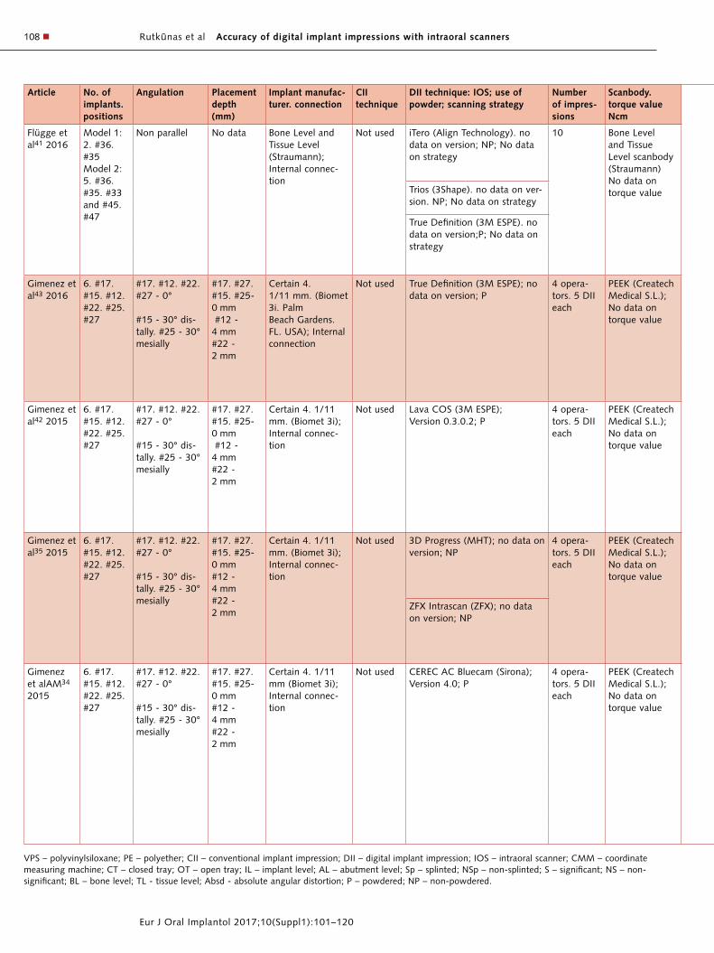
Rutkūnas et al Accuracy of digital implant impressions with intraoral scanners108 n
Eur J Oral Implantol 2017;10(Suppl1):101–120
Article No. of implants. positions
Angulation Placement depth (mm)
Implant manufac-turer. connection
CII technique
DII technique: IOS; use of powder; scanning strategy
Number of impres-sions
Scanbody. torque value Ncm
Reference scanner
Accuracy evaluation. results Conclusions
Flügge et al41 2016
Model 1:2. #36. #35Model 2:5. #36. #35. #33 and #45. #47
Non parallel No data Bone Level and Tissue Level (Straumann); Internal connec-tion
Not used iTero (Align Technology). no data on version; NP; No data on strategy
10 Bone Level and Tissue Level scanbody (Straumann)No data on torque value
D250 (3Shape)
Measurement location
Comparison: DII (digital model) distances and angles between two neighboring scanbodies (only statistically significant (p<0.05) data)
A smaller variation of the distance meas-urements was observed for the intraoral scanners True Definition and Trios. and a higher variation was seen for the iTero. Scanning precision worsened with increas-ing distance and angulation between scanbodies. Differences of mean distances between scanbodies comparing to dental lab scanning system (D250. 3Shape) were less than 40 µm.
Mean distance (mm) and stand-ard deviation (µm)
Mean angle and standard deviation
Trios (3Shape). no data on ver-sion. NP; No data on strategy
iTero True Definition iTero True Definition
#35 - #36 6.669 (28) 6.647 (4) 8.06º (0.18) 8.20º (0.04)
True Definition (3M ESPE). no data on version;P; No data on strategy
#35 - #36 (model 2)
8.19º (0.24) 8.12º (0.10)
#35 - #45 40.608 (28) 40.566 (44) 17.47º (0.21) 17.33º (0.09)
#36 - #47 50.479 (64) 50.405 (60) 23.09º (0.20) 23.28º (0.15)
Gimenez et al43 2016
6. #17. #15. #12. #22. #25. #27
#17. #12. #22. #27 - 0°
#15 - 30° dis-tally. #25 - 30° mesially
#17. #27. #15. #25- 0 mm #12 - 4 mm#22 - 2 mm
Certain 4. 1/11 mm. (Biomet 3i. PalmBeach Gardens. FL. USA); Internal connection
Not used True Definition (3M ESPE); no data on version; P
4 opera-tors. 5 DII each
PEEK (Createch Medical S.L.);No data on torque value
CMM Crista Apex (Mitu-toyo)
Comparison: DII (digital model) vs reference model Accuracy is clinically acceptable. Scanbody visibility. observer experience. and scan-ning area affect accuracy.
Distance Angulation
First quadrant 7.6 ± 17.6 µm (S) 0.21 ± 0.17º (S)
Second quadrant
-10.3 ± 39.2 µm (S) 0.28 ± 0.16º (S)
Gimenez et al42 2015
6. #17. #15. #12. #22. #25. #27
#17. #12. #22. #27 - 0°
#15 - 30° dis-tally. #25 - 30° mesially
#17. #27. #15. #25- 0 mm #12 - 4 mm#22 - 2 mm
Certain 4. 1/11 mm. (Biomet 3i); Internal connec-tion
Not used Lava COS (3M ESPE);Version 0.3.0.2; P
4 opera-tors. 5 DII each
PEEK (Createch Medical S.L.);No data on torque value
CMM Crista Apex (Mitu-toyo)
Comparison: DII (digital model) vs reference model Experienced operators delivered more accurate DII. Angulated implants and the deeply placed implants did not decrease the accuracy in digital impressions.
Group Mean (SD)
Experienced -30.8 ± 25.9 μm
Inexperienced 13.3 ± 51.2 μm
Angulated -20.2 ± 21.9 μm
Parallel -37.9 ± 26.2 μm
Deep implant -34.3 ± 18.7 μm
Gingival margin level -28.5 ± 29.8 μm
Gimenez et al35 2015
6. #17. #15. #12. #22. #25. #27
#17. #12. #22. #27 - 0°
#15 - 30° dis-tally. #25 - 30° mesially
#17. #27. #15. #25- 0 mm#12 - 4 mm#22 - 2 mm
Certain 4. 1/11 mm. (Biomet 3i); Internal connec-tion
Not used 3D Progress (MHT); no data on version; NP
4 opera-tors. 5 DII each
PEEK (Createch Medical S.L.);No data on torque value
CMM Crista Apex (Mitu-toyo)
Comparison: DII (digital model) vs reference model The 3D progress IOS performed signifi-cantly better in the first quadrant. ZFX Intrascan in the second quadrant. Tested scanners not suitable for multi-implant impressions.
Group ZFX Intrascan 3D Progress
Experienced -179 ± 601 μm 249 ± 702 μm
Inexperienced -101 ± 705 μm 224 ± 930 μm
ZFX Intrascan (ZFX); no data on version; NP
Angulated -125 ± 596 μm 257 ± 776 μm
Parallel -150 ± 693 μm 224 ± 854 μm
Deep implant (2 mm) -150 ± 397 μm 87 ± 403 μm
Gingival margin level -133 ± 782 μm 337 ± 997 μm
Gimenez et alAM34 2015
6. #17. #15. #12. #22. #25. #27
#17. #12. #22. #27 - 0°
#15 - 30° dis-tally. #25 - 30° mesially
#17. #27. #15. #25- 0 mm#12 - 4 mm#22 - 2 mm
Certain 4. 1/11 mm (Biomet 3i); Internal connec-tion
Not used CEREC AC Bluecam (Sirona);Version 4.0; P
4 opera-tors. 5 DII each
PEEK (Createch Medical S.L.);No data on torque value
CMM Crista Apex (Mitu-toyo)
Comparison: DII (digital model) vs reference model Tested scanner is clinically acceptable.The experience of the operator affected the accuracy. Angulation and location of the camera affect scanner results.The error increased from the first to the last implant scanned.
Group Mean (SD)
Experienced -85.4 ± 98.9 µm
Inexperienced -47.3 ± 75.7 µm
Angulated -72.7 ± 81.7 µm
Parallel -84.3 ± 99.9 µm
0 mm implant depth -89.47 ± 105.59 µm
2 mm implant depth -22.46 ± 30.92 µm
4 mm implant depth -107.25 ± 68.65 µm
First quadrant -17 ± 26.3 µm
Second quadrant -116 ± 103 µm
VPS – polyvinylsiloxane; PE – polyether; CII – conventional implant impression; DII – digital implant impression; IOS – intraoral scanner; CMM – coordinate measuring machine; CT – closed tray; OT – open tray; IL – implant level; AL – abutment level; Sp – splinted; NSp – non-splinted; S – significant; NS – non-significant; BL – bone level; TL - tissue level; Absd - absolute angular distortion; P – powdered; NP – non-powdered.

Rutkūnas et al Accuracy of digital implant impressions with intraoral scanners n 109
Eur J Oral Implantol 2017;10(Suppl1):101–120
Article No. of implants. positions
Angulation Placement depth (mm)
Implant manufac-turer. connection
CII technique
DII technique: IOS; use of powder; scanning strategy
Number of impres-sions
Scanbody. torque value Ncm
Reference scanner
Accuracy evaluation. results Conclusions
Flügge et al41 2016
Model 1:2. #36. #35Model 2:5. #36. #35. #33 and #45. #47
Non parallel No data Bone Level and Tissue Level (Straumann); Internal connec-tion
Not used iTero (Align Technology). no data on version; NP; No data on strategy
10 Bone Level and Tissue Level scanbody (Straumann)No data on torque value
D250 (3Shape)
Measurement location
Comparison: DII (digital model) distances and angles between two neighboring scanbodies (only statistically significant (p<0.05) data)
A smaller variation of the distance meas-urements was observed for the intraoral scanners True Definition and Trios. and a higher variation was seen for the iTero. Scanning precision worsened with increas-ing distance and angulation between scanbodies. Differences of mean distances between scanbodies comparing to dental lab scanning system (D250. 3Shape) were less than 40 µm.
Mean distance (mm) and stand-ard deviation (µm)
Mean angle and standard deviation
Trios (3Shape). no data on ver-sion. NP; No data on strategy
iTero True Definition iTero True Definition
#35 - #36 6.669 (28) 6.647 (4) 8.06º (0.18) 8.20º (0.04)
True Definition (3M ESPE). no data on version;P; No data on strategy
#35 - #36 (model 2)
8.19º (0.24) 8.12º (0.10)
#35 - #45 40.608 (28) 40.566 (44) 17.47º (0.21) 17.33º (0.09)
#36 - #47 50.479 (64) 50.405 (60) 23.09º (0.20) 23.28º (0.15)
Gimenez et al43 2016
6. #17. #15. #12. #22. #25. #27
#17. #12. #22. #27 - 0°
#15 - 30° dis-tally. #25 - 30° mesially
#17. #27. #15. #25- 0 mm #12 - 4 mm#22 - 2 mm
Certain 4. 1/11 mm. (Biomet 3i. PalmBeach Gardens. FL. USA); Internal connection
Not used True Definition (3M ESPE); no data on version; P
4 opera-tors. 5 DII each
PEEK (Createch Medical S.L.);No data on torque value
CMM Crista Apex (Mitu-toyo)
Comparison: DII (digital model) vs reference model Accuracy is clinically acceptable. Scanbody visibility. observer experience. and scan-ning area affect accuracy.
Distance Angulation
First quadrant 7.6 ± 17.6 µm (S) 0.21 ± 0.17º (S)
Second quadrant
-10.3 ± 39.2 µm (S) 0.28 ± 0.16º (S)
Gimenez et al42 2015
6. #17. #15. #12. #22. #25. #27
#17. #12. #22. #27 - 0°
#15 - 30° dis-tally. #25 - 30° mesially
#17. #27. #15. #25- 0 mm #12 - 4 mm#22 - 2 mm
Certain 4. 1/11 mm. (Biomet 3i); Internal connec-tion
Not used Lava COS (3M ESPE);Version 0.3.0.2; P
4 opera-tors. 5 DII each
PEEK (Createch Medical S.L.);No data on torque value
CMM Crista Apex (Mitu-toyo)
Comparison: DII (digital model) vs reference model Experienced operators delivered more accurate DII. Angulated implants and the deeply placed implants did not decrease the accuracy in digital impressions.
Group Mean (SD)
Experienced -30.8 ± 25.9 μm
Inexperienced 13.3 ± 51.2 μm
Angulated -20.2 ± 21.9 μm
Parallel -37.9 ± 26.2 μm
Deep implant -34.3 ± 18.7 μm
Gingival margin level -28.5 ± 29.8 μm
Gimenez et al35 2015
6. #17. #15. #12. #22. #25. #27
#17. #12. #22. #27 - 0°
#15 - 30° dis-tally. #25 - 30° mesially
#17. #27. #15. #25- 0 mm#12 - 4 mm#22 - 2 mm
Certain 4. 1/11 mm. (Biomet 3i); Internal connec-tion
Not used 3D Progress (MHT); no data on version; NP
4 opera-tors. 5 DII each
PEEK (Createch Medical S.L.);No data on torque value
CMM Crista Apex (Mitu-toyo)
Comparison: DII (digital model) vs reference model The 3D progress IOS performed signifi-cantly better in the first quadrant. ZFX Intrascan in the second quadrant. Tested scanners not suitable for multi-implant impressions.
Group ZFX Intrascan 3D Progress
Experienced -179 ± 601 μm 249 ± 702 μm
Inexperienced -101 ± 705 μm 224 ± 930 μm
ZFX Intrascan (ZFX); no data on version; NP
Angulated -125 ± 596 μm 257 ± 776 μm
Parallel -150 ± 693 μm 224 ± 854 μm
Deep implant (2 mm) -150 ± 397 μm 87 ± 403 μm
Gingival margin level -133 ± 782 μm 337 ± 997 μm
Gimenez et alAM34 2015
6. #17. #15. #12. #22. #25. #27
#17. #12. #22. #27 - 0°
#15 - 30° dis-tally. #25 - 30° mesially
#17. #27. #15. #25- 0 mm#12 - 4 mm#22 - 2 mm
Certain 4. 1/11 mm (Biomet 3i); Internal connec-tion
Not used CEREC AC Bluecam (Sirona);Version 4.0; P
4 opera-tors. 5 DII each
PEEK (Createch Medical S.L.);No data on torque value
CMM Crista Apex (Mitu-toyo)
Comparison: DII (digital model) vs reference model Tested scanner is clinically acceptable.The experience of the operator affected the accuracy. Angulation and location of the camera affect scanner results.The error increased from the first to the last implant scanned.
Group Mean (SD)
Experienced -85.4 ± 98.9 µm
Inexperienced -47.3 ± 75.7 µm
Angulated -72.7 ± 81.7 µm
Parallel -84.3 ± 99.9 µm
0 mm implant depth -89.47 ± 105.59 µm
2 mm implant depth -22.46 ± 30.92 µm
4 mm implant depth -107.25 ± 68.65 µm
First quadrant -17 ± 26.3 µm
Second quadrant -116 ± 103 µm

Rutkūnas et al Accuracy of digital implant impressions with intraoral scanners110 n
Eur J Oral Implantol 2017;10(Suppl1):101–120
Article No. of implants. positions
Angulation Placement depth (mm)
Implant manufac-turer. connection
CII technique
DII technique: IOS; use of powder; scanning strategy
Number of impres-sions
Scanbody. torque value Ncm
Reference scanner
Accuracy evaluation. results Conclusions
Gimenez et alAM33 2014
6. #17. #15. #12. #22. #25. #27
#17. #12. #22. #27 - 0°
#15 - 30° dis-tally. #25 - 30° mesially
#17. #27. #15. #25- 0 mm#12 - 4 mm#22 - 2 mm
Certain 4. 1/11 mm (Biomet 3i);Internal connec-tion
Not used iTero (Align Technology); Ver-sion 4.5.0.1.5.1; NP
4 opera-tors. 5 DII each
PEEK (Createch Medical S.L.);No data on torque value
CMM Crista Apex (Mitu-toyo)
Comparison: DII (digital model) vs reference model Angulated implants did not decrease digital impression accuracy. Impressions of implants placed at a depth of 0 mm?? were less accurate then deeper placed ones.
Implant depth Mean error and standard deviation
0 mm -23.1 ± 149.485 µm
2 mm -16.2 ± 34.569 µm
4 mm -27.9 ± 61.643 µm
First quadrant -28 ± 153 µm
Second quadrant -15 ± 30 µm
van der Meer et al37 2012
3. #36. #41. #46
- Gingival level
No information Not used CEREC Bluecam. (Sirona); Version 3.85 P
n = 10 PEEK (Createch Medical S.L.);No data on torque value
Contact scan-nerLeitz (PMM 12106)
Comparison: DII (digital model) vs reference model The Lava COS resulted in the smallest mean distance errors in full arch impres-sions.Lava COS had smallest angulation errors between cylinder 1–2 and the largest errors between cylinder 1–3. Although the absolute difference with the best mean value (iTero) was very small.
Absolute errors in distance between cylinders
CEREC Bluecam iTero Lava COS
iTero. (Align Technology); Version 3.5.0
1-2 1-3 1-2 1-3 1-2 1-3
79.6 ± 77.1 µm
81.6 ± 52.5 µm
70.5 ± 56.3 µm 61.1 ± 53.9 µm
14.6 ± 12.7 µm
23.5 ± 14.2 µm
Absolute errors in angle between cylinders
NP Lava COS (3M ESPE); Version 2.1 P
CEREC iTero Lava COS
1-2 1-3 1-2 1-3 1-2 1-3
0.6303 ± 0.5499º
0.4378 ± 0.3211º
0.3451 ± 0.3382º
0.4192 ± 0.1667º
0.2049 ± 0.0440º
0.4722 ± 0.1436º
Mangano et al40 2016
Model 1:3.#21. #24. #26;
Model 2:6.#16. #14. #11. #21. #24. #26
No data No data BTK implants (Dueville.Vicenza. Italy)
Not used Trios Color (3Shape); Version 2014 – 1. 1.3.3.1. NP
n = 5 PEEK; No data on torque value
IScan D104I (Imet-ric3D GmbH)
Comparison: DII (digital model) vs reference model No significant differences were found between partial and total edentulous models. CS 3500 intraoral scanner had the best result in terms of trueness and precision.
Scanner Model 1 Model 2 Significance
Mean trueness
CS 3500 (CarestreamHealth. Rochester. NY. US); Version 2016-4. 2.1.4.10. NP
Trios Color 72.2 ± 19.5 µm 71.6 ± 26.7 µm NS
CS 3500 47.8 ± 7.3 µm 63.2 ± 7.5 µm S
ZFX Intrascan 117.0 ± 28.6 µm 103.0 ± 26.9 µm S
ZFX Intrascan (MHT); Version 0.9 RC33 2.8. NP
Planscan 233.4 ± 62.6 µm 253.4 ± 13.6 µm S
Mean precision
Planmeca PlanScan (E4D Tech-nologies. LLC.
Trios Color 51.0 ± 18.5 µm 67.0 ± 32.2 µm S
CS 3500 40.8 ± 6.4 µm 55.2 ± 10.4 µm S
Richardson. TX. USA); Version 5 – 2015. NP
ZFX Intrascan 126.2 ± 21.2 µm 112.4 ± 22.6 µm S
Planscan 219.8 ± 59.1 µm 204.2 ± 22.7 µm S
Chew et al39 2016
2. #44.#45
Parallel No data Tissue and Bone Level Standard Plus (Straumann)
OT Trios Color (3Shape); Version 3.1.4 NP
n = 5 Core Scanbody 2077 RC and 2088 WN (Core 3D centres); Handtightened
CMM (Model Global Silver Edition. Brown and Sharpe)
Comparison: DII (digital model) vs reference model Between BL and TL groups BLCNV had the lowest global linear distortion. which was statistically significant. All TL groups were not significantly different. There were no significant differences in absolute angular distortions among all test groups.
Test group Global linear distortion Absolute angular distortion
AbsdΘ AbsdΘ
BLCII 35 ± 6 µm 0.058 ± 0.031º
0.09 ± 0.082º
iTero (Align Technology); Ver-sion HD 2.9; NP
BLTrios Color 64 ± 10 µm 0.105 ± 0.058º
0.206 ± 0.044º
BLiTero 62 ± 18 µm 0.191 ± 0.124º
0.154 ± 0.113º
BLTrue Definition 63 ± 17 µm 0.315 ± 0.138º
0.226 ± 0.143º
True Definition (3M ESPE);no data on version. P
TLCII 49 ± 10 µm 0.186 ± 0.161º
0.196 ± 0.147º
TLTrios Color 58 ± 11 µm 0.089 ± 0.039º
0.066 ± 0.033º
TLiTero 66 ± 34 µm 0.203 ± 0.094º
0.160 ± 0.121º
TLTrue Definition 64 ± 16 µm 0.206 ± 0.115º
0.195 ± 0.140º

Rutkūnas et al Accuracy of digital implant impressions with intraoral scanners n 111
Eur J Oral Implantol 2017;10(Suppl1):101–120
Article No. of implants. positions
Angulation Placement depth (mm)
Implant manufac-turer. connection
CII technique
DII technique: IOS; use of powder; scanning strategy
Number of impres-sions
Scanbody. torque value Ncm
Reference scanner
Accuracy evaluation. results Conclusions
Gimenez et alAM33 2014
6. #17. #15. #12. #22. #25. #27
#17. #12. #22. #27 - 0°
#15 - 30° dis-tally. #25 - 30° mesially
#17. #27. #15. #25- 0 mm#12 - 4 mm#22 - 2 mm
Certain 4. 1/11 mm (Biomet 3i);Internal connec-tion
Not used iTero (Align Technology); Ver-sion 4.5.0.1.5.1; NP
4 opera-tors. 5 DII each
PEEK (Createch Medical S.L.);No data on torque value
CMM Crista Apex (Mitu-toyo)
Comparison: DII (digital model) vs reference model Angulated implants did not decrease digital impression accuracy. Impressions of implants placed at a depth of 0 mm?? were less accurate then deeper placed ones.
Implant depth Mean error and standard deviation
0 mm -23.1 ± 149.485 µm
2 mm -16.2 ± 34.569 µm
4 mm -27.9 ± 61.643 µm
First quadrant -28 ± 153 µm
Second quadrant -15 ± 30 µm
van der Meer et al37 2012
3. #36. #41. #46
- Gingival level
No information Not used CEREC Bluecam. (Sirona); Version 3.85 P
n = 10 PEEK (Createch Medical S.L.);No data on torque value
Contact scan-nerLeitz (PMM 12106)
Comparison: DII (digital model) vs reference model The Lava COS resulted in the smallest mean distance errors in full arch impres-sions.Lava COS had smallest angulation errors between cylinder 1–2 and the largest errors between cylinder 1–3. Although the absolute difference with the best mean value (iTero) was very small.
Absolute errors in distance between cylinders
CEREC Bluecam iTero Lava COS
iTero. (Align Technology); Version 3.5.0
1-2 1-3 1-2 1-3 1-2 1-3
79.6 ± 77.1 µm
81.6 ± 52.5 µm
70.5 ± 56.3 µm 61.1 ± 53.9 µm
14.6 ± 12.7 µm
23.5 ± 14.2 µm
Absolute errors in angle between cylinders
NP Lava COS (3M ESPE); Version 2.1 P
CEREC iTero Lava COS
1-2 1-3 1-2 1-3 1-2 1-3
0.6303 ± 0.5499º
0.4378 ± 0.3211º
0.3451 ± 0.3382º
0.4192 ± 0.1667º
0.2049 ± 0.0440º
0.4722 ± 0.1436º
Mangano et al40 2016
Model 1:3.#21. #24. #26;
Model 2:6.#16. #14. #11. #21. #24. #26
No data No data BTK implants (Dueville.Vicenza. Italy)
Not used Trios Color (3Shape); Version 2014 – 1. 1.3.3.1. NP
n = 5 PEEK; No data on torque value
IScan D104I (Imet-ric3D GmbH)
Comparison: DII (digital model) vs reference model No significant differences were found between partial and total edentulous models. CS 3500 intraoral scanner had the best result in terms of trueness and precision.
Scanner Model 1 Model 2 Significance
Mean trueness
CS 3500 (CarestreamHealth. Rochester. NY. US); Version 2016-4. 2.1.4.10. NP
Trios Color 72.2 ± 19.5 µm 71.6 ± 26.7 µm NS
CS 3500 47.8 ± 7.3 µm 63.2 ± 7.5 µm S
ZFX Intrascan 117.0 ± 28.6 µm 103.0 ± 26.9 µm S
ZFX Intrascan (MHT); Version 0.9 RC33 2.8. NP
Planscan 233.4 ± 62.6 µm 253.4 ± 13.6 µm S
Mean precision
Planmeca PlanScan (E4D Tech-nologies. LLC.
Trios Color 51.0 ± 18.5 µm 67.0 ± 32.2 µm S
CS 3500 40.8 ± 6.4 µm 55.2 ± 10.4 µm S
Richardson. TX. USA); Version 5 – 2015. NP
ZFX Intrascan 126.2 ± 21.2 µm 112.4 ± 22.6 µm S
Planscan 219.8 ± 59.1 µm 204.2 ± 22.7 µm S
Chew et al39 2016
2. #44.#45
Parallel No data Tissue and Bone Level Standard Plus (Straumann)
OT Trios Color (3Shape); Version 3.1.4 NP
n = 5 Core Scanbody 2077 RC and 2088 WN (Core 3D centres); Handtightened
CMM (Model Global Silver Edition. Brown and Sharpe)
Comparison: DII (digital model) vs reference model Between BL and TL groups BLCNV had the lowest global linear distortion. which was statistically significant. All TL groups were not significantly different. There were no significant differences in absolute angular distortions among all test groups.
Test group Global linear distortion Absolute angular distortion
AbsdΘ AbsdΘ
BLCII 35 ± 6 µm 0.058 ± 0.031º
0.09 ± 0.082º
iTero (Align Technology); Ver-sion HD 2.9; NP
BLTrios Color 64 ± 10 µm 0.105 ± 0.058º
0.206 ± 0.044º
BLiTero 62 ± 18 µm 0.191 ± 0.124º
0.154 ± 0.113º
BLTrue Definition 63 ± 17 µm 0.315 ± 0.138º
0.226 ± 0.143º
True Definition (3M ESPE);no data on version. P
TLCII 49 ± 10 µm 0.186 ± 0.161º
0.196 ± 0.147º
TLTrios Color 58 ± 11 µm 0.089 ± 0.039º
0.066 ± 0.033º
TLiTero 66 ± 34 µm 0.203 ± 0.094º
0.160 ± 0.121º
TLTrue Definition 64 ± 16 µm 0.206 ± 0.115º
0.195 ± 0.140º

Rutkūnas et al Accuracy of digital implant impressions with intraoral scanners112 n
Eur J Oral Implantol 2017;10(Suppl1):101–120
Article No. of implants. positions
Angulation Placement depth (mm)
Implant manufac-turer. connection
CII tech-nique
DII technique: IOS; use of powder; scanning strategy
Number of impres-sions
Scanbody. torque value Ncm
Reference scanner
Accuracy evaluation. results Conclusions
Gintaute AM36 2015
4. #34. #32. #42. #44
Parallel No data Osseotite 2 Certain Implants (Biomet 3i); Inter-nal connection
OT. Sp. IL True Definition Scanner (3M ESPE); Version 4.0.3.1. P
PEEK (Createch Medical S.L.);No data on torque value
CMM Crista Apex (Mitu-toyo)
Comparison: DII (digital model) and CII (digitised VPS and PE models) vs reference model Digital and conventional impression-makingapproaches (with polyether and VPS materials) are applicable for straight and tilted dental implants.
Distance deviation Angulation deviation
Model 1
DII 9.46 ± 16.04 μm (NS) 0.17 ± 0.14° (S DII vs PE);S DII vs VPS)
VPS 12.74 ± 12.5 μm (NS) 0.07 ± 0.1°
PE 12.22 ± 16.93 μm (NS) 0.08 ± 0.07°
Model 2
2 anterior - parallel. 2 pos-terior - 40-45°
DII 35.78 ± 24.22 μm (S DII vs VPS) 0.22 ± 0.19°. (NS DII vs VPS)
VPS 4.87 ± 21.34 μm 0.04 ± 0.04°. (S)
PE 19.78 ± 21 μm 0.16 ± 0.1 6° (S)
VPS – polyvinylsiloxane; PE – polyether; CII – conventional implant impression; DII – digital implant impression; IOS – intraoral scanner; CMM – coordinate measuring machine; CT – closed tray; OT – open tray; IL – implant level; AL – abutment level; Sp – splinted; NSp – non-splinted; S – significant; NS – non-significant; BL – bone level; TL - tissue level; Absd - absolute angular distortion; P – powdered; NP – non-powdered.
The In vivo study evaluated accuracy of multi-unit DII (two implant-supported bar in the eden-tulous mandible) in 25 patients. The scanning pro-cedure was done with iTero IOS, after detaching the bars, using a defined scanning strategy. Definitive casts, which had been used for the fabrication of bars, served as reference casts. Authors presumed that the maximum acceptable horizontal misfit and angulation errors, considering two implant-sup-ported restoration, should not exceed 100 μm and 0.4° respectively32.
Of 15 included in vitro studies, two evaluated accuracy of single-unit and 13 evaluated the multi-unit DII. As for multi-unit DII, three studies used models with two implants46,49,39, two had three implants50,40, one was with 4 implants36, two were with five implants47,49, and seven used models with six implants33–35,42,43,48,40. Five of the studies evalu-ating the accuracy of full-arch DII from six implants, used the identical model33–35,42,43. Five studies evaluated the influence of operator experience and implant placement depth33–35,42,43, nine evaluated implant angulation33–36,41–43,46,47, eleven the dis-tance between the implants33–37,41–43,39–40, and one looked at the influence of scanning protocol42.
n Main findings
The majority of included studies indicated the impor-tance of error accumulation process throughout the
digital workflow. Lack of reference points, scanbody design, scanned surface characteristics, sensor size, scanning strategy, software and some other fac-tors were considered to affect accuracy. The factors potentially influencing the DII accuracy are summa-rised in Figure 2.
A workflow to produce indirect restoration in the laboratory starts from the time of impression. Therefore, accuracy of the impression is one of the most important aspects. If inaccuracies build up this could lead to misfits and strains in the final restor-ation. As the threshold for a clinically acceptable misfit is not defined clearly, it is difficult to judge the accuracy of DII, reported in the included studies, as clinically acceptable or not. In the literature, misfit of the implant-supported restoration of 100 µm or less is often considered as clinically acceptable51. However, level of the acceptable misfit could relate to the extent of the implant-supported restor-ation14. Different IOS utilising various data acquisi-tion principles were investigated in the included studies. A summary of separate accuracy measure-ments collected from included studies is presented in Figures 3 and 4.
In vivo study
According to the results of only one in vivo study included, due to a poor reference points caused by the mucosa of edentulous sites with little variation

Rutkūnas et al Accuracy of digital implant impressions with intraoral scanners n 113
Eur J Oral Implantol 2017;10(Suppl1):101–120
Article No. of implants. positions
Angulation Placement depth (mm)
Implant manufac-turer. connection
CII tech-nique
DII technique: IOS; use of powder; scanning strategy
Number of impres-sions
Scanbody. torque value Ncm
Reference scanner
Accuracy evaluation. results Conclusions
Gintaute AM36 2015
4. #34. #32. #42. #44
Parallel No data Osseotite 2 Certain Implants (Biomet 3i); Inter-nal connection
OT. Sp. IL True Definition Scanner (3M ESPE); Version 4.0.3.1. P
PEEK (Createch Medical S.L.);No data on torque value
CMM Crista Apex (Mitu-toyo)
Comparison: DII (digital model) and CII (digitised VPS and PE models) vs reference model Digital and conventional impression-makingapproaches (with polyether and VPS materials) are applicable for straight and tilted dental implants.
Distance deviation Angulation deviation
Model 1
DII 9.46 ± 16.04 μm (NS) 0.17 ± 0.14° (S DII vs PE);S DII vs VPS)
VPS 12.74 ± 12.5 μm (NS) 0.07 ± 0.1°
PE 12.22 ± 16.93 μm (NS) 0.08 ± 0.07°
Model 2
2 anterior - parallel. 2 pos-terior - 40-45°
DII 35.78 ± 24.22 μm (S DII vs VPS) 0.22 ± 0.19°. (NS DII vs VPS)
VPS 4.87 ± 21.34 μm 0.04 ± 0.04°. (S)
PE 19.78 ± 21 μm 0.16 ± 0.1 6° (S)
Fig 2 Main factors potentially affecting accuracy of digital im-plant impressions (DII). Items presented in red were investigated in the included studies.
Scanning strategy and protocol• Powder spray• Calibration• Scanning time• Number of images• Movement direction (straight,
zig-zag etc.)• Scanning mode• Sequence
Characteristics of the scanbodies• Size• Shape• Implant/abutment level• Repositioning accuracy• Construction (PEEK only or with
metallic connection; clipped or screw-retained etc.)
IOS hardware• Scanner type, generation• Scanning technology – Active wavefront sampling
with structured light projection principle
– Active triangulation principle – Confocal microscopy principle – Optical coherent tomography
principle – Other
3D printing/milling of the model• Printing technology, resolution
(x,y), layer thickness (z)• Milling technology• Production strategy• Material selection• Distortion with time• Implant analogue construction,
positioning accuracy
IOS software version
Experience and performance of the operator
DIGITAL IMPLANT IMPRESSION ACCURACY
Clinical factors• Movement of the object• Saliva, blood• Difficult to reach areas• Tongue, mobile mucosa• Fogging of the optics• Distortion of the mandible• Scanning location
(anterior/posterior etc.)• Distance between implants• Size of edentulous region• Angulation between implants• Implants insertion depth
in texture and height, digital impressions of four patients were impossible to perform32. Only in five cases were no optical irregularities of the IOS scans noticed. It was concluded that mean angular and distance errors were too large to be clinically accept-able.
In vitro studies
Two studies comparing models made from DII and CII for single-unit implant crowns reported different results. One study indicated significant change in the vertical position (93 µm) of the implant ana-logue in milled models45, while in another the mean error was comparatively small, but with a larger

Rutkūnas et al Accuracy of digital implant impressions with intraoral scanners114 n
Eur J Oral Implantol 2017;10(Suppl1):101–120
Fig 3 Digital implant impressions for: a) single-unit cases, b) FPD cases, c) fixed full-arch cases. Number of results reporting different linear absolute mean error intervals with certain IOS.
a
Planmeca Planscan
Cerec Omincam (Sirona)
Trios Color (3Shape)
CS 3500
3D Progress (MHT)
ZFX Intrascan (ZFX)
Lava COS (3M ESPE)
Trios (3Shape)
CEREC AC Bluecam
True Definition (3M ESPE)
iTero (Align Technology)
0 1 2 3 4
0–25 µm
25–50 µm
50–100 µm
100–150 µm
150–200 µm
>200 µm
b
0–25 µm
25–50 µm
50–100 µm
100–150 µm
150–200 µm
>200 µm
Planmeca Planscan
Cerec Omincam (Sirona)
Trios Color (3Shape)
CS 3500
3D Progress (MHT)
ZFX Intrascan (ZFX)
Lava COS (3M ESPE)
Trios (3Shape)
CEREC AC Bluecam
True Definition (3M ESPE)
iTero (Align Technology)
0 2 4 6 8 10 12 14
c
0–25 µm
25–50 µm
50–100 µm
100–150 µm
150–200 µm
>200 µm
Planmeca Planscan
Cerec Omincam (Sirona)
Trios Color (3Shape)
CS 3500
3D Progress (MHT)
ZFX Intrascan (ZFX)
Lava COS (3M ESPE)
Trios (3Shape)
CEREC AC Bluecam
True Definition (3M ESPE)
iTero (Align Technology)
0 1 2 3 4 5 6 7 8 9

Rutkūnas et al Accuracy of digital implant impressions with intraoral scanners n 115
Eur J Oral Implantol 2017;10(Suppl1):101–120
standard deviation: 14 ± 170 µm45. These results could not be considered as clinically acceptable, however reported deviations are the net result of inaccuracies introduced during digital impression taking, milling of the model, and positioning of the implant analogue. One of these studies also com-pared deviations between DII and the reference model (isolated assessment of only DII accuracy), and the difference was considerably smaller – -6 ± 40 µm46. Analysis of the accumulated errors in the digital workflow showed that the largest source of inaccuracy was the milling process (contributed SD is ± 98 μm) followed by the DII (contributed SD is ± 21 μm)44. Therefore, other factors besides DII could be responsible for less consistent results reported in these studies.
Thirteen studies investigated the accuracy of DII for multi-unit implant-supported restorations. Mean errors of several IOS used in five of these studies was higher than 100 µm34,35,46,40,48.
The results diverged with older generation IOS used in the included studies (Lava COS, iTero, Cerec Bluecam, 3D Progress, ZfX Intrascan), as there were studies reporting deviations above34,35,46,48 and below33,37,42 100 µm.
Different results could be explained by methodo-logical differences as well. One of the studies report-ing adequate results used less clinically relevant full-arch models, with between distantly oriented scanbodies, dentate segments, avoiding simulation of edentulous areas37. Remaining teeth in between the implants could help as reference areas, facilitat-ing stitching of the images and, possibly, improving
the accuracy. In contrast, a study utilising the single-implant model reported higher mean errors with iTero IOS for single-unit implant situation, as meas-urements were done on the model milled from pol-yurethane material based on DII data. Thus, error accumulation during fabrication of the model was inevitable46.
All studies analysing IOS of the newer gen-eration (Trios, Color, True Definition, Cerec Omnicam, CS3500) reported deviations of less than100 μm36,43,47,48,40. Interesting to note, was that all of these studies employed full-arch models with four to six implants. However, the accuracy of Planscan IOS was significantly less – 253,4 ± 13,6 µm for the full-arch situation40.
One study investigated only precision of DII with three different IOS41. It was concluded that the pre-cision of IOS tested (iTero, Trios, True Definition) was significantly different, and decreased with increasing distances between the scanbodies.
Some of the included studies explored the influ-ence of angulated implants on the accuracy of DII33–36,42,43,46,47. Reference models in these stud-ies had implants angulated from 10º47 to 45º36. The clinically acceptable threshold for the angle devia-tions generated during impression procedure is not defined in the literature. However, based on simple trigonometrical calculations (and assuming that the maximal lateral apex movement of 50 μm is accept-able), one study32 suggested that up to 0.4° angle deviation between implants could be acceptable, with total length of the implant of 14.8 mm. The majority of in vitro studies included in this systematic review
Fig 4 Number of results reporting linear absolute mean error intervals with different scanning technologies.
Optical coherent tomog-raphy, confocal microscopy
Active wavefront sampling
Active triangulation
Confocal microscopy
Linear absolute mean error intervals, µm
0–25 25–50 50–100 100–150 150–200 >200
35
30
25
20
15
10
5
0
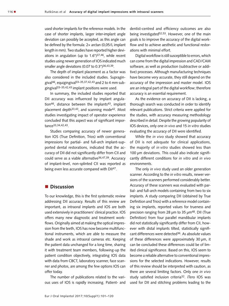
Rutkūnas et al Accuracy of digital implant impressions with intraoral scanners116 n
Eur J Oral Implantol 2017;10(Suppl1):101–120
used shorter implants for the reference models. In the case of shorter implants, larger inter-implant angle deviation can possibly be accepted, as this angle can be defined by the formula: 2× arctan (0,05/L implant length in mm). Two studies have reported higher devi-ations in angulation (up to 1.6°)37,46, while recent studies using newer generation of IOS indicated much smaller angle deviations (0.07 to 0.3°)36,43,39.
The depth of implant placement as a factor was also considered in the included studies. Supragin-gival46, equigingival33–35,37,42,43 and 2 to 4 mm sub-gingival33–35,42,43 implant positions were used.
In summary, the included studies reported that DII accuracy was influenced by implant angula-tion46, distance between the implants43, implant placement depth33,43, and scanning mode42. Most studies investigating impact of operator experience concluded that this aspect was of significant impor-tance33,34,42,43.
Studies comparing accuracy of newer genera-tion IOS (True Definition, Trios) with conventional impressions for partial- and full-arch implant-sup-ported dental restorations, indicated that the ac-curacy of DII did not significantly differ from CII and could serve as a viable alternative36,47,39. Accuracy of implant-level, non-splinted CII was reported as being even less accurate compared with DII47.
n Discussion
To our knowledge, this is the first systematic review addressing DII accuracy. Results of this review are important, as intraoral implants and IOS are both used extensively in practitioners’ clinical practice. IOS offers many new diagnostic and treatment work-flows. Originally aimed at making the optical impres-sion from the teeth, IOS has now become multifunc-tional instruments, which are able to measure the shade and work as intraoral cameras etc. Keeping the patient data unchanged for a long time, sharing it with treatment team members, following-up the patient condition objectively, integrating IOS data with data from CBCT, laboratory scanner, face scan-ner and photos, are among the few options IOS can offer today.
The number of publications related to the vari-ous uses of IOS is rapidly increasing. Patient- and
dentist-centred and efficiency outcomes are also being investigated52,53. However, one of the main goals is to improve the accuracy of the digital work-flow and to achieve aesthetic and functional restor-ations with minimal effort.
Digital workflow is still susceptible to errors, which can come from the digital impression and CAD/CAM software, as well as production (subtractive or addi-tive) processes. Although manufacturing techniques have become very accurate, they still depend on the accuracy of the impression and master model. IOS are an integral part of the digital workflow; therefore accuracy is an essential requirement.
As the evidence on accuracy of DII is lacking, a thorough search was conducted in order to identify relevant publications. Strict criteria were applied for the studies, with accuracy measuring methodology described in detail. Despite the growing popularity of IOS devices, only one in vivo and 15 in vitro studies evaluating the accuracy of DII were identified.
While the in vivo study showed that accuracy of DII is not adequate for clinical applications, the majority of in vitro studies showed less than 100 μm deviations. This could also indicate signifi-cantly different conditions for in vitro and in vivo environments.
The only in vivo study used an older generation scanner. According to the in vitro results, newer ver-sions of the scanners performed considerably better. Accuracy of these scanners was evaluated with par-tial- and full-arch models containing from two to six implants. A study comparing DII (obtained by True Definition and Trios) with a reference model contain-ing six implants, reported values for trueness and precision ranging from 28 µm to 35 µm48. DII (True Definition) from four parallel mandibular implants did not statistically significantly differ from CII, how-ever with distal implants tilted, statistically signifi-cant differences were detected36. As absolute values of these differences were approximately 30 µm, it can be concluded these differences could be of lim-ited clinical significance. Based on this, IOS seem to become a reliable alternative to conventional impres-sions for the selected indications. However, results of this review should be interpreted with caution, as there are several limiting factors. Only one in vivo study satisfied inclusion criteria32. iTero IOS was used for DII and stitching problems leading to the

Rutkūnas et al Accuracy of digital implant impressions with intraoral scanners n 117
Eur J Oral Implantol 2017;10(Suppl1):101–120
deformed image of the scan abutment, as described by the authors. The information is lacking if the ac-curacy of definitive models was rechecked by again fitting the bar to the model, as the true reference is difficult to obtain in in vivo studies, and this remains one of the challenges for the clinical evaluations. Hypothetically, trueness of the DII data could be bet-ter, but still deviate from the potentially less accurate model fabricated from the conventional impression. Moreover, at the time of this systematic review, a new version of the scanner used in this in vivo study became available, claiming much faster and more accurate scanning in colour. As the older version of the scanner and software were used, the findings of the study are therefore less relevant today.
In vivo use of IOS could be compromised by many aspects: movements of the object, saliva, fog-ging of the optics, and other patient-, operator- and device-related limiting factors. Scanning location can be important, as distant regions could be difficult to reach in a real clinical situation. Length of the eden-tulous ridge, lack of attached gingiva, tongue and cheek mobility could also negatively affect the ability to stitch the images. Scanning strategy and mode were also proved important aspects42,55. A recent study showed that intraoral scanning was less precise than model scanning54.
Comparison of the results of the in vitro studies could also be limited by disparities in study design, the models and techniques used. IOS can utilise several different technologies: confocal microscopy, optical coherence tomography, active and passive stereovision/triangulation, phase-shift principles, accordion fringe interferometry, etc36. Different IOS systems with different software versions compro-mise the comparisons further. Moreover, no stud-ies have been published with other new IOS sys-tems – DWIOS, Condor, CS3600, Aadva, Trios 3 and many others. In this regard, there is a big dif-ference between DII and CII, as the principles of conventional impression taking do not change that dramatically with time, and features of the products from different companies are relatively less different compared with IOS.
Accuracy of DII can also be affected by other factors. Characteristics of the scanbodies could be another source of errors. Shorter and less visible scanbodies can negatively influence the accuracy56.
It was recommended that longer scanbodies should be used with deep-placed implants43. One of the studies included in the systematic review used longer scanbodies, which could also contribute to better-measured accuracy47. Sharp angles of the scanbod-ies could negatively influence scan accuracy. One study was excluded from the review, as healing abut-ments instead of scanbodies were used, making the results of this study less relevant38.
Spraying of the scanbodies with powder is still needed for some of the IOS to reduce the reflections and aid the stitching of the images. Powdering could potentially influence the accuracy of scanning through homogeneity and thickness of spray. It was reported that experienced clinicians achieved greater homo-geneity and thinner coatings57. Therefore it is recom-mended to use only light dusting on the surfaces to be scanned. As powder could be inhaled by the patient and clinician or swallowed by patient, more informa-tion is needed about the effect of it on human health58.
Similarly, as with conventional impressions, type of the implant-abutment connection can influence the accuracy. External implant – abutment connection was reported to provide more consistent accuracy for CII25. Only one of the included studies investigated DII accuracy with external connection implants48.
A potential effect of embedment relaxation and manufacturing tolerances should be taken into con-sideration when selecting prosthetic components59. Repositioning accuracy of scanbodies could have an effect on the accuracy of the DII. It was reported that the ability of repositioning of the scanbody is better on lab analogues than on original implants60. However, other authors suggested that the precision of implant scanbody scanning was not significantly influenced by detachment and repositioning of the scanbody56. Not all the studies standardised the use of the scan-bodies (eg, tightening ranged from finger tightening to 15 Ncm) and this could act as an additional vari-able. Also, it could be hypothesised, that scanbodies with metallic base should have better repositioning accuracy as compared with fully plastic scanbodies.
Three studies evaluated accuracy of milled models obtained from DII44–46. It appeared that milling and positioning of implant analogues resulted in bigger deviations as compared with reference model. None of these studies described milling parameters they have used to fabricate the models. Also, information

Rutkūnas et al Accuracy of digital implant impressions with intraoral scanners118 n
Eur J Oral Implantol 2017;10(Suppl1):101–120
about implant analogues dedicated for milled models was lacking. No publication was identified, utilising 3D printing to fabricate the model from DII data. As models are necessary for layered restorations, occlusal adjustments etc., more research is needed to define milling and printing parameters in order to avoid inaccuracies and increase applications of IOS.
Despite good accuracy results reported by in vitro studies, digital workflow based on DII is still lacking some reliable conventional solutions – use of verification jigs to validate the master model, easy and reliable recording of the emergence profile, validated techniques to record static and dynamic occlusal relationships, etc. Recent studies also iden-tify the significance of inaccuracies of occlusal con-tacts of stereolithographic models fabricated, based on data from IOS61.
Although scientific literature is struggling to keep up with the newest IOS developments, due to positive clinical experience, constantly increasing opportunities with digital workflow and marketing, its use in clinical practice is growing fast. Further IOS improvements need to be done in order to replace conventional techniques and increase the potential of digital workflow, especially in partially and fully edentulous patients.
n Conclusions
1. Within the limitations of this systematic review (one in vivo and 15 in vitro studies identified), it can be concluded that digital implant impres-sions offer a valid alternative to conventional impressions for single- and multi-unit implant-supported restorations;
2. In vivo studies investigating the accuracy of new-est available IOS are needed to further define their clinical indications;
3. Factors potentially affecting accuracy of digital implant impressions should be more extensively described and investigated in clinical studies;
4. Due to the constant changes in IOS hardware and software, reliable methodology, represent-ing less forgiving in vivo situations should be defined to timely evaluate and compare trueness and precision of modern IOSs, and to provide clinical guidelines;
5. Digital implant impression techniques still have to be improved in order to fully substitute conven-tional ones;
6. Further studies are needed to investigate the ac-curacy of digital interocclusal records and master model production methods (milling, 3D printing) to ensure clinically acceptable results.
n Funding
No funding was received for conducting this system-atic review.
n References 1. Pjetursson BE, Thoma D, Jung R, Zwahlen M, Zembic A.
A systematic review of the survival and complication rates of implant-supported fixed dental prostheses (FDPs) after a mean observation period of at least 5 years. Clin Oral Implants Res 2012;23: 22–38.
2. Srinivasan M, Meyer S, Mombelli A, Müller F. Dental implants in the elderly population: a systematic review and meta-analysis. Clin Oral Implants Res 2016. Epub ahead of print.
3. Lemos CAA, de Souza Batista VE, Almeida DA de F, Santiago Júnior JF, Verri FR, Pellizzer EP. Evaluation of cement-retained versus screw-retained implant-supported restorations for marginal bone loss: A systematic review and meta-analysis. J Prosthet Dent 2016;115: 419–427.
4. Papaspyridakos P, Chen CJ, Chuang SK, Weber HP, Gallucci GO. A systematic review of biologic and technical complications with fixed implant rehabilitations for edentulous patients. Int J Oral Maxillofac Implants 2012;27:102–110.
5. Fasbinder DJ. Computerized technology for restorative den-tistry. Am J Dent 2013;26: 115–120.
6. Clinical Applications of Digital Dental Technology. Radi Masri (Editor), Carl F. Driscoll (Editor);May 2015, Wiley-Blackwell.
7. Joda T, Brägger U, Gallucci G. Systematic literature review of digital three-dimensional superimposition techniques to create virtual dental patients. Int J Oral Maxillofac Implants 2015;30: 330–337.
8. Abduo J, Lyons K, Bennani V, Waddell N, Swain M. Fit of screw-retained fixed implant frameworks fabricated by different methods: a systematic review. Int J Prosthodont 201;24: 207–220.
9. Boldt J, Knapp W, Proff P, Rottner K, Richter EJ. Measure-ment of tooth and implant mobility under physiological loading conditions. Ann Anat 2012;194: 185–189.
10. Hsieh WW, Luke A, Alster J, Weiner S. Sensory discrimina-tion of teeth and implant-supported restorations. Int J Oral Maxillofac Implants. 2010;25: 146–152.
11. Wee AG, Aquilino SA, Schneider RL. Strategies to achieve fit in implant prosthodontics: a review of the literature. Int J Prosthodont 1999;12: 167–178.
12. Manzella C, Bignardi C, Burello V, Carossa S, Schierano G. Method to improve passive fit of frameworks on implant-supported prostheses: An in vitro study. J Prosthet Dent 2016;116: 52–58.
13. Rutkunas V, Ignatovic J. A technique to splint and verify the accuracy of implant impression copings with light-polymer-izing acrylic resin. J Prosthet Dent 2014;111: 254–256.

Rutkūnas et al Accuracy of digital implant impressions with intraoral scanners n 119
Eur J Oral Implantol 2017;10(Suppl1):101–120
14. Mericske-Stern R, Worni A. Optimal number of oral implants for fixed reconstructions: a review of the literature. Eur J Oral Implantol 2014;7:S133-153.
15. Schwarz MS. Mechanical complications of dental implants. Clin Oral Implants Res 2000;11: 156–158.
16. Jemt T. Failures and complications in 391 consecutively inserted fixed prostheses supported by Brånemark implants in edentulous jaws: a study of treatment from the time of prosthesis placement to the first annual checkup. Int J Oral Maxillofac Implants 1991;6: 270–276.
17. Karl M, Taylor TD. Bone Adaptation Induced by Non-Pas-sively Fitting Implant Superstructures: A Randomized Clinical Trial. Int J Oral Maxillofac Implants 2016;31: 369–375.
18. Abduo J. Fit of CAD/CAM implant frameworks: a compre-hensive review. J Oral Implantol 2014;40: 758–766.
19. Rutkunas V, Sveikata K, Savickas R. Effects of implant angulation, material selection, and impression technique on impression accuracy: a preliminary laboratory study. Int J Prosthodont 2012;25: 512–515.
20. Siadat H, Alikhasi M, Beyabanaki E, Rahimian S. Comparison of Different Impression Techniques When Using the All-on-Four Implant Treatment Protocol. Int J Prosthodont. 2016;29: 265–270.
21. Lee H, So JS, Hochstedler JL, Ercoli C. The accuracy of implant impressions: A systematic review. J Prosthet Dent 2008;100: 285–291.
22. Papaspyridakos P, Chen CJ, Gallucci GO, Doukoudakis A, Weber HP, Chronopoulos V. Accuracy of implant impressions for partially and completely edentulous patients: a system-atic review. Int J Oral Maxillofac Implants 2014;29: 836–845.
23. Baig MR. Accuracy of impressions of multiple implants in the edentulous arch: a systematic review. Int J Oral Maxillofac Implants 2014;29: 869–880.
24. Baig MR. Multi-unit implant impression accuracy: A review of the literature. Quintessence Int 1985. 2014;45: 39–51.
25. Kim J-H, Kim KR, Kim S. Critical appraisal of implant impression accuracies: A systematic review. J Prosthet Dent 2015;114: 185–192.
26. Moreira AHJ, Rodrigues NF, Pinho ACM, Fonseca JC, Vilaça JL. Accuracy Comparison of Implant Impression Tech-niques: A Systematic Review. Clin Implant Dent Relat Res 2015;17:e751–764.
27. Kim J-H, Kim KR, Kim S. Critical appraisal of implant impression accuracies: A systematic review. J Prosthet Dent 2015;114: 185–192.
28. Papaspyridakos P, Chen C-J, Gallucci GO, Doukoudakis A, Weber H-P, Chronopoulos V. Accuracy of implant impressions for partially and completely edentulous patients: a systematic review. Int J Oral Maxillofac Implants 2014;29: 836–845.
29. Kim JH, Kim KR, Kim S. Critical appraisal of implant impres-sion accuracies: A systematic review. J Prosthet Dent 2015; 114: 185–192.
30. Bergin JM, Rubenstein JE, Mancl L, Brudvik JS, Raigrod-ski AJ. An in vitro comparison of photogrammetric and conventional complete-arch implant impression techniques. J Prosthet Dent 2013;110: 243–251.
31. Chochlidakis KM, Papaspyridakos P, Geminiani A, Chen CJ, Feng IJ, Ercoli C. Digital versus conventional impressions for fixed prosthodontics: A systematic review and meta-analysis. J Prosthet Dent 2016;116: 184–190.e12.
32. Andriessen FS, Rijkens DR, van der Meer WJ, Wismeijer DW. Applicability and accuracy of an intraoral scanner for scan-ning multiple implants in edentulous mandibles: a pilot study. J Prosthet Dent 2014;111: 186–194.
33. Giménez B, Özcan M, Martínez-Rus F, Pradíes G. Accuracy of a digital impression system based on parallel confocal laser technology for implants with consideration of operator experience and implant angulation and depth. Int J Oral Maxillofac Implants 2014;29: 853–862.
34. Giménez B, Özcan M, Martínez-Rus F, Pradíes G. Accuracy of a Digital Impression System Based on Active Triangula-tion Technology With Blue Light for Implants: Effect of Clinically Relevant Parameters. Implant Dent 2015;24: 498–504.
35. Giménez B, Pradíes G, Martínez-Rus F, Özcan M. Accuracy of two digital implant impression systems based on confo-cal microscopy with variations in customized software and clinical parameters. Int J Oral Maxillofac Implants 2015; 30:56–64.
36. Gintaute, Aiste. Accuracy of computerized and conventional impression-making procedures of straight and tilted dental implants [Internet]. [Freiburg]: Albert-Ludwigs-Universität; 2015. Available from: https://freidok.uni-freiburg.de/dnb/download/10655
37. van der Meer WJ, Andriessen FS, Wismeijer D, Ren Y. Appli-cation of intra-oral dental scanners in the digital workflow of implantology. PloS One 2012;7:e43312.
38. Ajioka H, Kihara H, Odaira C, Kobayashi T, Kondo H. Exam-ination of the Position Accuracy of Implant Abutments Reproduced by Intra-Oral Optical Impression. PloS One 2016;11:e0164048.
39. Chew AA, Esguerra RJ, Teoh KH, Wong KM, Ng SD, Tan KB. Three-Dimensional Accuracy of Digital Implant Impressions: Effects of Different Scanners and Implant Level. Int J Oral Maxillofac Implants 2017;32: 70–80.
40. Mangano FG, Veronesi G, Hauschild U, Mijiritsky E, Man-gano C. Trueness and Precision of Four Intraoral Scanners in Oral Implantology: A Comparative in Vitro Study. PloS One 2016;11:e0163107.
41. Flügge TV, Att W, Metzger MC, Nelson K. Precision of Dental Implant Digitization Using Intraoral Scanners. Int J Prosthodont 2016;29: 277–283.
42. Gimenez BD, Ozcan MD, Martinez-Rus FD, Pradies GD. Accuracy of a Digital Impression System Based on Active Wavefront Sampling Technology for Implants Considering Operator Experience, Implant Angulation, and Depth. Clin Implant Dent Relat Res 2015;17:e54 –64.
43. Gimenez-Gonzalez B, Hassan B, Özcan M, Pradíes G. An In Vitro Study of Factors Influencing the Performance of Digital Intraoral Impressions Operating on Active Wave-front Sampling Technology with Multiple Implants in the Edentulous Maxilla. J Prosthodont 2016. Epub ahead of print.
44. Koch GK, Gallucci GO, Lee SJ. Accuracy in the digital workflow: From data acquisition to the digitally milled cast. J Prosthet Dent 2016;115: 749–754.
45. Lee SJ, Betensky RA, Gianneschi GE, Gallucci GO. Accuracy of digital versus conventional implant impressions. Clin Oral Implants Res 2015;26: 715–9.
46. Lin WS, Harris BT, Elathamna EN, Abdel-Azim T, Morton D. Effect of implant divergence on the accuracy of defini-tive casts created from traditional and digital implant-level impressions: an in vitro comparative study. Int J Oral Maxil-lofac Implants 2015;30: 102–109.
47. Papaspyridakos P, Gallucci GO, Chen C-J, Hanssen S, Naert I, Vandenberghe B. Digital versus conventional im-plant impressions for edentulous patients: accuracy out-comes. Clin Oral Implants Res 2016;27: 465–472.
48. Vandeweghe S, Vervack V, Dierens M, De Bruyn H. Accur-acy of digital impressions of multiple dental implants: an in vitro study. Clin Oral Implants Res 2017;28: 648–653.
49. Flugge TV, Att W, Metzger MC, Nelson K. Precision of Dental Implant Digitization Using Intraoral Scanners. Int J Prosthodont 2016;29: 277–283.
50. van der Meer WJ, Andriessen FS, Wismeijer D, Ren Y. Appli-cation of intra-oral dental scanners in the digital workflow of implantology. PloS One 2012;7:e43312.
Rutkūnas et al Accuracy of digital implant impressions with intraoral scanners120 n
Eur J Oral Implantol 2017;10(Suppl1):101–120
51. Katsoulis J, Mericske-Stern R, Rotkina L, Zbären C, Enkling N, Blatz MB. Precision of fit of implant-supported screw-retained 10-unit computer-aided-designed and comput-er-aided-manufactured frameworks made from zirconium dioxide and titanium: an in vitro study. Clin Oral Implants Res 2014;25: 165–174.
52. Joda T, Brägger U. Digital vs. conventional implant pros-thetic workflows: a cost/time analysis. Clin Oral Implants Res 2015;26: 1430–1435.
53. Joda T, Brägger U. Patient-centered outcomes compar-ing digital and conventional implant impression proce-dures: a randomized crossover trial. Clin Oral Implants Res 2016;27:e185–e189.
54. Flügge TV, Schlager S, Nelson K, Nahles S, Metzger MC. Precision of intraoral digital dental impressions with iTero and extraoral digitization with the iTero and a model scan-ner. Am J Orthod Dentofacial Orthop 2013;144: 471–478.
55. Müller P, Ender A, Joda T, Katsoulis J. Impact of digital intraoral scan strategies on the impression accuracy using the TRIOS Pod scanner. Quintessence Int 2016;47: 343–349.
56. Fluegge T, Att W, Metzger M, Nelson K. A Novel Method to Evaluate Precision of Optical Implant Impressions with Commercial Scan Bodies-An Experimental Approach. J Pros-thodont 2015;26:34-41.
57. Dehurtevent M, Robberecht L, Béhin P. Influence of dentist experience with scan spray systems used in direct CAD/CAM impressions. J Prosthet Dent 2015;113: 17–21.
58. Patzelt SBM, Vonau S, Stampf S, Att W. Assessing the feasi-bility and accuracy of digitizing edentulous jaws. J Am Dent Assoc 2013;144: 914–920.
59. Yilmaz B, Gilbert AB, Seidt JD, McGlumphy EA, Clelland NL. Displacement of Implant Abutments Following Initial and Repeated Torqueing. Int J Oral Maxillofac Implants 2015;30: 1011–1018.
60. Stimmelmayr M, Güth J-F, Erdelt K, Edelhoff D, Beuer F. Digital evaluation of the reproducibility of implant scanbody fit--an in vitro study. Clin Oral Investig 2012;16: 851–856.
61. Wilding RJ, Adams LP, Lewin A. Absence of association between a preferred chewing side and its area of functional occlusal contact in the human dentition. Arch Oral Biol 1992;37: 423–428.
62. Ortorp A, Jemt T, Bäck T. Photogrammetry and conventional impressions for recording implant positions: a comparative laboratory study. Clin Implant Dent Relat Res 2005;7: 43–50.
63. Bergin JM, Rubenstein JE, Mancl L, Brudvik JS, Raigrodski AJ. An in vitro comparison of photogrammetric and con-ventional complete-arch implant impression techniques. J Prosthet Dent 2013;110: 243–251.
64. Eliasson A, Ortorp A. The accuracy of an implant impression technique using digitally coded healing abutments. Clin Im-plant Dent Relat Res 2012;14:e30-38.
65. Howell KJ, McGlumphy EA, Drago C, Knapik G. Comparison of the accuracy of Biomet 3i Encode Robocast Technology and conventional implant impression techniques. Int J Oral 2013;28: 228–240.
66. Lee SJ, Gallucci GO. Digital vs. conventional implant impres-sions: efficiency outcomes. Clin Oral Implants Res 2013;24: 111–115.
67. Lee SJD, MacArthur RXID, Gallucci GOD. An evaluation of student and clinician perception of digital and conventional implant impressions. J Prosthet Dent 2013;110: 420–423.
68. Wismeijer D, Mans R, van Genuchten M, Reijers HA. Patients’ preferences when comparing analogue implant impressions using a polyether impression material versus digital impressions (Intraoral Scan) of dental implants. Clin Oral Implants Res 2014;25: 1113–1118.
69. Calesini G, Zarone F, Sorrentino R, Micarelli C, Fabianelli A, Papacchini F, Gherlone E. Effect of 2 impression techniques on the dimensional accuracy of working implant prosthesis models: an in vitro study. J Craniofac Surg 2014;25: 822–827.
70. Schepke UD, Meijer HJAD, Kerdijk W, Cune MSD. Digital versus analog complete-arch impressions for single-unit pre-molar implant crowns: Operating time and patient prefer-ence. J Prosthet Dent 2015;114: 403–406.
71. Joda T, Lenherr P, Dedem P, Kovaltschuk I, Bragger U, Zitzmann NU. Time efficiency, difficulty, and operator’s pref-erence comparing digital and conventional implant impres-sions: a randomized controlled trial. Clin Oral Implants Res 2016, Epub ahead of print.
72. Aktas G, Özcan N, Aydin DH, Şahin E, Akça K. Effect of digitizing techniques on the fit of implant-retained crowns with different antirotational abutment features. J Prosthet Dent 2014;111: 367–372.
73. Abdel-Azim T, Zandinejad A, Elathamna E, Lin W, Morton D. The influence of digital fabrication options on the accur-acy of dental implant-based single units and complete-arch frameworks. Int J Oral Maxillofac Implants 2014;29: 1281–1288.
74. Karl M, Graef F, Schubinski P, Taylor T. Effect of intraoral scanning on the passivity of fit of implant-supported fixed dental prostheses. Quintessence Int 2012;43: 555–562.
75. Gherlone EF, Ferrini F, Crespi R, Gastaldi G, Capparé P. Digital impressions for fabrication of definitive “all-on-four” restorations. Implant Dent 2015;24: 125–129.
76. Lee CY, Wong N, Ganz SD, Mursic J, Suzuki JB. Use of an Intraoral Laser Scanner During the Prosthetic Phase of Implant Dentistry: A Pilot Study. J Oral Implantol 2015;41:e126-132.
77. Gherlone E, Capparé P, Vinci R, Ferrini F, Gastaldi G, Crespi R. Conventional Versus Digital Impressions for “All-on-Four” Restorations. Int J Oral Maxillofac Implants 2016;31: 324–330.

n 121
Eur J Oral Implantol 2017;10(Suppl1):121–138
REVIEW
Joannis Katsoulis Prof Dr med dent, MASAssociate Professor, Depart-ment of Reconstructive Dentistry and Gerodon-tology, School of Dental Medicine, University of Bern, Switzerland
Takuro Takeichi DDS, PhDLecturer, Department of Fixed Prosthodontics, School of Dentistry, Aichi Gakuin University, Nagoya, Japan
Ana Sol Gaviria Dr med dentResident, Department of Reconstructive Dentistry and Gerodontology, School of Dental Medicine, University of Bern, Switzerland
Lukas Peter Dr med dentResident, Department of Reconstructive Dentistry and Gerodontology, School of Dental Medicine, University of Bern, Switzerland
Konstantinos Katsoulis Dr med, Dr med dentSenior Lecturer, Department of Maxillofacial Surgery, University hospital, Bern, Switzerland
Correspondence to:Joannis Katsoulis, Prof Dr med dent, MASDepartment of Reconstruc-tive Dentistry and Gerodon-tology, School of Dental Medicine, University of Bern, SwitzerlandEmail: [email protected]
Joannis Katsoulis, Takuro Takeichi, Ana Sol Gaviria, Lukas Peter, Konstantinos Katsoulis
Misfit of implant prostheses and its impact on clinical outcomes. Definition, assessment and a systematic review of the literature
Key words clinical outcome, fixed dentures, implant prosthesis, interface, microgap, misfit, passive fit, precision, systematic review
Background and aim: Compromised fit between the contact surfaces of screw-retained implant-supported fixed dentures (IFDs) is thought to create uncontrolled strains in the prosthetic compo-nents and peri-implant tissues, thus evoking biological and technical complications such as bone loss, screw loosening, component fractures and, at worst, loss of implants or prostheses. The aim of this systematic review was to evaluate the impact of marginal misfit on the clinical outcomes of IFDs, and to elucidate definition and assessment methods for passive fit.Materials and methods: A systematic review of the literature was conducted with a PICO question: “For partially or complete edentulous subjects with screw-retained IFDs, does the marginal misfit at the implant-prosthesis interfaces have an impact on the clinical outcomes?”. A literature search was performed electronically in PubMed (MEDLINE) with the help of Boolean operators to combine key words, and by hand search in relevant journals. English written in vivo studies published before August 31, 2016 that reported on both clinical outcome and related implant prosthesis misfit (gap, strains, torque) were selected using predetermined inclusion criteria. Results: The initial search yielded 2626 records. After screening and a subsequent filtering process, five human and five animal studies were included in the descriptive analysis. The selected studies used different methods to assess misfit (linear distortion, vertical gap, strains, screw torque). While two human studies evaluated the biological response and technical complications prospectively over 6 and 12 months, the animal studies had an observation period < 12 weeks. Four human studies analysed retrospectively the 3 to 32 years’ outcomes. Screw-related complications were observed, but biological sequelae could not be confirmed. Although the animal studies had different designs, bone adaptation and implant displacement was found in histological analyses. Due to the small number of studies and the heterogenic designs and misfit assessment methods, no meta-analysis of the data could be performed.Conclusions: The current literature provides insufficient evidence as to the effect of misfit at the pros-thesis-implant interface on clinical outcomes of screw-retained implant-supported fixed dentures. Marginal gaps and static strains due to screw tightening were not found to have negative effects on initial osseointegration or peri-implant bone stability over time. Based on two clinical studies, the risk for technical screw-related complications was slightly higher. While the degree of tolerable misfit remains a matter of debate, the present data do not imply that clinicians neglect good fit, but aim to achieve the least misfit possible.
Conflict of interest statement: The authors declare no conflict of interest. The review was conducted as part of the 2016 Foundation of Oral Rehabilitation Consensus Conference on “Prosthetic Proto-cols in Implant-based Oral Rehabilitation”.
Katsoulis et al Passive fit and clinical outcomes122 n
Eur J Oral Implantol 2017;10(Suppl1):121–138
needed to be clarified before the actual literature was screened for studies comparing the clinical outcome of passively vs non-passively fitting implant pros-thesis. These are the different fabrication workflows including digital technologies, the definition of pas-sive fit, the clinical and in vitro assessment methods, as well as the characteristics and requirements of a screw-retained implant prostheses.
n Fabrication workflow
Before computer assisted design/computer assisted manufacturing (CAD/CAM) technology became commercially available, the lost wax technique for metal alloy frameworks was the gold standard. The accuracy of this conventional workflow depends mainly on physical material properties (impression, master model, casting) and human-related factors (timing, manual handling), thus, it is prone to an unpredictable degree of distortion17,18. In com-parison, the digital workflows are less influenced by manual errors and have fewer steps to follow, although also digital procedures (scanning, trans-fer) and milling may lead to minor imprecisions (Fig 1)19-21. The issue with casting, however, is its technique sensitivity and physical distortion, which can result in a poor prosthesis fit. The longer the span is, the greater the distortion and subsequently the misfit may occur. Therefore, short-span IFDs were preferred. The expert statements of an early International Symposium organised by the European Osseointegration Training Center in 1993 reflect the casting issues and the intensive thinking of 25 years ago22. In the early days of the computer numeric controlled (CNC) technology, a precise framework fabrication with the so-called “Procera-Method” was considered a promising alternative23 to the trad-itional cast technique (and its different approaches to overcome the distortion problem by sectioning and passively reconnect the separate segments). The Procera technology was considered to be accurate but required a great deal of apparatus and effort, rendering it unfavourable at the time23.
Meanwhile, CAD-software, CAM-machines, knowledge and experience have developed, and systems have become affordable and easier to use. In the past decade, the accessibility to CAD/CAM technology in dental offices and laboratories has
n Introduction
n Background
Edentulous and partially edentate patients may ben-efit from additional treatment options offered by implant-supported removable and fixed dentures1-5. Although the aesthetic aspects, especially in the an-terior maxilla, have often become of highest impor-tance in patients’ perspective, a predictable outcome with healthy and stable biomechanical conditions is a prerequisite for a successful long-term result. While multiple clinical studies have reported on the survival and success rates of various implant sys-tems6 and related reconstructions7,8, it is generally difficult to estimate the impact of the specific fac-tors that may have been associated, triggered by, or even causative to the adverse events. Compromised fit between the contact surfaces of screw-retained implant-supported fixed dentures (IFDs) is thought to create uncontrolled strains in the components and peri-implant tissues, thus evoking biological and technical complications such as bone loss, screw loosening, component fractures and, at worst, loss of the implants, the prosthesis, or both. The most frequent technical complications in full-arch recon-structions reported in a recent review article were screw fracture (yielding a 5-year complication rate of 10% and a 10-year rate of 21%) and chipping or fracture of the veneering material (33% at 5 years and 67% at 10 years)9. The role of passive fit or misfit of an implant reconstruction among the vari-ous factors has been controversially discussed in the past10-15. Opinion leaders have already stated in the early days of osseointegrated oral implants that mar-ginal discrepancies of 10 µm10 to 150 µm11 would be clinically acceptable in the long-term. From a bio-logical point of view, the gap size should be smaller than any periodontally harmful bacteria (< 2 µm)16. However, all these suggestions are of theoretical or empirical origin, lacking clear evidence. Thus, the aim of this review was to systematically evaluate the impact of marginal misfit on the clinical outcome of IFDs, and to elucidate definitions and assessment methods for passive fit.
As different opinions exist about what a “clini-cally acceptable” fit of a prosthesis is, the term “passive fit” and further aspects within this context

Katsoulis et al Passive fit and clinical outcomes n 123
Eur J Oral Implantol 2017;10(Suppl1):121–138
significantly increased. IFD frameworks and bars are milled out from one industrially produced homog-enous blank, providing highest material quality. As different studies18,24-32 have shown that the fabri-cation accuracy of short and long span CAD/CAM frameworks, bars or even full-contour reconstruc-tions made from different materials (titanium, zirco-nium dioxide, cobalt chromium) are more accurate than the cast ones, this fabrication method is consid-ered the standard method today.
n Definition of “passive fit”
Various definitions of “passive fit” can be found in literature10-13,33,34. Perfect passive fit is achieved when the opposing surfaces of the implants and the framework intaglio are in maximal spatial con-gruency, without strains in the components after tightening of all screws, provided the implant and framework surfaces are fabricated perfectly plain (Figs 2a to 2d). In consequence, non-passive fit may be; a) measured at this interface by linear25,30, volumetric17,35, and metrological36 methods before screw tightening using the one-screw test; b) by
Fig 1 Conventional and digital workflows for the fabrication of the working model (grey box) and the final implant reconstruction. Each box corresponds to a working step (blue: manual, red: digital, green: CNC machine) with potential dimen-sional errors. (EOS: Extra-oral scanner ( laboratory scanner); IOS: Intra-oral scanner).
Figs 2a to d Schematic illustration of a screw retained implant-supported fixed denture (IFD) and the two related implants. The perfectly fitting IFD shows passive fit in the one-screw test (a) and in the final position with all screws tightened (b). The ill-fitting IFD shows a certain gap at the interface in the one-screw test (c) and in the final position a non-passive fit with strains in the components (screw, framework, veneer, implant, bone) and a remaining micro-gap at the interface (d).
Framework/Bar/Reconstruction
Gypsum model Acrylic model
Virtual model
Virtual model
Virtual model
EOS EOS
Waxup
Incest/burnout
Cast alloy
Section/Reconnect
CAM
3D print
Conv. Impression IOS
Cast model
Patient
CAD
a b
c d

Katsoulis et al Passive fit and clinical outcomes124 n
Eur J Oral Implantol 2017;10(Suppl1):121–138
monitoring the screw torque during the tightening of each screw and the final rotation (degrees) to reach the recommended torque,14 and; c) by meas-uring the consequences after all screws are tightened and the prosthesis has reached its final position at the recommended screw torque, i.e. strains33,37,38
in the biological and prosthetic components (screw, framework, implant, peri-implant tissues). A lin-ear relationship between vertical misfit and strain magnitude was observed without influence by the framework material at a certain misfit39. The term “active fit” refers to this fit at the final position. In Table 1, a misfit classification is proposed against the background of today’s fabrication feasibility based on recent studies14,18,40.
n Clinical in situ assessment methods
The chairside possibilities to assess the fit of implant-retained prosthesis are limited. The pros-thesis may be positioned on the implant replicas in the master model before intraoral in situ try-in of the implants. As discussed earlier, several steps in the workflow, either conventional or digital, may lead to a distorted model and thus to an insufficient representation of the intraoral situation (Fig 1). In Table 2, the most important clinical assessment methods and their limitations are summarised. Other additional methods (disclosing media, floss) can be useful when the implant-prosthesis interface is not positioned sub-, but epi- or supra-gingivally, and abutments are used to correct for the axis and
connection level12,34. At a working distance of 25 cm, two points as close as 100 µm to each other can be distinguished as individual points by naked eye24. With the help of 2x magnification lenses a sensitivity of 50 µm can be expected at the same distance. However, visual assessment is difficult or impossible for subgingivally positioned and coni-cal type implants. For interfaces not accessible to direct vision, tactile discrimination methods with the help of an explorer may be useful to a certain degree. However, vertical gaps < 50 µm are diffi-cult to determine using a worn (> 100 µm tip) or even a brand new explorer with a tip as small as 40 µm41,42. The radiographic assessment allows visu-alising interfaces that are positioned deeply sub-gingival and impossible for direct vision and tac-tile exploration43,44. Although a paralleling device helps the clinician to more accurately evaluate the implant-abutment interface45, radiographic assess-ment with a tube angulation > 10 degrees becomes subjective for most implant types46,47. Further-more, conical implant connections may be more difficult to assess. During final prosthesis installa-tion, one screw is tightened after the other with a system depending on maximal torque. Screw resist-ance can be subjectively assessed by hand tighten-ing and, more objectively, with specific devices for torque-angle monitoring14,48,49.
The specificity and sensitivity to determine fit/misfit with one method alone may not be sufficient, but it can be improved by combining different chair-side techniques.
Table 1 Proposed fit and misfit classification according to reported assessment techniques.
Fit/misfit Before screw tighten-ing:18
Gap size at the interface(vertical and horizontal)
During screw tightening or loosening:14
Rotation (°) to final load(+ screw torque monitoring)
After screw tighten-ing:40 Strains in the pontic
Fabrication feasi-bility and clinical acceptance
Perfect 0 µm Small final rotation ° (Screw torque initial: low, final: steep increase)
0 µm/m Theoretical
(Very good) < 25 µm < 25 µm/m 3-unit IFD
Good < 50 µm < 45° final rot. < 50 µm/m 4-9-unit IFD
Fair 50–100 µm 50–100 µm/m Complete IFD
Moderate 100–150 µm 100–150 µm/m Not acceptable
Poor > 150 µm > 90 ° final rot. > 150 µm/m Not acceptable
(Very poor) > 200 µm Great final rotation ° (Screw torque initial to final: constant-ly high and increasing)
> 200 µm Not acceptable

Katsoulis et al Passive fit and clinical outcomes n 125
Eur J Oral Implantol 2017;10(Suppl1):121–138
Table 2 Clinical in situ assessment methods on the fit of prosthesis requiring only basic chairside equipment. One-ST: one screw test, All-ST: all screw test.
Assessment method Screw retention Criteria Limitations
Visual(Eye, binoculars)
None(One-ST)(All-ST)
Macroscopic gap visible
– Subgingival interface/mucosa interposition– Conical connection– Age > 40 years/visus– Experience– Light, angle, background– Quantitative discrimination
Tactile (Alternate finger pressure)
None(One-ST)
Lifting, Rocking, MotionSaliva movement
– Subgingival interface/mucosa interposition– Close implant position– Linear implant position– Conical connection– Experience– Inconsistent– Quantitative discrimination
Tactile(Explorer)
One-STAll-ST
Tactile discrimi-nation
– Subgingival interface/mucosa interposition– Conical connection– Clinician’s discriminatory ability– Explorer worn tip > 100 µm– Experience– Inconsistent– Quantitative discrimination
Radiographical(Periapical)
One-STAll-ST
Macroscopic gap visible
– Non–perpendicular alignment/angulation– Overlapping components– Radiolucent components– Knowledge on system– Experience– Analogue: size and contrast– Digital: filter effects
Screw retention Serial screwing Resistance while screwing up to final torque: none/steep vs consistent/flat
– Subgingival interface/mucosa interposition or pressure– Conical connection– Clinician’s discriminatory ability– Ranking of serial screwing– Experience– Inconsistent– Quantitative discrimination
n Laboratory in vitro assessment methods
Model-based assessment of prosthesis fit can be performed either with direct methods (light micro-scope, scanning electron microscope, strain meas-urements, screw-torque analysis) or with a virtual approach using accurate extraoral scanners and spe-cific software for superimposition. Virtual analyses (linear distance, volumetric gap, metrological visu-alisation) are usually based on a best-fit algorithm. However, model-based methods may only assess the second part of the workflow (Fig 1). If digitised patient data from an intraoral scanner are used, then the entire workflow may be included. The same goes for the photogrammetric 3D approach, as reported by Jemt50.
n Screw-retained implant-supported fixed dentures (IFDs)
Teeth have a physiological mobility of 100 to 150 µm within the periodontal ligament that may reduce stress patterns from an inaccurate framework fit. Additionally, cement-retained (dental or implant-borne) reconstructions require an internal gap of approximately 25 to 50 µm for the cement material, which also eliminates strains in the components. In contrast, screw-retained implant reconstructions will neither benefit from a cement layer, nor from a peri-implant ligament buffer. Thus, a maximal (theo-retically perfect) passive fit of the opposing surfaces at the implant-reconstruction interface should be the aim. Static stress resulting from a poorly fitting reconstruction will be directly transmitted to the im-plant components and the peri-implant bone, and

Katsoulis et al Passive fit and clinical outcomes126 n
Eur J Oral Implantol 2017;10(Suppl1):121–138
will be exacerbated by any dynamic loading in clinical use13 . The contact area in single implant restorations is circumferential and may have a flat horizontal or a conical surface, depending on the implant geometry. The maximal fit of the compo-nents (abutment) at a single implant site therefore depends on the possible fabrication accuracy at an industrial CAD/CAM centre. Adverse biological and technical events as an effect of the micro-gap and movement under dynamic loading condition have been investigated earlier. Crestal bone remodelling has been observed for either internal and external, or conical and butt–joint connections. There was a trend favouring the platform-switching concept51 and supracrestal positioning of the implant-abut-ment interface52 to prevent or minimise peri-implant marginal bone loss. In contrast, no evidence was identified in another systematic review about the effectiveness of these designs (scalloped implants, platform-switched implants and gingivally converg-ing or concave implant abutments) in preventing marginal bone loss and soft tissue recession53. Fur-thermore, the size of the micro-gap at single im-plant does not seem to influence the crestal bone resorption, unlike possible movements between implant and abutment54. The results of a recently published systematic review show that abutment screw loosening is a rare event in single-implant restorations regardless of the geometry of implant-abutment connection, provided that proper anti-rotational features and torque are employed55. Further in vitro studies investigate bacterial leak-age and colonisation of the interior implant space; mostly with single implant crowns that do not fully resist occlusal load56.
Zirconia frameworks milled in the pre-sintered stage have to anticipate subsequent final sintering shrinkage. This step may include a certain inac-curacy, depending on the span length18. Most im-plant systems offer a hybrid solution for zirconia reconstructions by inserting an additional (often, but not always, cemented) titanium base facing the implant platform. This base may be considered as a very flat standard abutment introducing a new cement interface that has been polished in the la-boratory. Although, this base is usually fixed on the master cast and not intraorally, it may reduce strains. Another aspect that needs to be mentioned
in mandibular full-arch implant prosthesis is the mandibular deformation of up to 420 µm in the posterior area and force transmission of up to 16 N as a result of jaw movement from the rest pos-ition57. Therefore, 3D implant relation is not stable during mandibular jaw movement and strains will inevitably occur.
n Materials and methods
This systematic literature review was conducted in accordance with the guidelines of Preferred Report-ing for Systematic Reviews and Meta-Analyses (PRISMA)58.
n PICO question
The PICO (patient, intervention, comparison, out-come) question was: “For partially or fully eden-tulous subjects with screw-retained IFDs, does the marginal misfit at the implant-prosthesis interfaces have an impact on the clinical outcomes?”.
n Search method
An electronic search was conducted independently by two reviewers (JK, TT) on the PubMed (MED-LINE) database for articles published in English lan-guage through to August 31, 2016. The following four groups of key words were combined using Boolean operators: (clinical outcome OR biological complication OR mechanical complication OR bone loss OR peri-implantitis OR screw loosening OR fracture OR failure) AND (framework OR prosthesis OR bridge OR bar OR component OR abutment) AND (fit OR fitting OR misfit OR gap OR pre-cision OR accuracy) AND (implant OR implants). Additional relevant articles were searched manually in the following journals accessible online at the University of Bern: Clinical Implant Dentistry and Related Research, Clinical Oral Implants Research, European Journal of Oral Implantology, Interna-tional Journal of Oral & Maxillofacial Implants, International Journal of Prosthodontics, and Quin-tessence International. The reference lists of the selected articles were assessed for possible further eligible studies.

Katsoulis et al Passive fit and clinical outcomes n 127
Eur J Oral Implantol 2017;10(Suppl1):121–138
n Inclusion criteria
Clinical studies with a prospective and retrospective design were included if they provided information on the clinical outcome and the related prosthesis fit together. Additionally, animal studies with the same information were also analysed. In detail, stud-ies were included if the following information was reported:• In vivo study (human or animal)• Screw-retained implant-supported prosthesis
(minimum two implants)• Misfit assessment at delivery (baseline) and/or
at follow-up appointments (gap size at interface, strains in reconstruction, screw torque)
• Biological changes and technical complications assessment (Table 3)
• Data extraction
The previously mentioned reviewers screened the titles and the abstracts of the records found. If the inclusion criteria were fulfilled, the full text of the article was reviewed. One investigator extracted the data. A study was excluded if the misfit gap size was missing or the clinical outcomes (in terms of biological changes or technical complications) were not reported clearly. The following information was collected: Author and year of publication, study design, human or animal study, number of subjects, prosthesis and implants, location of prosthesis, im-plant system and platform type, use of abutment, torque for screw tightening, misfit at baseline and/or follow-up, observation period, biological changes and implant survival, technical complications, and misfit surrogate assessment.
n Statistical analysis
Due to the small number of studies, the different misfit assessment methods and heterogenic study designs, a meta-analysis could not be performed. Thus, descriptive statistics were reported.
n Results
n Search results
A total of 2626 records were identified through elec-tronic databases and other sources. Based on their titles and abstracts, 2592 records had to be removed as they were not relevant to the topic. Out of 34 full-text articles assessed for eligibility, 24 articles were excluded because they did not meet the inclusion criteria. Most of the excluded studies were in vitro or single implant studies, or they did not clearly assess the misfit at the implant-prosthesis interface. Finally, a total of 10 studies (five human14,40,59-61 and five animal62-66) were included in the descriptive analysis
Table 3 Clinical outcome classification for the biological and technical complications.
Type Complication degree
Biological complication Technical complication Treatment options
I Catastrophic, non-repairable
- Peri-implantitis with extensive bone resorption
- Loss of osseointegration (aseptic)- Recurrent fistulae, etc.
- Fracture of implant body- Fracture of reconstruction- Major chipping
- Removal of implant and reconstruction
- New treatment/retreatment
II Major, repairable
Peri-implantitis with moderate bone resorption
- Fracture of occlusal screw- Minor chipping or fracture
of framework not at the interface
- Repair, corrective intervention, addition-al material required
III Minor, repairable
Mucositis/minor peri-implant inflammation without bone loss
- Screw loosening- Moving reconstruction
Hygiene and main-tenance instruction, adaptation
Fig 3 Flow chart showing the systematic selection process.
2626 records identified (database, other source)
34 full-text articles assessed for eligibility
10 articles (5 human, 5 animal) included in descriptive analysis
2592 records excluded
24 full-text articles excluded

Katsoulis et al Passive fit and clinical outcomes128 n
Eur J Oral Implantol 2017;10(Suppl1):121–138
Table 4a Characteristics of the included human studies evaluating biological and mechanical outcomes of implant prosthe-sis with different levels of misfit. IOD: implant-overdenture, IFD: implant-supported fixed denture, FW: framework.
Author, year Design Subjects Implants and plat-form
Prostheses
Karl & Taylor, 201640 Prospective 18 patients10 maxillae,10 mandibles
40 Straumann SP10/4.1 mmFlat + Synocta
20 IFDs 3-unitsHigh nobel alloyTest group: misfit (> 100 µm/m)Static and functional load
20 IFDs 3-unitsHigh nobel alloyControl group: fit (< 100 µm/m)
Jokstad & Shokati, 201561
Retrospective 30 patientsEdentulous mandibles
153BranemarkDiff. length/diam.Flat external hex+ Abutment
30 IFDs full-archAcrylic + metal alloy4-6 implants/prosthesisAbutment 35 Ncm, occl screw 15 Ncm
Hjalmarsson & Smedberg, 200514
Retrospective 46 patients30 maxillae20 mandibles
57 Astra78 Astra + Cresco76 Branemark65 Branemark + Cresco
10+14+14+12 IFDs full-arch Titanium or gold FW, acrylic teeth & veneer
Jemt & Book, 199660 Prospective group
7 patients7 maxillae
44 Nobel BiocareDiff. length/diam.Flat external hex+ Abutment
7 IFDs full-arch Prospective 1 year:10-15 Ncm
Retrospective group
7 patients7 maxillae
38Nobel BiocareDiff. length/diam.Flat external hex+ Abutment
7 IODs (0–1 year)/7 IFDs (1–5 years) Retrospective 5 years:10–15 Ncm
Kallus & Bessing, 199459
Retrospective 50 patients16 maxillae34 mandibles
278BranemarkDiff. length/diam.Flat external hex+ Abutment
50 IFDs full-archAcrylic + metal alloy4-6 implants / prosthesisAbutment max. manual force, occl screw max. manual force,
(Fig 3). The Cohen’s kappa coefficient for the final selection was 0.92, indicating excellent agreement between the reviewers.
n Design and observation period
In total, 194 and 65 prostheses with different levels of misfit were fabricated in human and animal stud-ies, respectively. While two human studies evaluated the biological response and technical complications prospectively over 640 and 12 months60, the animal studies had a short-term observation period of up to 12 weeks. Four human studies14,59-61 analysed retrospectively the clinical outcome over a period of 3 to 32 years (Tables 4 and 5).
n Misfit assessmentDifferent assessment methods were applied in the 10 included studies, depending on the technology available at the time.
Human studies
One study used an approximate estimation (0 = no visual discrepancy, 1 = slight discrepancy < 0.5 mm, 2 = moderate discrepancy 0.5 to 1 mm, 3 = pro-nounced discrepancy > 1 mm), without a measuring instrument for direct visual assessment of the vertical gap59. One clinical study calculated the linear hori-zontal distortion between the implants (3D photog-raphy, digital calculation)60. One study monitored the screw retention stability and the torque profiles14.

Katsoulis et al Passive fit and clinical outcomes n 129
Eur J Oral Implantol 2017;10(Suppl1):121–138
The most recent studies published in 2015 and 2016 used latest scanner technology61 and strain gauge measurements40 to assess the misfit.
Animal studies
Similarly, different assessment methods were applied, such as linear distance measurement between the implants (horizontal distortion)62,63, surface topo-graphical analysis64, screw torque65 and vertical gap size measurements66.
n Biological changes
Generally, no complications were reported con-cerning the soft tissues. Plaque accumulation
was not correlated to misfit values14,59. Neither vertical (> 1 mm)59 nor horizontal marginal gaps (≤ 275 µm)60, nor static strains (≤ 533 µm/m)40 due to screw tightening were found to have a negative effect on initial osseointegration or peri-implant bone stability over time in humans. Data from animal studies support a trend of bone remod-elling and not resorption. No significant correlation between level of misfit and crestal bone loss was found.
n Technical complications
In the retrospective study with the longest observa-tion period (mean 19 years, range 12 to 32 years) including 30 full-arch IFDs, the prostheses with a
Table 4b Characteristics of the included animal studies evaluating biological and mechanical outcomes of implant prosthesis with different levels of misfit. IFD: implant-supported fixed denture, FW: framework, SC: single crown.
Author and year
Design Subjects Implants and platform
Prostheses
Duyck et al 200566
Prospective 5 rabbits10 tibiae (left and right)
10 (+20)Branemark10/3.75 mmFlat external hex
5 FW Co-Crie 3 blocked SCs;test group: immediate load of medial implantstatic load from screw tighteningno functional load35 Ncm
5 FW Co-Cri.e. 3 blocked SCs;Control group: regular load of medial implant after osseointegration35 Ncm
Jemt et al 200065
Prospective 12 rabbits24 tibiae (left and right)
15 (+57)Branemark7/3 mmflat external hex+ abutment 4 mm
15 test FW + 9 control FWTitaniumi.e. 3 blocked SCs;Test groups: Static load from screw tightening LT: 15-18 Ncm (10)HT: 25-26 Ncm (5)no functional load
Jemt & Lekholm, 199864
Prospective 4 rabbits
4 tibiae
4 (+8)Branemark7/3 mmFlat external hex+ abutment 4 mm
4 barsi.e. 2-units(Laser-welded abutments)
Michaels et al 199763
Prospective 8 + 1 rabbit16 tibiae
32 + 4Steri-Oss 8/3.8 mmone-piece/cylindric (?)
8 bars misfit8 bars fiti.. 2-units(solder technique)Hand tightened screws
Carr et al199662
Prospective 6 baboons6 mandibles (bilat.)
12 + 10Steri-Oss 10/3.8 mmFlat external hex+ abutment
6 IFDs misfit5 IFDs fitCast alloy, solderedHand-tightened screws

Katsoulis et al Passive fit and clinical outcomes130 n
Eur J Oral Implantol 2017;10(Suppl1):121–138
Tabl
e 5a
D
escr
iptiv
e da
ta o
f th
e in
clud
ed h
uman
stu
dies
eva
luat
ing
biol
ogic
al a
nd m
echa
nica
l out
com
es o
f im
plan
t pr
osth
esis
with
diff
eren
t le
vels
of
mis
fit. C
SR: c
umul
ativ
e su
rviv
al r
ate,
IFD
: im
plan
t-su
ppor
ted
fixed
den
ture
, FW
: fra
mew
ork.
Aut
hor
and
year
Mis
fit
befo
re l
oad
Mis
fit
afte
r lo
adFo
llow
-up
tim
eM
isfi
t as
sess
men
t m
etho
dB
iolo
gica
l ou
tcom
eTe
chni
cal
outc
ome
Kar
l &
Tayl
or,
2016
40
In s
itu s
trai
ns a
t po
ntic
100–
533
µm/m
9 IF
Ds
(tes
t gr
oup)
5/9
IFD
s st
rain
re
duct
ion
(% in
re
gres
. cu
rve)
6 m
onth
sST
RA
INS
In v
itro:
Mod
el b
ased
str
ain
gaug
e an
alys
is m
esia
l and
dis
tal o
f im
plan
tIn
viv
o: s
trai
n de
velo
pmen
t in
IFD
po
ntic
CSR
impl
ant
100
%N
o sc
rew
loos
enin
g; C
SR I
FDs
100%
In v
ivo:
red
uctio
n of
str
ain
deve
lop-
men
ts o
ver
time
in 5
of
9 IF
Ds
In v
itro:
no
chan
ges
in s
trai
ns =
no
wea
r at
inte
rfac
e
In s
itu s
trai
ns a
t po
ntic
1–10
0 µm
/m10
IFD
s (c
ontr
ol
grou
p)
7/10
IFD
s st
rain
re
duct
ion
(% in
re
gres
. cu
rve)
6 m
onth
sST
RA
INS
“C
SR im
plan
t 10
0 %
No
scre
w lo
osen
ing
CSR
IFD
s 10
0 %
In v
ivo:
red
uctio
n of
str
ain
deve
lop-
men
ts o
ver
time
in 7
of
10 I
FDs
In v
itro:
no
chan
ges
in s
trai
ns =
no
wea
r at
inte
rfac
e
Joks
tad
&
Shok
ati
2015
61
-15
0 (9
5–23
2) µ
m19
(12
–32)
ye
ars
3 D
GA
PV
irtua
l sup
erim
posi
tion:
dig
itize
d in
tagl
io s
urfa
ce o
f IF
D (
D80
0 3s
hape
) vs
. di
gitiz
ed im
plan
t po
s-iti
ons
intr
aora
lly (
iTer
o)
CSR
impl
ant
96.7
%5
impl
ants
lost
in 4
pat
ient
sBo
ne lo
ss:
2.2
(SD
0.7
, ra
nge
0.6
- 5.
8) m
m
No
corr
elat
ion
with
mis
fit
(r2
= 0
.04,
p =
0.2
9)
Loos
enin
g or
fra
ctur
e of
abu
tmen
t or
oc
clus
al s
crew
in 4
7 %
(14
/30
IFD
s)C
SR I
FDs
73.3
%(8
IFD
s re
done
)Pr
osth
esis
with
his
tory
of
scre
w r
elat
ed
adve
rse
even
ts h
ad s
ign.
hig
her
mis
fit
valu
es (
169
µm v
s 13
4 µm
, p
= 0
.005
)
Hja
lmar
s-so
n &
Sm
ed-
berg
20
0514
-Sc
rew
sta
bilit
yA
: 18
–30°
A+
C:
35–5
0°B:
20–
50°
B+C
: 24
–38°
3 ye
ars
TOR
QU
ESt
abili
ty o
f re
tent
ion
scre
w,
mon
i-to
ring
of t
orqu
e pr
ofile
(O
sseo
care
to
rque
con
trol
ler)
Hig
her
° =
mis
fit
No
sign
. di
ffer
ence
bet
wee
n 4
grou
ps r
egar
ding
pla
que,
BO
P, m
argi
nal b
one
reso
rp-
tion.
A:
1 im
plan
t lo
ss
No
sign
. di
ffer
ence
bet
wee
n 4
grou
ps
rega
rdin
g m
obile
IFD
, FW
/scr
ew/ v
enee
r fr
actu
re,
scre
w lo
osen
ing
Jem
t &
Bo
ok
1996
60
-11
1 (5
9) µ
m(m
ean
cent
re p
oint
3D
)27
5 µm
max
.
1 ye
arH
OR
IZO
NTA
L G
AP
3 D
pho
togr
aphy
(ab
utm
ents
, in
trao
ral)
and
IFD
(ex
trao
ral),
dig
ital
supe
rimpo
sitio
n an
d m
easu
rem
ents
(c
entr
e po
int
and
angu
lar
dist
or-
tion)
; D
iffer
ence
of
linea
r di
stan
ce
betw
een
cent
re p
oint
of
FW
CSR
impl
ant
100%
Bone
loss
0.
5 m
m:
no s
ign.
Cor
rela
-tio
n be
twee
n bo
ne lo
ss a
nd
mis
fit
-91
(51
) µm
(mea
n ce
ntre
poi
nt
3D)
275
µm m
ax.
5 ye
ars
HO
RIZ
ON
TAL
GA
P “
CSR
impl
ant
100
%Bo
ne lo
ss 0
.2 m
m:
no s
ign.
C
orre
latio
n be
twee
n bo
ne
loss
and
mis
fit
Kal
lus
&
Bess
ing
1994
59
-Fi
t:0
mm
: 24
, <
0.5
mm
: 14
, 0
.5 –
1 m
m:
8,
> 1
mm
: 4
5 .5
yea
rsV
ERTI
CA
L G
AP
Vis
ual i
nspe
ctio
n/es
timat
ion
of
vert
ical
gap
siz
e (0
= n
o vi
sual
di
scre
panc
y, 1
= s
light
d. <
0.5
mm
, 2
= m
oder
ate
d. 0
.5–1
mm
, 3
= p
ro-
noun
ced
d. >
1 m
m)
Mod
if. C
AD
cla
ssifi
catio
n fo
r sc
rew
-tig
htne
ss r
atin
g: R
=
no lo
osen
ing,
S =
slig
ht l.
/ T
= o
bvio
us l.
, T
= e
xtre
me
l.
CSR
99.
6 %
(1
impl
ant
loss
)Bo
ne lo
ss:
no c
orre
latio
n to
fit
Plaq
ue a
ccum
ulat
ion:
no
corr
elat
ion
to fi
t
5/6
IFD
s w
ith e
xtre
me
loos
e sc
rew
had
m
oder
ate
or p
rono
unce
d ga
psSc
rew
loos
enin
g: c
orre
latio
n to
fit
and
to o
pera
tor;
ret
ight
enin
g af
ter
5
year
s re
com
men
ded
Int.
hex
agon
scr
ews
mor
e st
able
tha
n sl
ot s
crew
s

Katsoulis et al Passive fit and clinical outcomes n 131
Eur J Oral Implantol 2017;10(Suppl1):121–138
Tabl
e 5b
D
escr
iptiv
e da
ta o
f th
e in
clud
ed a
nim
al s
tudi
es e
valu
atin
g bi
olog
ical
and
mec
hani
cal o
utco
mes
of
impl
ant
pros
thes
is w
ith d
iffer
ent
leve
ls o
f m
isfit
. BIC
: bon
e-im
plan
t-co
ntac
t, C
SR:
cum
ulat
ive
surv
ival
rat
e, IF
D: i
mpl
ant-
supp
orte
d fix
ed d
entu
re, F
W: f
ram
ewor
k.
Aut
hor
and
year
Mis
fit
befo
re l
oad
Mis
fit
afte
r lo
adFo
llow
-up
tim
eM
isfi
t as
sess
men
t m
etho
dB
iolo
gica
l ou
tcom
eTe
chni
cal
outc
ome
Duy
ck
et a
l 20
0566
583
(358
–836
) µm
170
(8–3
20)
µm12
wee
ksV
ERTI
CA
LM
odel
bas
ed e
xam
inat
ion
of v
ert.
ga
p, d
irect
ligh
t m
icro
scop
e an
d di
gitiz
ed v
irtua
l mea
sure
men
t
Cra
ter
dept
h, s
urfa
ce,
and
BIC
(hi
stom
orph
omet
ry):
No
sign
. di
ff.
betw
een
test
and
co
ntro
l im
plan
tsC
SR im
plan
t 10
0 %
No
scre
w lo
osen
ing
CSR
FW
100
%Si
gn.
Dec
reas
e in
gap
siz
e Si
gn.
diff
. be
twee
n te
st a
nd c
ontr
ol
impl
ants
495
(406
–528
) µm
299
(74–
467)
µm
No
sign
. de
crea
se o
f ga
p si
ze
12w
eeks
VER
TIC
AL
C
SR im
plan
t 10
0 %
No
scre
w lo
osen
ing
CSR
FW
100
%
Jem
t et
al
2000
65Ti
ghte
ning
tor
que
in s
crew
of
inte
r-m
edia
te im
plan
t w
ith 1
mm
gap
LT:
17 N
cmH
T: 2
6 N
cm
Loos
enin
g to
rque
in
scre
w o
f in
term
edi-
ate
impl
ant
with
1
mm
gap
LT:
9 N
cmH
T: 1
3 N
cm
2–3
wee
ksTO
RQ
UE
Scre
w t
ight
enin
g/lo
osen
ing
torq
ue
(sta
tic p
relo
ad)
for
inte
rmed
iate
tes
t sc
rew
Bone
-met
al-c
onta
ct a
t th
read
s (B
ot-
tom
, up
per,
top,
low
er)
hist
omet
ry
CSR
impl
ant
100
%Si
gn.
corr
elat
ion
betw
een
BMC
% a
nd p
relo
ad in
duce
dBM
C %
at
tip o
f th
read
: si
gn m
ore
in H
T th
an L
T or
co
ntro
l
No
scre
w lo
osen
ing
No
abut
men
t lo
osen
ing
Jem
t &
Le
khol
m,
1998
64
Tigh
teni
ng t
orqu
e in
scr
ew o
f in
ter-
med
iate
impl
ant
with
1 m
m g
ap w
as
15 N
cm
0(be
fore
vs.
af
ter
scre
w
tight
enin
g)
TOR
QU
E3D
pho
togr
aphy
, st
ereo
scop
ic
mea
sure
men
t, s
urfa
ce t
opog
raph
y an
alys
is (
± 1
00 µ
m v
aria
tion)
100%
of
impl
ants
oss
eoin
-te
grat
ed.
Bone
def
orm
atio
n w
ith im
-pl
ant
disp
lace
men
t to
war
d FW
: 12
3 (6
0 -
200)
µm
Com
plex
, in
cons
iste
nt,
and
indi
vidu
al d
efor
mat
ion
patt
ern
Cal
cula
ted
load
on
impl
ant:
24
6 (2
38 -
252
) N
FW d
efor
mat
ion
with
rot
atio
n to
war
d im
plan
t: 1
77 (
100
- 30
0) µ
mC
ompl
ex,
inco
nsis
tent
, an
d in
divi
dual
de
form
atio
n pa
tter
n
Mic
hael
s et
al
1997
63
Mis
fit:
466
± 2
09 µ
mFi
t: 6
2 ±
35
µm
-12
wee
ksH
OR
IZO
NTA
LTr
avel
ling
mic
rosc
ope
Diff
eren
ce o
f lin
ear
dist
ance
be
twee
n ce
ntre
poi
nt o
f FW
CSR
impl
ant
100%
Clin
ical
ly n
o si
gns
of in
flam
-m
atio
n or
infe
ctio
nV
ertic
al h
isto
logi
cal s
ec-
tion
% le
ngth
and
% a
rea
of
osse
oint
egra
tion:
not
sig
n.
diff
. be
twee
n fit
vs.
mis
fit
grou
p
-
Car
r et
al
1996
62M
isfit
: 34
5 ±
203
(1
83–7
38)
µmFi
t: 6
2 ±
35
(2
–130
) µm
4 w
eeks
HO
RIZ
ON
TAL
Trav
ellin
g m
icro
scop
e D
iffer
ence
of
linea
r di
stan
ce b
etw
een
cent
re p
oint
of
IFD
CSR
impl
ant
100%
Clin
ical
ly n
o si
gns
of in
flam
-m
atio
n or
infe
ctio
nH
oriz
onta
l his
tolo
gica
l sec
-tio
n %
inte
grat
ion
and
%
area
of
osse
oint
egra
tion:
no
t si
gnifi
cant
diff
eren
ce
betw
een
fit v
s m
isfit
gro
upN
o di
ff.
betw
een
com
pres
-si
on v
s. t
ensi
on a
reas
-

Katsoulis et al Passive fit and clinical outcomes132 n
Eur J Oral Implantol 2017;10(Suppl1):121–138
history of screw-related adverse events had sig-nificantly higher misfit values (169 µm vs 134 µm, P = 0.005)61. The other clinical studies reported no complications40,60 or no differences in the frequency of adverse mechanical effects14. Slot screw types of the Brånemark implant system were more prone to screw loosening than int. hexagon screws59.
A tolerable degree of misfit could not be deter-mined for either gaps or strains.
n Discussion
The present literature review aimed to extract new information on the clinical outcome of implant recon-structions in relation to the prosthesis misfit. The PICO question was “For partially or fully edentulous subjects with screw retained IFDs, does the mar-ginal misfit at the implant-prosthesis interfaces have an impact on the clinical outcomes?” The results show that limited clinical evidence is available that supports the claimed fear for adverse biological and technical events as a direct and specific consequence of misfitting screw-retained IFDs.
In the preparation of this review, only five clin-ical human studies14,40,59-61 were found. Among these, one study40 was a prospective clinical trial with an experimental IFD including 18 patients and a 6-month observation time; one study60 had a group of seven patients who were prospectively followed for 1 year, while the others were retro-spective studies including a total of 133 patients over 3 to 32 years14,59-61. During the selection phase, another pilot study was excluded as only one single case and the methodology for strain measurements was reported67. For ethical reasons it is not possible to conduct clinical studies with intentionally poorly fitting implant reconstructions to investigate bio-logical changes and technical problems in the long-term. This is, on the other hand, acceptable with animals, provided animal welfare regulations are respected during the study. Five animal studies62-66
could be identified that fulfilled the inclusion criteria, and were therefore added in the present review to increase the power of the analysis.
A retrospective study by Jokstad and Shokati (2015)61 had the longest observation period (mean 19 years, range 12 to 32 years). Thirty patients with
an implant-supported fixed complete denture in the mandible were examined and data reported on both clinical outcomes and related misfit values, using sophisticated 3D metrological software for the mis-fit measurements. Apart from a slightly higher risk of screw-related adverse events, the effect of mis-fit up to 230 µm between the superstructures and their supporting implants appears to be minor on the long-term clinical outcomes.
While five implants were lost in four patients, no correlation of misfit values and marginal bone loss as a primary biological clinical outcome variable was found. There is no information available on the initial misfit at the time of loading, thus a potential implant displace-ment cannot be evaluated. It may be discussed if the torque of 15 Ncm applied through the occlusal screw on the abutment may induce implant displacement by micro-fractures of the surrounding bone or remodel-ling and orthodontic movement over time, as it is hypothesized elsewhere66. It is noteworthy that the misfit values at the clinical follow-up ranged between 95 and 232 µm. On the other hand, 14 of the 30 participants (47%) had experienced at least one inci-dence of screw loosening or fracture of prosthetic or abutment screw(s) over the long follow-up period. The occurrence of technical complications among the frameworks fabricated with different metal alloys did not alter. The abutment screws (tightened with 35 Ncm) and the occlusal gold screws (tightened with 15 Ncm) to retain the implant-supported fixed com-plete dentures on four to six implants both absorbed the strains induced by the misfit. However, in this spe-cific implant system the risk for screw loosening was increased with a shorter occlusal screw tightened with only 15 Ncm. The authors hypothesised that tighten-ing a non-passive superstructure on the transmucosal abutments imposed an uneven distribution of ten-sile stresses on the shank and threads of the pros-thetic screws resulting in screw loosening or fracture. According to the classification in Table 3, these would all be manageable events.
In the recently published prospective clinical study by Karl & Taylor (2016)40 the authors reported some bone adaptation around implants that were statically and functionally loaded with poorly and accurately fitting IFDs in terms of reduced strain values in the pontic of the three-unit restorations. The test group included 10 non-passively fitting

Katsoulis et al Passive fit and clinical outcomes n 133
Eur J Oral Implantol 2017;10(Suppl1):121–138
IFDs, i.e. initial strain development at the pontic of the restoration ≥ 100 µm/m. In the control group, nine passively fitting IFDs with strain values below 100 µm/m up to a perfect passive fit of theoreti-cally 0 µm/m were allocated. In contrast to another clinical animal study66, the IFDs were loaded not only statically (screw tightening), but also dynami-cally by functional loading (contacts in occlusion, articulation, and chewing), which may be well con-trolled only in humans. The results after 6 months of observation showed no negative effects on the primary biological and technical outcomes. Im-plant length, age, gender, and bone quality had no effect on the strain values. The osseous adaptation (remodelling and implant displacement) were not documented directly, but concluded by a surrogate measurement, i.e. reduction of the strain values in the IFD. Additionally, wear phenomena at the inter-face, or distortions of the restoration were excluded as no changes in the in vitro strains on the model could be observed. The clinical implication of this study remains unclear as the observation time was short (6 months), the number of patients enrolled with 19 (9 + 10) prostheses was rather small, and the strain developments did not correlate with the initial static load. However, the results support the conclusion that even IFDs with a greater level of misfit stress do not cause overload or peri-implant bone resorption, but lead to bone adaptation (with-out bony micro-fractures) and subsequently less misfit within the first couple of weeks. In a rabbit model, Duyck et al (2001)68 observed that exces-sive dynamic loads cause crater-like bone defects lateral to osseointegrated implants. The effect of cyclic occlusal dynamic load is difficult to quantify, but is hypothesised to have more influence on the bone than static load from prosthesis misfit. The conclusions of the clinical study by Jemt and Book in 199660 are in accordance with the two previ-ously mentioned studies40,61. No statistical correla-tion was observed between the change of marginal bone levels and the different parameters of pros-thesis misfit. The authors included 14 patients with four to six implant supporting a full-arch IFD. Two groups, each with seven patients, were enrolled in a prospective 1-year trial and a retrospective 5-year investigation. The misfit measurements were based on a 3D photography method and performed
only at the follow-up appointment. Thus, no com-parison with the initial misfit at the delivery can be done, nor may possible implant displacement be measured. The authors compared the misfit between the two groups measuring no differences and concluded that no bone adaptation could therefore have taken place between 1 and 5 years. This comparison and the conclusion are based on the assumption that the two groups with only seven patients apiece would have the same biological reaction, and that the IFDs were fabricated initially with the same precision. Thus, their interpretation is to be considered with caution, as it is not based on evidence. The results indicate that a certain bio-logical tolerance for misfit may be present. The degree of misfit reported in the study was esti-mated clinically acceptable with regard to observed marginal bone loss. The digitally measured misfit values of 100 µm and more at different implants of the full-arch IFDs indicate that the occlusal screws were under static (prosthesis misfit) and functional (occlusion) load, but resistant to mechanical fatigue over the whole period of observation, i.e. 1 and 5 years. A clinical study published in 1994 by Kal-lus and Bessing59 investigated retrospectively the frequency of gold screw loosening of complete IFDs after 5 years of loading and found a weak correla-tion to the framework misfit (and the operator). However, it has to be mentioned that the frame-works and the abutments were tightened manu-ally with maximum finger force, which may at best reach torque levels of 10 to 15 Ncm. In this context, loosening of the occlusal screws or the abutments is likely to occur even with perfectly fitting IFDs after functional loading of more than 5 years. It may be speculated that screw loosening would occur if the tightening was performed with a torque at 35 Ncm. Furthermore, clinical ratings of the screw-tightness and the gap size at the interface between abutment and IFD were not accurate enough and may have varied significantly between the three investigators. Although the interfaces must have been directly visible, a visual inspection was per-formed without measuring device, estimating the gap size in four categories (0 = no visual discrep-ancy, 1 = slight discrepancy < 0.5 mm, 2 = moder-ate discrepancy 0.5 to 1 mm, 3 = pronounced dis-crepancy > 1 mm). However, 500 to 1000 µm of

Katsoulis et al Passive fit and clinical outcomes134 n
Eur J Oral Implantol 2017;10(Suppl1):121–138
gap size (slight to moderate discrepancy according to the rating system) is far beyond the threshold of 100 µm considered to be clinically acceptable in lit-erature so far, and also way beyond the fabrication feasibility of today’s CAD/CAM systems (Table 1). Thus, the results and conclusions of this study may rather be considered of historical significance.
In an animal study Duyck et al (2005)66 investi-gated the effect of prosthesis misfit and static tensile load from screw tightening on the osseointegration of immediately loaded implants. Five test implants were immediately loaded with a screw retained ill-fitting Co-Cr framework. The measured gape sizes at base-line ranged between 358 to 836 µm (mean 583 µm) and were found to be significantly decreased after 12 weeks. One limitation of the study is the method of measurement that was applied to document the change of gap size. The implant position was trans-ferred at each stage to a model on which the vertical gap was measured with a light microscope. However, we do not know if the implant migrated slowly over the 12 weeks towards the framework platform or if rather the position change immediately while screw tightening at loading. No clinical or radiographic assessment was performed. The authors themselves estimate that the movement occurred most probably during fixation of the prosthesis inducing micro-frac-tures of the bone between and around the implant threads. This hypothesis was further supported by the fact that no screw loosening was observed nei-ther in the test group (immediate load) nor in the control group (osseointegrated implant), although the implant displacement was not statistically signifi-cant in the latter group. However, the fact that even an already osseointegrated implant may be pulled towards the platform only by regular tightening of the occlusal screw with 35 Ncm, means that micro-fractures of the peri-implant bone will be produced and, at the same time, strains in the implant com-ponents will decrease. Thus, the surrounding bone would absorb the initial misfit while the screws are tightened to a clinically perfect final fit. In conse-quence, the risk for technical complication would thus be minimal. However, using more rigid im-plant systems the implant itself would be exposed to higher preload stress with increased risk for fracture or peri-implant effects65. Wolff’s law was described as structural bone adaptation to mechanical usage69.
Strains transmitted to the implant surface would thus provoke adaptive changes of the trabecular archi-tecture of the peri-implant bone over time to resist load. However, compressive and tensile areas around the threads were described65. Furthermore, the pro-cess of immediate or slow implant displacement due to micro-fracture or orthodontic movement could occur before any structural bone adaptation. This issue is also discussed in the clinical study by Jok-stad and Shokati61, where a torque of 15 Ncm was applied for tightening the occlusal screw on the abutment. It remains unclear whether an implant displacement by micro-fractures of the surround-ing bone or remodelling and orthodontic movement over time was induced with only a 15 Ncm torque, or if the strains were absorbed from the prosthetic components. Jemt et al (2000)65 applied almost the same levels of preload torque in a study on rabbits. The study design included two test groups with low (15 to 18 Ncm) and high torque (25 Ncm) applied to tighten the screw of an intermediate implant con-nected to an ill-fitting titanium framework with a 1 mm (!) vertical gap at the interface. The authors focused on the effect of different preload levels than on the gap size itself, which was chosen to be extremely high (1 mm) without possibility to close the gap by screw tightening. However, the gap size was not measured after initial tightening or at the end of the observation time. Thus, only the screw loosening torque was documented and was found to have decreased by approximately 50% in both groups. Histologically, less bone-to-metal contact was observed at the tip of the thread of the unloaded implant and the implant with lower preload forces than at the tip of implants loaded with higher static forces. Against clinical expectations, there was a significant correlation between increasing degrees of preload stress and increased bone-to-metal con-tact at the tip of the threads of the loaded implants. Thus, a positive bone remodelling response was observed around the implant without difference in the thread areas (bottom, lower part) anticipated to be exposed to pressure or tensile forces. In a previous pilot study by the same group published in 199864, a complex deformation pattern of the framework and the peri-implant bone was described in four rabbit tibiae. The same study design was used with three implants and an artificially induced gap of

Katsoulis et al Passive fit and clinical outcomes n 135
Eur J Oral Implantol 2017;10(Suppl1):121–138
1 mm at the intermediate implant and a controlled screw tightening with 15 Ncm. The flexure of the framework may be explained by the fact that the three abutments were laser-welded, resulting in a bar type framework. The thin bar design and the superficial laser connections may have allowed for a bending distortion while screw tightening with 15 Ncm. However, based on only four specimens, the authors stated that the deformation pattern was inconsistent and individual. While increasing levels of vertical misfit (10, 50 and 100 µm) in a single implant study by Hermann et al (2001)54 did not lead to more crestal bone loss, possible movements between the abutment and the implant were found to lead to more bone loss. The implant with a previ-ously laser-welded and thus immobilised abutment-implant connection showed significantly less bone resorption. However, the abutment screws were tightened only manually, which is significantly less torque compared to tightening occlusal screws for final fit (system depending up to 35 Ncm). By con-trast, multiple-unit prostheses are splinted together and thus the possibility of repetitive distortion or motion at the interface due to functional loading is very small, if not impossible, with rigid framework materials, such as titanium or zirconia. The ques-tion remains unanswered as to whether immobilising IFDs by splinting would also yield a low bacterial colonisation under dynamic load like single implants with a rigid conical connection type70.
One of the first animal studies to investigate the effect of prosthetic superstructure on the bone interface of osseointegrated implants was performed by Michaels et al in199763. Eight rabbits each received a well and a poorly fitting soldered bar-type superstructure on two implants. The ill-fitting bars were fabricated with a linear distortion of 466 (mean) ± 209 (SD) µm. Interestingly, the horizontal measurements of the fitting group ranged from 20 to 116 µm (62 ± 35 µm). Today, these gap sizes would be considered to be moderately fitting at best. As the occlusal screws were tightened by hand (most probably with 10 to 15 Ncm max), and as there were no vertical gap size nor pontic strain measurements performed after screw tightening or after the obser-vation time of 12 weeks, no information is avail-able if and in which moment a gape size reduction occurred due to possible implant displacement. The
author did not histometrically find a significant dif-ference in percentage length and area of osseointe-gration between the fit and misfit group. This study reported very high horizontal misfit values (up to 747 µm) without effect on the peri-implant bone based on histologic findings. However, there was no functional loading and only a short observation period of 12 weeks.
Similarly, the same limitations are present in the first animal study found in this review by Carr et al (1996)62. Over 4 weeks the study investigated his-tologically the bone response of unloaded implants with different levels of (horizontal) misfit in six pri-mates (baboons). The misfit group included IFDs with a linear distortion of 183 to 738 µm (mean 345, SD 203 µm) compared to the fit group ranging from 2 to 130 µm (mean 38, SD 52 µm). Within the limita-tions of this study (low statistical power due to small sample size, no direct gap size or strain measure-ments at loading and follow-up, use of abutments, manual screw tightening) and contrary to clinical expectation, the data suggest a bone response trend in favour of the misfit group. However, the use of abutments may have biased the results. The static strains from screw tightening with the given misfit-ting IFDs may have been completely absorbed in the second interface and by the abutments eliminating tensile or compressive forces on the implant level.
Overall, no threshold for a clinically acceptable gap or strains could be determined. Biological and technical tolerance to non-passive fit with or without a remaining gap at the interface seems to be high. It may be discussed if a clinically acceptable misfit could be different from patient to patient depending on general and behavioural factors, similar to the risk for caries or periodontal disease. If misfit was a triggering factor, patients with a poor oral hygiene and genetic predisposition to pathologic periodontal bone resorption might be more susceptible to peri-implant bone loss from ill-fitting reconstructions. In such at risk patients with low misfit tolerance, the threshold for a clinically acceptable misfit would be very small. On the other hand, there seems to be patient resistant to pathologic processes, even in the absence of good oral hygiene. For this type of patient, a comparatively poor component fit would still be clinically acceptable. In this context, the age-related capacity to react on a disturbing factor and

Katsoulis et al Passive fit and clinical outcomes136 n
Eur J Oral Implantol 2017;10(Suppl1):121–138
to keep the bacterial balance in the oral cavity and at the interface should also be taken in consideration. Generally, it is hypothesised that older patients have a less intense and slower healing reaction. However, it is not clear whether a weak reaction to the mis-fit-stimulus would imply a greater misfit tolerance. On the contrary, young and healthy subjects with a stronger and faster local inflammatory defence on the misfit-stimulus could keep biological degrada-tion under control, which would imply a high misfit tolerance in the same way. The interpretation is con-troversial and remains a matter of debate.
Although this systematic review included most PRISMA guidelines, there are some limitations. The small number of in vivo studies and the heterogenic design with different assessment methods did not allow us to clearly compare the misfit values. There-fore, a meta-analysis was not possible and only a descriptive statistical analysis was performed. The fol-low-up duration for most studies was in the short to medium range (0.5 to 5.5 years), except for one retro-spective study61, which was from 12 to 32 years. The level of evidence of the included studies was rather poor according to the evidence pyramid71,72 because only one40 out of five human studies14, 40, 59-61 was a randomised controlled clinical trial, and five were animal studies62-66. The reporting quality of the pro-spective human study40 was good as the misfit assess-ment and the outcomes were clearly documented, which was not generally the case in the retrospective studies. The risk of bias was low in the prospective studies as the implant surgery, the prosthesis fabrica-tion and the assessments were performed by only one trained investigator. On the contrary, the retro-spective studies have to be considered as having a high risk for bias as multiple surgeons and investiga-tors were involved14,59-61, and for usage of subjective assessment methods in the visual gap estimation59.
n Conclusions
The current literature provides insufficient evidence about the effect of misfit at the prosthesis-implant interface on clinical outcomes of screw retained implant-supported fixed dentures. Marginal gaps and static strains due to screw tightening were not found to have negative effects on initial osseointegration or
peri-implant bone stability over time. Based on two clinical studies, the risk for technical screw-related complications was slightly higher. While the degree of tolerable misfit remains a matter of debate, the present data do not imply that clinicians should neglect good fit, but aim to achieve the least degree of misfit possible.
n References
1. Brånemark PI, Adell R, Albrektsson T, Lekholm U, Lund-kvist S, Rockler B. Osseointegrated titanium fixtures in the treatment of edentulousness. Biomaterials 1983;4: 25–28.
2. Adell R. Tissue integrated prostheses in clinical dentistry. Int Dent J 1985;35: 259–265.
3. Ueda T, Kremer U, Katsoulis J, Mericske-Stern R. Long-term results of mandibular implants supporting an overdenture: implant survival, failures, and crestal bone level changes. Int J Oral Maxillofac Implants 2011;26: 365–372.
4. Pjetursson BE, Thoma D, Jung R, Zwahlen M, Zembic A. A systematic review of the survival and complication rates of implant-supported fixed dental prostheses (IFDs) after a mean observation period of at least 5 years. Clin Oral Implants Res 2012;23: 22–38.
5. Mericske-Stern R, Worni A. Optimal number of oral implants for fixed reconstructions: a review of the literature. Eur J Oral Implantol 2014;7:S133–153.
6. Esposito M, Ardebili Y, Worthington HV. Interventions for replacing missing teeth: different types of dental implants. Cochrane Database Syst Rev 2014:CD003815.
7. Lang NP, Berglundh T, Heitz-Mayfield LJ, Pjetursson BE, Salvi GE, Sanz M. Consensus statements and recommended clinical procedures regarding implant survival and complica-tions. Int J Oral Maxillofac Implants 2004;19: 150–154.
8. Klinge B, Flemming T, Cosyn J, De Bruyn H, Eisner BM, Hultin M, Isodor F, Lang NP, Lund B, Meyle J, Mombelli A, Navarro JM, Pjetursson B, Renvert S, Schliephake H. The patient undergoing implant therapy. Summary and consen-sus statements. The 4th EAO Consensus Conference 2015. Clin Oral Implants Res 2015;26: 64–67.
9. Papaspyridakos P, Chen CJ, Chuang SK, Weber HP, Gal-lucci GO. A systematic review of biologic and technical com-plications with fixed implant rehabilitations for edentulous patients. Int J Oral Maxillofac Implants 2012;27: 102–110.
10. Brånemark PI. Osseointegration and its experimental back-ground. J Prosthet Dent 1983;50: 399–410.
11. Jemt T. Failures and complications in 391 consecutively inserted fixed prostheses supported by Branemark implants in edentulous jaws: a study of treatment from the time of prosthesis placement to the first annual checkup. Int J Oral Maxillofac Implants 1991;6: 270–276.
12. Kan JY, Rungcharassaeng K, Bohsali K, Goodacre CJ, Lang BR. Clinical methods for evaluating implant framework fit. J Prosthet Dent 1999;81: 7–13.
13. Sahin S, Cehreli MC. The significance of passive framework fit in implant prosthodontics: current status. Implant Dent 2001;10: 85–92.
14. Hjalmarsson L, Smedberg JI. A 3-year retrospective study of Cresco frameworks: preload and complications. Clin Implant Dent Relat Res 2005;7: 189–199.
15. Abduo J, Judge RB. Implications of implant framework mis-fit: a systematic review of biomechanical sequelae. Int J Oral Maxillofac Implants 2014;29: 608–621.

Katsoulis et al Passive fit and clinical outcomes n 137
Eur J Oral Implantol 2017;10(Suppl1):121–138
16. Schüpbach P. Oral communication, panel discussion. Nobel Biocare Study Club Bern, Switzerland. 2016.
17. Drago C, Saldarriaga RL, Domagala D, Almasri R. Volumetric determination of the amount of misfit in CAD/CAM and cast implant frameworks: a multicenter laboratory study. Int J Oral Maxillofac Implants 2010;25: 920–929.
18. Katsoulis J, Mericske-Stern R, Rotkina L, Zbaren C, Enkling N, Blatz MB. Precision of fit of implant-supported screw-retained 10-unit computer-aided-designed and comput-er-aided-manufactured frameworks made from zirconium dioxide and titanium: an in vitro study. Clin Oral Implants Res 2014;25: 165–174.
19. van der Meer WJ, Andriessen FS, Wismeijer D, Ren Y. Appli-cation of intra-oral dental scanners in the digital workflow of implantology. PLoS One 2012;7:e43312.
20. Patzelt SB, Emmanouilidi A, Stampf S, Strub JR, Att W. Ac-curacy of full-arch scans using intraoral scanners. Clin Oral Investig 2014;18: 1687–1694.
21. Patzelt SB, Bishti S, Stampf S, Att W. Accuracy of comput-er-aided design/computer-aided manufacturing-generated dental casts based on intraoral scanner data. J Am Dent Assoc 2014;145: 1133–1140.
22. Naert IE, Patterson EA, Wichmann M, Nilner K, Küpper H, Hobkirk JA, Quirynen M, Neuendorff G. Passive fit of implant supported superstructures: “Fiction or reality?”. Naert IE, editor. Leuven: Leuven University Press; 1995.
23. Küpper H. Passive fit of implant-superstructures: out of titanium. Naert IE, editor. Leuven: Leuven University Press; 1995.
24. Riedy SJ, Lang BR, Lang BE. Fit of implant frameworks fabricated by different techniques. J Prosthet Dent 1997;78: 596–604.
25. Ortorp A, Jemt T, Back T, Jalevik T. Comparisons of preci-sion of fit between cast and CNC-milled titanium implant frameworks for the edentulous mandible. Int J Prosthodont 2003;16: 194–200.
26. Takahashi T, Gunne J. Fit of implant frameworks: an in vitro comparison between two fabrication techniques. J Prosthet Dent. 2003;89: 256–260.
27. Al-Fadda SA, Zarb GA, Finer Y. A comparison of the accur-acy of fit of 2 methods for fabricating implant-prosthodontic frameworks. Int J Prosthodont 2007;20: 125–131.
28. Eliasson A, Wennerberg A, Johansson A, Ortorp A, Jemt T. The precision of fit of milled titanium implant frameworks (I-Bridge) in the edentulous jaw. Clin Implant Dent Relat Res 2010;12: 81–90.
29. Abduo J, Lyons K, Waddell N, Bennani V, Swain M. A com-parison of fit of CNC-milled titanium and zirconia frame-works to implants. Clin Implant Dent Relat Res 2012;14: e20–29.
30. Katsoulis J, Mericske-Stern R, Yates DM, Izutani N, Enkling N, Blatz MB. In vitro precision of fit of computer-aided design and computer-aided manufacturing titanium and zirconium dioxide bars. Dent Mater 2013;29: 945–953.
31. Katsoulis J, Mericske-Stern R, Enkling N, Katsoulis K, Blatz MB. In vitro precision of fit of computer-aided designed and computer-aided manufactured titanium screw-retained fixed dental prostheses before and after ceramic veneering. Clin Oral Implants Res 2015;26: 44–49.
32. Katsoulis J, Muller P, Mericske-Stern R, Blatz MB. CAD/CAM fabrication accuracy of long- vs. short-span implant-supported IFDs. Clin Oral Implants Res 2015;26: 245–249.
33. Karl M, Winter W, Taylor TD, Heckmann SM. In vitro study on passive fit in implant-supported 5-unit fixed partial den-tures. Int J Oral Maxillofac Implants 2004;19: 30–37.
34. Abduo J, Bennani V, Waddell N, Lyons K, Swain M. Assess-ing the fit of implant fixed prostheses: a critical review Int J Oral Maxillofac Implants 2010;25: 506–515.
35. Almasri R, Drago CJ, Siegel SC, Hardigan PC. Volumetric Misfit in CAD/CAM and Cast Implant Frameworks: A Uni-versity Laboratory Study. J Prosthodont 2011;20: 267–274.
36. Holst S, Karl M, Wichmann M, Matta RE. A technique for in vitro fit assessment of multi-unit screw-retained implant restorations: Application of a triple-scan protocol. J Dent Biomech 2012;3:1758736012452181.
37. Millington ND, Leung T. Inaccurate fit of implant superstruc-tures. Part 1: Stresses generated on the superstructure rela-tive to the size of fit discrepancy. Int J Prosthodont 1995;8: 511–516.
38. Karl M, Holst S. Strain development of screw-retained implant-supported fixed restorations: procera implant bridge versus conventionally cast restorations. Int J Prosthodont. 2012;25: 166–169.
39. Abduo J, Lyons K. Effect of vertical misfit on strain within screw-retained implant titanium and zirconia frameworks. J Prosthodont Res 2012;56: 102–109.
40. Karl M, Taylor TD. Bone Adaptation Induced by Non-Pas-sively Fitting Implant Superstructures: A Randomized Clinical Trial. Int J Oral Maxillofac Implants 2016;31: 369–375.
41. McLean JW, von Fraunhofer JA. The estimation of cement film thickness by an in vivo technique. Br Dent J 1971;131: 107–111.
42. Peter L, Katsoulis J. Preclinical fit assessment and gap esti-mation at the implant-abutment interface: an in vitro study. Master thesis (MAS), School of Dental Medicine, Medical Faculty, University of Bern, Switzerland. Unpublished data/under review.
43. Papavassiliou H, Kourtis S, Katerelou J, Chronopoulos V. Radiographical evaluation of the gap at the implant-abut-ment interface. J Esthet Restor Dent 2010;22: 235–250.
44. Sharkey S, Kelly A, Houston F, O’Sullivan M, Quinn F, O’Connell B. A radiographic analysis of implant component misfit. Int J Oral Maxillofac Implants 2011;26: 807–815.
45. Lin KC, Wadhwani CP, Cheng J, Sharma A, Finzen F. Assess-ing fit at the implant-abutment junction with a radiographic device that does not require access to the implant body. J Prosthet Dent 2014;112: 817–823.
46. Begona Ormaechea M, Millstein P, Hirayama H. Tube angulation effect on radiographic analysis of the implant-abutment interface. Int J Oral Maxillofac Implants 1999;14: 77–85.
47. Gaviria A, Katsoulis J. Digital versus analogue radiographical fit assessment at the implant-abutment interface of differ-ent implant systems. Master thesis (MAS), School of Dental Medicine, Medical Faculty, University of Bern, Switzerland. Unpublished data/under review.
48. Jemt T. In vivo measurements of precision of fit involving implant-supported prostheses in the edentulous jaw. Int J Oral Maxillofac Implants 1996;11: 151–158.
49. Calderini A, Maiorana C, Garlini G, Abbondanza T. A simpli-fied method to assess precision of fit between framework and supporting implants: a preliminary study. Int J Oral Maxillofac Implants 2007;22: 831–838.
50. Jemt T, Lie A. Accuracy of implant-supported prostheses in the edentulous jaw: analysis of precision of fit between cast gold-alloy frameworks and master casts by means of a three-dimensional photogrammetric technique. Clin Oral Implants Res 1995;6: 172–180.
51. Schwarz F, Alcoforado G, Nelson K, Schaer A, Taylor T, Beuer F, Strietzel FP. Impact of implant-abutment connection, position-ing of the machined collar/microgap, and platform switching on crestal bone level changes. Camlog Foundation Consensus Report. Clin Oral Implants Res 2014;25: 1301–1303.
52. Piattelli A, Vrespa G, Petrone G, Iezzi G, Annibali S, Scara-no A. Role of the microgap between implant and abutment: a retrospective histologic evaluation in monkeys. J Periodon-tol 2003;74: 346–352.

Katsoulis et al Passive fit and clinical outcomes138 n
Eur J Oral Implantol 2017;10(Suppl1):121–138
53. Bishti S, Strub JR, Att W. Effect of the implant-abutment interface on peri-implant tissues: a systematic review. Acta Odontol Scand 2014;72: 13–25.
54. Hermann JS, Schoolfield JD, Schenk RK, Buser D, Cochran DL. Influence of the size of the microgap on crestal bone changes around titanium implants. A histometric evaluation of unloaded non-submerged implants in the canine mandible. J Periodontol 2001;72: 1372–1383.
55. Theoharidou A, Petridis HP, Tzannas K, Garefis P. Abutment screw loosening in single-implant restorations: a systematic review. Int J Oral Maxillofac Implants 2008;23: 681–690.
56. Passos SP, Gressler May L, Faria R, Ozcan M, Bottino MA. Implant-abutment gap versus microbial colonization: Clinical significance based on a literature review. Journal of bio-medical materials research Part B. Appl Biomater 2013;101: 1321–1328.
57. Hobkirk JA, Schwab J. Mandibular deformation in subjects with osseointegrated implants. Int J Oral Maxillofac Implants 1991;6: 319–328.
58. Moher D, Liberati A, Tetzlaff J, Altman DG, Group P. Pre-ferred reporting items for systematic reviews and meta-anal-yses: the PRISMA statement. PLoS Med. 2009;6:e1000097.
59. Kallus T, Bessing C. Loose gold screws frequently occur in full-arch fixed prostheses supported by osseointegrated implants after 5 years. Int J Oral Maxillofac Implants. 1994;9: 169–178.
60. Jemt T, Book K. Prosthesis misfit and marginal bone loss in edentulous implant patients. Int J Oral Maxillofac Implants. 1996;11: 620–625.
61. Jokstad A, Shokati B. New 3D technologies applied to assess the long-term clinical effects of misfit of the full jaw fixed prosthesis on dental implants. Clin Oral Impl Res 2015;26: 1129–1134.
62. Carr AB, Gerard DA, Larsen PE. The response of bone in pri-mates around unloaded dental implants supporting prostheses with different levels of fit. J Prosthet Dent 1996;76: 500–509.
63. Michaels GC, Carr AB, Larsen PE. Effect of prosthetic super-structure accuracy on the osteointegrated implant bone
interface. Oral Surg Oral Med Oral Pathol Oral Radiol Endod 1997;83: 198–205.
64. Jemt T, Lekholm U. Measurements of bone and framework deformations induced by misfit of implant superstructures. A pilot study in rabbits. Clin Oral Implants Res 1998;9: 272–280.
65. Jemt T, Lekholm U, Johansson CB. Bone response to implant-supported frameworks with differing degrees of misfit preload: in vivo study in rabbits. Clin Implants Dent Relat Res 2000;2: 129–137.
66. Duyck J, Vrielinck L, Lambrichts I, Abe Y, Schepers S, Politis C, Naert I. Biologic response of immediately versus delayed loaded implants supporting ill-fitting prostheses: an animal study Clin Implants Dent Relat Res 2005;7: 150–158.
67. Karl M, Graef F, Heckmann S, Taylor T. A methodology to study the effects of prosthesis misfit over time: an in vivo model. Int J Oral Maxillofac Implants 2009;24: 689–694.
68. Duyck J, Ronold HJ, Van Oosterwyck H, Naert I, Vander Sloten J, Ellingsen JE. The influence of static and dynamic loading on marginal bone reactions around osseointegrated implants: an animal experimental study. Clin Oral Implants Res 2001;12: 207–218.
69. Frost HM. Wolff’s Law and bone’s structural adaptations to mechanical usage: an overview for clinicians. Angle Orthod 1994;64: 175–88.
70. Zipprich H, Miatke S, Hmaidouch R, Lauer HC. A New Experimental Design for Bacterial Microleakage Investiga-tion at the Implant-Abutment Interface: An In Vitro Study. Int J Oral Maxillofac Implants 2016;31: 37–44.
71. Pandis N. The evidence pyramid and introduction to rand-omized controlled trials. Am J Orthod Dentofacial Orthop: official publication of the American Association of Ortho-dontists, its constituent societies, and the American Board of Orthodontics. 2011;140: 446–447.
72. Brignardello-Petersen R, Carrasco-Labra A, Shah P, Azar-pazhooh A. A practitioner’s guide to developing critical appraisal skills: what is the difference between clinical and statistical significance? J Am Dent Assoc 2013;144: 780–786.
n 139
Eur J Oral Implantol 2017;10(Suppl1):139–146
REVIEW
Julian Conejo, DDS MScDepartment of Preventative and Restorative Sciences, Robert Schattner Center School of Dental Medicine, Philadelphia, USA
Toyoaki Kobayashi, DDSRobert Schattner CenterSchool of Dental MedicinePhiladelphia, USA
Evantia Anadioti, DDS MScRobert Schattner CenterSchool of Dental MedicinePhiladelphia, USA
Markus B Blatz, DMD, PhDRobert Schattner CenterSchool of Dental MedicinePhiladelphia, USA
Correspondence to: Dr Julian Conejo DDS MSc,Department of Preventive and Restorative Sciences,Robert Schattner Center School of Dental Medicine,240 South, 40th St. Philadelphia, PA 19104, USA.Email: [email protected]
Julian Conejo, Toyoaki Kobayashi, Evantia Anadioti, Markus B Blatz
Performance of CAD/CAM monolithic ceramic Implant-supported restorations bonded to titanium inserts: A systematic review
Key words CAD/CAM fabricated implant restorations, hybrid abutments, hybrid abutment crowns, implant hybrid restorations, resin bonding to titanium, TiBase, titanium im-plant insert, two piece CAD/CAM abutments
Aims: This review assessed the available evidence on the performance of CAD/CAM monolithic implant-supported restorations bonded to titanium (Ti) inserts and bases, which has become a popu-lar concept.Materials and Methods: An electronic and manual search of PubMed databases was conducted to identify studies published in English between 2000 and 2016 on the performance of monolithic cer-amic implant restorations with Ti inserts.Results: The initial search revealed 505 titles. Full-text screening was carried out for 70 studies, yielding 25 articles that met the inclusion criteria. No clinical studies could be identified regarding the performance of monolithic ceramic restorations bonded to Ti inserts. Laboratory studies on selected aspects and studies on similar prosthetic designs indicate that Ti inserts improve the overall fracture strength of ceramic abutments and crowns, protect the implant connection from wear, and offer a better marginal fit when compared with all-ceramic abutments.Conclusions: While laboratory studies and evaluations of similar designs indicated promising out-comes, clinical studies that evaluate the performance of CAD/CAM monolithic implant-supported restorations bonded to Ti inserts and bases are needed.
n Introduction
Traditionally, titanium abutments have been used to support single-implant dental restorations, showing high strength and biocompatibility with surround-ing soft tissues. Aesthetic results, however, are often compromised due to the grey colour of the abut-ment material and, consequently, the soft tissues1. With the introduction of zirconia in dentistry, implant manufacturers and CAD/CAM systems started to offer custom-made zirconia abutments1.
However, the significant differences in phys-ical properties between zirconia abutments and the titanium implants, especially hardness and modulus of elasticity, have caused detrimental effects at the
abutment-implant interface2. Zirconia abutments that directly engaged with the implant via internal connections, Morse conical connections, and narrow diameter external connections demonstrated a high number of fractures3. Titanium abutments revealed a significantly better fit than all titanium-implant-zirconia-abutment configurations, with mean gaps that were approximately three to seven times larger than those found with titanium abutment systems4.
Such findings led to the development of hybrid abutments that connect a titanium insert to the cer-amic mesostructure, which is typically held together by a resin cement5. These abutments can offer improved aesthetics and biological response without negatively affecting stability of the implant-abutment
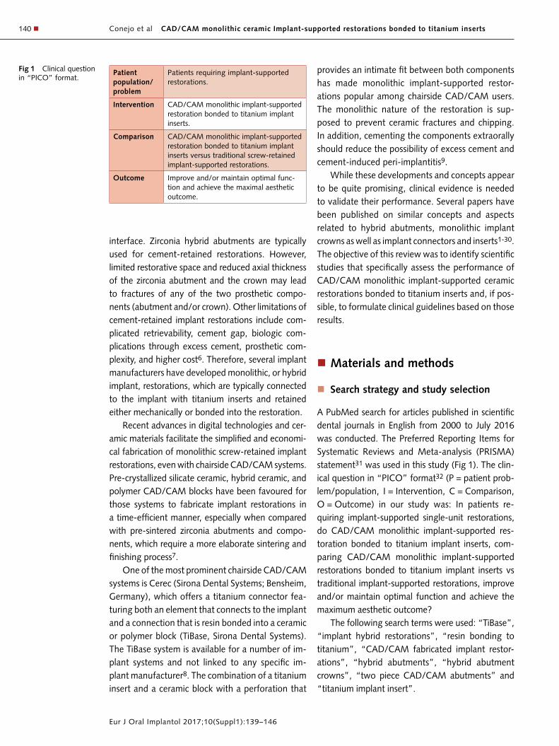
Conejo et al CAD/CAM monolithic ceramic Implant-supported restorations bonded to titanium inserts140 n
Eur J Oral Implantol 2017;10(Suppl1):139–146
provides an intimate fit between both components has made monolithic implant-supported restor-ations popular among chairside CAD/CAM users. The monolithic nature of the restoration is sup-posed to prevent ceramic fractures and chipping. In addition, cementing the components extraorally should reduce the possibility of excess cement and cement-induced peri-implantitis9.
While these developments and concepts appear to be quite promising, clinical evidence is needed to validate their performance. Several papers have been published on similar concepts and aspects related to hybrid abutments, monolithic implant crowns as well as implant connectors and inserts1-30. The objective of this review was to identify scientific studies that specifically assess the performance of CAD/CAM monolithic implant-supported ceramic restorations bonded to titanium inserts and, if pos-sible, to formulate clinical guidelines based on those results.
n Materials and methods
n Search strategy and study selection
A PubMed search for articles published in scientific dental journals in English from 2000 to July 2016 was conducted. The Preferred Reporting Items for Systematic Reviews and Meta-analysis (PRISMA) statement31 was used in this study (Fig 1). The clin-ical question in “PICO” format32 (P = patient prob-lem/population, I = Intervention, C = Comparison, O = Outcome) in our study was: In patients re-quiring implant-supported single-unit restorations, do CAD/CAM monolithic implant-supported res-toration bonded to titanium implant inserts, com-paring CAD/CAM monolithic implant-supported restorations bonded to titanium implant inserts vs traditional implant-supported restorations, improve and/or maintain optimal function and achieve the maximum aesthetic outcome?
The following search terms were used: “TiBase”, “implant hybrid restorations”, “resin bonding to titanium”, “CAD/CAM fabricated implant restor-ations”, “hybrid abutments”, “hybrid abutment crowns”, “two piece CAD/CAM abutments” and “titanium implant insert”.
interface. Zirconia hybrid abutments are typically used for cement-retained restorations. However, limited restorative space and reduced axial thickness of the zirconia abutment and the crown may lead to fractures of any of the two prosthetic compo-nents (abutment and/or crown). Other limitations of cement-retained implant restorations include com-plicated retrievability, cement gap, biologic com-plications through excess cement, prosthetic com-plexity, and higher cost6. Therefore, several implant manufacturers have developed monolithic, or hybrid implant, restorations, which are typically connected to the implant with titanium inserts and retained either mechanically or bonded into the restoration.
Recent advances in digital technologies and cer-amic materials facilitate the simplified and economi-cal fabrication of monolithic screw-retained implant restorations, even with chairside CAD/CAM systems. Pre-crystallized silicate ceramic, hybrid ceramic, and polymer CAD/CAM blocks have been favoured for those systems to fabricate implant restorations in a time-efficient manner, especially when compared with pre-sintered zirconia abutments and compo-nents, which require a more elaborate sintering and finishing process7.
One of the most prominent chairside CAD/CAM systems is Cerec (Sirona Dental Systems; Bensheim, Germany), which offers a titanium connector fea-turing both an element that connects to the implant and a connection that is resin bonded into a ceramic or polymer block (TiBase, Sirona Dental Systems). The TiBase system is available for a number of im-plant systems and not linked to any specific im-plant manufacturer8. The combination of a titanium insert and a ceramic block with a perforation that
Fig 1 Clinical question in “PICO” format.
Patient population/ problem
Patients requiring implant-supported restorations.
Intervention CAD/CAM monolithic implant-supported restoration bonded to titanium implant inserts.
Comparison CAD/CAM monolithic implant-supported restoration bonded to titanium implant inserts versus traditional screw-retained implant-supported restorations.
Outcome Improve and/or maintain optimal func-tion and achieve the maximal aesthetic outcome.

Conejo et al CAD/CAM monolithic ceramic Implant-supported restorations bonded to titanium inserts n 141
Eur J Oral Implantol 2017;10(Suppl1):139–146
We also reviewed the bibliographies and related searches of all selected full-text articles. A hand search of the literature was also conducted.
Inclusion criteria
• All study types related to the topic;• Articles in English; • Publication dates from 2000 to July 2016.
Exclusion criteria
• Studies related to implant placement;• CAD/CAM tooth-supported restorations;• Full-mouth implant-supported restorations;• Articles related to zirconia implants;• Articles that did not contain detailed information
about the topic of study;• Case reports.
n Selection of studies
From an initial 505 studies, 70 studies were selected for full-text analyses. Following detailed analysis by two independent reviewers, a final number of 25 articles met the inclusion criteria (Fig 2).
n Excluded studies
Of the 70 full-text articles, 45 were excluded from the final analysis.Reasons for exclusion:• Research question and purpose of the study
focused on parameters other than implant-supported monolithic restorations, hybrid abut-ments, or bonding to titanium abutments or inserts.
• Methods did not apply CAD/CAM for fabrication of the restorations.
n Data extraction
Characteristics of selected studies are listed in Tables 1 to 3. The 25 studies included were classified into three groups based on their key topic:• Zirconia hybrid abutments;• Bonding to titanium;
• Monolithic CAD/CAM implant restorations (cement-retained).
Due to the lack of clinical data and heterogeneity of the in-vitro studies, meta-analyses were not possible.
n Results
None of the selected studies reported on the clin-ical performance of CAD/CAM monolithic implant-supported restorations bonded to titanium inserts or bases. However, 25 studies met the broader inclu-sion criteria, evaluating key aspects of this protocol and similar restoration designs.
A total of 15 articles (Table 1) reported on the performance of zirconia hybrid abutments2-5,12-19, indicating that titanium inserts bonded to zirconia abutments improve the overall fracture strength,
Fig 2 Search strategy.
InclusionAll study types related to the topic
Publications were restricted to the English language and to publication dates from 2000 through to July 2016
ExclusionStudies related implant placement
CAD/CAM tooth supported restorationsFully endentulous implant restorations
Articles related to Zirconia implantsStudies that did not contain detailed information about
the topic of studyCase reports
PubMed430 Citation(s)
Hand search10 Citation(s)
0 Non-Duplicated Citations screened
70 articles retrieved
Inclusion/ Exclusion
Criteria applied
45 articles excluded after full text screen
25 articles included
Articles excluded during data extraction
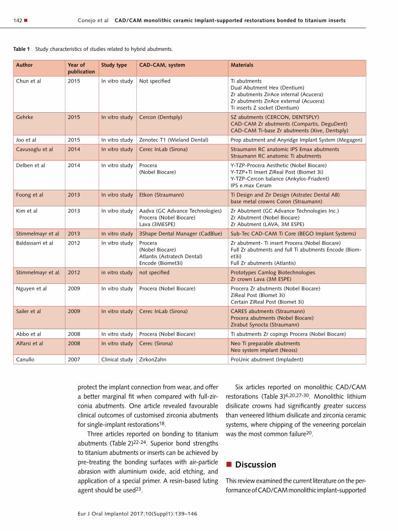
Conejo et al CAD/CAM monolithic ceramic Implant-supported restorations bonded to titanium inserts142 n
Eur J Oral Implantol 2017;10(Suppl1):139–146
Table 1 Study characteristics of studies related to hybrid abutments.
Author Year of publication
Study type CAD-CAM, system Materials
Chun et al 2015 In vitro study Not specified Ti abutments Dual Abutment Hex (Dentium) Zr abutments ZirAce internal (Acucera) Zr abutments ZirAce external (Acucera) Ti inserts Z socket (Dentium)
Gehrke 2015 In vitro study Cercon (Dentsply) SZ abutments (CERCON, DENTSPLY) CAD-CAM Zr abutments (Compartis, DeguDent) CAD-CAM Ti-base Zr abutments (Xive, Dentsply)
Joo et al 2015 In vitro study Zenotec T1 (Wieland Dental) Prop abutment and Anyridge Implant System (Megagen)
Cavusoglu et al 2014 In vitro study Cerec InLab (Sirona) Straumann RC anatomic IPS Emax abutments Straumann RC anatomic Ti abutments
Delben et al 2014 In vitro study Procera (Nobel Biocare)
Y-TZP-Procera Aesthetic (Nobel Biocare) Y-TZP+Ti Insert ZiReal Post (Biomet 3i) Y-TZP-Cercon balance (Ankylos-Friadent) IPS e.max Ceram
Foong et al 2013 In vitro study Etkon (Straumann) Ti Design and Zir Design (Astratec Dental AB) base metal crowns Coron (Straumann)
Kim et al 2013 In vitro study Aadva (GC Advance Technologies) Procera (Nobel Biocare) Lava (3MESPE)
Zr Abutment (GC Advance Technologies Inc.) Zr Abutment (Nobel Biocare) Zr Abutment (LAVA, 3M ESPE)
Stimmelmayr et al 2013 In vitro study 3Shape Dental Manager (CadBlue) Sub-Tec CAD-CAM Ti Core (BEGO Implant Systems)
Baldassarri et al 2012 In vitro study Procera (Nobel Biocare) Atlantis (Astratech Dental) Encode (Biomet3i)
Zr abutment- Ti insert Procera (Nobel Biocare) Full Zr abutments and full Ti abutments Encode (Biom-et3i) Full Zr abutments (Atlantis)
Stimmelmayr et al. 2012 in vitro study not specified Prototypes Camlog Biotechnologies Zr crown Lava (3M ESPE)
Nguyen et al 2009 In vitro study Procera (Nobel Biocare) Procera Zr abutments (Nobel Biocare) ZiReal Post (Biomet 3i) Certain ZiReal Post (Biomet 3i)
Sailer et al 2009 In vitro study Cerec InLab (Sirona) CARES abutments (Straumann) Procera abutments (Nobel Biocare) Zirabut Synocta (Straumann)
Abbo et al 2008 In vitro study Procera (Nobel Biocare) Ti abutments Zr copings Procera (Nobel Biocare)
Alfarsi et al 2008 In vitro study Cerec (Sirona) Neo Ti preparable abutments Neo system implant (Neoss)
Canullo 2007 Clinical study ZirkonZahn ProUnic abutment (Impladent)
protect the implant connection from wear, and offer a better marginal fit when compared with full-zir-conia abutments. One article revealed favourable clinical outcomes of customised zirconia abutments for single-implant restorations18.
Three articles reported on bonding to titanium abutments (Table 2)22-24. Superior bond strengths to titanium abutments or inserts can be achieved by pre-treating the bonding surfaces with air-particle abrasion with aluminium oxide, acid etching, and application of a special primer. A resin-based luting agent should be used23.
Six articles reported on monolithic CAD/CAM restorations (Table 3)6,20,27-30. Monolithic lithium disilicate crowns had significantly greater success than veneered lithium disilicate and zirconia ceramic systems, where chipping of the veneering porcelain was the most common failure20.
n Discussion
This review examined the current literature on the per-formance of CAD/CAM monolithic implant-supported

Conejo et al CAD/CAM monolithic ceramic Implant-supported restorations bonded to titanium inserts n 143
Eur J Oral Implantol 2017;10(Suppl1):139–146
restorations bonded to titanium inserts. The search period was defined from 2000 to July 2016. There were no studies that met the inclusion criteria pub-lished before 2004. None of the studies assessed the exact protocol of titanium inserts bonded into CAD/CAM monolithic crowns, neither in vivo nor in the laboratory. The 25 studies that met the broader inclu-sion criteria evaluated various restoration designs of hybrid abutments, bonding to titanium, and mono-lithic CAD/CAM cement-retained restorations. Except for one, all of them were in vitro studies.
n Monolithic hybrid abutment crown
Kurbad7 and Rauscher9 described the use of CAD/CAM lithium disilicate blocks (IPS e.max CAD, Ivo-clar Vivadent), which are specifically designed for
screw-retained implant-supported restorations. They also used CAD/CAM zirconia blocks (InCoris Meso, Sirona) to fabricate two-piece hybrid abutments. All of their restorations were completed with a TiBase connector (Sirona), which engages into the ceramic block’s perforation and features an anti-rotational component. Beuer and colleagues explained the dig-ital One-Abutment/One-Time Concept11. A digital intraoral impression is made on the day of the im-plant placement with a scan body and a CAD pro-gram (Dental Designer, 3shape) to design the final restoration. After bone healing, at the time of second stage surgery, the final screw-retained CAD/CAM-fabricated crown is inserted. This allows the soft tis-sue to heal around the definitive restoration and, therefore, avoids soft-tissue trauma through the repeated removal and re-insertion of abutments or
Table 2 Study characteristics of studies related to bonding to titanium abutments.
Author Year of publication
Study type CAD-CAM, system Materials
Maltzahn et al 2015 In vitro study CADSPEED GmbH Ti bases (Medentika) CAD/CAM Zr copings (CADSPEED) Panavia F 2.0 (Kuraray Noritake Dental Inc.) RelyX Unicem and Rocatec System (3MESPE)
Egoshi et al 2013 In vitro study not specified Estenia C&B primer and resin composite (Kuraray Noritake Dental)
Mehl et al 2012 In vitro study Autodesk Inventor (Autodesk) BEGO Medifacturing System (BEGO)
Ti universal abutments (Camlog Biotechnologies) Ketac-Cem, Durelon (3M ESPE) Multilink Implant (Ivoclar Vivadent)
Abbo et al 2008 In vitro study Procera (Nobel Biocare) Ti abutments Zr copings Procera (Nobel Biocare)
Table 3 Study characteristics of studies related to monolithic CAD/CAM implant restorations.
Author Year of publication
Study type CAD-CAM, system Materials
Lassle et al 2015 In vitro study Procera (Nobel Biocare) Snappy abutments 5.5 Conical connection (Nobel Biocare) IPS e.max crowns
Aktas et al 2014 In vitro study Cerec (Sirona) Solid and synOcta abutments (Straumann) inCoris ZI and Alumina silicate glass ceramic blocks (Sirona)
Cavusoglu et al 2014 In vitro study Cerec InLab (Sirona) Straumann RC anatomic IPS e.max abutments Straumann RC anatomic Ti abutments
Joda et al 2014 In vitro study Cerec (Sirona) synOcta prefabricated Ti CAD/CAM Ti CARES prefabricated Variobase Ti abutments (Straumann) Lava Ultimate (3M ESPE)
Martinez-Rus et al
2012 In vitro study Cerec InLab (Sirona) CARES ceramic abutments and CARES Ti abutments (Straumann) IPS e.max CAD,PRESS ZirCAD
Wolf et al 2008 In vitro study Cerec (Sirona) GingiHue Ti abutment ZiReal Zr abutment (Biomet 3i)
Conejo et al CAD/CAM monolithic ceramic Implant-supported restorations bonded to titanium inserts144 n
Eur J Oral Implantol 2017;10(Suppl1):139–146
healing screws. Monolithic screw-retained implant-supported restorations depend on an ideal implant position because of the access screw hole. If an angulated implant needs to be restored, a hybrid abutment designed with a shoulder that follows the soft tissue contours and a cement-retained crown should be considered. The shoulder position is crucial for excess cement removal. The prefabricated fitting surfaces of the TiBases (Sirona) and new implant solution blocks (Vita Zahn-Fabrik, Bad Sackingen, Germany, and Ivoclar Vivadent), the mechanical anti-rotational component, and the parallelism and height of the walls (4.0 mm) seem to provide a safe design for monolithic restoration design19.
A variety of materials are described in the litera-ture, ranging from polymer-based blocks for tem-porary restorations (VITA CAD Temp, Vita Zahn-Fabrik and Telio CAD, Ivoclar Vivadent) to silicate ceramic (VITA SUPRINITY IS, Vita Zahn-Fabrik and IPS e.max CAD A, Ivoclar Vivadent) and hybrid cer-amic block (VITA ENAMIC IS, Vita Zahn-Fabrik). A minimal marginal shoulder width of 0.4 mm, a circumferential wall thickness of 0.8 mm, and an occlusal thickness of 1.0 mm are recommended by the manufacturers.
n Zirconia hybrid abutments
Delben et al3 compared external hexagon, internal hexagon, and Morse taper full-zirconia abutment connections. Although the Morse taper connection was more prone to early abutment fracture, prob-ably due to the thin cross-section at the abutment neck, all three groups offer higher strength than mean functional load in the anterior region3.
According to Beuer et al, fracture strength of im-plant abutments increased with the implant diam-eter15. Zirconia implant abutments connected to titanium cores showed higher fracture strength than one-piece zirconia abutments. This hybrid abutment design was recommended as a safe option even for the posterior areas of the mouth15. Several other authors reported similar findings5,12,13,21.
In respect to the wear at the titanium-titanium and titanium-zirconia implant-abutment interface, Stimmelmayr et al17 reported a higher wear of tita-nium implants under cyclic loading when connected to one-piece zirconia abutments than when they
were connected to titanium abutments. Cavusoglu et al2 also concluded that the zirconia-abutment-tita-nium-implant interface is susceptible to wear of the abutment coupled with deformation of the implant neck greater than that associated with the conven-tional titanium-abutment-titanium-implant interface under dynamic loading. According to Baldassarri et al4, the implant-titanium abutment connection showed significantly better fit than all implant-zir-conia abutment configurations. Mean gaps were approximately three to seven times larger than those in the titanium abutment systems. In addition, Can-ullo18 reported that the gap in the zirconia-core-metal-abutment system was comparable to gap val-ues of the abutment-implant systems available on the market. Another study demonstrated that single implants restored with lithium disilicate crowns and zirconia abutments with titanium inserts could with-stand maximum masticatory force in the incisor area when the axial walls of the abutment were at least 0.5 mm thick19.
n Bonding to titanium abutments and inserts
The adhesive bonding of titanium inserts into the implant restorations is a key element of the pro-tocol under review. The combination of air-particle abrasion, acid etching and MDP-primer application seems to improve titanium bonding22. It was also concluded that surface modifications influence the retention forces between titanium and zirconia com-ponents in two-piece implant abutments23.
Mehl et al24 studied the influence of abut-ment height and thermocycling on retrievability of cemented implant-supported crowns. They con-cluded that crowns cemented with glass ionomer cement were potentially retrievable with a clinically applicable removable device when the abutment height ranged from 2 to 4 mm. Polycarboxylate or composite resin cements should be used as non-retrievable permanent cementation options. When restorations are cemented on abutments with a height of 2 mm or less, composite resin cement should be used to minimise the risk of crown loos-ening. Abbo et al25 reported that the resistance to tensile forces was significantly increased when the height of the abutment was increased by 1 mm.

Conejo et al CAD/CAM monolithic ceramic Implant-supported restorations bonded to titanium inserts n 145
Eur J Oral Implantol 2017;10(Suppl1):139–146
n Monolithic CAD/CAM restorations
A simplified approach to implant restorations with digital technologies was described by Brooks et al26. The benefits of this protocol for fabrication of multiple single crowns in one clinic session include: reduced chair and production time, greater ac-curacy, reduced cost, improved impression of the gingiva, fewer adjustments, ideal margin location and simplified cementation. Similarly, an in vitro study by Alfarsi et al27 concluded that chairside CAD/CAM fabrication of customised ceramic abutments and their associated ceramic crowns using pre-sintered feldspathic porcelain blocks was a viable treatment option. These outcomes were further supported by Stona et al28, who investi-gated the fracture resistance of CAD/CAM cer-amic crowns cemented on solid abutments. Cerec Vita Blocks Mark II (Vita Zahnfabrik), IPS Empress CAD, and IPS e.max CAD (both Ivoclar Vivadent) ceramic crowns cemented on solid abutments had sufficient resistance to withstand physiologi-cal chewing forces28. In addition, Martinez-Rus et al20 demonstrated that titanium abutments restored with monolithic lithium disilicate crowns presented the highest fracture resistance compared with manually veneered pressed lithium disilicate and zirconia copings. Some authors suggested the concept of cementing CAD/CAM lithium disilicate crowns to the abutments extraorally and deliver-ing them as screw-retained implant restorations. The preparation of a screw access channel into the lithium disilicate crowns significantly reduced the axial load capacity when compared with intact crowns without occlusal access6. However, the actual diameter of the screw access channel did not make a statistically significant difference in terms of load-bearing capacity6.
Anti-rotational abutment features positively affected the marginal fit of single implant-retained crowns29. Furthermore, digitizing techniques improved the fit of single-implant restorations29.
Joda et al30 presented a complete digital workflow for the fabrication of implant-supported single-unit monolithic crowns in the posterior jaw. However, the suggested application of a resin nanoceramic (Lava Ultimate Restorative, 3M ESPE, St Paul, Minneapolis, USA) as full-contour material has to be considered
experimental. Clinical investigations with long-term follow-up are necessary30.
Novel material blocks for CAD/CAM restorations supported by a single implant and bonded titanium inserts offer aesthetic advantages over conventional metal abutments and simpler processing than zir-conia hybrid abutments. Also, manufacturing costs are reduced with full-anatomic abutment crowns, in which the abutment and the crown are designed as one piece. The absence of a cement space at the abutment-crown interface may present a biologic advantage.
CAD/CAM monolithic implant-supported res-torations that are bonded to Ti inserts have several benefits and can be fabricated chairside. However, there is currently no scientific evidence to support the clinical application of this specific design. Several studies, most of them laboratory based, are available on select aspects and similar prosthetic designs. The lack of clinical data from several trials and heteroge-neity of the in vitro studies did not allow for further statistical analyses of the results. Controlled clinical trials and longitudinal studies are necessary before this type of restoration can be recommended for use in clinical practice without restrictions.
n Conclusions
While laboratory studies and evaluations of similar designs indicate promising outcomes, there is a strong need for clinical studies that evaluate the performance of CAD/CAM monolithic implant-supported restor-ations bonded to Ti inserts and bases before this pros-thetic design can be recommended for routine use in clinical practice without restrictions.
n References
1. Watkin A, Kerstein RB. Improving darkened anterior peri-implant tissue colour with zirconia custom implant abut-ments. Compend Contin Educ Dent 2008;29: 238–240, 242.
2. Cavusoglu Y, Akça K, Gurbuz R, Cehreli MC. A pilot study of joint stability at the zirconium or titanium abutment/titanium implant interface. Int J Oral Maxillofac Implants 2014;29: 338–343.
3. Delben JA, Barao VA, Ferreira MB, da Silva NR, Thompson VP, Assuncao WG. Influence of abutment-to-fixture design on reliability and failure mode of all-ceramic crown systems. Dent Mater 2014;30: 408–416.

Conejo et al CAD/CAM monolithic ceramic Implant-supported restorations bonded to titanium inserts146 n
Eur J Oral Implantol 2017;10(Suppl1):139–146
4. Baldassarri M, Hjerppe J, Romeo D, Fickl S, Thompson VP, Stappert CF. Marginal accuracy of three implant-ceramic abutment configurations. Int J Oral Maxillofac Implants 2012;27: 537–543.
5. Chun HJ, Yeo IS, Lee JH, Kim SK, Heo SJ, Koak JY, Han JS, Lee SJ. Fracture strength study of internally connected zir-conia abutments reinforced with titanium inserts. Int J Oral Maxillofac Implants 2015;30: 346–350.
6. Lassle MJ. CAD/CAM lithium disilicate crown performance cemented extraorally and delivered as a screw-retained im-plant restoration. [M.S.]. University of Minnesota; 2015.
7. Kurbad A. Provisional polymer-based CAD/CAM implant superstructures. Int J Comput Dent 2014;17: 239–251.
8. Kurbad A, Kurbad S. CAD/CAM-based implant abutments. Int J Comput Dent 2013;16: 125–141.
9. Rauscher O. Impressionless implant-supported restorations with cerec 4.2. Int J Comput Dent 2014;17: 159–168.
10. Rauscher O. Impression-free implant restorations with Cerec InLab. Int J Comput Dent 2011;14: 139–146.
11. Beuer F, Groesser J, Schweiger J, Hey J, Guth JF, Stimmel-mayr M. The digital one-abutment/one-time concept. A clin-ical report. J Prosthodont. 2015. doi: 10.1111/jopr.12256. [Epub ahead of print]
12. Foong JK, Judge RB, Palamara JE, Swain MV. Fracture resist-ance of titanium and zirconia abutments: An in vitro study. J Prosthet Dent 2013;109: 304–312.
13. Nguyen HQ, Tan KB, Nicholls JI. Load fatigue performance of implant-ceramic abutment combinations. Int J Oral Max-illofac Implants 2009;24: 636–646.
14. Kim JS, Raigrodski AJ, Flinn BD, Rubenstein JE, Chung KH, Mancl LA. In vitro assessment of three types of zirconia implant abutments under static load. J Prosthet Dent 2013;109: 255–263.
15. Gehrke P, Johannson D, Fischer C, Stawarczyk B, Beuer F. In vitro fatigue and fracture resistance of one- and two-piece CAD/CAM zirconia implant abutments. Int J Oral Maxillofac Implants 2015;30: 546–554.
16. Stimmelmayr M, Sagerer S, Erdelt K, Beuer F. In vitro fatigue and fracture strength testing of one-piece zirconia implant abutments and zirconia implant abutments connected to titanium cores. Int J Oral Maxillofac Implants 2013;28: 488–493.
17. Stimmelmayr M, Edelhoff D, Guth JF, Erdelt K, Happe A, Beuer F. Wear at the titanium-titanium and the titanium-zirconia implant-abutment interface: A comparative in vitro study. Dent Mater 2012;28: 1215–1220.
18. Canullo L. Clinical outcome study of customized zirconia abutments for single-implant restorations. Int J Prosthodont 2007;20: 489–493.
19. Joo HS, Yang HS, Park SW, Kim HS, Yun KD, Ji MK, Lim HP. Influence of preparation depths on the fracture load of customized zirconia abutments with titanium insert. J Adv Prosthodont 2015;7: 183–190.
20. Martinez-Rus F, Ferreiroa A, Ozcan M, Bartolome JF, Pradies G. Fracture resistance of crowns cemented on titanium and zirconia implant abutments: A comparison of monolithic versus manually veneered all-ceramic systems. Int J Oral Maxillofac Implants 2012;27: 1448–1455.
21. Sailer I, Sailer T, Stawarczyk B, Jung RE, Hämmerle CH. In vitro study of the influence of the type of connection on the fracture load of zirconia abutments with internal and exter-nal implant-abutment connections. Int J Oral Maxillofac Implants 2009;24: 850–858.
22. Egoshi T, Taira Y, Soeno K, Sawase T. Effects of sandblasting, H2SO4/HCl etching, and phosphate primer application on bond strength of veneering resin composite to commercially pure titanium grade 4. Dent Mater J 2013;32: 219–227.
23. von Maltzahn NF, Holstermann J, Kohorst P. Retention forces between titanium and zirconia components of two-part implant abutments with different techniques of sur-face modification. Clin Implant Dent Relat Res 2016;18: 735–744.
24. Mehl C, Harder S, Shahriari A, Steiner M, Kern M. Influence of abutment height and thermocycling on retrievability of cemented implant-supported crowns. Int J Oral Maxillofac Implants 2012;27: 1106–1115.
25. Abbo B, Razzoog ME, Vivas J, Sierraalta M. Resistance to dislodgement of zirconia copings cemented onto titanium abutments of different heights. J Prosthet Dent 2008;99: 25–29.
26. Brooks JH, Wicks RA, Hottel TL. A simplified approach to im-plant restorations using innovative digital technology. J Tenn Dent Assoc 2014;94:41–44.
27. Alfarsi MA, Okutan HM, Bickel M. CAD/CAM to fabricate ceramic implant abutments and crowns: A preliminary in vitro study. Aust Dent J 2009;54: 12–16.
28. Stona D, Burnett LH,Jr, Mota EG, Spohr AM. Fracture resist-ance of computer-aided design and computer-aided manu-facturing ceramic crowns cemented on solid abutments. J Am Dent Assoc 2015;146: 501–507.
29. Aktas G, Özcan N, Aydin DH, Şahin E, Akça K. Effect of digitizing techniques on the fit of implant-retained crowns with different antirotational abutment features. J Prosthet Dent 2014;111: 367–372.
30. Joda T, Huber S, Burki A, Zysset P, Bragger U. Influence of abutment design on stiffness, strength, and failure of implant-supported monolithic resin nano ceramic (RNC) crowns. Clin Implant Dent Relat Res 2015;17: 1200–1207.
31. Moher D, Shamseer L, Clarke M, Ghersi D, Liberati A, Pet-ticrew M, Shekelle P, Stewart LA; PRISMA-P Group. Preferred reporting items for systematic review and meta-analysis pro-tocols (PRISMA-P) 2015 statement. Syst Rev 2015;1:1.
32. da Costa Santos CM, de Mattos Pimenta CA, Nobre MR. The PICO strategy for the research question construction and evidence search. Rev Lat Am Enfermagem 2007;15: 508–511.


