Popliteal fossa
21
The popliteal fossa The distal continuation of the adductor canal Dr.Murali.M.S; M.B.A. Prof. of Surgery D Y Patil Medical College Mauritius.
-
Upload
uthamalingam-murali -
Category
Health & Medicine
-
view
96 -
download
4
Transcript of Popliteal fossa

The popliteal fossaThe distal continuation of the adductor canal
Dr.Murali.M.S; M.B.A.Prof. of SurgeryD Y Patil Medical CollegeMauritius.
Boundaries
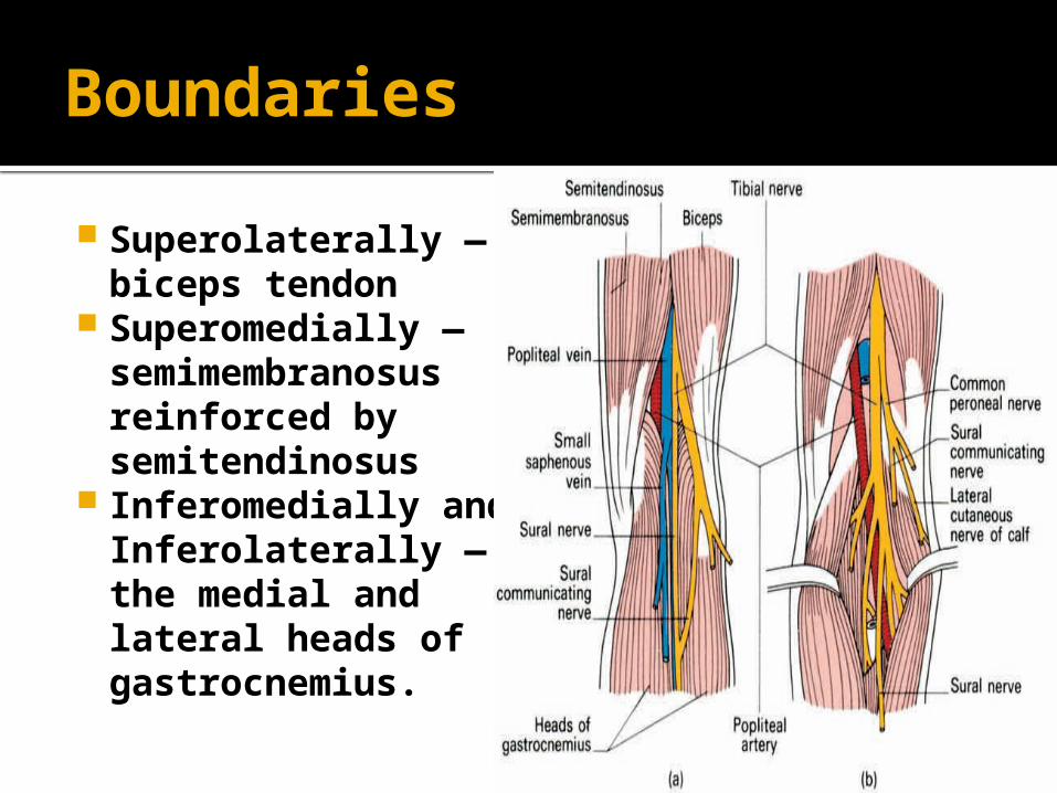
Superolaterally — biceps tendon
Superomedially — semimembranosus reinforced by semitendinosus
Inferomedially and Inferolaterally — the medial and lateral heads of gastrocnemius.
Roof
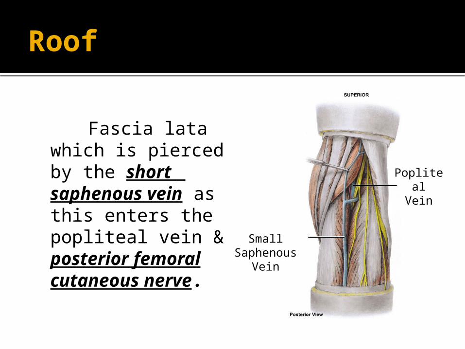
Fascia lata which is pierced by the short saphenous vein as this enters the popliteal vein & posterior femoral cutaneous nerve.
SmallSaphenous
Vein
PoplitealVein
Floor
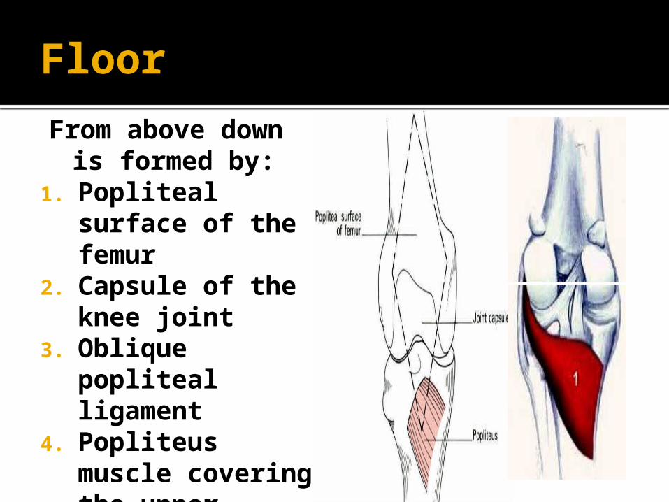
From above down is formed by:
1. Popliteal surface of the femur
2. Capsule of the knee joint
3. Oblique popliteal ligament
4. Popliteus muscle covering the upper posterior surface of the tibia.
LM
Popliteal Muscle
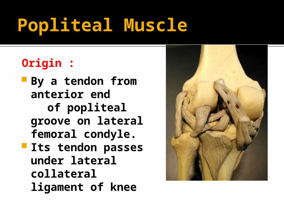
By a tendon from anterior end
of popliteal groove on lateral femoral condyle.
Its tendon passes under lateral collateral ligament of knee
Origin :
Popliteal Muscle
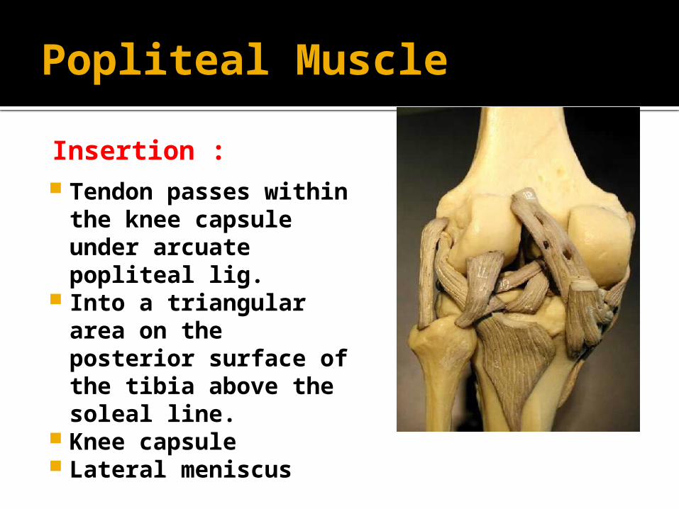
Tendon passes within the knee capsule under arcuate popliteal lig.
Into a triangular area on the posterior surface of the tibia above the soleal line.
Knee capsule Lateral meniscus
Insertion :
Contents
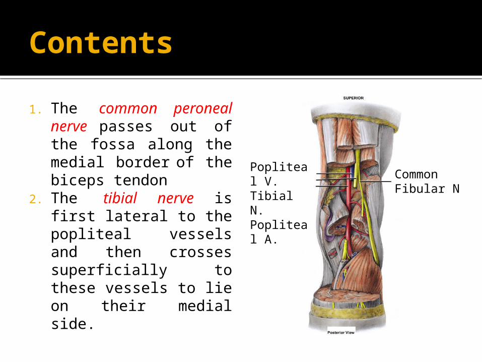
1. The common peroneal nerve passes out of the fossa along the medial border of the biceps tendon
2. The tibial nerve is first lateral to the popliteal vessels and then crosses superficially to these vessels to lie on their medial side.
Popliteal V.Tibial N.Popliteal A.
CommonFibular N
Contents
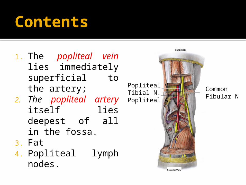
1. The popliteal vein lies immediately superficial to the artery;
2. The popliteal artery itself lies deepest of all in the fossa.
3. Fat 4. Popliteal lymph
nodes.
Popliteal V.Tibial N.Popliteal A.
CommonFibular N
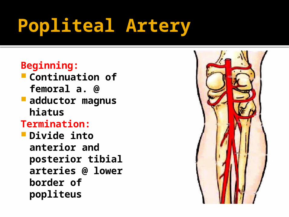
Popliteal Artery
Beginning: Continuation of
femoral a. @ adductor magnus
hiatusTermination: Divide into
anterior and posterior tibial arteries @ lower border of popliteus
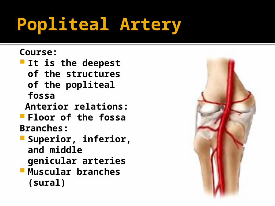
Popliteal Artery
Course: It is the deepest of
the structures of the popliteal fossa
Anterior relations: Floor of the fossaBranches: Superior, inferior,
and middle genicular arteries
Muscular branches (sural)
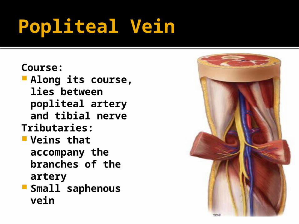
Popliteal Vein
Course: Along its course,
lies between popliteal artery and tibial nerve
Tributaries: Veins that
accompany the branches of the artery
Small saphenous vein
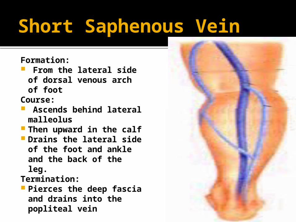
Short Saphenous Vein
Formation: From the lateral side
of dorsal venous arch of foot
Course: Ascends behind lateral
malleolus Then upward in the calf Drains the lateral side
of the foot and ankle and the back of the leg.
Termination: Pierces the deep fascia
and drains into the popliteal vein
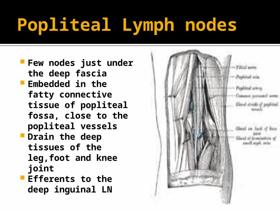
Popliteal Lymph nodes
Few nodes just under the deep fascia
Embedded in the fatty connective tissue of popliteal fossa, close to the popliteal vessels
Drain the deep tissues of the leg,foot and knee joint
Efferents to the deep inguinal LN
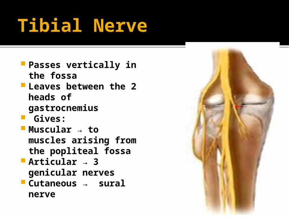
Tibial Nerve
Passes vertically in the fossa
Leaves between the 2 heads of gastrocnemius
Gives: Muscular → to
muscles arising from the popliteal fossa
Articular → 3 genicular nerves
Cutaneous → sural nerve
→
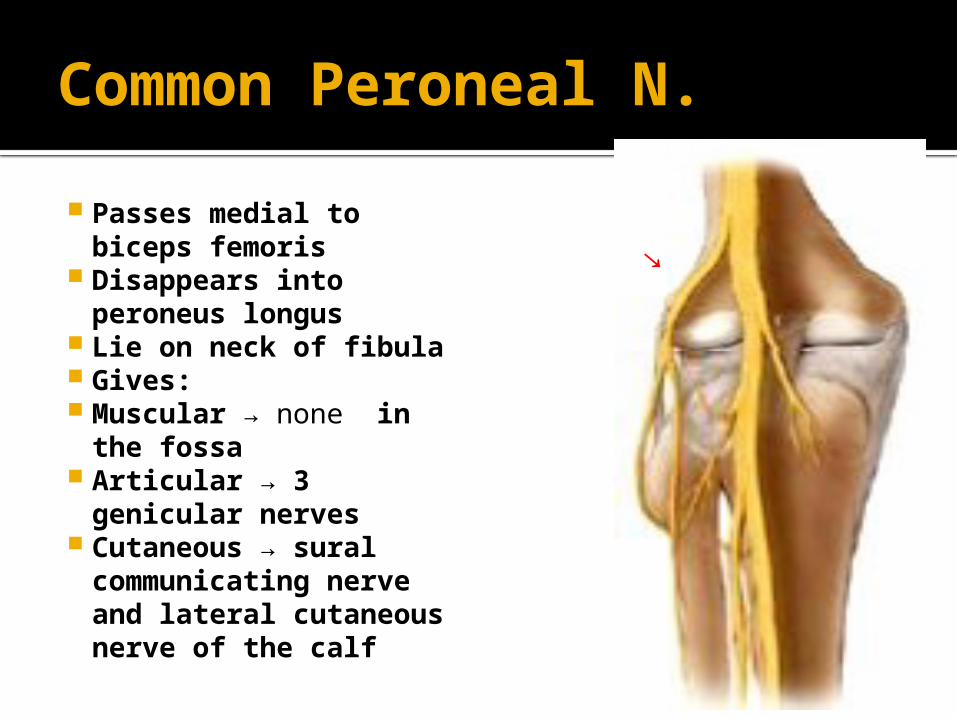
Common Peroneal N.
Passes medial to biceps femoris
Disappears into peroneus longus
Lie on neck of fibula Gives: Muscular → none in the
fossa Articular → 3
genicular nerves Cutaneous → sural
communicating nerve and lateral cutaneous nerve of the calf
→

APPLIED ANATOMY
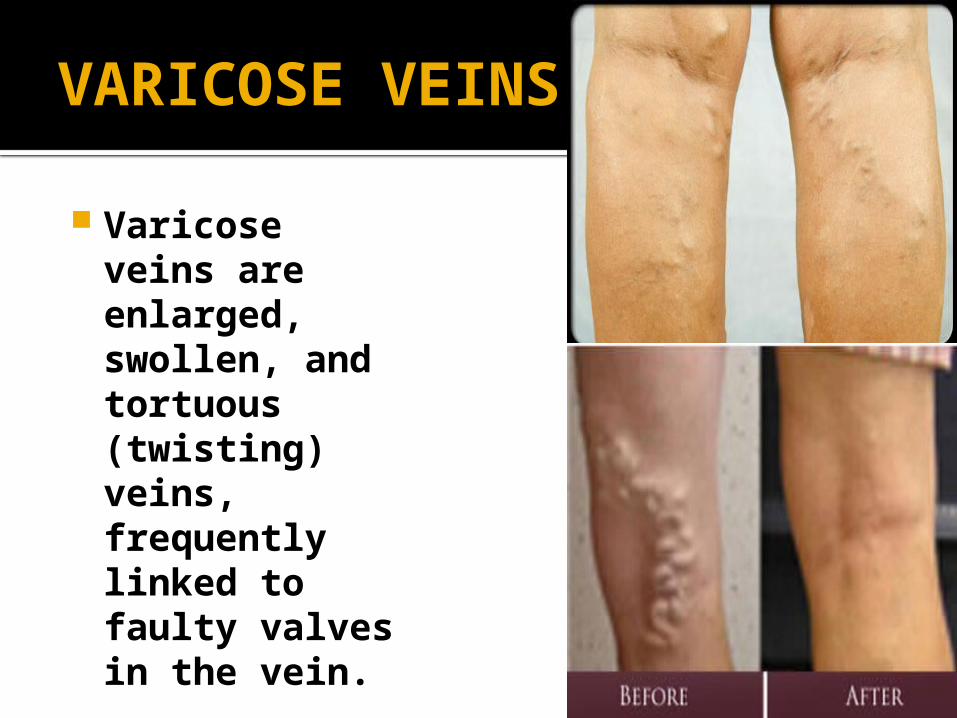
VARICOSE VEINS
Varicose veins are enlarged, swollen, and tortuous (twisting) veins, frequently linked to faulty valves in the vein.
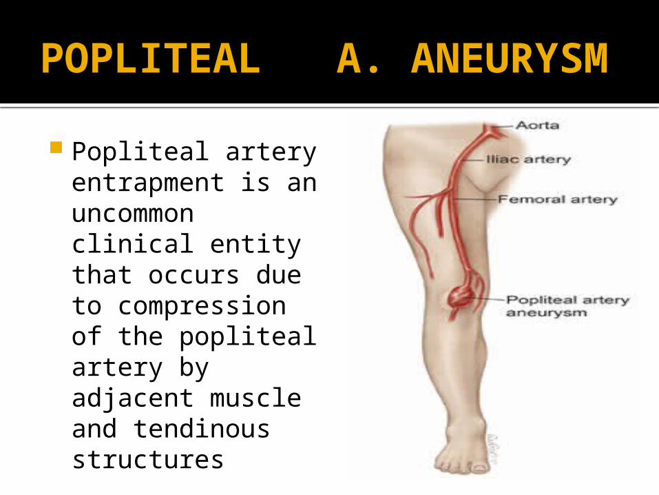
POPLITEAL A. ANEURYSM Popliteal artery
entrapment is an uncommon clinical entity that occurs due to compression of the popliteal artery by adjacent muscle and tendinous structures
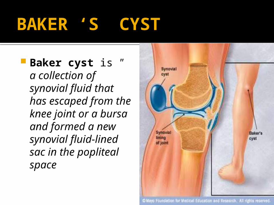
BAKER ‘S CYST
Baker cyst is " a collection of synovial fluid that has escaped from the knee joint or a bursa and formed a new synovial fluid-lined sac in the popliteal space