Pichia guilliermondii and in two filarial endemic
11
Molecular evidence on the occurrence of co-infection with Pichia guilliermondii and Wuchereria bancrofti in two filarial endemic districts of India Mukherjee et al. Mukherjee et al. Infectious Diseases of poverty 2014, 3:13 http://www.idpjournal.com/content/3/1/13
Transcript of Pichia guilliermondii and in two filarial endemic

Molecular evidence on the occurrence ofco-infection with Pichia guilliermondii andWuchereria bancrofti in two filarial endemicdistricts of IndiaMukherjee et al.
Mukherjee et al. Infectious Diseases of poverty 2014, 3:13http://www.idpjournal.com/content/3/1/13
Mukherjee et al. Infectious Diseases of poverty 2014, 3:13http://www.idpjournal.com/content/3/1/13
RESEARCH ARTICLE Open Access
Molecular evidence on the occurrence ofco-infection with Pichia guilliermondii andWuchereria bancrofti in two filarial endemicdistricts of IndiaSuprabhat Mukherjee, Niladri Mukherjee, Prasanta Saini, Prajna Gayen, Priya Roy and Santi P Sinha Babu*
Abstract
Background: Lymphatic filariasis (LF), a vector-borne parasitic disease, is endemic in several parts of India andmostly affects the poor or those with a low-income. The disease results in huge numbers of morbidities, disabilities,and deaths every year. Association of co-infection with other pathogens makes the condition more severe.Although co-infection is becoming a growing area of research, it is yet to emerge as a frontier research topic infilarial research specifically. This study reports the occurrence of a fungal infection in a large number of patientssuffering from bancroftian filariasis in two districts of West Bengal, India.
Methods: Nocturnal blood samples from filarial patients containing parasites and fungus were initially co-cultured,and further the fungus was isolated and characterized. Molecular identification of the isolate was carried out byPCR-based selective amplification and sequencing of highly-conserved D1/D2 region of 26S rDNA, whereaspathogenicity was determined by amplification of the RPS0 gene. A phylogenetic tree was constructed to study therelationship between the isolate and common pathogenic yeasts. The isolate was studied for antibiotic sensitivity,whereas morphological characterization was performed by microscopic techniques.
Results: The isolate was identified as Pichia guilliermondii and this fungus was found to exist in co-infection withWuchereria bancrofti in filarial patients. The fungus showed resistance to azole antifungals, griseofulvin, and,amphotericin B, whereas significant susceptibility was evident in cases of nystatin and cycloheximide. A total of 197out of 222 patients showed this co-infection.
Conclusion: This study revealed, for the first time, that P. guilliermondii exists as a co-infection in microfilaraemicindividuals living in a filarial endemic zone. The findings are important and have relevance to human health,especially for filarial patients.
Keywords: Lymphatic filariasis, Pichia guilliermondii, Co-infection, Polymerase chain reaction, Molecular identification
Multilingual abstractsPlease see Additional file 1: Multilingual abstracts in thesix official working languages of the United Nations.
BackgroundLymphatic filariasis (LF), a vector-borne disease mainlycaused by the filarial parasites– namely Wuchereria ban-crofti, Brugia malayi, and Brugia timori-has become a
* Correspondence: [email protected] Laboratory, Department of Zoology (Centre for AdvancedStudies), Visva-Bharati University, Santiniketan- 731 235, West Bengal, India
© 2014 Mukherjee et al.; licensee BioMed CenCreative Commons Attribution License (http:/distribution, and reproduction in any mediumDomain Dedication waiver (http://creativecomarticle, unless otherwise stated.
global problem that constitutes 120 million infectionsper year in 81 tropical countries [1]. It is the world’s sec-ond leading cause of long-term disability. Out of thetotal disease burden of LF, W. bancrofti alone accountsfor 90% and this form of LF is termed ‘bancroftian filar-iasis’ [2]. Currently, one-third of the affected persons arefrom South Asia and another third from Africa, whileone-sixth of the world’s population is at risk of infection[1]. It is a disease mostly of the poor, which significantlyaffects this group’s ability to earn an income, and thishas led to its inclusion on the list of neglected tropical
tral Ltd. This is an Open Access article distributed under the terms of the/creativecommons.org/licenses/by/2.0), which permits unrestricted use,, provided the original work is properly credited. The Creative Commons Publicmons.org/publicdomain/zero/1.0/) applies to the data made available in this

Mukherjee et al. Infectious Diseases of poverty 2014, 3:13 Page 2 of 10http://www.idpjournal.com/content/3/1/13
diseases [1]. It results in significant economic and psycho-social impacts wherever it is endemic; disfiguring and/orincapacitating more than 40 million individuals, their fam-ilies, and the endemic communities. Particularly in the In-dian subcontinent, the disease affects the work time ofinfected patients and thereby costs the National Treasurya minimum of US$842 million per year [3]. Lymphatic Fil-ariasis is endemic in several parts of India, including 250districts in 20 states and six union territories (UTs), con-tributing 40% to the global disease burden [3]. The causa-tive agents of LF i.e. filarial parasites live in the body cavityor tissues of vertebrate hosts where they parasitize thelymphatics, which results in the obstruction of the lymph-atic vessels, incompetence, lymphostasis, lymphatic dys-function (hydrocele and lymphedema), and interstitialfibrosis, followed by immunological dysfunction and in-flammation which results in elephantiasis [4]. This diseasealso promotes vulnerability to opportunistic infections [5],particularly during the progression of lymphedema fromchronic filarial infection. Development of elephantiasis iscaused by the long-term recurrent secondary infections byopportunistic microbes [4].Opportunistic microbial infections are very common
in infectious diseases that suppress the host immune sys-tem, and promote secondary infections that are of majorconcern as they make the host weaker or create a life-threatening condition [6,7]. Different bacteria, viruses, andfungi, especially Candida yeasts, have been characterizedas opportunistic pathogens [8]. The epidemiology of yeastinfections is rapidly evolving as co-infection in patientssuffering from primary infectious diseases [9]. Althoughrare, non-albicans Candida (NAC) spp. are emerging aspotential opportunistic pathogens, among which Pichiaguilliermondii (formerly known as Candida guilliermondiior Meyerozyma guilliermondii) is one of the 15 yeast spe-cies related to human diseases [9,10]. It is commonly iso-lated from clinical specimens such as phlegm, wounds,sputum, and blood [11]. Although P. guilliermondii is leastpathogenic compared to the other fungi of the Candidafamily, it can still be responsible for life-threatening in-fections in immunocompromised hosts [9,12]. It consti-tutes 35–65% of all candidaemias in the general patientpopulation and is mostly evident in cancer patients,bone marrow transplant recipients and, to a lesser de-gree, in intensive care unit patients, children, and surgi-cal and HIV-positive patients [13]. As reported by Hornet al. [14], 1–5% prevalence of Candida infection is con-tributed by P. guilliermondii. The large-scale studies ofcandidaemia between 1999 and 2006 found that around15% of a total of 9,717 cases were due to the P. guillier-mondii infection [12]. Dick et al. [15] previously reporteddeath due to disseminated candidiasis caused by the P.guilliermondii infection. Recently, this pathogen has beenreported to cause infection in the knee of a patient lacking
predisposing factors [16]. Occurrence of P. guilliermondiiout of the total Candida isolates in different geographiclocations is 1.1% in the Asia Pacific, 1.0% in Europe, 3.7%in Latin America, and 0.6% in North America [17].Particularly for filariasis, Ormerod et al. [18] reported
an unusual and chronic anaerobic urinary infection in thefilarial patients caused by Bacteroides melaninogenicus, B.fragilis, Peptococcus prevotii, and Propionibacterium gran-ulosum, passed from the abnormal lymphatics. Date et al.[19] reported severe lymphocytopenia, extensive mucosalcandidiasis, and disseminated cryptococcosis in patientswith long-standing filarial chyluria with immunologicalabnormalities. Recently, Metenou et al. [20] reviewedexperimental findings on filaria/mycobacteria or filaria/Plasmodium co-infections in filarial patients. Althoughstudied less, fungal infections are believed to cause prob-lems in LF such as edema-causing skin folds and skin tears[21]. However, occurrence of P. guilliermondii has neitherbeen reported from India nor from the peripheral bloodstream of any microfilaraemic patient. This study reports,for the first time, the co-infection of P. guilliermondii withW. bancrofti in the blood of microfilaraemic patients livingin the filaria endemic zone in West Bengal, India.
MethodsStudy area and populationThis study was conducted in two rural districts, namelyBankura (23° 14′ N and 87° 07′ E) and Birbhum (24° 35′N and 88° 1′ 40″ E), in West Bengal, India. These regionswere previously reported as endemic for lymphatic filaria-sis (LF), with more than 14% prevalence [22,23]. Thestudy was approved by the Human Ethical Committee ofthe Sub-Divisional Hospital, Bolpur, West Bengal, India,and by the Institutional Ethics Committee of the Visva-Bharati University, Santiniketan- 731 235, West Bengal,India. Before taking blood samples, written consents wereobtained from normal and infected persons.
Blood samplesThis study was carried out on the population residing in afilarial endemic region and suffering from bancroftian fil-ariasis. The study originated and was carried out while LFwas being studied for its prevalence and a clinical trial wasbeing conducted by Gayen et al. [22,23]. As reportedpreviously [22,23], all filarial patients were screened forthe presence of microfilaria of W. bancrofti in microscopicpreparation from finger-prick blood samples, whichformed a thick film over a glass slide and was followedby Giemsa staining. Initially, the study involved theentire population mentioned in the LF prevalence studyconducted between 2006 and 2008 by Gayen et al.[22,23]. Later, a total of 62 individuals from 31 endemicvillages from the districts of Bankura and Birbhum wereagain studied between 2011 and 2012. All the patients
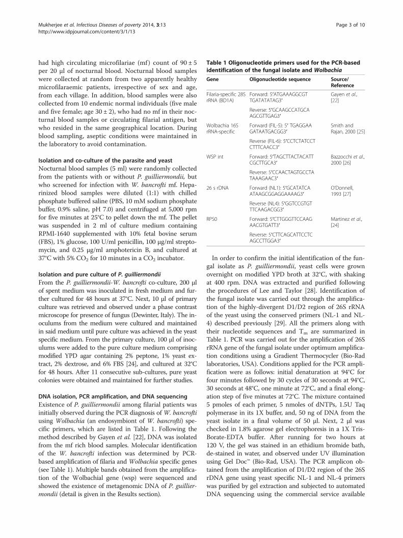
Table 1 Oligonucleotide primers used for the PCR-basedidentification of the fungal isolate and Wolbachia
Gene Oligonucleotide sequence Source/Reference
Filaria-specific 28SrRNA (BD1A)
Forward: 5′ATGAAAGGCGTTGATATATAG3′
Gayen et al.,[22]
Reverse: 5′GCAAGCCATGCAAGCGTTGAG3′
Wolbachia 16SrRNA-specific
Forward (FIL-5): 5′ TGAGGAAGATAATGACGG3′
Smith andRajan, 2000 [25]
Reverse (FIL-6): 5′CCTCTATCCTCTTTCAACC3′
WSP int Forward: 5′TAGCTTACTACATTCGCTTGCA3′
Bazzocchi et al.,2000 [26]
Reverse: 5′CCAACTAGTGCCTATAAAGAAC3′
26 s rDNA Forward (NL1): 5′GCATATCAATAAGCGGAGGAAAAG3′
O’Donnell,1993 [27]
Reverse (NL4): 5′GGTCCGTGTTTCAAGACGG3′
RPS0 Forward: 5′CTTGGGTTCCAAGAACGTGATT3′
Martinez et al.,[24]
Reverse: 5′CTTCAGCATTCCTCAGCCTTGGA3′
Mukherjee et al. Infectious Diseases of poverty 2014, 3:13 Page 3 of 10http://www.idpjournal.com/content/3/1/13
had high circulating microfilariae (mf) count of 90 ± 5per 20 μl of nocturnal blood. Nocturnal blood sampleswere collected at random from two apparently healthymicrofilaraemic patients, irrespective of sex and age,from each village. In addition, blood samples were alsocollected from 10 endemic normal individuals (five maleand five female; age 30 ± 2), who had no mf in their noc-turnal blood samples or circulating filarial antigen, butwho resided in the same geographical location. Duringblood sampling, aseptic conditions were maintained inthe laboratory to avoid contamination.
Isolation and co-culture of the parasite and yeastNocturnal blood samples (5 ml) were randomly collectedfrom the patients with or without P. guilliermondii, butwho screened for infection with W. bancrofti mf. Hepa-rinized blood samples were diluted (1:1) with chilledphosphate buffered saline (PBS, 10 mM sodium phosphatebuffer, 0.9% saline, pH 7.0) and centrifuged at 5,000 rpmfor five minutes at 25°C to pellet down the mf. The pelletwas suspended in 2 ml of culture medium containingRPMI-1640 supplemented with 10% fetal bovine serum(FBS), 1% glucose, 100 U/ml penicillin, 100 μg/ml strepto-mycin, and 0.25 μg/ml amphotericin B, and cultured at37°C with 5% CO2 for 10 minutes in a CO2 incubator.
Isolation and pure culture of P. guilliermondiiFrom the P. guilliermondii-W. bancrofti co-culture, 200 μlof spent medium was inoculated in fresh medium and fur-ther cultured for 48 hours at 37°C. Next, 10 μl of primaryculture was retrieved and observed under a phase contrastmicroscope for presence of fungus (Dewinter, Italy). The in-oculums from the medium were cultured and maintainedin said medium until pure culture was achieved in the yeastspecific medium. From the primary culture, 100 μl of inoc-ulums were added to the pure culture medium comprisingmodified YPD agar containing 2% peptone, 1% yeast ex-tract, 2% dextrose, and 6% FBS [24], and cultured at 32°Cfor 48 hours. After 11 consecutive sub-cultures, pure yeastcolonies were obtained and maintained for further studies.
DNA isolation, PCR amplification, and DNA sequencingExistence of P. guilliermondii among filarial patients wasinitially observed during the PCR diagnosis of W. bancroftiusing Wolbachia (an endosymbiont of W. bancrofti) spe-cific primers, which are listed in Table 1. Following themethod described by Gayen et al. [22], DNA was isolatedfrom the mf rich blood samples. Molecular identificationof the W. bancrofti infection was determined by PCR-based amplification of filaria and Wolbachia specific genes(see Table 1). Multiple bands obtained from the amplifica-tion of the Wolbachial gene (wsp) were sequenced andshowed the existence of metagenomic DNA of P. guillier-mondii (detail is given in the Results section).
In order to confirm the initial identification of the fun-gal isolate as P. guilliermondii, yeast cells were grownovernight on modified YPD broth at 32°C, with shakingat 400 rpm. DNA was extracted and purified followingthe procedures of Lee and Taylor [28]. Identification ofthe fungal isolate was carried out through the amplifica-tion of the highly-divergent D1/D2 region of 26S rRNAof the yeast using the conserved primers (NL-1 and NL-4) described previously [29]. All the primers along withtheir nucleotide sequences and Tm are summarized inTable 1. PCR was carried out for the amplification of 26SrRNA gene of the fungal isolate under optimum amplifica-tion conditions using a Gradient Thermocycler (Bio-Radlaboratories, USA). Conditions applied for the PCR ampli-fication were as follows: initial denaturation at 94°C forfour minutes followed by 30 cycles of 30 seconds at 94°C,30 seconds at 48°C, one minute at 72°C, and a final elong-ation step of five minutes at 72°C. The mixture contained5 pmoles of each primer, 5 nmoles of dNTPs, 1.5U Taqpolymerase in its 1X buffer, and, 50 ng of DNA from theyeast isolate in a final volume of 50 μl. Next, 2 μl waschecked in 1.8% agarose gel electrophoresis in a 1X Tris-Borate-EDTA buffer. After running for two hours at120 V, the gel was stained in an ethidium bromide bath,de-stained in water, and observed under UV illuminationusing Gel Doc™ (Bio-Rad, USA). The PCR amplicon ob-tained from the amplification of D1/D2 region of the 26SrDNA gene using yeast specific NL-1 and NL-4 primerswas purified by gel extraction and subjected to automatedDNA sequencing using the commercial service available
Mukherjee et al. Infectious Diseases of poverty 2014, 3:13 Page 4 of 10http://www.idpjournal.com/content/3/1/13
at Xcelris genomics (Xcelris Labs Ltd., Ahmedabad, India).All the nucleotide sequences were submitted to GenBank(www.ncbi.nlm.nih.gov) using Sequin software.
In silico phylogenetic analysisThe isolated organism was formally identified by a BLASTnsearch using the sequenced DNA against sequences inexisting DNA databases of reported organisms compiled bythe NCBI (www.ncbi.nlm.nih.gov). The BLASTn program(www.ncbi.nlm.nih.gov) was used to align 26S rDNA se-quence of the isolate to find the closest homologs. Atotal of 16 different yeast species, including P. guillier-mondii, were subjected to pair wise and multiple se-quence alignment using the ClustalW program [30]. Anunrooted phylogenetic tree was constructed for 26SrDNA using the maximum parsimony method employ-ing the subtree pruning and regrafting (SPR) algorithm[31] provided in the software package MEGA 5.1 [32].The SPR algorithm operated with search level 0 in whichthe initial trees were obtained by the random addition ofsequences (10 replicates). Included codon positions were1st + 2nd + 3rd +Noncoding; the positions containing gapsand missing data were eliminated. Furthermore, reliabilityof the maximum parsimony tree was tested by the boot-strap method (500 replicates), provided by the softwarepackage MEGA 5.1 [32].
Screening for pathogenicityPathogenicity of the fungal isolate was determined bythe PCR-based detection of RPS0 gene previously de-scribed by Martinez et al. [24]. Selective amplification ofthe RPS0 gene was carried out using the primers (seeTable 1) designed for the RPS0 exon region for P. guil-liermondii by Martinez et al. [24].
Scanning electron microscopy (SEM)Yeast cells were isolated by centrifugation and processedfor SEM analysis following the method described byHayat [33], with some modifications. In brief, cell pelletswere suspended in cold phosphate buffer (50 mM,pH 7.0) and incubated after adding 2.5% glutaraldehyde(Merck, Germany) for 24 hours at 4°C for fixation. Fixedcells were dehydrated by graded ethanol (10–99.9%;Merck, Germany) at room temperature (25 ± 5ᵒC) andcoated with 99.9% pure gold using a sputter gold coater,scanned and observed using a Scanning Electron Micro-scope (Hitachi, Japan).
Determination of antibiotic sensitivityThe antimicrobial profile of the isolated fungal strainwas determined by the disc diffusion method on modi-fied YPD agar plates (with serum) using freshly-preparedinoculums from the exponential phase of the growth.Antibiotic susceptibility of the yeast isolate was tested
using azole antifungals (fluconazole, clotrimazole, vori-conazole, posaconazole and miconazole), griseofulvin,amphotericin B, nystatin, nikkomycin Z, terbinafine, cas-pofungin, and cycloheximide. The antimicrobial discswere applied on the fungal culture plates and incubatedat 32°C for 24 hours. The inhibition zone appearingaround each disc was measured and the sensitivity was de-termined from the zone diameter appearing on the platefollowing CLSI (formerly NCCLS) guidelines [34]. A zonewith diameter of less than 13 mm in the presence of anantimicrobial was interpreted as resistant, a zone with adiameter of 15–16 mm was considered as having inter-mediate sensitivity, and a clear zone with a diameter of17 mm or more indicated a high degree of sensitivity to-wards that antimicrobial. All the data were representativeof five independent observations.
Prevalence of P. guilliermondii among microfilaraemicpatientsPrevalence is a common epidemiological measure of anyinfectious disease in a population. Since we have studiedthe prevalence of LF caused by the W. bancrofti infec-tion in the two districts of West Bengal [22], it was in-teresting to study the prevalence of this typical fungalinfection in the microfilaraemic patients under investiga-tion. We have investigated the occurrence of the P. guil-liermondii infection among 222 microfilaraemic patients(infected with W. bancrofti) in 32 different filarial endemicvillages in two rural districts (Birbhum and Bankura) ofWest Bengal. The % prevalence was calculated by dividingthe number of persons who harbor the W. bancrofti-P.guilliermondii co-infection by the number of microfilarae-mic individuals who have W. bancrofti but not P. guillier-mondii, and then multiplying this number by 100. Tostudy the prevalence of P. guilliermondii, both molecularidentification and culture analysis were employed to avoidartifact in the result.
Statistical analysisStatistical analysis was performed using GraphPad Prism5.0 and Minitab 16 in the Windows environment. The dif-ference between experimental data were analyzed by two-way ANOVA and further confirmed by the Tukey’s test.
ResultsMolecular identification of P. guilliermondiiIdentification of infectious agents through PCR diagnosisprovides a feasible option for the correct identificationof such pathogens, as well as of the accurate therapeuticstrategies that can be used to treat them. The result ofPCR and DNA sequencing based molecular identifica-tion studies revealed the fungal isolate as P. guilliermon-dii (see Table 2 and Figure 1). As we have mentioned inthe introduction, during identification of Wolbachial

Table 2 DNA sequencing based identification ofP. guilliermondii
Ampliconsize
Identified P. guilliermondii sequence/metagenome
GenBankaccession no.
630 bp NTS1, 5S rRNA gene, and partial NTS2. 96%identity with reference sequence (accessionno: FN554234.1).
KC970159
540 bp D1/D2 region of 26S rDNA. 99.9% similaritywith the reference nucleotide sequence(accession no: JX649967.1)
KC771883
504 bp 83% identical with the reference partialmRNA sequence of hypothetical protein(accession no: XM_001482915.1).
KC970158
252 bp 61% similarity with the reference partialmRNA sequence of hypothetical protein(accession no: XM_001486685.1).
KC970157
Mukherjee et al. Infectious Diseases of poverty 2014, 3:13 Page 5 of 10http://www.idpjournal.com/content/3/1/13
endosymbiont by conventional PCR-based amplificationof the wsp gene (a routine technique for determiningthe W. bancrofti infection), almost in every case threedifferent amplicons (size: 252, 504, and 630 bp), apartfrom the wsp amplicon (590 bp), were evident in the agar-ose electrophoresis (see Figure 1A). These amplicons weresequenced and subjected to a similarity search using the
Figure 1 Molecular identification and phylogeny of P. guilliermondii isof 18S rDNA of W. bancrofti, Wolbachial 16S rDNA, and Wolbachia surface pamplicon, L2: Amplified 16S rDNA of Wolbachia, L3: wsp-int specific amplic26S rDNA of P. guilliermondii. C. Agarose gel showing PCR amplicon of P. gMaximum Parsimony analysis of the phylogenetic relationship between P. gon the branches of the tree indicate % bootstrap value (BV). In the tree, BVrepresentative of experiments carried out in triplicates and repeated at leas
BLASTn program that showed 99% similarity with 5.8SrDNA, 99% of partial 26S rDNA, and NTS1 (RibosomalNon Transcribed Spacer 1), 5S rRNA gene and partialNTS2 of P. guilliermondii (see Table 2). These preliminaryresults have prompted us to identify the fungus from itspure culture. The PCR amplification of isolated DNA withyeast specific universal primers produced an amplicon ofhighly-conserved D1/D2 region of 26S rDNA of approxi-mately 540 bp size that primarily identified the organismas yeast (see Figure 1B and Table 2). This finding wasverified thrice and the amplicon was sequenced. BLASTnanalyses of nucleotide sequences obtained from DNA se-quencing using yeast specific universal NL-1 (forward)and NL-4 (reverse) primers showed a high degree (≥ 99%)of similarity with yeast specific 26S rDNA for P. guillier-mondii (C. guilliermondii or M. guilliermondii), availablein NCBI database (GenBank accession no: JX649967.1)(Table 2). Moreover, pair wise alignment of the sequencedDNA with reference 26S rDNA sequence of Pichia sp.(GenBank accession no: JX951173) showed 99% and 100%identity, respectively, for forward and reverse primer-based sequenced DNA (data not shown). The partial se-quencing of the D1/D2 region of 26S rDNA further
olated from filarial patients. A. Agarose gel showing PCR ampliconrotein. M: DNA ladder (100 bp to 3 kb), L1: filarial 28S rDNA specificon. B. Agarose gel representing the PCR amplicon of D1/D2 region ofuilliermondii DNA amplified with RPS0 gene specific primers. D.uilliermondii and some common yeast species. The numerals givenvalues less than 50 were considered low. Figure A, B, and C are thet five times.

Mukherjee et al. Infectious Diseases of poverty 2014, 3:13 Page 6 of 10http://www.idpjournal.com/content/3/1/13
identified the organism as Pichia guilliermondii and wasregistered in GenBank with the accession no. KC771883(see Table 2). As demonstrated in Figure 1C, an ampli-con size of approximately 610 bp of RPS0 indicated thepathogenicity of the isolate. Since the primers were de-signed from the RPS0 exon, one can therefore concludethat the RPS0 gene exists as exon in P. guilliermondii.Interestingly, the intensity and pattern of pathogenicityof P. guilliermondii is different from C. albicans, whichmight be due to the dissimilarity in the RPS0 gene se-quence [24].Major pathogenic Candida spp. including our isolate
along with S. cerevisiae, which were positioned distinctlyin the phylogenetic tree based on 26S rDNA, resembledC. albicans and C. tropicalis, respectively, as the closestand the most distant neighbor of P. guilliermondii (seeFigure 1D). The phylogenetic tree out of the six mostparsimonious trees (length = 141) is shown in Figure 1D.The consistency index is 0.773050 (0.757576), the reten-tion index is 0.853881 (0.853881), and the compositeindex is 0.660093 (0.646880) for all sites and parsimony-informative sites (in parentheses) (see Figure 1D). Boot-strapping values (% BVs) suggested that the genusCandida is not a monophyletic group and the speciescan be categorized into four monophyletic groups based
Figure 2 Micrographs of P. guilliermondii isolated from the blood streendemic districts in West Bengal, India. A. Phase contrast micrograph shco-cultured for a period of 24 hours. B. Phase contrast micrograph showingcondition in pure culture. C. Scanning electron micrograph showing typicaScanning electron micrograph showing fungal pseudohyphae after 72 hou
on BV% (see Figure 1D). The first group (BV 56%)constitutes two species pairs in which C. albicans/C.guilliermondii (P. guilliermondii) were phylogeneticallyclose with BV 41% (see Figure 1D). Thus, the bootstraptest also supported the result of the maximum parsi-mony analysis. As demonstrated by Hillis and Bull [35],BV 70% is considered to indicate well-established groups.In this tree, the pair comprising C. sake/P. caribbica wasinferred as a well-supported monophyletic group ac-cording to the bootstrap test (BV 99%), as depicted inFigure 1D. It is also worth noting that the C. orthopsilo-sis/C. tropicalis pair appeared as a cluster with a low BV(29%) (see Figure 1D).
Microscopic characterization of P. guilliermondiiMicroscopic characterization is a conventional techniquefor microbial characterization. Figure 2A depicts the phase-contrast micrograph showing P. guilliermondii along withthe mf of W. bancrofti in a co-culture, where both were vi-able. The characteristic microscopic morphology of thefungus was similar in both the co-culture and in the pureculture (see Figure 2A and B). Electron micrograph of P.guilliermondii showed characteristic single-cell morphologyof P. guilliermondii (see Figure 2B) and the micrograph wassimilar with a previous report [36]. However, microbes
am of microfilaraemic patient residing in the two studied filarialowing P. guilliermondii cells with W. bancrofti after they have beencharacteristic morphology of P. guilliermondii grown under optimum
l single cell morphology of P. guilliermondii grown in pure culture. D.rs of growth in pure culture on modified YPD agar.

Mukherjee et al. Infectious Diseases of poverty 2014, 3:13 Page 7 of 10http://www.idpjournal.com/content/3/1/13
from 72 hours of culture showed production of long un-branched pseudohyphae of the isolate cultivated on YPDagar (see Figure 2D) and the morphology of hyphae wasalso supported by a previous report [37].
The antibiotic sensitivity profile of the isolateThe fungal isolate described in this study showed resist-ance to common antifungals including azoles. The anti-biotic sensitivity profile of P. guilliermondii is given inFigure 3. It showed resistance to all azole antifungals,griseofulvin, and amphotericin B (see Figure 3A and B),whereas nystatin, nikkomycin Z, terbinafine, caspofungin,and cycloheximide were found to be effective against theisolate at a concentration of 10 μg/ml (see Figures 3Band C). These observations were corroborated by previousstudies by Pfaller et al. [17,38]. Among those effective an-tifungals, nystatin and cycloheximide were the antifungalsof choice with inhibition zones of 45 mm and 37.5 mm,respectively, whereas activity of other three drugs weremoderate at a concentration of 10 μg/ml. MIC values forthese most effective antimicrobials were 5.7 and 7.2 μg/ml, respectively for nystatin and cycloheximide.
Prevalence of P. guilliermondii among microfilaraemicpatientsWe found high prevalence (88.7%) of the P. guilliermon-dii infection in microfilaraemic patients co-existing withfilarial parasite. Out of the 222 microfilaraemic patientsstudied, 197 showed the P. guilliermondii co-infection,whereas the rest were devoid of the P. guilliermondiiinfection (see Figure 4). As shown in Figure 4A and B,88 out of 100 microfilaraemic patients in Bankuraand 109 out of 122 microfilaraemic patients in theBirbhum district showed parasite-fungus co-infection.
Figure 3 Antibiotic sensitivity of P. guilliermondii grown in presence oshowed resistance to voriconazole (VOR), griseofulvin (GRI), and miconazoleamphotericin B (AMB) and posaconazole (POS). B. The fungal isolate showewhereas detectable susceptibility was evident for nystatin (NYS) and cyclohrespectively. C. Caspofungin (CAP), nikkomycin Z (NIK), and terbinafine (TER1.5, and 15.0 ± 1.5 mm, respectively. Experiments were carried out in triplicdose of 10 μg/ml.
The difference between the number of microfilaraemicpatients and microfilaraemic patients having the P. guil-liermondii co-infection was not statistically significant(p > 0.05). Therefore, microfilaraemic patients were likelyto acquire the P. guilliermondii co-infection in the men-tioned areas. However, individuals without a filarial infec-tion (endemic normal) did not show any such infection.Moreover, a large number of patients did not possess anydetectable symptom externally.
DiscussionBancroftian filariasis, caused by the W. bancrofti infec-tion, covers about two-thirds of the global filarial popu-lation [39]. This form of filariasis is considered to be animportant public health issue in India [39]. The diseaseis very ancient in the country as evident from the twooldest medical books of India namely the Susruta Samhita(6th century BC), and the Rug-vinischaya, also known asthe Nidāna, written by the physician Madhava-kara (7th
century AD) [3]. The endemic map constitutes 20 statesand six UTs in India, where 23 million people are sufferingfrom the disease, 31 million are mf carriers, and about 553million are at risk of infection [3]. Previously, we have re-ported the prevalent status of bancroftian filariasis in thefilarial endemic rural areas of the two districts, Bankuraand Birbhum, in West Bengal, India [22].Individuals suffering from bancroftian filariasis exhibit
demonstrable clinical pathology that includes lymphedema,hydrocele, and elephantiasis [39]. Progressive lymphaticdamage and pathology caused by the filarial parasite is dueto immense tissue alteration and immunomodulation,which also promotes secondary infection by bacteriaand fungus [40,41]. Previous researchers have reportedbacterial and fungal co-infections in LF patients during
f common antifungals on YPD agar Plate. A. P. guilliermondiinitrate (MIN), whereas mild susceptibility (<5 mm) was observed ford resistance to both fluconazole (FLU) and clotrimazole (CLO),eximide (CYC), with inhibition zones of 45.0 ± 2.5 and 37.5 ± 2.3 mm,) were found to be effective with inhibition zones of 22.5 ± 2.0, 20.0 ±ates and all the antifungal drugs were tested for susceptibility at a

Figure 4 Prevalence study of P. guilliermondii co-infection in microfilaraemic patients. A. P. guilliermondii-W. bancrofti co-infection amongthe microfilaraemic patients of the Bankura district, India. B. P. guilliermondii-W. bancrofti co-infection among the microfilaraemic patients living inthe filarial endemic villages of the Birbhum district, India.
Mukherjee et al. Infectious Diseases of poverty 2014, 3:13 Page 8 of 10http://www.idpjournal.com/content/3/1/13
the progression of elephantiasis [18,19,21]. However,the study of the diagnosis of fungal co-infection hasbeen neglected to date.Co-infection is a very important topic in relation to
human health and still lacks a lot of epidemiological andexperimental data. To date, there are very few re-searchers who have studied the co-infections associatedwith any form of filariasis. There has been a report ofco-infection with filarial parasites and Mycobacteria orPlasmodium spp. in filarial patients, which indicated thatimmunomodulation and suppression of pro-inflamma-tory response are the principal reasons behind suchco-infection [20]. However, fungal co-infection withbancroftian filariasis has not been reported to date. Ourexperimental and epidemiological data revealed theoccurrence and molecular identification of non-albicansCandida (P. guilliermondii) co-infection in a statisticallysignificant number of individuals suffering from bancrof-tian filariasis.The identification of P. guilliermondii-W. bancrofti co-
infection in microfilaraemic individuals and its prevalencewere studied using PCR-based molecular diagnosis.The method of choice for molecular diagnosis of ban-
croftian filariasis is the PCR-based selective amplificationof the Wolbachia (a filarial endosymbiont) specific gene(wsp int gene). Interestingly, during the study of the LFprevalence in the mentioned districts, in most of thecases (88.7%), we observed three intense bands of 630,504, and 252 bp, along with the wsp amplicon (590 bp).After sequencing all the bands, we came to the conclu-sion that, apart from wsp, the nonspecifically amplifiedbands belonged to a fungus i.e. P. guilliermondii (seeTable 2). All the DNA sequences obtained from thebands shared significant similarities (up to 90%) with P.guilliermondii, which is synonymous to C. guilliermondiior M. guilliermondii [42]. Existence/appearance of suchfungal metagenomic DNA in a number of occasions hadprompted us to identify and investigate the occurrence
of this organism in filarial patients. Amplification andsequencing of the D1/D2 region of the 26S rRNAgene from fungal DNA, followed by a similaritysearch using BLASTn, confirmed that the organism isP. guilliermondii. Molecular identification based onD1/D2 region of the 26S/28S rRNA gene is a reliableand robust means for identifying clinically relevantyeast isolates [43]. This PCR amplification basedidentification is specific, sensitive and does not in-volve complex and expensive equipment [24]. Sequen-cing of the internal transcribed regions (ITS) of thenuclear rRNA gene had especially been used to iden-tify and discriminate between 40 species of medicallyimportant yeasts [44]. These regions have evolvedslowly and show high degrees of conservation amongfungi, and are thus used for molecular identificationand to study the phylogenetic relationships amongthe isolates [43]. In this study, the phylogenetic treebased on D1/D2 region of 26S rDNA sequence ofP. guilliermondii and its close neighbors presented acomprehensive view of their distribution, as wellas their evolutionary relationship (see Figure 1D). P.guilliermondii was placed closely with C. albicans inthe tree. However, the position of P. guilliermondii-C.albicans was supported by a low bootstrap value (BV% 41). In addition, selective amplification of the RPS0gene with P. guilliermondii specific PCR primers fur-ther supported our finding. An amplicon of 620 bp(approximately) indicated the pathogenicity of the iso-lated species, and this finding corroborates with aprevious report [24]. Screening of pathogenicitythrough a selective amplification of RPS0 has beenreported as an efficient approach for the determin-ation of pathogenicity of yeast isolates from a clinicalspecimen [24]. Identification and differentiation ofpathogenicity of a number of Candida spp., includingP. guilliermondii, by selective amplification of RPS0has been reported previously [45].

Mukherjee et al. Infectious Diseases of poverty 2014, 3:13 Page 9 of 10http://www.idpjournal.com/content/3/1/13
The antifungal susceptibility profile of pathogenic yeastvaries greatly and the widespread use of antifungals mighthave contributed to the alteration in the species distribu-tion through antibiotic resistance [13]. According to ourstudy, P. guilliermondii tends to be resistant to commonantifungals (mostly azoles). Therefore, treatment with anappropriate antifungal is required to improve survivalrates in these patients. Previously, we have reported effect-ive combinatorial chemotherapy using doxycycline (anantibacterial antibiotic) and albendazole (an anthelmintic)for the control of bancroftian filariasis in India [23]. How-ever, neither of these drugs could eliminate this secondaryinfection. In vitro susceptibility of the isolate suggestedthat nystatin or cycloheximide could be the drugs totreat this fungal co-infection in filarial patients. Exten-sive in vivo studies and clinical trials are welcome tooptimize the dose and duration needed to treat patients.The high prevalence of P. guilliermondii in W. bancrofti
infected individuals, inferred through first-time experi-mental findings, establishes the occurrence of the yeast-filarial parasite co-infection in India. Therefore, the studyprovides the platform to investigate the role of such co-infection in pathology and disease progression in LF.
ConclusionWe address the PCR-based molecular identification andassociation of a yeast species i.e. P. guilliermondii co-infection in a significant number of individuals sufferingfrom lymphatic filariasis in the districts of Birbhum andBankura, West Bengal, India. The findings showed thepresence of a new species of fungus in bancroftian filar-ial patients. These findings are particularly important inrelation to human health, especially for filarial patients.Investigations of the P. guilliermondii and W. bancroftico-infection in other endemic zones (involving a largenumber of patients), to arrive at a molecular under-standing of the two species, are currently underway.
Additional file
Additional file 1: Multilingual abstracts in the six official workinglanguages of the United Nations.
Competing interestsThe authors declare that they have no competing interests.
Authors’ contributionsSM designed and performed all the experiments, identified P. guilliermondii,analyzed the data, and wrote the manuscript. NM and PR performed theantibiotic susceptibility test and culture of test samples. PS and PG carriedout the prevalence study. SPS designed and supervised the study, analyzedthe data, and wrote the manuscript. All authors read and approved the finalmanuscript.
AcknowledgementsThis study was funded by the Department of Biotechnology, Ministry ofScience and Technology, (Grant No. BT/PR8779/Med/14/1282/2007) and
Council of Scientific and Industrial Research (Grant No. 37(1516)/11/EMR-II),Govt. of India. SM, NM and PS thank University Grants Commission, Govt. ofIndia for awarding their fellowships. Grateful thanks to Dr. Swadesh RanjanBiswas and Prof. Narayan Ch. Mandal, Dept. of Botany, Visva-Bharati; and Dr.Surjya Kumar Saikia, Dept. of Zoology, Visva-Bharati for their valuable support.We also thank Dr. Dipankar Roy, Dept. of English & Other Modern EuropeanLanguages, Visva-Bharati, India for critically reviewing the manuscript.
Received: 16 November 2013 Accepted: 12 March 2014Published: 7 April 2014
References1. Behm CA, Bendig MM, McCarter JP, Sluder AE: WHO Report - WHO/TDR
Scientific Working Group on ‘RNA interference as a means of identifying drugtargets for filariasis’. Geneva, Switzerland: WHO; 2003:1–15.
2. Taylor MJ, Hoerauf A, Bockarie M: Lymphatic filariasis and onchocerciasis.Lancet 2010, 376:1175–1185.
3. Sabesan S, Vanamail P, Raju K, Jambulingam P: Lymphatic filariasis in India:epidemiology and control measures. J Postgrad Med 2010, 56:232–238.
4. Dreyer G, Norões J, Figueredo-Silva J, Piessens WF: Pathogenesis ofLymphatic Disease in Bancroftian Filariasis: A Clinical Perspective.Parasitol Today 2000, 16:544–548.
5. Fernando RJ, Fernando SSE, Leong ASY: Tropical infectious diseases:epidemiology, investigation, diagnosis and management. Cambridge:Cambridge University Press; 2001.
6. Armstrong D: History of opportunistic infection in theimmunocompromised host, controversies in the management of infectionsin immunocompromised patients. Clin Infect Dis 1993, 17:318–321.
7. Pasquau F, Ena J, Sanchez R, Cuadrado JM, Amador C, Flores J, Benito C,Redondo C, Lacruz J, Abril V, Onofre J, Leishmania HIV, MediterreaneanCo-operative Group: Leishmaniasis as an opportunistic infection inHIV-infected patients: determinants of relapse and mortality in acollaborative study of 228 episodes in a mediterreanean region. Eur J ClinMicrobiol Infect Dis 2005, 24:411–418.
8. Richardson MD: Opportunistic and pathogenic fungi. J AntimicrobChemother 1991, 28:1–11.
9. Miceli MH, Díaz JA, Lee SA: Emerging opportunistic yeast infections.Lancet Infect Dis 2011, 11:142–151.
10. Pasqualotto AC, Antunes AGV, Severo LC: Candida guilliermondii as theaetiology of candidosis. Rev Inst Med Trop S Paulo 2006, 48:123–127.
11. Martini AV, Kurtzman CP, Meyer SA, O’Neill EB: Two new species in thePichia guilliermondii clade: Pichia caribbica sp. nov., the ascosporic stateof Candida fermentati, and Candida carpophila comb. nov. FEMS Yeast Res2005, 5:463–469.
12. Pang SYM, Tristram S, Brown S: The Contribution of Growth Rate to thePathogenicity of Candida spp. Int J Biol Life Sci 2012, 8:80–86.
13. Krcmery V, Barnes AJ: Non-albicans Candida spp. causing fungaemia:pathogenicity and antifungal resistance. J Hosp Infect 2002, 50:243–260.
14. Horn DL, Neofytos D, Anaissie EJ, Fishman JA, Steinbach WJ, Olyaei AJ, MarrKA, Pfaller MA, Chang CH, Webster KM: Epidemiology and outcomes ofcandidemia in 2019 patients: data from the prospective antifungaltherapy alliance registry. Clin Infect Dis 2009, 48:1695–1703.
15. Dick JD, Merz WG, Saral R: Incidence of polyene resistant yeast recoveredfrom clinical specimens. Antimicrob Agents Chemother 1980, 18:158–163.
16. Lee GW, Kim TH, Son JH: Primary Candida guilliermondii infection of theknee in a patient without predisposing Factors. Case Report Med 2012,2012:375682.
17. Pfaller MA, Diekema DJ, Mendez M, Kibbler C, Erzsebet P, Chang SC,Gibbs DL, Newell VA: Candida guilliermondii, an opportunistic fungalpathogen with decreased susceptibility to fluconazole: geographic andtemporal trends from the ARTEMIS DISK antifungal surveillance program.J Clin Microbiol 2006, 44:3551–3556.
18. Ormerod AD, Petersen J, Hussey JK, Weir J, Edward N: Immunecomplex glomerulonephritis and chronic anaerobic urinaryinfection-complications of filariasis. Postgrad Med J 1983, 59:730–733.
19. Date A, Chandy M, Pulimood BM: Filarial chyluria with opportunisticinfections. Trans R Soc Trop Med Hyg 1983, 77:112–113.
20. Metenou S, Babu S, Nutman TB: Impact of filarial infections on coincidentintracellular pathogens: Mycobacterium tuberculosis and Plasmodiumfalciparum. Curr Opin HIV AIDS 2012, 7:231–238.

Mukherjee et al. Infectious Diseases of poverty 2014, 3:13 Page 10 of 10http://www.idpjournal.com/content/3/1/13
21. Pani SP, Srividya A: Clinical manifestations of bancroftian filariasis withspecial reference to lymphoedema grading. Indian J Med Res 1995,102:114–118.
22. Gayen P, Maitra S, Datta S, Sinha Babu SP: Evidence for Wolbachiasymbiosis in microfilariae of Wuchereria bancrofti from West Bengal.J Biosci 2010, 35:73–77.
23. Gayen P, Nayak A, Saini P, Mukherjee N, Maitra S, Sarkar P, Sinha Babu SP: Adouble-blind controlled field trial of doxycycline and albendazole incombination for the treatment of bancroftian filariasis in India. Acta Trop2013, 125:150–156.
24. Martinez JM, Gomez EV, Peman J, Canton E, Garcia MG, Del CastilloAgudo L: Identification of pathogenic yeast species by polymerase chainreaction amplification of the RPS0 gene intron fragment. J Appl Microbiol2010, 108:1917–1927.
25. Smith HL, Rajan TV: Tetracycline inhibits development of the infective-stagelarvae of filarial nematodes in vitro. Exp Parasitol 2000, 95:265–270.
26. Bazzocchi C, Ceciliani F, McCall JW, Ricci I, Genchi C, Bandi C: Antigenic roleof the endosymbionts of filarial nematodes: IgG response against theWolbachia surface protein in cats infected with Dirofilaria immitis.Proc Biol Sci 2000, 267:2511––2525.
27. O’Donnell K: Fusarium and its near relatives. In The Fungal Holomorph:Mitotic, Meiotic and Pleomorphic Speciation in Fungal Systematics. Edited byReynolds DR, Taylor JW. Wallingford: CAB International; 1993:225–233.
28. Lee SB, Taylor JW: PCR protocols: A Guide to Methods and Applications.In Isolation of DNA from fungal mycelia and single spores. Edited by Innis MA,Gelfand DH, Sninsky JJ, White TJ. Orlando, Florida: Academic Press;1990:282–287.
29. Porter TM, Golding GB: Factors that affect large subunit ribosomal DNAamplicon sequencing studies of fungal communities: classificationmethod, primer choice, and error. PLoS ONE 2012, 7:e35749.
30. Thompson JD: CLUSTAL W: improving the sensitivity of progressivemultiple sequence alignment through sequence weighting,position-specific gap penalties and weight matrix choice. Nucleic AcidsRes 1994, 22:4673–4680.
31. Nei M, Kumar S: Molecular Evolution and Phylogenetics. New York: OxfordUniversity Press; 2000.
32. Tamura K, Peterson D, Peterson N, Stecher G, Nei M, Kumar S: MEGA5:molecular evolutionary genetics analysis using maximum likelihood,evolutionary distance, and maximum parsimony methods. Mol Biol Evol2011, 28:2731–2739.
33. Hayat MA: Principles and Techniques of Electron Microscopy: BiologicalApplications. Cambridge: Cambridge University Press; 2000.
34. Clinical and Laboratory Standard Institute: Methods for dilution antimicrobialsusceptibility tests for bacteria that grow aerobically; approved standard.Wayne: Clinical and Laboratory Standard Institute; 2006.
35. Hillis DM, Bull JJ: An empirical test of bootstrapping as a method forassessing confidence in phylogenetic analysis. Syst Biol 1993, 42:182–192.
36. Beresa C, De Nazaréa FV, De Souzaa NC, Miguel MA, Werneck MM: Taperedplastic optical fiber-based biosensor – tests and application.Biosens Bioelectron 2011, 30:328–332.
37. Zullo BA, Cioccia G, Ciafardini G: Distribution of dimorphic yeast species incommercial extra virgin olive oil. Food Microbiol 2010, 27:1035–1042.
38. Pfaller MA, Diekema DJ, Rinaldi MG, Barnes R, Hu B, Veselov AV, Tiraboshi N,Nagy E, Gibbs DL: Results from the ARTEMIS DISK global antifungalsurveillance study: a 6.5-year analysis of susceptibilities of Candida andother yeast species to fluconazole and voriconazole by standardizeddisk diffusion testing. J Clin Microbiol 2005, 43:5848–5859.
39. Babu S, Anuradha R, Kumar NP, George PJ, Kumaraswami V, Nutman TB:Toll- like receptor-and filarial antigen-mediated, mitogen-activatedprotein kinase-and NF-κB dependent regulation of angiogenic growthfactors in filarial lymphatic pathology. Infect Immun 2012, 80:2509–2518.
40. Figueredo-Silva J, Noroes J, Cedenho A, Dreyer G: The histo-pathology ofbancroftian filariasis revisited: the role of the adult worm in thelymphatic-vessel disease. Ann Trop Med Parasitol 2002, 96:531–541.
41. Taylor MJ: Wolbachia in the inflammatory pathogenesis of humanfilariasis. Ann NY Acad Sci 2003, 990:444–449.
42. Kurtzman CP, Fell JW, Boekhout T: The yeasts: a taxonomic study. New York:Elsevier; 2011.
43. Linton CJ, Borman AM, Cheung G, Holmes AD, Szekely A, Palmer MD,Bridge PD, Campbell CK, Johnson EM: Molecular identification of unusualpathogenic yeast isolates by large ribosomal subunit gene sequencing:
2 years of experience at the United Kingdom mycology referencelaboratory. J Clin Microbiol 2007, 45:1152–1158.
44. Chen YC, Eisner JD, Kattar MM, Rassoulian-Barrett SL, Lafe K, Bui U, LimayeAP, Cookson BT: Polymorphic internal transcribed spacer region 1 DNAsequences identify medically important yeasts. J Clin Microbiol 2001,39:4042–4051.
45. Del Pilar VM, Garcia Martínez JM, Canton E, Peman J, Gomez Garcia MM,Gomez EV, Del Castillo AL: Differentiation of Candida parapsilosis, C.orthopsilosis and C. metapsilosis by specific PCR amplification of the RPS0intron. Int J Med Microbiol 2011, 301:531–535.
doi:10.1186/2049-9957-3-13Cite this article as: Mukherjee et al.: Molecular evidence on theoccurrence of co-infection with Pichia guilliermondii and Wuchereriabancrofti in two filarial endemic districts of India. Infectious Diseases ofpoverty 2014 3:13.
Submit your next manuscript to BioMed Centraland take full advantage of:
• Convenient online submission
• Thorough peer review
• No space constraints or color figure charges
• Immediate publication on acceptance
• Inclusion in PubMed, CAS, Scopus and Google Scholar
• Research which is freely available for redistribution
Submit your manuscript at www.biomedcentral.com/submit