EasySelect Pichia Expression Kit - Life Technologies
95
User Manual EasySelect ™ Pichia Expression Kit For Expression of Recombinant Proteins Using pPICZ and pPICZα in Pichia pastoris Cat. no. K1740-01 Rev. Date 18 June 2010 Manual part no. 25-0172 MAN0000042
Transcript of EasySelect Pichia Expression Kit - Life Technologies
User Manual
EasySelect
™ Pichia Expression Kit For Expression of Recombinant Proteins Using pPICZ and pPICZα in Pichia pastoris
Cat. no. K1740-01
Rev. Date 18 June 2010 Manual part no. 25-0172 MAN0000042

ii
iii
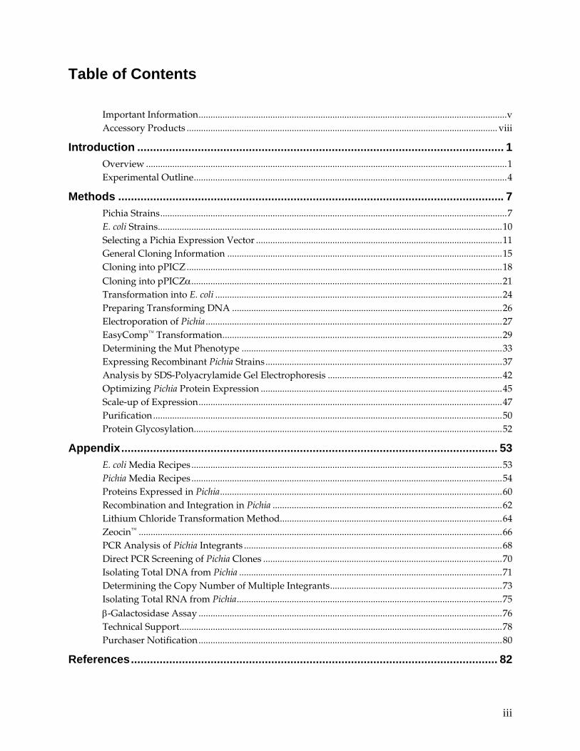
Table of Contents
Important Information.................................................................................................................................v Accessory Products .................................................................................................................................. viii
Introduction ................................................................................................................... 1 Overview .......................................................................................................................................................1 Experimental Outline...................................................................................................................................4
Methods ......................................................................................................................... 7 Pichia Strains.................................................................................................................................................7 E. coli Strains................................................................................................................................................10 Selecting a Pichia Expression Vector .......................................................................................................11 General Cloning Information ...................................................................................................................15 Cloning into pPICZ....................................................................................................................................18 Cloning into pPICZα..................................................................................................................................21 Transformation into E. coli ........................................................................................................................24 Preparing Transforming DNA .................................................................................................................26 Electroporation of Pichia ............................................................................................................................27 EasyComp™ Transformation.....................................................................................................................29 Determining the Mut Phenotype .............................................................................................................33 Expressing Recombinant Pichia Strains...................................................................................................37 Analysis by SDS-Polyacrylamide Gel Electrophoresis .........................................................................42 Optimizing Pichia Protein Expression .....................................................................................................45 Scale-up of Expression...............................................................................................................................47 Purification..................................................................................................................................................50 Protein Glycosylation.................................................................................................................................52
Appendix...................................................................................................................... 53 E. coli Media Recipes..................................................................................................................................53 Pichia Media Recipes..................................................................................................................................54 Proteins Expressed in Pichia......................................................................................................................60 Recombination and Integration in Pichia ................................................................................................62 Lithium Chloride Transformation Method.............................................................................................64 Zeocin™ ........................................................................................................................................................66 PCR Analysis of Pichia Integrants ............................................................................................................68 Direct PCR Screening of Pichia Clones ....................................................................................................70 Isolating Total DNA from Pichia ..............................................................................................................71 Determining the Copy Number of Multiple Integrants........................................................................73 Isolating Total RNA from Pichia...............................................................................................................75 β-Galactosidase Assay ...............................................................................................................................76 Technical Support.......................................................................................................................................78 Purchaser Notification...............................................................................................................................80
References................................................................................................................... 82

iv
v
Important Information
Kit Contents The EasySelect™ Pichia Expression Kit contains the following components.
The Pichia EasyComp™ Kit. This kit contains sufficient reagents for 6 preparations of competent cells. Each competent cell preparation yields enough cells for 20 transformations.
Upon receipt, store Solutions I and III at 4°C. You may store Solution II at 4°C or at room temperature.
Component Description Quantity
Solution I Sorbitol solution containing ethylene glycol and DMSO for the preparation of competent cells
75 ml
Solution II PEG solution for the transformation of competent cells
150 ml (2 × 75 ml)
Solution III Salt solution for washing and plating transformed cells
150 ml (2 × 75 ml)
Stab Vials: Pichia and E. coli stabs. Store at 4°C.
Strain Genotype Phenotype (Pichia only)
X-33 wild-type Mut+
GS115 his4 His–, Mut+
KM71H arg4 aox1::ARG4 MutS, Arg+
GS115/Albumin HIS4 MutS
GS115/pPICZ/lacZ his4 His–, Mut+
TOP10F´ E. coli F´ {proAB, lacIq, lacZΔM15, Tn10 (TetR)} mcrA, Δ(mrr-
hsdRMS-mcrBC), φ80lacZΔM15, ΔlacX74, deoR, recA1, λ–
araD139, Δ(ara-leu)7697, galU, galK, rpsL(StrR), endA1, nupG
Box 3: Vectors and Zeocin™. Store at –20°C
Reagent Amount
pPICZ A, B, and C 20 μg of each vector in TE buffer, pH 8.0* (40 μl at 500 ng/μl)
pPICZα A, B, and C 20 μg of each vector in TE buffer, pH 8.0 (40 μl at 500 ng/μl)
Zeocin™ 2 × 1.25 ml, 100 mg/ml
*TE buffer, pH 8.0: 10 mM Tris-HCl, 1 mM EDTA, pH 8.0
Continued on next page
vi
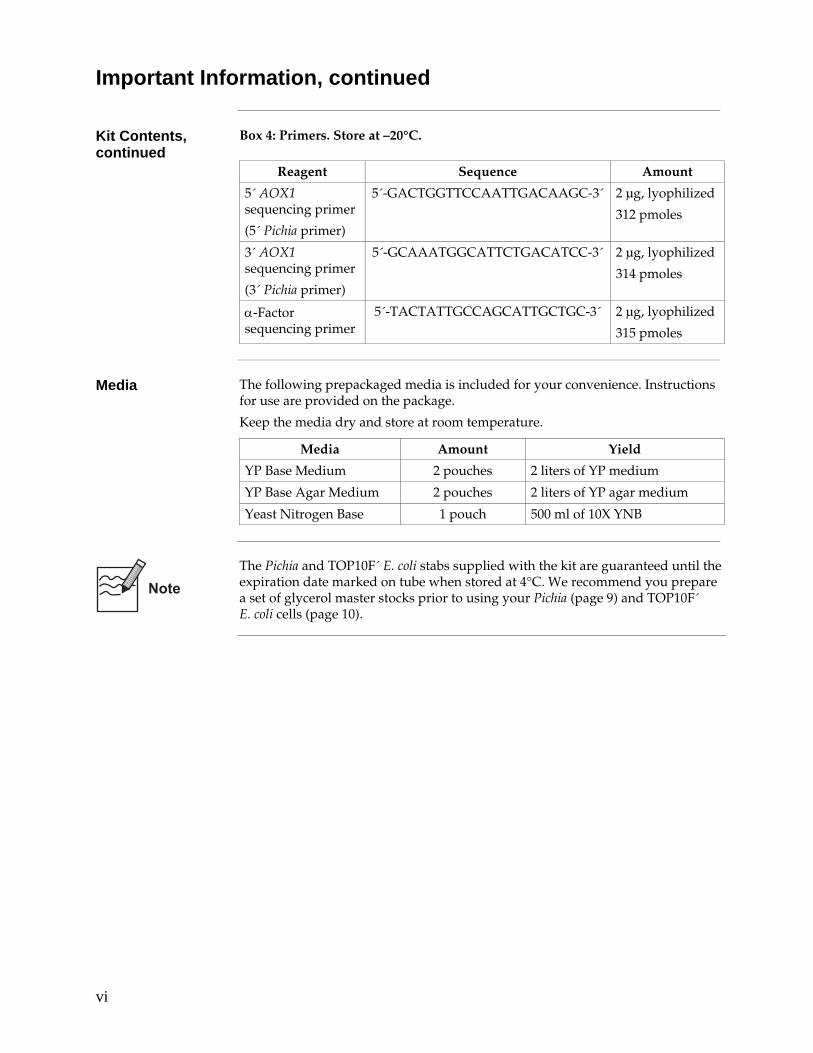
Important Information, continued
Kit Contents, continued
Box 4: Primers. Store at –20°C.
Reagent Sequence Amount
5´ AOX1 sequencing primer
(5´ Pichia primer)
5´-GACTGGTTCCAATTGACAAGC-3´ 2 μg, lyophilized
312 pmoles
3´ AOX1 sequencing primer
(3´ Pichia primer)
5´-GCAAATGGCATTCTGACATCC-3´ 2 μg, lyophilized
314 pmoles
α-Factor sequencing primer
5´-TACTATTGCCAGCATTGCTGC-3´ 2 μg, lyophilized
315 pmoles
Media The following prepackaged media is included for your convenience. Instructions
for use are provided on the package.
Keep the media dry and store at room temperature.
Media Amount Yield
YP Base Medium 2 pouches 2 liters of YP medium
YP Base Agar Medium 2 pouches 2 liters of YP agar medium
Yeast Nitrogen Base 1 pouch 500 ml of 10X YNB
The Pichia and TOP10F´ E. coli stabs supplied with the kit are guaranteed until the expiration date marked on tube when stored at 4°C. We recommend you prepare a set of glycerol master stocks prior to using your Pichia (page 9) and TOP10F´ E. coli cells (page 10).
vii
Important Information, continued
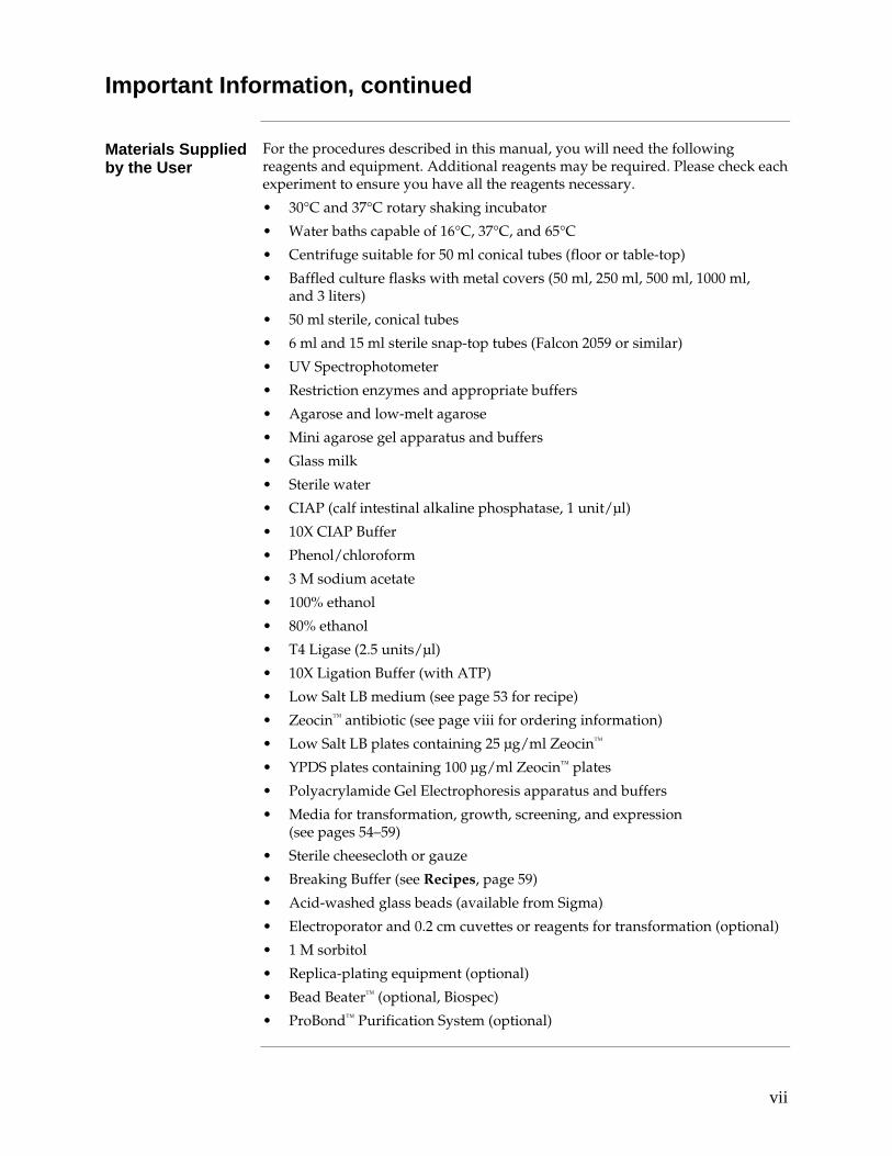
Materials Supplied by the User
For the procedures described in this manual, you will need the following reagents and equipment. Additional reagents may be required. Please check each experiment to ensure you have all the reagents necessary.
• 30°C and 37°C rotary shaking incubator
• Water baths capable of 16°C, 37°C, and 65°C
• Centrifuge suitable for 50 ml conical tubes (floor or table-top)
• Baffled culture flasks with metal covers (50 ml, 250 ml, 500 ml, 1000 ml, and 3 liters)
• 50 ml sterile, conical tubes
• 6 ml and 15 ml sterile snap-top tubes (Falcon 2059 or similar)
• UV Spectrophotometer
• Restriction enzymes and appropriate buffers
• Agarose and low-melt agarose
• Mini agarose gel apparatus and buffers
• Glass milk
• Sterile water
• CIAP (calf intestinal alkaline phosphatase, 1 unit/μl)
• 10X CIAP Buffer
• Phenol/chloroform
• 3 M sodium acetate
• 100% ethanol
• 80% ethanol
• T4 Ligase (2.5 units/μl)
• 10X Ligation Buffer (with ATP)
• Low Salt LB medium (see page 53 for recipe)
• Zeocin™ antibiotic (see page viii for ordering information)
• Low Salt LB plates containing 25 μg/ml Zeocin™
• YPDS plates containing 100 μg/ml Zeocin™ plates
• Polyacrylamide Gel Electrophoresis apparatus and buffers
• Media for transformation, growth, screening, and expression (see pages 54–59)
• Sterile cheesecloth or gauze
• Breaking Buffer (see Recipes, page 59)
• Acid-washed glass beads (available from Sigma)
• Electroporator and 0.2 cm cuvettes or reagents for transformation (optional)
• 1 M sorbitol
• Replica-plating equipment (optional)
• Bead Beater™ (optional, Biospec)
• ProBond™ Purification System (optional)

viii
Accessory Products
Introduction The products listed in this section are intended for use with the EasySelect™ Pichia Expression Kit. For more information, refer to our website (www.invitrogen.com) or call Technical Support (see page 78).
Accessory Products
Many of the reagents supplied in the EasySelect™ Pichia Expression Kit are available separately from Invitrogen. Ordering information is provided below.
Product Reactions or Amount Cat. no.
Pichia EasyComp™ Kit 20 transformations K1730-01 Zeocin™ 1 g R250-01 5 g R250-05 pPICZ A, B, and C 20 μg of each vector in TE buffer,
pH 8.0 (40 μl at 500 ng/μl) V190-20
pPICZα A, B, and C 20 μg of each vector in TE buffer, pH 8.0 (40 μl at 500 ng/μl)
V195-20
Antibodies for Detection of Fusion Protein
A number of antibodies are available from Invitrogen to detect expression of your fusion protein from the pPICZ and pPICZα vectors. Horseradish peroxidase (HRP)-conjugated antibodies allow one-step detection using colorimetric or chemiluminescent detection methods. The amount of antibody supplied is sufficient for 25 Westerns.
Antibody Epitope Cat. no.
Anti-myc R950-25
Anti-myc-HRP
Detects the 10 amino acid epitope derived from c-myc (Evan et al., 1985): EQKLISEEDL R951-25
Anti-His(C-term) R930-25
Anti-His(C-term)-HRP
Detects the C-terminal polyhistidine (6×His) tag (requires the free carboxyl group for detection) (Lindner et al., 1997): HHHHHH-COOH
R931-25
Purification of Fusion Protein
The polyhistidine (6×His) tag allows purification of the recombinant fusion protein using metal-chelating resins such as ProBond™. Ordering information for ProBond™ resin is provided below.
Item Quantity Cat. no.
ProBond™ Purification System 6 purifications K850-01
ProBond™ Purification System with Anti-myc-HRP Antibody
1 kit K852-01
ProBond™ Purification System with Anti-His(C-term)-HRP Antibody
1 kit K853-01
ProBond™ Resin 50 ml R801-01
150 ml R801-15
Purification Columns (10 ml polypropylene columns)
50 columns R640-50
1
Introduction Overview
Review Articles The information presented here is designed to give you a concise overview of the
Pichia pastoris expression system. It is by no means exhaustive. For further information, please read the articles cited in the text along with recent review articles (Buckholz and Gleeson, 1991; Cregg and Higgins, 1995; Cregg et al., 1993; Nico-Farber et al., 1995; Sreekrishna et al., 1988; Wegner, 1990). A general review of foreign gene expression in yeast is also available (Romanos et al., 1992).
General Characteristics of Pichia pastoris
As a eukaryote, Pichia pastoris has many of the advantages of higher eukaryotic expression systems such as protein processing, protein folding, and posttranslational modification, while being as easy to manipulate as E. coli or Saccharomyces cerevisiae. It is faster, easier, and less expensive to use than other eukaryotic expression systems and generally gives higher expression levels. As a yeast, it shares the advantages of molecular and genetic manipulations with Saccharomyces, and it has the added advantage of 10- to 100-fold higher heterologous protein expression levels. These features make Pichia very useful as a protein expression system.
Similarity to Saccharomyces
Many of the techniques developed for Saccharomyces may be applied to Pichia. These include: • transformation by complementation • gene disruption • gene replacement
In addition, the genetic nomenclature used for Saccharomyces has been applied to Pichia. For example, histidinol dehydrogenase is encoded by the HIS4 gene in both Saccharomyces and Pichia. There is also cross-complementation between gene products in both Saccharomyces and Pichia. Several wild-type genes from Saccharomyces complement comparable mutant genes in Pichia. Genes such as HIS4, LEU2, ARG4, TRP1, and URA3 all complement their respective mutant genes in Pichia.
Pichia pastoris as a Methylotrophic Yeast
Pichia pastoris is a methylotrophic yeast, capable of metabolizing methanol as its sole carbon source. The first step in the metabolism of methanol is the oxidation of methanol to formaldehyde using molecular oxygen by the enzyme alcohol oxidase. In addition to formaldehyde, this reaction generates hydrogen peroxide. To avoid hydrogen peroxide toxicity, methanol metabolism takes place within a specialized cell organelle, called the peroxisome, which sequesters toxic by-products away from the rest of the cell. Alcohol oxidase has a poor affinity for O2, and Pichia pastoris compensates by generating large amounts of the enzyme. The promoter regulating the production of alcohol oxidase is the one used to drive heterologous protein expression in Pichia.
Continued on next page

2
Overview, continued
Two Alcohol Oxidase Proteins
Two genes in Pichia pastoris code for alcohol oxidase – AOX1 and AOX2. The majority of alcohol oxidase activity in the cell is attributable to the product of the AOX1 gene. Expression of the AOX1 gene is tightly regulated and induced by methanol to very high levels, typically > 30% of the total soluble protein in cells grown with methanol. The AOX1 gene has been isolated and a plasmid-borne version of the AOX1 promoter is used to drive expression of the gene of interest encoding the desired heterologous protein (Ellis et al., 1985; Koutz et al., 1989; Tschopp et al., 1987a). While AOX2 is about 97% homologous to AOX1, growth on methanol is much slower than with AOX1. This slow growth on methanol allows isolation of MutS strains (aox1) (Cregg et al., 1989; Koutz et al., 1989).
Expression Expression of the AOX1 gene is controlled at the level of transcription. In
methanol-grown cells approximately 5% of the polyA+ RNA is from the AOX1 gene. The regulation of the AOX1 gene is a two step process: a repression/derepression mechanism plus an induction mechanism (e. g. GAL1 gene in Saccharomyces (Johnston, 1987)). Briefly, growth on glucose represses transcription, even in the presence of the inducer methanol. For this reason, growth on glycerol is recommended for optimal induction with methanol. Note that growth on glycerol alone (derepression) is not sufficient to generate even minute levels of expression from the AOX1 gene. The inducer, methanol, is necessary for even detectable levels of AOX1 expression (Ellis et al., 1985; Koutz et al., 1989; Tschopp et al., 1987a).
Phenotype of aox1 Mutants
Loss of the AOX1 gene, and thus a loss of most of the cell's alcohol oxidase activity, results in a strain that is phenotypically MutS (Methanol utilization slow). This has in the past been referred to as Mut–. The MutS designation has been chosen to accurately describe the phenotype of these mutants. This results in a reduction in the cells' ability to metabolize methanol. The cells, therefore, exhibit poor growth on methanol medium. Mut+ (Methanol utilization plus) refers to the wild type ability of strains to metabolize methanol as the sole carbon source. These two phenotypes are used when evaluating Pichia transformants for integration of your gene (Experimental Outline, page 4).
Continued on next page
3
Overview, continued
Intracellular and Secretory Protein Expression
Heterologous expression in Pichia pastoris can be either intracellular or secreted. Secretion requires the presence of a signal sequence on the expressed protein to target it to the secretory pathway. While several different secretion signal sequences have been used successfully, including the native secretion signal present on some heterologous proteins, success has been variable. The secretion signal sequence from the Saccharomyces cerevisiae factor prepro peptide has been used with the most success (Cregg et al., 1993; Scorer et al., 1993).
The major advantage of expressing heterologous proteins as secreted proteins is that Pichia pastoris secretes very low levels of native proteins. Since there is very low amount of protein in the minimal Pichia growth medium, the secreted heterologous protein comprises the vast majority of the total protein in the medium and serves as the first step in purification of the protein (Barr et al., 1992). Note, however, that if there are recognized glycosylation sites (Asn-X-Ser/Thr) in your protein's primary sequence, glycosylation may occur at these sites.
Posttranslational Modifications
In comparison to Saccharomyces cerevisiae, Pichia may have an advantage in the glycosylation of secreted proteins because it may not hyperglycosylate. Both Saccharomyces cerevisiae and Pichia pastoris have a majority of N-linked glycosylation of the high-mannose type; however, the length of theoligosaccharide chains added posttranslationally to proteins in Pichia (average 8–14 mannose residues per side chain) is much shorter than those in Saccharomyces cerevisiae (50–150 mannose residues) (Grinna and Tschopp, 1989; Tschopp et al., 1987b). Very little O-linked glycosylation has been observed in Pichia.
In addition, Saccharomyces cerevisiae core oligosaccharides have terminal α1,3 glycan linkages whereas Pichia pastoris does not. It is believed that the α1,3 glycan linkages in glycosylated proteins produced from Saccharomyces cerevisiae are primarily responsible for the hyper-antigenic nature of these proteins making them particularly unsuitable for therapeutic use. Although not yet proven, this is predicted to be less of a problem for glycoproteins generated in Pichia pastoris, because it may resemble the glycoprotein structure of higher eukaryotes (Cregg et al., 1993).
4
Experimental Outline
Selection of Vector and Cloning
To utilize the strong, highly-inducible PAOX1 promoter for expression of your protein, there are two expression vectors included in this kit. One vector, pPICZ, is for intracellular expression while the other vector, pPICZα, is for secreted expression. Each vector is provided in three reading frames to facilitate cloning in frame with the C-terminal polyhistidine tag. All vectors contain the Zeocin™ resistance gene for positive selection in E. coli and Pichia. See pages 11–14 for more information on these vectors.
Transformation and Integration
Two different phenotypic classes of recombinant strains can be generated Mut+ and MutS. MutS refers to the "Methanol utilization slow" phenotype caused by the loss of alcohol oxidase activity encoded by the AOX1 gene. A strain with a MutS phenotype has a mutant aox1 locus, but is wild type for AOX2. This results in a slow growth phenotype on methanol medium. Both X-33 and GS115 are Mut+, and KM71H is MutS. Transformation of X-33 or GS115 with plasmid DNA linearized in the 5´ AOX1 region will yield Mut+ transformants, while KM71H will yield only MutS transformants. Both Mut+ and MutS recombinants are useful to have as one phenotype may favor better expression of your protein than the other. You should test between 6–10 recombinants per phenotype because the site of recombination may affect expression. There is no way to predict beforehand which construct or isolate will better express your protein. For more information on recombination in Pichia, see page 62.
Once you have successfully cloned your gene behind the AOX1 promoter, you will then linearize your plasmid to stimulate recombination when the plasmid is transformed into Pichia.
Continued on next page
5
Experimental Outline, continued
Expression and Scale-up
After isolating your Pichia recombinants, you will then test expression of both Mut+ and MutS recombinants. This will involve growing a small culture of each recombinant, inducing with methanol, and taking time points. If looking for intracellular expression, analyze the cell pellet from each time point by SDS polyacrylamide gel electrophoresis (SDS-PAGE). If looking for secreted expression, analyze both the cell pellet and supernatant from each time point. We recommend that you analyze your SDS-PAGE gels by both Coomassie staining and Western blot. We also suggest checking for protein activity by assay if one is available. Not all proteins express to the level of grams per liter, so it is advisable to check by Western blot or activity assay, and not just by Coomassie staining of SDS-PAGE gels for production of your protein.
Choose the Pichia recombinant strain which best expresses your protein and optimize induction based on the suggestions on pages 45–46. Once expression is optimized, scale-up your expression protocol to produce more protein for purification.
Purification Both pPICZ and pPICZα contain a polyhistidine tag that binds divalent cations
like Ni2+ to facilitate purification. Metal-binding resins such as ProBond™ can be used to purify proteins expressed from pPICZ or pPICZα. We recommend that you use the ProBond™ Purification System (Cat. no. K850-01) to purify fusion proteins expressed using pPICZ or pPICZα. Note that instructions for equilibration of and chromatography on ProBond™ resin are contained in the ProBond™ Purification System Kit manual. Preliminary preparation steps are described on pages 50–51.
If you are using a metal-chelating resin other than ProBond™, please follow the manufacturer's recommendations for fusion proteins expressed in yeast.
Continued on next page
6
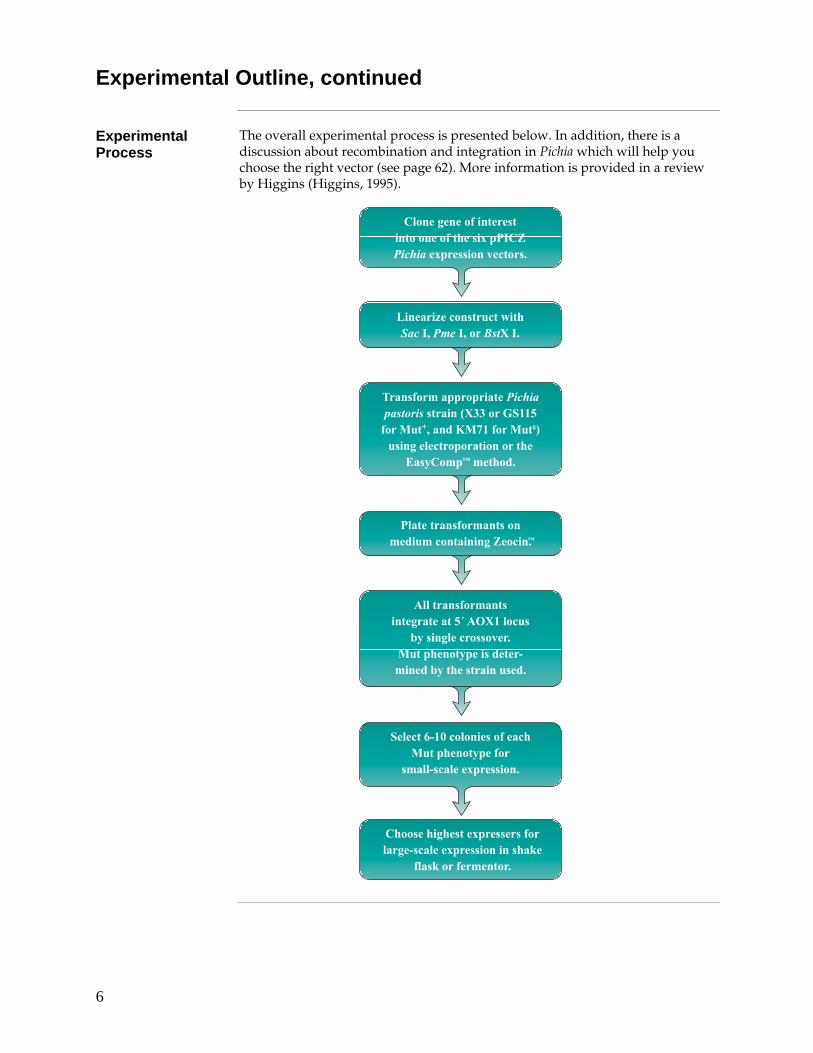
Experimental Outline, continued
Experimental Process
The overall experimental process is presented below. In addition, there is a discussion about recombination and integration in Pichia which will help you choose the right vector (see page 62). More information is provided in a review by Higgins (Higgins, 1995).
������������ �� �� �����������������
������������������ ������� ����� �������������������� ���!"�#������ �$����%�&� ������ ����� ��
'��()���*�� ��!�
��&� +,�-�&������������ ����� (�����
���&&,��&��.���������
)�������%��� �.�����������&��%�,��&��.�������������/�
�&��/�������� ���
)&���%������� ���� �� ������ ����.�0�)1�������.��������2� ����
0&� � ���������� �����!����� �����%1�����
3&& ���������� ��� �%�� �� �435��&���
6(���%&������2����� ����� (����!� ��,����!6( ��� �������!�
*
7
Methods Pichia Strains
Introduction Pichia pastoris is quite similar to Saccharomyces cerevisiae as far as general growth
conditions and handling. You should be familiar with basic microbiological and sterile techniques before attempting to grow and manipulate any microorganism. You should also be familiar with basic molecular biology and protein chemistry. Some general references to consult are Guide to Yeast Genetics and Molecular Biology, (Guthrie and Fink, 1991), Current Protocols in Molecular Biology, (Ausubel et al., 1994), Molecular Cloning: A Laboratory Manual, (Sambrook et al., 1989), Protein Methods, (Bollag and Edelstein, 1991), and Guide to Protein Purification, (Deutscher, 1990).
Genotypes of Pichia Strains
X-33 is a wild-type Pichia strain that is useful for selection on Zeocin™ and large-scale growth. It will grow in YPD and in minimal media.
The Pichia host strain GS115 has a mutation in the histidinol dehydrogenase gene (his4) that prevents it from synthesizing histidine. GS115 will grow on complex medium such as YPD (also known as YEPD) and on minimal media supplemented with histidine.
The parent strain of KM71H has a mutation in the argininosuccinate lyase gene (arg4) that prevents the strain from growing in the absence of arginine. The wild-type ARG4 gene was used to disrupt AOX1, creating KM71H, a MutS, Arg+ strain.
Construction of KM71H
The ARG4 gene (~2 kb) was inserted into the cloned, wild-type AOX1 gene between the BamH I site (codons 15/16 of AOX1) and the Sal I site (codons 227/228 of AOX1). ARG4 replaces codons 16 through 227 of AOX1. This construct was transformed into the parent strain of KM71 (arg4 his4) and Arg+ transformants were isolated and analyzed for the MutS phenotype. Genetic analysis of Arg+ transformants showed that the wild-type AOX1 gene was replaced by the aox1::ARG4 construct. To create KM71H, KM71 was transformed with a gene fragment encoding the HIS4 gene and a His+ convertant was isolated.
��������
The advantage of using KM71H is that there is no need to screen for the Mut phenotype on methanol minimal medium. All transformants will be MutS. Secondly, since the AOX1 locus was completely deleted, it is theoretically possible to replace aox1::ARG4 with your construct by gene replacement. The phenotype of this strain would be MutS Arg–. This means the recombinant strain would require arginine in the medium to grow. Unfortunately, simple inclusion of arginine does not totally alleviate the effects of the arg4 mutation, and arg4 strains do not grow well on minimal medium supplemented with arginine. Therefore, we do not recommend that you generate transformants in KM71H by replacing the aox1::ARG4 construct.
Continued on next page
8
Pichia Strains, continued
Control Expression Strains
GS115/His+ MutS Albumin: This strain is a control for secreted expression (page 41) and the MutS phenotype when characterizing Pichia transformants (page 34). The gene for serum albumin was cloned with its native secretion signal, then integrated into Pichia at the AOX1 locus. This strain secretes albumin (67 kDa) into the medium at levels > 1 gram/liter.
GS115/pPICZ/lacZ Mut+ β-galactosidase: The strain GS115/pPICZ/lacZ expresses β-galactosidase fused at the C-terminus to the myc epitope and the polyhistidine tag. Expression of the 119 kDa fusion protein is driven by the PAOX1 promoter and is inducible by methanol. The fusion protein is visible on a Coomassie-stained SDS-polyacrylamide gel and can be detected antigenically using the Anti-myc Antibody (see page viii) or enzymatically using an ONPG assay (β-Gal Assay Kit, Cat. no. K1455-01). GS115/pPICZ/lacZ is provided as a positive control for Zeocin™ resistance in Pichia, Mut+ expression (page 33 and 41) and purification.
Growth of Pichia Strains
The growth temperature of Pichia pastoris is 28–30°C for liquid cultures, plates, and slants. Growth above 32°C during induction can be detrimental to protein expression and can even lead to cell death. Other important facts:
• Doubling time of log phase Mut+ or MutS Pichia in YPD is ~2 hours
• Mut+ and MutS strains do not differ in growth rates unless grown on methanol
• Doubling time of log phase Mut+ Pichia in methanol medium (MM) is 4–6 hours
• Doubling time of log phase MutS Pichia in MM is ~18 hours
• One OD600 = ~5 × 10⁷ cells/ml
Note that growth characteristics may vary depending on the recombinant protein expressed.
Growth on Methanol
When plates or medium containing methanol are used as growth medium, it is advisable to add methanol every day to compensate for loss because of evaporation or consumption.
• For plates add 100 μl of 100% methanol to the lid of the inverted plate.
• For liquid medium add 100% methanol to a final concentration of 0.5%.
Some researchers have had success adding methanol to 1% every day for MutS strains and up to 3% for Mut+ without any negative effect to their liquid culture.
Continued one next page

9
Pichia Strains, continued
Storing Pichia Strains
To store cells for weeks to months, use YPD medium and YPD agar slants (see page 55).
• Streak each strain for single colonies on YPD.
• Transfer one colony to a YPD stab and grow for 2 days at 30°C.
• The cells can be stored on YPD for several weeks at 4°C.
To store cells for months to years, store frozen at –80°C.
• Culture a single colony of each strain overnight in YPD.
• Harvest the cells and suspend in YPD containing 15% glycerol at a final OD600 of 50–100 (approximately 2.5 × 10⁹–5.0 × 10⁹ cells/ml).
• Cells are frozen in liquid nitrogen or a dry ice/ethanol bath and then stored at –80°C.
After extended storage at 4°C or –80°C, it is recommended that ZeoR transformants be checked for correct phenotype and protein expression.
10
E. coli Strains
Genotype of E. coli Strain
The E. coli strain, TOP10F´ is provided in case no suitable E. coli strain is available. Other strains which may be suitable are TOP10, DH5αF´, JM109, or any other strain which is recombination deficient (recA) and deficient in endonuclease A (endA).
F´ {proAB, lacIq, lacZΔM15, Tn10 (TetR)} mcrA, Δ(mrr-hsdRMS-mcrBC),
φ80lacZΔM15, ΔlacX74, deoR, recA1, λ–araD139, Δ(ara-leu)7697, galU, galK, rpsL(StrR), endA1, nupG λ–
��������
Any E. coli strain that contains the complete Tn5 transposable element (i.e., DH5αF´IQ, SURE, SURE2) encodes the ble (bleomycin) resistance gene. These strains will confer resistance to Zeocin™. For the most efficient selection it is highly recommended that you choose an E. coli strain that does not contain the Tn5 gene (i.e., TOP10, DH5, DH10, etc.).
����
�����
���
We recommend that you make a frozen stock of TOP10F´ to keep on hand.
• Culture TOP10F´ in 5 ml LB with 10 μg/ml tetracycline. Grow overnight.
• Mix thoroughly 0.85 ml of culture with 0.15 ml sterile glycerol.
• Transfer to a freezer vial and freeze in liquid nitrogen or a dry ice/ethanol bath.
• Store at –80°C.
11
Selecting a Pichia Expression Vector
Selecting a Vector If your protein is cytosolic and non-glycosylated, you may elect to express the protein intracellularly using one of the pPICZ vectors. If your protein is normally secreted, glycosylated, or directed to an intracellular organelle, you may wish to try secreting your protein using one of the pPICZα vectors. We recommend that you try both the native secretion signal and the α-factor signal sequence in order to secrete your protein.
There is no yeast origin of replication in any of the Pichia expression vectors included in this kit. ZeoR transformants can only be isolated if recombination occurs between the plasmid and the Pichia genome.
Features of pPICZ A, B, and C
pPICZ A (3329 bp), pPICZ B (3328 bp), and pPICZ C (3329 bp) contain the following elements. All features have been functionally tested.
Feature Benefit
5´ AOX1 A 942 bp fragment containing the AOX1 promoter that allows methanol-inducible, high-level expression in Pichia Targets plasmid integration to the AOX1 locus
Multiple cloning site with 10 unique restriction sites
Allows insertion of your gene into the expression vector
C-terminal myc epitope tag
(Glu-Gln-Lys-Leu-Ile-Ser-Glu-Glu-Asp-Leu-Asn) Permits detection of the fusion protein by the Anti-myc Antibody or Anti-myc-HRP Antibody (see page viii for ordering information) (Evan et al., 1985)
C-terminal polyhistidine tag Permits purification of your recombinant fusion protein on metal-chelating resin such as ProBond™ In addition, the C-terminal polyhistidine tag is the epitope for the Anti-His(C-term) Antibody and the Anti-His (C-term)-HRP Antibody (see page viii) (Lindner et al., 1997)
AOX1 Transcription Termination (TT)
Native transcription termination and polyadenylation signal from AOX1 gene (~260 bp) that permits efficient 3´ mRNA processing, including polyadenylation, for increased mRNA stability
TEF1 promoter
Transcription elongation factor 1 gene promoter from Saccharomyces cerevisiae that drives expression of the Sh ble gene in Pichia, conferring Zeocin™ resistance (GenBank Acc. no. D12478, D01130).
EM7 (synthetic prokaryotic promoter)
Constitutive promoter that drives expression of the Sh ble gene in E. coli, conferring Zeocin™ resistance
Sh ble gene (Streptoalloteichus hindustanus ble gene)
Zeocin™ resistance gene for selection in E. coli
CYC1 transcription termination region
3´ end of the Saccharomyces cerevisiae CYC1 gene that allows efficient 3´ mRNA processing of the Sh ble gene for increased stability (GenBank Acc. no. M34014)
pUC origin Allows replication and maintenance of the plasmid in E. coli
Sac I, Pme I, BstX I Unique restriction sites that permit linearization of the vectors at the AOX1 locus for efficient integration into the Pichia genome
Continued on next page
12
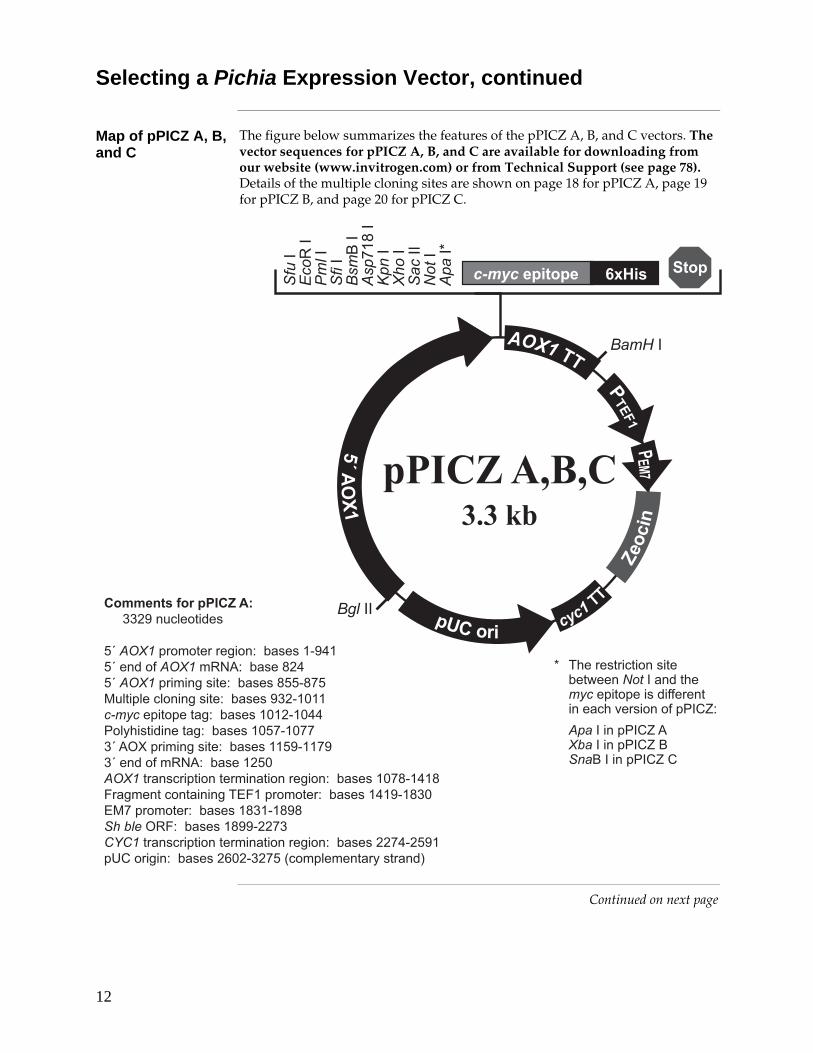
Selecting a Pichia Expression Vector, continued
Map of pPICZ A, B, and C
The figure below summarizes the features of the pPICZ A, B, and C vectors. The vector sequences for pPICZ A, B, and C are available for downloading from our website (www.invitrogen.com) or from Technical Support (see page 78). Details of the multiple cloning sites are shown on page 18 for pPICZ A, page 19 for pPICZ B, and page 20 for pPICZ C.
����������� �������������������� ��
������������������ ���������������������������������������� ������������ � ���� ��������� ��� !�"�� ����� ���� ��������������#�������� ����������������#����#��$�%& �� � ��������������#�!��#!!����'(��� � ���� ����������������!������������������������#������������ �� ������ ��� ����� �����������#! ���� )������������ � ���*+)����������������������� �#+"!����������������� ���� � �� ���'�)��������� �����!������������� �� ������ ��� ����� ������������!�������,-��� � ����������.#����!��/���������%�������0
1 *&����� �� ���� ���2������3������&���� ���� ��� ����� ����&�4�� �������$3-5�
����3� ���$3-5��� ��3� ���$3-5�6��6�3� ���$3-5�-
�0�)13�7�)���/6
���� ��
����
���� �
�
��� ���
������
���
������
����������� � !�����3
�����3
����3
���3
���
6�3
���!� �3
����3
����3
���33
����3
����31
"���
��� �3
�!��33
Continued on next page
13
Selecting a Pichia Expression Vector, continued
Features of pPICZα A, B, and C
pPICZα A (3593 bp), pPICZα B (3597 bp), and pPICZα C (3598 bp) contain the following elements. All features have been functionally tested.
Feature Benefit
5´ AOX1 A 942 bp fragment containing the AOX1 promoter that allows methanol-inducible, high-level expression in Pichia
Targets plasmid integration to the AOX1 locus.
Native Saccharomyces cerevisiae α-factor secretion signal
Allows for efficient secretion of most proteins from Pichia
Multiple cloning site with 10 unique restriction sites
Allows insertion of your gene into the expression vector
C-terminal myc epitope tag
(Glu-Gln-Lys-Leu-Ile-Ser-Glu-Glu-Asp-Leu-Asn)
Permits detection of the fusion protein by the Anti-myc Antibody or Anti-myc-HRP Antibody (see page viii for ordering information) (Evan et al., 1985)
C-terminal polyhistidine tag Permits purification of your recombinant fusion protein on metal-chelating resin such as ProBond™
In addition, the C-terminal polyhistidine tag is the epitope for the Anti-His(C-term) Antibody and the Anti-His(C-term)-HRP Antibody (see page viii for ordering information) (Lindner et al., 1997)
AOX1 Transcription Termination (TT)
Native transcription termination and polyadenylation signal from AOX1 gene (~260 bp) that permits efficient 3´ mRNA processing, including polyadenylation, for increased mRNA stability
TEF1 promoter
Transcription elongation factor 1 gene promoter from Saccharomyces cerevisiae that drives expression of the Sh ble gene in Pichia, conferring Zeocin™ resistance (GenBank Acc. no. D12478, D01130)
EM7 (synthetic prokaryotic promoter)
Constitutive promoter that drives expression of the Sh ble gene in E. coli, conferring Zeocin™ resistance
Sh ble gene (Streptoalloteichus hindustanus ble gene)
Zeocin™ resistance gene
CYC1 transcription termination region
3´ end of the Saccharomyces cerevisiae CYC1 gene that allows efficient 3´ mRNA processing of the Sh ble gene for increased stability (GenBank Acc. no. M34014)
pUC origin Allows replication and maintenance of the plasmid in E. coli
Sac I, Pme I, BstX I Unique restriction sites that permit linearization of the vectors at the AOX1 locus for efficient integration into the Pichia genome
Continued on next page
14
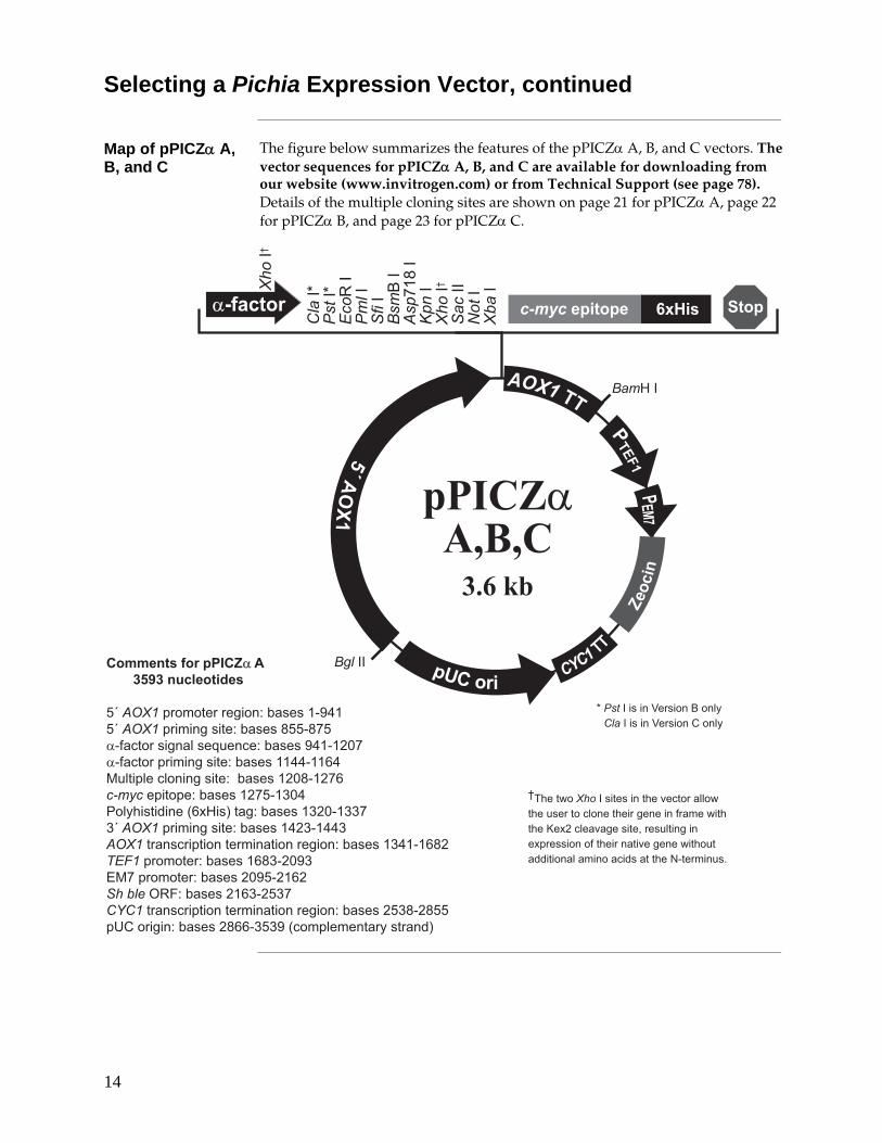
Selecting a Pichia Expression Vector, continued
Map of pPICZα A, B, and C
The figure below summarizes the features of the pPICZα A, B, and C vectors. The vector sequences for pPICZα A, B, and C are available for downloading from our website (www.invitrogen.com) or from Technical Support (see page 78). Details of the multiple cloning sites are shown on page 21 for pPICZα A, page 22 for pPICZα B, and page 23 for pPICZα C.
����������� �����#�$#�%�&����'��
������������������ ������������������������ � ���� �������� ��� !����������� �����7����������������#!����������� � ���� ���������������.�"�� ����� ���� �����������# ���!.������ ������������!����#�$�%& �� � ��/.89 �0��������������#����!���������� � ���� ����������������������������� �� ������ ��� ����� ���������������. �"�#�����������������. ���#��+"!����������������#�����.��� ���'�)���������.�����!������������ �� ������ ��� ����� ������������ �� ���,-��� � ��������� ..������/���������%�������0
����3� �� ��:�� ���6���%����3� �� ��:�� ���-���%
1
���� ��
����
���� ��
��� ���
������
���
������
�0�)1�3�7�)��+/6
����31
����31
�����3
����3
���3
���
6�3
���!� �3
����3
����3;
���33
����3
� ��3
"���
���9�3
�!��33
�()���� ����������� � !��
����3;
;*&��2������3�� ��� ���&�4�������2�&�������������& ����� �������2 �&�&�<8����4���� �=����� ��� �8���� �������& ����� 4����2 �&������ � ������ ����� �������&������ ���>
15
General Cloning Information
Introduction Before cloning your gene into one of the pPICZ or pPICZα vectors, consider some of the general guidelines presented below. If you are cloning into pPICZα, it is important to clone your gene in frame with the α-factor signal sequence. The multiple cloning sites for all vectors are presented on pages 18–23 to help you develop a cloning strategy.
General Considerations
The following are some general considerations applicable to pPICZ or pPICZα. • The codon usage in Pichia is believed to be similar to Saccharomyces cerevisiae.
• Many Saccharomyces genes have proven to be functional in Pichia. • Maintain plasmid constructions in a recA, endA E. coli strain such as TOP10. • The BsmB I site in the multiple cloning site has been specifically engineered
to be compatible with inserts that have BamH I and/or Bgl II ends. The BamH I and Bgl II sites will be destroyed upon ligation, but the insert can be released by digestion with BsmB I.
• The premature termination of transcripts because of "AT rich regions" has been observed in Pichia and other eukaryotic systems (Henikoff and Cohen, 1984; Irniger et al., 1991; Scorer et al., 1993; Zaret and Sherman, 1984). If you have problems expressing your gene, check for premature termination by Northern analysis and check your sequence for AT rich regions. It may be necessary to change the sequence in order to express your gene (Scorer et al., 1993).
• The native 5´ end of the AOX1 mRNA is noted in each multiple cloning site. This is needed to calculate the size of the expressed mRNA of the gene of interest if you need to analyze mRNA for any reason.
For pPICZ only:
• For proper initiation of translation, your insert should contain an initiation ATG codon as part of a yeast consensus sequence (Romanos et al., 1992). An example of a yeast consensus sequence is provided below. The ATG initiation codon is shown underlined.
(G/A)NNATGG
Note that other sequences are also possible. Although not as strong as the mammalian Kozak translation initiation sequence, the yeast consensus sequence is thought to have a 2–3-fold effect on the efficiency of translation initiation.
• To express your gene as a recombinant fusion protein, you must clone your gene in frame with the C-terminal peptide containing the c-myc epitope and the polyhistidine tag. The vector is supplied in three reading frames to facilitate cloning. Refer to the diagrams on pages 18–20 to develop a cloning strategy.
• If you wish to express your protein without the C-terminal peptide, be sure to include a stop codon.
Continued on next page
16
General Cloning Information, continued
General Considerations
For pPICZα only:
• The initiation ATG in the α-factor signal sequence in pPICZα corresponds to the native initiation ATG of the AOX1 gene.
• If you are using pPICZα, the open reading frame (ORF) of the mature gene of interest should be cloned in frame and downstream of the α-factor signal sequence and in frame with the C-terminal tag (if desired).
Note: Cloning of your gene of interest in frame with the signal sequence does not automatically guarantee that your protein will be in-frame with the C-terminal tag. Please consider both the frame of the signal sequence and the C-terminal fusion tag when designing a cloning strategy.
• If you wish to express your gene of interest without the C-terminal peptide, be sure your gene contains a stop codon.
• The predicted protease cleavage sites for the α-factor signal sequence are indicated in the figures on pages 21–23.
Cloning Procedures
Refer to Ausubel, et al., 1990, pages 3.16.1 to 3.17.3. or Sambrook, et al., 1989, pages 5.10 to 5.13. for help with cloning.
Constructing Multimeric Plasmids
pPICZ and pPICZα contain unique Bgl II and BamH I sites to allow construction of plasmids containing multiple copies of your gene. For information on how to construct multimers, please contact Technical Support (see page 78).
����
�����
���
For preparing competent E. coli cells for transformation, use your own procedure or refer to Current Protocols in Molecular Biology (Ausubel et al., 1994) or Molecular Biology: A Laboratory Manual (Sambrook et al., 1989). Note that electrocompetent TOP10F´ cells are available from Invitrogen.
Item Amount Cat. no.
TOP10F´ Electrocomp™ 6 × 20 reactions C665-24
TOP10F´ Electrocomp™ 2 × 20 reactions C665-11
Continued on next page
17
General Cloning Information, continued
��������
To propagate pPICZ and pPICZα or select ZeoR transformants in E. coli, you will need to prepare Low Salt LB. For Zeocin™ to be active, the salt concentration of the medium must remain low (< 90 mM) and the pH must be 7.5. Prepare Low Salt LB broth and plates using the recipe on page 53.
Failure to lower the salt content of your LB medium will result in non-selection due to inactivation of the drug.
To propagate vectors:
• Resuspend the plasmid in 20 μl sterile water to make a 1 μg/μl solution
• Dilute 1 μl of the plasmid (1 μg/μl) to 10–100 pg/μl using sterile water or TE buffer.
• Transform competent E. coli with 1–2 μl of the diluted plasmid and select on Low Salt LB with 25 μg/ml Zeocin™.
Signal Sequence Processing
The processing of the α-factor mating signal sequence in pPICZα occurs in two steps:
1. The preliminary cleavage of the signal sequence by the KEX2 gene product, with the final Kex2 cleavage occurring between arginine and glutamine in the sequence Glu-Lys-Arg * Glu-Ala-Glu-Ala, where * is the site of cleavage.
2. The Glu-Ala repeats are further cleaved by the STE13 gene product.
Optimizing Signal Cleavage
In Saccharomyces cerevisiae, the Glu-Ala repeats are not necessary for cleavage by Kex2, but cleavage after Glu-Lys-Arg may be more efficient when followed by Glu-Ala repeats. A number of amino acids are tolerated at site X instead of Glu in the sequence Glu-Lys-Arg-X. These amino acids include the aromatic amino acids, small amino acids, and histidine. Proline, however, will inhibit Kex2 cleavage. For more information on Kex2 cleavage, see (Brake et al., 1984)
There are some cases where Ste13 cleavage of Glu-Ala repeats is not efficient, and Glu-Ala repeats are left on the N-terminus of the expressed protein of interest. This is generally dependent on the protein of interest.
Expressing Recombinant Protein with Native N-terminus
To express your protein with a native N-terminus, use the Xho I site at bp 1184–1189 to clone your gene flush with the Kex2 cleavage site. Use PCR to rebuild the sequence from the Xho I site to the arginine codon at nucleotides 1193–1195. Remember to include the first amino acid(s) of your protein, if necessary, for correct fusion to the Kex2 cleavage site.
Continued on next page
18
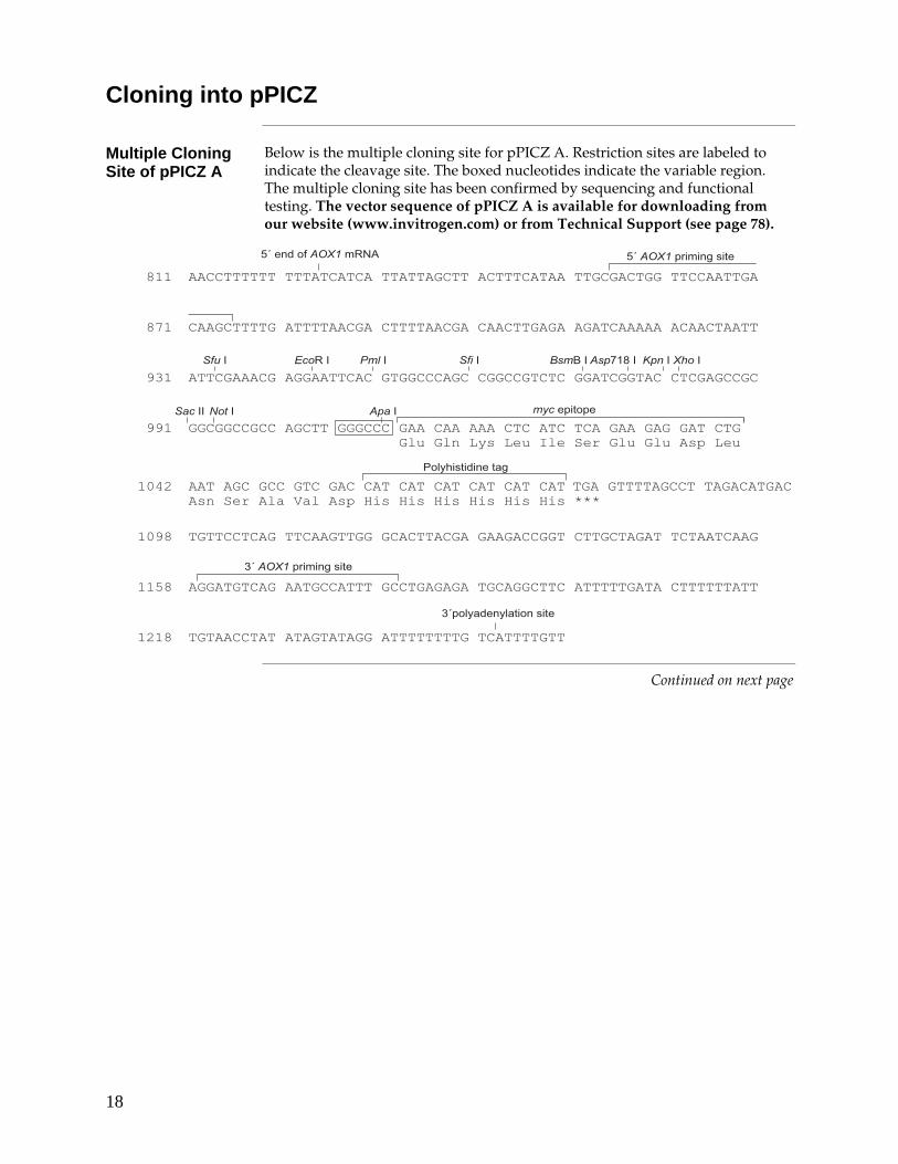
Cloning into pPICZ
Multiple Cloning Site of pPICZ A
Below is the multiple cloning site for pPICZ A. Restriction sites are labeled to indicate the cleavage site. The boxed nucleotides indicate the variable region. The multiple cloning site has been confirmed by sequencing and functional testing. The vector sequence of pPICZ A is available for downloading from our website (www.invitrogen.com) or from Technical Support (see page 78).
�������������������������������������������
�����������������������������������������������������������������
�����������������������������������������������������������������
����������������������������������������������
���� ����������������������������������
���������� � ���� �
�������������������������
�����������������������������������������������������������������
�����������������������������������������������������������������
�����������������������������������������������������������������
���������������������������������������������������������������
��
������������ ���� �� �������������� �
���������� � ���� �
���� ���
����%���%�� ���� �
$�%& �� � �����
������������������
���3 �����3 ����3 ���3 ���6�3 ���!� �3 ����3 ����3
���33 ����3 ����3
����
���
���
��
���
���
Continued on next page
19
Cloning into pPICZ, continued
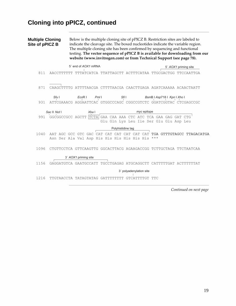
Multiple Cloning Site of pPICZ B
Below is the multiple cloning site of pPICZ B. Restriction sites are labeled to indicate the cleavage site. The boxed nucleotides indicate the variable region. The multiple cloning site has been confirmed by sequencing and functional testing. The vector sequence of pPICZ B is available for downloading from our website (www.invitrogen.com) or from Technical Support (see page 78).
���������������������������������������������������������������������
�����������������������������������������������������������������
�����������������������������������������������������������������
��������������������������������������������������
���� ����������������������������������
���������� � ���� �
�������������������������
�����������������������������������������������������������������
�����������������������������������������������������������������
�����������������������������������������������������������������
�������������������������������������������������������������
��
������������ ���� �� �������������� �
���������� � ���� �
���� ���
�����%���%�� ���� �
$�%& �� � �����
������������������
���3 �����3 ����3 ���3 ���6�3 ���!� �3 ����3 ����3
���33 ����3 � ��3
����
���
���
��
���
���
Continued on next page
20
Cloning into pPICZ, continued
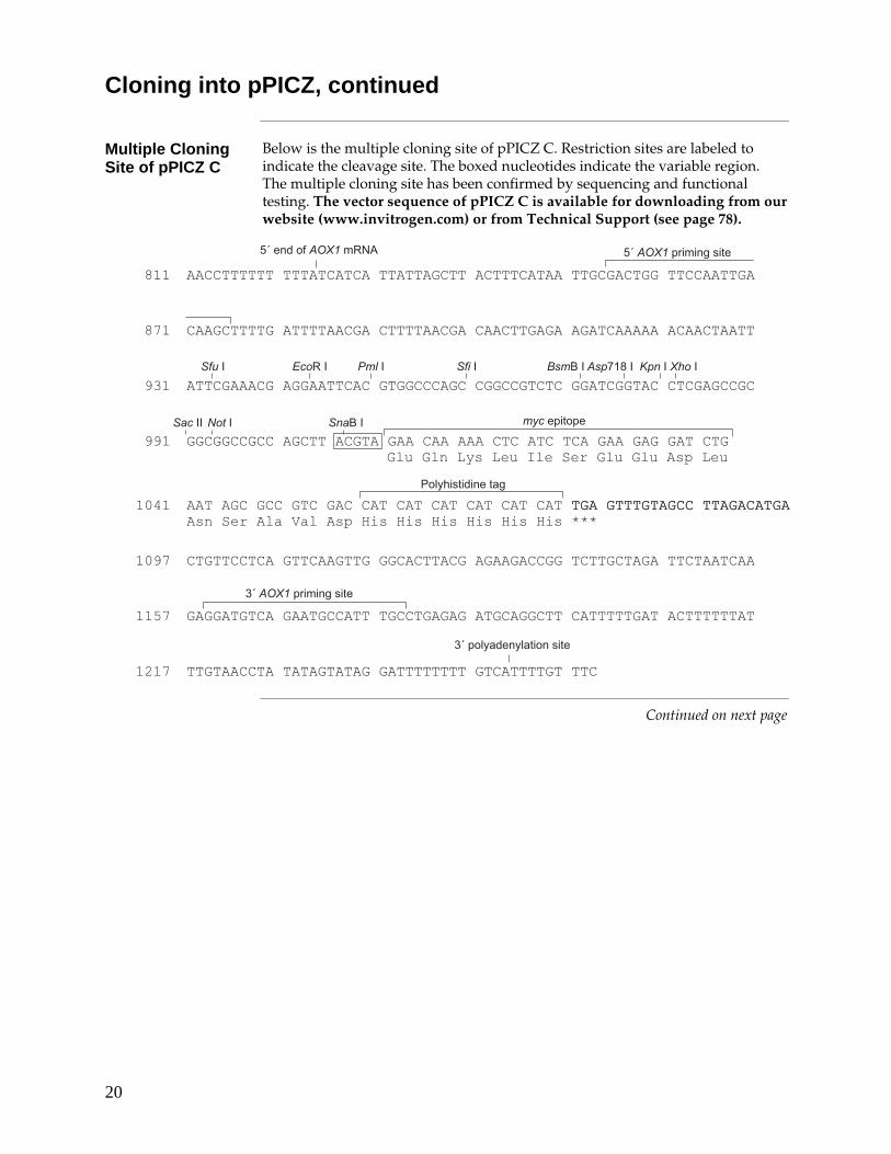
Multiple Cloning Site of pPICZ C
Below is the multiple cloning site of pPICZ C. Restriction sites are labeled to indicate the cleavage site. The boxed nucleotides indicate the variable region. The multiple cloning site has been confirmed by sequencing and functional testing. The vector sequence of pPICZ C is available for downloading from our website (www.invitrogen.com) or from Technical Support (see page 78).
���������������������������������������������������������������������
�����������������������������������������������������������������
�����������������������������������������������������������������
���������������������������������������������������
���� ����������������������������������
���������� � ���� �
�������������������������
�����������������������������������������������������������������
�����������������������������������������������������������������
�����������������������������������������������������������������
��������������������������������������������������������������
��
������������ ���� �� �������������� �
���������� � ���� �
���� ���
�����%���%�� ���� �
$�%& �� � �����
������������������
���3 �����3 ����3 ���3 ���6�3 ���!� �3 ����3 ����3
���33 ����3 ��6�3
����
����
����
��
���
���
Continued on next page
21
Cloning into pPICZα
Multiple Cloning Site of pPICZα A
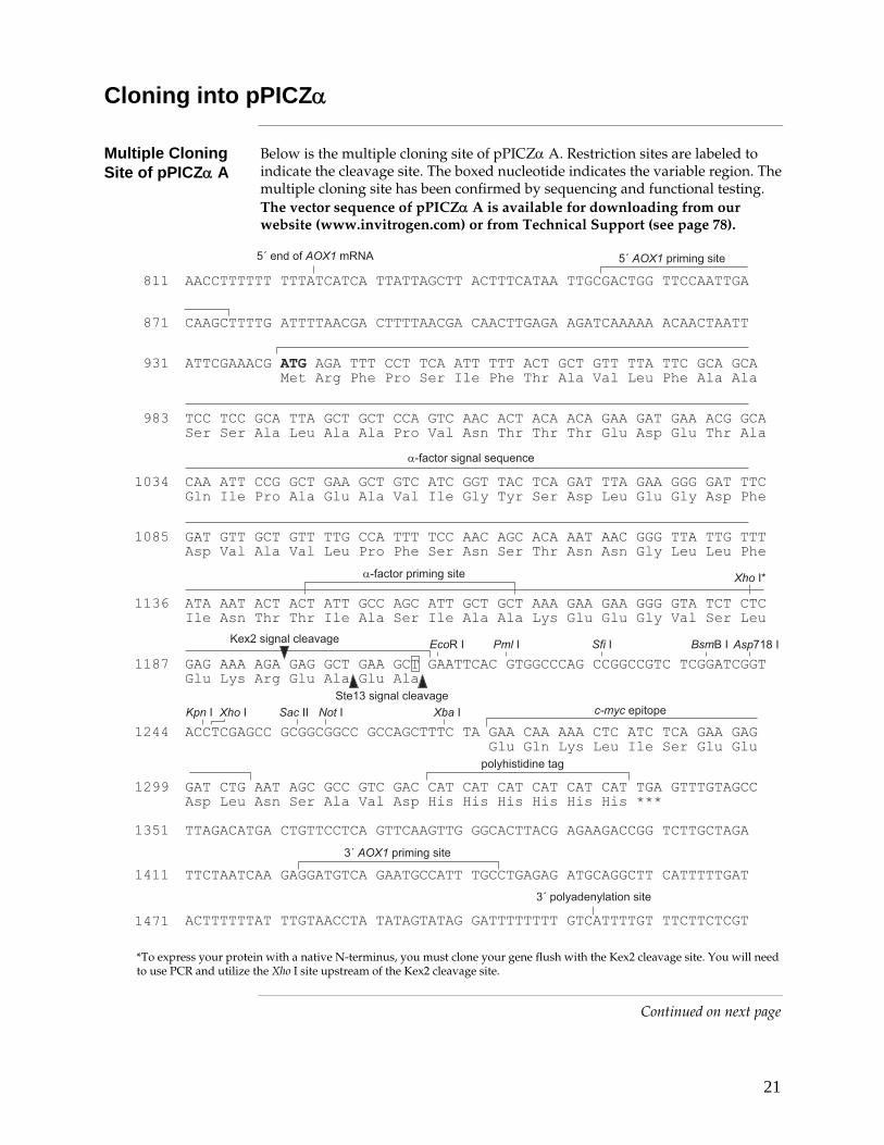
Below is the multiple cloning site of pPICZα A. Restriction sites are labeled to indicate the cleavage site. The boxed nucleotide indicates the variable region. The multiple cloning site has been confirmed by sequencing and functional testing. The vector sequence of pPICZα A is available for downloading from our website (www.invitrogen.com) or from Technical Support (see page 78).
�������������������������������������������������������������������
�������������������������������������������������������������������
�������������������������������������������������������������������
����������������������������������������������������������������������
������������������������������������������������������������������
�������������������������������������������������������������������
����
���
���
���
��
���������� �����7���
����������� � ���� �
����3 ���6�3 ���!� �3
�� �����!���!���� ������ ���� ����������������������������� ��� �
������ �"�#������������������� ���������� ������ �������������"!
���������������� ��"�#�"! �� ������ ���!������������� ��� ��"!
���������$����������������
� ��� ������� ����������"�#���������!���!���!��������������!�����
% &���$�"! �"�#�� ���� �"! ��!����������� ��"! ��������
����31
<8��� ������4��
?����� ������4��
������������������������������������������������������������������
������������������������������������������������������������������
���������������������������������������������������������������������
����
����
���������������������������������������������������������������������
���������������������������������������������������������������������
�����%���%�� ���� �
����3 ����3 ����3���33 � ��3 ������ ���
��%& �� � �����
���������� � ���� �
���� ������ ����������������������������������
������������ ���� �� ���������
�������������������������������������������������������������������
���������� � ���� �������������������
�������������������������������������������������������������������
�����3 ���3
*To express your protein with a native N-terminus, you must clone your gene flush with the Kex2 cleavage site. You will need to use PCR and utilize the Xho I site upstream of the Kex2 cleavage site.
Continued on next page
22
Cloning into pPICZα, continued
Multiple Cloning Site of pPICZα B
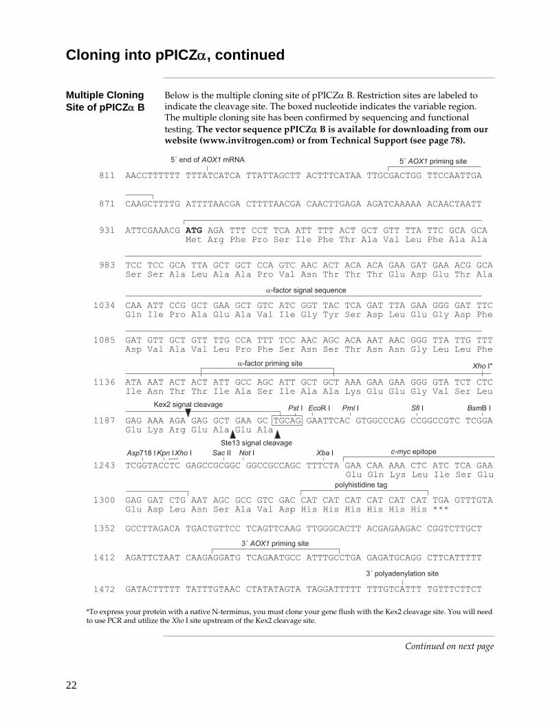
Below is the multiple cloning site of pPICZα B. Restriction sites are labeled to indicate the cleavage site. The boxed nucleotide indicates the variable region. The multiple cloning site has been confirmed by sequencing and functional testing. The vector sequence pPICZα B is available for downloading from our website (www.invitrogen.com) or from Technical Support (see page 78).
�������������������������������������������������������������������
�������������������������������������������������������������������
���������� � ���� �������������������
�������������������������������������������������������������������
�������������������������������������������������������������������
����������������������������������������������������������������������
�����������������������������������������������������������������
������������������������������������������������������������������
�������������������������������������������������������������������
����
���
���
��
���
��
���������� �����7���
����������� � ���� �
����3 ���3 ���6�3
�� �����!���!���� ������ ���� ����������������������������� ��� �
������ �"�#������������������� ���������� ������ �������������"!
���������������� ��"�#�"! �� ������ ���!������������� ��� ��"!
���������$����������������
� ��� ������� ����������"�#���������!���!���!��������������!�����
% &���$�"! �"�#�� ���� �"! ��!����������� ��"! ��������
����31
<8��� ������4��
�����%���%�� ���� �
��%& �� � �����
�������������������������������������������������������������������
�������������������������������������������������������������������
���������������������������������������������������������������������
����
����
���������������������������������������������������������������������
���������������������������������������������������������������������
����3����3 ����3���33 � ��3 ������ ���
���������� � ���� �
�������� ������ ����������������������������������
������������ ���� �� �����
?����� ������4�����!� �3
�����3����3
*To express your protein with a native N-terminus, you must clone your gene flush with the Kex2 cleavage site. You will need to use PCR and utilize the Xho I site upstream of the Kex2 cleavage site.
Continued on next page
23
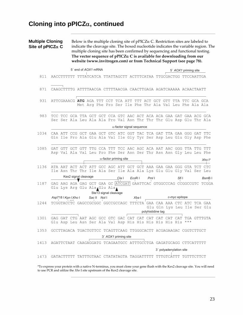
Cloning into pPICZα, continued
Multiple Cloning Site of pPICZα C
Below is the multiple cloning site of pPICZα C. Restriction sites are labeled to indicate the cleavage site. The boxed nucleotide indicates the variable region. The multiple cloning site has been confirmed by sequencing and functional testing. The vector sequence of pPICZα C is available for downloading from our website (www.invitrogen.com) or from Technical Support (see page 78).
�������������������������������������������������������������������
�������������������������������������������������������������������
���������� � ���� �������������������
�������������������������������������������������������������������
�������������������������������������������������������������������
�����������������������������������������������������������������������
�����������������������������������������������������������������
������������������������������������������������������������������
�������������������������������������������������������������������
����
���
���
��
���
��
���������� �����7���
����������� � ���� �
����3 ���3 ���6�3
���!� �3
�� �����!���!���� ������ ���� ����������������������������� ��� �
������ �"�#������������������� ���������� ������ �������������"!
���������������� ��"�#�"! �� ������ ���!������������� ��� ��"!
���������$����������������
� ��� ������� ����������"�#���������!���!���!��������������!�����
% &���$�"! �"�#�� ���� �"! ��!����������� ��"! ��������
����31
<8��� ������4��
�����%���%�� ���� �
��%& �� � �����
�������������������������������������������������������������������
�������������������������������������������������������������������
���������������������������������������������������������������������
����
����
���������������������������������������������������������������������
���������������������������������������������������������������������
����3����3 ����3���33 � ��3 ������ ���
���������� � ���� �
�������� ������ ����������������������������������
������������ ���� �� �����
?����� ������4��
�����3����3
*To express your protein with a native N-terminus, you must clone your gene flush with the Kex2 cleavage site. You will need to use PCR and utilize the Xho I site upstream of the Kex2 cleavage site.
24
Transformation into E. coli
Introduction Ligation mixtures may be transformed into E. coli and selected on Low Salt LB medium (see below) with Zeocin™. Transformants are isolated and analyzed for the presence and orientation of insert. There is no blue/white screening for the presence of insert with pPICZ or pPICZα. After obtaining the desired recombinant plasmid, you will be ready to transform into Pichia.
��������
For Zeocin™ to be active, the salt concentration of the medium must remain low (< 90 mM) and the pH must be 7.5. Prepare Low Salt LB broth and plates using the recipe in the Appendix, page 53.
Failure to lower the salt content of your LB medium will result in non-selection due to inactivation of the drug.
Transformation Guidelines are as follows:
• Transformation may be performed by either electroporation or chemical methods. Use your preferred method or refer to general molecular biology references (Ausubel et al., 1994; Sambrook et al., 1989)
• Add either Low Salt LB or LB medium to the cells after heat shock or electroporation to allow them to recover.
• Plate on Low Salt LB medium with 25 μg/ml Zeocin™.
Note: You may also use SOB, 2XYT, or TB medium, but you may have to increase the concentration of Zeocin™ to 50 μg/ml to compensate for differences in the salt concentration.
• Incubate overnight at 37°C.
Analyzing Transformants
1. After transformation, plate 10 μl and 100 μl of the transformation mix onto Low Salt LB plates with 25 μg/ml Zeocin™ (see above) and select Zeocin™-resistant colonies.
2. Pick 10 Zeocin™-resistant transformants and inoculate into 2 ml Low Salt LB medium with 25 μg/ml Zeocin™. Grow overnight at 37°C with shaking.
3. Isolate plasmid DNA by miniprep for restriction analysis and sequencing (see next page).
4. Be sure to make a glycerol stock of your purified clone for safekeeping.
Continued on next page
25
Transformation into E. coli, continued
Sequencing Recombinant Clones
We strongly recommend that you sequence your construct to confirm that your gene is in frame with the C-terminal peptide before transforming into Pichia. Use the sequencing primers included in the kit to sequence your construct.
To sequence your construct in pPICZ, use the 5´ AOX1 and the 3´ AOX1 Sequencing Primers.
To sequence your construct in pPICZα, use the α-factor or the 5´ AOX1 and the 3´ AOX1 Sequencing Primers.
To use the primers, resuspend each lyophilized primer in 20 μl sterile water. This will yield a stock solution of 0.1 μg/μl.
For sequencing protocols, Refer to Unit 7 in Current Protocols in Molecular Biology (Ausubel et al., 1994) or Chapter 13 in Molecular Cloning: A Laboratory Manual (Sambrook et al., 1989).
Plasmid Preparation
Once you have cloned and sequenced your insert, generate enough plasmid DNA to transform Pichia (5–10 μg of each plasmid per each transformation). We recommend the S.N.A.P.™ Miniprep Kit (Cat. no. K1900-01) or the PureLink™ HiPure Plasmid DNA Purification Kit (Cat. no. K2100-01) for isolation of pure plasmid DNA. Once you have purified plasmid DNA, proceed to Preparing Transforming DNA, next page.
26
Preparing Transforming DNA
Introduction At this point, you should have your gene cloned into one of the pPICZ or pPICZα vectors. Your construct should contain a yeast consensus sequence (A/YAA/TAATGTCT) and be correctly fused to the secretion signal (pPICZα) and/or the C-terminal peptide.
To transform Pichia, prepare 5–10 μg of plasmid DNA, and linearize the plasmid prior to transformation and selection in Pichia. Plate the transformants on YPDS plates containing 100 μg/ml Zeocin™ to isolate Zeocin™-resistant (ZeoR) clones.
Remember also to isolate two control strains for background protein expression in Pichia. Linearize pPICZ or pPICZα and transform into GS115 to generate a Mut+ control and KM71H to generate a MutS control.
Method of Transformation
We recommend electroporation or chemical methods for transformation of Pichia with pPICZ or pPICZα. Electroporation yields 103 to 104 transformants per μg of linearized DNA and does not destroy the cell wall of Pichia. If you do not have access to an electroporation device, use the Pichia EasyComp™ procedure on page 29.
We do not recommend spheroplasting for transformation of Pichia with plasmids containing the Zeocin™ resistance marker. Spheroplasting involves removal of the cell wall to allow DNA to enter the cell. Cells must first regenerate the cell wall before they are able to express the Zeocin™ resistance gene. For this reason, plating spheroplasts directly onto selective medium containing Zeocin™ does not yield any transformants.
��������
Integration can only occur at the AOX1 locus. Vector linearized within the 5´ AOX1 region will integrate by gene insertion into the host 5´ AOX1 region. Therefore, the Pichia host that you use will determine whether the recombinant strain is able to metabolize methanol (Mut+) or not (MutS). To generate a Mut+ recombinant strain, you must use a Pichia host that contains the native AOX1 gene (i.e., X-33, GS115, SMD1168). If you choose to generate a MutS recombinant strain, then use a Pichia host that has a disrupted AOX1 gene (i.e., KM71H). Information on recombination in Pichia is available on page 62.
Restriction Digest 1. Digest ~5–10 μg of plasmid DNA with one of the restriction enzymes listed
below. Each enzyme cuts one time in the 5´ AOX1 region to linearize the either pPICZ or pPICZα.
Note: Choose the enzyme that does not cut within your gene: Sac I (209 bp), Pme I (414 bp), and BstX I (707 bp).
2. We recommend that you check a small aliquot of your digest by agarose gel electrophoresis for complete linearization.
3. If the vector is completely linearized, heat inactivate or add EDTA to stop the reaction, phenol/chloroform extract once, and ethanol precipitate using 1/10 volume 3 M sodium acetate and 2.5 volumes of 100% ethanol.
4. Centrifuge the solution to pellet the DNA, wash the pellet with 80% ethanol, air-dry, and resuspend in 10 μl sterile, deionized water. Use immediately or store at –20°C.
27
Electroporation of Pichia
Introduction We strongly recommend electroporation if you are specifically interested in isolating multi-copy integrants of your gene in Pichia. The frequency of multi-copy insertions ranges from 1 to 10%, requiring hundreds to thousands of transformants to isolate a suitable number of multi-copy clones to test for expression. Electroporation yields some of the highest transformation frequencies in Pichia and is the method of choice to isolate multi-copy integrants.
��������
Traditionally, spheroplasting has been used to transform Pichia, but this method of transformation does not allow direct selection on Zeocin™. Damage to the cell wall leads to increase sensitivity to Zeocin™, causing putative transformants to die before they express the Zeocin™ resistance gene.
Before Starting You will need the following materials for transforming Pichia and selecting
transformants on Zeocin™.
Note: Inclusion of sorbitol in YPD plates stabilizes electroporated cells as they appear to be somewhat osmotically sensitive.
• 5–10 μg pure pPICZ or pPICZα containing your insert
Note: For transforming with circular DNA, you will need 50–100 μg plasmid DNA. If you have constructed multimers in pPICZ or pPICZα, you will not be able to linearize the plasmid.
• YPD Medium
• 50-ml conical polypropylene tubes
• 1 liter cold (4°C) sterile water (place on ice the day of the experiment)
• 25 ml cold (4°C) sterile 1 M sorbitol (place on ice the day of the experiment)
• 30°C incubator
• Electroporation device and 0.2 cm cuvettes
• YPDS plates containing 100 μg/ml Zeocin™ (See page 56 for recipe)
Preparing Pichia for Electroporation
1. Grow 5 ml of your Pichia pastoris strain in YPD in a 50 ml conical at 30°C overnight.
2. Inoculate 500 ml of fresh medium in a 2 liter flask with 0.1–0.5 ml of the overnight culture. Grow overnight again to an OD600 = 1.3–1.5.
3. Centrifuge the cells at 1,500 × g for 5 minutes at 4°C. Resuspend the pellet with 500 ml of ice-cold, sterile water.
4. Centrifuge the cells as in Step 3, then resuspend the pellet with 250 ml of ice-cold, sterile water.
5. Centrifuge the cells as in Step 3, then resuspend the pellet in 20 ml of ice-cold 1 M sorbitol.
6. Centrifuge the cells as in Step 3, then resuspend the pellet in 1 ml of ice-cold 1 M sorbitol for a final volume of approximately 1.5 ml. Keep the cells on ice and use that day. Do not store cells.
Continued on next page
28
Electroporation of Pichia, continued
Transformation by Electroporation
1. Mix 80 μl of the cells from Step 6 (previous page) with 5–10 μg of linearized DNA (in 5–10 μl sterile water) and transfer them to an ice-cold 0.2 cm electroporation cuvette. Note: For circular DNA, use 50–100 μg.
2. Incubate the cuvette with the cells on ice for 5 minutes.
3. Pulse the cells using the manufacturer’s instructions for Saccharomyces cerevisiae.
4. Immediately add 1 ml of ice-cold 1 M sorbitol to the cuvette. Transfer the cuvette contents to a sterile 15-ml tube and incubate at 30°C without shaking for 1 to 2 hours.
5. Spread 10, 25, 50, 100, and 200 μl each on separate, labeled YPDS plates containing 100 μg/ml Zeocin™. Plating at low cell densities favors efficient Zeocin™ selection.
6. Incubate plates from 3–10 days at 30°C until colonies form.
7. Pick 10–20 colonies and purify (streak for single colonies) on fresh YPD or YPDS plates containing 100 μg/ml Zeocin™.
Isolating Multi-copy Recombinants in vivo
A quick, direct way to select putative multi-copy recombinants is to plate the transformation mix on increasing concentrations of Zeocin™.
1. Prepare YPDS plates containing 500, 1000, and 2000 μg/ml Zeocin™
2. Plate 100 to 200 μl of the transformation mix on each plate and incubate at 30°C for 2 days
3. Test transformants for the Mut phenotype (page 33) and expression of your protein (page 37)
Generally several hundred to several thousand Zeocin™-resistant (ZeoR ) colonies are generated using the above protocol. For more colonies, you may modify the protocol as described below. Note that you will need ~20 150-mm plates with YPDS agar containing 100 μg/ml Zeocin™.
1. Set up two transformations per construct and follow Steps 1 through 5 of the Transformation by Electroporation protocol, above.
2. After 1 hour in 1 M sorbitol at 30°C (Step 4, above), add 1 ml YPD medium to each tube. Shake (~200 rpm) the cultures at 30°C.
3. After 1 hour, take one of the tubes and plate out all of the cells by spreading 200 μl on 150-mm plates containing 100 μg/ml Zeocin™.
4. Optional: Continue incubating the other culture for three more hours (for a total of four hours) and then plate out all of the cells by spreading 200 μl on 150-mm plates containing 100 μg/ml Zeocin™.
5. Incubate plates for 2 to 4 days at 30°C until colonies form.
Analyzing Pichia Transformants
Select 6–10 of your ZeoR Pichia transformants and confirm the Mut phenotype as described on page 33. You may also analyze for the presence of insert using PCR (page 68), or for copy number using Southern analysis (page 74).
29
EasyComp™ Transformation
Introduction The Pichia EasyComp™ Kit produces chemically competent Pichia cells and is included to provide an alternative to electroporation and a rapid, convenient method for transformation. However, because of the low transformation efficiency (3 μg plasmid DNA yields about 50 colonies), it is very difficult to isolate multi-copy integrants. In instances where multi-copy integrants are desired, please use electroporation (page 28) for best results. Note that cells are prepared differently for electroporation. Do not use cells prepared using the EasyComp™ protocol for electroporation.
Required Reagents and Equipment
• 30°C rotary shaking incubator • YPD (Yeast Extract Peptone Dextrose) medium (see Recipes, page 55) • 50 ml, sterile conical tubes • Centrifuge suitable for 50 ml conical tubes (floor or table-top) • 1.5 ml sterile screw-cap microcentrifuge tubes • –80°C freezer • Styrofoam box or paper towels
Before Beginning • Streak a YPD plate with your Pichia pastoris strain such that isolated, single
colonies will grow. Incubate the plate at 28–30°C for 2 days.
• Equilibrate Solution I to room temperature.
Preparing Competent Cells
1. Inoculate 10 ml of YPD with a single colony of your Pichia strain. Grow overnight at 28–30°C in a shaking incubator (250–300 rpm).
2. Dilute cells from the overnight culture to an OD600 of 0.1–0.2 in 10 ml of YPD. Grow the cells at 28–30°C in a shaking incubator until the OD600 is 0.6–1.0. This will take approximately 4 to 6 hours.
3. Pellet the cells by centrifugation at 500 × g for 5 minutes at room temperature. Discard the supernatant.
4. Resuspend the cell pellet in 10 ml of Solution I. No incubation time is required.
5. Pellet the cells by centrifugation at 500 × g for 5 minutes at room temperature. Discard the supernatant.
6. Resuspend the cell pellet in 1 ml of Solution I. The cells are now competent.
7. Aliquot 50 to 200 μl of competent cells into labeled 1.5 ml sterile screw-cap microcentrifuge tubes.
Note: Use 50 μl of cells for each transformation. You can thaw the cells and refreeze several times without significant loss in transformation efficiency.
8. At this point, the cells may be kept at room temperature and used directly for transformation or frozen for future use. To freeze cells, place tubes in a Styrofoam box or wrap in several layers of paper towels and place in a –80°C freezer. It is important that you freeze the cells slowly. Do not snap-freeze the cells in liquid nitrogen.
9. Proceed to the transformation procedure.
Continued on next page
30
EasyComp™ Transformation, continued
We have observed that higher transformation efficiencies are often obtained with frozen versus freshly prepared cells. You may choose to use some of the cells immediately following preparation and freeze the remaining cells in small aliquots.
Transformation You may use the following protocol to transform freshly prepared or frozen
competent Pichia cells. Transformation efficiency may vary with each strain and vector used.
Required Reagents and Equipment
• 30°C incubator • Water baths or heat blocks at 30°C and 42°C • Microcentrifuge at room temperature • YPDS with 100 μg/ml Zeocin™ plates (see Recipes, page 56)
Before Beginning • The PEG in Solution II may precipitate at temperatures below 27°C. If you
see a precipitate, warm the solution at 37°C, swirling occasionally, until the precipitate dissolves. To prevent formation of a precipitate, store Solution II at room temperature.
• Equilibrate Solution III to room temperature.
• Equilibrate the appropriate number and type of plates to room temperature. You will need one plate for each transformation.
• You may want to include controls to check for contamination. We recommend a no DNA and a plasmid only control.
Continued on next page
31
EasyComp™ Transformation, continued
Transformation Protocol
1. For each transformation, thaw one tube of competent cells at room temperature and aliquot 50 μl into a sterile microcentrifuge tube. If transforming fresh cells, use 50 μl of cells from Preparing Competent Cells, Step 7, page 29.
2. Add 3 μg of linearized Pichia expression vector DNA to the competent cells. Note: Using greater than 3 μg of DNA may increase transformation efficiencies in
some cases. The volume of DNA should not exceed 5 μl. Linearized DNA can be used directly from a restriction digest reaction without affecting transformation efficiency. Phenol chloroform extraction and ethanol precipitation are not necessary.
3. Add 1 ml of Solution II to the DNA/cell mixture and mix by vortexing or flicking the tube.
4. Incubate the transformation reactions for 1 hour at 30°C in a water bath or incubator. Mix the transformation reaction every 15 minutes by vortexing or flicking the tube. Failure to mix the transformation reaction every 15 minutes will result in decreased transformation efficiency.
5. Heat shock the cells in a 42°C heat block or water bath for 10 minutes.
6. Split the cells into 2 microcentrifuge tubes (approximately 525 μl per tube) and add 1 ml of YPD medium to each tube.
7. Incubate the cells at 30°C for 1 hour to allow expression of Zeocin™ resistance.
8. Pellet the cells by centrifugation at 3,000 × g for 5 minutes at room temperature. Discard the supernatant.
9. Resuspend each tube of cells in 500 μl of Solution III and combine the cells into one tube.
10. Pellet the cells by centrifugation at 3,000 × g for 5 minutes at room temperature. Discard the supernatant.
11. Resuspend the cell pellet in 100 to 150 μl of Solution III.
12. Plate the entire transformation on appropriate selection plates using a sterile spreader. Incubate the plates for 3 to 10 days at 30°C. Each transformation should yield approximately 50 colonies.
Continued on next page
32
EasyComp™ Transformation, continued
Analyzing Pichia Transformants
Select 6–10 of your Zeocin™-resistant Pichia transformants and confirm the Mut phenotype as described on page 33. You may also wish to analyze for the presence of insert using PCR (page 68). Note: When selecting Zeocin™-resistant Pichia transformants, it is normal to observe a low amount of background (~10–30%).
Troubleshooting The table below provides solutions to possible problems you may encounter
when preparing and transforming competent Pichia pastoris cells.
Problem Probable Cause Possible Solution
Low efficiency of transformation
The pH of Solution I or Solution III may have drifted. The pH of both solutions should be 8.0
Check the pH of Solutions I and III. If the pH is low, increase it by adding NaOH. If the pH is high, decrease it by adding HCl. Store solutions at 4°C in order to minimize drift in pH.
Transformation reaction not mixed during incubation
Be sure to mix the transformation reaction every 15 minutes throughout the 1 hour incubation at 30°C. Vortexing works best.
Incubation time is too short or temperature is too low.
Pichia pastoris transformations may be incubated for longer periods of time (up to 3 hours) and at higher temperature (35–37°C). This may, in some instances, result in higher transformation efficiencies.
Cell density is too low (OD600 <0.6)
Resuspend cells from Preparing Competent Cells, Step 6, page 29, in a smaller volume (i.e., 500 μl).
33
Determining the Mut Phenotype
Introduction If you used X-33 or GS115 as the host, the transformants should be Mut+. To confirm the expected phenotype, two strains are included in the kit that will provide examples of Mut+ and MutS phenotypes. GS115 Albumin is MutS and GS115/pPICZ/lacZ is Mut+. Note that KM71H recombinants do not need to be screened for their Mut phenotype as they all will be MutS.
Screening for Mut+ in X-33 and GS115
Transformation of X-33 or GS115 with linearized constructs favor single crossover recombination at the AOX1 locus. Most of the transformants should be Mut+; however, with the presence of the AOX1 sequences in the plasmid, there is a chance that recombination will occur in the 3´AOX1 region also, disrupting the wild-type AOX1 gene and creating MutS transformants. Testing on MDH and MMH plates will allow you to confirm the Mut+ phenotype (see below).
MutS in KM71H All ZeoR transformants in KM71H will be MutS because of the disruption of the
AOX1 gene (aox1::ARG4). There is no need to test recombinants for the Mut phenotype; all recombinants will be MutS. Transformants need to be purified on minimal plates to ensure pure clonal isolates before either testing for expression (see page 37) or confirming integration by PCR (see page 68).
��������
Host strains containing the his4 allele (e.g., GS115) and transformed with the pPICZ or pPICZα vectors require histidine when grown in minimal media. Add histidine to a final concentration of 0.004% to ensure growth of your transformant.
Preparation You may prepare the following media (see page 57) and materials several days
in advance and store at 4°C:
Minimal Dextrose with histidine (MDH) agar plates, 1 liter Minimal Methanol with histidine (MMH) agar plates, 1 liter Sterile toothpicks and Scoring Templates (see page 36)
Streak out the strains GS115 Albumin (MutS) and GS115/pPICZ/lacZ (Mut+) on an MDH or MGYH plate as controls for Mut+ and MutS growth.
Continued on next page
34
Determining the Mut Phenotype, continued
Mut+ in GS115 or X-33
Use the plates containing the ZeoR transformants and confirm the Mut+ phenotype as described below. See page 36 for scoring templates. Note: Instructions are for GS115 strains. These strains require histidine for growth. X-33 does not require histidine for growth, so you may leave it out of the medium.
1. Using a sterile toothpick, pick one colony and streak or patch one ZeoR transformant in a regular pattern on both an MMH plate and an MDH plate, making sure to patch the MMH plate first.
2. Use a new toothpick for each transformant and continue until 10 transformants have been patched (1 plate).
3. To differentiate Mut+ from MutS, make one patch for each of the controls (GS115/MutS Albumin and GS115/pPICZ/lacZ Mut+) onto the MDH and MMH plates.
4. Incubate the plates at 30°C for 2 days.
5. After 2 days or longer at 30°C, score the plates. Mut+ strains will grow normally on both plates, while MutS strains will grow normally on the MDH plate but show little or no growth on the MMH plate.
��������
We recommend purifying your ZeoR transformants to ensure isolation of a pure clonal isolates. This is done by streaking for single colonies on YPD or minimal plates with histidine. You may do this before or after testing for the Mut phenotype.
Replica-Plating Procedure
This procedure gives a lower rate of misclassifications, but it increases the overall Mut+/MutS screening procedure by 2 days. You will need equipment to replica-plate.
1. Using sterile toothpicks, patch 10 ZeoR transformants on an MDH plate. For controls, make one patch from each of the strains GS115/MutS Albumin and GS115/pPICZ/lacZ Mut+ onto the MDH plate.
2. Incubate the plate at 28–30°C for 2 days.
3. After 2 days, replica-plate the patches from the MDH plate onto fresh MMH and MDH plates to screen for MutS transformants.
4. Incubate the replica plates at 28–30°C for 2 days.
5. After 2 days at 28–30°C, score the replica plates. Look for patches that grow normally on the MDH replica plate but show little or no growth on the MMH replica plate; these are your MutS transformants. Including Mut+ and MutS control patches on each plate will provide examples of Mut+ and MutS phenotypes.
Continued on next page
35
Determining the Mut Phenotype, continued
Screening by Functional Assay
Some researchers have used a functional assay to directly screen for high expressing Pichia recombinant clones without first screening for MutS or Mut+ phenotypes. If you elect to screen directly for high-expressing recombinants, be sure to also check the Mut phenotype. This will help you optimize expression of your recombinant clone.
Multiple Integration Events
Pichia pastoris is capable of integrating multiple copies of transforming DNA via recombination into the genome at sites of sequence homology (see page 63 for figure). Although the exact mechanism of multiple integration events is not fully understood, such events are reasonably common among selected transformants.
Successful expression of the gene of interest to useful levels may depend upon the generation of a recombinant strain that contains multiple copies integrated at the AOX1 locus. In addition to simply screening expression levels among several MutS or Mut+ recombinants via SDS-PAGE analysis, it may be desirable to determine the existence of strains that have multiple integrants in the MutS or Mut+ recombinant strain.
See the Appendix, page 73 for methods to determine copy number.
The Next Step After confirming the Mut phenotype, you may proceed to small-scale expression
(page 37) to test for expression of your gene.
Continued on next page
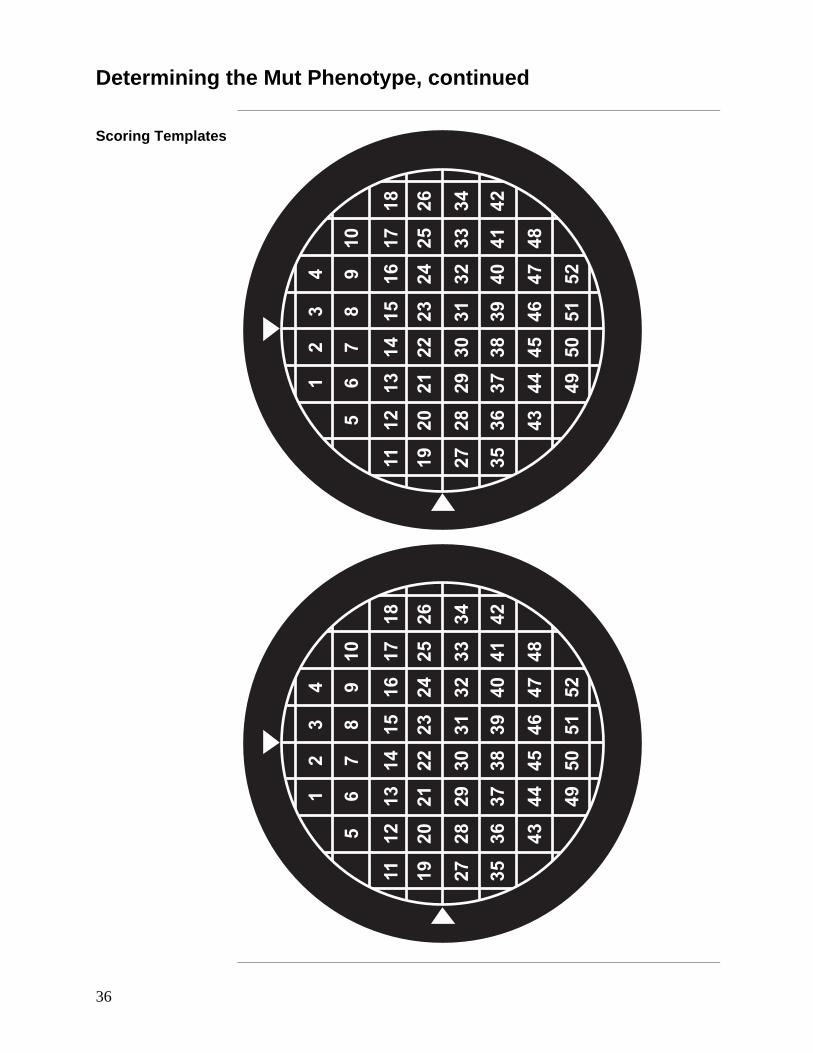
36
Determining the Mut Phenotype, continued
Scoring Templates
�*
#+
�$
,�
�#��
���*
��-
�+��
���,
+�+*
+#++
+-+�
�$+�
+$#+
#�#-
+,##
+�#*
#�*-
#$#,
#�*�
#�*+
***�
*�*�
*#*,
*$�+
���-
�*
#+
�$
,�
�#��
���*
��-
�+��
���,
+�+*
+#++
+-+�
�$+�
+$#+
#�#-
+,##
+�#*
#�*-
#$#,
#�*�
#�*+
***�
*�*�
*#*,
*$�+
���-
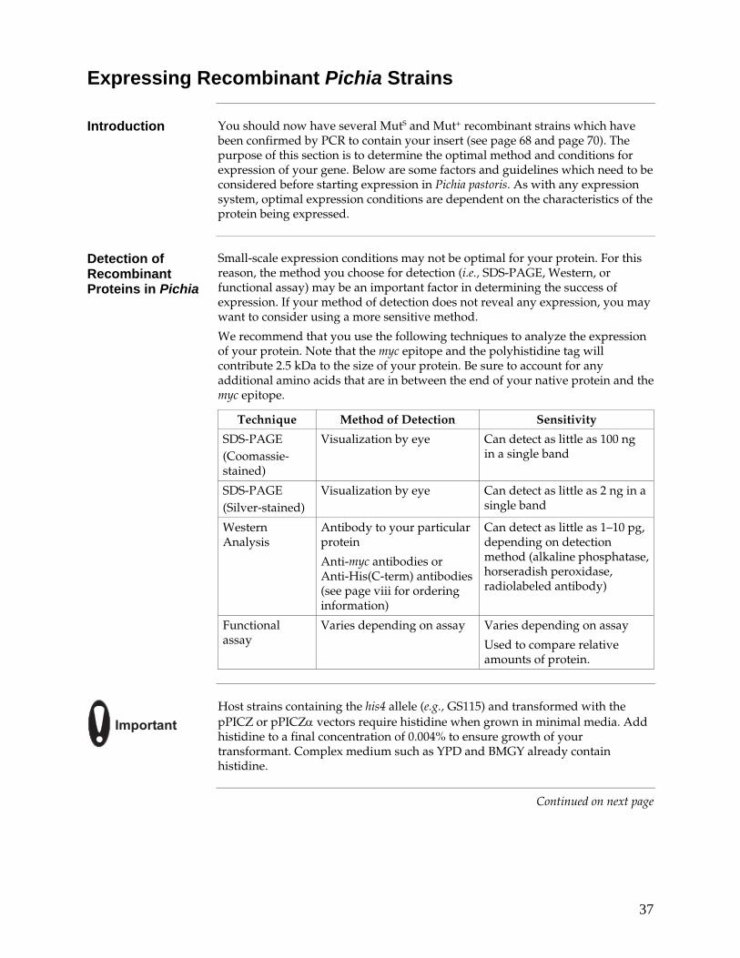
37
Expressing Recombinant Pichia Strains
Introduction You should now have several MutS and Mut+ recombinant strains which have been confirmed by PCR to contain your insert (see page 68 and page 70). The purpose of this section is to determine the optimal method and conditions for expression of your gene. Below are some factors and guidelines which need to be considered before starting expression in Pichia pastoris. As with any expression system, optimal expression conditions are dependent on the characteristics of the protein being expressed.
Detection of Recombinant Proteins in Pichia
Small-scale expression conditions may not be optimal for your protein. For this reason, the method you choose for detection (i.e., SDS-PAGE, Western, or functional assay) may be an important factor in determining the success of expression. If your method of detection does not reveal any expression, you may want to consider using a more sensitive method.
We recommend that you use the following techniques to analyze the expression of your protein. Note that the myc epitope and the polyhistidine tag will contribute 2.5 kDa to the size of your protein. Be sure to account for any additional amino acids that are in between the end of your native protein and the myc epitope.
Technique Method of Detection Sensitivity
SDS-PAGE (Coomassie-stained)
Visualization by eye Can detect as little as 100 ng in a single band
SDS-PAGE (Silver-stained)
Visualization by eye Can detect as little as 2 ng in a single band
Western Analysis
Antibody to your particular protein
Anti-myc antibodies or Anti-His(C-term) antibodies (see page viii for ordering information)
Can detect as little as 1–10 pg, depending on detection method (alkaline phosphatase, horseradish peroxidase, radiolabeled antibody)
Functional assay
Varies depending on assay Varies depending on assay
Used to compare relative amounts of protein.
��������
Host strains containing the his4 allele (e.g., GS115) and transformed with the pPICZ or pPICZα vectors require histidine when grown in minimal media. Add histidine to a final concentration of 0.004% to ensure growth of your transformant. Complex medium such as YPD and BMGY already contain histidine.
Continued on next page
38
Expressing Recombinant Pichia Strains, continued
Media For expressing recombinant Pichia strains, you need BMGY/BMMY (buffered
complex glycerol or methanol medium), BMGH/BMMH (buffered minimal glycerol or methanol medium containing histidine), or MGYH/MMH (minimal glycerol or minimal methanol medium containing histidine) (see Recipes, pages 57–58). BMGH, BMMH, BMGY, and BMMY are usually used for expressing secreted proteins, particularly if pH is important for the activity of your protein. Unlike MGYH and MMH, they are all buffered media. Because these media are buffered with phosphate buffer, you may use a wider range of pH values to optimize protein production. BMGY/BMMY contain yeast extract and peptone to stabilize secreted proteins and to prevent or decrease proteolysis of secreted proteins. Inclusion of yeast extract and peptone allow better growth and biomass accumulation.
Proteases There are some proteins specifically susceptible to proteases that have optimal
activity at neutral pH. If this is the case, expression using MGYH and MMH media may be indicated. As Pichia expression progresses in an unbuffered medium such as MMH, the pH drops to 3 or below, inactivating many neutral pH proteases (Brierley et al., 1994). Pichia is resistant to low pH, so the low pH will not affect growth. In contrast, it has been reported that by including 1% Casamino acids (Difco) and buffering the medium at pH 6.0, extracellular proteases were inhibited, increasing the yield of mouse epidermal growth factor (Clare et al., 1991b).
If you know your protein of interest is especially susceptible to neutral pH proteases, express your protein in an unbuffered medium (MMH). If there is no evidence that your secreted protein of interest is susceptible to proteases at neutral pH, we recommend you do your initial expressions in BMMY. If the expressed protein is degraded, try expressing your protein in an unbuffered medium.
Aeration The most important parameter for efficient expression in Pichia is adequate
aeration during methanol induction. As a general rule when inducing expression, never allow cultures to be more than 10–30% of your total flask volume. It is strongly recommended that baffled flasks be used. See Recipes, page 59 for suppliers of baffled flasks. Cover the flasks with cheesecloth (2–3 layers) or another loose fitting cover. Never use tight fitting covers. (Aeration is not as critical when generating biomass before induction.)
Kinetics of Growth Note that while Mut+ and MutS strains will grow at essentially the same rate in
YPD or glycerol media, Mut+ will grow faster than MutS when both are grown on methanol because of the presence of the AOX1 gene product.
Temperature and Shaking
All expression is done at 30°C, in a shaking incubator. It is critical that the temperature does not exceed 30°C. If your incubator temperature fluctuates, set the temperature at 28°C. If using a floor shaking incubator, shake at 225–250 rpm. If using a table-top shaker that sits inside an incubator, shake at 250–300 rpm.
Continued on next page
39
Expressing Recombinant Pichia Strains, continued
Before Starting When performing your expression, it is important to run the proper controls so
that you will be able to interpret your expression results. Use the following expression controls:
GS115 MutS albumin MutS – Secretion control GS115/pPICZ/lacZ Mut+ Mut+ – Intracellular control GS115 or KM71H/Vector (no insert) background control
Since recombination can occur in many different ways that can effect expression (clonal variation), we recommend that you screen 6–10 verified recombinant clones for expression levels. Start with colonies from the freshest plates available. Colony viability drops over time, so if you have any doubts, it is better to streak out your strain. (You may also start the cultures with a small sample from a frozen glycerol stock that was generated from a single colony.)
Guidelines for Expression
The following steps are guidelines and are presented to get you started with expression. You may have to change the conditions to optimize expression for your particular protein. Use bottom or side baffled flasks whenever possible. These are available in a variety of sizes (50–2000 ml). If you are analyzing a number of recombinants, you can try 50 ml conical tubes. Be sure that the medium is well-aerated by increasing the rate of shaking or placing the tubes at an angle in the shaker.
Continued on next page
40
Expressing Recombinant Pichia Strains, continued
Mut+ Intracellular or Secreted
You can test the effectiveness of your expression conditions by growing GS115/pPICZ/lacZ which is Mut+ and expresses β-Galactosidase intracellularly. Remember to include GS115 transformed with the parent vector as a control for background intracellular expression.
1. Using a single colony, inoculate 25 ml of MGYH, BMGH, or BMGY in a 250 ml baffled flask. Grow at 28–30°C in a shaking incubator (250–300 rpm) until culture reaches an OD600 = 2–6 (approximately 16–18 hours). The cells will be in log-phase growth.
2. Harvest the cells by centrifuging at 1,500–3,000 × g for 5 minutes at room temperature. Decant supernatant and resuspend cell pellet to an OD600 of 1.0 in MMH, BMMH, or BMMY medium to induce expression (approximately 100–200 ml).
3. Place culture in a 1 liter baffled flask. Cover the flask with 2 layers of sterile gauze or cheesecloth and return to incubator to continue growth.
4. Add 100% methanol to a final concentration of 0.5% methanol every 24 hours to maintain induction.
5. At each of the times indicated below, transfer 1 ml of the expression culture to a 1.5 ml microcentrifuge tube. Use these samples to analyze expression levels and determine the optimal time post-induction to harvest. Centrifuge at maximum speed in a tabletop microcentrifuge for 2–3 minutes at room temperature.
Time points (hours): 0, 6, 12, 24 (1 day), 36, 48 (2 days), 60, 72 (3 days), 84, and 96 (4 days).
6. For secreted expression, transfer the supernatant to a separate tube. Store the supernatant and the cell pellets at –80°C until ready to assay. Freeze quickly in liquid N2 or a dry ice/alcohol bath.
For intracellular expression, decant the supernatant and store just the cell pellets at –80°C until ready to assay. Freeze quickly in liquid N2 or a dry ice/alcohol bath.
7. Analyze the supernatants and cell pellets for protein expression by Coomassie-stained SDS-PAGE and Western blot or functional assay (see Analysis by SDS-Polyacrylamide Gel Electrophoresis, page 42).
Continued on next page
41
Expressing Recombinant Pichia Strains, continued
MutS Intracellular or Secreted
You can test the effectiveness of your expression conditions by growing GS115 which is MutS and secretes albumin to the medium. Remember to include X-33, GS115 or KM71H transformed with the parent vector as a control for background intracellular expression.
1. Using a single colony, inoculate 100 ml of MGYH, BMGH, or BMGY in a 1 liter baffled flask. Grow at 28–30°C in a shaking incubator (250–300 rpm) until the culture reaches an OD600 = 2–6 (approximately 16–18 hours).
2. Harvest the cells by centrifuging at 1,500–3,000 × g for 5 minutes at room temperature. To induce expression, decant the supernatant, and resuspend cell pellet in MMH, BMMH, or BMMY medium using 1/5 to 1/10 of the original culture volume (approximately 10–20 ml).
3. Place in a 100 ml baffled flask. Cover the flask with 2 layers of sterile gauze or cheesecloth and return to incubator to continue to grow.
4. Add 100% methanol to a final concentration of 0.5% every 24 hours to maintain induction.
5. At each of the times indicated below transfer 1 ml of the expression culture to a 1.5 ml microcentrifuge tube. These samples will be used to analyze expression levels and determine the optimal time post-induction to harvest. Centrifuge at maximum speed in a tabletop microcentrifuge for 2–3 minutes at room temperature.
Time points (hours): 0, 24 (1 day), 48 (2 days), 72 (3 days), 96 (4 days), 120 (5 days), and 144 (6 days).
6. For secreted expression, transfer the supernatant to a separate tube. Store the supernatant and the cell pellets at –80°C until ready to assay. Freeze quickly in liquid N2 or a dry ice/alcohol bath.
For intracellular expression, decant the supernatant, and store just the cell pellets at –80°C until ready to assay. Freeze quickly in liquid N2 or a dry ice/alcohol bath.
7. Analyze the cell pellets for protein expression by Coomassie-stained SDS-PAGE and Western blot or functional assay (see Analysis by SDS-Polyacrylamide Gel Electrophoresis, next page).
42
Analysis by SDS-Polyacrylamide Gel Electrophoresis
Introduction This section provides guidelines to prepare and analyze your samples using SDS-polyacrylamide gel electrophoresis.
Polyacrylamide Gel Electrophoresis
If you are pouring your own polyacrylamide gels, please note that any standard SDS-polyacrylamide gel apparatus and protocol will work. For example, a 12% polyacrylamide gel with a 5% stacking gel is recommended for proteins ranging in size from 40–100 kDa. For other recommendations, See standard texts such as Molecular Cloning: A Laboratory Manual (Sambrook et al., 1989), Current Protocols in Molecular Biology (Ausubel et al., 1994), Guide to Protein Purification (Deutscher, 1990), or Protein Methods (Bollag and Edelstein, 1991).
Alternatively, a wide range of pre-cast NuPAGE® and Tris-Glycine polyacrylamide gels and electrophoresis apparatus are available from Invitrogen. The patented NuPAGE® Gel System avoids the protein modifications associated with laemmli-type SDS-PAGE, ensuring optimal separation for protein analysis. In addition, Invitrogen also carries a large selection of molecular weight protein standards and staining kits. For more information about the appropriate gels, standards, and stains to use, refer to our website (www.invitrogen.com) or call Technical Support (see page 78).
Preparing Samples
You will need to prepare Breaking Buffer (see page 59) and have acid-washed 0.5 mm glass beads on hand.
Preparing cell pellets (Intracellular and Secreted Expression):
1. Thaw cell pellets quickly and place on ice.
2. For each 1 ml sample, add 100 μl Breaking Buffer to the cell pellet and resuspend.
3. Add an equal volume of acid-washed glass beads (size 0.5 mm). Estimate equal volume by displacement.
4. Vortex 30 seconds, then incubate on ice for 30 seconds. Repeat for a total of 8 cycles.
5. Centrifuge at maximum speed for 10 minutes at 4°C. Transfer the clear supernatant to a fresh microcentrifuge tube.
6. Take 50 μl of supernatant (cell lysate) and mix with 50 μl 2X SDS-PAGE Gel Loading buffer (Sample Buffer).
7. Boil for 10 minutes and load 10–20 μl per well. Thickness of the gel and number of wells will determine the volume you load. You may store the remaining sample at –20°C for Western blots, if necessary. You may store the cell lysates at –80°C for further analysis.
Continued on next page
43
Analysis by SDS-Polyacrylamide Gel Electrophoresis, continued
Preparing Samples, continued
Preparing supernatant (Secreted Expression only):
1. Thaw supernatants and place on ice.
2. Mix 50 μl of the supernatant with 50 μl of SDS-PAGE Gel Loading buffer.
3. Boil 10 minutes, then load 10–30 μl onto the gel. You may store the remaining sample at –20°C for Western blots, if necessary. You may store the supernatants at –80°C for further analysis.
4. If you do not see any protein by Coomassie or by Western blot, then concentrate the supernatant 5–10 fold and analyze samples again by Western blot. Centricon and Centriprep filters (Amicon) are very useful for this purpose.
Concentrating Protein
You may perform Lowry, BCA (Pierce) or Bradford protein assays to quantify the amounts of protein in the cell lysates and medium supernatants. In general, Pichia cell lysates contain 5–10 μg/μl protein. Pichia medium supernatants will vary in protein concentration primarily due to the amount of your secreted protein. Pichia secretes very few native proteins. If the protein concentration of the medium is > 50 μg/ml, 10 μl of medium will give a faint band on a Coomassie-stained SDS-PAGE gel.
Controls Include the following samples as controls on your SDS-PAGE:
• Molecular weight standards appropriate for your desired protein
• A sample of your protein as a standard (if available)
• A sample of X-33, GS115, or KM71H with the parent plasmid transformed into it. This shows the background of native Pichia proteins that are present intracellularly. Inclusion of this sample will help you differentiate your protein from background if you express it intracellularly.
• Analyze the GS115/pPICZ/lacZ and Albumin controls also as they should indicate any problems with the media or expression conditions.
����
�����
���
In addition to Coomassie-stained SDS-PAGE, we strongly recommend performing a Western blot or another more sensitive assay to detect your protein. Visualization of the expressed protein will depend on several factors including its expression level, its solubility, its molecular weight, and whether it will be masked by an abundant cellular protein of the same size. Western blot analysis, enzymatic activities, or a defined purification profile, if available, may help to identify the expressed protein among the native Pichia cellular proteins.
Continued on next page
44
Analysis by SDS-Polyacrylamide Gel Electrophoresis, continued
Western Blot Analysis
To detect expression of your recombinant fusion protein by Western blot analysis, you may use Anti-myc or Anti-His(C-term) antibodies available from Invitrogen (see page viii) or an antibody to your protein of interest. In addition, the Positope™ Control Protein (Cat. no. R900-50) is available for use as a positive control for detection of fusion proteins containing a c-myc epitope or a polyhistidine tag. The ready-to-use WesternBreeze™ Chromogenic Kits and WesternBreeze™ Chemiluminescent Kits are available from Invitrogen to facilitate detection of antibodies by colorimetric or chemiluminescent methods. For more information, refer to our website (www.invitrogen.com) or call Technical Support (see page 78).
Analyzing Protein Expression
Inspection of your Coomassie-stained SDS-PAGE should reveal the induction over time of your protein co-migrating with your standard. If you are satisfied with the level of expression, try a test purification (page 50) or scale-up expression (page 47).
If there is no recombinant protein visible, then perform either a Western blot or a functional assay if available.
If you detect low expression of your recombinant protein, see Optimizing Pichia Protein Expression, page 45, for guidelines to optimize expression.
Test your expression conditions with the one of the two control strains included in the kit (GS115/pPICZ/lacZ or GS115/Albumin).
If there is no indication of expression at all, use PCR to analyze your recombinants for the correctly sized PCR product (page 69). If you find that you have recombinants, perform a Northern analysis to see if and how much full-length mRNA is induced. See page 75 for an RNA isolation protocol.
45
Optimizing Pichia Protein Expression
Introduction Based on available data, there is approximately a 50 to 75% chance of expressing your protein of interest in Pichia pastoris at reasonable levels. The biggest hurdle seems to be generating initial success--i.e., expression of your protein at any level. While there are relatively few examples of expression of >10 grams/liter, there are many examples of expression in the >1 gram/liter range, making the Pichia pastoris expression system one of the most productive eukaryotic expression systems available. Likewise, there are several examples of proteins that have been successfully expressed in Pichia pastoris that were completely unsuccessful in baculovirus or Saccharomyces cerevisiae, suggesting that the Pichia pastoris system is an important alternative to have available. If you obtain no or low protein expression in your initial expression experiment, use the following guidelines to optimize expression.
Proteolysis or Degradation
• Do a time course study of expression. Check to see if there is a time point that yields a larger percentage of full-length protein.
• If secreting your protein, check to see if your protein is susceptible to neutral pH proteases by expressing in unbuffered medium (MMH). In addition, try 1% Casamino acids with buffered medium to inhibit extracellular proteases.
Low Secreted Expression Levels
• Check cell pellet to see if overall expression is low or if the protein did not secrete. If it did not secrete, try a different signal sequence (e.g., a native or α-factor signal sequence).
• Concentrate your supernatant by ammonium sulfate precipitation or ultrafiltration (see page 49).
• For Mut+, induce expression with a higher density culture.
Low Expression Levels
• Look for multi-copy recombinants (i.e., jackpot clones) by dot blot (see page 73). There are quite a few examples of increasing the expression levels of a particular protein by increasing the gene dosage. See (Clare et al., 1991a; Clare et al., 1991b; Romanos et al., 1991).
• Check both Mut+ and MutS recombinants for increased expression. Some proteins express better in one type of genetic background than another.
• If secreting your protein, try intracellular expression. The protein may not be processed correctly and fail to secrete. Be sure you check your cell pellets for evidence of expression. If you are having problems with intracellular expression, try secreting your protein. It probably will glycosylate, which may be desirable or not. If glycosylation is undesirable, oligosaccharides can be removed with Peptide:N-Glycosidase F (New England BioLabs).
• Scale up to fermentation (page 49). Pichia is a yeast, and is particularly well suited to growth in a fermentor.
Continued on next page
46
Optimization of Pichia Protein Expression, continued
No Expression Be sure to try some of the easier things listed above as no expression can be the
same thing as very low expression. If none of these things improve protein expression, use PCR to check for insertion of your gene into the Pichia genome (page 68). If your gene is present, perform a Northern blot analysis to check for transcription of your gene. There is a protocol in the Appendix for RNA isolation from Pichia (see page 75).
If you see premature transcriptional termination, check the AT content of your gene. In Saccharomyces, there are a few consensus sequences which promote premature termination. One of these, TTTTTATA, resembles a sequence in HIV-1 gp120, ATTATTTTAT AAA, which prematurely terminatates mRNA when expressed in Pichia. When this sequence was changed, longer transcripts were found (Scorer et al., 1993).
Hyper-glycosylation
If your protein is hyperglycosylated:
• Try intracellular expression as your protein will not go through the secretion pathway and therefore, not be modified.
• Try deglycosylating the protein with Peptide:N-Glycosidase F or other enzymes (see page 52).
47
Scale-up of Expression
Guidelines for Expression
Once expression is optimized, you will want to scale-up your expression protocol to produce more protein. This may be done by increasing the culture volume using larger baffled flasks (below) or fermentation. Use the guidelines below to scale-up your expression protocol. To purify your protein, see page 50.
Mut+ Intracellular or Secreted
1. Using a single colony, inoculate 25 ml of MGYH, BMGH, or BMGY in a 250 ml baffled flask. Grow at 28–30°C in a shaking incubator (250–300 rpm) until culture reaches an OD600 = 2–6 (approximately 16–18 hours).
2. Use this 25 ml culture to inoculate 1 liter of MGYH, BMGH, or BMGY in a 3 or 4 liter baffled flask and grow at 28–30°C with vigorous shaking (250–300 rpm) until the culture reaches log phase growth (OD600 = 2–6).
3. Harvest the cells using sterile centrifuge bottles by centrifuging at 1500–3000 × g for 5 minutes at room temperature. To induce expression, decant the supernatant and resuspend cell pellet to an OD600 = 1.0 (2–6 liters) in MMH, BMMH, or BMMY medium to start induction.
4. Aliquot the culture between several 3 or 4 liter baffled flask. Cover the flasks with 2 layers of sterile gauze or cheesecloth and return to incubator. Continue to grow at 28–30°C with shaking.
5. Add 100% methanol to 0.5% every 24 hours until the optimal time of induction is reached as determined from the time course study.
6. Harvest cells by centrifuging at 1,500–3,000 × g for 5 minutes at room temperature.
For intracellular expression, decant the supernatant and store the cell pellets at –80°C until ready to process. For secreted expression, save the supernatant, chill to 4°C, and concentrate it down if desired (see page 49). Proceed directly to purification (page 50) or store the supernatant at –80°C until ready to process further.
Continued on next page
48
Scale-up of Expression, continued
MutS Intracellular or Secreted
1. Using a single colony, inoculate 10 ml of MGYH, BMGH, or BMGY in a 100 ml baffled flask. Grow at 28–30°C in a shaking incubator (250–300 rpm) until the culture reaches an OD600 = 2–6 (approximately 16–18 hours).
2. Use this 10 ml culture to inoculate 1 liter of MGYH, BMGH, or BMGY in a 3 or 4 liter baffled flask and grow at 28–30°C with vigorous shaking (250–300 rpm) until the culture reaches log phase growth (OD600 = 2–6).
3. Harvest the cells by centrifuging at 1,500–3,000 × g for 5 minutes at room temperature. To induce expression, decant the supernatant and resuspend cell pellet in 1/5 to 1/10 of the original culture volume of MMH, BMMH, or BMMY medium (approximately 100–200 ml).
4. Place the culture in a 1 liter baffled flask. Cover the flask with 2 layers of sterile gauze or cheesecloth and return to incubator. Continue to grow at 28–30°C with shaking.
5. Add 100% methanol to 0.5% every 24 hours until the optimal time of induction is reached.
6. Harvest cells by centrifuging at 1,500–3,000 × g for 5 minutes at room temperature.
For intracellular expression, decant the supernatant and store the cell pellets at –80°C until ready to process. For secreted expression, save the supernatant, chill to 4°C, and concentrate it down if desired (see next page). Proceed directly to purification (page 50) or store the supernatant at –80°C until ready to process further.
To increase the amount of cells for MutS recombinants, increase the number of flasks, put 200–300 ml in a 3 liter flask, or try fermentation.
Continued on next page

49
Scale-up of Expression, continued
Concentration of Proteins
Proteins secreted into the media are usually > 50% homogeneous and will require some additional purification (see page 50 or 52). It is optimal to concentrate the protein if the expression level is not particularly high. There are several general methods to concentrate proteins secreted from Pichia. These general methods include:
• Ammonium sulfate precipitation
• Dialysis
• Centrifuge concentrator for small volumes (e.g., Centricon or Centriprep devices available from Amicon)
• Pressurized cell concentrators for large volumes (Amicon ultrafiltration devices)
• Lyophilization
A general guide to protein techniques is Protein Methods (Bollag and Edelstein, 1991).
Cell Lysis A general procedure for cell lysis using glass beads is provided on the next page.
There is also a cell lysis protocol in Current Protocols in Molecular Biology, page 13.13.4. (Ausubel et al., 1994) and in Guide to Protein Purification (Deutscher, 1990). We also recommend lysis by French Press (follow the manufacturer's suggestions for yeast).
Fermentation Basic guidelines are available for fermentation of Pichia from Invitrogen. We
recommend that only those with fermentation experience or those who have access to people with experience attempt fermentation. Contact Technical Support (see page 78) for more information.
50
Purification
Introduction In this section, you will grow and induce a 10–200 ml culture of your Pichia transformant for trial purification on a metal-chelating resin such as ProBond™. You may harvest the cells and store them at –80°C until you are ready to purify your fusion protein, or you may proceed directly with protein purification. Note that this section only describes preparation of cell lysates and sample application onto ProBond™. For instructions on how to prepare and use ProBond™ resin, refer to the ProBond™ Purification System manual.
ProBond™ Resin We recommend that you use the ProBond™ Purification System (Cat. no. K850-01)
for purifying fusion proteins expressed from pPICZ or pPICZα. Note that instructions for equilibration of and chromatography on ProBond™ resin are contained in the ProBond™ Purification System.
If you are using a metal-chelating resin other than ProBond™, follow the manufacturer's recommendations for fusion proteins expressed in bacteria or yeast.
Binding Capacity of ProBond™
One milliliter of ProBond™ resin binds at least 1 mg of recombinant protein. This amount can vary depending on the nature of the protein.
��������
Throughout the following protocol, be sure to keep the cell lysate and fractions on ice. Small-scale purifications using the 2 ml ProBond™ columns and buffers can be performed at room temperature on the bench top. For large scale purifications, all reagents must be at 4°C.
Preparing Cell Lysates
Express your protein using a small-scale culture (10–20 ml for MutS strains; 100–200 ml for Mut+) and the optimal conditions for expression (if determined). Refer to the Pichia Expression Kit manual for details. Once your protein is expressed, follow the protocol below to prepare a cell lysate for chromatography on ProBond™.
Prepare Breaking Buffer (BB) as described in the Recipes, page 59.
1. Wash cells once in BB by resuspending them and centrifuging 5–10 minutes at 3,000 × g at 4°C.
2. Resuspend the cells to an OD600 of 50–100 in BB.
3. Add an equal volume of acid-washed glass beads (0.5 mm). Estimate volume by displacement.
4. Vortex the mixture for 30 seconds, then incubate on ice for 30 seconds. Repeat 7 more times. Alternating vortexing with cooling keeps the cell extracts cold and reduces denaturation of your protein.
5. Centrifuge the sample at 4°C for 5–10 minutes at 12,000 × g.
6. Transfer the clear supernatant to a fresh container and analyze for your protein. The total protein concentration should be around 2–3 mg/ml.
7. Save the pellet and extract with 6 M urea or 1% Triton® X-100 to check for insoluble protein.
Continued on next page

51
Purification, continued
Sample Application (Native Conditions)
For sample application onto ProBond™, you will need Native Binding Buffer, pH 7.8 and a 2 ml ProBond™ column, pre-equilibrated using native conditions.
1. Combine 1 ml (2–3 mg/ml total protein) of Pichia lysate with 7 ml Native Binding Buffer.
2. Take a pre-equilibrated ProBond™ column and resuspend the resin in 4 ml of the diluted lysate from Step 1.
3. Seal the column and batch-bind by rocking gently at room temperature for 10 minutes.
4. Let the resin settle by gravity or low speed centrifugation (800 × g) and carefully remove the supernatant. Save the supernatant to check for unbound protein.
5. Repeat Steps 2 through 4 with the remaining 4 ml of diluted lysate. Proceed to Column Washing and Elution Under Native Conditions in the ProBond™ Purification manual. Use the recommendations noted for bacterial cell lysates.
Sample Application (Denaturing Conditions)
Use the protocol above except pre-equilibrate the ProBond™ column using Denaturing Binding Buffer and combine 1 ml of the Pichia cell lysate with 7 ml of the Denaturing Binding Buffer.
We have observed some Pichia proteins may be retained on the ProBond™ column using native purification conditions. Optimization of the purification or using denaturing purification may remove these non-specific Pichia proteins (see ProBond™ Purification System manual).
Analysis of Purification
Be sure to save all fractions, washes, and flow-through for analysis by SDS-PAGE. You may need to use Western blot analysis to detect your protein if expression is low or not enough protein was loaded onto the column. Refer to the ProBond™ Purification System manual for a guide to troubleshoot chromatography.
Scale-up You may find it necessary to scale-up your purification to obtain sufficient
amounts of purified protein. Adjust the pH and NaCl concentration of your lysate with 1/10 volume of 10X Stock Solution B (ProBond™ Purification System) before adding it to the column. The pH should be greater than or equal to 7.5 and the NaCl concentration should be ~500 mM. Using 10X Stock Solution B to adjust the pH and the ionic strength keeps the total volume small for sample application.
52
Protein Glycosylation
Analyzing Glycoproteins
When expressing and purifying a glycosylated protein in a heterologous expression system, it is desirable to quickly determine whether the protein is glycosylated properly. There are published protocols for carbohydrate analysis of proteins to allow the molecular biologist to characterize glycosylated proteins of interest (Ausubel et al., 1994), Unit 17. Further information about glycosylation in eukaryotes is available in a review by Varki and Freeze (Varki and Freeze, 1994).
Enzymes for Analyzing Glycoproteins
These are just a few of the enzymes available for carbohydrate analysis. Abbreviations are as follows: Asn - Asparagine, Gal - Galactose, GlcNAc - N-acetylglucosamine, GalNAc - N-acetylgalactosamine, and NeuAc - N-acetylneuraminic acid.
Enzyme Type of enzyme
Specificity
Endoglycosidase D Endo Cleaves various high mannose glycans
Endoglycosidase F Endo Cleaves various high mannose glycans
Endoglycosidase H Endo Cleaves various high mannose glycans
β-galactosidase Exo Removes terminal galactosides from Gal-β1,3-GlcNAc, Gal-β1,4-GlcNAc or Gal-β1,3 GalNAc.
Peptide:N-Glycosidase F Endo Glycoproteins between Asn and GlcNAc (removes oligosaccharides)
Sialidases (Neuraminidases)
Vibrio cholerae Clostridium perfringens Arthobacter ureafaciens Newcastle disease virus
Exo NeuAc-α2,6-Gal, NeuAc-α2,6-GlcNAc or NeuAc-α2,3-Gal
Commercial Carbohydrate Analysis
There are a number of commercial vendors who will contract to analyze proteins for glycosylation. A number of companies also supply kits and reagents for researchers to do carbohydrate analysis in their own laboratories. A partial list is provided below:
Company Type of Service Phone Number
Glyko Kits for Carbohydrate Analysis, Reagents, Contract Services
1-800-334-5956
Oxford GlycoSystems Kits for Carbohydrate Analysis, Reagents, Contract Services
1-800-722-2597
New England BioLabs Reagents 1-800-632-5227
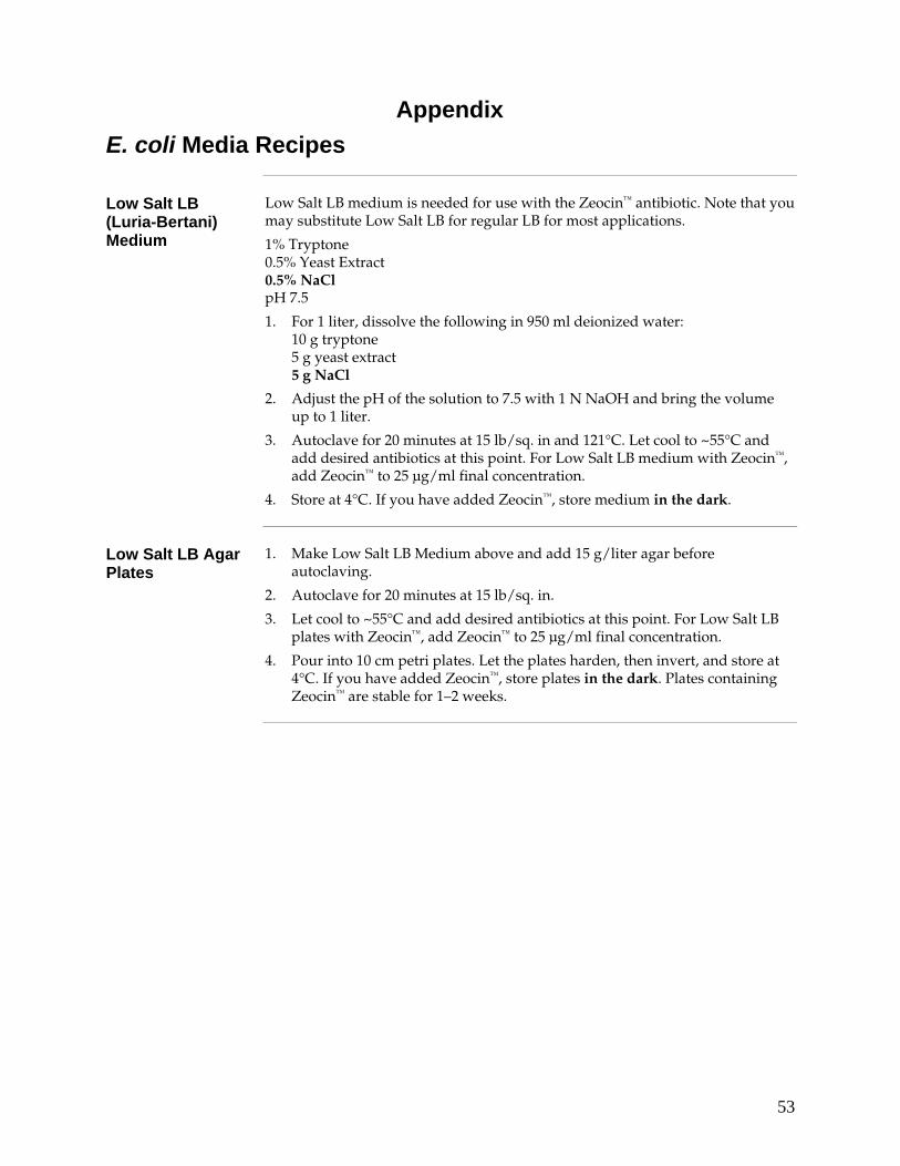
53
Appendix E. coli Media Recipes
Low Salt LB (Luria-Bertani) Medium
Low Salt LB medium is needed for use with the Zeocin™ antibiotic. Note that you may substitute Low Salt LB for regular LB for most applications.
1% Tryptone 0.5% Yeast Extract 0.5% NaCl pH 7.5
1. For 1 liter, dissolve the following in 950 ml deionized water: 10 g tryptone 5 g yeast extract 5 g NaCl
2. Adjust the pH of the solution to 7.5 with 1 N NaOH and bring the volume up to 1 liter.
3. Autoclave for 20 minutes at 15 lb/sq. in and 121°C. Let cool to ~55°C and add desired antibiotics at this point. For Low Salt LB medium with Zeocin™, add Zeocin™ to 25 μg/ml final concentration.
4. Store at 4°C. If you have added Zeocin™, store medium in the dark.
Low Salt LB Agar Plates
1. Make Low Salt LB Medium above and add 15 g/liter agar before autoclaving.
2. Autoclave for 20 minutes at 15 lb/sq. in.
3. Let cool to ~55°C and add desired antibiotics at this point. For Low Salt LB plates with Zeocin™, add Zeocin™ to 25 μg/ml final concentration.
4. Pour into 10 cm petri plates. Let the plates harden, then invert, and store at 4°C. If you have added Zeocin™, store plates in the dark. Plates containing Zeocin™ are stable for 1–2 weeks.
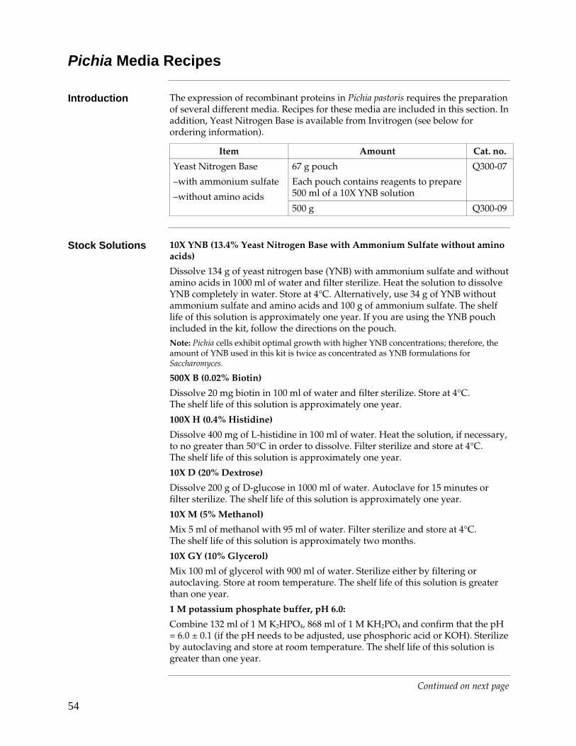
54
Pichia Media Recipes
Introduction The expression of recombinant proteins in Pichia pastoris requires the preparation of several different media. Recipes for these media are included in this section. In addition, Yeast Nitrogen Base is available from Invitrogen (see below for ordering information).
Item Amount Cat. no.
67 g pouch
Each pouch contains reagents to prepare 500 ml of a 10X YNB solution
Q300-07 Yeast Nitrogen Base
–with ammonium sulfate
–without amino acids 500 g Q300-09
Stock Solutions 10X YNB (13.4% Yeast Nitrogen Base with Ammonium Sulfate without amino
acids)
Dissolve 134 g of yeast nitrogen base (YNB) with ammonium sulfate and without amino acids in 1000 ml of water and filter sterilize. Heat the solution to dissolve YNB completely in water. Store at 4°C. Alternatively, use 34 g of YNB without ammonium sulfate and amino acids and 100 g of ammonium sulfate. The shelf life of this solution is approximately one year. If you are using the YNB pouch included in the kit, follow the directions on the pouch. Note: Pichia cells exhibit optimal growth with higher YNB concentrations; therefore, the amount of YNB used in this kit is twice as concentrated as YNB formulations for Saccharomyces.
500X B (0.02% Biotin)
Dissolve 20 mg biotin in 100 ml of water and filter sterilize. Store at 4°C. The shelf life of this solution is approximately one year.
100X H (0.4% Histidine)
Dissolve 400 mg of L-histidine in 100 ml of water. Heat the solution, if necessary, to no greater than 50°C in order to dissolve. Filter sterilize and store at 4°C. The shelf life of this solution is approximately one year.
10X D (20% Dextrose)
Dissolve 200 g of D-glucose in 1000 ml of water. Autoclave for 15 minutes or filter sterilize. The shelf life of this solution is approximately one year.
10X M (5% Methanol)
Mix 5 ml of methanol with 95 ml of water. Filter sterilize and store at 4°C. The shelf life of this solution is approximately two months.
10X GY (10% Glycerol)
Mix 100 ml of glycerol with 900 ml of water. Sterilize either by filtering or autoclaving. Store at room temperature. The shelf life of this solution is greater than one year.
1 M potassium phosphate buffer, pH 6.0:
Combine 132 ml of 1 M K2HPO4, 868 ml of 1 M KH2PO4 and confirm that the pH = 6.0 ± 0.1 (if the pH needs to be adjusted, use phosphoric acid or KOH). Sterilize by autoclaving and store at room temperature. The shelf life of this solution is greater than one year.
Continued on next page
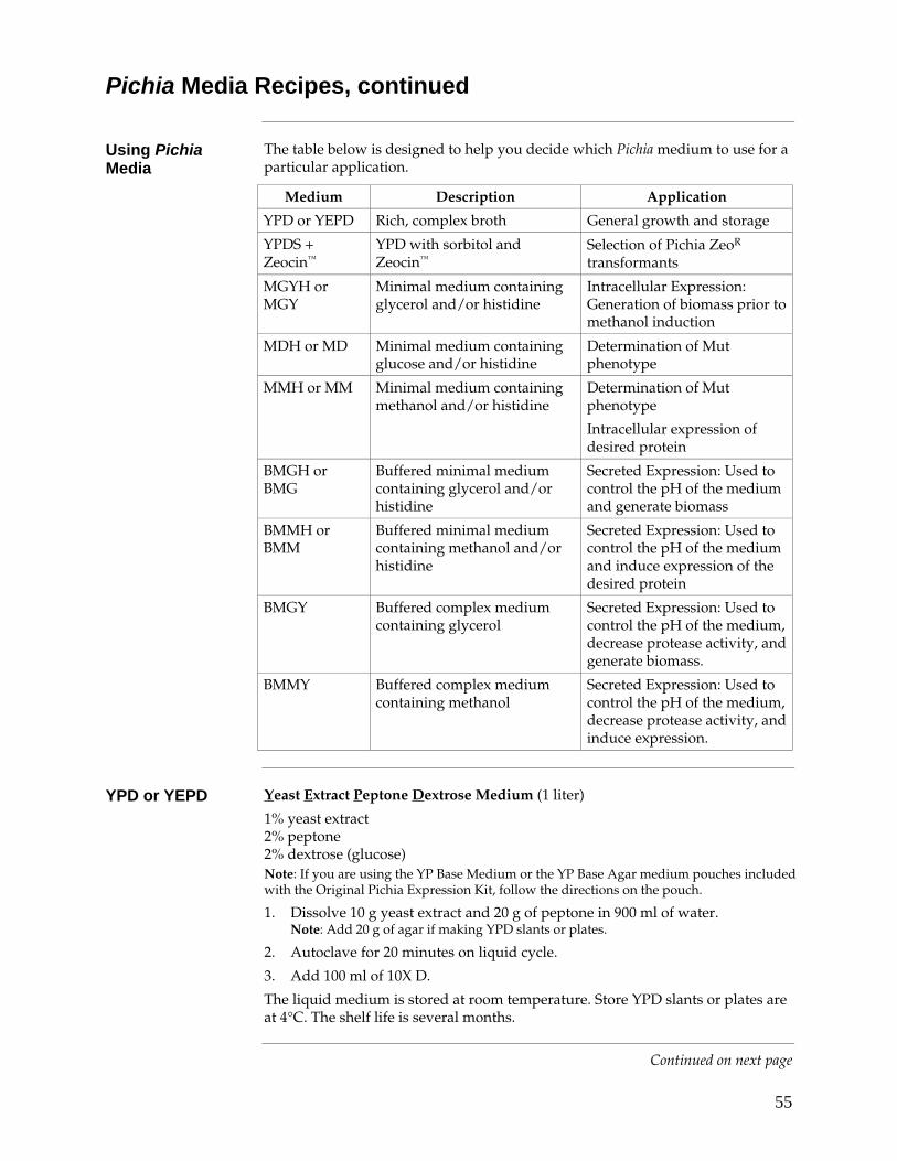
55
Pichia Media Recipes, continued
Using Pichia Media
The table below is designed to help you decide which Pichia medium to use for a particular application.
Medium Description Application
YPD or YEPD Rich, complex broth General growth and storage
YPDS + Zeocin™
YPD with sorbitol and Zeocin™
Selection of Pichia ZeoR transformants
MGYH or MGY
Minimal medium containing glycerol and/or histidine
Intracellular Expression: Generation of biomass prior to methanol induction
MDH or MD Minimal medium containing glucose and/or histidine
Determination of Mut phenotype
MMH or MM Minimal medium containing methanol and/or histidine
Determination of Mut phenotype
Intracellular expression of desired protein
BMGH or BMG
Buffered minimal medium containing glycerol and/or histidine
Secreted Expression: Used to control the pH of the medium and generate biomass
BMMH or BMM
Buffered minimal medium containing methanol and/or histidine
Secreted Expression: Used to control the pH of the medium and induce expression of the desired protein
BMGY Buffered complex medium containing glycerol
Secreted Expression: Used to control the pH of the medium, decrease protease activity, and generate biomass.
BMMY Buffered complex medium containing methanol
Secreted Expression: Used to control the pH of the medium, decrease protease activity, and induce expression.
YPD or YEPD Yeast Extract Peptone Dextrose Medium (1 liter)
1% yeast extract 2% peptone 2% dextrose (glucose) Note: If you are using the YP Base Medium or the YP Base Agar medium pouches included with the Original Pichia Expression Kit, follow the directions on the pouch.
1. Dissolve 10 g yeast extract and 20 g of peptone in 900 ml of water. Note: Add 20 g of agar if making YPD slants or plates.
2. Autoclave for 20 minutes on liquid cycle.
3. Add 100 ml of 10X D.
The liquid medium is stored at room temperature. Store YPD slants or plates are at 4°C. The shelf life is several months.
Continued on next page
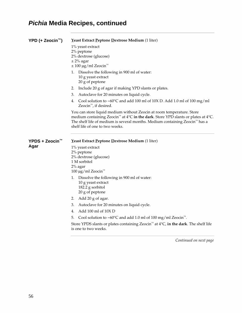
56
Pichia Media Recipes, continued
YPD (+ Zeocin™) Yeast Extract Peptone Dextrose Medium (1 liter)
1% yeast extract 2% peptone 2% dextrose (glucose) ± 2% agar ± 100 μg/ml Zeocin™
1. Dissolve the following in 900 ml of water: 10 g yeast extract 20 g of peptone
2. Include 20 g of agar if making YPD slants or plates.
3. Autoclave for 20 minutes on liquid cycle.
4. Cool solution to ~60°C and add 100 ml of 10X D. Add 1.0 ml of 100 mg/ml Zeocin™, if desired.
You can store liquid medium without Zeocin at room temperature. Store medium containing Zeocin™ at 4°C in the dark. Store YPD slants or plates at 4°C. The shelf life of medium is several months. Medium containing Zeocin™ has a shelf life of one to two weeks.
YPDS + Zeocin™ Agar
Yeast Extract Peptone Dextrose Medium (1 liter)
1% yeast extract 2% peptone 2% dextrose (glucose) 1 M sorbitol 2% agar 100 μg/ml Zeocin™
1. Dissolve the following in 900 ml of water: 10 g yeast extract 182.2 g sorbitol 20 g of peptone
2. Add 20 g of agar.
3. Autoclave for 20 minutes on liquid cycle.
4. Add 100 ml of 10X D
5. Cool solution to ~60°C and add 1.0 ml of 100 mg/ml Zeocin™.
Store YPDS slants or plates containing Zeocin™ at 4°C, in the dark. The shelf life is one to two weeks.
Continued on next page
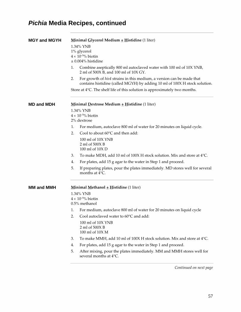
57
Pichia Media Recipes, continued
MGY and MGYH Minimal Glycerol Medium + Histidine (1 liter)
1.34% YNB 1% glycerol 4 × 10⁻⁵% biotin ± 0.004% histidine
1. Combine aseptically 800 ml autoclaved water with 100 ml of 10X YNB, 2 ml of 500X B, and 100 ml of 10X GY.
2. For growth of his4 strains in this medium, a version can be made that contains histidine (called MGYH) by adding 10 ml of 100X H stock solution.
Store at 4°C. The shelf life of this solution is approximately two months.
MD and MDH Minimal Dextrose Medium + Histidine (1 liter)
1.34% YNB 4 × 10⁻⁵% biotin 2% dextrose
1. For medium, autoclave 800 ml of water for 20 minutes on liquid cycle.
2. Cool to about 60°C and then add:
100 ml of 10X YNB 2 ml of 500X B 100 ml of 10X D
3. To make MDH, add 10 ml of 100X H stock solution. Mix and store at 4°C.
4. For plates, add 15 g agar to the water in Step 1 and proceed.
5. If preparing plates, pour the plates immediately. MD stores well for several months at 4°C.
MM and MMH Minimal Methanol + Histidine (1 liter)
1.34% YNB 4 × 10⁻⁵% biotin 0.5% methanol
1. For medium, autoclave 800 ml of water for 20 minutes on liquid cycle
2. Cool autoclaved water to 60°C and add:
100 ml of 10X YNB 2 ml of 500X B 100 ml of 10X M
3. To make MMH, add 10 ml of 100X H stock solution. Mix and store at 4°C.
4. For plates, add 15 g agar to the water in Step 1 and proceed.
5. After mixing, pour the plates immediately. MM and MMH stores well for several months at 4°C.
Continued on next page
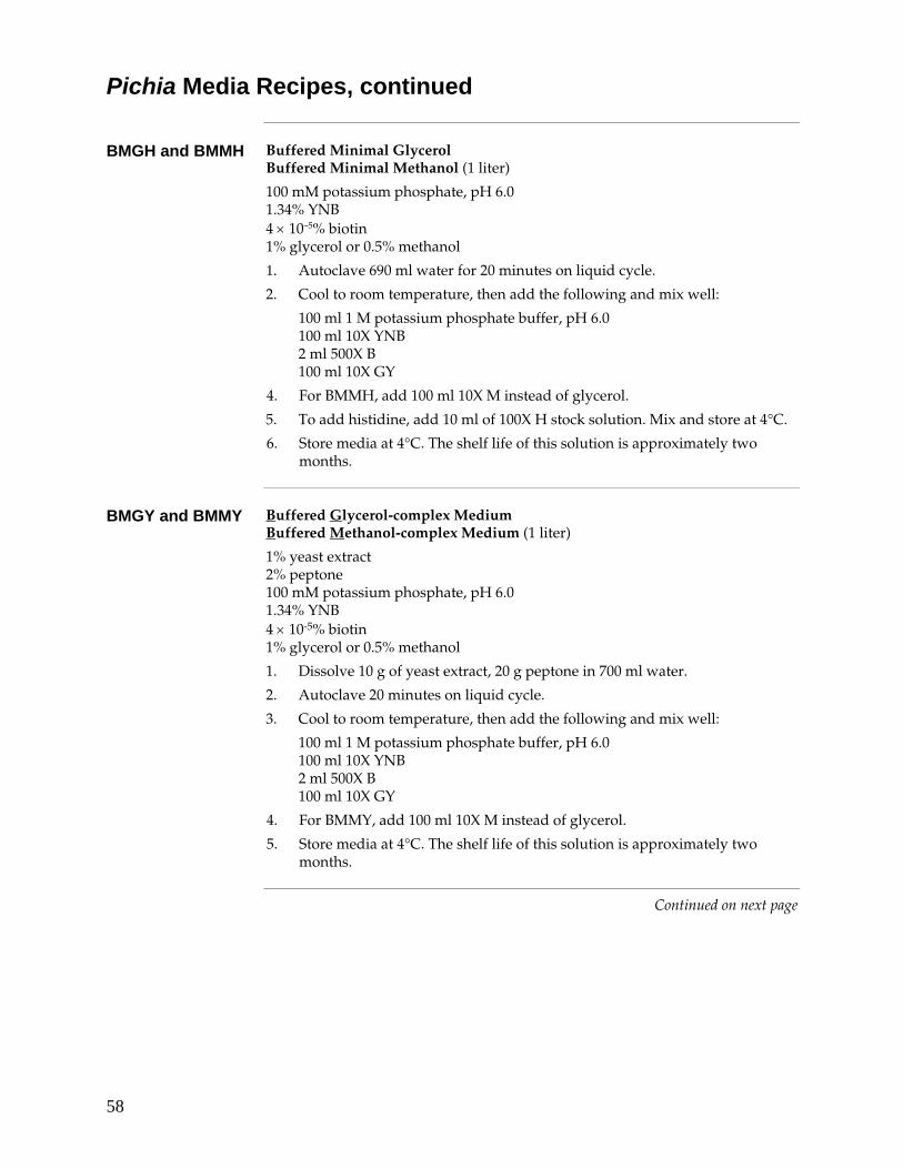
58
Pichia Media Recipes, continued
BMGH and BMMH Buffered Minimal Glycerol
Buffered Minimal Methanol (1 liter)
100 mM potassium phosphate, pH 6.0 1.34% YNB 4 × 10⁻⁵% biotin 1% glycerol or 0.5% methanol
1. Autoclave 690 ml water for 20 minutes on liquid cycle.
2. Cool to room temperature, then add the following and mix well: 100 ml 1 M potassium phosphate buffer, pH 6.0 100 ml 10X YNB 2 ml 500X B 100 ml 10X GY
4. For BMMH, add 100 ml 10X M instead of glycerol.
5. To add histidine, add 10 ml of 100X H stock solution. Mix and store at 4°C.
6. Store media at 4°C. The shelf life of this solution is approximately two months.
BMGY and BMMY Buffered Glycerol-complex Medium
Buffered Methanol-complex Medium (1 liter)
1% yeast extract 2% peptone 100 mM potassium phosphate, pH 6.0 1.34% YNB 4 × 10⁻⁵% biotin 1% glycerol or 0.5% methanol
1. Dissolve 10 g of yeast extract, 20 g peptone in 700 ml water.
2. Autoclave 20 minutes on liquid cycle.
3. Cool to room temperature, then add the following and mix well:
100 ml 1 M potassium phosphate buffer, pH 6.0 100 ml 10X YNB 2 ml 500X B 100 ml 10X GY
4. For BMMY, add 100 ml 10X M instead of glycerol.
5. Store media at 4°C. The shelf life of this solution is approximately two months.
Continued on next page
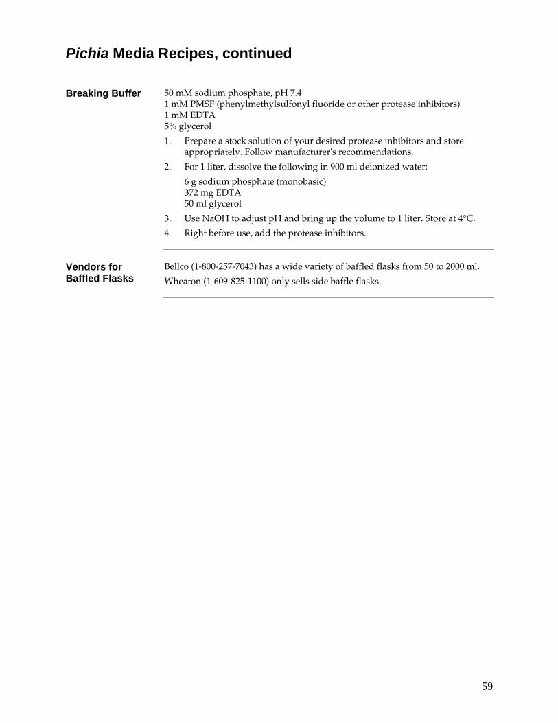
59
Pichia Media Recipes, continued
Breaking Buffer 50 mM sodium phosphate, pH 7.4
1 mM PMSF (phenylmethylsulfonyl fluoride or other protease inhibitors) 1 mM EDTA 5% glycerol
1. Prepare a stock solution of your desired protease inhibitors and store appropriately. Follow manufacturer's recommendations.
2. For 1 liter, dissolve the following in 900 ml deionized water:
6 g sodium phosphate (monobasic) 372 mg EDTA 50 ml glycerol
3. Use NaOH to adjust pH and bring up the volume to 1 liter. Store at 4°C.
4. Right before use, add the protease inhibitors.
Vendors for Baffled Flasks
Bellco (1-800-257-7043) has a wide variety of baffled flasks from 50 to 2000 ml.
Wheaton (1-609-825-1100) only sells side baffle flasks.
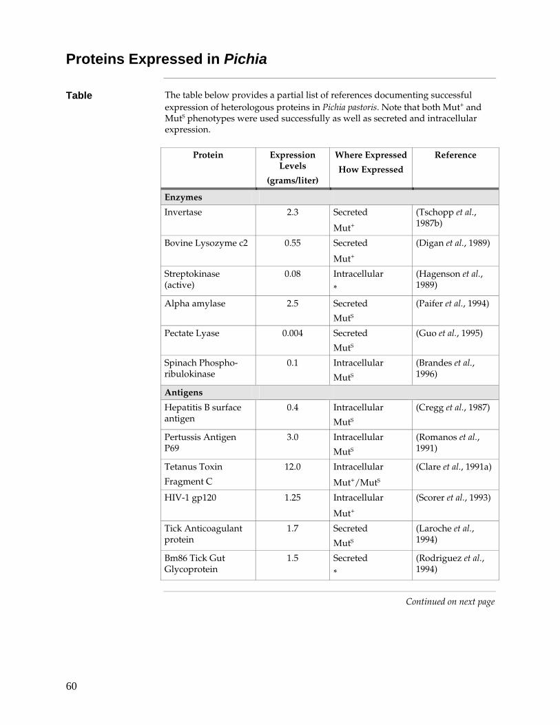
60
Proteins Expressed in Pichia
Table The table below provides a partial list of references documenting successful expression of heterologous proteins in Pichia pastoris. Note that both Mut+ and MutS phenotypes were used successfully as well as secreted and intracellular expression.
Protein Expression
Levels
(grams/liter)
Where Expressed
How Expressed
Reference
Enzymes
Invertase 2.3 Secreted
Mut+
(Tschopp et al., 1987b)
Bovine Lysozyme c2 0.55 Secreted
Mut+
(Digan et al., 1989)
Streptokinase (active)
0.08 Intracellular
*
(Hagenson et al., 1989)
Alpha amylase 2.5 Secreted
MutS
(Paifer et al., 1994)
Pectate Lyase 0.004 Secreted
MutS
(Guo et al., 1995)
Spinach Phospho-ribulokinase
0.1 Intracellular
MutS
(Brandes et al., 1996)
Antigens
Hepatitis B surface antigen
0.4 Intracellular
MutS
(Cregg et al., 1987)
Pertussis Antigen P69
3.0 Intracellular
MutS
(Romanos et al., 1991)
Tetanus Toxin
Fragment C
12.0 Intracellular
Mut+/MutS
(Clare et al., 1991a)
HIV-1 gp120 1.25 Intracellular
Mut+
(Scorer et al., 1993)
Tick Anticoagulant protein
1.7 Secreted
MutS
(Laroche et al., 1994)
Bm86 Tick Gut Glycoprotein
1.5 Secreted
*
(Rodriguez et al., 1994)
Continued on next page
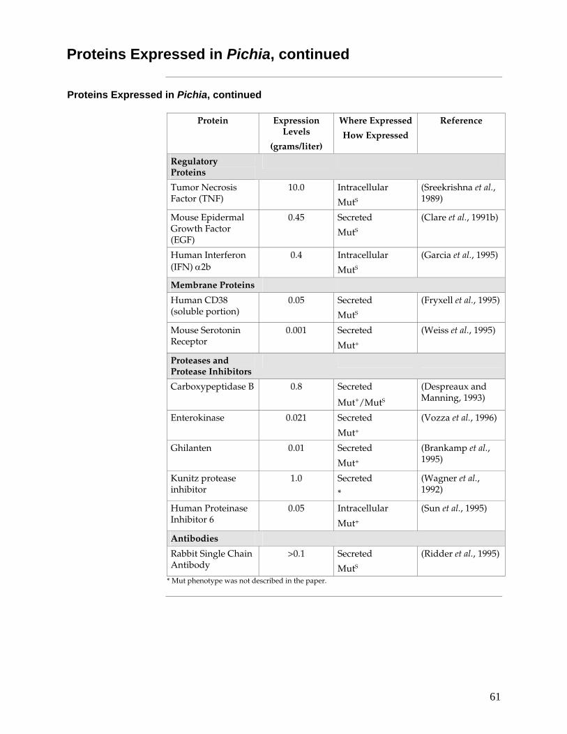
61
Proteins Expressed in Pichia, continued
Proteins Expressed in Pichia, continued
Protein Expression Levels
(grams/liter)
Where Expressed
How Expressed
Reference
Regulatory Proteins
Tumor Necrosis Factor (TNF)
10.0 Intracellular
MutS
(Sreekrishna et al., 1989)
Mouse Epidermal Growth Factor (EGF)
0.45 Secreted
MutS
(Clare et al., 1991b)
Human Interferon (IFN) α2b
0.4 Intracellular
MutS
(Garcia et al., 1995)
Membrane Proteins
Human CD38 (soluble portion)
0.05 Secreted
MutS
(Fryxell et al., 1995)
Mouse Serotonin Receptor
0.001 Secreted
Mut+
(Weiss et al., 1995)
Proteases and Protease Inhibitors
Carboxypeptidase B 0.8 Secreted
Mut+/MutS
(Despreaux and Manning, 1993)
Enterokinase 0.021 Secreted
Mut+
(Vozza et al., 1996)
Ghilanten 0.01 Secreted
Mut+
(Brankamp et al., 1995)
Kunitz protease inhibitor
1.0 Secreted
*
(Wagner et al., 1992)
Human Proteinase Inhibitor 6
0.05 Intracellular
Mut+
(Sun et al., 1995)
Antibodies
Rabbit Single Chain Antibody
>0.1 Secreted
MutS
(Ridder et al., 1995)
* Mut phenotype was not described in the paper.
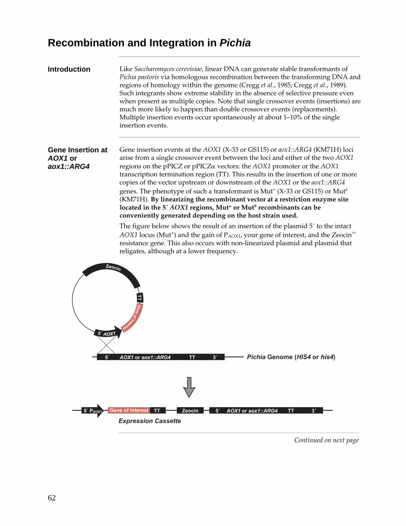
62
Recombination and Integration in Pichia
Introduction Like Saccharomyces cerevisiae, linear DNA can generate stable transformants of Pichia pastoris via homologous recombination between the transforming DNA and regions of homology within the genome (Cregg et al., 1985; Cregg et al., 1989). Such integrants show extreme stability in the absence of selective pressure even when present as multiple copies. Note that single crossover events (insertions) are much more likely to happen than double crossover events (replacements). Multiple insertion events occur spontaneously at about 1–10% of the single insertion events.
Gene Insertion at AOX1 or aox1::ARG4
Gene insertion events at the AOX1 (X-33 or GS115) or aox1::ARG4 (KM71H) loci arise from a single crossover event between the loci and either of the two AOX1 regions on the pPICZ or pPICZα vectors: the AOX1 promoter or the AOX1 transcription termination region (TT). This results in the insertion of one or more copies of the vector upstream or downstream of the AOX1 or the aox1::ARG4 genes. The phenotype of such a transformant is Mut+ (X-33 or GS115) or MutS (KM71H). By linearizing the recombinant vector at a restriction enzyme site located in the 5´ AOX1 regions, Mut+ or MutS recombinants can be conveniently generated depending on the host strain used.
The figure below shows the result of an insertion of the plasmid 5´ to the intact AOX1 locus (Mut+) and the gain of PAOX1, your gene of interest, and the Zeocin™ resistance gene. This also occurs with non-linearized plasmid and plasmid that religates, although at a lower frequency.
������.���
���������
��
�� ����
�����.�����/����������0
������.������������
������������������
��
�� ���������������� � #�
�� ���� �� �� #��������������� �
Continued on next page

63
Recombination and Integration in Pichia, continued
Multiple Gene Insertion Events
Multiple gene insertion events at a single locus in a cell do occur spontaneously with a low, but detectable frequency--between 1 and 10% of all selected ZeoR transformants. Because of the low frequency of multiple gene insertion events, you will need to screen hundreds to thousands of Zeocin™-resistant transformants to locate these "jack-pot" clones. We recommend that you use electroporation to generate ZeoR transformants for screening.
Multi-copy events can occur as gene insertions either at the AOX1 or the aox1::ARG4 loci. This results in a Mut+ phenotype in X-33 or GS115 and a MutS phenotype in KM71H. Multiple gene insertion events can be detected by quantitative dot blot analysis, Southern blot analysis, and differential hybridization. See page 73 for a protocol to screen for multiple inserts.
/ /������.������������ ��/
#1����
� ���������)�������
!�"����������#���
� ���������)�������
$�"����������#���%���&
/ /
/ /
� ���������)������+
������.������������ ��
#1���� �� ����
�� ����
������
.������������
��
�� ���������������� � #�
�� ���������������� � #�
�� ����
/ /������.������������ ��/
#1����
� ���������)�������
!�"����������#���
� ���������)�������
$�"����������#���%���&
/ /
/ /
� ���������)������+
������.������������ ��
#1���� �� ����
�� ����
������
.������������
��
�� ���������������� � #�
�� ���������������� � #�
�� ����
( (ZeocinGene of Interest TT(
3' AOX1
Expression Cassette 1
2nd Insertion Event
Expression Cassette 1
3rd Insertion Event, etc.
( (
( (
Expression Cassette 2
ZeocinGene of Interest TT
5´ P AOX1
5´ P AOX1
Zeocin
Gene of Interest
TT
5´ TTAOX1 or aox1::ARG4 3´
5´ TTAOX1 or aox1::ARG4 3´
5´ P A OX1

64
Lithium Chloride Transformation Method
Introduction This is a modified version of the procedure described for S. cerevisiae (Gietz and Schiestl, 1996). This protocol is provided as an alternative to transformation by electroporation. Transformation efficiency is between 102 to 103 cfu/μg linearized DNA.
Preparation of Solutions
Lithium acetate does not work with Pichia pastoris. Use only lithium chloride.
1 M LiCl in distilled, deionized water. Filter sterilize. Dilute as needed with sterile water.
50% polyethylene glycol (PEG-3350) in distilled, deionized water. Filter sterilize. Store in a tightly capped bottle.
2 mg/ml denatured, fragmented salmon sperm DNA in TE (10 mM Tris-HCl, pH 8.0, 1.0 mM EDTA). Store at –20°C.
Preparing Cells 1. Grow a 50 ml culture of Pichia pastoris in YPD at 30°C with shaking to an
OD600 of 0.8 to 1.0 (approximately 108 cells/ml).
2. Harvest the cells and wash with 25 ml of sterile water and centrifuge at 1,500 × g for 10 minutes at room temperature.
3. Decant the water and resuspend the cells in 1 ml of 100 mM LiCl.
4. Transfer the cell suspension to a 1.5 ml microcentrifuge tube.
5. Pellet the cells at maximum speed for 15 seconds and remove the LiCl with a pipet.
6. Resuspend the cells in 400 μl of 100 mM LiCl.
7. Dispense 50 μl of the cell suspension into a 1.5 ml microcentrifuge tube for each transformation and use immediately. Do not store on ice or freeze at –20°C.
Continued on next page
65
Lithium Chloride Transformation Method, continued
Transformation 1. Boil a 1 ml sample of single-stranded DNA for five minutes, then quickly
chill in ice water. Keep on ice.
Note: It is not necessary nor desirable to boil the carrier DNA prior to each use. Store a small aliquot at –20°C and boil every 3–4 times the DNA is thawed.
2. Centrifuge the LiCl-cell solution from Step 7, above, and remove the LiCl with a pipet.
3. For each transformation sample, add the following reagents in the order given to the cells. PEG shields the cells from the detrimental effects of the high concentration of LiCl.
240 μl 50% PEG 36 μl 1 M LiCl 25 μl 2 mg/ml single-stranded DNA Plasmid DNA (5–10 μg) in 50 μl sterile water
4. Vortex each tube vigorously until the cell pellet is completely mixed (~1 minute).
5. Incubate the tube at 30°C for 30 minutes without shaking.
6. Heat shock in a water bath at 42°C for 20–25 minutes.
7. Centrifuge the tubes at 6,000 to 8,000 rpm and remove the transformation solution with a pipet.
8. Resuspend the pellet in 1 ml of YPD and incubate at 30°C with shaking.
9. After 1 hour and 4 hours, plate 25 μl to 100 μl on YPD plates containing 100 μg/ml Zeocin™. Incubate the plates for 2–3 days at 30°C. Proceed to Analysis of Pichia Transformants, page 28.

66
Zeocin™
Description Zeocin™ belongs to a family of structurally related bleomycin/phleomycin-type antibiotics isolated from Streptomyces. Antibiotics in this family are broad spectrum antibiotics that act as strong antibacterial and anti-tumor drugs. They show strong toxicity against bacteria, fungi (including yeast), plants, and mammalian cells. Zeocin™ is not as toxic as bleomycin on fungi.
Chemical Properties
Zeocin™ is a basic, water-soluble compound isolated from Streptomyces verticillus as a copper-chelated glycopeptide. The presence of copper gives the solution its blue color. The chemical formula for Zeocin™ is C55H83N19O21S2Cu. It contains several unique amino acids, sugars, and aliphatic amines. For general information about the family of bleomycin antibiotics, See Berdy, 1980. The general structure of Zeocin™ is shown below.
���
��
�
��� ��
� �
�
���
�
������
��
�
�
���
������
�
���� ��
��
��
��
�
��
�����
��
����
���
� �
�
�
�
���
�
�
�
�
�
�� �
�
��
��
�� �
���������
Mechanism of Action
The exact mechanism of action of Zeocin™ is not known; however, it is thought to be the same as bleomycin and phleomycin due to its similarity to these drugs and its inhibition by the Sh ble resistance protein (see next section). The copper/glycopeptide complex is selective and involves chelation of copper (Cu2+) by the amino group of the α-carboxamide, single nitrogen atoms of both the pyrimidine chromophore and the imidazole moiety, and the carbamoyl group of mannose. The copper-chelated form is inactive. When the antibiotic enters the cell, the copper cation is reduced from Cu2+ to Cu1+ and removed by sulfhydryl compounds in the cell. Upon removal of the copper, Zeocin™ is activated to bind DNA and cleave it causing cell death (Berdy, 1980). High salt concentrations and acidity or basicity inactivate Zeocin™; therefore, it is necessary to reduce the salt in bacterial medium to 90 mM (5 g/liter) or less and adjust the pH to 7.5 to make sure the drug remains active.
Continued on next page
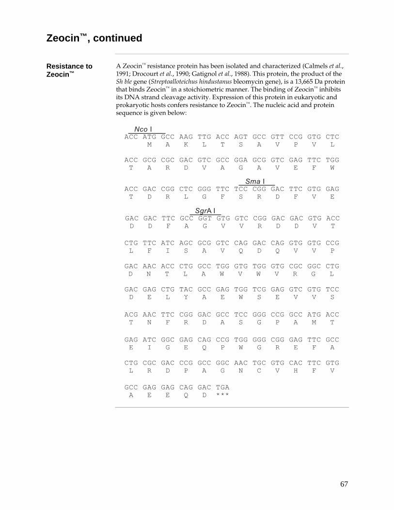
67
Zeocin™, continued
Resistance to Zeocin™
A Zeocin™ resistance protein has been isolated and characterized (Calmels et al., 1991; Drocourt et al., 1990; Gatignol et al., 1988). This protein, the product of the Sh ble gene (Streptoalloteichus hindustanus bleomycin gene), is a 13,665 Da protein that binds Zeocin™ in a stoichiometric manner. The binding of Zeocin™ inhibits its DNA strand cleavage activity. Expression of this protein in eukaryotic and prokaryotic hosts confers resistance to Zeocin™. The nucleic acid and protein sequence is given below:
�������������������������������������� �������������������������������������
������� ������������������������������������
��������������������������������������������
��������������������������������
������������������������������������
�������� ������������������������������������������������������������������������
���������������������������������������� ���������������������������������������
�������������������
����3
���3
!$��3
�����������������������
���������������������������������������������������������������������������������
�������������������������������������������������������� ��������������� ��������
����������������������������������������� ����
������������������������������������
��������������������������������������
��������������������������������������
����� ����������������������������������������
����������������������������������������������
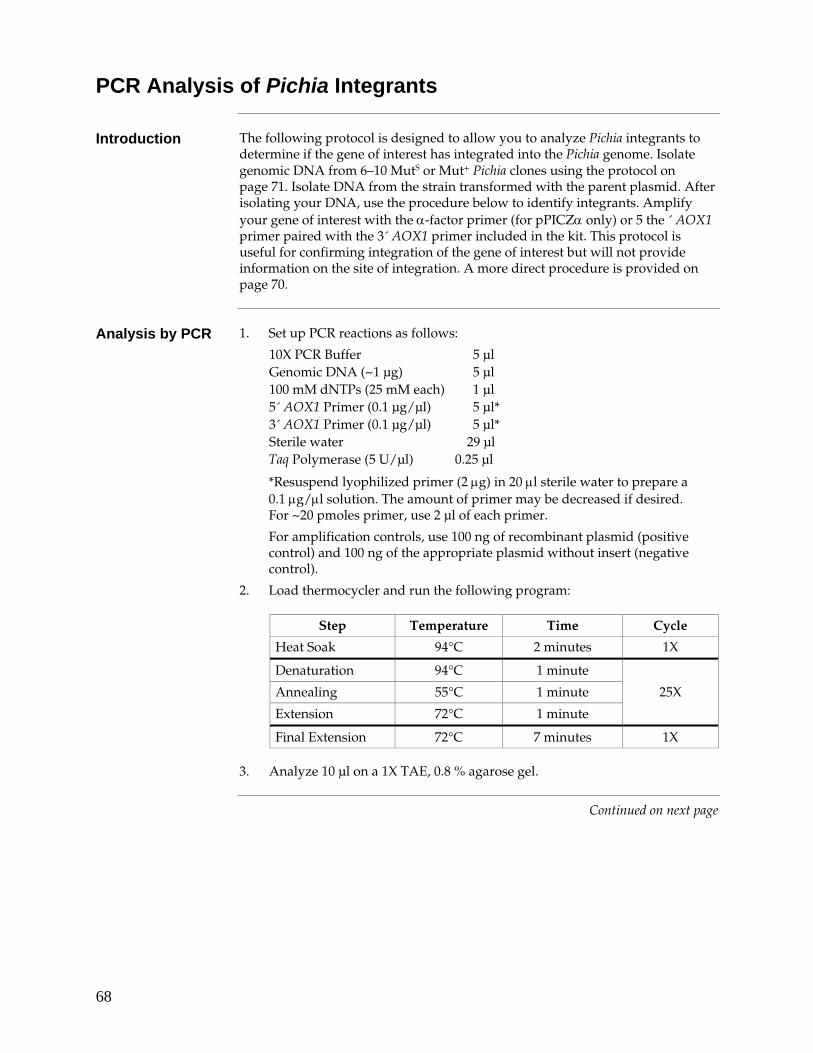
68
PCR Analysis of Pichia Integrants
Introduction The following protocol is designed to allow you to analyze Pichia integrants to determine if the gene of interest has integrated into the Pichia genome. Isolate genomic DNA from 6–10 MutS or Mut+ Pichia clones using the protocol on page 71. Isolate DNA from the strain transformed with the parent plasmid. After isolating your DNA, use the procedure below to identify integrants. Amplify your gene of interest with the α-factor primer (for pPICZα only) or 5 the ´ AOX1 primer paired with the 3´ AOX1 primer included in the kit. This protocol is useful for confirming integration of the gene of interest but will not provide information on the site of integration. A more direct procedure is provided on page 70.
Analysis by PCR 1. Set up PCR reactions as follows:
10X PCR Buffer 5 μl Genomic DNA (~1 μg) 5 μl 100 mM dNTPs (25 mM each) 1 μl 5´ AOX1 Primer (0.1 μg/μl) 5 μl* 3´ AOX1 Primer (0.1 μg/μl) 5 μl* Sterile water 29 μl Taq Polymerase (5 U/μl) 0.25 μl
*Resuspend lyophilized primer (2 μg) in 20 μl sterile water to prepare a 0.1 μg/μl solution. The amount of primer may be decreased if desired. For ~20 pmoles primer, use 2 μl of each primer.
For amplification controls, use 100 ng of recombinant plasmid (positive control) and 100 ng of the appropriate plasmid without insert (negative control).
2. Load thermocycler and run the following program:
Step Temperature Time Cycle
Heat Soak 94°C 2 minutes 1X
Denaturation 94°C 1 minute
Annealing 55°C 1 minute 25X
Extension 72°C 1 minute
Final Extension 72°C 7 minutes 1X 3. Analyze 10 μl on a 1X TAE, 0.8 % agarose gel.
Continued on next page

69
PCR Analysis of Pichia Integrants, continued
Interpreting PCR If screening Mut+ integrants, you should see two bands, one corresponding to
the size of your gene of interest, the other to the AOX1 gene (approximately 2.2 kb). In KM71H, the PCR product is 3.6 kb because of the ARG4 insert in AOX1. Parent plasmids will produce the following sized PCR products. Add the size of these products to the size of your insert to interpret your PCR results.
Vector PCR Product
pPICZ 325 bp (A), 323 bp (B), 324 bp (C)
pPICZα (using the 5´ AOX1 primer) 588 bp (A), 592 bp (B), 593 bp (C)
pPICZα (using the α-Factor primer) 299 bp (A), 303 bp (B), 304 bp (C)
��������
If you use the α-factor primer as a PCR primer, you will not see a band with either GS115 or KM71H. This is because there is no α-factor signal associated with the chromosomal AOX1 gene.
Sometimes there will be ghost bands appearing in your PCR. These do not seem to be significant as they have not been shown to be a problem.
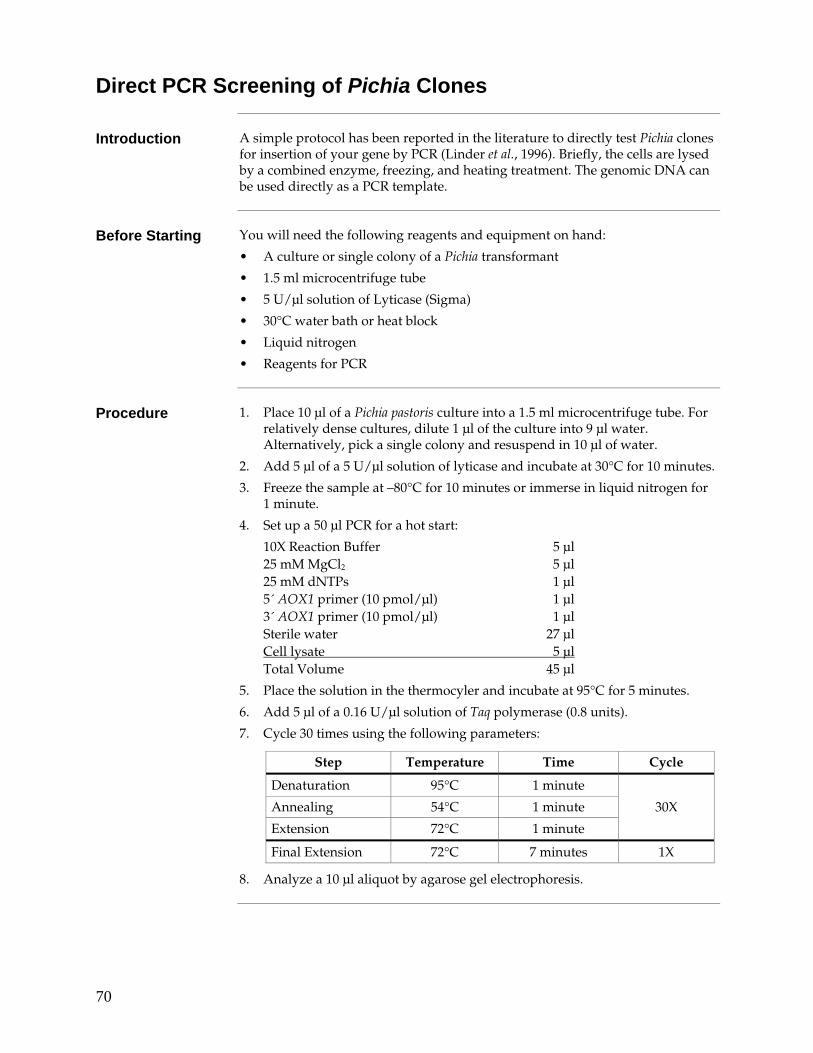
70
Direct PCR Screening of Pichia Clones
Introduction A simple protocol has been reported in the literature to directly test Pichia clones for insertion of your gene by PCR (Linder et al., 1996). Briefly, the cells are lysed by a combined enzyme, freezing, and heating treatment. The genomic DNA can be used directly as a PCR template.
Before Starting You will need the following reagents and equipment on hand:
• A culture or single colony of a Pichia transformant
• 1.5 ml microcentrifuge tube
• 5 U/μl solution of Lyticase (Sigma)
• 30°C water bath or heat block
• Liquid nitrogen
• Reagents for PCR
Procedure 1. Place 10 μl of a Pichia pastoris culture into a 1.5 ml microcentrifuge tube. For
relatively dense cultures, dilute 1 μl of the culture into 9 μl water. Alternatively, pick a single colony and resuspend in 10 μl of water.
2. Add 5 μl of a 5 U/μl solution of lyticase and incubate at 30°C for 10 minutes.
3. Freeze the sample at –80°C for 10 minutes or immerse in liquid nitrogen for 1 minute.
4. Set up a 50 μl PCR for a hot start:
10X Reaction Buffer 5 μl 25 mM MgCl2 5 μl 25 mM dNTPs 1 μl 5´ AOX1 primer (10 pmol/μl) 1 μl 3´ AOX1 primer (10 pmol/μl) 1 μl Sterile water 27 μl Cell lysate 5 μl Total Volume 45 μl
5. Place the solution in the thermocyler and incubate at 95°C for 5 minutes.
6. Add 5 μl of a 0.16 U/μl solution of Taq polymerase (0.8 units).
7. Cycle 30 times using the following parameters:
Step Temperature Time Cycle
Denaturation 95°C 1 minute
Annealing 54°C 1 minute 30X
Extension 72°C 1 minute
Final Extension 72°C 7 minutes 1X
8. Analyze a 10 μl aliquot by agarose gel electrophoresis.
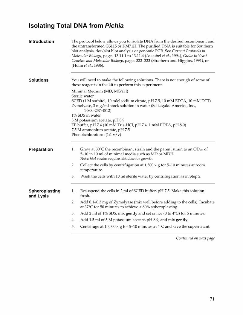
71
Isolating Total DNA from Pichia
Introduction The protocol below allows you to isolate DNA from the desired recombinant and the untransformed GS115 or KM71H. The purified DNA is suitable for Southern blot analysis, dot/slot blot analysis or genomic PCR. See Current Protocols in Molecular Biology, pages 13.11.1 to 13.11.4 (Ausubel et al., 1994), Guide to Yeast Genetics and Molecular Biology, pages 322–323 (Strathern and Higgins, 1991), or (Holm et al., 1986).
Solutions You will need to make the following solutions. There is not enough of some of
these reagents in the kit to perform this experiment.
Minimal Medium (MD, MGYH) Sterile water SCED (1 M sorbitol, 10 mM sodium citrate, pH 7.5, 10 mM EDTA, 10 mM DTT) Zymolyase, 3 mg/ml stock solution in water (Seikagaku America, Inc.,
1-800-237-4512) 1% SDS in water 5 M potassium acetate, pH 8.9 TE buffer, pH 7.4 (10 mM Tris-HCl, pH 7.4, 1 mM EDTA, pH 8.0) 7.5 M ammonium acetate, pH 7.5 Phenol:chloroform (1:1 v/v)
Preparation 1. Grow at 30°C the recombinant strain and the parent strain to an OD600 of
5–10 in 10 ml of minimal media such as MD or MDH. Note: his4 strains require histidine for growth.
2. Collect the cells by centrifugation at 1,500 × g for 5–10 minutes at room temperature.
3. Wash the cells with 10 ml sterile water by centrifugation as in Step 2.
Spheroplasting and Lysis
1. Resuspend the cells in 2 ml of SCED buffer, pH 7.5. Make this solution fresh.
2. Add 0.1–0.3 mg of Zymolyase (mix well before adding to the cells). Incubate at 37°C for 50 minutes to achieve < 80% spheroplasting.
3. Add 2 ml of 1% SDS, mix gently and set on ice (0 to 4°C) for 5 minutes.
4. Add 1.5 ml of 5 M potassium acetate, pH 8.9, and mix gently.
5. Centrifuge at 10,000 × g for 5–10 minutes at 4°C and save the supernatant.
Continued on next page
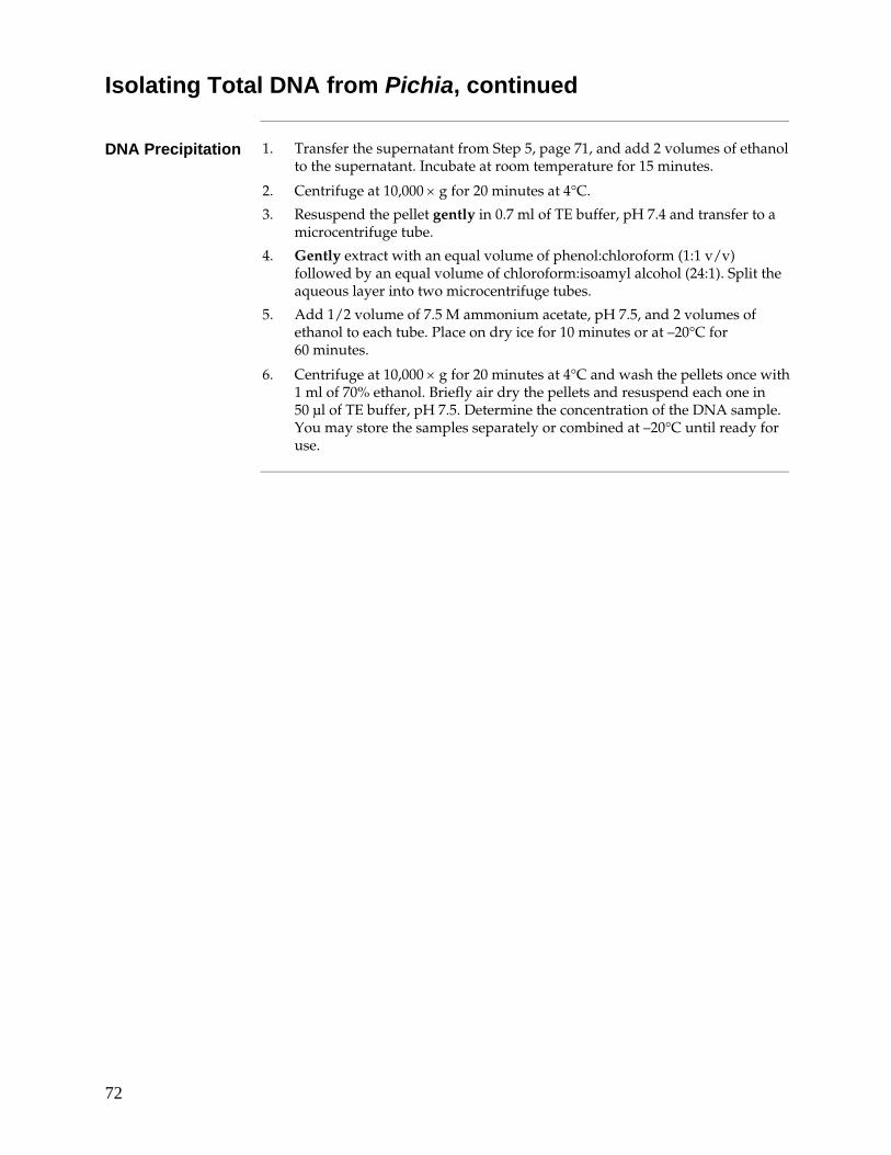
72
Isolating Total DNA from Pichia, continued
DNA Precipitation 1. Transfer the supernatant from Step 5, page 71, and add 2 volumes of ethanol
to the supernatant. Incubate at room temperature for 15 minutes.
2. Centrifuge at 10,000 × g for 20 minutes at 4°C.
3. Resuspend the pellet gently in 0.7 ml of TE buffer, pH 7.4 and transfer to a microcentrifuge tube.
4. Gently extract with an equal volume of phenol:chloroform (1:1 v/v) followed by an equal volume of chloroform:isoamyl alcohol (24:1). Split the aqueous layer into two microcentrifuge tubes.
5. Add 1/2 volume of 7.5 M ammonium acetate, pH 7.5, and 2 volumes of ethanol to each tube. Place on dry ice for 10 minutes or at –20°C for 60 minutes.
6. Centrifuge at 10,000 × g for 20 minutes at 4°C and wash the pellets once with 1 ml of 70% ethanol. Briefly air dry the pellets and resuspend each one in 50 μl of TE buffer, pH 7.5. Determine the concentration of the DNA sample. You may store the samples separately or combined at –20°C until ready for use.
73
Determining the Copy Number of Multiple Integrants
Introduction You may wish to determine the actual number of gene copies in your Pichia recombinant. You may either use quantitative dot blots or Southern hybridization to analyze gene copy number (Brierley, et al., 1994; Clare, et al., 1991a; Romanos, et al., 1991; Scorer, et al., 1993; Scorer, et al., 1994). This requires you to isolate genomic DNA from Pichia recombinants transformed with the parent vector (0 copies of your gene), pPICZ or pPICZα containing 1 copy of your gene (single copy control), and the Pichia recombinants containing multiple copies of your gene. Use the protocol detailed on pages 71–72 to isolate genomic DNA.
Quantitative Dot Blot Solutions
For each dot blot, you need 10–15 ml of each of the following solutions. 50 mM EDTA, 2.5% β-mercaptoethanol pH 9 1 mg/ml Zymolyase 100T in water (Seikagaku America, Inc., 1-800-237-4512) 0.1 N NaOH, 1.5 M NaCl 2X SSC You will also need 3MM paper.
Quantitative Dot Blot Procedure
The following protocol is a summary of a rapid DNA dot blot technique to detect multiple integrants (Romanos, et al., 1991). It is very important to spot equivalent numbers of cells onto filters to quantify the copy number. Alternatively, you may isolate genomic DNA, spot it directly onto nitrocellulose or nylon, fix, and analyze for copy number.
1. Grow Mut+ or MutS transformants in individual wells of a 96-well microtiter plate in 200 μl of YPD broth at 30°C until all wells have approximately the same density. This may necessitate several passages. Alternatively, you may grow individual transformants in culture tubes and normalize the absorbance at 600 nm by adding medium.
2. Filter 50 μl of each sample onto a nitrocellulose or nylon filter placed into a dot (slot) blot apparatus using multi-channel pipettor. Air dry filters.
3. To lyse the cells on the filter, treat the filter with four solutions as follows: place two sheets of 3 MM paper in a tray and soak with 10–15 ml of 50 mM EDTA, 2.5% β-mercaptoethanol pH 9. Make sure that the paper is uniformly soaked and that there are no puddles. Place the nitrocellulose filter face down on the treated 3MM paper. Incubate for 15 minutes at room temperature.
4. Remove the nitrocellulose filter from the 3MM paper and replace the 3MM paper with two new sheets. Soak with 10–15 ml of 1 mg/ml Zymolyase 100T as described in Step 3. Place the nitrocellulose filter face down on the 3MM paper and incubate for 4 hours at 37°C.
Continued on next page
74
Determining the Copy Number of Multiple Integrants, continued
Quantitative Dot Blot Procedure, continued
5. Remove the nitrocellulose filter from the paper and replace the paper with two new sheets. Soak with 10–15 ml of 0.1 N NaOH, 1.5 M NaCl. Place the nitrocellulose filter face down on the paper and incubate for 5 minutes at room temperature.
6. Remove the nitrocellulose filter and replace with two new 3MM sheets. Soak with 10–15 ml of 2X SSC. Place the nitrocellulose filter face down on the 3MM paper and incubate for 5 minutes at room temperature. Repeat.
7. Bake nitrocellulose filters at 80°C or UV-crosslink DNA to nylon. You may probe the filters with a nonradioactive-labeled or random-primed, 32P-labeled probe complementary to your gene.
You can identify multi-copy integrants by a strong hybridization signal relative to the single copy control. You can then quantify dot blots for copy number by densitometry of the film or blot, or by using a β-scanner (if radiolabeled).
Southern Blot Analysis
For a detailed description of this technique as applied to Pichia pastoris, see (Clare, et al., 1991a). It is very important to digest your DNA with the right restriction enzyme(s) to generate a blot of digested and gel-separated genomic DNA. We recommend that you use a restriction enzyme that cuts outside of the expression cassette. This will generate a restriction fragment whose size reflects the number of multimers. For example, if you have cloned a 1.2 kb fragment into pPICZ and transformed into Pichia, you can digest the DNA from this recombinant with Nhe I (which does not cut within the vector or the insert). The size of the fragment will increase in additions of 4.7 kb over the size of the fragment containing 1 copy of the gene.
Alternatively, digestion of DNA from Pichia recombinants containing multiple copies will produce a band that will vary in intensity depending on the number of copies of your gene. It is very important to include a control to show the intensity of a single copy gene. You can quantify the relative band intensities using densitometry to estimate gene dosage.
Controls It is very important to include DNA from the host strain alone (X-33, GS115 or
KM71H), the host strain transformed with the parent vector, and the host strain transformed with a vector containing one copy of your gene.
General Guidelines
• Use standard procedures and solutions for Southern blotting as outlined in Molecular Cloning: A Laboratory Manual (Sambrook, et al., 1989), pages 9.31–9.58.
• Isolate genomic DNA and quantify using fluorometry. Be sure to eliminate RNA. It is very important to load the same amount of DNA into each lane to accurately determine copy number if you are using relative intensities.
• Probe your Southern blot with a fragment complementary to your gene.
75
Isolating Total RNA from Pichia
Introduction This protocol is designed to isolate 60–300 μg total RNA (Schmitt et al., 1990) from Pichia, which is suitable for mRNA isolation using Invitrogen's FastTrack® 2.0 or Micro FastTrack™ 2.0 mRNA Isolation Kit. If you wish to use another protocol, scale-up the reaction to yield about 2 mg of total RNA per time point. The mRNA is for northern blot analysis of Pichia recombinants to determine if the gene of interest is being induced and transcribed. You should isolate RNA from induced cultures using an uninduced culture as a negative control.
Solutions Prepare the following solutions. Remember to use DEPC-treated water and to use
equipment free of RNase.
MGYH or BMGY medium DEPC-treated water 3 M sodium acetate, pH 5.3 Buffered phenol 10% SDS in DEPC treated water Phenol:chloroform (1:1) Chloroform:isoamyl alcohol (24:1) 65°C water bath AE buffer (50 mM sodium acetate, pH 5.3, 1 mM EDTA)
Growth of Cells 1. Grow two cultures (100–200 ml in MGYH or BMGY), but induce only one of
them. Use the same protocol for induction that you used when expressing recombinant Pichia strains (pages 37–41).
2. Take 10 ml time points at 1, 2, 3, 4, and 6 days by centrifuging at 1,500 × g for 10 minutes at room temperature.
3. Resuspend cell pellet in 400 μl AE buffer and transfer to a microcentrifuge tube.
Lysing Cells 1. Add 40 μl 10% SDS and vortex for ~ 20 seconds.
2. Add an equal volume (450–500 μl) of buffer saturated phenol and vortex for ~20 seconds.
3. Incubate at 65°C for 4 minutes.
4. Incubate in a dry ice/ethanol bath until crystals show (~1 minute). Centrifuge at maximum speed for 2 minutes at 4°C.
5. Transfer aqueous phase to new centrifuge tube and add an equal volume of phenol/chloroform and vortex for ~20 seconds. Centrifuge at maximum speed for 2 minutes at 4°C.
6. Remove upper phase to a new tube and add 40 μl of 3 M sodium acetate, pH 5.3 and 2.5 volumes of 100% ethanol (–20°C). Centrifuge at maximum speed for 15 minutes at 4°C. Remove ethanol.
7. Wash pellet with 80% ethanol and air dry briefly. Resuspend total RNA in 20 μl DEPC-treated water and store at –80°C. Yield is 60–300 μg total RNA.
mRNA Isolation and Northern Analysis
See (Ausubel et al., 1994) for a protocol for mRNA isolation and Northern analysis. The FastTrack® 2.0 mRNA Kit (Cat. no. K1593-02) is designed to isolate mRNA from 0.2 to 1 mg total RNA. The Micro-FastTrack™ 2.0 Kit (Cat. no. K1520-02) is designed to isolate mRNA from ~100 μg total RNA. You will need ~1–5 μg mRNA per time point.
76
β-Galactosidase Assay
Introduction The GS115/pPICZ/lacZ strain is provided as a His+ Mut+ intracellular expression control. Growth of the strain during Mut+ expression provides a positive control for expression conditions. You may use the cell-free β-galactosidase assay adapted from (Miller, 1972), page 403, to evaluate expression of β-galactosidase.
Preparation of Solutions
Prepare the following:
• A fresh crude cell lysate of GS115/pPICZ/lacZ (see page 40)
• Z buffer
• ONPG solution
• 1 M sodium carbonate solution
Recipes for the solutions are found below.
Z Buffer 60 mM Na2HPO4-7H2O
40 mM NaH2PO4-H2O 10 mM KCl 1 mM MgSO4-7H2O 50 mM β-mercaptoethanol pH 7.0
1. Dissolve the following in 950 ml deionized water:
16.1 g Na2HPO4-7H2O 5.5 g NaH2PO4-H2O 0.75 g KCl 0.246 g MgSO4-7H2O 2.7 ml β-mercaptoethanol
2. Adjust pH to 7.0 with either NaOH or HCl and bring the volume up to 1 liter with water.
3. Do not autoclave. Store at 4°C.
ONPG Solution 4 mg/ml in 100 mM phosphate buffer, pH 7.0
1. Dissolve the following in 90 ml deionized water: 1.61 g Na2HPO4-7H2O 0.55 g NaH2PO4-H2O
2. Adjust pH to 7.0 with either NaOH or HCl and add 400 mg of ONPG. Stir to dissolve and bring the volume up to 100 ml with water.
3. Store at 4°C in the dark.
1 M Sodium Carbonate
Dissolve 12.4 g sodium carbonate in 100 ml of deionized water. Store at room temperature.
Continued on next page

77
β-Galactosidase Assay, continued
Procedure 1. Determine protein concentration of your lysate by Lowry, Bradford, or BCA
assay.
2. Equilibrate Z buffer, ONPG solution, and sodium carbonate solution to 28°C.
3. Add 10–50 μl of your crude assay to 1 ml of Z buffer and equilibrate at 28°C. As a control for spontaneous hydrolysis of ONPG, add an aliquot of your lysis buffer to 1 ml of Z buffer.
4. To initiate the reaction, add 0.2 ml 4 mg/ml ONPG to each of the tubes in Step 2.
5. Incubate the samples and the control at 28°C until a faint yellow color develops. This should occur at least 10 minutes after the start of the assay to ensure accurate data. Note that the tube with no lysate may not change color.
6. Stop the reaction by adding 0.5 ml of 1 M sodium carbonate to each tube. Record the length of incubation for each sample.
7. Read the OD420 against the control containing buffer alone.
8. Determine the protein concentration of your lysate in mg/ml.
If the reaction turns yellow too quickly, you need to dilute your lysate. Try successive 10-fold dilutions of the lysate using your lysis buffer until the reaction starts turning yellow after 10 minutes. This is to ensure that you are measuring a true initial rate.
Determination of Specific Activity
Use the following formula to determine the specific activity of the β-galactosidase in units/mg total protein:
OD420 × 380 β-galactosidase units/mg total protein =
minutes at 28°C × mg protein in reaction
Remember to take into account the volume of lysate added to the reaction and any dilutions made to the lysate when calculating the amount of protein in the reaction. The number 380 is the constant used to convert the OD420 reading into units. One unit is defined as the amount of enzyme that will hydrolyze 1 nmole of ONPG per minute at 28°C. The molar extinction coefficient of ONPG under these conditions is 4,500. For a sample calculation, See below.
Sample Calculation
Here is a sample calculation: Extract concentration = 10 mg/ml Assay 10 μl of a 1/100 dilution Time = 10 minutes OD420 = 0.4
The amount of protein in the reaction = 0.01 ml × 0.01 (dilution factor) × 10 mg/ml = 0.001 mg protein in the reaction
The specific activity = 0.400 × 380 = 15,200 units/mg protein 10 × 0.001 mg
Pure β-galactosidase has an activity of 300,000 units/mg protein.
78
Technical Support
Web Resources
Visit the Invitrogen website at www.invitrogen.com for: • Technical resources, including manuals, vector maps and sequences,
application notes, SDSs, FAQs, formulations, citations, handbooks, etc. • Complete technical support contact information • Access to the Invitrogen Online Catalog • Additional product information and special offers
Contact Us For more information or technical assistance, call, write, fax, or email. Additional
international offices are listed on our website (www.invitrogen.com).
Corporate Headquarters: 5791 Van Allen Way Carlsbad, CA 92008 USA Tel: 1 760 603 7200 Tel (Toll Free): 1 800 955 6288 Fax: 1 760 602 6500 E-mail: [email protected]
Japanese Headquarters: LOOP-X Bldg. 6F 3-9-15, Kaigan Minato-ku, Tokyo 108-0022 Tel: 81 3 5730 6509 Fax: 81 3 5730 6519 E-mail: [email protected]
European Headquarters: Inchinnan Business Park 3 Fountain Drive Paisley PA4 9RF, UK Tel: +44 (0) 141 814 6100 Tech Fax: +44 (0) 141 814 6117 E-mail: [email protected]
SDS SDSs ( Safety Data Sheets) are available on our website at
www.invitrogen.com/sds.
Certificate of Analysis
The Certificate of Analysis (CofA) provides detailed quality control information for each product. The CofA for each product is available on our website at www.invitrogen.com/cofa, and is searchable by product lot number, which is printed on each box.
Continued on next page
79
Technical Support, continued
Limited Warranty Invitrogen (a part of Life Technologies Corporation) is committed to providing
our customers with high-quality goods and services. Our goal is to ensure that every customer is 100% satisfied with our products and our service. If you should have any questions or concerns about an Invitrogen product or service, contact our Technical Support Representatives. All Invitrogen products are warranted to perform according to specifications stated on the certificate of analysis. The Company will replace, free of charge, any product that does not meet those specifications. This warranty limits the Company’s liability to only the price of the product. No warranty is granted for products beyond their listed expiration date. No warranty is applicable unless all product components are stored in accordance with instructions. The Company reserves the right to select the method(s) used to analyze a product unless the Company agrees to a specified method in writing prior to acceptance of the order. Invitrogen makes every effort to ensure the accuracy of its publications, but realizes that the occasional typographical or other error is inevitable. Therefore the Company makes no warranty of any kind regarding the contents of any publications or documentation. If you discover an error in any of our publications, please report it to our Technical Support Representatives. Life Technologies Corporation shall have no responsibility or liability for any special, incidental, indirect or consequential loss or damage whatsoever. The above limited warranty is sole and exclusive. No other warranty is made, whether expressed or implied, including any warranty of merchantability or fitness for a particular purpose.
80
Purchaser Notification
Limited Use Label License No: 22 Vectors and Clones Encoding Histidine Hexamer
This product is licensed under U.S. Patent Nos. 5,284,933 and 5,310,663 and foreign equivalents from Hoffmann-LaRoche, Inc., Nutley, NJ and/or Hoffmann-LaRoche Ltd., Basel, Switzerland and is provided only for use in research. Information about licenses for commercial use is available from QIAGEN GmbH, Max-Volmer-Str. 4, D-40724 Hilden, Germany.
Limited Use Label License No. 74: Pichia Pastoris Expression System
The Pichia Expression System is based on the yeast Pichia pastoris. Pichia pastoris was developed into an expression system by scientists at Salk Institute Biotechnology/ Industry Associates (SIBIA) and Phillips Petroleum for high-level expression of recombinant proteins. All patents for Pichia pastoris and licenses for its use as an expression system are owned by Research Corporation Technologies (RCT), Inc., Tucson, Arizona. Life Technologies has an exclusive license to sell Pichia expression kits and vectors to scientists for research purposes only, under the terms described below. Use of Pichia pastoris by commercial entities for any commercial purpose requires the user to obtain a commercial license as detailed below. Before using any Pichia expression product, please read the following license agreement. If you do not agree to be bound by its terms, contact Life Technologies within 10 days for authorization to return the unused Pichia expression products and to receive a full refund. If you do agree to the terms of this license agreement, please complete the User Registration Card and return it to Life Technologies before using the product. Life Technologies Corporation ("Life Technologies") grants you a non-exclusive license to use the enclosed Pichia expression vectors ("Expression Vector") for academic research or for evaluation purposes only. The Expression Vectors are being transferred to you in furtherance of, and reliance on, such license. You may not use the Expression Vectors for any commercial purpose without a license for such purpose from Research Corporation Technologies, Inc., Tucson, Arizona. Commercial purposes include: any use of Expression Products or Expression Vectors in a Commercial Product; any use of Expression Products or Expression Vectors in the manufacture of a Commercial Product; any sale of Expression Products; any use of Expression Products or the Expression Kit to facilitate or advance research or development directed to a Commercial Product; and any use of Expression Products or the Expression Kit to facilitate or advance any research or development program the results of which will be directly applied to the development or manufacture of a Commercial Product. "Expression Products" means products expressed with the Expression Kit, or with the use of any Pichia expression vectors (including the Expression Vector) or host strains. "Commercial Product" means any product intended for sale or commercial use.
Continued on next page
81
Purchaser Notification, continued
Limited Use Label License No. 74: Pichia Pastoris Expression System, continued
Commercial entities may conduct their evaluation for one year at which time this license automatically terminates. Commercial entities will be contacted by Research Corporation Technologies during the evaluation period regarding their desire for a commercial license. Access to the Expression Kit and Vector must be limited solely to those officers, employees and students of your institution who need access to perform the above-described research or evaluation. You must inform each such officer, employee and student of the provisions of this license agreement and require them to agree, in writing, to be bound by the provisions of this license agreement. You may not distribute any Expression Vector or host strain contained herein or in the Expression Kit to others, even those within your own institution. You may only transfer modified, altered, or original material from the Expression Kit or Vector to a third party following written notification of, and written approval from, Life Technologies so that the recipient can be licensed. You may not assign, sub-license, rent, lease or otherwise transfer this license agreement or any of the rights or obligation there under, except as expressly permitted by Life Technologies and RCT. This license agreement is effective until terminated. You may terminate it at any time by destroying all Pichia Expression products in your control. It will also terminate automatically if you fail to comply with the terms and conditions of the license agreement. You shall, upon termination of the license agreement, destroy all Pichia Expression products in your control, and so notify Life Technologies in writing. You may contact Research Corporation Technologies at the following address: Bennett Cohen, Ph.D., Research Corporation Technologies, 101 North Wilmot Road, Suite 600, Tucson, Arizona 85711-3335. Tel: 520-748-4443, Fax: 520-748-0025.
82
References
Ausubel, F. M., Brent, R., Kingston, R. E., Moore, D. D., Seidman, J. G., Smith, J. A., and Struhl, K. (1994).
Current Protocols in Molecular Biology (New York: Greene Publishing Associates and Wiley-Interscience).
Barr, K. A., Hopkins, S. A., and Sreekrishna, K. (1992). Protocol for Efficient Secretion of HSA Developed
from Pichia pastoris. Pharm. Eng. 12, 48-51. Berdy, J. (1980) Bleomycin-Type Antibiotics. In Amino Acid and Peptide Antibiotics, J. Berdy, ed. (Boca
Raton, FL: CRC Press), pp. 459-497. Bollag, D. M., and Edelstein, S. J. (1991). Protein Methods (New York: Wiley-Liss). Brake, A. J., Merryweather, J. P., Coit, D. G., Heberlein, U. A., Masiarz, G. R., Mullenbach, G. T., Urdea,
M. S., Valenzuela, P., and Barr, P. J. (1984). a-Factor-Directed Synthesis and Secretion of Mature Foreign Proteins in Saccharomyces cerevisiae. Proc. Natl. Acad. Sci. USA 81, 4642-4646.
Brandes, H. K., Hartman, F. C., Lu, T.-Y. S., and Larimer, F. W. (1996). Efficient Expression of the Gene
for Spinach Phosphoribulokinase in Pichia pastoris and Utilization of the Recombinant Enzyme to Explore the Role of Regulatory Cysteinyl Residues by Site-directed Mutagenesis. J. Biol. Chem. 271, 6490-6496.
Brankamp, R. G., Sreekrishna, K., Smith, P. L., Blankenship, D. T., and Cardin, A. D. (1995). Expression of
a Synthetic Gene Encoding the Anticoagulant-Antimetastatic Protein Ghilanten by the Methylotropic Yeast Pichia pastoris. Protein Expression and Purification 6, 813-820.
Brierley, R. A., Davis, G. R., and Holtz, G. C. (1994). Production of Insulin-Like Growth Factor-1 in
Methylotrophic Yeast Cells. United States Patent 5,324,639. Buckholz, R. G., and Gleeson, M. A. G. (1991). Yeast Systems for the Commercial Production of
Heterologous Protein. Bio/Technology 9, 1067-1072. Calmels, T., Parriche, M., Burand, H., and Tiraby, G. (1991). High Efficiency Transformation of
Tolypocladium geodes Conidiospores to Phleomycin Resistance. Curr. Genet. 20, 309-314. Clare, J. J., Rayment, F. B., Ballantine, S. P., Sreekrishna, K., and Romanos, M. A. (1991a). High-level
Expression of Tetanus Toxin Fragment c in Pichia pastoris Strains Containing Multiple Tandem Integrations of the Gene. Bio/Technology 9, 455-460.
Clare, J. J., Romanos, M. A., Rayment, F. B., Rowedder, J. E., Smith, M. A., Payne, M. M., Sreekrishna, K.,
and Henwood, C. A. (1991b). Production of Epidermal Growth Factor in Yeast: High-Level Secretion Using Pichia pastoris Strains Containing Multiple Gene Copies. Gene 105, 205-212.
Cregg, J. M., Barringer, K. J., and Hessler, A. Y. (1985). Pichia pastoris as a Host System for
Transformations. Mol. Cell. Biol. 5, 3376-3385. Cregg, J. M., and Higgins, D. R. (1995). Production of Foreign Proteins in the Yeast Pichia pastoris.
Canadian J. Botany Supp. 73, 5981-5987.
Continued on next page
83
References, continued
Cregg, J. M., Madden, K. R., Barringer, K. J., Thill, G., and Stillman, C. A. (1989). Functional
Characterization of the Two Alcohol Oxidase Genes from the Yeast, Pichia pastoris. Mol. Cell. Biol. 9, 1316-1323.
Cregg, J. M., Tschopp, J. F., Stillman, C., Siegel, R., Akong, M., Craig, W. S., Buckholz, R. G., Madden, K.
R., Kellaris, P. A., Davis, G. R., Smiley, B. L., Cruze, J., Torregrossa, R., Velicelebi, G., and Thill, G. P. (1987). High-Level Expression and Efficient Assembly of Hepatitis B Surface Antigen in the Methylotrophic Yeast Pichia pastoris. Bio/Technology 5, 479-485.
Cregg, J. M., Vedvick, T. S., and Raschke, W. C. (1993). Recent Advances in the Expression of Foreign
Genes in Pichia pastoris. Bio/Technology 11, 905-910. Despreaux, C. W., and Manning, R. F. (1993). The dacA Gene of Bacillus stearothermophilus Coding for D-
Alanine carboxypeptidase: Cloning, Structure, and Expression in Escherichia coli and Pichia pastoris. Gene 131, 35-41.
Deutscher, M. P. (1990) Guide to Protein Purification. In Methods in Enzymology, Vol. 182. (J. N. Abelson
and M. I. Simon, eds.) Academic Press, San Diego, CA. Digan, M. E., Lair, S. V., Brierley, R. A., Siegel, R. S., Williams, M. E., Ellis, S. B., Kellaris, P. A., Provow, S.
A., Craig, W. S., Velicelebi, G., Harpold, M. M., and Thill, G. P. (1989). Continuous Production of a Novel Lysozyme via Secretion from the Yeast Pichia pastoris. Bio/Technology 7, 160-164.
Drocourt, D., Calmels, T. P. G., Reynes, J. P., Baron, M., and Tiraby, G. (1990). Cassettes of the
Streptoalloteichus hindustanus ble Gene for Transformation of Lower and Higher Eukaryotes to Phleomycin Resistance. Nucleic Acids Res. 18, 4009.
Ellis, S. B., Brust, P. F., Koutz, P. J., Waters, A. F., Harpold, M. M., and Gingeras, T. R. (1985). Isolation of
Alcohol Oxidase and Two other Methanol Regulatable Genes from the Yeast, Pichia pastoris. Mol. Cell. Biol. 5, 1111-1121.
Evan, G. I., Lewis, G. K., Ramsay, G., and Bishop, V. M. (1985). Isolation of Monoclonal Antibodies
Specific for c-myc Proto-oncogene Product. Mol. Cell. Biol. 5, 3610-3616. Fryxell, K. B., O'Donoghue, K., Graeff, R. M., Lee, H. C., and Branton, W. D. (1995). Functional Expression
of Soluble Forms of Human CD38 in Escherichia coli and Pichia pastoris. Protein Expression and Purification 6, 329-336.
Garcia, J. N., Aguiar, J. A., Gill, M., Alvarez, A., Morales, J., Ferrero, J., Gonzalez, B., Padron, G., and
Menendez, A. (1995). High Level Expression of Human IFN-a2b in Pichia pastoris. Biotecnologia Aplicada 12, 152-155.
Gatignol, A., Durand, H., and Tiraby, G. (1988). Bleomycin Resistance Conferred by a Drug-binding
Protein. FEB Letters 230, 171-175. Gietz, R. D., and Schiestl, R. H. (1996) Transformation of Lithium-Treated Yeast Cells and the Selection of
Auxotrophic and Dominant Markers. In Methods in Molecular Biology (I.H.Evans, ed) Humana Press, Totowa, NJ.
Continued on next page
84
References, continued
Grinna, L. S., and Tschopp, J. F. (1989). Size Distribution and General Structural Features of N-Linked
Oligosaccharides from the Methylotrophic Yeast, Pichia pastoris. Yeast 5, 107-115. Guo, W., Gonzalez-Candelas, L., and Kolattukudy, P. E. (1995). Cloning of a New Pectate Lyase Gene
pelC from Fusarium solani f. sp. pisi (Nectria haematococca, Mating Type VI) and Characterization of the Gene Product Expressed in Pichia pastoris. Arch. Biochem. Biophys 323, 352-360.
Guthrie, C., and Fink, G. R. (1991) Guide to Yeast Genetics and Molecular Biology. In Methods in
Enzymology, Vol. 194. (J. N. Abelson and M. I. Simon, eds.) Academic Press, San Diego, CA. Hagenson, M. J., Holden, K. A., Parker, K. A., Wood, P. J., Cruze, J. A., Fuke, M., Hopkins, T. R., and
Stroman, D. W. (1989). Expression of Streptokinase in Pichia pastoris Yeast. Enzyme Microbiol. Technol. 11, 650-656.
Henikoff, S., and Cohen, E. H. (1984). Sequences Responsible for Transcription Termination on a Gene
Segment in Saccharomyces cerevisiae. Mol. Cell. Biol. 4, 1515-1520. Higgins, D. R. (1995) Heterologous Protein Expression in the Methylotrophic Yeast Pichia pastoris. In
Current Protocols in Protein Science, Supplement 2, P. T. Wingfield, ed., pp. 5.7.1-5.7.16. Holm, C., Meeks-Wagner, D. W., Fangman, W. L., and Botstein, D. (1986). A Rapid, Efficient Method for
Isolating DNA from Yeast. Gene 42, 169-173. Irniger, S., Egli, C. M., and Braus, G. H. (1991). Different Classes of Polyadenylation Sites in the Yeast
Saccharomyces cerevisiae. Mol. Cell. Bio. 11, 3060-3069. Johnston, M. (1987). A Model Fungal Gene Regulatory Mechanism: the GAL Genes of Saccharomyces
Cerevisiae. Microbiol. Rev 51, 458-476. Koutz, P. J., Davis, G. R., Stillman, C., Barringer, K., Cregg, J. M., and Thill, G. (1989). Structural
Comparison of the Pichia pastoris Alcohol Oxidase Genes. Yeast 5, 167-177. Laroche, Y., Storme, V., Meutter, J. D., Messens, J., and Lauwereys, M. (1994). High-Level Secretion and
Very Efficient Isotopic Labeling of Tick Anticoagulant Peptide (TAP) Expressed in the Methylotrophic Yeast, Pichia pastoris. Bio/Technology 12, 1119-1124.
Linder, S., Schliwa, M., and Kube-Granderath, E. (1996). Direct PCR Screening of Pichia pastoris Clones.
BioTechniques 20, 980-982. Miller, J. H. (1972). Experiments in Molecular Genetics (Cold Spring Harbor, New York: Cold Spring
Harbor Laboratory). Nico-Farber, K., Harder, W., Ab, G., and Veenhuis, M. (1995). Review: Methylotrophic Yeasts as Factories
for the Production of Foreign Proteins. Yeast 11, 1331-1344. Paifer, E., Margolles, E., Cremata, J., Montesino, R., Herrera, L., and Delgado, J. M. (1994). Efficient
Expression and Secretion of Recombinant Alpha Amylase in Pichia pastoris Using Two Different Signal Sequences. Yeast 10, 1415-1419.
Continued on next page
85
References, continued
Ridder, R., Schmitz, R., Legay, F., and Gram, H. (1995). Generation of Rabbit Monoclonal Antibody
Fragments from a Combinatorial Phage Display Library and Their Production in the Yeast Pichia pastoris. Bio/Technology 13, 255-260.
Rodriguez, M., Rubiera, R., Penichet, M., Montesinos, R., Cremata, J., Falcon, V., Sanchez, G., Bringas, R.,
Cordoves, C., Valdes, M., Lleonart, R., Herrera, L., and delaFuente, J. (1994). High Level Expression of the B. microplus Bm86 Antigen in the Yeast Pichia pastoris Forming Highly Immunogenic Particles for Cattle. J. Biotechnol. 33, 135-146.
Romanos, M. A., Clare, J. J., Beesley, K. M., Rayment, F. B., Ballantine, S. P., Makoff, A. J., Dougan, G.,
Fairweather, N. F., and Charles, I. G. (1991). Recombinant Bordetella pertussis Pertactin p69 from the Yeast Pichia pastoris High Level Production and Immunological Properties. Vaccine 9, 901-906.
Romanos, M. A., Scorer, C. A., and Clare, J. J. (1992). Foreign Gene Expression in Yeast: A Review. Yeast
8, 423-488. Sambrook, J., Fritsch, E. F., and Maniatis, T. (1989). Molecular Cloning: A Laboratory Manual, Second
Edition (Plainview, New York: Cold Spring Harbor Laboratory Press). Schmitt, M. E., Brown, T. A., and Trumpower, B. L. (1990). A Rapid and Simple Method for Preparation
of RNA from Saccharomyces cerevisiae. Nucleic Acids Res. 18, 3091. Scorer, C. A., Buckholz, R. G., Clare, J. J., and Romanos, M. A. (1993). The Intracellular Production and
Secretion of HIV-1 Envelope Protein in the Methylotrophic Yeast Pichia pastoris. Gene 136, 111-119. Sreekrishna, K., Nelles, L., Potenz, R., Cruse, J., Mazzaferro, P., Fish, W., Fuke, M., Holden, K., Phelps, D.,
Wood, P., and Parker, K. (1989). High Level Expression, Purification, and Characterization of Recombinant Human Tumor Necrosis Factor Synthesized in the Methylotrophic Yeast Pichia pastoris. Biochemistry 28, 4117-4125.
Sreekrishna, K., Potenz, R. H. B., Cruze, J. A., McCombie, W. R., Parker, K. A., Nelles, L., Mazzaferro, P.
K., Holden, K. A., Harrison, R. G., Wood, P. J., Phelps, D. A., Hubbard, C. E., and Fuke, M. (1988). High Level Expression of Heterologous Proteins in Methylotrophic Yeast Pichia pastoris. J. Basic Microbiol. 28, 265-278.
Strathern, J. N., and Higgins, D. R. (1991) Recovery of Plasmids from Yeast into Escherichia coli: Shuttle
Vectors. In Guide to Yeast Genetics and Molecular Biology, C. Guthrie and G. R. Fink, eds. (San Diego, CA: Academic Press).
Sun, J., Coughlin, P., Salem, H. H., and Bird, P. (1995). Production and Characterization of Recombinant
Human Proteinase Inhibitor 6 Expressed in Pichia pastoris. Biochim. Biophys. ACTA 1252, 28-34. Tschopp, J. F., Brust, P. F., Cregg, J. M., Stillman, C., and Gingeras, T. R. (1987a). Expression of the lacZ
Gene from Two Methanol Regulated Promoters in Pichia pastoris. Nucleic Acids Res. 15, 3859-3876. Tschopp, J. F., Sverlow, G., Kosson, R., Craig, W., and Grinna, L. (1987b). High Level Secretion of
Glycosylated Invertase in the Methylotrophic Yeast Pichia pastoris. Bio/Technology 5, 1305-1308. Varki, A., and Freeze, H. H. (1994). The Major Glycosylation Pathways of Mammalian Membranes. A
Summary. Subcell. Biochem. 22, 71-100.
Continued on next page
86
References, continued
Vozza, L. A., Wittwer, L., Higgins, D. R., Purcell, T. J., Bergseid, M., Collins-Racie, L. A., LaVallie, E. R.,
and Hoeffler, J. P. (1996). Production of a Recombinant Bovine Enterokinase Catalytic Subunit in the Methylotrophic Yeast Pichia pastoris. Bio/Technology 14, 77-81.
Wagner, S. L., Siegel, R. S., Vedvick, T. S., Raschke, W. C., and VanNostrand, W. E. (1992). High-level
Expression, Purification, and Characterization of the Kunitz-type Protease Inhibitor Domain of Protease Nixin-2/amyloid b-Protein Precursor. Biochem. Biophys. Res. Commun. 186, 1138-1145.
Wegner, G. H. (1990). Emerging Applications of the Methylotrophic Yeasts. FEMS Microbiology Reviews
87, 279-284. Weiss, H. M., Haase, W., Michel, H., and Reilander, H. (1995). Expression of Functional Mouse 5-HT5A
Serotonin Receptor in the Methylotrophic Yeast Pichia pastoris: Pharmacological Characterization and Localization. FEBS 377, 451-456.
Zaret, K. S., and Sherman, F. (1984). Mutationally Altered 3´ Ends of Yeast CYC1 mRNA Affect Transcript
Stability and Translational Efficiency. J. Mol. Biol. 177, 107-136. ©2010 Life Technologies Corporation. All rights reserved. For research use only. Not intended for any animal or human therapeutic or diagnostic use. The trademarks mentioned herein are the property of Life Technologies Corporation or their respective owners. The trademarks mentioned herein are the property of Life Technologies Corporation or their respective owner. Zeocin™ is a trademark of CAYLA.

Corporate Headquarters5791 Van Allen WayCarlsbad, CA 92008T: 1 760 603 7200F: 1 760 602 6500E: [email protected]
For country-specific contact information, visit our web site at www.invitrogen.com