Peripheral Circadian Oscillators
13
Peripheral Circadian Oscillators Interesting Mechanisms and Powerful Tools LUDMILA CUNINKOVA AND STEVEN A. BROWN Institute for Pharmacology and Toxicology, University of Zurich, Zurich, Switzerland The lives of plants, animals, and human beings are all regulated by circadian clocks. In mammals, 24-hour rhythms of physiology and behavior are directed by a master clock in the suprachiasmatic nucleus (SCN) of the brain hypothalamus, which in turn entrains “slave” oscillators of similar molecular composition in most cells of the body. These peripheral clocks are interesting not only because they control many aspects of circadian physiology, but also because they are model systems through which we understand how the SCN regulates complex behavior. To this end, peripheral oscillators have been exploited both biochemically to understand the proteins that make up biological clocks, and genetically to decipher the ways in which individual differences in human chronotype might arise. Key words: circadian; clock; peripheral; chronotype; genetics Introduction Rhythm, by definition, is change that is repeated in a similar pattern. In the environment, these changes occur in temperature, light/dark cycles, tidal rhythms, and in the seasons. Organisms have evolved mecha- nisms for anticipating these changes to maximize their survival and improve fitness. Thus, life moves in syn- chrony to the beat of clocks and calendars, some out- side the body and some within the very cells of all living things. Circadian clocks are those that have an intrin- sic period length of approximately 24 hours (from the Latin circa diem, “about a day”). The earliest circadian clocks probably evolved in Kingdom Archaea. At least, modern diazotrophic cyanobacteria display daily rhythms of nitrogen fixa- tion in light/dark cycles and in constant darkness. 1–3 These oscillations have been shown to be important to adaptive fitness in normal light/dark conditions, prob- ably because of the chemical incompatibility of photo- synthesis and nitrogen-fixation pathways. 4 While the adaptive benefit of circadian clocks to more complex eukaryotes is less clearly defined and tested, their pres- ence and conservation in most branches of Domain Eukarya is clear. Thus, the molecular genetic bases of circadian rhythms have been investigated extensively in many model organisms. Address for correspondence: Steven A. Brown, Institute for Pharma- cology and Toxicology, University of Zurich, Winterthurerstrasse 190, CH-8057 Zurich, Switzerland. [email protected] In mammals, the circadian oscillations of gene ex- pression that are orchestrated by these clocks influence nearly all aspects of physiology and behavior, includ- ing sleep/wake cycles, the cardiovascular system, body temperature, endocrinology, renal and hepatic func- tion, and the activity of the digestive tract. In total, 10% of all genes are expressed in circadian fashion. 5–7 Recent studies suggest that posttranscriptional regula- tion may increase this fraction even further, as high as 20% in some tissues, such as liver. 8 Concepts of Molecular Clockwork Core clock components are defined as genes whose protein products are essential for the generation of cir- cadian rhythms. 9 The process of understanding the architecture of these components as an oscillating clock began with the discovery of clock mutants in the filamentous fungus Neurospora crassa and the fruitfly Drosophila psuedobscura. The identification of Neurospora clock mutants 10 led to the discovery and cloning of the Frequency locus (frq), one of the first known circadian clock-genes. 11 Similarly, in Drosophila, the pioneering forward genetic screens of Roland Konopka and Sey- mour Benzer led to the discovery of the Period (per ) locus. 12,13 Subsequent research in both organisms showed that their circadian oscillators are based upon autoregula- tory feedback loops of transcription and translation. 14 This basic structure has been conserved in all species studied. Interestingly, however, these loops are not es- sential in cyanobacterial clocks, which are based upon Ann. N.Y. Acad. Sci. 1129: 358–370 (2008). C 2008 New York Academy of Sciences. doi: 10.1196/annals.1417.005 358
Transcript of Peripheral Circadian Oscillators
Peripheral Circadian OscillatorsInteresting Mechanisms and Powerful Tools
LUDMILA CUNINKOVA AND STEVEN A. BROWN
Institute for Pharmacology and Toxicology, University of Zurich, Zurich, Switzerland
The lives of plants, animals, and human beings are all regulated by circadian clocks. In mammals,24-hour rhythms of physiology and behavior are directed by a master clock in the suprachiasmaticnucleus (SCN) of the brain hypothalamus, which in turn entrains “slave” oscillators of similarmolecular composition in most cells of the body. These peripheral clocks are interesting notonly because they control many aspects of circadian physiology, but also because they are modelsystems through which we understand how the SCN regulates complex behavior. To this end,peripheral oscillators have been exploited both biochemically to understand the proteins thatmake up biological clocks, and genetically to decipher the ways in which individual differences inhuman chronotype might arise.
Key words: circadian; clock; peripheral; chronotype; genetics
Introduction
Rhythm, by definition, is change that is repeated ina similar pattern. In the environment, these changesoccur in temperature, light/dark cycles, tidal rhythms,and in the seasons. Organisms have evolved mecha-nisms for anticipating these changes to maximize theirsurvival and improve fitness. Thus, life moves in syn-chrony to the beat of clocks and calendars, some out-side the body and some within the very cells of all livingthings. Circadian clocks are those that have an intrin-sic period length of approximately 24 hours (from theLatin circa diem, “about a day”).
The earliest circadian clocks probably evolvedin Kingdom Archaea. At least, modern diazotrophiccyanobacteria display daily rhythms of nitrogen fixa-tion in light/dark cycles and in constant darkness.1–3
These oscillations have been shown to be important toadaptive fitness in normal light/dark conditions, prob-ably because of the chemical incompatibility of photo-synthesis and nitrogen-fixation pathways.4 While theadaptive benefit of circadian clocks to more complexeukaryotes is less clearly defined and tested, their pres-ence and conservation in most branches of DomainEukarya is clear. Thus, the molecular genetic bases ofcircadian rhythms have been investigated extensivelyin many model organisms.
Address for correspondence: Steven A. Brown, Institute for Pharma-cology and Toxicology, University of Zurich, Winterthurerstrasse 190,CH-8057 Zurich, Switzerland.
In mammals, the circadian oscillations of gene ex-pression that are orchestrated by these clocks influencenearly all aspects of physiology and behavior, includ-ing sleep/wake cycles, the cardiovascular system, bodytemperature, endocrinology, renal and hepatic func-tion, and the activity of the digestive tract. In total,10% of all genes are expressed in circadian fashion.5–7
Recent studies suggest that posttranscriptional regula-tion may increase this fraction even further, as high as20% in some tissues, such as liver.8
Concepts of Molecular Clockwork
Core clock components are defined as genes whoseprotein products are essential for the generation of cir-cadian rhythms.9 The process of understanding thearchitecture of these components as an oscillatingclock began with the discovery of clock mutants inthe filamentous fungus Neurospora crassa and the fruitflyDrosophila psuedobscura. The identification of Neurospora
clock mutants10 led to the discovery and cloning of theFrequency locus (frq), one of the first known circadianclock-genes.11 Similarly, in Drosophila, the pioneeringforward genetic screens of Roland Konopka and Sey-mour Benzer led to the discovery of the Period (per)locus.12,13
Subsequent research in both organisms showed thattheir circadian oscillators are based upon autoregula-tory feedback loops of transcription and translation.14
This basic structure has been conserved in all speciesstudied. Interestingly, however, these loops are not es-sential in cyanobacterial clocks, which are based upon
Ann. N.Y. Acad. Sci. 1129: 358–370 (2008). C© 2008 New York Academy of Sciences.doi: 10.1196/annals.1417.005 358
Cuninkova & Brown: Peripheral Circadian Oscillators 359
cyclical rhythms of phosphorylation of clock compo-nents.15 Although cyclical posttranslational modifica-tions of clock components play a prominent role inmetazoan clocks as well, current evidence suggests thattranscription/translation feedback loops consisting ofboth positive and negative elements are essential totheir primary mechanism.16
In mammals, the positive elements of these loopsinclude members of helix-loop-helix (bHLH)-PAS(Period-Arnt-Single-minded) transcription-factor fam-ily, CLOCK and BMAL1. In some tissues includ-ing brain and liver, the NPAS2 protein can alsoplay an important role.17,18 CLOCK or NPAS2 andBMAL1 heterodimerize and initiate transcription oftarget genes that contain E-box cis- regulatory se-quences. Negative feedback is achieved by a com-plex of other components including the PERIOD1–3 (PER) and CRYPTOCHROME1–2 (CRY) proteinproducts. The genes encoding these products are acti-vated by CLOCK:BMAL1 heterodimers via E-boxes,and their cognate proteins then translocate back tothe nucleus to repress their own transcription by pre-venting CLOCK:BMAL1 complex binding.14,19 TheCIPC protein, which interacts with CLOCK:BMAL1complexes and inhibits their activation activity, proba-bly also plays a role.20
A second type of loop is formed byCLOCK:BMAL1 heterodimers that activate tran-scription of retinoic acid-related orphan nuclear-receptor genes Rev-Erbα and RORα.21 REV-ERBα
and RORα subsequently compete dramatically tobind retinoic acid-related orphan receptor-responseelements (ROREs), which are present in the Bmal1
promoter. RORα (as well as related proteins RORβ
and γ) activate transcription of Bmal1, and REV-ERBα
(and probably its sister protein REV-ERBβ) repressit.21,22
Posttranslational modifications of clock componentsand of other proteins play an important role in bothloops. These modifications include phosphorylationand ubiquitiniation of clock components, chromatinmodifications, and possibly even direct acetylation ofsome clock components by others. For example, PERproteins are phosphorylated by casein kinase 1 ε andδ, and probably by other kinases as well, and thesephosphorylations affect both nuclear localization anddegradation via ubiquitination.23 Ubiquitin ligase cou-pling via the FBXL3 protein also affects degradationof other clock proteins such as CRYs.24,25 At the levelof chromatin structure, circadian loci such as Dbp andRev-Erbα change each day from a repressive to an ac-tive chromatin structure via histone acetylation andmethylation.26 Finally, the CLOCK protein itself pos-
sesses a histone acetyltransferase activity and can acety-late BMAL1.27 Although it has been speculated thatposttranslational modifications might themselves suf-fice for circadian oscillations in metazoans (as in thecyanobacterial clock, which can operate independentof transcription), no experimental evidence exists so farto support this idea.
Central Clocks and PeripheralOscillators
Both conceptually and physically, biological clockscan be divided into three main parts: an input path-way that relays signals from the external environmentto the clock; a central oscillator or pacemaker thatis able to generate and sustain rhythms, as well as re-ceive and integrate signals from input pathway; and anoutput pathway by which the oscillator can affect phys-iology. In the absence of external timing cues, the cen-tral oscillator continues to cycle with a “free-running”period of approximately 24 hours, and the many bi-ological processes controlled by the output pathwayremain rhythmic. Under normal conditions, however,the pacemaker is continuously adjusted to external 24-h light/dark cycles, the “photoperiod.” Light is a verystrong zeitgeber (timing cue), and light-induced phaseshifts reset the pacemaker’s oscillation. Advances ordelays occur because the pacemaker is differentiallysensitive to light exposure at different times of the free-running circadian cycle.
The master pacemaker in mammals is located in thesuprachiasmatic nuclei (SCN), approximately 16,000neurons located in the ventral part of the hypotha-lamus.28,29 Electrophysiological studies have demon-strated that circadian oscillations in the SCN are gen-erated in individual neurons in a cell-autonomous fash-ion.30 Photic information is received by the cells of theretina and reaches the SCN via the optic nerve and theretinohypothalamic tract. Here, it adjusts the phase ofthe molecular clock in the SCN. This phase adjust-ment may involve the activation of the clock-genes Per1
and Per2 in immediate-early fashion—that is, withoutthe need for prior protein synthesis—upon light stim-ulation.31,32 The SCN then communicates this timinginformation to the rest of the body. The mechanismby which this signal relay occurs has been a subject ofmuch recent debate.
Core clock-genes are expressed rhythmically notonly in the SCN but also in most cells of the body.In fact, oscillators that are capable of generating atleast several regular cycles of circadian gene expressionwere found in peripheral, nonneural tissues of multiple
360 Annals of the New York Academy of Sciences
animals, including Drosophila,33 Zebrafish,34 and mam-mals.35 Furthermore, Balsalobre et al. found that brieftreatment of immortalized fibroblasts with high con-centration of serum, induces circadian gene expressionthat persists for several days.36 Similar results were ob-tained when the cells were incubated with chemicalsthat can activate different signal transduction pathways[e.g., tissue plasminogen activator (TPA), which acti-vates protein kinase C and MAP kinases;37 forskolin,which activates protein kinase A; and dexamethasone,a glucocorticoid hormone analog38,39]. From thesedata, it was immediately imagined that the SCN clockcan entrain the phase of peripheral clocks throughchemical cues, and that these oscillators in turn controlcircadian physiology. In fact, the situation proved to bemuch more complicated, and much more interesting.In simpler animals such as Drosophila and zebrafish,nearly all organs of the body are independently lightsensitive. Thus, dismembered organs not only continueto display circadian patterns of gene expression but alsoentrain independently to environmental light/dark cy-cles.40 In these organisms, it is believed that the centralclock tissue (in Drosophila, the lateral neurons) controlslocomotor behavior,41 but peripheral clocks indepen-dently control physiology of their respective organs andcells in synchrony with the environment.
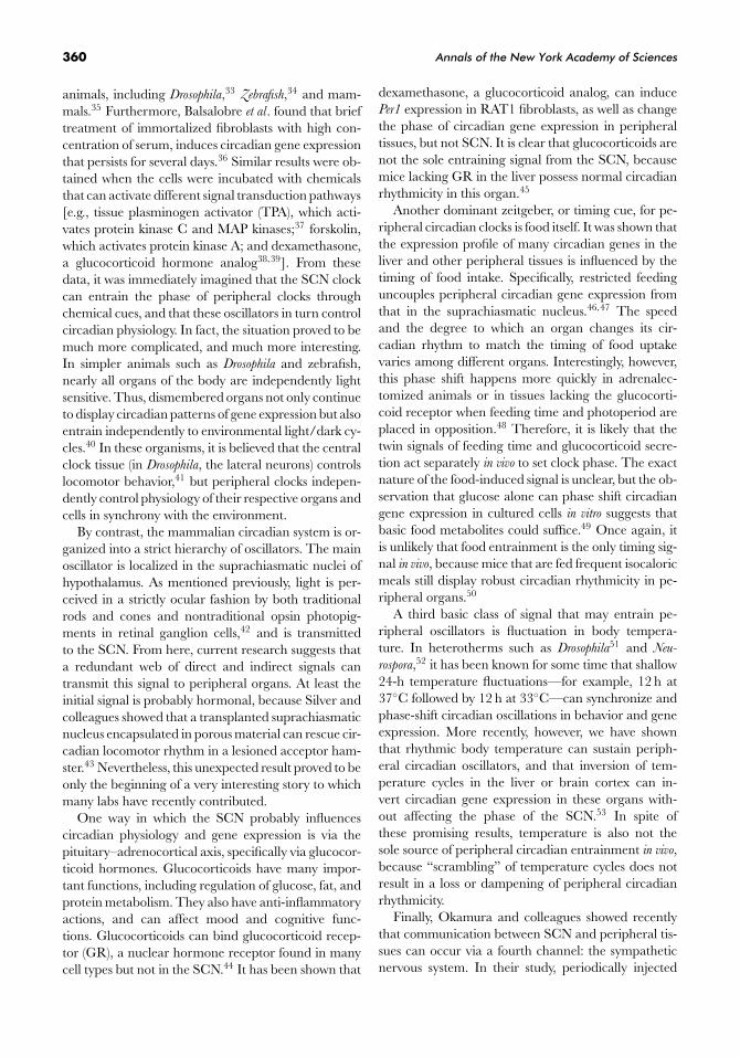
By contrast, the mammalian circadian system is or-ganized into a strict hierarchy of oscillators. The mainoscillator is localized in the suprachiasmatic nuclei ofhypothalamus. As mentioned previously, light is per-ceived in a strictly ocular fashion by both traditionalrods and cones and nontraditional opsin photopig-ments in retinal ganglion cells,42 and is transmittedto the SCN. From here, current research suggests thata redundant web of direct and indirect signals cantransmit this signal to peripheral organs. At least theinitial signal is probably hormonal, because Silver andcolleagues showed that a transplanted suprachiasmaticnucleus encapsulated in porous material can rescue cir-cadian locomotor rhythm in a lesioned acceptor ham-ster.43 Nevertheless, this unexpected result proved to beonly the beginning of a very interesting story to whichmany labs have recently contributed.
One way in which the SCN probably influencescircadian physiology and gene expression is via thepituitary–adrenocortical axis, specifically via glucocor-ticoid hormones. Glucocorticoids have many impor-tant functions, including regulation of glucose, fat, andprotein metabolism. They also have anti-inflammatoryactions, and can affect mood and cognitive func-tions. Glucocorticoids can bind glucocorticoid recep-tor (GR), a nuclear hormone receptor found in manycell types but not in the SCN.44 It has been shown that
dexamethasone, a glucocorticoid analog, can inducePer1 expression in RAT1 fibroblasts, as well as changethe phase of circadian gene expression in peripheraltissues, but not SCN. It is clear that glucocorticoids arenot the sole entraining signal from the SCN, becausemice lacking GR in the liver possess normal circadianrhythmicity in this organ.45
Another dominant zeitgeber, or timing cue, for pe-ripheral circadian clocks is food itself. It was shown thatthe expression profile of many circadian genes in theliver and other peripheral tissues is influenced by thetiming of food intake. Specifically, restricted feedinguncouples peripheral circadian gene expression fromthat in the suprachiasmatic nucleus.46,47 The speedand the degree to which an organ changes its cir-cadian rhythm to match the timing of food uptakevaries among different organs. Interestingly, however,this phase shift happens more quickly in adrenalec-tomized animals or in tissues lacking the glucocorti-coid receptor when feeding time and photoperiod areplaced in opposition.48 Therefore, it is likely that thetwin signals of feeding time and glucocorticoid secre-tion act separately in vivo to set clock phase. The exactnature of the food-induced signal is unclear, but the ob-servation that glucose alone can phase shift circadiangene expression in cultured cells in vitro suggests thatbasic food metabolites could suffice.49 Once again, itis unlikely that food entrainment is the only timing sig-nal in vivo, because mice that are fed frequent isocaloricmeals still display robust circadian rhythmicity in pe-ripheral organs.50
A third basic class of signal that may entrain pe-ripheral oscillators is fluctuation in body tempera-ture. In heterotherms such as Drosophila51 and Neu-
rospora,52 it has been known for some time that shallow24-h temperature fluctuations—for example, 12 h at37◦C followed by 12 h at 33◦C—can synchronize andphase-shift circadian oscillations in behavior and geneexpression. More recently, however, we have shownthat rhythmic body temperature can sustain periph-eral circadian oscillators, and that inversion of tem-perature cycles in the liver or brain cortex can in-vert circadian gene expression in these organs with-out affecting the phase of the SCN.53 In spite ofthese promising results, temperature is also not thesole source of peripheral circadian entrainment in vivo,because “scrambling” of temperature cycles does notresult in a loss or dampening of peripheral circadianrhythmicity.
Finally, Okamura and colleagues showed recentlythat communication between SCN and peripheral tis-sues can occur via a fourth channel: the sympatheticnervous system. In their study, periodically injected
Cuninkova & Brown: Peripheral Circadian Oscillators 361
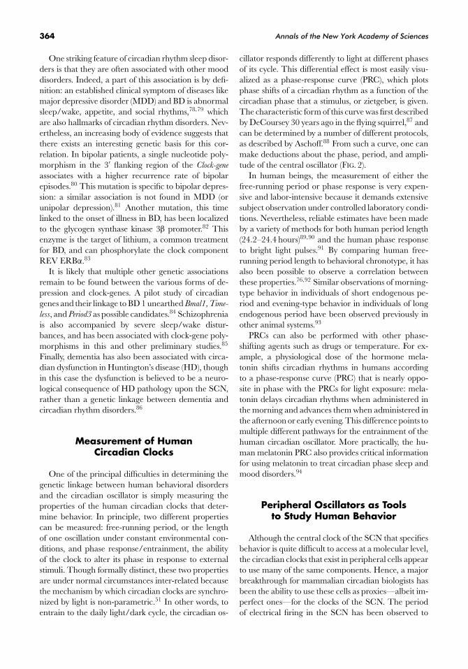
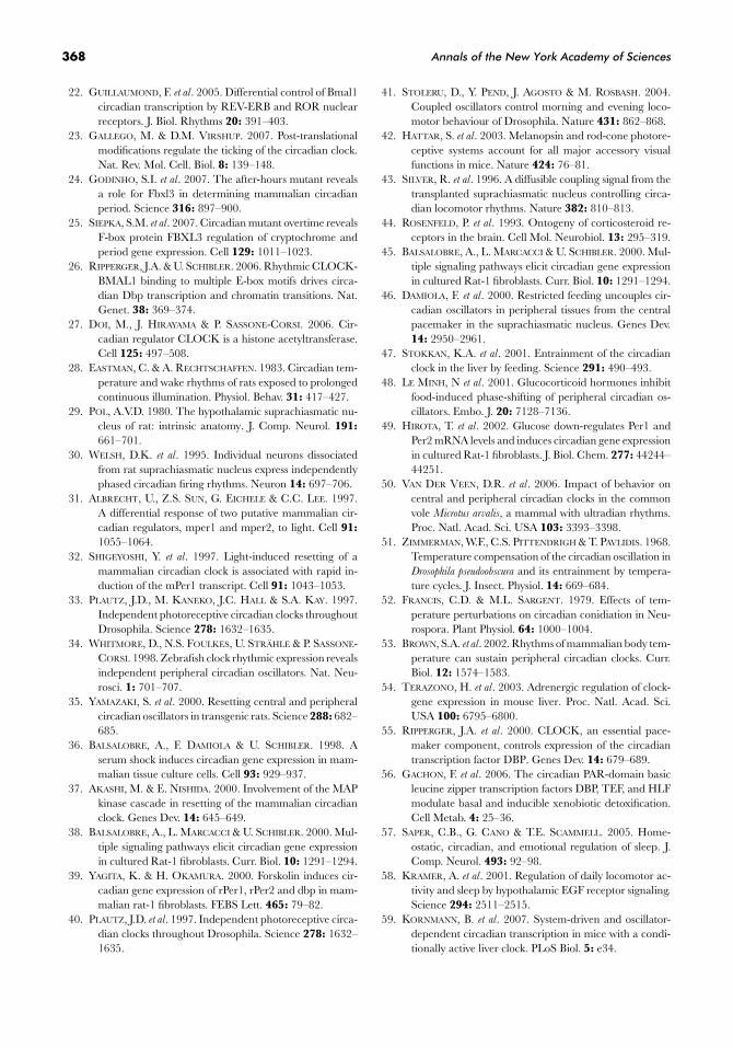
FIGURE 1. Organization of central and peripheral oscillators. Light is the principal timing cue thatsynchronizes the neurons located in the suprachiasmatic nucleus (SCN) of the anterior hypothalamus,whereas the SCN communicates with peripheral oscillators via a redundant web of direct and indirectsignals. For the circadian system, light is received by both rod and cone cells containing traditional opsinpigments and retinal ganglion cells containing melanopsin. This message is sent to the SCN via the opticnerve and the retinohypothalamic tract. From the SCN, a variety of signals are transmitted to peripheraloscillators. These include direct signals from the sympathetic nervous system, hormonal cues, and indirectsignals such as food metabolites and body temperature that are relayed via the control of the SCN overappetite and locomotor activity.
adrenaline in SCN-lesioned mice induced strong Per2
expression in the liver. This result suggested adrenergicregulation influences circadian clock-gene oscillationsin peripheral tissues. Moreover, electrical stimulationof the sympathetic nerve increased mPer1 transcriptionmainly in the liver of these mice. As a confirmation thatthe sympathetic nerve is important for sustaining Per
gene expression in peripheral organs, the mice were in-jected by 6-hydroxydopamine-HCl, which causes thedestruction of sympathetic nerves. The expression ofthe Per2 gene was significantly reduced. Together, thesestudies demonstrated that sympathetic nerve activityplays an important role in the delivery of the centralclock information to at least some peripheral tissues.54
Of course, although we have evoked four distinctchannels of communication in the preceding discus-sion, significant overlap is possible and even likely. Forexample, corticosterone release from the adrenal glandcan occur via the sympathetic nervous system, and foodmetabolites can be brain-signaling molecules. Overall,the important message is that the SCN conveys itstiming information to peripheral circadian clocks by aredundant mix of direct hormonal and nervous signals,and indirect environmental ones, such as feeding and
body temperature, that are influenced by rest/activitycycles (FIG. 1).
Circadian Physiology
A related but separate question is how the SCN di-rects circadian physiology and gene expression. cDNAmicroarray technologies suggest that the circadianclock regulates the transcription of about 10% of allgenes expressed in each tissue.5–7 Given the pervasiveand cell-autonomous nature of peripheral circadianoscillators, it was largely assumed that they would beresponsible for the direction of most circadian physiol-ogy and gene expression. Certainly, this hypothesis is atleast partially true. Many clock-controlled genes are di-rected by the same cis-acting elements such as E-boxesthat control expression of clock-genes themselves. Forsuch “direct” regulation, the Dbp gene has served asan excellent model system.55 Ripperger et al. have dis-sected this gene and shown that it possesses multipleE-boxes in both promoter and intronic regions. Byclock proteins and chromatin modifying factors thatbind to these regulatory elements, the entire locus is
362 Annals of the New York Academy of Sciences
switched from an active to an inactive chromatin con-formation to turn the locus on and off in circadianfashion.26 Meanwhile, DBP is itself a member of afamily of circadian transcription factors that direct theexpression of other circadian output genes importantfor the liver’s role in xenobiotic detoxication.56 Thus,one way in which the circadian clock controls circadianphysiology is by transcription factor cascades directedby peripheral oscillators.
A second way in which the SCN drives circa-dian physiology is via direct nervous connections toother centers of the brain. The major output fromthe suprachiasmatic nuclei is the dorsomedial nucleusand supraventricular zone. The dorsal supraventricu-lar zone can organize circadian regulation of body tem-perature. The ventral suprachiasmatic zone regulatecircadian rhythms of sleep and wakefulness, and thedorsomedial nucleus is necessary for feeding activity.57
Since an SCN encapsulated in porous plastic materialcan rescue behavioral arrhythmicity in lesioned ani-mals,43 multiple laboratories have also tried to purifyneuropeptides important for SCN signaling. Kramerand Weitz identified one such factor as transforminggrowth factor α (TGF-α). This growth factor activatesthe epidermal growth-factor receptor (EGFR) on neu-rons in the hypothalamic supraventricular zone, andthese may mediate circadian locomotor activity.58
Surprisingly, recent tissue-specific clock knockoutshave shown that the SCN can also directly regulatesome peripheral circadian physiology at the gene-expression level. The elegant experiments of Korn-mann and colleagues showed that some circadian ex-pression of genes in peripheral organs can be drivendirectly by systemic circadian signals and others by lo-cal oscillators. To show this, they generated mice withtetracycline-dependent hepatocyte clocks by placingthe clock-gene Rev-Erbα under the control of the tetra-cycline operator. Its overexpression in the absence ofdoxycycline thereby leads to silencing of Bmal tran-scription, and with it circadian clock function in thistissue. When the hepatocyte clocks are turned off, thebulk of circadian transcription in the liver is stronglyattenuated. This result indicates that most expressionof circadian liver genes is driven by local cellular clocks.By contrast, a smaller subset of genes—which includedthe clock-gene mPer2—continued to oscillate. Hence,these genes must be regulated by systemic signals suchas hormones, metabolites, or temperature.59
Circadian Behavior
The circadian system has a very important influ-ence on human physiology and behavior. Indeed, con-
sidering the extent of circadian regulation describedearlier, it is perhaps not surprising that disruption ofbiological clocks has a negative effect. One of the mostobvious manifestations is jet lag, misadjustment of cir-cadian phase due to travel. Links have also been es-tablished between circadian irregularities and psychi-atric disorders, including various forms of depressionand mania. Prolonged disruption of circadian rhythmsis believed to have significant adverse health conse-quences on peripheral organs outside the brain aswell, particularly in the development or exacerbationof cardiovascular disease and cancer.60–62 Conversely,chronopharmacology—the timing of treatment in co-ordination with the body clock—may significantly in-crease efficacy of various therapies, and reduce drugtoxicity or adverse reactions.63
Even under normal conditions, the complex natureof circadian behavior is evident from the fact that phas-ing of the cycle during the day varies widely for indi-viduals, resulting in extremes colloquially called larksand owls. Morningness/eveningness, or “chronotype,”is an individual characteristic that refers mostly to thephase of sleep timing.64 Because of this effect of the cir-cadian system upon sleep, most circadian rhythm dis-orders are therefore classified as sleep disorders. Nev-ertheless, circadian sleep disorders and true sleep dis-orders are likely to be mechanistically unrelated, andtherefore it is both scientifically and clinically relevantto distinguish between them.
Sleep is an active and as yet poorly understoodprocess, during which many physiological and cere-bral events occur. Indeed, even sleep itself is actuallyan ultradian process represented by the alternationof different electrophysiologically defined sleep states.In general, the daily sleep/wake cycle is under circa-dian control, although the urge to sleep appears to becontrolled by brain functions that are independent ofthe circadian system.65 This independence led Bor-bely and colleagues to propose a model for the regu-lation of sleep that includes a homeostatic process thataccumulates during wakefulness and diminishes dur-ing sleep, as well as an independent circadian drive.66
Each of these processes can operate independently;thus, sleep duration is not correlated with sleep phasein humans.67
So-called “circadian rhythm sleep disorders” canresult from alterations in the properties of the endoge-nous circadian clock (e.g., delayed sleep phase and ad-vanced sleep phase) or changes in the physical environ-ment in relation to the endogenous clock (shift workdisorders and jet lag). In the former class, which is unre-lated to human choice, genetic variations in circadiangenes have been found to associate with these disorders.
Cuninkova & Brown: Peripheral Circadian Oscillators 363
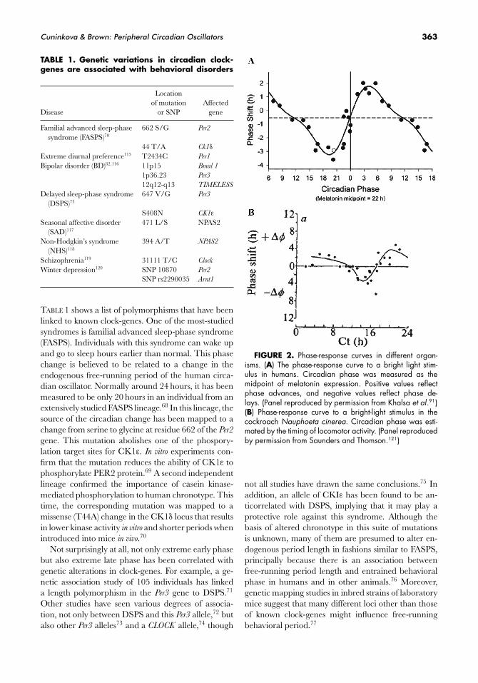
TABLE 1. Genetic variations in circadian clock-genes are associated with behavioral disorders
Locationof mutation Affected
Disease or SNP gene
Familial advanced sleep-phase 662 S/G Per2
syndrome (FASPS)70
44 T/A Ck1δ
Extreme diurnal preference115 T2434C Per1
Bipolar disorder (BD)82,116 11p15 Bmal 1
1p36.23 Per3
12q12-q13 TIMELESS
Delayed sleep-phase syndrome 647 V/G Per3
(DSPS)73
S408N CK1ε
Seasonal affective disorder 471 L/S NPAS2(SAD)117
Non-Hodgkin’s syndrome 394 A/T NPAS2
(NHS)118
Schizophrenia119 31111 T/C Clock
Winter depression120 SNP 10870 Per2
SNP rs2290035 Arnt1
TABLE 1 shows a list of polymorphisms that have beenlinked to known clock-genes. One of the most-studiedsyndromes is familial advanced sleep-phase syndrome(FASPS). Individuals with this syndrome can wake upand go to sleep hours earlier than normal. This phasechange is believed to be related to a change in theendogenous free-running period of the human circa-dian oscillator. Normally around 24 hours, it has beenmeasured to be only 20 hours in an individual from anextensively studied FASPS lineage.68 In this lineage, thesource of the circadian change has been mapped to achange from serine to glycine at residue 662 of the Per2
gene. This mutation abolishes one of the phospory-lation target sites for CK1ε. In vitro experiments con-firm that the mutation reduces the ability of CK1ε tophosphorylate PER2 protein.69 A second independentlineage confirmed the importance of casein kinase-mediated phosphorylation to human chronotype. Thistime, the corresponding mutation was mapped to amissense (T44A) change in the CK1δ locus that resultsin lower kinase activity in vitro and shorter periods whenintroduced into mice in vivo.70
Not surprisingly at all, not only extreme early phasebut also extreme late phase has been correlated withgenetic alterations in clock-genes. For example, a ge-netic association study of 105 individuals has linkeda length polymorphism in the Per3 gene to DSPS.71
Other studies have seen various degrees of associa-tion, not only between DSPS and this Per3 allele,72 butalso other Per3 alleles73 and a CLOCK allele,74 though
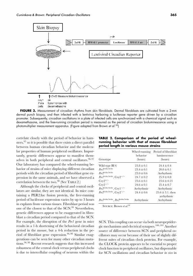
FIGURE 2. Phase-response curves in different organ-isms. (A) The phase-response curve to a bright light stim-ulus in humans. Circadian phase was measured as themidpoint of melatonin expression. Positive values reflectphase advances, and negative values reflect phase de-lays. (Panel reproduced by permission from Khalsa et al.91)(B) Phase-response curve to a bright-light stimulus in thecockroach Nauphoeta cinerea. Circadian phase was esti-mated by the timing of locomotor activity. (Panel reproducedby permission from Saunders and Thomson.121)
not all studies have drawn the same conclusions.75 Inaddition, an allele of CKIε has been found to be an-ticorrelated with DSPS, implying that it may play aprotective role against this syndrome. Although thebasis of altered chronotype in this suite of mutationsis unknown, many of them are presumed to alter en-dogenous period length in fashions similar to FASPS,principally because there is an association betweenfree-running period length and entrained behavioralphase in humans and in other animals.76 Moreover,genetic mapping studies in inbred strains of laboratorymice suggest that many different loci other than thoseof known clock-genes might influence free-runningbehavioral period.77
364 Annals of the New York Academy of Sciences
One striking feature of circadian rhythm sleep disor-ders is that they are often associated with other mooddisorders. Indeed, a part of this association is by defi-nition: an established clinical symptom of diseases likemajor depressive disorder (MDD) and BD is abnormalsleep/wake, appetite, and social rhythms,78,79 whichare also hallmarks of circadian rhythm disorders. Nev-ertheless, an increasing body of evidence suggests thatthere exists an interesting genetic basis for this cor-relation. In bipolar patients, a single nucleotide poly-morphism in the 3′ flanking region of the Clock-gene
associates with a higher recurrence rate of bipolarepisodes.80 This mutation is specific to bipolar depres-sion: a similar association is not found in MDD (orunipolar depression).81 Another mutation, this timelinked to the onset of illness in BD, has been localizedto the glycogen synthase kinase 3β promoter.82 Thisenzyme is the target of lithium, a common treatmentfor BD, and can phosphorylate the clock componentREV ERBα.83
It is likely that multiple other genetic associationsremain to be found between the various forms of de-pression and clock-genes. A pilot study of circadiangenes and their linkage to BD 1 unearthed Bmal1, Time-
less, and Period3 as possible candidates.84 Schizophreniais also accompanied by severe sleep/wake distur-bances, and has been associated with clock-gene poly-morphisms in this and other preliminary studies.85
Finally, dementia has also been associated with circa-dian dysfunction in Huntington’s disease (HD), thoughin this case the dysfunction is believed to be a neuro-logical consequence of HD pathology upon the SCN,rather than a genetic linkage between dementia andcircadian rhythm disorders.86
Measurement of HumanCircadian Clocks
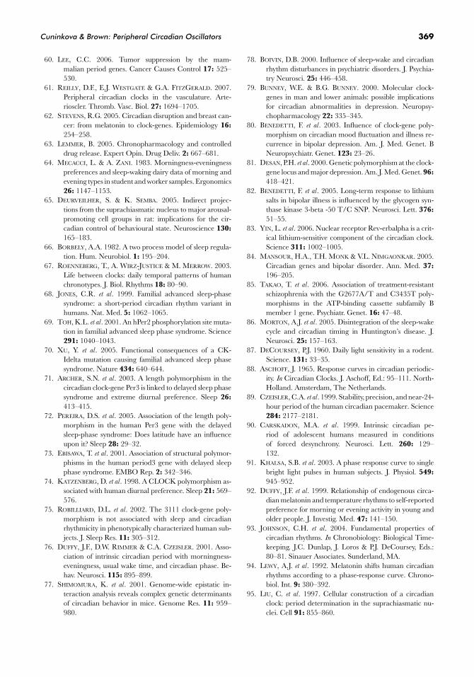
One of the principal difficulties in determining thegenetic linkage between human behavioral disordersand the circadian oscillator is simply measuring theproperties of the human circadian clocks that deter-mine behavior. In principle, two different propertiescan be measured: free-running period, or the lengthof one oscillation under constant environmental con-ditions, and phase response/entrainment, the abilityof the clock to alter its phase in response to externalstimuli. Though formally distinct, these two propertiesare under normal circumstances inter-related becausethe mechanism by which circadian clocks are synchro-nized by light is non-parametric.51 In other words, toentrain to the daily light/dark cycle, the circadian os-
cillator responds differently to light at different phasesof its cycle. This differential effect is most easily visu-alized as a phase-response curve (PRC), which plotsphase shifts of a circadian rhythm as a function of thecircadian phase that a stimulus, or zietgeber, is given.The characteristic form of this curve was first describedby DeCoursey 30 years ago in the flying squirrel,87 andcan be determined by a number of different protocols,as described by Aschoff.88 From such a curve, one canmake deductions about the phase, period, and ampli-tude of the central oscillator (FIG. 2).
In human beings, the measurement of either thefree-running period or phase response is very expen-sive and labor-intensive because it demands extensivesubject observation under controlled laboratory condi-tions. Nevertheless, reliable estimates have been madeby a variety of methods for both human period length(24.2–24.4 hours)89,90 and the human phase responseto bright light pulses.91 By comparing human free-running period length to behavioral chronotype, it hasalso been possible to observe a correlation betweenthese properties.76,92 Similar observations of morning-type behavior in individuals of short endogenous pe-riod and evening-type behavior in individuals of longendogenous period have been observed previously inother animal systems.93
PRCs can also be performed with other phase-shifting agents such as drugs or temperature. For ex-ample, a physiological dose of the hormone mela-tonin shifts circadian rhythms in humans accordingto a phase-response curve (PRC) that is nearly oppo-site in phase with the PRCs for light exposure: mela-tonin delays circadian rhythms when administered inthe morning and advances them when administered inthe afternoon or early evening. This difference points tomultiple different pathways for the entrainment of thehuman circadian oscillator. More practically, the hu-man melatonin PRC also provides critical informationfor using melatonin to treat circadian phase sleep andmood disorders.94
Peripheral Oscillators as Toolsto Study Human Behavior
Although the central clock of the SCN that specifiesbehavior is quite difficult to access at a molecular level,the circadian clocks that exist in peripheral cells appearto use many of the same components. Hence, a majorbreakthrough for mammalian circadian biologists hasbeen the ability to use these cells as proxies—albeit im-perfect ones—for the clocks of the SCN. The periodof electrical firing in the SCN has been observed to
Cuninkova & Brown: Peripheral Circadian Oscillators 365
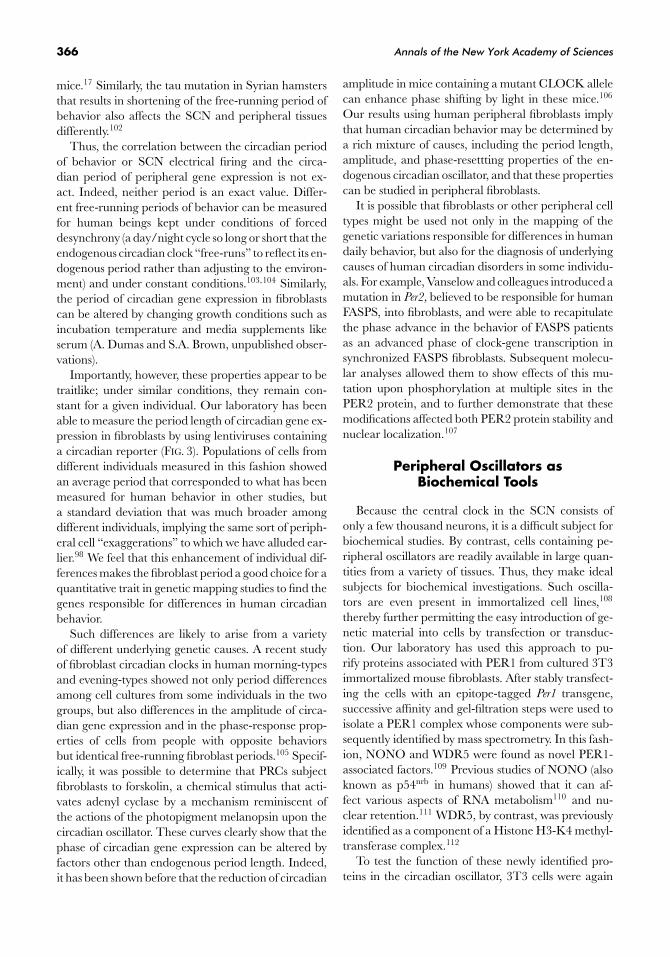
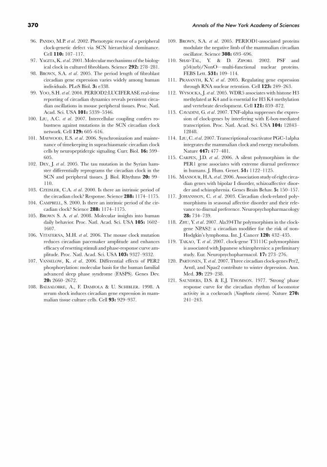
FIGURE 3. Measurement of circadian rhythms from skin fibroblasts. Dermal fibroblasts are cultivated from a 2-mmdermal punch biopsy, and then infected with a lentivirus harboring a luciferase reporter gene driven by a circadianpromoter. Subsequently, circadian oscillations in a plate of infected cells are synchronized with a chemical signal such asdexamethasone, and the free-running circadian period is measured as the period of circadian bioluminescence using aphotomultiplier measurement apparatus. (Figure adapted from Brown et al.98)
correlate closely with the period of behavior in ham-sters,95 so it is possible that there exists a direct parallelbetween human circadian behavior and the molecu-lar properties of human peripheral oscillators. Impor-tantly, genetic differences appear to manifest them-selves in both peripheral and central oscillators.96,97
Our laboratory has compared the wheel-running be-havior of strains of mice displaying different circadianperiods with the circadian period of fibroblast gene ex-pression in the same animals, and we have observed acorrelation between the two.98 (See TABLE 2.)
Although the clocks of peripheral and central oscil-lators are similar, they are not identical. In mice con-taining a PER2:luc fusion protein, the free-runningperiod of luciferase expression varies by up to 3 hoursin explants from various tissues. Fibroblast period wasone of the closest to that of the SCN.99 Nevertheless,genetic differences appear to be exaggerated in fibro-blast a circadian period compared to that of the SCN.For example, the disruption of the Per1 gene in miceresults in a 1-h shortening of the behavioral circadianperiod in the mouse, but a 4-h reduction in the pe-riod of fibroblast gene expression.96,98 Similar exag-gerations can be seen for many other circadian muta-tions.96,98 Recent research suggests that this increasedrobustness of the central clock versus peripheral clocksis due to intercellular coupling of neurons within the
TABLE 2. Comparison of the period of wheel-running behavior with that of mouse fibroblastperiod length in various mouse strains
Wheel-running Period of fibroblastbehavior luminescence
Genotype (hours) (hours)
Wild type BI 6 23.8 ± 0.1 24.4 ± 0.4Per1brdm/brdm 23.4 ± 0.1 20.0 ± 0.1Per2brdm/brdm 23.0 ± 0.6 ArrhythmicPer1brdm/brdm, Cry2−/− 24.7 ± 0.2 25.9 ± 0.8Cry2+/− 23.6 ± 0.1 23.6 ± 0.21Cry2−/− 24.6 ± 0.1 25.4 ± 0.7Per2brdm/brdm, Cry1−/− Arrhythmic ArrhythmicPer2brdm/brdm, Cry2−/− 24.4 ± 0.6 25.6 ± 3.1. then
arrhythmicPer1brdm/brdm, Per2brdm/brdm Arrhythmic Arrhythmic
SOURCE: Brown et al.98
SCN. This coupling can occur via both neuropeptider-gic mechanisms and electrical synapses.100,101 Anothersource of difference between SCN and peripheral os-cillators may occur because of their use of slightly dif-ferent suites of circadian clock proteins. For example,the CLOCK protein appears to be essential to properclock function in peripheral oscillators, but dispensablefor SCN oscillations and circadian behavior in vivo in
366 Annals of the New York Academy of Sciences
mice.17 Similarly, the tau mutation in Syrian hamstersthat results in shortening of the free-running period ofbehavior also affects the SCN and peripheral tissuesdifferently.102
Thus, the correlation between the circadian periodof behavior or SCN electrical firing and the circa-dian period of peripheral gene expression is not ex-act. Indeed, neither period is an exact value. Differ-ent free-running periods of behavior can be measuredfor human beings kept under conditions of forceddesynchrony (a day/night cycle so long or short that theendogenous circadian clock “free-runs” to reflect its en-dogenous period rather than adjusting to the environ-ment) and under constant conditions.103,104 Similarly,the period of circadian gene expression in fibroblastscan be altered by changing growth conditions such asincubation temperature and media supplements likeserum (A. Dumas and S.A. Brown, unpublished obser-vations).
Importantly, however, these properties appear to betraitlike; under similar conditions, they remain con-stant for a given individual. Our laboratory has beenable to measure the period length of circadian gene ex-pression in fibroblasts by using lentiviruses containinga circadian reporter (FIG. 3). Populations of cells fromdifferent individuals measured in this fashion showedan average period that corresponded to what has beenmeasured for human behavior in other studies, buta standard deviation that was much broader amongdifferent individuals, implying the same sort of periph-eral cell “exaggerations” to which we have alluded ear-lier.98 We feel that this enhancement of individual dif-ferences makes the fibroblast period a good choice for aquantitative trait in genetic mapping studies to find thegenes responsible for differences in human circadianbehavior.
Such differences are likely to arise from a varietyof different underlying genetic causes. A recent studyof fibroblast circadian clocks in human morning-typesand evening-types showed not only period differencesamong cell cultures from some individuals in the twogroups, but also differences in the amplitude of circa-dian gene expression and in the phase-response prop-erties of cells from people with opposite behaviorsbut identical free-running fibroblast periods.105 Specif-ically, it was possible to determine that PRCs subjectfibroblasts to forskolin, a chemical stimulus that acti-vates adenyl cyclase by a mechanism reminiscent ofthe actions of the photopigment melanopsin upon thecircadian oscillator. These curves clearly show that thephase of circadian gene expression can be altered byfactors other than endogenous period length. Indeed,it has been shown before that the reduction of circadian
amplitude in mice containing a mutant CLOCK allelecan enhance phase shifting by light in these mice.106
Our results using human peripheral fibroblasts implythat human circadian behavior may be determined bya rich mixture of causes, including the period length,amplitude, and phase-resettting properties of the en-dogenous circadian oscillator, and that these propertiescan be studied in peripheral fibroblasts.
It is possible that fibroblasts or other peripheral celltypes might be used not only in the mapping of thegenetic variations responsible for differences in humandaily behavior, but also for the diagnosis of underlyingcauses of human circadian disorders in some individu-als. For example, Vanselow and colleagues introduced amutation in Per2, believed to be responsible for humanFASPS, into fibroblasts, and were able to recapitulatethe phase advance in the behavior of FASPS patientsas an advanced phase of clock-gene transcription insynchronized FASPS fibroblasts. Subsequent molecu-lar analyses allowed them to show effects of this mu-tation upon phosphorylation at multiple sites in thePER2 protein, and to further demonstrate that thesemodifications affected both PER2 protein stability andnuclear localization.107
Peripheral Oscillators asBiochemical Tools
Because the central clock in the SCN consists ofonly a few thousand neurons, it is a difficult subject forbiochemical studies. By contrast, cells containing pe-ripheral oscillators are readily available in large quan-tities from a variety of tissues. Thus, they make idealsubjects for biochemical investigations. Such oscilla-tors are even present in immortalized cell lines,108
thereby further permitting the easy introduction of ge-netic material into cells by transfection or transduc-tion. Our laboratory has used this approach to pu-rify proteins associated with PER1 from cultured 3T3immortalized mouse fibroblasts. After stably transfect-ing the cells with an epitope-tagged Per1 transgene,successive affinity and gel-filtration steps were used toisolate a PER1 complex whose components were sub-sequently identified by mass spectrometry. In this fash-ion, NONO and WDR5 were found as novel PER1-associated factors.109 Previous studies of NONO (alsoknown as p54nrb in humans) showed that it can af-fect various aspects of RNA metabolism110 and nu-clear retention.111 WDR5, by contrast, was previouslyidentified as a component of a Histone H3-K4 methyl-transferase complex.112
To test the function of these newly identified pro-teins in the circadian oscillator, 3T3 cells were again
Cuninkova & Brown: Peripheral Circadian Oscillators 367
probed, this time via RNA interference and chromatinimmunoprecipitation techniques. Such tools providepowerful methods to analyze the importance of bothcis-acting elements and trans-acting factors to the cir-cadian clock. For example, Ripperger et al.26 identifiedthe functions of various E-box elements within the cir-cadian gene DBP by introducing marked and modifiedcopies of the gene into fibroblast cells and studying theirfunction. Similarly, Cavadini et al. showed that TNFα isable to suppress the expression of clock-genes in fibro-blasts by inhibiting E-box-mediated transcription.113
Fibroblast-based studies also demonstrate PGC-1α,a transcriptional coactivator important for energymetabolism, to be necessary for cell-autonomous clockfunction,114 and highlighted the importance of thenovel protein CIPC to circadian function.20
Conclusions
Circadian clocks have pervasive effects upon humanphysiology and behavior. Nevertheless, the complexand hierarchical nature of the mammalian circadianoscillator has long proven a barrier to its understand-ing at a molecular level. The discovery a decade ago of“slave” oscillators in most of the cells of the body hasproven a turning point in our understanding. Periph-eral clocks, and their communication with the SCNmaster clock, are essential to the regulation of circa-dian physiology in mammals. Equally important, theyhave proven a useful model system to probe the molec-ular roles of novel proteins within the oscillator. Finally,the similarity between the timing of peripheral circa-dian gene expression and the timing of daily humanbehavior may even render them useful in the quest forthe genetic origins of human chronotype.
Acknowledgments
The authors thank the Swiss National Science Foun-dation, the Desiree and Neils Yde Foundation for Neu-ropsychiatric Research, and the 6th EU frameworkprogram EUCLOCK for their support.
Competing Interest
The authors declare no competing interest.
References
1. GROBBELAAR, N., T.-C. HUANG, H.Y. LIN & T.J. CHOW.1986. Dinitrogen-fixing endogenous rhythm in Syne-chococcus RF-1. FEMS Microbial. Lett. 37: 173–177.
2. MITSUI, A. et al. 1986. Strategy by which nitrogen-fixing unicellular cyanobacteria grow photoautotrophi-cally. Nature 323: 721–722.
3. STAL, L.J. & W.E. KRUMBEIN. 1985. Nitrogenase activ-ity in non-heterocystous cyanobacterium Oscillatoria sp.grown under alternating light-dark cycles. Arch. Micro-biol. 143: 67–71.
4. WOELFLE, M.A. et al. 2004. The adaptive value of circa-dian clocks: an experimental assessment in cyanobacte-ria. Curr. Biol. 14: 1481–1486.
5. STORCH, K.F. et al. 2002. Extensive and divergent circadiangene expression in liver and heart. Nature 417: 78–83.
6. AKHTAR, R.A. et al. 2002. Circadian cycling of the mouseliver transcriptome, as revealed by cDNA microarray, isdriven by the suprachiasmatic nucleus. Curr. Biol. 12:540–550.
7. PANDA, S. et al. 2002. Coordinated transcription of keypathways in the mouse by the circadian clock. Cell 109:307–320.
8. REDDY, A.B. et al. 2006. Circadian orchestration of thehepatic proteome. Curr. Biol. 16: 1107–1115.
9. TAKAHASHI, J.S. 2004. Finding new clock components: pastand future. J. Biol. Rhythms 19: 339–347.
10. FELDMAN, J.F. & M.N. HOYLE. 1973. Isolation of circa-dian clock mutants of Neurospora crassa. Genetics 75: 605–613.
11. MCCLUNG, C.R., B.A. FOX & J.C. DUNLAP. 1989. TheNeurospora clock-gene frequency shares a sequence el-ement with the Drosophila clock-gene period. Nature339: 558–562.
12. KONOPKA, R.J. & S. BENZER. 1971. Clock mutants ofDrosophila melanogaster. Proc. Natl. Acad. Sci. USA 68:2112–2116.
13. ZEHRING, W.A. et al. 1984. P-element transformation withperiod locus DNA restores rhythmicity to mutant, ar-rhythmic Drosophila melanogaster. Cell 39: 369–376.
14. SHEARMAN, L.P. et al. 2000. Interacting molecular loopsin the mammalian circadian clock. Science 288: 1013–1019.
15. KONDO, T. & M. ISHIURA. 2000. The circadian clock ofcyanobacteria. Bioessays 22: 10–15.
16. SATO, T.K. et al. 2006. Feedback repression is required formammalian circadian clock function. Nat. Genet. 38:312–319.
17. DEBRUYNE, J.P. et al. 2006. A clock shock: mouse CLOCKis not required for circadian oscillator function. Neuron50: 465–477.
18. ZHOU, Y.D. et al. 1997. Molecular characterization of twomammalian bHLH-PAS domain proteins selectively ex-pressed in the central nervous system. Proc. Natl. Acad.Sci. USA 94: 713–718.
19. SATO, T.K. et al. 2006. Feedback repression is required formammalian circadian clock function. Nat. Genet. 38:312–319.
20. ZHAO, W.N. et al. 2007. CIPC is a mammalian circadianclock protein without invertebrate homologues. Nat. CellBiol. 9: 286–275.
21. PREITNER, N. et al. 2002. The orphan nuclear receptorREV-ERBalpha controls circadian transcription withinthe positive limb of the mammalian circadian oscillator.Cell 110: 251–260.
368 Annals of the New York Academy of Sciences
22. GUILLAUMOND, F. et al. 2005. Differential control of Bmal1circadian transcription by REV-ERB and ROR nuclearreceptors. J. Biol. Rhythms 20: 391–403.
23. GALLEGO, M. & D.M. VIRSHUP. 2007. Post-translationalmodifications regulate the ticking of the circadian clock.Nat. Rev. Mol. Cell. Biol. 8: 139–148.
24. GODINHO, S.I. et al. 2007. The after-hours mutant revealsa role for Fbxl3 in determining mammalian circadianperiod. Science 316: 897–900.
25. SIEPKA, S.M. et al. 2007. Circadian mutant overtime revealsF-box protein FBXL3 regulation of cryptochrome andperiod gene expression. Cell 129: 1011–1023.
26. RIPPERGER, J.A. & U. SCHIBLER. 2006. Rhythmic CLOCK-BMAL1 binding to multiple E-box motifs drives circa-dian Dbp transcription and chromatin transitions. Nat.Genet. 38: 369–374.
27. DOI, M., J. HIRAYAMA & P. SASSONE-CORSI. 2006. Cir-cadian regulator CLOCK is a histone acetyltransferase.Cell 125: 497–508.
28. EASTMAN, C. & A. RECHTSCHAFFEN. 1983. Circadian tem-perature and wake rhythms of rats exposed to prolongedcontinuous illumination. Physiol. Behav. 31: 417–427.
29. POL, A.V.D. 1980. The hypothalamic suprachiasmatic nu-cleus of rat: intrinsic anatomy. J. Comp. Neurol. 191:661–701.
30. WELSH, D.K. et al. 1995. Individual neurons dissociatedfrom rat suprachiasmatic nucleus express independentlyphased circadian firing rhythms. Neuron 14: 697–706.
31. ALBRECHT, U., Z.S. SUN, G. EICHELE & C.C. LEE. 1997.A differential response of two putative mammalian cir-cadian regulators, mper1 and mper2, to light. Cell 91:1055–1064.
32. SHIGEYOSHI, Y. et al. 1997. Light-induced resetting of amammalian circadian clock is associated with rapid in-duction of the mPer1 transcript. Cell 91: 1043–1053.
33. PLAUTZ, J.D., M. KANEKO, J.C. HALL & S.A. KAY. 1997.Independent photoreceptive circadian clocks throughoutDrosophila. Science 278: 1632–1635.
34. WHITMORE, D., N.S. FOULKES, U. STRAHLE & P. SASSONE-CORSI. 1998. Zebrafish clock rhythmic expression revealsindependent peripheral circadian oscillators. Nat. Neu-rosci. 1: 701–707.
35. YAMAZAKI, S. et al. 2000. Resetting central and peripheralcircadian oscillators in transgenic rats. Science 288: 682–685.
36. BALSALOBRE, A., F. DAMIOLA & U. SCHIBLER. 1998. Aserum shock induces circadian gene expression in mam-malian tissue culture cells. Cell 93: 929–937.
37. AKASHI, M. & E. NISHIDA. 2000. Involvement of the MAPkinase cascade in resetting of the mammalian circadianclock. Genes Dev. 14: 645–649.
38. BALSALOBRE, A., L. MARCACCI & U. SCHIBLER. 2000. Mul-tiple signaling pathways elicit circadian gene expressionin cultured Rat-1 fibroblasts. Curr. Biol. 10: 1291–1294.
39. YAGITA, K. & H. OKAMURA. 2000. Forskolin induces cir-cadian gene expression of rPer1, rPer2 and dbp in mam-malian rat-1 fibroblasts. FEBS Lett. 465: 79–82.
40. PLAUTZ, J.D. et al. 1997. Independent photoreceptive circa-dian clocks throughout Drosophila. Science 278: 1632–1635.
41. STOLERU, D., Y. PEND, J. AGOSTO & M. ROSBASH. 2004.Coupled oscillators control morning and evening loco-motor behaviour of Drosophila. Nature 431: 862–868.
42. HATTAR, S. et al. 2003. Melanopsin and rod-cone photore-ceptive systems account for all major accessory visualfunctions in mice. Nature 424: 76–81.
43. SILVER, R. et al. 1996. A diffusible coupling signal from thetransplanted suprachiasmatic nucleus controlling circa-dian locomotor rhythms. Nature 382: 810–813.
44. ROSENFELD, P. et al. 1993. Ontogeny of corticosteroid re-ceptors in the brain. Cell Mol. Neurobiol. 13: 295–319.
45. BALSALOBRE, A., L. MARCACCI & U. SCHIBLER. 2000. Mul-tiple signaling pathways elicit circadian gene expressionin cultured Rat-1 fibroblasts. Curr. Biol. 10: 1291–1294.
46. DAMIOLA, F. et al. 2000. Restricted feeding uncouples cir-cadian oscillators in peripheral tissues from the centralpacemaker in the suprachiasmatic nucleus. Genes Dev.14: 2950–2961.
47. STOKKAN, K.A. et al. 2001. Entrainment of the circadianclock in the liver by feeding. Science 291: 490–493.
48. LE MINH, N et al. 2001. Glucocorticoid hormones inhibitfood-induced phase-shifting of peripheral circadian os-cillators. Embo. J. 20: 7128–7136.
49. HIROTA, T. et al. 2002. Glucose down-regulates Per1 andPer2 mRNA levels and induces circadian gene expressionin cultured Rat-1 fibroblasts. J. Biol. Chem. 277: 44244–44251.
50. VAN DER VEEN, D.R. et al. 2006. Impact of behavior oncentral and peripheral circadian clocks in the commonvole Microtus arvalis, a mammal with ultradian rhythms.Proc. Natl. Acad. Sci. USA 103: 3393–3398.
51. ZIMMERMAN, W.F., C.S. PITTENDRIGH & T. PAVLIDIS. 1968.Temperature compensation of the circadian oscillation inDrosophila pseudoobscura and its entrainment by tempera-ture cycles. J. Insect. Physiol. 14: 669–684.
52. FRANCIS, C.D. & M.L. SARGENT. 1979. Effects of tem-perature perturbations on circadian conidiation in Neu-rospora. Plant Physiol. 64: 1000–1004.
53. BROWN, S.A. et al. 2002. Rhythms of mammalian body tem-perature can sustain peripheral circadian clocks. Curr.Biol. 12: 1574–1583.
54. TERAZONO, H. et al. 2003. Adrenergic regulation of clock-gene expression in mouse liver. Proc. Natl. Acad. Sci.USA 100: 6795–6800.
55. RIPPERGER, J.A. et al. 2000. CLOCK, an essential pace-maker component, controls expression of the circadiantranscription factor DBP. Genes Dev. 14: 679–689.
56. GACHON, F. et al. 2006. The circadian PAR-domain basicleucine zipper transcription factors DBP, TEF, and HLFmodulate basal and inducible xenobiotic detoxification.Cell Metab. 4: 25–36.
57. SAPER, C.B., G. CANO & T.E. SCAMMELL. 2005. Home-ostatic, circadian, and emotional regulation of sleep. J.Comp. Neurol. 493: 92–98.
58. KRAMER, A. et al. 2001. Regulation of daily locomotor ac-tivity and sleep by hypothalamic EGF receptor signaling.Science 294: 2511–2515.
59. KORNMANN, B. et al. 2007. System-driven and oscillator-dependent circadian transcription in mice with a condi-tionally active liver clock. PLoS Biol. 5: e34.
Cuninkova & Brown: Peripheral Circadian Oscillators 369
60. LEE, C.C. 2006. Tumor suppression by the mam-malian period genes. Cancer Causes Control 17: 525–530.
61. REILLY, D.F., E.J. WESTGATE & G.A. FITZGERALD. 2007.Peripheral circadian clocks in the vasculature. Arte-rioscler. Thromb. Vasc. Biol. 27: 1694–1705.
62. STEVENS, R.G. 2005. Circadian disruption and breast can-cer: from melatonin to clock-genes. Epidemiology 16:254–258.
63. LEMMER, B. 2005. Chronopharmacology and controlleddrug release. Expert Opin. Drug Deliv. 2: 667–681.
64. MECACCI, L. & A. ZANI. 1983. Morningness-eveningnesspreferences and sleep-waking dairy data of morning andevening types in student and worker samples. Ergonomics26: 1147–1153.
65. DEURVEILHER, S. & K. SEMBA. 2005. Indirect projec-tions from the suprachiasmatic nucleus to major arousal-promoting cell groups in rat: implications for the cir-cadian control of behavioural state. Neuroscience 130:165–183.
66. BORBELY, A.A. 1982. A two process model of sleep regula-tion. Hum. Neurobiol. 1: 195–204.
67. ROENNEBERG, T., A. WIRZ-JUSTICE & M. MERROW. 2003.Life between clocks: daily temporal patterns of humanchronotypes. J. Biol. Rhythms 18: 80–90.
68. JONES, C.R. et al. 1999. Familial advanced sleep-phasesyndrome: a short-period circadian rhythm variant inhumans. Nat. Med. 5: 1062–1065.
69. TOH, K.L. et al. 2001. An hPer2 phosphorylation site muta-tion in familial advanced sleep phase syndrome. Science291: 1040–1043.
70. XU, Y. et al. 2005. Functional consequences of a CK-Idelta mutation causing familial advanced sleep phasesyndrome. Nature 434: 640–644.
71. ARCHER, S.N. et al. 2003. A length polymorphism in thecircadian clock-gene Per3 is linked to delayed sleep phasesyndrome and extreme diurnal preference. Sleep 26:413–415.
72. PEREIRA, D.S. et al. 2005. Association of the length poly-morphism in the human Per3 gene with the delayedsleep-phase syndrome: Does latitude have an influenceupon it? Sleep 28: 29–32.
73. EBISAWA, T. et al. 2001. Association of structural polymor-phisms in the human period3 gene with delayed sleepphase syndrome. EMBO Rep. 2: 342–346.
74. KATZENBERG, D. et al. 1998. A CLOCK polymorphism as-sociated with human diurnal preference. Sleep 21: 569–576.
75. ROBILLIARD, D.L. et al. 2002. The 3111 clock-gene poly-morphism is not associated with sleep and circadianrhythmicity in phenotypically characterized human sub-jects. J. Sleep Res. 11: 305–312.
76. DUFFY, J.F., D.W. RIMMER & C.A. CZEISLER. 2001. Asso-ciation of intrinsic circadian period with morningness-eveningness, usual wake time, and circadian phase. Be-hav. Neurosci. 115: 895–899.
77. SHIMOMURA, K. et al. 2001. Genome-wide epistatic in-teraction analysis reveals complex genetic determinantsof circadian behavior in mice. Genome Res. 11: 959–980.
78. BOIVIN, D.B. 2000. Influence of sleep-wake and circadianrhythm disturbances in psychiatric disorders. J. Psychia-try Neurosci. 25: 446–458.
79. BUNNEY, W.E. & B.G. BUNNEY. 2000. Molecular clock-genes in man and lower animals: possible implicationsfor circadian abnormalities in depression. Neuropsy-chopharmacology 22: 335–345.
80. BENEDETTI, F. et al. 2003. Influence of clock-gene poly-morphism on circadian mood fluctuation and illness re-currence in bipolar depression. Am. J. Med. Genet. BNeuropsychiatr. Genet. 123: 23–26.
81. DESAN, P.H. et al. 2000. Genetic polymorphism at the clock-gene locus and major depression. Am. J. Med. Genet. 96:418–421.
82. BENEDETTI, F. et al. 2005. Long-term response to lithiumsalts in bipolar illness is influenced by the glycogen syn-thase kinase 3-beta -50 T/C SNP. Neurosci. Lett. 376:51–55.
83. YIN, L. et al. 2006. Nuclear receptor Rev-erbalpha is a crit-ical lithium-sensitive component of the circadian clock.Science 311: 1002–1005.
84. MANSOUR, H.A., T.H. MONK & V.L. NIMGAONKAR. 2005.Circadian genes and bipolar disorder. Ann. Med. 37:196–205.
85. TAKAO, T. et al. 2006. Association of treatment-resistantschizophrenia with the G2677A/T and C3435T poly-morphisms in the ATP-binding cassette subfamily Bmember 1 gene. Psychiatr. Genet. 16: 47–48.
86. MORTON, A.J. et al. 2005. Disintegration of the sleep-wakecycle and circadian timing in Huntington’s disease. J.Neurosci. 25: 157–163.
87. DECOURSEY, P.J. 1960. Daily light sensitivity in a rodent.Science. 131: 33–35.
88. ASCHOFF, J. 1965. Response curves in circadian periodic-ity. In Circadian Clocks. J. Aschoff, Ed.: 95–111. North-Holland. Amsterdam, The Netherlands.
89. CZEISLER, C.A. et al. 1999. Stability, precision, and near-24-hour period of the human circadian pacemaker. Science284: 2177–2181.
90. CARSKADON, M.A. et al. 1999. Intrinsic circadian pe-riod of adolescent humans measured in conditionsof forced desynchrony. Neurosci. Lett. 260: 129–132.
91. KHALSA, S.B. et al. 2003. A phase response curve to singlebright light pulses in human subjects. J. Physiol. 549:945–952.
92. DUFFY, J.F. et al. 1999. Relationship of endogenous circa-dian melatonin and temperature rhythms to self-reportedpreference for morning or evening activity in young andolder people. J. Investig. Med. 47: 141–150.
93. JOHNSON, C.H. et al. 2004. Fundamental properties ofcircadian rhythms. In Chronobiology: Biological Time-keeping. J.C. Dunlap, J. Loros & P.J. DeCoursey, Eds.:80–81. Sinauer Associates. Sunderland, MA.
94. LEWY, A.J. et al. 1992. Melatonin shifts human circadianrhythms according to a phase-response curve. Chrono-biol. Int. 9: 380–392.
95. LIU, C. et al. 1997. Cellular construction of a circadianclock: period determination in the suprachiasmatic nu-clei. Cell 91: 855–860.
370 Annals of the New York Academy of Sciences
96. PANDO, M.P. et al. 2002. Phenotypic rescue of a peripheralclock-genetic defect via SCN hierarchical dominance.Cell 110: 107–117.
97. YAGITA, K. et al. 2001. Molecular mechanisms of the biolog-ical clock in cultured fibroblasts. Science 292: 278–281.
98. BROWN, S.A. et al. 2005. The period length of fibroblastcircadian gene expression varies widely among humanindividuals. PLoS Biol. 3: e338.
99. YOO, S.H. et al. 2004. PERIOD2:LUCIFERASE real-timereporting of circadian dynamics reveals persistent circa-dian oscillations in mouse peripheral tissues. Proc. Natl.Acad. Sci. USA 101: 5339–5346.
100. LIU, A.C. et al. 2007. Intercellular coupling confers ro-bustness against mutations in the SCN circadian clocknetwork. Cell 129: 605–616.
101. MAYWOOD, E.S. et al. 2006. Synchronization and mainte-nance of timekeeping in suprachiasmatic circadian clockcells by neuropeptidergic signaling. Curr. Biol. 16: 599–605.
102. DEY, J. et al. 2005. The tau mutation in the Syrian ham-ster differentially reprograms the circadian clock in theSCN and peripheral tissues. J. Biol. Rhythms 20: 99–110.
103. CZEISLER, C.A. et al. 2000. Is there an intrinsic period ofthe circadian clock? Response. Science 288: 1174–1175.
104. CAMPBELL, S. 2000. Is there an intrinsic period of the cir-cadian clock? Science 288: 1174–1175.
105. BROWN S. A. et al. 2008. Molecular insights into humandaily behavior. Proc. Natl. Acad. Sci. USA 105: 1602–1607.
106. VITATERNA, M.H. et al. 2006. The mouse clock mutationreduces circadian pacemaker amplitude and enhancesefficacy of resetting stimuli and phase-response curve am-plitude. Proc. Natl. Acad. Sci. USA 103: 9327–9332.
107. VANSELOW, K. et al. 2006. Differential effects of PER2phosphorylation: molecular basis for the human familialadvanced sleep phase syndrome (FASPS). Genes Dev.20: 2660–2672.
108. BALSALOBRE, A., F. DAMIOLA & U. SCHIBLER. 1998. Aserum shock induces circadian gene expression in mam-malian tissue culture cells. Cell 93: 929–937.
109. BROWN, S.A. et al. 2005. PERIOD1-associated proteinsmodulate the negative limb of the mammalian circadianoscillator. Science 308: 693–696.
110. SHAV-TAL, Y. & D. ZIPORI. 2002. PSF andp54(nrb)/NonO—multi-functional nuclear proteins.FEBS Lett. 531: 109–114.
111. PRASANTH, K.V. et al. 2005. Regulating gene expressionthrough RNA nuclear retention. Cell 123: 249–263.
112. WYSOCKA, J. et al. 2005. WDR5 associates with histone H3methylated at K4 and is essential for H3 K4 methylationand vertebrate development. Cell 121: 859–872.
113. CAVADINI, G. et al. 2007. TNF-alpha suppresses the expres-sion of clock-genes by interfering with E-box-mediatedtranscription. Proc. Natl. Acad. Sci. USA 104: 12843–12848.
114. LIU, C. et al. 2007. Transcriptional coactivator PGC-1alphaintegrates the mammalian clock and energy metabolism.Nature 447: 477–481.
115. CARPEN, J.D. et al. 2006. A silent polymorphism in thePER1 gene associates with extreme diurnal preferencein humans. J. Hum. Genet. 51: 1122–1125.
116. MANSOUR, H.A. et al. 2006. Association study of eight circa-dian genes with bipolar I disorder, schizoaffective disor-der and schizophrenia. Genes Brain Behav. 5: 150–157.
117. JOHANSSON, C. et al. 2003. Circadian clock-related poly-morphisms in seasonal affective disorder and their rele-vance to diurnal preference. Neuropsychopharmacology28: 734–739.
118. ZHU, Y. et al. 2007. Ala394Thr polymorphism in the clock-gene NPAS2: a circadian modifier for the risk of non-Hodgkin’s lymphoma. Int. J. Cancer 120: 432–435.
119. TAKAO, T. et al. 2007. clock-gene T3111C polymorphismis associated with Japanese schizophrenics: a preliminarystudy. Eur. Neuropsychopharmacol. 17: 273–276.
120. PARTONEN, T. et al. 2007. Three circadian clock-genes Per2,Arntl, and Npas2 contribute to winter depression. Ann.Med. 39: 229–238.
121. SAUNDERS, D.S. & E.J. THOMSON. 1977. ‘Strong’ phaseresponse curve for the circadian rhythm of locomotoractivity in a cockroach (Nauphoeta cinerea). Nature 270:241–243.


![S ynchronization Feature of Coupled Cell -Cycle …oscillators are the cell cycle oscillators [11] and circadian cloc ks [12]. The synchronization analysis of these oscillators have](https://static.fdocuments.in/doc/165x107/5fe209ee315d045f1d150ab0/s-ynchronization-feature-of-coupled-cell-cycle-oscillators-are-the-cell-cycle-oscillators.jpg)