Regulation and modification of peripheral circadian ...
167
Regulation and modification of peripheral circadian molecular clocks in 13-lined ground squirrels during hibernation. By Alexander Watts M.Sc. 2016 Carleton University Title Page A thesis submitted to the Faculty of Graduate and Postdoctoral Affairs in partial fulfillment of the requirements for the degree of Doctor of Philosophy Department of Biology Carleton University Ottawa, Ontario, Canada © Copyright 2020 Alexander Watts
Transcript of Regulation and modification of peripheral circadian ...

Regulation and modification of peripheral circadian
molecular clocks in 13-lined ground squirrels during
hibernation.
By
Alexander Watts
M.Sc. 2016
Carleton University
Title Page
A thesis submitted to the Faculty of Graduate and Postdoctoral
Affairs in partial fulfillment of the requirements for the degree of
Doctor of Philosophy
Department of Biology
Carleton University
Ottawa, Ontario, Canada
© Copyright 2020
Alexander Watts

The undersigned hereby recommend to the Faculty of Graduate Studies and Research
acceptance of this thesis:
Acceptance Page
Regulation and modification of peripheral circadian
molecular clocks in 13-lined ground squirrels during
hibernation.
Submitted by:
Alexander Watts, M.Sc.
in partial fulfillment of the requirements for the degree of Doctor of Philosophy
__________________________________
Chair, Department of Biology
__________________________________
Thesis Supervisor
__________________________________
External Examiner
Carleton University

iii
Abstract
During winter, hibernators are able to conserve energy during times of limited
resources through the virtual cessation of energetically expensive processes that are
thought to be intrinsic to the cell in homeostasis. During prolonged hibernation, these
mammals, such as the 13-lined ground squirrel (Ictidomys tridecemlineatus), shut down
the bulk of transcription and translation in order to preserve resources yet still require the
expression of subsets of genes to assist with the challenges encountered during hibernation.
Hibernators provide a unique opportunity for examining the dynamics of circadian clock
activation in a system that requires the selection of groups of transcripts against a backdrop
of suppressed cellular activity. This research shows that peripheral circadian clocks are
regulated and have adapted to function in a tissue-specific manner that is congruent with
the tissues functions during hibernation.
In addition, substantial transcriptional and post-transcriptional machineries are
required to endure deep torpor and low body temperature, including increased regulation
over genomic activity by epigenetic enzymes. Both RNA adenosine and protein arginine
methylation act to regulate activity within the circadian clock via epigenetic mechanisms
and provide novel opportunities to uncover information about the post-translational
modifications used during hibernation. RNA N6-methyladenosine (m6A) dynamics were
maintained during hibernation and levels of m6A were increased on mRNA transcripts
during torpor in liver. Responses by protein arginine methyltransferase (PRMT) enzymes
were tissue-specific and within liver and white adipose, revealed responses that
characterized metabolic reprogramming, whereas skeletal muscle PRMT activity was
centered around transcriptional regulation. This research suggests that dynamic epigenetic

iv
modifications provide a mechanism for maintaining translation of selected groups of
necessary transcripts during hibernation, including core circadian clock genes, against a
backdrop of stunted transcript processing. These data also provide evidence that the
circadian clock is an important and integral regulator of peripheral tissues within the
mammalian hibernation phenotype.

v
Acknowledgments
I wish to thank everyone who has helped me get to this point.
But I have the space to be a bit more specific – so here goes:
I first, foremost, and most-respectfully want to thank Dr. Kenneth B. Storey and
Janet Storey for their thankless, amazing, and continuous support and guidance throughout
my graduate career. I have no doubt that without their help, I would not be in the position
I am in today.
My family, biological or otherwise: you have supported me through thick and thin
and through the hardest of times no matter what stood in the way. I am infinitely grateful
to you.
An unluckier and much less fortunate version of myself would not have had the
luck to have landed in such a great lab, surrounded by some of the greatest graduate and
undergraduate students Carleton has known. I want to thank all the current members of the
lab, especially those who contributed to the thoughts and discussions required to make this
work a reality; Rasha, Hanane, Sam, Liam and Stuart – you guys rock! There were even
some people who I wasn’t able to spend an entire six(!) years with but still greatly impacted
my graduate life and made the Storey lab a great place to work: Bryan, Michael, Christie,
Sanoji, Zephanie, and Jessica – THANKS.
I must of course also thank those people who were not involved in my daily life in
the lab but still gave me a reason to cheer. As I embark on a career in law I am reminded
that people like Jeff Sessions and Bob Mueller should get a shout-out for making his life a
misery; BoJo for being a “conservative I would think about,” and oh sure, Nancy Pelosi
and the squad, I guess, for being awesome.

vi
Table of Contents
Title Page .................................................................................................................................................. i
Acceptance Page ...................................................................................................................................... ii
Abstract .................................................................................................................................................. iii
Acknowledgments.................................................................................................................................... v
Table of Contents .................................................................................................................................... vi
List of Abbreviations .............................................................................................................................. vii
List of Figures .......................................................................................................................................... xi
List of Appendices ................................................................................................................................. xiii
Chapter 1 General Introduction ...................................................................................................... 1
Chapter 2 Goin’ down slow: Peripheral circadian gene activity is altered during hibernation in
the thirteen-lined ground squirrel ........................................................................................................ 27
Chapter 3 RNA methylation dynamics are maintained during hibernation in the thirteen-lined
ground squirrel ..................................................................................................................................... 57
Chapter 4 Arginine methylation produces differential regulation of epigenetic marks during
hibernation in the thirteen-lined ground squirrel ................................................................................ 89
Chapter 5 General Discussion ...................................................................................................... 114
Appendices .......................................................................................................................................... 130
References ........................................................................................................................................... 136
vii
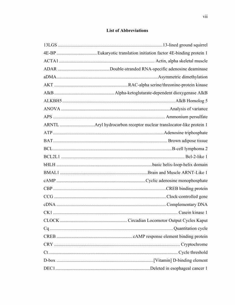
List of Abbreviations
13LGS ........................................................................................... 13-lined ground squirrel
4E-BP ................................... Eukaryotic translation initiation factor 4E-binding protein 1
ACTA1 ................................................................................... Actin, alpha skeletal muscle
ADAR ............................................. Double-stranded RNA-specific adenosine deaminase
aDMA ........................................................................................Asymmetric dimethylation
AKT ................................................................ RAC-alpha serine/threonine-protein kinase
AlkB .................................................... Alpha-ketoglutarate-dependent dioxygenase AlkB
ALKBH5 .................................................................................................. AlkB Homolog 5
ANOVA .............................................................................................. Analysis of variance
APS ................................................................................................. Ammonium persulfate
ARNTL ..............................Aryl hydrocarbon receptor nuclear translocator-like protein 1
ATP ................................................................................................ Adenosine triphosphate
BAT ................................................................................................... Brown adipose tissue
BCL ....................................................................................................... B-cell lymphoma 2
BCL2L1 ........................................................................................................... Bcl-2-like 1
bHLH ...................................................................................basic helix-loop-helix domain
BMAL1 ............................................................................Brain and Muscle ARNT-Like 1
cAMP ............................................................................. Cyclic adenosine monophosphate
CBP ..................................................................................................CREB binding protein
CCG ................................................................................................. Clock-controlled gene
cDNA ............................................................................................... Complementary DNA
CK1 ............................................................................................................ Casein kinase 1
CLOCK .......................................................... Circadian Locomotor Output Cycles Kaput
Cq ........................................................................................................... Quantitation cycle
CREB .................................................................. cAMP response element binding protein
CRY ............................................................................................................. Cryptochrome
Ct ................................................................................................................ Cycle threshold
D-box .................................................................................... [Vitamin] D-binding element
DEC1 .................................................................................. Deleted in esophageal cancer 1

viii
DNA ................................................................................................ Deoxyribonucleic acid
DTT ................................................................................................................ Dithiothreitol
E-box ............................................................................................................. Enhancer box
EA .................................................................................................................. Early arousal
EC ............................................................................................................Euthermic control
ECL ..................................................................................... Enhanced chemiluminescence
EDTA ............................................................................. Ethylenediamine tetra-acetic acid
EGTA ....................... Ethylene glycol-bis(β-aminoethyl ether)-N,N,N′,N′-tetraacetic acid
eIF ............................................................................................ Eukaryotic initiation factor
ELISA ..................................................................... Enzyme-linked immunosorbent assay
EN ................................................................................................................ Entry to torpor
FABP .......................................................................................... Fatty acid binding protein
Fos ..................................................................... FBJ murine osteosarcoma viral oncogene
FTO ...................................................................... Fat mass and obesity-associated protein
GAPDH ......................................................... Glyceraldehyde 3-phosphate dehydrogenase
HDAC .................................................................................................. Histone deacetylase
HRP ................................................................................................ Horseradish peroxidase
HSP ....................................................................................................... Heat shock protein
IA ............................................................................................................. Interbout arousal
IgG ........................................................................................................ Immunoglobulin G
JMJD6 .................................................................... Jumonji-Domain Containing 6 protein
KMT ............................................................................................ Lysine methyltransferase
L:D ................................................................................................. Light-dark photoperiod
LT ....................................................................................................................... Late torpor
m6A .................................................................................................... N6-methyladenosine
m7G ...................................................................................................... 7-Methylguanosine
MAPK ............................................................................. Mitogen-activated protein kinase
METTL ............................................................................................. Methytransferase like
miRNA ............................................................................................................. micro-RNA
MMA ....................................................................................................... Monomethylation
mRNA ....................................................................................................... Messenger RNA

ix
MyoD ....................................................................................... Myogenic differentiation 1
ncRNA ..................................................................................................... Noncoding RNA
NF-κB .............................. Nuclear Factor kappa-light-chain-enhancer of activated B cells
NR1D1 ................................................ Nuclear receptor subfamily 1, group D, member 1
NR1D2 ................................................ Nuclear receptor subfamily 1, group D, member 2
OD ............................................................................................................... Optical density
PAGE .......................................................................... Polyacrylamide gel electrophoresis
PAI-1 .............................................................................. Plasminogen activator inhibitor-1
PAR bZIP .................................... Proline and acidic amino acid-rich basic leucine zipper
PAS ................................................................................................... Per-Arnt-Sim domain
PBS ............................................................................................ Phosphate-buffered Saline
PCR ........................................................................................... Polymerase chain reaction
PDK ................................................................................... Pyruvate dehydrogenase kinase
PER ........................................................................................................................... Period
PMSF .................................................................................. Phenylmethylsulfonyl fluoride
PPAR ................................................................ Peroxisome proliferator-activated receptor
PRMT ........................................................................... Protein arginine methyltransferase
PTL ................................................................................... Pancreatic triacylglycerol lipase
PTM ................................................................................... Post-translational modification
PVDF ........................................................................ Polyvinylidene fluoride [membrane]
RAR ................................................................................................. Retinoic acid receptor
RNA ..........................................................................................................Ribonucleic acid
RORA ............................................................................. RAR Related Orphan Receptor A
RPP .............................................................................. Reversible protein phosphorylation
rRNA ......................................................................................................... Ribosomal RNA
RT ...................................................................................................... Reverse transcription
RT-PCR ..................................................................................... Reverse transcription PCR
RT-qPCR ............................................................... Quantitative reverse transcription PCR
SCN .............................................................................................. Suprachiasmatic nucleus
sDMA .......................................................................................... Symmetric dimethylation
SDS ................................................................................................ Sodium dodecyl sulfate

x
SEM .......................................................................................... Standard error of the mean
Sin3A .................................................... SIN3 Transcription Regulator Family Member A
SMAD ................................................................... Small mothers against decapentaplegic
SSC ..................................................................................................... Saline sodium citrate
Tb ............................................................................................................. Body temperature
TBP ................................................................................................. TATA-binding protein
TBST .......................................................................... Tris-buffered Saline with Tween-20
TGFβ ............................................................................... Transforming growth factor beta
TMB ................................................................................... 3,3′,5,5′-Tetramethylbenzidine
tRNA ............................................................................................................ Transfer-RNA
UCP1 ................................................................................................. Uncoupling protein 1
UTR ...................................................................................................... Untranslated region
WAT ................................................................................................... White adipose tissue
WT1.................................................................................................... Wilms tumor protein
WTAP .......................................................................................... WT1 Associated Protein
YTH .................................................................................................... YT521-B homology
YTHDC ............................................................................ YTH domain containing protein
YTHDF ...................................................................................YTH domain family protein

xi
List of Figures
Figure 1.1 Representative image of the hibernation season in the 13-lined 23
ground squirrel.
Figure 1.2 The circadian transcription/translation feedback loop in mammals. 24
Figure 1.3 Modification reactions for adenosine methylation and mechanisms 25
for affecting mRNA transcript functioning.
Figure 1.4 Arginine methylation by PRMTs. 26
Figure 2.1 Expression of circadian and CCG mRNAs in liver as evaluated by 50
RT-qPCR in control (EC) and late torpor (LT) hibernating 13-lined ground
squirrels.
Figure 2.2 Expression of circadian and CCG mRNAs in white adipose tissue. 51
Figure 2.3 Expression of circadian and CCG mRNAs in skeletal muscle. 52
Figure 2.4 Changes in binding by the CLOCK transcription factor to a DNA- 53
binding element designed for the E-box consensus sequence in liver, white
adipose tissue (WAT) and skeletal muscle in EC and LT animals.
Figure 2.5 Relative total protein levels of core circadian clock proteins in 54
liver of hibernating 13-lined ground squirrels.
Figure 2.6 Relative total protein levels of core circadian clock proteins in 55
white adipose tissue of hibernating 13-lined ground squirrels.
Figure 2.7 Relative total protein levels of core circadian clock proteins in 56
skeletal muscle of hibernating 13-lined ground squirrels.
Figure 3.1 Relative levels of N6-methyladenosine (m6A) in total RNA 83
extracts (global) from liver, WAT and skeletal muscle tissues, comparing
ground squirrels in euthermic control (EC) and late torpor (LT) conditions.
Figure 3.2 Relative total protein levels of m6A writer proteins in liver, 84
white adipose (WAT) and skeletal muscle of hibernating 13-lined ground
squirrels.

xii
Figure 3.3 Relative total protein levels of m6A eraser proteins in 85
three tissues of hibernating 13-lined ground squirrels.
Figure 3.4 Relative levels of m6A demethylase activity over 4 time points 86
of the torpor-arousal cycle in hibernating 13-lined ground squirrels.
Figure 3.5 Relative total protein levels of m6A reader proteins in 87
three tissues of hibernating 13-lined ground squirrels.
Figure 3.6 Relative levels of eukaryotic translation initiation factor 3, 88
subunit D (eIF3d) over the torpor-arousal cycle in hibernating 13-lined
ground squirrels.
Figure 4.1 Relative Type II-specific PRMT methyltransferase activity 109
in total soluble protein extracts of liver, white adipose tissue, and skeletal
muscle at selected time points of the torpor‐arousal cycle.
Figure 4.2 Relative protein abundance of PRMT enzymes in liver over 110
the torpor‐arousal cycle.
Figure 4.3 Relative protein abundance of PRMT enzymes in white adipose 111
tissue over the torpor‐arousal cycle.
Figure 4.4 Relative protein abundance of PRMT enzymes in skeletal muscle 112
over the torpor‐arousal cycle.
Figure 4.5 Relative amounts of histone H3 showing asymmetric di- 113
methylated R2 (H3R2me2a) and symmetric dimethylated R8 (H3R8me2s),
as well as histone H4 showing symmetric dimethylated R3 (H4R3me2s) in
liver, WAT, and skeletal muscle over the torpor‐arousal cycle.

xiii
List of Appendices
Appendix A: Circadian controlled gene primer sequences .................................................................... 130
Appendix B: Western Blot Considerations ............................................................................................ 131
Appendix C: Communications at scientific meetings ............................................................................ 132
Appendix D: Publication List................................................................................................................. 134

1
Chapter 1
Chapter 1 General Introduction
General Introduction

2
Introduction
Time is a central feature of anyone's life, and the same may be said for almost any
organism. Time or more specifically, the earth’s rotation on its axis and around the sun,
and the resulting changes in daylight, temperature, and seasons that this causes, gives rise
to an interesting feature of animal physiology and behaviour – the occurrence of both
circadian and circannual rhythms that regulate diverse aspects of metabolism on a daily
or seasonal basis. The circadian rhythm (from the Latin circa, about; diem, day) is the
name given to physiological rhythms that occur on a daily basis, not just sleep-wake
cycles but also body temperature (Tb), heart rate or blood pressure, and cognitive
abilities, to name a few. Even childbirth or cardiac arrest are more likely to occur at
certain times of day than others in humans. Indeed, most of our body's physiological or
biochemical parameters show rhythmic variability over the 24-hour day, and this fact is
taught to young medical students early on in their career, indicating its real importance
(Kreitzman and Foster 2011). It is important to note that the daily physiological rhythms
that we experience, occur in anticipation of, and not as a result of circadian rhythm
changes, i.e., shifting one's circadian rhythm results in modulated physiological cycles
that require a certain catch-up period giving rise to the colloquially-termed 'jet-lag.' The
endogenicity of circadian rhythms in daily activity cycles was first commented on by
Curt Richter in 1922, although Colin Pittendriegh is often credited with being the first to
spur the systematic study of biological rhythm generation (Pittendrigh 1993). In the
following years, chronobiological research explored biological rhythms in a plethora of
animals showing that (i) many biological processes show close to 24-hour rhythms, (ii)
these rhythms will persist under constant conditions, (iii) it is necessary for rhythms to

3
synchronize with the astronomical day [entrainment], and (iv) unlike many biochemical
reactions, environmental changes in temperature do not noticeably affect the 'speed'
[period] of biological rhythms.
In mammals, the control of many cyclical behaviours is centered in the
suprachiasmatic nucleus (SCN), a small area of the anterior hypothalamus which receives
information about the environment from sensory receptors, especially photoreceptors
within the retina, and entrains [synchronizes] activity rhythms within single SCN neurons
to the solar day. Efferent connections from the SCN influence endocrine (e.g., melatonin
and arginine vasopressin) and neuronal signaling, ultimately synchronizing peripheral
oscillators located throughout the body in most visceral organs and in several separate
locations within the brain (Brown and Azzi 2013). The generation of behavioural
rhythms may be exemplified by the roles of neuropeptides or humoral factors released by
SCN efferents in regulating behaviour. For instance, transforming growth factor-alpha
(TGF-alpha) acts on hypothalamic receptors to inhibit locomotor activity in response to
light in nocturnal species, while prokineticin-2 (PK2) is regulated by circadian clock-
controlled transcription and acts on receptors during the day to also decrease locomotor
activity (Kramer et al. 2001; Cheng et al. 2002). Through separate diffusible factors
regulating behaviours synergistically and being responsive to different environmental
inputs, integration of information about physiological state is allowed.
Mammalian Hibernation
As winter approaches some small mammals display a remarkable phenotypic
shift, lowering their metabolic rate and entering a state of torpor to survive seasonal
stresses including extreme cold and limited food availability. This altered physiological

4
state, known as hibernation, is displayed annually by many animals that inhabit extreme
climates, particularly at high latitudes or altitudes. The involvement of circadian controls
in regulating adaptations to environmental stresses, such as those encountered by small
mammals that overwinter in harsh climates, has been studied in a variety of systems and
species making it clear that core molecular clock machinery must also be involved in the
cellular responses to hibernation. Many animals show pronounced circannual rhythms
that govern their behaviour and metabolism across the seasons of the year, influencing or
defining the timing of major events including reproduction, migration, hibernation, etc.
Hibernation (Figure 1.1) is exhibited by diverse species ranging from temperate
zones to the high Arctic in the Northern Hemisphere, and conversely in most southerly
parts of Australia and Africa. Through the concerted effects of major physiological and
biochemical changes, many small mammals can sink into a profound state of torpor,
allowing their body temperature to fall to near ambient, and survive solely on stored body
fats for days or weeks at a time. It has been estimated that hibernators can potentially
conserve about 90% of the energy that they would normally require to remain active and
euthermic all winter (Wang and Wolowyk 1988). Therefore, hibernation serves as a
drastic mechanism for surviving harsh winter conditions in small mammals.
Molecular regulation of hibernation
For the thirteen-lined ground squirrel (13LGS), Ictidomys tridecemlineatus,
preparation for hibernation begins when animals enter a phase of hyperphagia in early
August during which they accumulate enough fat to increase body weight by around
50%. Pre-hibernation mass gain is accomplished through increased food consumption
during the late summer and fall seasons, and is linked to increased circulating insulin

5
concentrations following its release from the pancreas (Boyer et al., 1993; Boyer and
Barnes, 1999; Florant et al., 1990). Insulin then binds membrane receptors initiating a
signaling cascade which ultimately promotes glycogen synthesis and deposition within
the liver and muscle, as well as lipid synthesis and triglyceride production. During entry
into torpid states, insulin serves to increase the activity of lipogenic enzymes and proteins
that deposit fat into white adipose tissue (WAT) stores and build up metabolic fuel stores
(Woods and Porte, 1978). For example, protein levels and enzyme activities of
lipoprotein lipase, fatty acid synthase, and diacylglycerol acetyltransferase are all
elevated during ground squirrel seasonal mass gain (Mostafa et al., 1993; Wang et al.,
1997). In response to increased adipose stores and increased WAT cell size (Dark, 2005;
Otis et al., 2011), digestive satiety signals (i.e., leptin) are released by adipocytes. While
leptin typically has the effect of suppressing appetite and enhancing lipid oxidation,
interestingly, its anorexigenic effects are not experienced by hibernators during the
seasonal pre-hibernation mass gain (Florant and Healy, 2012; Healy and Florant, 2012).
Therefore, while levels of serum insulin increase during the fall, typical of any
hyperphagic state, a resistance to satiety signals is also experienced in hibernators that
ultimately allows for increases in WAT mass that give the 13LGS adequate fuel supplies
through lipid catabolism to last through the hibernation fast (Schwartz et al., 2015; Wu et
al., 2013).
During the shift to a hypometabolic state, the majority of carbohydrate
metabolism is suspended during hibernation and instead lipolytic enzyme activity is
upregulated in order to ensure that a majority of the hibernator’s energy demands are met
by lipid oxidation derived from built-up adipose stores (Hittel and Storey, 2001; Storey

6
and Storey, 2010). Similarly, increased levels of pancreatic triacylglycerol lipase (PTL)
stimulate lipolysis via the breakdown of circulating adiposomes. Levels of fatty acid
binding protein, fatty acid transporter, and enzymes involved in ketone production such
as hydroxymethylglutaryl-CoA synthase, are also increased and support the switch to
increased reliance on the beta-oxidation pathway (Epperson et al., 2010a). Similarly,
levels of pyruvate dehydrogenase kinase isozyme 4, which inhibits glycolysis through
reversible protein phosphorylation (RPP) of pyruvate dehydrogenase, are upregulated
within muscle, heart, liver and WAT tissues (Andrews et al., 1998).
Due to increased fatty acid catabolism, and concurrent decreases in glycolysis,
blood glucose levels fall to an annual minimum during winter (Buck and Barnes, 1999),
in turn causing reductions in insulin levels (Bauman et al., 1987; Woods and Porte,
1978). Interestingly, increased levels of glucagon, expected to occur in the face of
lowered circulating glucose levels, are not seen during mammalian hibernation (Bauman
et al., 1987; Hoo-Paris et al., 1985); however, a shift in the plasma glucagon to insulin
ratio does occur which favors the effects of glucagon and in turn, poises catabolism
towards the breakdown of glycogen through the activation of WAT lipolytic enzymes
(i.e., hormone sensitive lipase), and inhibition of pyruvate kinase (Dark, 2005; Wilson et
al., 1992). In fact, the shift towards fatty acid catabolism is so extensive that even the
breakdown of glycogen is inhibited following decreases in activating-RPP of glycogen
phosphorylase (Storey, 1987; 1997). Clearly, multiple systems ensure that during torpor
in the 13LGS, glycolysis is suppressed while metabolic fuel requirements are fulfilled
almost entirely via lipid oxidation.

7
Small mammalian hibernator species descend into torpor in either a facultative or
obligate nature. Hibernation occurs in several groups of mammals, including elephant
shrews, rodents, bats, several marsupials and even some primates, however the most
commonly used model hibernators are the ground squirrel family. The thirteen-lined
ground squirrel, Ictidomys tridecemlineatus, is an obligate hibernator on an annual cycle,
displaying the traits mentioned above each year. Obligate hibernators, like the 13-lined
ground squirrel (13LGS) and other species in the Sciuridae family enter and arouse from
hibernation on an annual cycle regardless of environmental cues (Hut et al. 2014;
Pengelley and Asmundson 1969; Kondo et al. 2006).
Torpor and arousals
During torpor in the 13LGS, characteristics of mammalian life are significantly
decreased for days at a time – e.g., for animals hibernating at a Tb near 0°C (as compared
with Tb of 37°C) breathing rates are reduced from 100-200 breaths/min to 4-6
breaths/min, and heart rates drop from 200-300 beats/min to 3-5 beats/min (Boyer and
Barnes, 1999; Nedergaard et al., 1990; Refinetti, 1996; Storey, 2010). These
physiological changes enable the animal’s metabolic rate to plummet often to just 2-4%
of resting summer values (Carey et al., 2003).
Decreased insulin levels further inhibit glucose uptake in peripheral tissues via
decreased stimulation of glucose transporter type 4 (Tessier and Storey, 2010; Wu et al.,
2013). Glucose uptake is further inhibited by decreased phosphoinositide-3-kinase (PI3-
K) mediated activation of the serine/threonine kinase Akt, resulting from decreased
insulin binding to membrane receptors, which causes a significant regulatory shift within
the liver and skeletal muscle tissues (Abnous et al., 2008; 2010). Furthermore, levels of

8
downstream targets of Akt, including mammalian target of rapamycin (mTOR) and
tuberin (TSC2), are not changed during torpor but their activated forms are significantly
suppressed during torpor – lowering rates of gene transcription and protein synthesis (Wu
and Storey, 2012a).
Decreased rates of gene transcription and protein translation are common findings
in studies done in a variety of hibernating mammals (Morin and Storey, 2009; Storey,
2003; Storey et al., 2010), including 13LGS (Frerichs et al., 1998; Morin and Storey,
2006; Tessier and Storey, 2014). Protein synthesis is a major consumer of a euthermic
mammal’s energy expenditure, and its downregulation during hibernation aids the
conservation of fuel reserves for the most essential cellular tasks (Heldmaier et al., 2004).
This suppression has been shown to occur in almost all 13LGS tissues during torpor,
including brain, liver, kidney, brown adipose tissue (BAT) and digestive organs (Biggar
and Storey, 2014; Hittel and Storey, 2002). Decreases in gene expression and protein
synthesis are, however, not global over the entire genome but rather are specific to genes
that are nonessential for the hibernator’s switch between euthermia and torpor or the
maintenance of either physiological state (Epperson et al., 2010a; 2010b; O'Hara et al.,
1999). As an example, within the heart of hibernating 13LGS, the transcription factor
myocyte enhancer factor 2 (MEF2) is upregulated and activated through RPP leading to
increased levels of the cardioprotective proteins desmin and myomesin (Tessier and
Storey, 2012). Similarly, changes in RPP of ribosomal initiation and elongation factors
(Frerichs et al., 1998; van Breukelen and Martin, 2001), as well as proteins that lower
messenger RNA (mRNA) turnover (i.e., poly(A) binding protein) and assist in stabilizing
and folding existing proteins (i.e., heat-shock proteins) contribute to decreasing cellular

9
energy usage during hibernation (Fahlman et al., 2000; Knight et al., 2000; Wu et al.,
2015). During hibernation, regulation over genome suppression is seen in combination
with upregulation of certain genes whose products function in either cellular metabolism
or preservation, and therefore an intricate level of selection over biological pathways is
clearly required.
Intermittent arousals from torpor occur regularly over the hibernation season
(Fons et al., 1997; Lovegrove et al., 1999; Mzilikazi et al., 2002; Wang, 1979) and, as
such, mammals must have a way to reversibly return their bodies to euthermic Tb values
and facilitate the necessary increases in cellular metabolism that will increase Tb above
ambient temperature (Ta). Increased reliance on carbohydrate oxidation is seen during the
brief interbout arousal periods and disruption of rewarming is seen when animals are
given an inhibitor of glycolysis but not when given an inhibitor of lipid oxidation (Dark
and Miller, 1997; Karpovich et al., 2009). Furthermore, levels of mitochondrial
respiration are significantly increased as are activity measurements of succinate
dehydrogenase during arousal from torpor, as compared to torpid animals, when
measured in either liver or muscle (Armstrong and Staples, 2010; Brown et al., 2013).
Increased cellular respiration during arousal shows that the favourability of lipid
oxidation over carbohydrate oxidation is limited only to the torpid stages of hibernation.
Rewarming from torpor and required metabolism during interbout arousals account for
more than half of the use of a hibernator’s entire winter-time fuel store (French, 1985;
Wang, 1979).
As would be expected following a sudden reversal of hypometabolism,
mammalian hibernators must also find ways to protect tissues in the face of enormous

10
increases in oxygen consumption and free-radical generation, as well as increased
carbohydrate consumption following uncoupled cellular respiration supporting
thermogenesis in BAT and shivering thermogenesis in skeletal muscle (Carey et al.,
2003; Kloner et al., 1998; Meyer et al., 2012). Increased levels of antioxidant defences
are observed during arousal from hibernation including heme oxygenase 1, and
associated effector proteins including nuclear factor (erythroid-derived 2)-like 2 (Nrf2)
and MAF BZIP Transcription Factor G (MafG), in the liver, kidney, brain and heart of
aroused 13LGSs (Ni and Storey, 2010). Antioxidant defences stemming from the
transcriptional activity of nuclear factor kappa-light-chain-enhancer of activated B cells
(NF-κB) are also significantly increased in skeletal muscle tissue from 13LGSs
transitioning to the arousal phase of the torpor/arousal cycle (Allan and Storey, 2012;
Morin et al., 2008; Vucetic et al., 2013). In a similar fashion, anti-apoptotic protein
expression is increased within a variety of tissues throughout the torpor cycle, as
compared to summer euthermic animals (Fleck and Carey, 2005; Logan et al., 2016a;
Rouble et al., 2013), as is heat shock protein expression (Feder and Hofmann, 1999;
Storey and Storey, 2011; Vermillion et al., 2015).
Thanks to multiple cytoprotective mechanisms that are upregulated during
arousal, 13LGSs may survive multiple bouts of torpor and subsequent arousal via
protection from the harmful consequences of decreased tissue use (i.e., muscle atrophy),
free-radical generation, as well as shifts in metabolic fuel or energy requirements and
metabolite buildup or depletion. Since mammalian hibernators have these protective
mechanisms, their abilities to defend themselves from cellular stresses that are atypical of
normal mammalian life are of great interest to human medical research – especially with

11
regards to obesity and diabetes (Kirchner et al., 2013; Sookoian and Pirola, 2013; Wu et
al., 2013), aging and longevity (Storey and Storey, 2004a; Wu and Storey, 2016) and
neurodegenerative damage or diseases (Drew et al., 2007; Logan et al., 2016b; Wood,
2015).
Circadian rhythmicity during hibernation
In order for the 13LGS to progress through the phases of the torpor/arousal cycle,
control over a variety of cellular processes is required in the form of genomic control
over transcription, protein synthesis, as well as covalent modification of metabolic
enzymes and structural proteins (as previously described).
Contemporary analyses of the persistence of circadian rhythms during hibernation
have been limited. Circadian rhythmicity is controlled by a master circadian clock formed
by neurons within the suprachiasmatic nucleus (SCN) of the anterior hypothalamus.
Interestingly, cellular activity within the SCN is increased in torpid animals compared to
euthermic animals and similarly the area shows increased glucose uptake during
hibernation in contrast to the majority of brain areas (Kilduff et al. 1990; Bratincsák et al.
2007). An early hypothesis suggested that torpor duration results from normal circadian
functioning under conditions that slow its progression including reduced ambient and
body temperatures. The idea that low Tb and impaired temperature compensation of the
circadian system may extend the cycle experienced by hibernating mammals may be
compelling, but it is not substantiated by persistent, temperature-compensated rhythms
within the SCN in hibernators even though torpor bout duration shifts with reduced Tb
(Twente et al. 1977; Grahn et al. 1994; Malan 2010). Studies of the regulation of torpor
bout duration by the SCN showed that fully-lesioned and arrhythmic ground squirrels
12
would present torpor-arousal cycles with different durations, and that changing the
rhythm within the SCN does not change the duration of torpor-bouts (Ruby et al. 1996;
Oklejewicz et al. 2001). Finally, the persistence of circadian rhythms during prolonged
torpor is indicated in species whose arousals remain entrained to a time of day and that
periodically emerge from their hibernaculum, such as pygmy possums (Körtner et al.
1998; Turner and Geiser 2017) and foraging bats (Park et al. 2000; Hope and Jones
2013), but not in a number of rodent species whose arousals commence at seemingly
random times of day (Oklejewicz et al. 2001; Hut et al. 2002; Russell et al. 2010). It may
be that opportunistic hibernators, who take advantage of favorable conditions during the
hibernation season have a greater need for entrained arousals. By contrast, most rodent
hibernators typically dwell in deep burrows and are not exposed to astronomical and
environmental cues, and generally don’t eat over the winter season; hence, they have lost
this sort of rhythmicity. The insignificance of daily cues among most rodents that
hibernate in deep burrows is further indicated by the lack of rhythmic melatonin levels or
reduced pineal size during hibernation (Stanton et al. 1986; Florant et al. 1984; Vanĕcek
et al. 1984), a lack of rhythmic clock gene transcription in the SCNs (as seen in European
hamsters, Cricetus cricetus L. and Arctic ground squirrels, Urocitellus parryii) (Revel et
al. 2007; Ikeno et al. 2017), and the absence of circadian rhythms of Tb in Arctic ground
squirrels (Williams, Radonich, et al. 2017; Williams, Barnes, et al. 2017). These results
clearly show that the circadian clock is inhibited during torpor in these rodent
hibernators, but the question of whether these adaptation changes extend to the peripheral
oscillators is still unanswered.

13
Molecular regulation of the circadian clock
In mammals, the molecular circadian clock is based around a transcription factor
heterodimer complex: Brain and Muscle ARNT-Like 1 (BMAL1) and Circadian
Locomotor Output Cycles Kaput (CLOCK), as shown in Figure 1.2. BMAL1 and
CLOCK are basic helix-loop-helix-PAS domain containing transcription factors which
can bind E-box regulatory elements to stimulate transcription. Importantly, the canonical
downstream targets of the BMAL1/CLOCK complex are Per and Cry genes that encode
the inhibitory proteins PER1, PER2, PER3, CRY1 and CRY2, and can block
BMAL1/CLOCK from activating transcription (i.e., of Per or Cry) (Gekakis et al. 1998;
Shearman et al. 2000). Transcriptional activation by BMAL1/CLOCK occurs during the
daytime, whereas PER and CRY proteins are transcribed and translated so that their
expression peaks in the evening and night hours. Translocation of PER or CRY proteins
occurs via their interaction with one another and a nuclear import receptor via their
nuclear localization signals (Lee et al. 2015; Miyazaki et al. 2001), and is stimulated by
their accumulation in the cytoplasm as the day progresses. Cytoplasmic accumulation of
PER protein is the rate-limiting step of circadian clock ‘ticking,’ before translocation into
the nucleus as part of the PER/CRY complex, where it may block BMAL1/CLOCK-
controlled gene activation after 12 or so hours (Lee et al. 2001; Vielhaber et al. 2001).
The degradation of PER and CRY proteins is mediated by their phosphorylation via
serine/threonine kinases casein kinase 1δ (CK1δ) or CK1, which activate binding by E3
ubiquitin ligase complexes and targeting the proteins for proteasomal degradation either
within the cytoplasm or nucleus (Gallego and Virshup 2007). The half-lives of PER and
CRY proteins are short in the presence of phosphorylation (Eide et al. 2005) and so

14
transcriptional repression by the nuclear PER/CRY complex is soon relieved allowing
BMAL1/CLOCK to bind and activate transcription, beginning the cycle anew.
Circadian activation of gene transcription is, of course, not limited to control over
its own feedback repression. The BMAL1/CLOCK complex also drives transcription of
two nuclear receptor families. The Rev-ErbA nuclear receptors (Rev-ErbA and Rev-
ErbA, encoded by NR1D1 and NR1D2, respectively) are activated by BMAL1/CLOCK
binding to an E-box element in the promotor of Rev-ErbA genes, allowing for a second
transcriptional feedback loop. By acting on RevDR2 and retinoic acid-related orphan
receptor (ROR)-binding elements (ROREs) in the promoter sequences of the CLOCK and
BMAL1 genes, Rev-ErbA receptor proteins can repress transcription of CLOCK and
BMAL1 when transcriptional activation of NR1D1 or NR1D2 [by BMAL1/CLOCK] is
highest and the receptors accumulate in the nucleus (Preitner et al. 2002). A third class of
proteins belonging to the BMAL1/CLOCK complex are known simply as clock-
controlled genes (CCGs) and may have wide-reaching and numerous effects throughout
the cell. The three best known of members of this class are the proline and acidic-rich
(PAR) domain transcription factors: D-box binding protein (DBP); thryotroph-embryonic
factor (TEF); and hepatic leukemia factor (HLF); all of which interact with D-box
elements in the promotor regions of target genes (Mitsui et al. 2001). By acting on D-box
elements within gene promoter regions these transcription factors can drive the output of
multiple other CCGs and also serve somewhat as tissue-specific factors of circadian
output. For instance, the RAR-related orphan receptor (ROR) transcription factor genes
contain a D-box element in their promoter regions. The ROR transcription factors, ROR
and ROR, also act on ROREs in gene promoter regions and serve as a circadian output

15
oscillator by controlling the transcriptional activation of CCGs, including BMAL1 (Ueda
et al. 2005; J. Lee et al. 2016). By acting on the promoter regions of several transcription
factors, the BMAL1/CLOCK complex clearly controls the rhythmic output of numerous
CCGs and integrates tissue-specificity into the regulation of CCGs.
Interactions between CCGs and core circadian clock members within gene
promoter or enhancer regions produce phases of peak transcriptional activation for CCGs
depending on the DNA binding elements present within the regulatory region of each
CCG. For instance, binding of the BMAL1/CLOCK complex to E-box elements is anti-
phasic (i.e., separated by 12 hours) to the repressor PER/CRY complex binding whereas
DBP binding to D-box elements is directly preceded by the binding of a repressor
protein, nuclear-factor interleukin-3 (NFIL-3) (Panda et al. 2002; Rey et al. 2011). The
combination of DNA binding elements within genic regions further allows for co-
operative or interfering interactions between CCGs, serving to extend or restrict
transcriptional activation, respectively.
mRNA Adenosine Methylation
In comparison to the circadian regulation of, and by, methylation of DNA or
proteins, RNA methylation is nearly uncharted territory. The most studied RNA
methylation modification, aside from the structural m7G-cap placed on nascent mRNA
molecules, is modification of N6 on adenosine residues (m6A). Introduced through the
use of DEAE-cellulose (borate) chromatography (Desrosiers et al. 1974), RNA m6A
modifications (Figure 1.3) have been shown to affect nearly all aspects of the mRNA life-
cycle including stability, processing and splicing, translational activity and subcelllar
localization (Meyer and Jaffrey 2014). Mapping of m6A to specific RNA molecules

16
within the transcriptome has further enhanced our understanding of the modification’s
dynamic and regulatory nature. The majority of m6A modifications placed upon mRNA
are deposited within the untranslated regions (UTR) adjacent to the coding region (Meyer
et al. 2015), whereas m6A located within coding regions makes up only about 35% of all
m6A modifications (Mao et al. 2019). Moreover, m6A-mapping studies, accomplished
through m6A-RNA immunoprecipitation followed by sequencing the resulting
transcripts, has revealed that m6A is enriched selectively within certain transcripts, and is
typically constitutive across the transcriptome within the UTR and near stop codons
(Meyer et al. 2012; Dominissini et al. 2012; Schwartz et al. 2014). This modification can
also be a dynamic modification following cellular stressors or changes in the cellular
environment (Ke et al. 2017; Geula et al. 2015).
It is important to understand the control of mRNA methylation that occurs in
response to environmental changes since it can be said to have regulatory influences over
all the genetic output of a cell. For example, novel work by Fustin et al. (2013)
investigated the ratio of AdoMet to its inhibitory by-product and found that circadian
cycle duration was altered in both cultured cells and in mice upon manipulation of global
methylation using an inhibitor, 3-deazaadenosine (DAA). Treatment with 3-DAA was
associated with reduced m6A content within exons of Per1, Per2, Per3, Dbp and Nr1d1
and nuclear retention of PER2 mRNA due to a significant delay in its half-life. To
validate these results as due to downregulated mRNA methylation, the authors used
METTL3 siRNA to reduce catalytic subunit activity in the mRNA methylation complex
that, in turn, reduced the number of m6A modifications on circadian transcripts.
Similarly, overexpression of METTL3 protein accelerated the nuclear exit of Per2 and

17
BMAL1 mRNA. These results provide a fine demonstration of the regulatory power
possessed by the mRNA methyltransferase complex over mRNA processing that is
relevant not to just circadian rhythms but to countless other cellular events (Hastings
2013). It is unsurprising that nearly all epigenetic mechanisms, including
[epitranscriptomic] mRNA methylation, show regulatory crosstalk with the circadian
clock’s functioning.
Protein Arginine Methylation
Methylation of arginine residues on histones has been shown to play a role in the
remodelling of chromatin and regulate protein dynamics and functioning in several
cellular pathways, including the circadian clock. Arginine is well positioned to interact
with other biological molecules, since it has five potential hydrogen bond donors and has
been shown to form predictable, defined, and frequent interactions with RNA, DNA and
proteins (Luscombe et al. 2001; Najbauer et al. 1993; Hughes and Waters 2006).
Methylation of arginine (Figure 1.4) may also change the likelihood of a protein binding
to certain interacting partners by increasing the protein’s affinity for certain molecular
features including aromatic residues (Sprangers et al. 2003). Since methylation replaces
available hydrogen bond donor atoms, it can alter the interactions of arginine residues
with various binding partners (Pahlich et al. 2006) and, indeed, the discovery of this
modification has been shown to be increasingly relevant to cellular dynamics.
Arginine methylation of histone proteins is directly modulated by the circadian
clock. Na et al. (2012) showed that the type II PRMT5 methyltransferase enzyme is a
binding partner of CRY1 and that this interaction is rhythmic and dependent on CRY1
accumulation. The authors then showed that, upon binding the CLOCK/BMAL1 complex

18
while bound to CRY1, levels of arginine-3 on histone H4 (H4R3) were increased in a
rhythmic fashion (i.e., correlated to rhythmic circadian output), whereas histone
acetylation was reduced. Finally, the authors showed that these mechanisms were absent
in synchronized cells transfected with PRMT5 shRNA. Given the far-reaching effects that
PRMT enzymes may have that indirectly affect circadian regulation, as well as the direct
mechanism detailed here, it is clear these enzymes are important for the regulation and
temporal organization of chromatin modifications and architecture.
Objectives and Hypothesis
The research reported in this thesis is an investigation of the molecular circadian
clock and its regulators in the 13LGS in the hopes of understanding their relevance to
torpor-arousal cycles in an obligate hibernator as well as post-translational and post-
transcriptional modifications that contribute to the functioning of circadian rhythmicity at
the molecular level. Through characterization of the circadian pathway’s expression and
post-transcriptional and post-translational regulators during hibernation, an increased
understanding of circadian clock functioning in hibernation as well the discovery of
tissue-specific adaptations at such times is achieved.
Mammalian hibernators are subject to substantial genomic regulation as they
transition to and from the torpid state, therefore my Ph.D. thesis explores three avenues
of transcriptional and translational control. The core circadian clock, RNA adenosine
methylation and protein arginine methylation are each able to influence either RNA and
protein processing, or both, in order to help a hibernator adapt to and overcome the
stresses of prolonged cycles of torpor and arousal. This research was achieved through
characterization of the molecular responses to torpor-arousal cycles by both the circadian

19
clock (Chapter 2) and the enzymes controlling some of the post-transcriptional (Chapter
3) or post-translational modifications (Chapter 4) that affect the activity of the canonical
circadian transcription-translation feedback loop. These studies focused on liver, WAT
and skeletal muscle tissues of 13LGS and involved analysis of ground squirrels sampled
at five different points on the torpor-arousal time course, allowing investigation of
changes that are made while entering torpor as well as during arousal from torpor. Liver
and WAT were chosen for their established roles in lipid metabolism and the
maintenance of homeostasis during hibernation whereas skeletal muscle was chosen for
its almost complete cessation of activity during torpor and for its importance in
rewarming the animal via shivering thermogenesis during interbout arousals. Each
chapter deepens our understanding of the control required for successful hibernation
using novel approaches.
Objective 1: Expression and Activity of the Molecular Circadian Clock
As mentioned, many rhythmic behaviors are inhibited during torpor as the animal’s
metabolic rate falls to less than 10% of resting euthermic rate and only the most essential
cellular tasks are sustained. This likely involves control over and from the core circadian
clock within peripheral tissues. Previous studies completed in other small hibernating
mammals have evaluated the existence of rhythmic physiological responses as well as
responses by the neural circadian clock to hibernation and determined that most outward
appearance of circadian rhythmicity is diminished or absent (Körtner and Geiser 2000).
This thesis greatly extends the analysis of the circadian clock by focusing on the
expression and activity of core clock components and CCGs involved in regulating
homeostatic mechanisms within peripheral cells/tissues of the body.

20
Hypothesis 1: The molecular circadian clock responds to global metabolic rate
depression during torpor cycles by contributing to decreased cellular transcriptional and
translational activity.
Chapter 2 tests this hypothesis by assessing changes in the relative transcript and
protein levels of canonical molecular circadian clock members as well as their protein
regulators to examine the tissue-specific responses and implications of their roles during
hibernation. Levels of 10 circadian clock transcripts were analyzed using qRT-PCR to
examine the differential transcription induced by hibernation. Secondly, nuclear
transcription-factor binding activity to circadian-clock response elements (E-box) was
analyzed via transcription-factor ELISAs to determine if the core transcription factor
CLOCK shows differential DNA binding during torpor. Western blot analysis of proteins
involved in regulating the circadian clock were completed for BMAL1, CRY2, PER2,
and CK1δ/.
Objective 2: Response of mRNA methyltransferases to suppressed transcriptional
activity
The mechanisms controlling post-translational modifications during hibernation
are unknown to date. On the other hand, it is known that during hibernation a complex
selection of gene activity is required since selected pathways are enhanced while the
majority of genomic activity is depressed. This clearly necessitates a role for molecular
controls that are able to shut down or activate gene products that are produced and
preserved until required or expressed at drastically lower amounts. The METTL RNA
methyltransferases (METTL3, METT14), as well as their regulator WTAP, control the
deposition of m6A on RNA molecules whereas eraser proteins (FTO and ALKBH5) are

21
in charge of their removal and influence m6A dynamics. Furthermore, reader proteins
(YTHDF1/3, YTHDC2, eIF3d) bind to and exert control over the processing and stability
of RNA molecules, and mRNA in particular.
Hypothesis 2: Adenosine methylation provides a means of reducing
transcriptional rates through modification of mRNA and other types of RNA during
torpor. Relative levels of proteins in control of depositing or removing adenosine
methylation, as well as m6A reader proteins, accommodate increased m6A dynamics
across cycles of torpor and arousal.
Chapter 3 tests this hypothesis by assessing total methyl-adenosine levels using an
m6A ELISA procedure to assess global differences in RNA and mRNA methylation
during torpor. Protein levels of mRNA methyltransferases and levels of m6A binding
partners, were analyzed using Western blotting or ELISAs in search of responses to, or
preceding, the inhibition of transcriptional and translational activity during torpor and
arousal.
Objective 3: Arginine methylation and its role in chromatin remodeling
Arginine methyltransferase proteins such as PRMT1-7, play crucial roles in the
deposition of PTMs and epigenetic marks in all tissues and in response to almost all
cellular environmental changes. Importantly, these enzymes are able to methylate
arginine residues on histones H3 and H4 which can influence global levels of
transcription, genomic reorganization and the recruitment of transcription factors or
structural proteins to the genome-chromatin assembly. Notably, dimethylation of R2 and
R8 on histone H3, or R3 on histone H4 is repressive over genomic activity. Moreover, the
dimethylation abilities of PRMT enzymes allow their categorization (Figure 1. 4) into
22
those enzymes that are able to methylate already modified guanidine groups (Type I)
from those that are only able to methylate unmodified guanidine groups on arginine
(Type II).
Hypothesis 3: Arginine methylation provides a means of reducing transcriptional
rates through modification of histone proteins and is involved in modifying the activity of
non-histone proteins during torpor. These responses are then reversed during arousal.
Chapter 4 tests this hypothesis by performing Western blot measurements of
arginine methyltransferase enzymes to provide an indication of tissue reliance on arginine
methylation for manipulating chromatin binding site accessibility or protein activity
during hibernation. Furthermore, antibodies specific for dimethyl-arginine histone
residues indicative of this PTM’s role in manipulating chromatin accessibility were used
in Western blotting to determine if the aforementioned residues are differentially
regulated across torpor-arousal cycles. The activity levels of PRMT enzymes was assayed
using an enzyme activity ELISA specific to Type-II enzymes, including PRMT5 and 7.
23
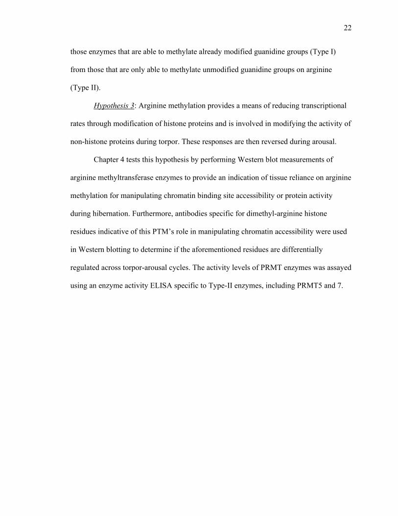
Figure 1.1: A representative image of the hibernation season of 13-lined ground squirrels
displaying the relationship between hibernating body temperature and time. The
hibernation season typically begins in late September with animals entering shallow
torpor periods known as ‘test-drops’. Torpor bouts lengthen over time as body
temperature drops to lower levels and begin to shorten again as the hibernation season
ends, seemingly without external cues in laboratory environments (animals held at
constant 5°C in a cold room). A comparable pattern occurs in nature, associated with
progressive lowering of ambient temperature (Ta) as autumn-winter progresses and a
shortening of torpor bouts with spring warming. Each cycle is characterized by specific
phases: EC, euthermic in the cold room (control animals); EN, entrance into torpor
(approximately 12h); LT, late torpor (>5 consecutive days in a torpor bout); EA, early
arousal (duration ~3h); and IA, interbout arousal (within 24h of returning to euthermic
physiological conditions). Image modified from Tessier et al. (2016).

24
Figure 1.2: The circadian transcription/translation feedback loop in mammals is focused
around the transcriptional activity of the BMAL1/CLOCK complex. This core
transcription factor complex acts on the E-box element of the Period and Cryptochrome
genes, whose products inhibit the transcription by BMAL1 and CLOCK. Genes for the
inhibitor proteins Rev-ErbA, Rev-ErbA and the activator protein ROR are also
regulated by an E-box, creating a secondary regulatory loop controlling the transcription
of BMAL1. Period and Cryptochrome degradation is mediated by their interaction with
Casein Kinases, which phosphorylate both proteins (red triangles) and lead to shunting
them into E3 ubiquitin ligase pathways. Interlocked transcriptional feedback loops whose
products inhibit their activators produces a cyclic pattern of transcription and translation.
In this way transcriptional regulation of downstream and tissue-specific rhythmic clock-
controlled genes (CCGs) is possible. Figure modified from Buhr et al. (2013).
BMAL1 CLOCK
Rev-Erb’sE-box
RORɑE-box
Rev-
Erbɑ
RORɑ
E-boxPER1,2,3
E-boxCRY1,2
PER2 CRY2
RREBMAL1
CRY2
PER2
P
P
PROTEASOME
Casein Kinase I! /"
CCG’sE-box

25
Figure 1.3: Top two panels: Modification reactions for adenosine methylation (top) by
the METTL3, METTL14, and WTAP enzymatic complex, and demethylation (bottom)
by either FTO or ALKBH5 demethylases. m6A modifications interfere with base triples
by removing a free proton that may have bound a nucleic acid via Hoogsteen base
pairing, while H-bonds that follow Watson-Crick base pairing will be unaffected. Bottom
panels: Four mechanisms that allow mRNA-methylation to interfere with typical
functions of mRNA, or their interactions with either catalytic, stabilizing or degradative
proteins. Methylation of m6A may facilitate or block interactions with proteins, either in
exon or intronic regions, or interactions with ribosomes leading to changes in translation
efficiency. Image modified from Meyer et al. (2014).
Adenosine
Adenosine

26
Figure 1.4: Arginine methylation by PRMTs catalyze the transfer of a methyl group to
the side chain of arginine residues, thereby generating mono-methylarginine. Addition of
another methyl group to the same guanidine group generates an asymmetric di-methyl-
arginine, a reaction catalyzed by type I PRMTs, whereas addition of a second methyl
group to another guanidine group generates symmetric di-methyl-arginine, which is
catalyzed by type II PRMTs. Figure modified from Basso et al. (2015).

27
Chapter 2
Chapter 2 Goin’ down slow: Peripheral circadian gene
activity is altered during hibernation in the thirteen-lined
ground squirrel
Goin’ down slow: Peripheral circadian gene activity is
altered during hibernation in the thirteen-lined ground
squirrel

28
Introduction
Over the course of a year, many animals fundamentally shift major phenotypic
traits to best adapt to the changing environments they reside in. These changes, brought
about and shaped by contextual clues that indicate seasonality from environmental
conditions, allow these animals to meet changing demands in their environment (i.e.,
drought, food shortage), usually by introducing a helpful trait or removing unnecessary or
harmful traits during stressful seasons (Visser et al. 2010; Schwartz et al. 2013; Boyce
1979). Hibernation in small mammals describes a predictable adaptation to
environmental conditions that include cold temperatures and reduced resource supplies,
and allows animals to survive these conditions through the almost complete arrest of
unnecessary metabolic processes and descent into a hypometabolic state (Humphries et
al. 2003; Heldmaier et al. 2004). By entering and using hibernation, small mammals can
reduce their global metabolism by more than 90%, while decreasing cardiopulmonary
rates to fewer than a dozen beats or breaths per minute and allowing body temperature
(Tb) to fall to near ambient values (Wang and Wolowyk 1988; Nedergaard et al. 1990).
Through major physiological and biochemical changes via reductions in the activity of
nearly all cellular functions, hibernating mammals can sink into a state of torpor and
survive solely on stored body fuels (chiefly lipids) for days or weeks at a time,
interrupted by brief (~24 h) arousal periods where body temperature (Tb) returns to the
euthermic level (Boyer and Barnes 1999).
The predictability of a small mammal’s requirement to adapt to such stressful
conditions (i.e. on a seasonal basis) have caused numerous researchers to investigate the
extent to which endogenous timing mechanisms contribute to hibernation (for review, see
Williams et al. 2014). Many animals show pronounced circannual rhythms that govern

29
their behaviour and metabolism across the seasons of the year, influencing major events
including weight gain, hibernation, and reproduction, as well as molecular rhythms
(Kondo et al. 2006). Further, these circannual clocks have an integral role in the
persistence and sequence of seasonal life-cycle events, as shown in hibernating ground
squirrels that continue characteristic behaviors even when removed from their natural
environment and its accompanying external cues (Pengelley et al. 1976). For hibernators
that store their fuel chiefly as fats for the duration of hibernation, preparation begins in
early August as animals enter a phase of hyperphagia during which they accumulate
enough fat to increase body weight by around 50%. Once peak body weight is reached
after 1-2 months, the animals retreat into their burrows (a cool and dark hibernaculum)
and soon begin hibernation, usually by late September (Mrosovsky and Fisher 1970;
Dark 2005). Most hibernators, first go through multiple shallow bouts of torpor, known
as a 'test-drops' that serve to shift their metabolic profile. These gradually deepen and
lengthen until a rhythm is established typically accompanied by the animal's descent into
their hibernacula but occurring also while exposed to ambient room temperatures in the
lab (Wang 1973; Kisser and Goodwin 2012; Vaughan et al. 2006). This pattern gives rise
to the "two switch" theory of mammalian hibernation - the first switch occurring as
summer turns to autumn/winter, allowing shallow descents into heterothermy, and
enabling test-drops to occur. A second switch is then responsible for alternating entries
into torpor or arousal and is necessary for extended periods of torpor to take place
(Russell et al. 2010; Serkova et al. 2007).
During the winter, metabolic rate inhibition, lowered rates of breathing and
heartbeat and Tb set-point lead to the 'flipping' of the second switch and a steep drop in

30
body temperature as the animal enters the torpid phase of true hibernation (Jastroch et al.
2016; Malan 1973). It is during this phase that the most significant reductions in
metabolic rate and physiological parameters take place, as energetically-expensive
cellular processes (i.e., aerobic metabolism, transcription/translation, ion pumping,
among others) become inhibited and the animal’s cells redirect fuel sources to maintain
regulatory abilities and cytoprotective or homeostatic mechanisms (Grabek, Martin, et al.
2015; Melvin and Andrews 2009). Hibernating mammals show characteristic, extended
periods of torpor during which time body temperature can decrease to only a few degrees
above 0°C. These torpor bouts typically last between three and seven days for ground
squirrels and are interspersed with shorter [arousal] periods when body temperature
returns to euthermic values. A switch in fuel utilization is also displayed. Hibernating
animals primarily utilize lipids as fuels; these are retrieved from white adipose tissue
(WAT) and delivered via the blood to organs that use aerobic lipid oxidation to generate
ATP. To survive the long winter season only on resources stored within their bodies,
hibernating mammals must also attenuate energy-intensive processes to minimal levels.
The animal’s metabolic rate may decrease to just 1-5% of euthermic resting metabolism,
and physiological parameters such as respiration rate and heartbeat are also greatly
reduced. Among hibernators in deep torpor, common metabolic responses have also been
identified including: i) extensive protein post-translational modifications (PTMs) that
provide a reversible switch to turn enzyme/protein activities down or off during torpor, as
well as suppress processes such as global or gene-specific transcription/translation by
mechanisms such as global histone-modifications or specific PTMs on transcription
factors (Storey 2015; Tessier, Zhang, et al. 2017); ii) the selected upregulation of some

31
genes and proteins including stress-responsive transcription factors, cell-preservation
factors, and antioxidant defenses in preparation for a surge in ROS production during
rewarming (Eddy et al. 2006; Mamady and Storey 2008; Morin and Storey 2009; Rouble
et al. 2013); and iii) differential expression of microRNA molecules and their
downstream influences on mRNA translation (Frigault et al. 2017; Biggar and Storey
2017). All of these contribute to regulating the cellular environment during long-term
hypometabolic states.
Given that circadian clock mechanisms are driven by transcription/translation
feedback loops (Koike et al. 2012), and that both transcription and translation are
globally suppressed during hibernation, save for selective genes which remain active
during torpor (Hittel and Storey 2002; Tessier and Storey 2014), it is clear that the core
molecular clock machinery must also be implicated in the cellular responses to
hibernation. In mammals, the molecular circadian clock is based around a transcription
factor heterodimer complex: Brain and Muscle ARNT-Like 1 (BMAL1) and Circadian
Locomotor Output Cycles Kaput (CLOCK), which are basic helix-loop-helix-PAS
domain containing transcription factors that bind E-box regulatory elements on
chromosomes to stimulate transcription. Importantly, the canonical downstream targets of
the BMAL1/CLOCK complex are Per and Cry genes that encode the inhibitory proteins
PER1, PER2, PER3, CRY1 and CRY2. PER and CRY proteins also bind E-box elements
and can block BMAL1/CLOCK from activating transcription (i.e., of Per or Cry)
(Gekakis et al. 1998; Shearman et al. 2000). Transcriptional activation by
BMAL1/CLOCK occurs during the active period, whereas PER and CRY proteins are
transcribed and translated so that their expression peaks during the resting phase.

32
Degradation of PER and CRY proteins is mediated by their phosphorylation via the
serine/threonine kinases casein kinase 1δ (CK1δ) or CK1ε, which activate binding by E3
ubiquitin ligase complexes and target the proteins for proteasomal degradation within the
cytoplasm or nucleus (Gallego and Virshup 2007). The half-lives of PER and CRY
proteins are short in the presence of phosphorylation (Eide et al. 2005) and so
transcriptional repression by the nuclear PER/CRY complex is soon relieved allowing
BMAL1/CLOCK to bind and activate transcription, beginning the cycle anew. By acting
on E-box elements within gene promoter regions these transcription factors can drive the
output of multiple other protein feedback loops and can even regulate the activation of
tissue-specific factors in a circadian fashion within peripheral tissues (Mitsui et al. 2001;
Rey et al. 2011). Peripheral or 'slave' oscillators integrate circadian information from the
'master' circadian clock in the brain with physiological signals and serve to maintain and
coordinate metabolism, appetite and activity in a rhythmic fashion. The physical
separation of peripheral oscillators throughout the body allows for the generation of
independent daily rhythms to influence the rhythmic signals generated by other
peripheral oscillators. Multiple circadian oscillators exist throughout the body and core
molecular clock machinery is expressed and active in nearly all cells and tissues as
demonstrated by the generation of circadian rhythmicity in explanted rodent tissue cell
cultures both before and after SCN ablation (Yamazaki et al. 2000; Yoo et al. 2004),
although these rhythms typically degrade after a few days without regular input.
Circadian rhythmicity within peripheral oscillators allows tissues to control their local
environments and the genetic activity needed for regulating physiology or behaviour
(Balsalobre et al. 1998; Yagita et al. 2001). Given the complexity and number of systems

33
regulated by homeostatic mechanisms, the hierarchical and multi-oscillatory nature of
peripheral circadian rhythms is well-suited to its role.
The present study tested the hypothesis that the molecular circadian clock
responds to global metabolic rate depression during torpor cycles and contributes to
decreased cellular transcription and translation activity. This was achieved by
characterizing the responses of the molecular circadian-clock within peripheral organs
(liver, WAT, skeletal muscle) across torpor-arousal cycles. It is likely that during
hibernation decreased transcriptional and translational activities influence the expression
or activity of the molecular circadian clock, either due to altered transcription and
translation of proteins involved in the required clock mechanisms, or proteins that
influence the clock. The involvement of peripheral circadian clock mechanisms in
regulating adaptations to environmental stresses has been shown in a variety of systems
and species (van der Veen et al. 2017; Dumbell et al. 2016), but has never been explored
in the context of hibernation, although it is likely that core molecular clock machinery is
involved in the cellular responses required for mammalian torpor. The 13-lined ground
squirrel (13LGS), Ictidomys tridecemlineatus, engages in hibernation to survive
prolonged periods of resource scarcity during the winter and has been a commonly
studied model organism for understanding the adaptations required for, and consequences
of, prolonged hypometabolism. By examining changes in the protein levels of core
molecular clock proteins or mRNA levels of downstream clock-controlled genes (CCGs),
activity and turnover of the molecular clock may be analyzed during torpor and arousal.
Activity levels of the molecular clock can be further gauged by assaying the DNA-
binding potential of CLOCK during hibernation within peripheral tissues in order to

34
assess the potential for changes in downstream transcriptional activity. While it was
hypothesized that, due to global changes in transcription and translation, circadian clock
activity would be downregulated during hibernation, it was instead found that circadian
clock activity is persistent during torpor.
Methods
Animal and Experimental Procedures
Wild 13LGS weighing at least 150 g were caught in summer by a trapper (TLS
Research, Bloomingdale, IL) licensed by the US Department of Agriculture and were
transported to the National Institute of Neurological Diseases and Stroke (NIH, Bethesda,
MD, USA) where they were housed and cared for as described previously (Cai et al.
2004). The NINDS animal care and use committee (ACUC) approved all holding and
experimental procedures (protocol number ASP 1223-05). Briefly, squirrels were housed
individually in plastic shoebox cages in a vivarium with an ambient temperature of 21°C,
a 12:12 L:D photoperiod, and were provided with standard rodent diet and water ad
libitum until they gained sufficient lipid stores to enter hibernation. Well before
experimental work began, squirrels were briefly anesthetized with 5% isofluorane and
fitted with a telemetry device (IPTT-200; Bio Medic Data Systems, Seaford, DE)
implanted subcutaneously to collect body temperature (Tb) measurements. The animals
were moved into a hibernaculum for the months of November to March with an ambient
temperature of 4-6°C and 60% humidity under 24-hour darkness. A photographic red
safelight (3–5 lx) was used when necessary and the chamber was only accessible through
a darkened anteroom.

35
Hibernation in ground squirrels is identifiable by minimal activity, a curled body
position and near-ambient Tb. Animals were sampled at several points over the torpor-
arousal cycle (as determined by changes in Tb and requiring quick removal from the
hibernaculum, anesthesia and decapitation within two minutes. Whole organ samples
were dissected and immediately frozen in liquid nitrogen and then shipped to Carleton
University on dry ice, where tissues were immediately stored at -80°C until required.
Samples were collected from animals at different stages: (i) euthermic in the cold room
(EC) animals showed euthermic physiology (i.e., stable euthermic body Tb and normal
metabolic rate) for at least 72 hours; (ii) entrance into torpor (EN) animals showed a
declining Tb between 12-31°C; (iii) late torpid (LT) animals displayed a stable Tb of 5-
8°C for longer than 72 h; (iv) early arousal (EA) animals displayed a persistent rising Tb
between 9-12°C and more than 60 breaths per min; (v) interbout arousal (IA) animals had
naturally returned to euthermic conditions for less than 24 h after a prolonged torpor
bout.
RNA isolation, cDNA synthesis, and primers
Total RNA was isolated from liver of control and torpid squirrels as described
previously (Hadj-Moussa and Storey 2018). Oligo‐dT primer ligation and reverse
transcription were performed as described previously (Hoyeck et al. 2017). Thirteen‐lined
ground squirrel mRNA sequences were identified by aligning CCGs against the ground
squirrel genome to find orthologous mRNA sequences using NCBI BLAST
(https://blast.ncbi.nlm.nih.gov/Blast.cgi). Primers for 13‐lined ground squirrel genes were
designed for BMAL1, CLOCK, CRY1, CRY2, PER1, PER3, RORA, NR1D1, NR1D2 and
DEC1. For a complete list of all the primer sequences refer to Appendix C.

36
Relative mRNA quantification and data analysis
Liver RT‐qPCR reactions were performed as previously described (Hoyeck et al.
2017) following MIQE guidelines (Bustin et al. 2009) and using a BioRad CFX
Detection System (Mississauga, ON, Canada). Post‐run melt‐curve analyses were
performed to ensure the amplification of a single product. Reactions that amplified
multiple products were not used. Relative mRNA expression levels were calculated using
the comparative ΔΔCq method (Rao et al. 2013). Gene expression was standardized
against a single reference gene (TBP in liver, ACTA1 in WAT, and BCL2L1 in muscle).
Soluble protein extract preparation
Frozen samples of liver, visceral WAT, and hind leg muscle tissue from four
individual animals at each time point of the torpor-arousal cycle were crushed and
homogenized as described previously (Watts and Storey 2019) in ice-cold homogenizing
buffer (20 mM HEPES pH 7.8, 200 mM NaCl, 0.1 mM EDTA, 10 mM NaF, 1 mM
Na3VO4, 10 mM β-glycerophosphate) using a Polytron PT 10-35 homogenizer with
sonication (Kinematica; #11010104); dilutions were 1:3 (w/v) in the case of WAT and
1:5 (w/v) for liver and muscle. Protein concentrations were standardized to 10 μg/μL
before storage at −80°C. A portion of the extracts were removed and prepared for
Western blotting, as described below, whereas other portions were immediately stored at
-80C for use in other assays.
Western Immunoblotting
SDS-polyacrylamide gel electrophoresis and immunoblotting were accomplished as
previously described (Watts and Storey 2019). Aliquots of total soluble protein extracts
(10 μg/μL) were mixed 1:1 v/v with 2X SDS loading buffer (100 mM Tris-base H 6.8,

37
4% w:v SDS, 20% v:v glycerol, 0.2% w:v bromophenol blue, 10% v:v 2-
mercaptoethanol) and boiled for 10 min giving final standardized sample concentrations
of 5 μg/μL that were then stored at −40°C until use. 20 μg of protein sample (which was
adjusted as required in keeping with the linear dynamic range of the antibody reaction)
was loaded on SDS-polyacrylamide gels and run at 180 V for 45-180 min. Transfer onto
a 0.45 μm PVDF membrane was accomplished using target-specific conditions (typically
1.0 A for 5-15 min) in the Trans-Blot® Turbo™ Transfer System (Bio-Rad; Mississauga,
ON, Canada).
Membranes were blocked using skim milk powder (0.5-5.0% w:v) dissolved in
Tris-buffered saline with Tween (TBST; 50 mM Tris-HCl pH 7.5, 150 mM NaCl, 0.05%
v:v Tween-20) for 30 min. Primary antibodies for BMAL1 (#A302-616A), PER2
(#A303-109A), CRY2 (#A302-615A), CK1δ (#A302-136A) and CK1ε (#A302-135A)
were purchased from Bethyl Laboratories (Montgomery, TX) and are polyclonal
antibodies raised in rabbit. Epitopes for all antibodies used in this study were available
from the producer. These antibodies, which typically target human proteins, were
confirmed to recognize the orthologous ground squirrel proteins using the NCBI protein
database to assemble protein alignments of the human and squirrel proteins. Primary
antibodies were diluted 1:1000 v/v in TBST (to 0.1 ng/nl) and then incubated with
membranes on a rocker at 4°C overnight. Membranes were then washed before probing
with HRP-linked affinity purified anti-rabbit IgG secondary antibody (BioShop;
Burlington, ON, Canada; #APA007P.2). Specific immunoreactive bands were visualized
via enhanced chemiluminescence (ECL; 1:1 hydrogen peroxide and luminol) using a
Chemi-Genius Bioimager (Syngene, Frederick, MD). All antibodies cross-reacted with

38
bands on the immunoblots at the expected subunit molecular weight of their respective
protein. Chemiluminescent band density in each lane was quantified using GeneTools
(version 4.02, Syngene, Frederick, MD). Relative band density was then determined by
standardizing all values to the first control sample that was arbitrarily set to one.
Membranes were then stained using Coomassie blue (0.25% w:v Coomassie brilliant
blue, 7.5% v:v acetic acid, 50% methanol) to visualize total proteins in each lane. To
account for possible minor variations in sample loading between different lanes, the
densities of immunoreactive bands corresponding to the protein of interest were
standardized against the summed intensity of a group of Coomassie stained bands in the
same lane that were well separated from the immunoreactive band of interest (Eaton et al.
2013); the same group of bands was used for all trials with a given antibody.
DNA-protein interaction ELISA
An ELISA was performed as described previously (Y. Zhang and Storey 2016) to
determine the protein binding ability of CLOCK protein. Biotinylated probe
oligonucleotides (CLOCK 5’- Biotin-AGTATTTAGCCACCGTGACAGTGTAAG-3’;
and compliment 5'-CTTACACTGTCACGTGGCTAAATACT-3') were based on probes
designed for the DNA binding elements of CLOCK, i.e., the canonical E-box sequence
(CACGTG) surrounded by a 10 bp of flanking sequence (Gillessen et al. 2017). The
probes were diluted in water and annealed at 94°C before dilution in 1X PBS. Double-
stranded probe was then added to streptavidin-coated wells on a microplate, and the
excess washed away in wash buffer (1X PBS containing 0.1% Tween-20).
Test trials were run with negative controls containing no probe, no protein or no
primary antibody added, and the ELISA was optimized so that negative control wells

39
showed < 50% absorbance relative to positive (i.e., sample) wells. In order to assay
CLOCK-DNA interactions, 15.5 µg of soluble protein extract was added to each well in
the case of liver and muscle, while 30 µg was added for WAT, all in the presence of
transcription factor binding buffer (10 mM HEPES, 50 mM KCl, 0.5 mM EDTA, 3 mM
MgCl2,10%v/v glycerol, 0.5 mg/mL bovine serum albumin, 0.05 % NP-40, 0.5 mM DTT,
20 pg/μL salmon sperm DNA, 44 mM NaCl, pH 7.9), and incubated for 90 minutes at
room temperature. Diluted primary antibody (1:500) was then added (60 μL/well) and
incubated for 1 hour. A primary antibody specific for mammalian CLOCK (#Y058186)
was purchased from Applied Biological Materials (Richmond, BC, Canada) and used for
the primary antibody incubations. Assay wells were rinsed three times with wash buffer
before incubation with diluted anti-rabbit secondary antibodies (1:1000, 60μL/well) for 1
hour. The wells were again rinsed three times with wash buffer before TMB application
for 15 minutes and then stopping with 1M HCl. Absorbance was measured at 450 nm
(reference wavelength of 655 nm) using a Multiskan spectrophotometer.
Statistical testing
Fold change values were calculated by comparing the difference between a
particular time-point, which has been standardized to the control value, and the
appropriate control value. For example, the fold change of EN compared to EC for any
target protein is expressed as EN/EC, where a fold change > 1 represents an increase, and
a fold decrease is expressed as the percentage decrease and represents a fold change <1.
All numerical data are expressed as mean ± SEM (n = 4 samples from independent
animals). Statistical analysis of differences between experimental hibernation time-points
was performed using a one-way ANOVA and post-hoc Tukey tests or a Student’s t-test

40
using RBioPlot statistical package (J. Zhang and Storey 2016) with p<0.05 accepted as
significant.
Results and Discussion
In all cells, circadian rhythmicity is dependent on regulated oscillations of the
expression and activity of a number of core circadian clock proteins. At the center of the
molecular circadian clock is the CLOCK and BMAL1 protein heterodimer that activates
gene transcription by binding to CCGs that possess an E-box in their promoter region. In
addition to numerous transcription factors and regulatory proteins, CLOCK/BMAL1 also
act upon the Cry and Per genes that repress the activation of CCGs by binding to and
repressing the CLOCK/BMAL1 complex in a rhythmic fashion that gives rise to the
cyclic nature of the circadian molecular pathway (Chiou et al. 2016; For review, see
Takahashi 2017). The transcription of BMAL1 is regulated by other CCGs, specifically
via RORα binding to a ROR-response element in the Bmal1 promoter region or inhibition
by one of the REV-ERB proteins (J. Lee et al. 2016). Finally, an additional layer of
complexity emerges from the activation of other transcription factors by
CLOCK/BMAL1 which act upon D-boxes, E-boxes, ROR-elements or cAMP response
elements to participate in the regulation of rhythmically expressed genes (Ueda et al.
2005). The actions of other accessory proteins are similarly required to regulate and keep
the clock temporally regulated, including DEC1, an inhibitory binding partner of BMAL1
(Honma et al. 2002), and CK1δ/ε, that catalyze both the translocation of cytoplasmic
PER/CRY protein heterodimers as well as the degradation of PER monomers (Zheng et
al. 2014; Eng et al. 2017).
Circadian clock and CCG transcript abundance was investigated in three ground
squirrel tissues comparing EC and LT to determine whether activity within the core

41
molecular clock is inhibited during deep torpor. Figures 2.1-2.3 show that during LT in
liver, WAT and muscle, respectively, the majority of CCG transcripts were maintained at
levels comparable with control (EC) animals. Within liver, in fact, all measured
transcripts were unchanged from controls, showing that at least with respect to the 10
transcripts studied, transcriptional activity within the core molecular clock is maintained
during torpor. Importantly, the transcript levels of CRY and PER in LT were comparable
to those in control animals in both liver and WAT tissues, whereas transcription of
NR1D1 in WAT was increased 3.2-fold over EC levels (Figures 2.1-2.2), and hence
downstream activation of CCGs by CLOCK is likely maintained in both. On the other
hand, BMAL1 and NR1D1 transcript levels in skeletal muscle were significantly
heightened by 4.8- and 2.7-fold over EC levels, respectively, whereas levels of PER1
were reduced to 37% of EC values (Figure 2.3). This pattern in muscle is not surprising
since a reduction in the level of one of either CRY or PER proteins would release the
inhibition imposed upon CLOCK and BMAL1 and allow activation of CCG
transcription. CLOCK expression was also increased significantly 3.2-fold within muscle
tissue, likely owing to the increased oxidation of lipids within muscle during torpor
(Chen and Yang 2014). Notably, similar results were seen in the SCN of hibernating
European hamsters where PER2 fell to levels that were comparable with the active period
in euthermic hamsters (Revel et al. 2007). Similar results were also observed within the
SCN of Arctic ground squirrels in the case of PER1 and PER2 transcript levels (Ikeno et
al. 2017).
With respect to circadian transcription, the CLOCK and BMAL1 protein
heterodimer activates transcription of Per and Cry genes, as well numerous other

42
transcription factor genes that are involved in regulating several other networks including
metabolic, immunological, and endocrine signaling (For review, see Bozek et al. 2009).
For instance, RORα transcript levels were maintained during LT in all three tissues;
RORα protein inhibits the activation of PPARγ downstream genes via interactions with
HDAC3 that deacetylates histone lysine residues in the vicinity of PPARγ promoters
(Kim et al. 2017). Previous work by our group has shown that in 13LGS muscle tissue,
total HDAC activity significantly increased during LT with a resulting reduction in
acetylation of lysine 23 on histone H3 (Morin and Storey 2006) and that acetylation of
lysine 9 was diminished in WAT (Rouble et al. 2018). If levels of RORA transcripts are
indeed indicative of maintained levels of RORα protein during hibernation, it is likely
that this mechanism contributes to regulating the activation of lipid homeostasis by
PPARγ. Notably, RORα deficient animals were shown to have increased thermogenic
capacity, due to increased uncoupling protein 1 (UCP1) activity in both WAT and BAT
(Monnier et al. 2018). Whereas increased WAT ‘browning’ in theory could be helpful to
an animal during arousal, the effects of uncoupled energy generation in WAT would
likely be deleterious to a hibernator given their reliance on lipids that need to be rationed
to sustain life over the full winter season. Interestingly, NR1D1 transcript abundance
(which translates to REV-ERBα) was heightened by 3.2-fold above EC levels in WAT,
and 2.7-fold in muscle. REV-ERB proteins interfere with activation of the Bmal1 gene, as
well as CLOCK (Crumbley and Burris 2011). However, transcript levels of both genes
were unchanged during LT in WAT and rose significantly in muscle (by 4.8-fold in the
case of BMAL1 and 3.2-fold for CLOCK). Therefore, increased levels of NR1D1, and
potentially REV-ERBα, are not likely to be involved in inhibiting circadian transcript

43
activation during hibernation. Instead, REV-ERBα may play a role in the differential
regulation of lipid homeostasis genes, including plasminogen activator inhibitor-1 (PAI-
1) that becomes unnecessary during hibernation (Wang et al. 2006; Bonis et al. 2019).
Following observations that the majority of circadian and CCG transcripts maintain
their abundance levels during LT, the relative levels of CLOCK DNA binding were
explored to determine if differences in the protein level or robustness of CLOCK occur
during hibernation. Despite the strong increase in CLOCK transcript levels in muscle, no
difference in CLOCK DNA binding was seen during LT, with a similar result for liver
(Figure 2.4). On the other hand, CLOCK DNA binding increased 1.4-fold above EC
levels in WAT. The mechanisms for and functional consequence of increased CLOCK
DNA binding during hibernation are unexplored to date, since studies of circadian
rhythmicity during hibernation so far have been limited to circadian activity within the
brain and therefore have focused on expression of the neural activity marker, c-FOS, as
evidence of circadian clock activity. Analogously however, activity within WAT during
torpor is maintained as evidenced increased pyruvate dehydrogenase kinase isoform 4
(PDK4) and pancreatic triacylglycerol lipase (PTL) protein levels that inhibit the entry of
pyruvate into the mitochondria, and promote increased lipid catabolism, respectively
(Andrews 2004). One study that assessed the network of protein interactions of CCGs
found numerous interactions with proteins involved in fatty acid, amino acid, and
carbohydrate metabolism during the active period when NPAS2 (which is paralogous to
CLOCK) and BMAL1 levels peaked (Christou et al. 2019). Interestingly, a circadian
rhythm in fatty acid metabolism (i.e., regulation of lipid degradation pathways) was
predicted by their study, wherein metabolic reactions are restricted to certain times, likely

44
to prevent the reaction from occurring at times when the necessary enzymes or cofactors
are not expressed or active. During LT, CLOCK and BMAL1 expression, as well as all
investigated CCGs were maintained at EC levels (Figure 2.2), except for NR1D1, which
as mentioned previously, plays an integral role in regulating lipid metabolism in WAT.
The mechanisms that control increased CLOCK DNA binding in WAT during LT may
rely on the presence of co-activators, such as BMAL1, or on post-translational
modifications to CLOCK, including phosphorylation by AKT which negatively impacts
CLOCK activity (Luciano et al. 2018). In fact AKT levels within WAT are significantly
reduced during hibernation in 13LGS (Eddy and Storey 2003), making it possible that
reduced levels of CLOCK phosphorylation by AKT lead to the increased DNA binding
activity observed in this study. Protein phosphorylation is a tool frequently employed by
hibernators to allow for the rapid and reversible modifications of protein activity (Storey
2010), and therefore are a likely regulator of circadian protein activity during hibernation.
Finally, circadian protein levels were investigated at five timepoints over the
torpor-arousal cycle, to grasp the effects of the differential expression and CLOCK-DNA
binding results observed earlier. Within liver, results were as would be expected given
that cellular activity is more-or-less maintained during hibernation, since liver is the
central processing hub for lipid mobilization and metabolism (Dark 2005; Epperson et al.
2010). BMAL1 protein levels were maintained during all stages of torpor and early
arousal (EA) but were reduced to 34% of EC levels during interbout arousal (IA) (Figure
2.5). Notably, IA is the time when squirrels revert back to a dependence on carbohydrates
for fuel and Tb returns to euthermic values. Additionally, PER2 levels fell to 22% and
19% during torpor entry (EN) and LT, respectively, but returned to control euthermic

45
levels during EA. Given the significantly reduced amounts of PER protein, it is not
surprising that the protein abundance of CK1δ was elevated 2.9-fold above EC levels in
LT, whereas at every other timepoint CK1δ and CK1ε were maintained at EC levels. On
the other hand, CRY2 was maintained at euthermic levels during EN and was
significantly increased 1.9-fold over EC levels in LT. The fact that CRY2 levels were
maintained or even raised is intriguing since without PER2 as a binding partner, the only
role for CRY2 within the circadian pathway is AMPK-mediated phosphorylation and
ubiquitination. Furthermore, hepatic AMPK activity is increased during hibernation in
13LGS (Lanaspa et al. 2015), which would increase the likelihood that CRY2 be destined
for degradation, unless it had some necessary role within the tissue. In fact, studies with
human liver tissue show that CRY2 mRNA is associated and correlated with hepatic
triacylglyceride content and aids in limiting glucose production (Machicao et al. 2016).
Whether or not CRY2 is specifically required for allowing the increased storage and
throughput of lipids during hibernation is an interesting avenue that is yet to be explored.
Lastly, an interesting question is posed by the results during IA specifically, when both
BMAL1 and PER2 levels were significantly decreased, although, maintained levels of
CRY2 likely point towards inhibited circadian clock output during IA. Given that while
aroused, the animal is both in constant darkness and asleep (Strijkstra and Daan 1997) it
may be that peripheral circadian clocks need to be ‘reset’ to realign with potential outputs
from the master SCN clock during arousal, following the prolonged depression of neural
temperature and circadian clock activity within the SCN that occurs during a torpor bout
(Malan 2010; Malan et al. 2018).
46
Lipid metabolism and mobilization is dependent on efficient regulation of the
protein networks involved within WAT, and so it is not surprising that levels of core
circadian clock proteins which are intricately linked to lipid metabolism, are also tightly
regulated during hibernation (Figure 6). During EN, all investigated proteins were
maintained at levels that were comparable to EC control animals, whereas during LT, the
levels of CRY2 and PER2 were significantly raised by 1.9-fold and 3.8-fold above EC
levels, respectively. On the other hand, levels of CK1δ were diminished to 57% of EC
values in LT, likely due to its redundancy in function with CK1ε. Levels of BMAL1 and
CRY2 were elevated in EA, 1.6-fold and 2.6-fold above levels seen during EC,
respectively, as were both CK1δ/ε by 1.5-fold and 3.5-fold, respectively. PER2 protein
levels, on the other hand, fell back to levels comparable with EC. During IA, all proteins
returned to levels that were not statistically different from protein levels in EC. Based on
these results, it is clear that activity within the core circadian clock is maintained, given
the shared patterns of protein expression that arise out of the tension between
transcription factor and inhibitor activity (Christou et al. 2019). In essence, the results
appear to show that levels of molecular clock proteins share a pattern of expression and
abundance, leading to turnover and ultimately activation of the involved proteins.
Analysis of clock protein abundance in skeletal muscle depict a different cellular
environment than the one described for liver. During EN and LT, BMAL1 was
maintained at EC levels, and the inhibitory heterodimer proteins also showed maintained
or increased levels (Figure 2.7). PER2 protein levels increased by 2.8- and 2.7-fold above
levels in EC animals during EN and LT, respectively, and were still above EC levels in
EA, although not significantly. Similarly, CK1δ, necessary for the phosphorylation-

47
mediated nuclear translocation of the PER/CRY heterodimer, was elevated in EN by 3.5-
fold above EC levels but fell back to control levels during LT. However, CK1δ protein
levels increased again during EA (by 2.8-fold above EC levels), and then increased a
third time during IA, by 8.6-fold above EC (and 3.1-fold above EA values). In this case,
it is clear that the inhibitory arm of the circadian clock is dominant and active in
inhibiting circadian output within skeletal muscle. Indeed, the loss of circadian activity is
likely intensified during EA and IA by the strong reduction in BMAL1 levels to less than
32% of EC values. Inhibited circadian output during hibernation within skeletal muscle is
unsurprising given that this tissue is largely inactive during hibernation, aside from
during arousals when skeletal muscle is important in generating heat through shivering
thermogenesis (Cotton and Harlow 2009). At the same time, muscle tissue must protect
itself from the potential negative consequences of disuse that typically lead to atrophy in
non-hibernators (including humans). Indeed, hibernators show little to no atrophy, even
though levels of myostatin, the main regulator of muscle mass, showed no increase
during hibernation in 13LGS (Brooks et al. 2011). Important to the current study, the
circadian network includes proteins that also act to maintain muscle tissue mass,
requiring the complex interplay of CCG products (Chatterjee and Ma 2016). Studies of
CLOCK or BMAL1 knockout mice have shown blunted responses by the MyoD
transcription factor and disrupted myofilament organization (Andrews et al. 2010),
whereas MyoD mRNA and protein increased within skeletal muscle during EA in 13LGS
(Tessier and Storey 2010). Increased MyoD production despite reduced levels of BMAL1
during EA and IA may represent an adaptation by hibernators to the specific needs of
skeletal muscle during arousal from hibernation. Alternately, this may simply represent a

48
‘fine-tuning’ of the clock for optimal cytoprotective functioning. This is certainly worth
investigation, given that MyoD contains an E-box within its promoter region.
As measurements of circadian rhythmicity have become more robust, studies of
hibernating mammals have recently begun to turn back to the question of circadian
activity, as measured within the brain or through Tb rhythmicity observations. Typically,
the results of these studies observe that circadian behavior, as measured by Tb
rhythmicity (Williams, Radonich, et al. 2017; Hut et al. 2002) or transcription of core
molecular clock genes within the SCN (Ikeno et al. 2017; Revel et al. 2007) is diminished
during hibernation. Notably, a study by Ruby et al., (Ruby et al. 2002) provided a
mechanistic link by measuring Tb rhythmicity in SCN-ablated ground squirrels for 2.5
years and found that rhythmic Tb was lost and was not recovered in ground squirrels kept
on a 12 hr light/dark cycle, and that this loss was not dependent on hibernation.
Importantly however, both wild-type and SCN-ablated animals had synchronized torpor
entry times, whereas arousal from torpor remained desynchronized across the groups,
showing that some element of temporal regulation over hibernation (i.e., the mechanism
that controlled torpor entry) remains even following ablation of the master circadian
pacemaker. At the same time, the SCN must certainly impose a degree of regulation over
hibernation, since SCN-ablated animals showed increased numbers of torpor bouts,
longer arousal durations, as well as more frequent and shorter torpor bouts, as compared
with control animals (Ruby et al. 1996). Future studies that seek to understand the degree
to which peripheral oscillators play a role in regulating not just the temporal control of
hibernation, but also the necessary protein networks and endocrinological systems linking
the SCN to peripheral tissues, will be helpful in uncovering additional roles for the core

49
molecular clock, as well as the adaptations that hibernators employ to successfully
survive extreme winter environments.

50
Figure 2.1: Expression of circadian and CCG mRNAs in liver as evaluated by RT-qPCR
in euthermic control (EC) and late torpor (LT) hibernating 13-lined ground squirrels.
Relative expression of the indicated mRNAs was standardized to the expression of TBP
amplified from the same sample. Relative mRNA expression is expressed relative to the
EC value which was set to 1.0. Data are displayed as mean ± SEM (n = 4 independent
trials) and were analyzed using Students t-test (with a p < 0.05 cutoff). Significant
differences from EC are shown with an asterisk (*) above the appropriate column.
0
0.5
1
1.5
2
BMAL1 CLOCK CRY1 CRY3 PER1 PER3 RORA NR1D1 NR1D2 DEC1
Re
lati
ve
Tra
ns
cri
pt
Am
ou
nt
EC LT
0
0.5
1
1.5
2
BMAL1 CLOCK CRY1 CRY3 PER1 PER3 RORA NR1D1 NR1D2 DEC1
Re
lati
ve
Tra
ns
cri
pt
Am
ou
nt
EC LT

51
Figure 2.2: Expression of circadian and CCG mRNAs in white adipose tissue
standardized to the expression of ACTA1 amplified from the same sample. All other
information is as in Figure 2.1.
*
0
0.5
1
1.5
2
2.5
3
3.5
4
BMAL1 CLOCK CRY1 CRY2 PER1 PER3 RORA NR1D1 NR1D2 DEC1
Re
lati
ve
Tra
ns
cri
pt
Am
ou
nt
Control Torpor
0
0.5
1
1.5
2
BMAL1 CLOCK CRY1 CRY3 PER1 PER3 RORA NR1D1 NR1D2 DEC1
Re
lati
ve
Tra
ns
cri
pt
Am
ou
nt
EC LT

52
Figure 2.3: Expression of circadian and CCG mRNAs in skeletal muscle standardized to
the expression of BCL2L1 amplified from the same sample. All other information is as in
Figure 2.1.
*
*
*
*
0
0.5
1
1.5
2
2.5
3
3.5
4
4.5
5
5.5
6
6.5
BMAL1 CLOCK CRY1 CRY2 PER1 PER3 RORA NR1D1 NR1D2 DEC1
Re
lati
ve
Tra
ns
cri
pt
Am
ou
nt
EC LT
0
0.5
1
1.5
2
BMAL1 CLOCK CRY1 CRY3 PER1 PER3 RORA NR1D1 NR1D2 DEC1
Re
lati
ve
Tra
ns
cri
pt
Am
ou
nt
EC LT

53
Figure 2.4: Changes in binding by the CLOCK transcription factor to a DNA-binding
element designed for the E-box consensus sequence in liver, white adipose tissue (WAT)
and skeletal muscle in EC and LT animals. DNA-protein interaction ELISA absorbance
readings were corrected by subtracting the negative controls containing no protein, and
values were expressed relative to EC. Data are displayed as mean ± SEM (n = 4
independent trials) and were analyzed using Students t-test (with a p < 0.05 cutoff).
Significant differences from EC are shown with an asterisk (*) above the appropriate
column.
*
0
0.5
1
1.5
2
LIVER WAT MUSCLE
Rela
tiv
e D
NA
Bin
din
g
EC LT
0
0.5
1
1.5
2
BMAL1 CLOCK CRY1 CRY3 PER1 PER3 RORA NR1D1 NR1D2 DEC1
Re
lati
ve
Tra
ns
cri
pt
Am
ou
nt
EC LT

54
Figure 2.5: Relative total protein levels of core circadian clock proteins in liver of
hibernating 13-lined ground squirrels. Sampling points are euthermic in the cold room
(EC), entrance into torpor (EN), late torpor (LT), late arousal (LA), and interbout arousal
(IA). Data are displayed as mean ± SEM (n = 4 independent trials) and were analyzed
using ANOVA followed by a Tukey post hoc test and labeled so that different letters
denote values that are significantly different from each other (p < 0.05). Representative
immunoblot images below the histogram are for the EC and LT timepoints.

55
Figure 2.6: Relative total protein levels of core circadian clock proteins in white adipose
tissue of hibernating 13-lined ground squirrels. All other information is as in Figure 2.5.

56
Figure 2.7: Relative total protein levels of core circadian clock proteins in skeletal
muscle of hibernating 13-lined ground squirrels. All other information is as in Figure 2.5.

57
Chapter 3
Chapter 3 RNA methylation dynamics are maintained
during hibernation in the thirteen-lined ground squirrel
RNA methylation dynamics are maintained during
hibernation in the thirteen-lined ground squirrel

58
Introduction
Many mammals show pronounced circannual rhythms that govern their behaviour and
metabolism across the seasons of the year, influencing or defining the timing of major events
including reproduction, migration, hibernation, etc. As winter approaches some small
mammals display a remarkable phenotypic shift, lowering their metabolic rate and entering a
state of torpor to survive seasonal stresses including extreme cold and limited food
availability. This altered physiological state, known as hibernation, is exhibited annually by
diverse species that inhabit extreme climates, ranging from temperate zones to the high
Arctic in the Northern Hemisphere, and oppositely in most southerly parts of Australia and
Africa. Through the concerted effects of major physiological and biochemical changes, these
small mammals can sink into a state of torpor, allowing their body temperature to fall to near
ambient, and survive solely on stored body fats for days or weeks at a time. Therefore,
hibernation serves as an important mechanism for surviving harsh winter conditions in small
mammals. It has been estimated that these animals, by employing hibernation, can potentially
conserve about 90% of the energy that they would normally require to remain active and
euthermic all winter (Wang and Wolowyk 1988).
Among hibernators in deep torpor, common metabolic responses have been identified
including: i) extensive protein post-translational modifications (PTMs) that provide a
reversible switch to turn enzyme/protein activities down or off during torpor, as well as
heavily suppressing transcription by placing PTMs on histones and specific transcription
factors (Storey 2015; Tessier, Luu, et al. 2017; Tessier, Zhang, et al. 2017); ii) the selected
upregulation of some genes or proteins including stress-responsive transcription factors, cell-
preservation factors, and antioxidant defenses in preparation for a surge in ROS production
during rewarming (Eddy et al. 2006; Mamady and Storey 2008; Morin and Storey 2009;

59
Rouble et al. 2013); and iii) differential expression of microRNA molecules and influences
on mRNA translation (Frigault et al. 2017; Biggar and Storey 2017). These mechanisms all
contribute to regulating the cellular environment during long-term hypometabolic states.
It is well established that gene expression is regulated in response to environmental
change, and that this regulation may be imposed at various levels that are integrated with
each other (Maniatis and Reed 2002; Orphanides and Reinberg 2002; Proudfoot et al. 2002;
McKee and Silver 2007). Briefly, gene expression regulation can be divided into three main
categories: (i) transcriptional control, (ii) post-transcriptional control, and (iii) post-
translational control. While the first and last of these categories have been explored deeply, a
burgeoning interest in post-transcriptional control has been insightful for our understanding
of the regulation of hypometabolic states. A new player in this area is methylation of
adenosine (m6A) that is an important PTM regulator of RNA transcripts. m6A RNA
methylation is targeted to a GAC consensus sequence, especially near the stop codon and in
linear regions but less-so in loops or bulge RNA structures. Methylation has increasingly
been associated with multiple effects specifically altering mRNA stability and translational
activity. For instance, localization of m6A to intron areas on pre-mRNA transcripts suggest
its role in splicing control (Ping et al. 2014), whereas altered translational activity of
methylated transcripts suggests that these modifications interact with other factors when
needed (Heilman et al. 1996; Karikó et al. 2008). Furthermore, altered transcript stability
among m6A-bearing mRNAs is likely a result of m6A binding to or blocking of mRNA
stabilizing proteins from interacting with the transcript (Wang et al. 2014; Wang and He
2014). The manipulation of mRNA by methylation can therefore have far-reaching effects,
and as such it has been shown to modify the expression of thousands of genes in almost every
cellular pathway through altered transcript stability (Berulava et al. 2013). The most studied
of these modifications, adenosine methylation (m6A) on nitrogen-6 of the basic group is

60
enriched near the stop codons and in the 5’ UTR region (Meyer et al. 2012), and shows
tissue-specific differences (Dominissini et al. 2012).Thus, RNA methylation clearly serves an
important role in metabolic control by acting as a gatekeeper of transcript processing and
translation.
RNA adenosine residue methylation is catalyzed by a very large multiprotein complex
(approximately 1 MDa in size) containing methyltransferase-like 3 and 14 (METTL3,
METTL14) enzymes and their required regulatory subunit Wilms Tumor 1 Associated
Protein (WTAP). Notably, WTAP knockdown blocks the localization of the RNA
methyltransferase complex to nuclear speckles (a major site of mRNA splicing), although
mRNA methylation readily occurs within the nucleus or cytoplasm of wild type cells (Ping et
al. 2014) and obviously will have different roles depending on the modified transcript’s
location.. Demethylation of mRNA is also possible, through the activities of fat mass and
obesity-associated protein (FTO) or AlkB homologue 5 (ALKBH5) and their activities are
necessary for effective mRNA processing and metabolism (Jia et al. 2011; Bartosovic et al.
2017; Kang et al. 2018).
The downstream effects of m6A methylation are regulated mainly via transcript
binding to m6A reader proteins, typically proteins containing a YTH (YT521-B homology)
domain. Just about every aspect of RNA metabolism is modulated by the presence of m6A
modifications where effects are conferred to the transcript by m6A reader proteins; for
example, sub-nuclear localization, splicing, nuclear export, and transcript stability and
degradation are all modified by the presence of reader proteins binding m6A and altering
these processes (for review see Dai et al. 2018). Furthermore, gene expression can ultimately
be affected by this layer of regulation through the promotion of translation or transcript
instability when m6A-bound reader proteins recruit transcripts to the ribosome or for
deadenylation, respectively (Du et al. 2016; Wang et al. 2015). The regulation of downstream

61
functions in each individual m6A reader protein has yet to be determined, however m6A
reader protein binding to transcripts is clearly an effective method for rapidly altering
translation efficiency.
Similarly, m6A modifications can recruit the eukaryotic initiation factor 3D (eIF3d),
which is remarkable for its ability to form a ribosome on transcripts that lack an m7G 5’ cap
and in the absence of eIF4 (Meyer et al. 2015). Normally, eukaryotic translation begins with
the recruitment of the 43S pre-initiation complex to the 5’ cap by eIF4F and allowing 48S
initiation complex formation. The presence of an m6A modification within the 5’ UTR is
enough to allow for translation-competent 48S complexes to form however, in a way that is
not dependent on eIF4F binding the 5’ cap. This activity is elicited by eIF3D which binds the
5’ UTR m6A-modification and allows the participation of transcripts lacking an m7G cap in
forming the 48S complex (de la Parra et al. 2018; Meyer et al. 2015). In this way, the specific
transcripts that are poised for translation may be dynamically altered by changing the
proportion of transcripts that contain m6A modifications in their 5’ UTR (Shah et al. 2016; J.
Zhou et al. 2015) and can serve important roles as cells undergo rapid changes – as during
heat shock for instance.
By testing the hypothesis that adenosine methylation provides a means of reducing
transcriptional rates, greater understanding of the role of post-transcriptional regulations in
controlling hibernation is to be gained by assessing the functioning of the m6A modification
enzymes and related proteins, as well as the amount of modification occurring in a
hypometabolic state. Using the 13-lined ground squirrels (Ictidomys tridecemlineatus;
13LGS) as the model hibernator, the present study assesses the relative protein levels of
adenosine-methyltransferases (METTL3 and METTL14) and their regulator, WTAP, via
Western immunoblotting, as well as relative protein levels of the demethylase enzymes FTO
and ALKBH5, and reader proteins YTHDF1, YTHDC2 and YTHDF3 in liver, white adipose

62
tissue (WAT) and skeletal muscle of ground squirrels at multiple points over the torpor-
arousal cycle. eIF3D responses are also analyzed using an ELISA. Characterization of
relative adenosine demethylation activity over the torpor-arousal cycles was also assessed
using a commercial ELISA-like activity assay to quantify demethylase enzymatic activity.
Relative protein levels and enzymatic activity give a broad sense of the role that adenosine
methylation has on the cell’s ability to preserve mRNA transcripts until required during
torpor-arousal cycles. In a similar fashion, analysis of total amounts of m6A using an
immuno-dot blotting procedure was used to probe RNA samples for global differences in
tissue m6A levels between euthermic and torpid states.
Methods
Animal and Experimental Procedures
Wild 13-lined ground squirrel (Ictidomys tridecemlineatus) weighing at least 150 g,
were caught by a trapper (TLS Research, Bloomingdale, IL) licensed by the US
Department of Agriculture, and transported to a National Institute of Health facility,
where their care has been described previously (Cai et al. 2004). NINDS animal care and
use committee (ACUC) approved all holding and experimental procedures were followed
(protocol number ASP 1223-05). Briefly, squirrels were housed individually in plastic
shoebox cages in a vivarium with an ambient temperature of 21°C, a 12:12 L:D
photoperiod, and were fed standard rodent diet. Animals were anesthetized with 5%
isofluorane and fitted with a telemetry device (IPTT-200; Bio Medic Data Systems,
Seaford, DE) implanted subcutaneously for Tb measurements before experimental work
began. 13LGS were moved to a hibernaculum for the months of November to March with
an ambient temperature of 4-6°C and 60% humidity under 24-hour darkness. A

63
photographic red safelight (3–5 lx) was used when necessary and the animal’s chamber
was only accessible through a darkened anteroom.
Hibernation in ground squirrels is identifiable by minimal activity, a curled body
position and near-ambient Tb. Animals were sampled at several points over the torpor-
arousal cycle (as determined by changes in Tb), requiring quick removal from the
hibernaculum, anesthetization and decapitation within two minutes. Whole organ samples
were dissected and immediately frozen in liquid nitrogen and then shipped to Carleton
University on dry ice, where tissues were immediately stored at -80°C until required.
Samples were collected from: (i) euthermic in the cold room (EC) animals showed
euthermic physiology (i.e., stable euthermic body Tb and normal metabolic rate) for at
least 72 hours; (ii) entrance into torpor (EN) animals showed a declining Tb between 12-
31°C; (iii) late torpid (LT) animals displayed a stable Tb of 5-8°C for longer than 72 h;
(iv) early arousal (EA) animals displayed a persistent rising Tb between 9-12°C and more
than 60 breaths per min; (v) interbout arousal (IA) animals had naturally returned to
euthermic conditions for less than 24 h after a prolonged torpor bout.
Nucleic acid extraction and m6A quantification
Total RNA was isolated from samples of frozen liver, visceral WAT, and hind leg
muscle using Trizol (Invitrogen; Cat# 15596-018) and homogenized with a Polytron PT
1200 homogenizer (Kinematica; #11010026) as per the manufacturer’s instructions.
Assessment of RNA contamination was done by measuring the 260/280 nm ratio (> 1.8)
using a Take3 micro-volume quantification plate (BioTek). Additionally, mRNA was
isolated from the RNA pool in some of the liver samples using the NEBNext® Poly(A)

64
mRNA Magnetic Isolation Module (New England Biolabs; #E7490) according to the
manufacturer’s instructions and assessed as described above.
To compare the relative amounts of m6A, the EpiQuik m6A RNA Methylation
Quantification Kit (Epigentek; #P-9005) was used. RNA amounts of up to 5 μg were
appropriate for the final load amounts chosen for the assays (0.2 μg in liver, 0.l μg in
skeletal muscle and 0.5 μg in WAT). Assays were run according to manufacturer’s
instructions, while all other instructions are as described for the m6A demethylase
activity assays. OD values were interpolated to an m6A concentration according to a
reproduced standard curve of ΔOD/[m6A].
Soluble protein extract preparation
Frozen liver, visceral WAT, and hind leg muscle tissue from four individual
animals at each time point of the torpor-arousal cycle were crushed and homogenized as
described previously (Watts and Storey 2019) in ice-cold homogenizing buffer using a
Polytron PT 10-35 homogenizer with sonication (Kinematica; #11010104); dilutions
were 1:3 (w/v) in the case of WAT and 1:5 (w/v) for liver and muscle. Protein
concentrations were standardized to 10 μg/μL before storage at −80°C. A portion of the
extracts were removed and prepared for Western blotting, as described below, whereas
other portions were immediately stored at -80C for use in other assays.
Western Immunoblotting
SDS-polyacrylamide gel electrophoresis and immunoblotting were accomplished as
previously described (Watts and Storey 2019). Aliquots of total soluble protein extracts
(10 μg/μL) were mixed 1:1 v/v with 2X SDS loading buffer (100 mM Tris-base H 6.8,
4% w:v SDS, 20% v:v glycerol, 0.2% w:v bromophenol blue, 10% v:v 2-
65
mercaptoethanol) and boiled for 10 min giving final standardized sample concentrations
of 5 μg/μL that were then stored at −40°C until use. 20 μg of protein sample (which was
adjusted as required in keeping with the linear dynamic range of the antibody reaction)
was loaded on SDS-polyacrylamide gels and run at 180 V for 45-180 min. Transfer onto
a 0.45 μm PVDF membrane was accomplished using target-specific conditions (typically
1.0 A for 5-15 min) in the Trans-Blot® Turbo™ Transfer System (Bio-Rad; Mississauga,
ON, Canada).
Membranes were blocked using skim milk powder (0.5-5.0% w:v) dissolved in
Tris-buffered saline with Tween (TBST; 50 mM Tris-HCl pH 7.5, 150 mM NaCl, 0.05%
v:v Tween-20) for 30 min. All primary antibodies were polyclonal and raised in rabbit.
METTL3/MT-A70 (cat#A301-567A) and WTAP (#A301-435A) were purchased from
Bethyl Laboratories (Montgomery, TX). Antibodies for YTHDF1 (#A13260), YTHDC2
(#A15004) and YTHDF3 (#A8395) were from ABclonal (Woburn, MA). The primary
antibodies for METTL14 (#abx123810), ALKBH5 (#abx125516) and FTO (#abx125855)
were purchased from Abbexa (Cambridge, UK). Epitopes for all antibodies used in this
study were available from the producer. These antibodies, which typically target human
proteins, were confirmed to recognize the orthologous ground squirrel proteins using the
NCBI protein database to assemble protein alignments of the human and squirrel
proteins. Primary antibodies were diluted 1:1000 v/v in TBST and then incubated with
membranes on a rocker at 4°C overnight. Membranes were then washed before probing
with HRP-linked affinity purified anti-rabbit IgG secondary antibody (BioShop;
Burlington, ON, Canada; #APA007P.2).

66
Specific immunoreactive bands were visualized via enhanced chemiluminescence
(ECL; 1:1 hydrogen peroxide and luminol) using a Chemi-Genius Bioimager (Syngene,
Frederick, MD). All antibodies cross-reacted with bands on the immunoblots at the
expected subunit molecular weight of each protein. Chemiluminescent band density in
each lane was quantified using GeneTools (version 4.02, Syngene, Frederick, MD).
Relative band density was then determined by standardizing all values to the first control
sample that was arbitrarily set to one. Membranes were then stained using Coomassie
blue (0.25% w:v Coomassie brilliant blue, 7.5% v:v acetic acid, 50% methanol) to
visualize total proteins in each lane. To account for possible minor variations in sample
loading between different lanes, the densities of immunoreactive bands corresponding to
the protein of interest were standardized against the summed intensity of a group of
Coomassie stained bands in the same lane that were well separated from the
immunoreactive band of interest (Eaton et al. 2013).
Demethylase activity assay
To determine the relative activity of m6A demethylases in control, torpid and
aroused ground squirrels, the Epigenase m6A Demethylase Activity/Inhibition Assay Kit
from Epigentek (Farmingdale, NY; #P-9013) was employed to measure m6A
demethylation (KMT) activity.
Total soluble protein extracts from animals sampled from EC, EN, LT, and IA
time-points (N=4) were standardized to a concentration of 10 μg/μL. A standard curve
and dilution curve were created before data collection to demonstrate that protein
amounts below 80 μg did not produce signal saturation and were appropriate for the final
load amounts chosen for the assays (30 μg in liver, 75 μg in WAT and 40 μg in skeletal

67
muscle). Assays were run according to manufacturer’s instructions. Absorbances were
measured at 450 nm using a PowerWave HT microplate spectrophotometer (BioTek,
Winooski, VT) and Gen5 Microplate Reader and Imager Software (Version 1.09;
BioTek). To calculate m6A demethylation activity, OD values were manipulated using a
manufacturer-provided formula and the ΔOD/ng un-demethylated m6A, from the
reproduced standard curve. KMT enzyme activity was expressed as nanograms of un-
demethylated m6A remaining per hour per mg protein (ng/h/mg).
ELISA of eIF3d protein amount
To compare the relative amounts of eIF3D, the Human Eukaryotic Translation
Initiation Factor 3 Subunit D (EIF3D) ELISA Kit (Abbexa; #abx387095) was used.
Protein amounts of up to 5 μg were appropriate for the final load amounts chosen for the
assays (0.2 μg in liver, 0.l μg in skeletal muscle and 0.5 μg in WAT). Assays were run
according to manufacturer’s instructions, while all other instructions were as described
for the m6A demethylase activity assays. OD values were interpolated to an eIF3D
concentration according to a reproduced standard curve of ΔOD/[eIF3D].
Statistical testing
Fold change values were calculated by comparing the difference between a
particular time-point, which has been standardized to the control value, and the
appropriate control value. For example, the fold change of EN compared to EC for any
target protein is expressed as EN/EC, where a fold change > 1 represents an increase, and
a fold decrease is expressed as the percentage decrease and represents a fold change <1.
All numerical data are expressed as mean ± SEM (n = 4 samples from independent
animals). Statistical analysis of differences between experimental hibernation time-points

68
was performed using a one-way ANOVA and post-hoc Tukey tests or a Student’s t-test
using RBioPlot statistical package (J. Zhang and Storey 2016) with p<0.05 accepted as
significant.
Results
Levels of m6A are significantly altered during torpor
Antibody-mediated capture of m6A-laden transcripts was achieved from total RNA
extracts of liver, WAT and skeletal muscle (Figure 3.1). It was determined that the
percentage of m6A RNA modification increased in WAT by 5.5-fold during late torpor
(LT) as compared with the euthermic in the cold room condition (EC) but decreased in
muscle (to 59% of EC values) during LT. Total RNA extracted from liver did not show a
change in m6A RNA during hibernation, but when mRNA specifically was extracted
from the total pool of liver RNA, a decrease in the percentage of m6A RNA modification
was observed within the mRNA pool from the LT condition, a reduction of 38% as
compared with EC values.
Relative levels of enzymes involved in m6A-deposition and removal are regulated
across the torpor-arousal cycle
The relative protein levels of several widely studied enzymes involved in methyl-
RNA deposition (METTL3, METTL14, WTAP), removal (FTO, ALKBH5) or
interactions (YTHDF1, YTHDC2, YTHDF3, eIF3d) were assessed in liver, WAT, and
skeletal muscle of 13LGS across the torpor‐arousal cycle. Preliminary analysis confirmed
that each of the commercial mammalian antibodies used bound to protein at the expected
molecular mass for the target.

69
In liver, METTL3 protein levels were unaffected during entrance into torpor (EN)
or during LT as compared with EC values. However, METTL3 decreased significantly
during the early arousal (EA) phase after torpor, falling to 41% in comparison to EC
values (Figure 3.2). Levels of liver METTL3 then recovered strongly during interbout
arousal (IA) to a level 2.39-fold greater than EC levels. METTL3 levels in WAT showed
a different pattern with no significant difference from the EC value over the EN, EA and
IA phases, but a large increase occurred during LT, 4.9-fold greater than EC levels.
Skeletal muscle METTL3 levels were unchanged compared with EC in three phases but
showed a significant decrease to just 31% of EC values during EA. The second writer
enzyme, METTL14, displayed a different pattern over torpor/arousal. In liver, a
significant increase occurred during LT, 1.6-fold greater than EC levels, but then fell
during arousal (IA) to just 43% of EC values. In WAT, METTL14 also showed a
significant increase during LT, 1.7-fold greater than EC, although this followed a strong
significant decrease during entry to torpor (EN) when METTL14 levels in WAT fell to
just 30% of EC levels. METTL14 levels in skeletal muscle showed a similar pattern as in
liver; levels also increased during LT, 1.9-fold greater than EC, and then decreased
during IA to just 11% of EC values. Lastly, the protein levels of WTAP in liver were
constant over EN and LT phases compared to EC but decreased significantly during both
arousal stages (EA and IA) to about 33% as compared to the earlier stages of torpor (EN
or LT). On the other hand, WTAP levels in both WAT and skeletal muscle showed strong
significant 4.4-fold and 2.2-fold increases during LT, respectively as compared to EC.
WTAP levels were not significantly different from EC at any of the other timepoints in
either tissue.

70
Relative protein levels of RNA removal enzymes are displayed in Figure 3.3 over
the same timepoints of the torpor-arousal cycle. When compared to EC, levels of FTO in
liver significantly decreased upon EN, and remained low at 56% of the EC value during
LT and then rose back to values not significantly different from EC during arousal.
Levels of FTO in WAT also fell significantly during EN to just 38% compared to EC,
although protein levels rebounded during LT. However, during arousal FTO protein
again fell significantly, and was as low as 29% of the EC value during IA. Skeletal
muscle FTO showed a largely opposite response to that of liver and WAT. Skeletal
muscle FTO levels did not change significantly during both EN and LT, but rose during
the arousal stages, 2.2-fold above EC during EA and 4.1-fold above EC during IA (as
well as 1.8-fold above EA levels). The second RNA-demethylase, ALKBH5, showed a
significant increase during EN in liver, 1.2-fold over EC levels, before falling to values
not significantly different from EC during arousal (EA, IA). In WAT, ALKBH5 levels
were not significantly different with respect to EC over the EN, LT and EA timepoints,
but ALKBH5 levels were significantly reduced during IA to just 20% of LT values. In
skeletal muscle, levels of ALKBH5 remained constant over four timepoints but rose
strongly during IA to values 7.7-fold greater than EC.
Levels of m6A demethylase activity are decreased or unchanged during hibernation
Total RNA m6A demethylase activity was assessed at four timepoints: EC, EN, LT
and IA. Use of a bound m6A-bearing substrate allows a way to quantify the total
activities of m6A demethylases that can bind to and demethylate the substrate, producing
an immuno-signal that is proportional to the amount of remaining m6A. With this
method, the measured m6A demethylase activity in tissues of control (EC) animals was
71
determined to be 42.97 ± 4.1 ng/min/mg, 6.22 ± 0.36 ng/min/mg, and 31.57 ± 4.86
ng/min/mg in liver, WAT, and muscle, respectively. Demethylase activities at the EN, LT
and IA timepoints were then expressed relative to these EC values which were
standardized to 1.0 (Figure 3.4).
Within liver, a significant decrease in m6A demethylase activity was observed
during LT (a reduction to 35% of EC activity) and during IA (46% of EC activity). A
decrease in demethylase activity was also observed in WAT, although this occurred
during EN (44% of EC activity). On the other hand, no significant changes in m6A
demethylase activity occurred in skeletal muscle over the torpor-arousal cycle.
Proteins involved in binding m6A are regulated across the torpor-arousal cycle
Three YTH domain-containing proteins involved in recognizing m6A
modifications were quantified and the effects of the torpor/arousal cycle on these proteins
are shown in Figure 3.5. YTHDF1 in liver showed a pattern that was largely constant
over most of the torpor-arousal cycle, save for during EA when levels rose to 1.7-fold
higher than EC. However, in both WAT and skeletal muscle, YTHDF1 levels rose
significantly during EN and remained significantly higher than EC in LT; levels of
YTHDF1 increased in the torpid period (EN, LT) by between 1.8 and 2.2-fold and in
WAT and between 5.3 and 6.4-fold skeletal muscle. Levels of YTHDF1 in both WAT
and skeletal muscle then fell back to euthermic values during the arousal from torpor.
Levels of YTHDF1 continued to fall in WAT during IA when values for YTHDF1 were
only 36% of EC values. Turning to YTHDC2, protein levels within liver were
significantly lower during both LT and IA (reduced to 65% and 41%, respectively, as
compared to EC values). In WAT, YTHDC2 levels were reduced in transitory periods

72
(EN and EA) to 44% and 60% of EC values, respectively, but lowest levels (28%
compared to EC) occurred when squirrels were fully aroused during IA. In skeletal
muscle, however, YTHDC2 levels increased significantly in EN, to 3.7-fold above EC
levels but were not significantly different from EC values at other points of the torpor-
arousal cycle. Finally, levels of YTHDF3 in liver increased strongly beginning in EN and
remained elevated during LT and EA by 2.7 and 4-fold, respectively in comparison to EC
values. On the other hand, levels of YTHDF3 in WAT were significantly reduced during
EN to 30% of EC levels, but were re-established in LT, before falling again during EA
and IA to 37% and 28% of EC levels, respectively. Skeletal muscle values for YTHDF3
showed no significant changes until IA, when levels rose 2.5-fold above EC values.
Finally, protein levels of eIF3d were analyzed using an ELISA to assay changes in
this mRNA-binding protein involved in non-canonical translation initiation (Figure 3.6).
Levels of eIF3d in liver rose significantly during EN to 1.3-fold higher than EC levels but
were reduced again during LT and IA. Tissue levels of eIF3d were unchanged in WAT
over the topor-arousal course. However, in skeletal muscle, levels of eIF3d rose
significantly during LT and IA, by 1.8-fold and 2.2-fold, respectively, compared to EC
eIF3d levels.
Discussion
The mechanisms that regulate posttranscriptional processing of genes mRNA
transcripts in order to allow cells to adapt to stress signals are complex and still poorly
defined. In particular, little is known about RNA dynamics during mammalian
hibernation although the regulatory adaptations that control gene transcription and protein
synthesis, the processes that come before and after RNA processing, respectively, have
73
received much attention (Storey 2015). The role of microRNA in determining the fate of
mRNA transcripts has also been well studied in recent years. However, within the
hibernation field, little or no attention has been given to mRNA itself and the impact of
direct modifications of mRNA molecules as a factor in their control over the
torpor/arousal cycles of hibernation. Methylation of adenosine residues (m6A) is a well-
known post-translational modification of mRNA transcripts with strong regulatory impact on
the fate of mRNA molecules. Understanding how functionally related proteins control
m6A modification dynamics is a key missing part to our full understanding of the
molecular mechanisms controlling the characteristic changes in gene expression that
occur during hibernation. The question of RNA dynamics during hibernation is a
complicated one, since the typical assumption about the genome of a torpid animal is that
it is largely quiescent and inactive (van Breukelen and Martin 2002). In reality, however,
parts of the genome must remain active during torpor in a bid to maintain key
homeostatic and cytoprotective functions at a level that is capable of greater fuel-savings
when compared to active, euthermic animals (for review, see Morin and Storey 2009). To
date, the changes in genomic activity that occur during hibernation have, in large part,
been linked to the activities of epigenetic programs that regulate post-translational
modifications on the histone proteins that guard DNA (Tessier, Luu, et al. 2017; Biggar
and Storey 2014). While much attention has been given to genomic reorganization during
hibernation, the regulation of RNAs has been much less studied. One recent study
showed that RNAs present within neural tissue of hibernating 13LGS are subject to
transcript-specific regulation in the form of A-to-I editing by the ADAR family of
deaminases (Riemondy et al. 2018) and that this mechanism contributes to increased
RNA stability at the low body temperatures during torpor by avoiding double-stranded

74
RNA formation. In another study, increased polyadenylation of the C-terminal ends of
mRNAs (a stabilizing modification) was observed to take place on a group of transcripts
that are important to heat production by brown adipose tissue (Grabek, Diniz Behn, et al.
2015), likely by increasing the translational activation of modified transcripts. Certainly,
a mechanism allowing the torpid genome greater flexibility in modifying the stability or
the transcription and translation of related groups of RNA species during hibernation
would be advantageous.
The results of the present study show that during hibernation in ground squirrels,
the dynamic nature of RNA is further modulated by the deposition of methylation
modifications on adenosine residues during torpor. Global levels of m6A-modified RNA
were significantly altered in both WAT (strongly increased) and skeletal muscle
(decreased), and specific m6A modifications on mRNA molecules were significantly
decreased in liver (Figure 3.1). Since m6A modifications decreased on liver mRNA, and
on global RNA in skeletal muscle, a role for this posttranslational modification in
reducing the translational activity of most RNA species in these two tissues is suggested,
which can be achieved via modifications of mRNA or other RNA species. However, the
opposite was true of RNA species in WAT that showed greatly elevated m6A levels
during late torpor (LT). Whether the purpose of a single m6A-modification is structural
or functional appears to depend on the placement of that modification on each individual
transcript (For review, see Meyer 2019), however this is beyond the scope of the present
study. Briefly, m6A modifications present within the 5’UTR of gene transcripts typically
increase translational activity or stability by increasing cap-binding to alternative
translation initiation factors (i.e., eIF3d), whereas m6A modifications within the 3’UTR

75
increase transcription efficiency and alternative polyadenylation (Zhou et al. 2018; Yue et
al. 2018). For instance, m6A modifications in the 5’UTR of hsp70 can enable translation
initiation independent of other necessary post-transcriptional modifications in times of
cell stress (J. Zhou et al. 2015). On the other hand, m6A modifications within the coding
region are largely subject to the functioning of reader proteins which have either
activating or inhibiting effects on a transcript’s activity depending on the function of the
reader proteins (i.e., YTH family proteins) (Mao et al. 2019; Du et al. 2016). Clearly
then, a more detailed picture of the dynamics underpinning m6A modifications during
hibernation is required to fully understand the meaning of the present results.
Assessing the protein levels of m6A writers provides an understanding of the
important enzymes that control m6A deposition and the potential for RNA methylation
activity. The majority of transcripts bearing m6A sites are modified by an RNA
methyltransferase complex containing a METTL3-METTL14 heterodimer, whereby
METTL3 provides the enzymatic component and METTL14 provides allosteric
regulation (X. Wang et al. 2016; Geula et al. 2015). The roles of both complex members
are crucial to the deposition of m6A modifications onto almost all RNA species, as the
number of m6A residues catalyzed by other writer enzymes is comparatively very small.
Other complex members, such as WTAP, provide a selection mechanism for the
methyltransferase complex, by preferentially binding and presenting certain transcripts
for modification over others (Ping et al. 2014; Liu et al. 2014). For instance, WTAP is
able to recruit METTL3-METTL14 to sites of active transcription by binding
transcription factors bound to specific promoters. In 13LGS, levels of the METTL3-
METTL14 heterodimer appear to be maintained in all three tissues during the torpor

76
phase of the hibernation cycle, since only during EN in WAT does the level of either
member, in this case METTL14, decrease significantly in comparison to euthermic (EC)
protein levels (Figure 3.2). Furthermore, during arousal from torpor, in both liver and
skeletal muscle, levels of METTL3 are significantly reduced in EA and the same is true
of METTL14 in IA. Apparently, the regulation of the core RNA methyltransferase
complex is remarkably consistent within skeletal muscle and in liver, whereas WAT
seems to be under the effects of a different tissue-specific regulatory mechanism. Since
the METTL3-METTL14 complex requires the synergistic activities of both members
(Śledź and Jinek 2016; P. Wang et al. 2016), it is likely that a decrease in m6A site
creation occurs when either member is significantly reduced; this is the case during EN in
WAT and arousal in both liver and skeletal muscle. Relative protein levels of WTAP are
regulated in a congruent pattern within WAT and skeletal muscle, whereas WTAP is
significantly decreased in liver during both arousal periods. Both WTAP and METTL14
have intrinsic RNA-binding activities (Liu et al. 2014; X. Wang et al. 2016), and so
increased levels of either complex member may serve to increase the number of m6A
sites on certain groups of transcripts over others. Within human stem cells WTAP has
been shown to preferentially modify SMAD2/3-induced transcripts via interactions with
METTL3-METTL14 in response to TGFβ signaling (Bertero et al. 2018). This
interaction could prove to be important within hibernation as the TGFβ/SMAD signaling
pathway is known to be intricately regulated in a tissue specific manner during
hibernation (Wu and Storey 2018; Brooks et al. 2011; Talaei et al. 2011), and is a
worthwhile candidate for future studies seeking to identify the transcript-specific nature
of m6A modifications during 13LGS hibernation.

77
Eraser proteins control the demethylation of RNA transcripts and contribute to the
dynamics of m6A sites, although increased protein expression of these enzymes appears
to be limited to specific stress-relevant conditions (Zhao et al. 2017; Darnell et al. 2018).
For both FTO and ALKBH5, ferrous iron and α-ketoglutarate are needed as co-factors to
oxidize the N-methyl group of m6A to a hydroxymethyl group. Depletion of FTO or
ALKBH5 leads to increased levels of m6A on RNA species (Li et al. 2017; Zheng et al.
2013). Protein levels of FTO were reduced in liver during torpor and across the torpor-
arousal cycle in WAT except during LT (Figure 3.3). However, ALKBH5 protein levels
were maintained at euthermic levels in torpor and EA, and in fact were significantly
increased during EN in liver. By contrast, levels of both FTO and ALKBH5 in skeletal
muscle were maintained during torpor and were increased during arousal, with respect to
EC animals. When comparing the results for the relative protein levels with the data on
RNA demethylase activity (Figure 3.4), the data for liver show a significant decrease in
demethylase activity during arousal which is not explained by the changes in the protein
levels of FTO and ALKBH5. Furthermore, significantly decreased levels of FTO and
ALKBH5 protein in WAT during IA were not reflected in the constant levels of
demethylase activity. It could be that m6A dynamics are less required during arousal as
cells re-establish typical mechanisms for transcript selection (Wu and Storey 2012;
Tessier and Storey 2014). Importantly, ALKBH5 and FTO are substrates for serine and
tyrosine phosphorylation, as well as lysine acetylation and these posttranslational
modifications have the potential to effect changes in demethylase activity and
downstream RNA interactions (Choudhary et al. 2009; Lin et al. 2014; Aik et al. 2014).
For instance, phosphorylation of FTO prevents its polyubiquitination and leads to altered

78
expression levels (Lin et al. 2014), whereas another group posited that FTO interacts with
calcium/calmodulin-dependent protein kinase 2 leading to increased phosphorylation
activity on CREB and altered levels of CREB-downstream mRNA (Faulds et al. 2018).
Mammalian hibernation employs post-translational modifications (PTMs) to rapidly and
reversibly alter enzyme/protein activity on a number of substrates (Morin and Storey
2009). Since PTMs on FTO or ALKBH5 can change their activity levels, it is possible
that even though protein levels of demethylase enzymes were maintained in liver during
hibernation, the activities of these proteins could be reduced during arousal; the opposite
could be true in WAT or muscle.
In order to fully appreciate the downstream consequences for mRNA molecules
targeted for N6-A methylation, levels of m6A reader proteins must be analyzed. Proteins
that specifically recognize and bind m6A control the fate of methylated mRNA by
interacting with other protein factors that recognize the RNA sequence or structure in
order for downstream processing to be regulated (Dominissini et al. 2012). In vertebrates,
the role of YTHDF1, YTHDC2 or YTHDF3 following methylated-mRNA binding is to
act in the cytosol to increase the translation of the bound molecule by facilitating
ribosomal loading, as is the case for YTHDF1 and YTHDF3 (Shi et al. 2017), or possibly
by acting as an RNA helicase, as has been proposed for YTHDC2 (Hsu et al. 2017). For
these reasons, changes in protein levels of m6A readers during hibernation likely indicate
changes in the transcripts being selected for translation. Indeed, the transcriptome of the
13LGS changes drastically during hibernation (Andrews 2019) and leads to major
changes to physiology as compared with the euthermic state. In assessing the changes in
protein levels of YTHDF1 and YTHDF3 (Figure 3.5), it becomes evident that an opposite

79
pattern emerges in liver and muscle where each protein is significantly increased during
hibernation or are maintained at euthermic levels. YTHDF1 is significantly increased
within liver during EA, however the same is true in muscle during EN and LT. This is
interesting as YTHDF1 is significantly increased in each tissue at the times that the tissue
is thought to be relatively inactive during the torpor-arousal cycle; By the same token,
YTHDF3 levels were significantly increased at the times when tissues are comparatively
active: throughout the heterothermic period in liver (i.e., during EN, LT and to a certain
extent in EA), and during IA in muscle. Liver and muscle tissues become active at
different phases of hibernation: muscle activity is known to increase during the arousal
period to enable shivering-thermogenesis, whereas the liver proteome is known to shift to
favor and actively metabolize lipids while in torpor (for reviews, see Tessier and Storey
2016; Lang-Ouellette et al. 2014). Additionally, YTHDF1 is significantly increased in
WAT during both phases of torpor that YTHDF1 levels in muscle are elevated (EN and
LT), pointing to a role in the mobilization and metabolism of fats that actively occur
while the animal is torpid. During IA, both YTHDF1 and YTHDF3 are significantly
reduced in WAT, when the preference for lipid-based metabolism is reversed (Wu et al.
2013). Notably, adipogenesis within intramuscular adipocytes is promoted through a
mechanism dependent on the methylation of mitochondrial carrier homology 2 (MTCH2)
mRNA and its binding by YTHDF1 (Jiang et al. 2019), showing that mRNA binding
proteins can play an important role in regulating lipid metabolism during normal
physiology. Finally, while the exact mRNAs targeted by YTHDC2 are as yet unknown, it
has been shown that m6A localized to the mRNA coding region can regulate the
formation of mRNA post-secondary structures and surprisingly can increase transcription

80
via reliance on YTHDC2 (Mao et al. 2019). This makes it possible that the significant
changes in protein levels of YTHDC2 which occur during IA in all three tissues are
indicative of changes to the m6A modifications being deposited within the coding regions
of mRNAs required at that time. In fact, the effects of temperature on mRNA interactions
has been predicted to change significantly during torpor (Hadj-Moussa and Storey 2018),
adding to the proposition that an RNA helicase would aid the selection of mRNAs for
processing.
The fact that these m6A reader proteins are significantly increased only at specific
times of the torpor-arousal cycle in any tissue is important, and mirrors increases in
transcription factors that are typically active at only selected timepoints during the torpor-
arousal cycle (Srere et al. 1992). If these transcription factors, whose role is to favor the
transcription of a group of transcripts when active, also interact with an RNA
methyltransferase, then those transcripts could be selected by the m6A readers which are
most abundant. While the literature is still unclear on how a YTH protein can mediate
different functions, the question remains of why either reader needs to be more abundant
during hibernation. It could be that this need arises from differences in the processing,
stability and metabolism required of the bound transcripts (Shi et al. 2017), given the
differences in cells within a tissue that require increased output at a certain timepoint
within the torpor-arousal cycle (i.e., intricate regulation of mRNA fate and the need for
non-canonical mechanisms of translation initiation) (Pan and van Breukelen 2011;
Tessier and Storey 2014).
In addition, eIF3 is a known m6A reader and enables m6A-dependent recruitment
and formation of the ribosomal 48S complex in a non-canonical mechanism for initiating

81
translation, and notably this occurred even in the absence of group 4 eIFs (Meyer et al.
2015). The fact that eIF3d, as a subunit of, eIF3 allows ribosomal formation without the
requirement for an eIF4 cap-binding complex is interesting in this context, as the
formation of the eIF4 complex is known to be disrupted by eIF4E hyperphosphorylation,
and significantly increased levels of 4E-binding protein-1 (4E-BP) and activating
phosphorylation during torpor in the 13LGS (van Breukelen et al. 2004; Frerichs et al.
1998). Through recognition of m6A within the 5’UTR, eIF3 is able to initiate translation
independent of eIF4E recruitment, using a cap-recognition mechanism by eIF3d (A. S.
Lee et al. 2016; Coots et al. 2017). When protein levels of eIF3d were assayed, they were
seen to be maintained in both liver and WAT with a significant increase during EN in
liver, making it possible that eIF3d participates in cap-dependent initiation during torpor
(Figure 3.6). Levels of eIF3d were also significantly increased during IA in muscle and
likely contribute to translation initiation in this context, given the need for protein
turnover in IA (Tessier and Storey 2016), but also surprisingly during LT, when
hibernator muscle is not thought of to be active. An eIF3d-dependent mechanism for cap-
recognition operates as an alternate form of translation initiation that operates
independent of the eIF4E-cap recognition mechanism and facilitates the translation of
approximately 20% of mRNAs when eIF4 is disrupted (de la Parra et al. 2018). Within
muscle tissue during LT, 4E-BP is hypophosphorylated suggesting increased binding to
eIF4 and inactivation of eIF4-dependent translation initiation (Wu and Storey 2012).
Given the ubiquity and redundancy of an eIF3-dependent mechanism for cap-recognition
and translation initiation, it follows that increased levels of eIF3d in muscle during LT

82
may be required for the translation of necessary (e.g., cytoprotective) transcripts while
canonical mechanisms for translation initiation are inhibited.
It is clear from these results that RNA methylation is maintained during hibernation
in ground squirrels within the three tissues investigated. In a majority of cases, protein
levels of RNA writers, readers and erasers were seen to be at euthermic values, or were
elevated from those levels, indicating their potential requirement for hibernation to take
place in a coordinated and efficient manner. Hibernation is a reversible hypometabolic
state that requires numerous changes to the transcriptome and proteome at specific times.
The mechanisms available to a hibernator doubtless include all manner of regulation, and
surely include RNA editing in the form of methylation modifications. Future research
would be wise to include sequencing of m6A on transcripts during hibernation in order to
further investigate the roles of proteins that recognize, and process essential RNA species
and transcripts needed for cellular adaptation, as well as to uncover novel regulatory
control points that control and enable mammalian hibernation.

83
Figure 3.1: Relative levels of N6-methyladenosine (m6A) in total RNA extracts (global)
from liver, WAT and skeletal muscle tissues, comparing ground squirrels in euthermic
control (EC) and late torpor (LT) conditions. For liver, the mRNA fraction was further
isolated from the global RNA pool and m6A levels within this subset are also shown.
Histogram shows the relative mean levels of m6A ± SEM (n = 4 independent RNA
isolations from different animals). Data were analyzed using the Student’s t-test (p <
0.05) and labeled with an asterisk shown above the LT bar when the result was
statistically significant.
*
*
*
0
0.5
1
1.5
2
2.5
3
3.5
4
4.5
5
5.5
6
Liver − Global Liver − mRNA WAT − Global Muscle − Global
Re
lati
ve
m6
A m
eth
yla
tio
n
EC LT
*
*
*
0
0.5
1
1.5
2
2.5
3
3.5
4
4.5
5
5.5
6
Liver − Global Liver − mRNA WAT − Global Muscle − Global
Re
lati
ve
m6
A m
eth
yla
tio
n
EC LT

84
Figure 3.2: Relative total protein levels of m6A writer proteins in liver, white adipose
(WAT) and skeletal muscle of hibernating 13-lined ground squirrels. Sampling points are
euthermic in the cold room (EC), entrance into torpor (EN), late torpor (LT), late arousal
(LA), and interbout arousal (IA) and used 20 𝜇g of protein per lane. Histograms show
mean standardized band densities ± SEM (n = 4 independent trials from different
animals). Data were analyzed using ANOVA followed by a Tukey post hoc test and
labeled so that different letters denote values that are significantly different from each
other (p < 0.05). Representative immunoblot images below the histogram are for the EC
and LT timepoints.
ProteinLIVEC
LIVLT
WATEC
WATLT
MUSEC
MUSLT
METTL3
METTL14
WTAP

85
Figure 3.3: Relative total protein levels of m6A eraser proteins in three tissues of
hibernating 13-lined ground squirrels. All other information is as in Figure 3.2.
ProteinLIVEC
LIVLT
WATEC
WATLT
MUSEC
MUSLT
FTO
ALKBH5
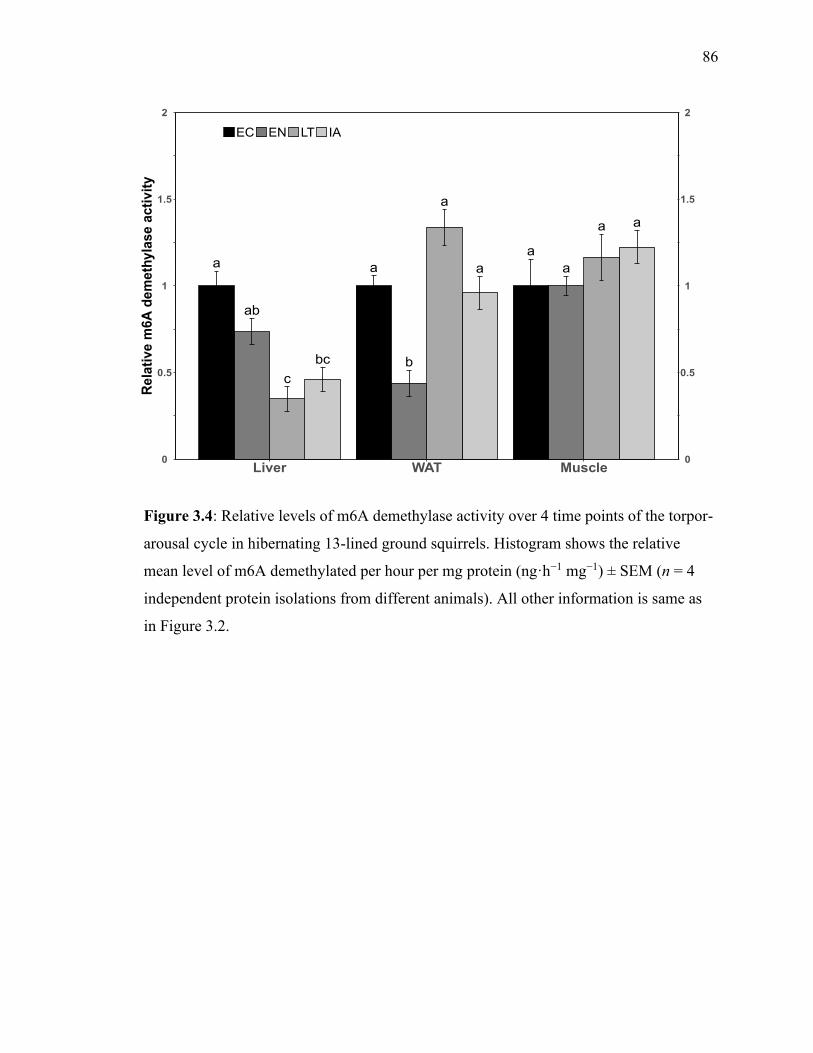
86
Figure 3.4: Relative levels of m6A demethylase activity over 4 time points of the torpor-
arousal cycle in hibernating 13-lined ground squirrels. Histogram shows the relative
mean level of m6A demethylated per hour per mg protein (ng·h−1 mg−1) ± SEM (n = 4
independent protein isolations from different animals). All other information is same as
in Figure 3.2.
bc
c
ab
a a
a
b
a
aa
a
a
0
0.5
1
1.5
2
Liver WAT Muscle
Re
lati
ve
m6
A d
em
eth
yla
se
acti
vit
y
EC EN LT IA
0
0.5
1
1.5
2
a
a
b
a
aa
aa
b
b
ab
a
0
0.5
1
1.5
2
2.5
Liver WAT Muscle
Re
lati
ve e
IF3
d a
mo
un
t
EC EN LT IA

87
Figure 3.5: Relative total protein levels of m6A reader proteins in three tissues of
hibernating 13-lined ground squirrels. Blots images compare EC and interbout arousal
(IA) time points. All other information is as in figure 3.2.
ProteinLIVEC
LIVIA
WATEC
WATIA
MUSEC
MUSIA
YTHDF1
YTHDC2
YTHDF3

88
Figure 3.6: Relative levels of eukaryotic translation initiation factor 3, subunit D (eIF3d)
over the torpor-arousal cycle in hibernating 13-lined ground squirrels. Histogram shows
the relative mean level eIF3d ± SEM (n = 4 independent protein isolations from different
animals). All other information is same as in Figure 3.2.
a
a
b
a
aa
aa
b
b
ab
a
0
0.5
1
1.5
2
2.5
Liver WAT Muscle
Re
lati
ve e
IF3
d a
mo
un
t
EC EN LT IA
a
a
b
a
aa
aa
b
b
ab
a
0
0.5
1
1.5
2
2.5
Liver WAT Muscle
Re
lati
ve e
IF3
d a
mo
un
t
EC EN LT IA
89
Chapter 4
Chapter 4 Arginine methylation produces differential
regulation of epigenetic marks during hibernation in the
thirteen-lined ground squirrel
Arginine methylation produces differential regulation
of epigenetic marks during hibernation in the thirteen-
lined ground squirrel

90
Introduction
Some sciurid rodents such as the 13-lined ground squirrel (13LGS, Ictidomys
tridecemlineatus) engage in hibernation to survive prolonged periods of resource scarcity
(e.g., winter). In mammals, hibernation is characterized by prolonged periods of
hypometabolism (i.e., torpor) wherein global reductions in fuel and energy requirements
reduce the resting metabolic rate to <5% of euthermic (i.e., summer) values (Ruf and
Geiser 2015). Attenuation of nearly all cellular functions may be achieved via passive
suppression through lowering the hypothalamic thermal set-point, combined with active
suppression imposed upon many biochemical pathways. During the hibernation season,
the squirrels undergo long periods of deep torpor that are interspersed with brief arousal
periods where body temperature (Tb) returns to the euthermic level (Carey et al. 2003),
indicating that the mechanisms involved in controlling torpor/arousal must be readily
reversible. For these reasons, the 13LGS has become a major model organism for
investigating the molecular controls of natural mammalian hibernation.
The majority of 13LGS tissues show evidence of strong metabolic rate depression
during torpor, allowing them to survive hibernation through increased regulation over
protein production and protein/enzyme activity which provides intricate selection of
necessary, tissue-specific cytoprotective biochemical pathways (for review, see Storey
and Storey 2010). This feat is achieved, in large part, through the programmed use of
post-translational modifications (PTMs), which effect a pattern of down-regulation over
protein production that rapidly and reversibly modulates target proteins during torpor and
arousal. For example, selected gene transcription patterns are achieved through
acetylation and phosphorylation of activated transcription factors during torpor, and

91
allow greater regulatory plasticity over the genome (Morin and Storey 2009; Srere et al.
1992).
Similarly, changes to PTMs on histone proteins are a crucial factor in the regulation
imposed on protein production during hibernation. Methylation of arginine residues on
histones has been shown to play a role in the remodelling of chromatin and regulates
aspects of protein dynamics and functioning in several cellular pathways. Arginine is
well positioned to interact with other biological molecules, since the residue has five
potential hydrogen bond donors and has been shown to form predictable, defined, and
frequent interactions with RNA, DNA and proteins (Luscombe et al. 2001; Najbauer et
al. 1993; Hughes and Waters 2006). In addition, methylation of arginine may also
increase the likelihood of a protein binding to certain interacting partners by increasing a
protein’s affinity for selected molecular features including aromatic residues (Sprangers
et al. 2003). Since methylation replaces available hydrogen bond donor atoms, it can
thereby alter the interactions of arginine residues with various binding partners (Pahlich
et al. 2006) and, indeed, the discovery of this modification has been shown to be
increasingly relevant to cellular dynamics.
Arginine methylation is catalyzed by protein arginine methyltransferase (PRMT)
enzymes that remove a methyl-group from a donor molecule [S-adenosyl-L-methionine,
AdoMet] and place the modification onto terminal nitrogen atoms of arginine residues. In
mammals this can occur on two of the three nitrogen atoms within the guanidinium
group. Arginine methylation comes in three forms, depending on the enzyme that places
the modification: monomethylarginine (MMA), symmetric dimethylarginine (sDMA) and
asymmetric dimethylarginine (aDMA). These are named for the number of methyl groups

92
placed on the arginine and their placement on either one or both terminal nitrogen atoms
(Wolf 2009; Bachand 2007). This difference in enzymatic products separates PRMT
enzymes into two groups based on their ability to place MMA as well as either aDMA
(type I PRMTs) or sDMA (type II PRMTs) modifications. Seven PRMT enzymes have
been found (PRMT1-7) in mammals, of which PRMT5 and PRMT7 are the only type II
enzymes. Arginine methylation has also been shown to be reversible via the demethylase
activity of Jumonji-Domain Containing 6 protein (JMJD6) (Chang et al. 2007), although
this mechanism is still hotly debated (Blanc and Richard 2017). Protein arginine
methylation has major implications for the cell’s transcriptional regulatory capacity
through actions on several binding partners.
When placed upon histone proteins, arginine methylation acts as a stable and
reversible epigenetic modification, that can regulate gene transcription by altering the
degree of chromatin relaxation (activating) or condensation (inhibitory). The PRMT4
enzyme is perhaps the best characterized and is associated with the recruitment of nuclear
receptor transcription factors to DNA due to methylation of histone H3 on R2, -17 and -
26 (Schurter et al. 2001; Lee et al. 2002). Activation following arginine methylation and
nuclear receptor binding is further increased via synergistic effects of histone acetylation.
PRMT1-3 seemingly act via similar mechanisms (e.g., H4R3 methylation) to activate
gene transcription in cooperation with other histone modifications and are thus
characterized as transcription coactivators (Lee et al. 2005). PRMT5, on the other hand,
was associated with repression of gene activity, in association with the co-repressor
proteins histone deacetylase 2 (HDAC2) and Sin3A or with chromatin-remodelling
factors following methylation of histone H4 on R8 (Pal et al. 2003; Pal et al. 2004). By

93
modifying histone proteins, the PRMT enzymes alter the chromatin accessibility and
transcriptional activation of nearby genes.
The present study aims to understand the role played by arginine protein
methylation in mammalian hibernation and metabolic rate depression. It is thought that
arginine methylation provides a means of reducing transcriptional rates through
modification of histone proteins and is involved in modifying the activity of non-histone
proteins during torpor. Therefore, the protein levels of several PRMT enzymes that play a
role in chromatin condensation and relaxation were analyzed along with total type-II
arginine methyltransferase activity, and changes in methylarginine modifications on
histone proteins over the torpor‐arousal cycle. These results allow for conclusions to be
made about the roles that arginine methylation may provide in regulating hibernation.
Methods
Animal Preparations
As described by Cai et al. (2004), 13LGS were caught in the wild between the
months of August and September in Illinois by a trapper licensed by the US Department
of Agriculture. Squirrels were transported to a National Institute of Health facility where
they were quarantined during antiparasitic treatment. NINDS animal care and use
committee (ACUC) approved animal housing and experimental procedures were
followed (protocol number ASP 1223-05). Squirrels were housed individually in plastic
shoebox cages in a vivarium with an ambient temperature of 21°C and a 12:12 h L:D
photoperiod. Animals were fed standard rodent diet. Weight gain before hibernation
served as a cue for subcutaneous implantation of telemetry devices for Tb measurements
(IPTT-200; Bio Medic Data Systems, Seaford, DE) while the squirrels were anesthetized
with 5% isofluorane. Animals were housed in a hibernaculum during the months of

94
November to March with an ambient temperature of 4-6°C and 60% humidity under 24-
hour darkness. A photographic red safelight (3–5 lx) was used when necessary and the
animal’s chamber was only accessible through a darkened anteroom.
Hibernation in 13LGS is identifiable by a near-ambient Tb, minimal activity and
curled body position. Animals were sampled at several points over the torpor-arousal
cycle, requiring quick removal from the hibernaculum, anesthetization and decapitation
within two minutes. Whole organ samples were dissected and immediately frozen in
liquid nitrogen for subsequent transport to Carleton University on dry ice. Tissue samples
were then stored at -80°C until required.
Total Soluble Protein Extraction
Frozen liver, visceral white adipose tissue (WAT), and hind leg skeletal muscle
tissue from four individual animals at each time point of the torpor-arousal cycle were
crushed and homogenized as described previously (Watts and Storey 2019) in ice-cold
homogenizing buffer using a Polytron PT 10-35 homogenizer with sonication
(Kinematica; #11010104); dilutions were 1:3 (w/v) in the case of WAT and 1:5 (w/v) for
liver and muscle. Protein concentrations were stored at -80°C and standardized to 10
μg/μL before use.
Arginine Methyltransferase Assay
Arginine methyltransferase activity was examined in total soluble protein extracts
using the Epigenase PRMT Methyltransferase (Type II-specific) Activity/Inhibition
Assay Kit (Epigentek, Cat# P-3088) according to manufacturer’s instructions. A standard
curve was first produced to find the optimal amount of input material required (10 µg
protein for liver or WAT, and 30 protein µg for muscle). The assay involves addition of
protein samples to wells coated with a substrate compatible with type-II PRMT enzymes.

95
The modified, methylated products were detected using the provided primary and
secondary antibodies before exposure using a developing solution. Methylated products
are proportional to the absorbance measured in a microplate spectrophotometer at a
wavelength of 450nm. Specific methyltransferase activity is reported as ng of
methylarginine produced per hour per mg of protein sample added.
Western Blotting
SDS-polyacrylamide gel electrophoresis and immunoblotting were accomplished as
previously described (Watts and Storey 2019), using total soluble protein extracts
standardized to 5 μg/μL by mixing 1:1 v/v with 2X SDS loading buffer (100 mM Tris-
base H 6.8, 4% w:v SDS, 20% v:v glycerol, 0.2% w:v bromophenol blue, 10% v:v 2-
mercaptoethanol). 20 μg of protein sample (which was adjusted as required in keeping
with the linear dynamic range of the antibody reaction) was loaded on SDS-
polyacrylamide gels and run at 180 V for ~60 min and transferred onto a 0.45 μm PVDF
membrane using the Trans-Blot® Turbo™ Transfer System (Bio-Rad; Mississauga, ON,
Canada). Membranes were blocked using skim milk powder (1.0-3.0% w:v) dissolved in
Tris-buffered saline with Tween (TBST; 50 mM Tris-HCl pH 7.5, 150 mM NaCl, 0.05%
v:v Tween-20) for 30 min. Primary antibodies were diluted 1:1000 v/v in TBST and then
incubated with membranes at 4°C overnight. Primary antibodies for PRMT1 (#sc-
166963), PRMT2 (#sc-393254), and PRMT7 (#sc-376077) were purchased from Santa-
Cruz Biotechnology (Dallas, TX); these were monoclonal and raised in mice. The
primary antibodies for PRMT3 (#A302-526A), PRMT4 (#A300-420A), and PRMT5
(#A300-850A) were purchased from Bethyl Laboratories (Montgomery, TX), and
antibodies targeting methylarginine-modified histones, H3R2me2a (#MBS9402172),
H3R8me2s (#MBS9607605), and H4R3me2s (#MBS126222) were purchased from

96
MyBioSource (San Diego, CA); these antibodies were polyclonal and raised in rabbit.
Epitopes for all antibodies used in this study are available from the producer. Membranes
were probed with HRP-linked affinity purified anti-rabbit IgG secondary antibody
(#APA007P) or with HRP-linked affinity purified anti-mouse IgG secondary antibody
(#APA005P) purchased from BioShop (Burlington, ON, Canada). Specific
immunoreactive bands were visualized via enhanced chemiluminescence (ECL; 1:1
hydrogen peroxide and luminol).
To account for potential minor variations in sample loading between different
lanes, the densities of immunoreactive bands corresponding to the protein of interest were
standardized against the summed intensity of a group of Coomassie stained bands in the
same lane, these bands being consistently present across all lanes and well-separated
from the immunoband of interest (Eaton et al. 2013). Coomassie blue-stained membranes
(0.25% w:v Coomassie brilliant blue, 7.5% v:v acetic acid, 50% methanol) allowed
visualization of the total protein loaded, which was constant across all lanes.
Quantification and Statistics
Chemiluminescent band density was quantified using GeneTools (version 4.02,
Syngene, Frederick, MD). Relative band density was determined by standardizing all
values to the first control sample that was arbitrarily set to one.
Fold change values were calculated by comparing the difference between a
particular time-point, which as standardized against the control value, and the appropriate
control value. For example, the fold change of EN compared to EC for any target protein
is expressed as EN/EC, where a fold change > 1 represents an increase, while a fold
decrease is expressed as the percentage decrease and represents a fold change <1. All
numerical data are expressed as mean ± SEM (n = 4 samples from independent animals).

97
Statistical analysis of differences between experimental hibernation time-points was
performed using a one-way ANOVA and post-hoc Tukey tests or a Student’s t-test using
RBioPlot statistical package (J. Zhang and Storey 2016) with p < 0.05 accepted as
significant.
Results
The relative protein levels of six widely studied methyltransferase enzymes
(PRMT1, 2, 3, 4, 5, 7) involved in regulating chromatin condensation were assessed in
skeletal muscle and liver of 13LGS across five time points of the torpor‐arousal cycle.
Samples were collected from (i) euthermic in the cold room (EC) animals which showed
euthermic physiology (i.e., stable euthermic body Tb and normal metabolic rate) for at
least 72 hours; (ii) entrance into torpor (EN) animals which showed a declining Tb
between 12-31°C; (iii) late torpor (LT) animals that displayed 5-8°C Tb for longer than
72 hours; (iv) early arousal (EA) animals that had begun to rewarm (persistent rising Tb
between 9-12°C) and displaying more than 60 breaths per minute; and (v) interbout
aroused (IA) animals that had naturally returned to euthermic conditions for no more than
24 hours after a prolonged torpor bout. Preliminary analysis confirmed that each of the
commercial antibodies used for western blotting reacted with an immunoblot band at the
at the expected molecular mass of each monomeric PRMT.
Type-II PRMT methylation activity was assessed in liver, white adipose (WAT)
and skeletal muscle of squirrels sampled from EC, EN, LT and IA conditions. Active
type-II PRMTs transfer a methyl group to methylate a substrate that is detected by a
specific antibody. Using this method, the measured PRMT methylation activity in control
(EC) animals was determined to be 53.4 ± 3.7 ng·min−1·mg−1, 29 ± 5.2 ng·min−1·mg−1
and 50.8 ± 5.2 ng·min−1·mg−1 in liver, white adipose, and muscle tissues, respectively.

98
Methylation activity in the EN, LT and IA timepoints are expressed as fold change values
relative to these EC values, which were standardized to 1.0 (Figure 4.1).
Methyltransferase activity was increased significantly in liver during IA, 2.3-fold above
levels seen in EC, and a comparable result was seen in muscle tissue, where PRMT
activity was 2.0-fold above EC levels. Similarly, in both liver and muscle,
methyltransferase activity did not change significantly from EC levels during either of the
torpor timepoints (i.e., EN and LT). On the other hand, methyltransferase activity in
WAT was not significantly different from euthermic levels across hibernation time-
course, but a significant change in activity was seen between EN and LT time points
when levels increased by 2.5-fold in LT as compared with the EN value that was just
44% of EC levels.
Liver PRMT protein levels were observed to change significantly over the torpor-
arousal cycle (Figure 4.2). PRMT1 levels decreased by 45% upon torpor entrance, but re-
established protein levels that were comparable to control EC animals during LT. These
levels were maintained across EA and IA timepoints. Levels of PRMT2 were maintained
throughout EN and LT, as was also true for protein levels of PRMT4 and for PRMT5
throughout the torpor phases. However, levels of PRMT2 rose sharply during arousal,
values in EA increasing by 2.3-fold over control levels in EA. The opposite was seen for
PRMT4, which decreased to 45% of EC levels during EA, and then to 39% during IA.
Protein levels of PRMT3 and 7 both increased significantly compared to EC values
during EN, by 1.4-fold and 2.2-fold for PRMT3 and 7, respectively, and this increase was
maintained in LT, at 1.4-fold and 2.3-fold for PRMT3 and 7, respectively.

99
In WAT, the protein levels of PRMT1, 2, 4 and 7, (i.e., all except for PRMT3 and
PRMT5), were observed to decrease significantly during EN, to between 15% and 39%
(which was the case for PRMT2 and 7, respectively) (Figure 4.3). Subsequently, values
returned to control EC levels in LT, or exceeded them as was the case for PRMT7 that
rose to levels that were 2.5-fold above EC levels (a 6.5-fold increase over EN levels). On
the other hand, levels of PRMT3 were maintained during EN, but decreased to just 7% of
EC levels in LT, whereas levels of PRMT5 were not significantly different than EC
values in both EN and LT. As opposed to the decreases observed during EN, protein
levels of all PRMTs, except PRMT3, returned to values not significantly different from
controls (EC) at both arousal timepoints. Levels of PRMT3, on the other hand, continued
to be significantly reduced in both EA and IA at values ~40% of EC levels.
PRMT protein levels were also investigated in skeletal muscle tissue (Figure 4.4)
where it was seen that PRMT1 levels decreased significantly in LT to 34% of EC levels,
and remain decreased in EA at 45% of EC levels. Levels of PRMT1 enzyme were seen to
be comparable to control protein levels in both EN and IA. Interestingly, PRMT2 also
showed a decrease in enzyme levels in LT (49% of EC levels), but were then elevated
2.6-fold above EC levels in EA (representing a 5.3-fold increase over levels in the
preceding LT timepoint), and continued to rise, showing a 3.8-fold increase over EC
levels in IA. Similarly, levels of PRMT3 and PRMT5 showed a significant decrease in
protein levels in LT, to 34% and 32% of EC levels, respectively, however their protein
levels then rose during EA and were returned to values comparable to EC controls during
IA. Remarkably, both PRMT4 and PRMT7 showed an increasing trend during EN, which
was significant in the case of PRMT7 (increased 1.8-fold above EC levels). Levels of

100
PRMT7 then fell back to EC levels by LT and remained low until IA when protein levels
were elevated 3.3-fold above EC levels (and 1.8-fold above EN levels). Levels of
PRMT4 on the other hand, continued to increase and were raised significantly in LT by
1.6-fold over EC levels. This was reversed during arousal when PRMT4 levels decreased
to 40% of EC levels during EA, and remained reduced in IA compared to the earlier
peaks during torpor but were not significantly different as compared to control animals.
The relative levels of methylated arginine residues present on histone proteins
under EC, LT and IA conditions in the three tissues was also investigated by
immunoblotting (Figure 4.5) by targeting the asymmetric demethylated R2 and
symmetric demethylated R8 on histone H3 (H3R2me2a and H3R8me2s), as well as
symmetric demethylated R3 on histone H4 (H4R3me2s). In liver, H3R2me2a showed
significantly greater methylation in IA compared to EC (a 2.3-fold increase), whereas
methylation of H4R3me2s decreased significantly during LT (to 29% of EC levels). The
pattern of methylation seen in WAT was almost completely opposite to that seen in liver;
in WAT methylation of H3R2me2a was significantly reduced during LT (to 54% of
H3R2me2a levels in EC), whereas methylation of H4R3me2s increased significantly
during LT, 1.8-fold above EC levels. H3R8me2s levels also rose significantly in WAT to
values at least 2.4-fold over EC levels during both LT and IA. Finally, skeletal muscle
levels of H3R2me2a were elevated 1.6-fold above EC levels during LT, but fell back to
control levels during arousal. On the other hand, LT levels of both H3R8me2s and
H4R3me2s were reduced to 7% and 33% of control levels, respectively. H3R8me2s
remained at a reduced level (30%) compared to EC during IA, whereas levels of
H4R3me2s were significantly elevated 2.1-fold above EC levels during arousal.

101
Discussion
Studies have previously investigated the protein levels and activity of various DNA
or protein methyltransferase enzymes during hibernation of 13-lined ground squirrels
(13LGS), but this is the first study to examine the arginine methyltransferase enzymes
themselves and, as such, this study provides support for PRMT enzymes having a
regulatory role in the metabolic control of mammalian hibernation. The significant,
tissue-specific, changes in PRMT protein levels and methylarginine residues on histone
proteins seen over the torpor-arousal cycle demonstrate that PRMT enzymes are
modulated in their downstream regulatory abilities during hibernation, as posited by
previous studies of methylation activity during hibernation (Morin and Storey 2009).
Reversible methylation of cytosine residues on DNA, long thought to be a mechanism for
widespread genomic changes, was significantly altered in the muscle of hibernating
13LGS and likely contributes to the dynamic nature of DNA methylation across the
seasons (Alvarado et al. 2015), and to a somewhat maintained level of transcriptional
activity in skeletal muscle during hibernation (Allan and Storey 2012; Malatesta et al.
2009). Similarly, changes in posttranslational methylation modifications to proteins
provide rapid and reversible control over diverse metabolic functions in cells. A previous
analysis of lysine methyltransferase protein levels and histone PTMs by our lab found
that lysine methylation is potentially an important regulator of the mammalian
hibernation phenotype in both liver and muscle (Watts and Storey 2019). Results from
this study contribute to our understanding of how changes to protein methylation and the
“histone code” control the adjustments in metabolic and genomic activity necessary for
proper and efficient hibernation.
102
In adjusting to hibernation, tissues such as liver and WAT must switch to a
prolonged fasting mode and regulate lipid metabolism, which is favored over
carbohydrate as the fuel supporting torpor (Dark 2005). PTMs have long been known to
be commonly employed in these tissues during hibernation in order to force shifts in
protein/enzyme activity, stability, or localization (Grabek, Martin, et al. 2015; Storey
1987). As such, the role of methylation as a dynamic and stable protein modification is
worth investigating. In both liver and WAT, type-II PRMT activity levels were consistent
with control levels throughout torpor but were heightened strongly during IA in liver
(Figure 4.1). Both PRMT5 and PRMT7 are type-II PRMT enzymes responsible for
symmetric dimethylation of histones at H3R8 and H4R3. Protein levels of PRMT5 were
maintained across hibernation in liver whereas PRMT7 levels showed a significant
increase in both stages of torpor (Figure 4.2). H3R8me2s, which was also maintained at
control levels within liver during hibernation (Figure 4.5), is the product of PRMT5
activity and possibly plays a role in the prevention of oxidation following increased lipid
catabolism via necessary interactions with Oxidation Resistance gene 1 (OXR1A) that
promotes H3R8 methylation and prevents the development of fatty liver disease (Yang et
al. 2020). The fact that neither PRMT5 or PRMT7 protein levels corresponded to the
decrease in H4R3me2s within liver, make it likely that a non-histone protein serves as a
more favorable substrate during torpor (Wei et al. 2014). Further, methylation of
H4R3me2s is be inhibited by the presence of other neighboring histone modifications that
are increased in liver during torpor, such as H3K27 (Watts and Storey 2019; Liu et al.
2020). Interestingly, the increase in type-II methylation occurred at the same time that
levels of H3R2me2a were also significantly increased in liver, pointing to the
103
involvement of PRMT enzymes in shifting metabolism back towards carbohydrate
metabolism during interbout arousal through interactions with histone proteins.
Dimethylation of H3R2 by PRMT6 is typically localized to regions of repressed
transcriptional activity and interferes with the modification of nearby lysine residues
available for methylation or acetylation (Guccione et al. 2007; Bouchard et al. 2018). Our
previous analysis (Watts and Storey 2019) showed that monomethylation of H3K4, an
inherently repressive modification that serves as the scaffold for activating marks (Cheng
et al. 2014), is kept at control levels or significantly increased during torpor. Whereas
determination of whether localization of these marks on the genome is targeted to
enhancer or promoter regions is outside the scope of the present study, it is evident that
the interaction of histone modifications serves integral roles in the metabolic shift that
takes place during hibernation. Unfortunately, PRMT6 did not produce quantifiable
protein abundance results in the tissues investigated, however significant increases in
H3R2me2a are enough to conclude that the enzyme is active in liver.
A similar story may be inferred from the data in WAT, as PRMT activity and
protein levels (with the exception of PRMT3 and PRMT7) were, unchanged from at EC
control levels between LT and IA (Figures 4.1, 4.3). Indications of the downstream
significance of these conserved protein activity and abundance levels are found in the
heightened levels of both H3R8me2s and H4R3me2s during LT and IA (Figure 4.5).
PRMT5 can di-methylate either of these modifications, and interestingly has been found
to interact with mitochondrial biogenesis and peroxisomal lipid metabolism via
regulation of PPARα and PGC-1α. In mice fed a high-fat diet, PRMT5 levels were
increased in liver, whereas hepatic PRMT5 knock-down led to increased levels of

104
phosphatidylinositol 3-kinase/AKT signaling, but these changes were not seen in WAT or
other tissues that are metabolically-relevant in hibernation (Huang et al. 2018). Given the
difference in cellular and biochemical responses between a hibernating mammal and
other mammals in terms of lipid-metabolism, specifically in regards to PPARα (El
Kebbaj et al. 2009; Han et al. 2015), it is remarkable that PRMT5 levels were maintained
in both liver and WAT. However, the question remains as to why H3R8me2s and
H4R3me2s increase only within WAT, despite significant increases in PRMT5 within
both WAT and liver. Levels of activated AKT in liver are known to decrease during
torpor and increase during arousal despite maintained PRMT5 levels, and in contrast to
mice fed a high-fat diet (McMullen and Hallenbeck 2010; Wei et al. 2012), raising the
possibility that this correlation may be less relevant in hibernators. While the mechanism
underlying the correlation between AKT activation and PRMT5 abundance is still
unclear, their links to PPAR signaling and the active role that PPAR proteins play in
hibernation, makes investigating the methylation status of proteins that regulate these
pathways, such as mTOR, warranted. Localization of PRMT5 bound to the WAT genome
has previously been carried out with adipocytes undergoing adipogenesis, where it was
determined that the enzyme acts on histone proteins to regulate the binding of chromatin-
remodeling enzymes and activate sequences downstream of a PRMT5 binding site
(LeBlanc et al. 2012). Given that WAT undergoes extensive remodelling in the pre-
hibernation season and during hibernation in at least one hibernating species (Chayama et
al. 2018), it is very likely that maintained levels of PRMT5 in WAT are required for
genomic reorganization, as is evident from the large increase in WAT H3R8me2s and
H4R3me2s (Figure 5).
105
Meanwhile, muscle type-II PRMT activities indicate that much of the enzyme
activity by PRMT5 and PRMT7 occurs during IA (Figure 4.1), but in contrast to liver
tissue, is targeted towards H4R3me2s (Figure 4.5). Increased levels of H4R3me2s
typically localize to areas of DNA damage (Karkhanis et al. 2012) and its significant
increase during arousal in muscle likely represents the hibernators remarkable ability to
avoid genomic instability during hibernation by repairing the DNA when the tissue is
able to during arousal (Yancey 2018). Furthermore, while PRMT7 levels show
maintained or significantly increased levels across torpor, levels of PRMT5 decreased in
LT and remained somewhat reduced throughout arousal (Figure 4.4), explaining the
decrease in levels of its product, H3R8me2s, during hibernation. However, in a similar
vein to the role of type-II methylation in liver, the role of PRMT7 methylation may
combat increased levels of oxidative metabolism occurring in muscle during arousal as
the proteome shifts and the tissue becomes more active as shivering thermogenesis is
triggered to rewarm the animal (Hindle et al. 2011). PRMT7 depletion within muscle
affects levels of PGC-1α and inhibits its interaction with p38 MAPK, leading to reduced
endurance and energy expenditure (Jeong et al. 2016). Thus, increased levels of PRMT7
during arousal could accommodate the protection of muscle tissue through activation of
the endurance exercise pathway during hibernation (Xu et al. 2013). Finally, given that
none of the PRMT proteins studied showed increased levels during LT, drastic reductions
in symmetric dimethylation of both H3R8 and H4R3 are more likely to be a result of
antagonism by other histone modifications than by asymmetric methylation of these
residues by a different PRMT enzyme (Guccione et al. 2007; Liu et al. 2020).

106
While the analysis of type-II PRMT activity and products are certainly instructive
to our understanding of the biochemical and molecular responses of cells to hibernation,
analysis of PRMT protein levels during hibernation can also be useful for increasing our
understanding of the purpose of arginine methylation during hibernation. This is true
given that differential gene expression during hibernation represents the expenditure of
resources at a time when there is increased pressure for conservation (Srere et al. 1992).
Therefore, levels of PRMT1, 2, 3, and 4 were also investigated to assess arginine
methylation writer enzymes for tissue-specific responses during torpor and arousal.
Both PRMT1 and PRMT4 (CARM1) are known to methylate arginine residues on
histone tails and are typically located in areas surrounding activated gene transcription.
Specifically PRMT1 acts on H4R3 and H2AR3 while PRMT4 acts upon H3R17 and R26,
and either enzyme can dimethylate these residues but only in an asymmetric form (Daujat
et al. 2002; Huang et al. 2005). Through their interactions with histone targets, both
PRMT1 and PRMT4 are transcriptional co-activators responsible for recruiting chromatin
modifiers following their recruitment to the genome by various transcription factors
(Bedford and Richard 2005). Upon entry to torpor, protein levels of PRMT4 were
maintained in liver whereas PRMT1 protein levels fell (Figure 4.2), however this trend
was reversed in arousal as PRMT1 levels climbed to return to EC values. Protein levels
of both enzymes are maintained in WAT, except during EN when they were strongly
reduced by about 70% (Figure 4.3). PRMT1 is important for the maintenance and
deposition of histone modifications. Through interactions with histone H4, its product
(H4R3me2a), is influential over surrounding lysine methylation or acetylation
modifications on neighbouring histone tails and allows PRMT1 to activate transcription

107
(Huang et al. 2005). The fact that PRMT1 is maintained between LT and IA in both liver
and WAT is evidence of the active roles that these tissues have in regulating lipid
metabolism during hibernation, and the role that epigenetics plays in supporting this task
(Storey 2015). Notably, PRMT1 levels decreased during LT and EA in skeletal muscle
(Figure 4.4), and perhaps influenced the histone code by releasing the inhibition over
H3K9 and H3K27 methylation which previous work observed to rise significantly during
LT in skeletal muscle (Watts and Storey 2019). The role of PRMT4 may be significant in
the regulation of lipid metabolism due to interactions with both PPARγ and GAPDH. By
acting as a co-activator of PPARγ (Yadav et al. 2008), PRMT4 is likely to be influential
in maintaining energy balance during torpor in liver and arousal in WAT (Eddy et al.
2005). On the other hand, PRMT4 methylation of GAPDH is important in signaling
cellular glucose levels and suppresses GAPDH activity during glucose starvation (X.-Y.
Zhong et al. 2018; Bell et al. 2014). This interaction could be especially relevant in
skeletal muscle during torpor, since predicted levels of PRMT4 rise following depletion
of glucose in muscle tissue (Wu et al. 2013). By acting as gene co-activators or through
non-histone interactions, both PRMT1 and PRMT4 are able to influence the regulation of
both lipid and carbohydrate metabolism, and these interactions are likely to be important
during hibernation.
Finally, the enzymes PRMT2 and PRMT3 are responsible for histone modifications
typically found in areas surrounding activate gene transcription, namely asymmetric
dimethylation of H3R8 and H4R3, respectively (Dong et al. 2018; Min et al. 2019), and
are likely responsible for methylating non-histone proteins as well. Significant elevations
in PRMT2 in both liver and skeletal muscle, and control levels in WAT, likely serve to

108
influence the regulation of damaged cell turnover during arousals by promoting apoptosis
in affected cells (Ganesh et al. 2006) via activation of the NF-kB pathway during
hibernation (Hadj-Moussa et al. 2020). PRMT3 levels were elevated only in liver,
whereas both WAT and muscle showed significant decreases in PRMT3 protein levels
LT and reduced levels as well in EA. When overexpressed, PRMT3 is associated with the
increased expression of lipogenic proteins within liver and a high fat diet is known to
increase PRMT3 levels in liver of animal models (Kim et al. 2015). It seems probable
that the role of PRMT3 is associated with the increased lipid metabolism occurring in
liver during torpor, given its significant increase in both EN and LT. The roles of both
PRMT2 and PRMT3 are certainly also involved in regulating the metabolic needs of the
animal during hibernation, as their suitability for this role is clear given their known roles
and protein interactions.
In summary, arginine methylation plays an important role in controlling the vast
reorganization of metabolic regulation which is required in hibernation. Moreover, these
mechanisms are likely necessary for proper and efficient hibernation due to their
influence over cell and organ-specific responses. Arginine methylation certainly
contributes to altering and adapting the cellular environment to support hibernation
through multiple mechanisms as described.
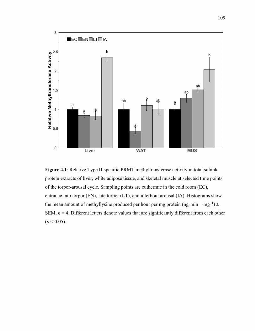
109
Figure 4.1: Relative Type II-specific PRMT methyltransferase activity in total soluble
protein extracts of liver, white adipose tissue, and skeletal muscle at selected time points
of the torpor‐arousal cycle. Sampling points are euthermic in the cold room (EC),
entrance into torpor (EN), late torpor (LT), and interbout arousal (IA). Histograms show
the mean amount of methyllysine produced per hour per mg protein (ng·min−1·mg−1) ±
SEM, n = 4. Different letters denote values that are significantly different from each other
(p < 0.05).
b
aa
aab
b
a
ab
b
ab
ab
a
0
0.5
1
1.5
2
2.5
3
Liver WAT MUS
Re
lati
ve
Me
thylt
ran
sfe
ras
e A
cti
vit
y
EC EN LT IA
b
aa
aab
b
a
ab
b
ab
ab
a
0
0.5
1
1.5
2
2.5
3
Liver WAT MUS
Re
lati
ve
Me
thylt
ran
sfe
ras
e A
cti
vit
y
EC EN LT IA
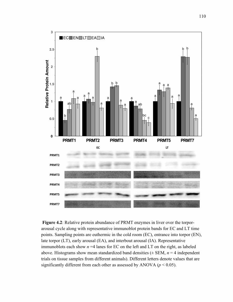
110
Figure 4.2: Relative protein abundance of PRMT enzymes in liver over the torpor‐
arousal cycle along with representative immunoblot protein bands for EC and LT time
points. Sampling points are euthermic in the cold room (EC), entrance into torpor (EN),
late torpor (LT), early arousal (EA), and interbout arousal (IA). Representative
immunoblots each show n =4 lanes for EC on the left and LT on the right, as labeled
above. Histograms show mean standardized band densities (± SEM, n = 4 independent
trials on tissue samples from different animals). Different letters denote values that are
significantly different from each other as assessed by ANOVA (p < 0.05).
EC LT
PRMT1
PRMT2
PRMT3
PRMT4
PRMT5
PRMT7

111
Figure 4.3: Relative protein abundance of PRMT enzymes in white adipose tissue over
the torpor‐arousal cycle along with representative immunoblot protein bands for EC and
LT time points. All other information is as in figure 4.2.
EC LT
PRMT1
PRMT2
PRMT3
PRMT4
PRMT5
PRMT7

112
Figure 4.4: Relative protein abundance of PRMT enzymes in skeletal muscle over the
torpor‐arousal cycle along with representative immunoblot protein bands for EC and LT
time points. All other information is as in figure 4.2.
EC LT
PRMT1
PRMT2
PRMT3
PRMT4
PRMT5
PRMT7

113
Figure 4.5: Relative amounts of histone H3 showing asymmetric dimethylated R2
(H3R2me2a) and symmetric dimethylated R8 (H3R8me2s), as well as histone H4 showing
symmetric dimethylated R3 (H4R3me2s) in liver, WAT, and skeletal muscle over the
torpor‐arousal cycle. Representative immunoblots each show n =4 lanes for EC on the
left and IA on the right, as labeled above. All other information is as in figure 4.1.
LIVEC
LIVIA
WATEC
WATIA
MUSEC
MUSIA
H3R2me2a
H3R8me2s
H4R3me2s

114
Chapter 5
Chapter 5 General Discussion
General Discussion

115
Two themes of mammalian hibernation biology are explored within this thesis: i)
the dependence of hibernators on rhythmic activation of gene transcription and activation,
and ii) the use and repurposing of epigenetic mechanisms for adapting and overcoming
massive changes in cellular activity by adding an additional layer of control over protein
expression and activation. Obviously, these two themes are interlinked, and so both aid
our understanding of the complexity involved in successfully employing hibernation to
overcome winter-time stresses.
In an effort to save as much energy as possible during winter and survive prolonged
resource scarcity coupled with increased energetic demands, the 13-lined ground squirrel
(13LGS, Ictidomys tridecemlineatus) undergoes huge phenotypic plasticity in an effort to
conserve stored fuels for necessary purposes. In turn, the vast majority of metabolic
functions are turned down or turned off, whereas regulatory demands which are
intertwined with survival are prioritized and upregulated. As an example, thermogenic
and fatty acid catabolism pathways, crucial for the hibernator’s survival, were seen to be
prioritized in all cases studied within this thesis. By modulating the requirements for gene
activation by transcription factors, chromatin remodeling enzymes, and post-
transcriptional modification enzymes, cells can increase the transcriptional activity of
required genes groups.
Preparation for an almost complete dependence on lipid fuels during the winter is
first achieved in late summer to early autumn, when ground squirrels double their body
weight on average and increase their fat mass by up to 4-fold (Sheriff et al. 2013). This
leads to large increases in white adipose tissue (WAT) stores and alters the number,
shape, size and function of cells as depots increase in triglyceride levels for use during

116
hibernation (Chayama et al. 2018). During torpor, WAT and liver support vital functions
by mobilizing fat stores as Tb falls to ambient temperatures during torpor throughout the
hibernation season. Within liver, levels of long-chain fatty-acid transport proteins such as
apolipoprotein A1 as well as lipid catabolism and mobilization pathways are selectively
activated, in addition to proteins involved in supporting and maintaining protein stability
and folding (Epperson et al. 2010). For these reasons, dietary lipids impact the efficacy
and success of hibernation as a survival strategy – animals that consume larger amounts
of polyunsaturated fatty-acids (PUFAs), and especially n-6 class PUFAs hibernate more
successfully due to these lipids intrinsic ability to maintain membrane fluidity and
promote energy savings (Munro and Thomas 2004; Jastroch et al. 2016).
As opposed to the active roles of liver and WAT in lipid metabolism during torpor,
transcriptional suppression and global decreases in metabolic rate define the environment
within skeletal muscle tissue. Overall energy expenditure within muscle tissue during
torpor is modulated through reductions in non-essential cellular processes such as new
protein synthesis and cell division, whereas cytoprotective and chaperone proteins remain
activated to accommodate shivering thermogenesis that is an important component of
rewarming during arousals as well as for emergence from hibernation in the spring
(Hindle et al. 2011). Remarkably, hibernators are able to fend off and negate the effects
of prolonged muscle inactivity, showing preserved muscle morphology and mass upon
re-emergence and maintained muscle strength (Cotton 2016), mainly due to increased
stability and preservation of existing cellular proteins while in torpor.
Although this is not the first study to assess and uncover changes in the
mechanisms controlling metabolic and cytoprotective pathways in hibernator cells, only a

117
select few have touched on the roles of peripheral circadian oscillators and their
modifications when an organism undergoes suppressed metabolism events (i.e.,
translational and transcriptional arrest, regulation of post-transcriptional and post-
translational modifications, etc). The aim of my thesis was to investigate regulatory
patterns of signaling pathways that have previously been shown to contribute to the
rhythmic output of a cell, based on previous studies in other mammalian models. The
thesis aimed to address two goals: 1. Conservation of regulatory pathways in ground
squirrels, and 2. Differential regulation of these pathways in energy conservation. To
address these goals, I undertook a molecular approach, utilizing a variety of tools to
characterize genomic and proteomic regulation of peripheral circadian clock pathways, as
well as assessing structure and function of key enzyme regulators of the molecular clock.
Metabolism Interacts with the Molecular Circadian Clock
In Chapter 2 the regulation of key components of the molecular circadian clock
were assessed during torpor, focusing on the BMAL1/CLOCK complex due to its direct
link in regulating protein synthesis. Emphasis was placed on comparing the regulatory
patterns observed in each of liver, WAT and muscle tissues due to their differing
activities during hibernation. The levels of nearly all clock components were present at
the transcript level in all three tissues, and levels of only one transcript, PER1 in muscle,
was observed to decrease. Similarly, CLOCK DNA-binding activity was maintained in
the investigated tissues showing significant increases in WAT during late torpor (LT).
The results in all tissues suggested that circadian mechanisms remain intact in these
peripheral tissues during hibernation. However, the circadian transcription/translation
feedback loop likely swings towards members of the activating arm (i.e.,

118
CLOCK/BMAL1 are more active) within liver and WAT, whereas the opposite is true in
muscle tissue, where PER2 and CRY2 inhibition seemed to dominate. This pattern of
regulation suggests a state of maintenance of protein transcription in liver and WAT, but
transcriptional decreases in skeletal muscle during torpor.
It is perhaps not surprising that major integrators of cellular energy information
affect transcriptional activity within both the activating and repressive arms of the
circadian clock transcription/translation feedback loop. Integration of metabolic sensor
elements into the regulatory DNA regions of the Per genes, such as cAMP Response
Elements (CREs), allows signals of low cellular fuel availability to activate the repressive
arm of the core transcription/translation feedback loop so that rhythmic pathway
activation becomes muted after entrainment. In an elegant experiment performed by
Vollmers et al. (2009) it was shown that phosphorylation of CRE Binding protein
(CREB) coincides and is in phase with periods of fasting in animals lacking circadian
rhythms. CREB activation of Per1 and Per2 transcription, attenuates downstream CCG
activation when the animal is under adverse metabolic conditions by repressing circadian
activation in numerous pathways. Similarly, increased levels of 5’AMP-activated protein
kinase (AMPK) activity are correlated with nutrient/energy levels within most tissues,
and alter CRY protein rhythmicity and localization, as well as CCG activation within
peripheral oscillators (Lamia et al. 2009; Vieira et al. 2008). Clearly, signaling of
rhythmic behavioural and environmental changes, including feeding, has direct effects on
gene expression controlled by the circadian clock and provides additional regulatory
layers on important cellular events.

119
The Peroxisome Proliferator-Activated Receptor (PPAR) family of transcription
factors are another class of CCGs that show daily changes in expression levels in a
number of peripheral tissues (Lemberger et al. 1996; Yang et al. 2006; Gachon et al.
2011). There are three PPAR isoforms in mammals [PPAR//] which share largely
similar structural and functional homology, but differ in their tissue expressions
(Desvergne and Wahli 1999). PPARs bind several endogenous ligands including lipids
and are recruited to PPAR response elements, PPREs, after binding retinoid X receptors
(RXRs) to control and activate downstream gene transcription of targets that most
prominently serve to regulate lipid metabolism and adipogenesis (Wu et al. 2009). The
integration of the PPAR family into the circadian clock is significant and reciprocal,
meaning that many genes activated by PPAR family members show circadian variation,
but also that modulation of PPAR transcriptional activity feeds back to alter the
transcriptional activity of core circadian clock genes (Chen and Yang 2014; Canaple et al.
2006). For example, expression of PPRE-regulated genes in the intestine and in
fibroblasts, were modulated in a manner that correlated to the expression of BMAL1
(Inoue et al. 2005; Oishi et al. 2005) through interactions with an E-box element within
an intron of the PPAR gene. On the other hand, mice fed a PPAR agonist showed
advanced circadian locomotor activity and core molecular clock gene expression in
peripheral tissues due to action on a PPRE within the BMAL1 promoter region, but this
does not affect gene expression within the SCN (Canaple et al. 2006; Shirai et al. 2007).
Notably, levels of PPAR, which have been assessed previously in 13LGS, show
increased levels in liver and WAT, and correlate with protein levels of BMAL1 during
torpor (Eddy et al. 2005). While similar findings have already shown a relationship

120
between PPAR and the control of rhythmic protein levels in peripheral oscillators or
rhythmic physiological parameters (Yang et al. 2012; Wang et al. 2008), the same
connectedness may not be true for PPAR. Whereas PPAR does not show a direct link
to transcriptional activation of the circadian clock, it is the main target for a liver-specific
microRNA that is regulated by the oscillating Rev-ErbA repressor. By adding a layer of
circadian regulation over lipid metabolism in the liver, PPAR can show variations in
diurnal expression and affect fatty acid metabolism in the liver (Liu et al. 2013; Gatfield
et al. 2009). Finally, any one of the PPAR family transcription factors can be a binding
partner for the CLOCK transcription factor and in this way inhibit CLOCK’s interaction
with BMAL1 (McNamara et al. 2001). Since PPARs play an intricate role as regulators
of circadian lipid metabolism, it is likely that changes in rhythmic metabolic homeostasis,
such as those that occur during hibernation (McGinnis and Young 2016; Wu et al. 2013),
will alter both PPAR and circadian clock protein levels.
Modifications of Circadian Clock Molecules
Both torpor and circadian rhythmicity are dynamic processes that must be rapidly
controlled in a coordinated manner. PTMs serve as an indispensable mechanism for
quickly and effectively altering cellular activities via changes to the status of covalent
modifications on pathway regulatory proteins/enzymes. Given the already noted
importance of PTMs in the regulation of torpor-arousal cycles, their massive effects on
biochemical pathway activity and their ubiquity throughout the cell make their
investigation during hibernation warranted. PTMs modify numerous cellular regulators of
transcription/translation, protein-protein or protein-transcript interactions and protein or
transcript stability or localization (Lee and Stallcup 2009; Di Lorenzo and Bedford 2011;

121
Ma et al. 2017; Engel and Chen 2018) and play a major role in regulating protein
turnover and activity in almost all molecular pathways, including the circadian clock. The
repressive complex of the core clock is modified by several mechanisms, such as the
balance between kinase (CK1δ or CK1ε) and phosphatase (phosphatase 1, PP1) activities
(Lee et al. 2011) as well as ubiquitination by the β‑TrCP E3 ubiquitin ligase complex (M.
Zhou et al. 2015). An inactivating mutation in the gene encoding CK1δ lengthens the
circadian cycle by increasing the stability of PER1/2 as a result of attenuating
phosphorylation-dependent ubiquitination (Etchegaray et al. 2009; Meng et al. 2010), in
contrast to CK1ε which regulates the nuclear entry of the repressive complex (Vielhaber
et al. 2000; Eide and Virshup 2001). The CRY1/2 or BMAL1 proteins may also be
phosphorylated by casein kinase 1 enzymes, following kinase association with PER1/2/3
(Akashi et al. 2002; Eide et al. 2002). Phosphorylated PER2 proteins bound to CRY are
actually stabilized by inhibiting PER ubiquitination (Lee et al. 2001). While the
inhibitory phase of the transcriptional loop is active, phosphorylated CRY/PER
complexes within the nucleus bind the BMAL1/CLOCK complex (which remains bound
to the DNA) and serve as a scaffold for hyperphosphorylation of the complex members
by CK1δ or CK1ε, forming the transcriptionally repressive ‘timesome’ complex (Reppert
and Weaver 2002). In this way, rhythmic binding of the repressors for the DNA-anchored
transcriptional activator pushes the circadian clock into the night phase of the cycle.
Acetylation of BMAL1 is a second major PTM mechanism that regulates
transcriptional activity of the core clock complex. A second role for the CLOCK
transcription factor is as an activator of its own binding partner via inherent
acetyltransferase activity. Hirayama et al. (2007) showed that a conserved lysine residue,

122
K537 in mice, is acetylated in the liver and that this modification occurs in preparation
for, and is necessary for, binding of the repressive complex. By further showing that
mutant BMAL1 lacking the conserved lysine residue is unable to drive rhythmic
transcription of PER2, the authors showed that this modification is essential for effective
circadian transcription/translation feedback.
While numerous PTMs modify the circadian clock, epigenetic mechanisms, so-
called for their ability to transfer through the germline, also play a major role in the
regulation of cellular timing. For instance, the acetyltransferase activity of CLOCK may
acetylate histone H3 on lys9 and lys14 residues, and the deacetylase sirtuin 1 regulates
circadian functioning by associating with the core clock complex (Doi et al. 2006;
Nakahata et al. 2008; Asher et al. 2008). The coordinated activities of core circadian
proteins and their abilities to recruit and bind chromatin remodelling enzymes has been
noted to play an intricate role in the regulation of rhythmic gene transcription within
multiple pathways and cell types (Ripperger and Schibler 2006; Yeung et al. 2018).
Undoubtedly, the clock’s ability to recognize and place epigenetic modifications is
crucial to its functioning and depends on the actions of histone-modification mechanisms.
Maintenance of RNA methylation dynamics during torpor
Regulatory crosstalk between the deposition of m6A methylation with the
circadian clock has so far been established in only a limited number of systems. As
mentioned in Chapter 1, some circadian-controlled gene products contain m6A sites, and
moreover, inhibiting m6A methylation prolongs the circadian clock period through
uncoupled rhythms of BMAL1 and PER2 production (Fustin et al. 2013). At least one of
the mechanisms responsible for modulating the circadian clock period depends on m6A

123
methylation of circadian mRNA products. In addition to PER2, CK1δ mRNA, that codes
for one of the critical kinases responsible for translocation of the PER/CRY inhibitory
complex, also contains sites for m6A modification which inhibit its translation.
Modulation of the abundance of m6A deposited within the coding regions of CK1δ can
modulate circadian clock period by regulating the amount of CK1δ and ultimately
phosphorylation of PER2 proteins (Fustin et al. 2018). The deposition of m6A also
affects proteins that regulate the circadian clock, such as PPAR (discussed above)
which controls Bmal1 gene activity via PPAR-response elements in the upstream
promoter region. Following modification of PPAR by METTL3, and binding by
YTHDF2, the transcription factor is recruited to the genome where it can affect and alter
the production of numerous gene products (X. Zhong et al. 2018). Importantly, this last
result reveals not only the interconnectedness of the circadian and epigenetic
mechanisms, but also the link between both pathways and the regulation of metabolism
following changes in the cellular environment.
Chapter 3 examined the global levels of m6A within the transcriptome, where it
was seen that m6A shows differential abundance during torpor – increasing in WAT and
decreasing in skeletal muscle. Whereas liver showed no changes in RNA methylation,
m6A levels did show decreases when assessing levels within mRNA selectively. When
comparing levels of m6A writer enzymes, all tissues appeared to show maintenance of
m6A deposition during torpor although to different degrees in each tissue.
Writer, eraser and reader enzymes show rhythmicity in their gene expression, and
similarly, in the deposition of m6A (X. Zhong et al. 2018). Within 13LGS, levels of one
of the two m6A eraser enzymes were decreased in both liver and WAT, whereas both

124
were maintained in skeletal muscle. These results, as well as observations of reduced
m6A removal activity during LT in liver and EN in WAT, appear to mirror the
maintenance of transcriptional activity within these tissues in the context of molecular
circadian clock activity. On the other hand, skeletal muscle levels of eraser proteins and
activity indicate a paucity that is apparent in circadian transcriptional activity during
torpor. Through crosstalk with the circadian pathway and metabolism, mRNA
methylation is able to impact the homeostatic balance of multiple metabolic cellular
functions. Precisely how m6A methylation coordinates circadian clock functioning and
cellular metabolism is currently still debated.
Overall, chapters 2 and 3 indicate the continued production or preservation of
selected groups of gene transcripts during torpor-arousal cycles within three central
tissues of the 13LGS. The regulatory control over genomic activity inherent in post-
transcriptional modifications likely functions in co-operation with the regulatory abilities
of gene activators through recruitment to the genome, or localization of transcripts
towards processing or degradation sites within the cell. Future studies of hibernation or
circadian rhythmicity would be wise to include investigations of m6A deposition and
reader enzyme complexes when studying apparent transcriptional activity. Furthermore,
the localization of m6A within the coding regions of selected transcripts during torpor, as
investigated by me-RIP-seq (Meyer 2019; Saletore et al. 2012), would allow a greater
understanding of the prioritization of gene products within the phenotypically plastic cell
and during the observed physiological changes inherent in hibernation.

125
Modulation of protein arginine methylation during torpor
Contemporary biological research, especially within the context of organisms
adapting to environmental changes, has shown a sustained interest in seeking to
understand epigenetic mechanisms as well as the underlying “histone code” (Keating and
El-Osta 2015; Storey 2015). Epigenetic mechanisms regulate levels of genomic activity
on a global scale via chromatin remodeling but can also act selectively on specific genes
or proteins via recruitment or inhibition of transcription factor-DNA binding. In
particular, histone methylation that is thought to be a relatively neutral modification when
compared to the much larger or charged PTMs (i.e., acetylation, phosphorylation), is a
key regulator of gene expression through co-operativity with methyl binding domains and
effects on the deposition or removal of modifications on neighbouring amino acids (Miao
and Natarajan 2005). Chapter 4 surveyed the landscape of arginine methylation on a
global level during hibernation by employing assays of type-II specific arginine
methyltransferase activity in three tissues which showed maintenance during torpor, and
significant elevations during IA in both liver and muscle tissue, whereas WAT showed
reduced methylation activity during torpor. These changes were substantiated by levels of
PRMT enzymes that in many cases mirrored the levels of type-II specific
methyltransferase activity, as measured by Western blotting. While measurements of
methylation activity within liver and muscle appeared congruent, the downstream effects
on histone methylation differed between the two tissues during hibernation, with liver
showing increases in symmetric dimethylation of H3R8, whereas muscle tissue showed
the same increased modification but targeted to H4R3 residues instead. The differential
regulation of histone methylation during hibernation can have multitude effects as

126
discussed in Chapter 4 and previously (Tessier, Luu, et al. 2017; Watts and Storey 2019)
and, in this case, seem to indicate differences in the levels of activity or cellular damage
that occur in these tissues during torpor. Additionally, relative levels of PRMT enzymes
and downstream histone modifications in WAT indicate the importance of maintaining
lipid catabolism via apparent upregulation of PPAR-related pathways.
Similar to arginine methylation of histone proteins, non-histone protein methylation
may have wide-reaching effects on cellular mechanisms. For instance, methylation of the
chromatin binding protein high-mobility group protein A1 (HMGA1a) by PRMT1, -3 or -
6 is associated with blocking its association with DNA and activating apoptosis (Sgarra et
al. 2006; Zou et al. 2007). With regard to transcriptional control, PRMT4 also plays an
interesting role by both activating transcription (as mentioned earlier) via histone
methylation while also blocking transcription activation by CREB. Activation of genes
by CREB, relies on a coactivator, CREB binding partner (CBP) and CBP may be
methylated on a conserved arginine residue by PRMT4. This inhibits its interaction with
CREB and thereby leads to repression of CREB-mediated gene transcription (Xu et al.
2001). Hence, by affecting protein interacting partners and enzymatic activity, arginine
methylation clearly has numerous effects throughout the cell.
Both arginine methylation and the circadian clock are regulated by metabolic
signaling and cellular energy levels, a pattern which is evidenced in both Chapters 2 and
4. In particular, PRMT3 and PRMT5 that have integral links with lipid metabolism
(Huang et al. 2018; Hsu et al. 2019) showed similar patterns of protein abundance with
BMAL1 protein across hibernation. As previously mentioned, PRMT5 can methylate the
CRY1 protein and is rhythmically recruited to the Per1 gene promoter to increase levels

127
of H4R3 dimethylation at times when the inhibitory arm of the circadian
transcription/translation feedback loop is dominant (Na et al. 2012). Interestingly,
H4R3me2 was significantly increased in muscle tissue, where results from Chapter 2
indicated activation of the inhibitory circadian proteins, whereas the opposite occurred in
liver where significant decreases in H4R3me2 were seen. For these reasons, future
studies of arginine methylation research in the context of hibernation must include
measurements of methylarginine localization within the genome, that could potentially be
done by ChIP-seq using antibodies targeted towards methylarginine histone-residues
(Kirmizis et al. 2007), PRMT enzymes (Ma et al. 2001), or methyl-binding proteins
containing Tudor domain (Sprangers et al. 2003). Furthermore, measurements of arginine
methylation readers and erasers, responsible for carrying out the downstream effects of
arginine methylation and removing methylation modifications, respectively, would be of
interest in understanding the dynamics of epigenetic or cytoplasmic mechanisms during
environmental adaptation. While the full landscape of methylation-specific binding
domains and the enzymes responsible for arginine demethylation are still an area of
current research (Blanc and Richard 2017; Biggar and Li 2015), these proteins clearly
would play an integral role in the mechanisms discussed in this thesis.
Outlook
This thesis aimed to characterize the regulatory processes of circadian related
metabolic or epigenetic functions in response to hibernation in ground squirrels. The
components of the circadian clock pathway were found to be differentially regulated in
each of liver, WAT and skeletal muscle during torpor. Similarly, m6A methylation by
METTL enzymes, as well as mechanisms controlling protein arginine methylation

128
dynamics displayed tissue specific modes of regulation, functioning to maintain cellular
activity within liver and WAT, while increasing cellular paucity within skeletal muscle
tissue. The dependence of complex cellular pathways on energy levels as shown in other
hibernating models or in cells exposed to changes in their environment, leads one to
expect that energy status and metabolic functions are important to hibernators and
deserve investigation. This relationship is a fundamental principle of how animals adapt
to various types of environments via metabolic reprogramming, not only under winter
stress conditions but also in various metabolic diseases or dysregulations.
While the work within this thesis is comprehensive of the details contained, these
experiments may be supplemented in future through use of larger sample sizes, more
complex cell sorting mechanisms or lab-models of torpor regulation. This thesis makes
clear that the 13LGS is a natural model of hibernation which clearly makes use of the
mechanisms detailed in this work, opening the door to further experiments that
manipulate these pathways to uncover and understand the implications and consequences
of the manipulation. For example, in this work tissue organization and complexity is
reduced in various biochemical assays used whereas cell- and gene-specific changes are
clearly at play.
This thesis provides evidence of transcriptional activation occurring despite the
metabolic rate of hibernating animals being reduced by >90% and vast numbers of
pathways being inhibited during torpor. These results reinforce the idea that a
hypometabolic state should be considered an actively regulated process wherein
coordination of metabolic and molecular controls contribute to a state of regulated
dormancy. Just as remarkable, however, is the animal’s ability to emerge from

129
hibernation and arouse back to euthermic Tb and metabolic rate – providing evidence of
the reversibility of circadian and PTM systems in regulating hibernation, as well as
numerous other systems likely to be required during hibernation. Whereas further
evidence remains to be gathered about the biological cues that induce entry into
hibernation, it is undoubtable that circadian regulation over gene activity, as well as its
related controls, are involved. The involvement of circadian rhythmicity of cellular
activity in inducing hibernation is certainly only one question that still needs to be
answered in hibernation research. The resulting research will enlighten future scientists
seeking to appreciate this incredible natural model of phenotypic plasticity.

130
Appendix A: Circadian controlled gene primer sequences
The primer sequences (purchased from Integrated DNA Technologies, Inc.) were as
follows:
Itr BMAL forward 1 5' - TGACTACCAAGAAAGCATGGACA - 3'
Itr BMAL reverse 1 5' - ACTGTGAGCTTCCCTTGCAT - 3'
Itr CLOK forward 1 5' - GGGTTGGTGGAAGAAGATGAC - 3'
Itr CLOK reverse 1 5' - AGCATTACCAGGAAGCATGGAT - 3'
Itr CRY1 forward 1 5' - CATGGGGTATGCACCTGGAG - 3'
Itr CRY1 reverse 1 5' - AGGTGAGTTTGCTGACTGTCT - 3'
Itr CRY2 forward 2 5' - AGGTCTCATGGGGTATGCAC - 3'
Itr CRY2 reverse 2 5' - CCATGGAGCTTCTTCTTCCACT - 3'
Itr PER1 forward 2 5' - AGCCAGATTGGTGGAGGTAAC - 3'
Itr PER1 reverse 2 5' - CTGACTGCTGCCAGAACGC - 3'
Itr PER3 forward 1 5' - CCTAAGCCACCTGTGTCCAG - 3'
Itr PER3 reverse 2 5' - GATACGGGGGTTGTGCAAGA - 3'
Itr RORA forward 1 5' - ACCTACATCGATGGGCACAC - 3'
Itr RORA reverse 1 5' - AGAGGGCTGTATGTCCAGGT - 3'
Itr NRD1 forward 1 5' - TCTCGAGATGCTGTGCGTTT - 3'
Itr NRD1 reverse 1 5' - CTGCTCAACTGGTTGTTGGC - 3'
Itr NRD2 forward 1 5' - TGTCAGCAGTGTCGCTTCAA - 3'
Itr NRD2 reverse 1 5' - TACGACCAAACCGAACAGCG - 3'
Itr DEC1 forward 1 5' - GTCATCCAGCGGACATTTGC - 3'
Itr DEC1 reverse 1 5' - GAAGTACGGCTGCTCATTGC - 3'
Appendices Primers were designed using NCBI Primer Designer to amplify all known isoforms of CCG transcripts.
s

131
Appendix B: Western Blot Considerations
The antibodies used for PTMs and m6A are likely to give robust data owing to the known
epitope sequence and the company’s specificity guarantees. All epitope sequences were
aligned with the homologous sequence in 13LGS to assess identity, with all primary
antibodies showing at least 85% identity in epitope sequence.
Representative western blot images were shown to display to the reader the results of an
optimization procedure which accomplished the blocking of non-specific bands and the
dilution of primary and secondary antibodies to keep ECL signals within the linear
dynamic range. A representative Western blot image is shown below.

132
Appendix C: Communications at scientific meetings
Oral Presentations
“Investigation of the Molecular Clock Circadian During Hibernation in the Thirteen-
Lined Ground Squirrel,” at the 10th International Congress of Comparative
Physiology and Biochemistry, 2019 August, Ottawa, ON
“Genes of the undead: hibernation and death display different gene profiles” at the 10th
International Congress of Comparative Physiology and Biochemistry, 2019 August,
Ottawa, ON
“Lysine Methylation and Methyltransferase Enzymes in Mammalian Hibernation,” at the
13th Ottawa-Carleton Institute of Biology Symposium, 2016 May, Ottawa, ON
“Hibernation in the 13-Lined Ground Squirrel, (I. tridecemlineatus), Involves Differences
In Lysine Methyltransferases That Modulate Transcription,” at the 12th Ottawa-
Carleton Institute of Biology Symposium, 2016 April, Ottawa, ON
Poster Presentations
Watts, A., Storey, K.B. m6A Methylation Modulates Transcriptional Activity during
Hibernation in a Small Mammal, the 13-Lined Ground Squirrel. Poster presented
at: 10th International Congress of Comparative Physiology and Biochemistry, 2019
Aug; Ottawa, ON.
Watts, A., Storey, K.B. m6A Methylation Modulates Transcriptional Activity during
Hibernation in a Small Mammal, the 13-Lined Ground Squirrel. Poster presented
at: TREnD – Toronto RNA Enthusiasts’ Day, 2019 July; Toronto, ON.
Hadj-Moussa‡, H., Watts‡, A., Storey, K.B. Genes of the Undead: Hibernation and Death
display different Gene Profiles. Poster presented at: Young Researchers’ Brain
Health Research Day, 2019 May; Ottawa, ON.
Watts, A., Storey, K.B. m6A Methylation Modulates Transcriptional Activity in a Small
Mammalian Hibernator. Poster presented at: 21st Chemistry and Biochemistry
Graduate Research Conference, 2018 Nov; Montreal, PQ.

133
Watts, A., Storey, K.B. Lysine methylation provides epigenetic control over cellular
processes during hibernation in the thirteen-lined ground squirrel. Poster presented
at: Lung Development, Injury and Repair, Gordon Research Conference, 2017 Aug;
New London, NH.
Watts, A., Storey, K.B. Lysine methylation provides epigenetic control over cellular
processes during hibernation in the thirteen-lined ground squirrel. Poster presented
at: The Summit on Organ Banking through Converging Technologies, 2017 Aug;
Boston, MA.
Watts, A., Storey, K.B. Lysine Methylation Regulates Transcriptional Control during
Hibernation in the Thirteen-Lined Ground Squirrel. Poster presented at: 60th
Canadian Society for Molecular Biosciences, 2017 May; Ottawa, ON.
Watts, A., Storey, K.B. Lysine Methylation Regulates Transcriptional Control during
Hibernation in Ictidomys tridecemlineatus. Poster presented at: CRYO2016 – 53rd
Meeting of the Society for Cryobiology, 2016 July; Ottawa, ON.
Watts, A., Storey, K.B. Differences in Lysine Methylation and Associated
Methyltransferase Enzymes in Mammalian Hibernation in the Thirteen-Lined
Ground Squirrel, Ictidomys tridecemlineatus. Poster presented at: 13th Ottawa-
Carleton Institute of Biology Symposium, 2016 May; Ottawa, ON.
Watts, A., Storey, K.B. Regulation of Lysine Methyltransferases and Lysine Methylation
during Torpor cycle in 13-Lined Ground Squirrel, Ictidomys tridecemlineatus.
Poster presented at: 18th Chemistry and Biochemistry Graduate Research
Conference, 2015 Nov; Montreal, PQ.
Watts, A., Storey, K.B. Hibernation in the 13-Lined Ground Squirrel, Ictidomys
tridecemlineatus, Involves Differences in Lysine Methyltransferases that Modulate
Transcription. Poster presented at: 12th Ottawa-Carleton Institute of Biology
Symposium, 2015 Apr; Ottawa, ON.

134
Appendix D: Publication List
Research Articles Published
Logan, S.M., Watts, A.J., Posautz, A., et al. 2020. The ratio of linoleic and linolenic acid
in the pre-hibernation diet influences NFκB signaling in garden dormice during
torpor. Frontiers in molecular biosciences 7, p. 97. doi: 10.3389/fmolb.2020.00097
Hadj-Moussa‡, H., Watts‡, A., Storey, K.B. (2019) Genes of the undead: hibernation and
death display different gene profiles. FEBS Letters. 593: 527-532. doi:
10.1002/1873-3468.13338
Watts, A., Storey, K.B. (2019) Hibernation impacts lysine methylation dynamics in the
13‐lined ground squirrel, Ictidomys tridecemlineatus. Journal of Experimental
Zoology Part A. 331: 234-244. doi: 10.1002/jez.2259
Watts, A., Storey, K.B. (2016) Lysine methylation regulates transcriptional control
during hibernation in Ictidomys tridecemlineatus. Cryobiology. 73: 435. doi:
10.1016/j.cryobiol.2016.09.140
MacKay, H., Charbonneau, V.R., St-Onge, V., Murray, E., Watts, A., Wellman, M.K.,
Abizaid, A. (2016) Rats with a truncated ghrelin receptor (GHSR) do not respond
to ghrelin, and show reduced intake of palatable, high-calorie food. Physiology &
Behavior. 163:88-96. doi: 10.1016/j.physbeh.2016.04.048
King, S.J., Rodrigues, T., Watts, A., Murray, E., Abizaid, A. (2016) Investigation of a
Role for Ghrelin Signaling in Binge-Like Feeding in Mice Under Limited Access to
High-Fat Diet. Neuroscience. 319:233-45 doi: 10.1016/j.neuroscience.2016.01.004
St. Onge, V.; Watts, A.; Abizaid, A. (2015) Ghrelin enhances cue-induced bar pressing
for high fat food. Hormones and Behavior. 78:141-9 doi: 10.1016/j.yhbeh.2015.
Book Chapters Published
Logan, S.M., Watts, A.J., and Storey, K.B. (2018) Brain dead: the dynamic
neuroendocrinological adaptations during hypometabolism in mammalian
hibernators. In: Ludwig, M. and Levkowitz, G. eds. Model animals in

135
neuroendocrinology: from worm to mouse to man. Chichester, UK: John Wiley &
Sons Ltd, pp. 207–231.
Research Articles in Preparation
Watts, A.J., Logan, S.M., Kübber-Heiss, A., et al. Regulation of Peroxisomal Lipid
Metabolism Pathway during Torpor in the Garden Dormouse, Eliomys quercinus.
Watts, A.J. Childers, C., Storey, K.B. Regulation of Signal Transduction Pathways during
Torpor within the Grey Mouse Lemur Lung.
Watts, A.J. and Storey, K.B. Goin’ down slow: Peripheral circadian gene activity is
altered during hibernation in the thirteen-lined ground squirrel.
Watts, A.J. and Storey, K.B. RNA methylation dynamics are maintained during
mammalian hibernation.
Watts, A.J. and Storey, K.B. Arginine methylation produces differential regulation of
epigenetic marks during hibernation in the 13-lined ground squirrel.

136
References
Abnous, K., Dieni, C.A. and Storey, K.B. 2008. Regulation of Akt during hibernation in
Richardson’s ground squirrels. Biochimica et Biophysica Acta 1780(2), pp. 185–193.
Aik, W., Scotti, J.S., Choi, H., et al. 2014. Structure of human RNA N6-methyladenine
demethylase ALKBH5 provides insights into its mechanisms of nucleic acid
recognition and demethylation. Nucleic Acids Research 42(7), pp. 4741–4754.
Akashi, M., Tsuchiya, Y., Yoshino, T. and Nishida, E. 2002. Control of intracellular
dynamics of mammalian period proteins by casein kinase I epsilon (CKIepsilon) and
CKIdelta in cultured cells. Molecular and Cellular Biology 22(6), pp. 1693–1703.
Allan, M.E. and Storey, K.B. 2012. Expression of NF-κB and downstream antioxidant
genes in skeletal muscle of hibernating ground squirrels, Spermophilus
tridecemlineatus. Cell Biochemistry and Function 30(2), pp. 166–174.
Alvarado, S., Mak, T., Liu, S., Storey, K.B. and Szyf, M. 2015. Dynamic changes in
global and gene-specific DNA methylation during hibernation in adult thirteen-lined
ground squirrels, Ictidomys tridecemlineatus. The Journal of Experimental Biology
218(Pt 11), pp. 1787–1795.
Andrews, J.L., Zhang, X., McCarthy, J.J., et al. 2010. CLOCK and BMAL1 regulate
MyoD and are necessary for maintenance of skeletal muscle phenotype and function.
Proceedings of the National Academy of Sciences of the United States of America
107(44), pp. 19090–19095.
Andrews, M.T. 2004. Genes controlling the metabolic switch in hibernating mammals.
Biochemical Society Transactions 32(Pt 6), pp. 1021–1024.
Andrews, M.T. 2019. Molecular interactions underpinning the phenotype of hibernation
in mammals. The Journal of Experimental Biology 222(2), p. jeb160606.
Asher, G., Gatfield, D., Stratmann, M., et al. 2008. SIRT1 regulates circadian clock gene
expression through PER2 deacetylation. Cell 134(2), pp. 317–328.
Bachand, F. 2007. Protein arginine methyltransferases: from unicellular eukaryotes to
humans. Eukaryotic Cell 6(6), pp. 889–898.
Balsalobre, A., Damiola, F. and Schibler, U. 1998. A serum shock induces circadian gene
expression in mammalian tissue culture cells. Cell 93(6), pp. 929–937.
Bartosovic, M., Molares, H.C., Gregorova, P., Hrossova, D., Kudla, G. and Vanacova, S.
2017. N6-methyladenosine demethylase FTO targets pre-mRNAs and regulates
alternative splicing and 3’-end processing. Nucleic Acids Research 45(19), pp. 11356–
11370.
Basso, M. and Pennuto, M. 2015. Serine phosphorylation and arginine methylation at the
crossroads to neurodegeneration. Experimental Neurology 271, pp. 77–83.
Bedford, M.T. and Richard, S. 2005. Arginine methylation an emerging regulator of
protein function. Molecular Cell 18(3), pp. 263–272.
Bell, R.A.V., Smith, J.C. and Storey, K.B. 2014. Purification and properties of
glyceraldehyde-3-phosphate dehydrogenase from the skeletal muscle of the
hibernating ground squirrel, Ictidomys tridecemlineatus. PeerJ 2, p. e634.
Bertero, A., Brown, S., Madrigal, P., et al. 2018. The SMAD2/3 interactome reveals that
TGFβ controls m6A mRNA methylation in pluripotency. Nature 555(7695), pp. 256–
259.
137
Berulava, T., Ziehe, M., Klein-Hitpass, L., et al. 2013. FTO levels affect RNA
modification and the transcriptome. European Journal of Human Genetics 21(3), pp.
317–323.
Biggar, K.K. and Li, S.S.-C. 2015. Non-histone protein methylation as a regulator of
cellular signalling and function. Nature Reviews. Molecular Cell Biology 16(1), pp. 5–
17.
Biggar, K.K. and Storey, K.B. 2017. Functional impact of microRNA regulation in
models of extreme stress adaptation. Journal of Molecular Cell Biology 10(2), pp. 93–
101.
Biggar, Y. and Storey, K.B. 2014. Global DNA modifications suppress transcription in
brown adipose tissue during hibernation. Cryobiology 69(2), pp. 333–338.
Blanc, R.S. and Richard, S. 2017. Arginine methylation: the coming of age. Molecular
Cell 65(1), pp. 8–24.
Bonis, A., Anderson, L., Talhouarne, G., et al. 2019. Cardiovascular resistance to
thrombosis in 13-lined ground squirrels. Journal of Comparative Physiology. B,
Biochemical, Systemic, and Environmental Physiology 189(1), pp. 167–177.
Bouchard, C., Sahu, P., Meixner, M., et al. 2018. Genomic location of PRMT6-dependent
H3R2 methylation is linked to the transcriptional outcome of associated genes. Cell
reports 24(12), pp. 3339–3352.
Boyce, M.S. 1979. Seasonality and patterns of natural selection for life histories. The
American Naturalist 114(4), pp. 569–583.
Boyer, B. and Barnes, B. 1999. Molecular and metabolic aspects of mammalian
hibernation. Bioscience 49(9), pp. 713–724.
Bozek, K., Relógio, A., Kielbasa, S.M., et al. 2009. Regulation of clock-controlled genes
in mammals. Plos One 4(3), p. e4882.
Bratincsák, A., McMullen, D., Miyake, S., Tóth, Z.E., Hallenbeck, J.M. and Palkovits,
M. 2007. Spatial and temporal activation of brain regions in hibernation: c-fos
expression during the hibernation bout in thirteen-lined ground squirrel. The Journal
of Comparative Neurology 505(4), pp. 443–458.
van Breukelen, F. and Martin, S.L. 2002. Reversible depression of transcription during
hibernation. Journal of Comparative Physiology. B, Biochemical, Systemic, and
Environmental Physiology 172(5), pp. 355–361.
van Breukelen, F., Sonenberg, N. and Martin, S.L. 2004. Seasonal and state-dependent
changes of eIF4E and 4E-BP1 during mammalian hibernation: implications for the
control of translation during torpor. American Journal of Physiology. Regulatory,
Integrative and Comparative Physiology 287(2), pp. R349-53.
Brooks, N.E., Myburgh, K.H. and Storey, K.B. 2011. Myostatin levels in skeletal muscle
of hibernating ground squirrels. The Journal of Experimental Biology 214(Pt 15), pp.
2522–2527.
Brown, S.A. and Azzi, A. 2013. Peripheral circadian oscillators in mammals. Handbook
of experimental pharmacology (217), pp. 45–66.
Buhr, E.D. and Takahashi, J.S. 2013. Molecular components of the Mammalian circadian
clock. Handbook of experimental pharmacology (217), pp. 3–27.
Bustin, S.A., Benes, V., Garson, J.A., et al. 2009. The MIQE guidelines: minimum
information for publication of quantitative real-time PCR experiments. Clinical
Chemistry 55(4), pp. 611–622.

138
Cai, D., McCarron, R.M., Yu, E.Z., Li, Y. and Hallenbeck, J. 2004. Akt phosphorylation
and kinase activity are down-regulated during hibernation in the 13-lined ground
squirrel. Brain Research 1014(1–2), pp. 14–21.
Canaple, L., Rambaud, J., Dkhissi-Benyahya, O., et al. 2006. Reciprocal regulation of
brain and muscle Arnt-like protein 1 and peroxisome proliferator-activated receptor
alpha defines a novel positive feedback loop in the rodent liver circadian clock.
Molecular Endocrinology 20(8), pp. 1715–1727.
Carey, H.V., Andrews, M.T. and Martin, S.L. 2003. Mammalian hibernation: cellular and
molecular responses to depressed metabolism and low temperature. Physiological
Reviews 83(4), pp. 1153–1181.
Chang, B., Chen, Y., Zhao, Y. and Bruick, R.K. 2007. JMJD6 is a histone arginine
demethylase. Science 318(5849), pp. 444–447.
Chatterjee, S. and Ma, K. 2016. Circadian clock regulation of skeletal muscle growth and
repair. [version 1; peer review: 3 approved]. F1000Research 5, p. 1549.
Chayama, Y., Ando, L., Sato, Y., et al. 2018. Molecular basis of white adipose tissue
remodeling that precedes and coincides with hibernation in the Syrian hamster, a food-
storing hibernator. Frontiers in physiology 9, p. 1973.
Chen, L. and Yang, G. 2014. PPARs integrate the mammalian clock and energy
metabolism. PPAR Research 2014, p. 653017.
Cheng, J., Blum, R., Bowman, C., et al. 2014. A role for H3K4 monomethylation in gene
repression and partitioning of chromatin readers. Molecular Cell 53(6), pp. 979–992.
Cheng, M.Y., Bullock, C.M., Li, C., et al. 2002. Prokineticin 2 transmits the behavioural
circadian rhythm of the suprachiasmatic nucleus. Nature 417(6887), pp. 405–410.
Chiou, Y.-Y., Yang, Y., Rashid, N., Ye, R., Selby, C.P. and Sancar, A. 2016. Mammalian
Period represses and de-represses transcription by displacing CLOCK-BMAL1 from
promoters in a Cryptochrome-dependent manner. Proceedings of the National
Academy of Sciences of the United States of America 113(41), pp. E6072–E6079.
Choudhary, C., Kumar, C., Gnad, F., et al. 2009. Lysine acetylation targets protein
complexes and co-regulates major cellular functions. Science 325(5942), pp. 834–840.
Christou, S., Wehrens, S.M.T., Isherwood, C., et al. 2019. Circadian regulation in human
white adipose tissue revealed by transcriptome and metabolic network analysis.
Scientific Reports 9(1), p. 2641.
Coots, R.A., Liu, X.-M., Mao, Y., et al. 2017. m6A facilitates eIF4F-independent mRNA
translation. Molecular Cell 68(3), p. 504–514.e7.
Cotton, C.J. 2016. Skeletal muscle mass and composition during mammalian hibernation.
The Journal of Experimental Biology 219(Pt 2), pp. 226–234.
Cotton, C.J. and Harlow, H.J. 2009. Do hypothermic tissue tolerances limit torpor
expression? Journal of thermal biology 34(4), pp. 206–211.
Crumbley, C. and Burris, T.P. 2011. Direct regulation of CLOCK expression by REV-
ERB. Plos One 6(3), p. e17290.
Dai, D., Wang, H., Zhu, L., Jin, H. and Wang, X. 2018. N6-methyladenosine links RNA
metabolism to cancer progression. Cell death & disease 9(2), p. 124.
Dark, J. 2005. Annual lipid cycles in hibernators: integration of physiology and behavior.
Annual review of nutrition 25, pp. 469–497.

139
Darnell, R.B., Ke, S. and Darnell, J.E. 2018. Pre-mRNA processing includes N6
methylation of adenosine residues that are retained in mRNA exons and the fallacy of
“RNA epigenetics”. RNA (New York) 24(3), pp. 262–267.
Daujat, S., Bauer, U.-M., Shah, V., Turner, B., Berger, S. and Kouzarides, T. 2002.
Crosstalk between CARM1 methylation and CBP acetylation on histone H3. Current
Biology 12(24), pp. 2090–2097.
Desrosiers, R., Friderici, K. and Rottman, F. 1974. Identification of methylated
nucleosides in messenger RNA from Novikoff hepatoma cells. Proceedings of the
National Academy of Sciences of the United States of America 71(10), pp. 3971–3975.
Desvergne, B. and Wahli, W. 1999. Peroxisome proliferator-activated receptors: nuclear
control of metabolism. Endocrine Reviews 20(5), pp. 649–688.
Di Lorenzo, A. and Bedford, M.T. 2011. Histone arginine methylation. FEBS Letters
585(13), pp. 2024–2031.
Doi, M., Hirayama, J. and Sassone-Corsi, P. 2006. Circadian regulator CLOCK is a
histone acetyltransferase. Cell 125(3), pp. 497–508.
Dominissini, D., Moshitch-Moshkovitz, S., Schwartz, S., et al. 2012. Topology of the
human and mouse m6A RNA methylomes revealed by m6A-seq. Nature 485(7397),
pp. 201–206.
Dong, F., Li, Q., Yang, C., et al. 2018. PRMT2 links histone H3R8 asymmetric
dimethylation to oncogenic activation and tumorigenesis of glioblastoma. Nature
Communications 9(1), p. 4552.
Du, H., Zhao, Y., He, J., et al. 2016. YTHDF2 destabilizes m(6)A-containing RNA
through direct recruitment of the CCR4-NOT deadenylase complex. Nature
Communications 7, p. 12626.
Dumbell, R., Matveeva, O. and Oster, H. 2016. Circadian clocks, stress, and immunity.
Frontiers in endocrinology 7, p. 37.
Eaton, S.L., Roche, S.L., Llavero Hurtado, M., et al. 2013. Total protein analysis as a
reliable loading control for quantitative fluorescent Western blotting. Plos One 8(8), p.
e72457.
Eddy, S.F., Morin, P. and Storey, K.B. 2005. Cloning and expression of PPAR-gamma
and PGC-1alpha from the hibernating ground squirrel, Spermophilus tridecemlineatus.
Molecular and Cellular Biochemistry 269(1–2), pp. 175–182.
Eddy, S.F., Morin, P. and Storey, K.B. 2006. Differential expression of selected
mitochondrial genes in hibernating little brown bats, Myotis lucifugus. Journal of
experimental zoology. Part A, Comparative experimental biology 305(8), pp. 620–
630.
Eddy, S.F. and Storey, K.B. 2003. Differential expression of Akt, PPARgamma, and
PGC-1 during hibernation in bats. Biochemistry and Cell Biology (Biochimie et
Biologie Cellulaire) 81(4), pp. 269–274.
Eide, E.J., Vielhaber, E.L., Hinz, W.A. and Virshup, D.M. 2002. The circadian regulatory
proteins BMAL1 and cryptochromes are substrates of casein kinase Iepsilon. The
Journal of Biological Chemistry 277(19), pp. 17248–17254.
Eide, E.J. and Virshup, D.M. 2001. Casein kinase i: another cog in the circadian
clockworks. Chronobiology International 18(3), pp. 389–398.

140
Eide, E.J., Woolf, M.F., Kang, H., et al. 2005. Control of mammalian circadian rhythm
by CKIepsilon-regulated proteasome-mediated PER2 degradation. Molecular and
Cellular Biology 25(7), pp. 2795–2807.
El Kebbaj, Z., Andreoletti, P., Mountassif, D., et al. 2009. Differential regulation of
peroxisome proliferator-activated receptor (PPAR)-alpha1 and truncated PPARalpha2
as an adaptive response to fasting in the control of hepatic peroxisomal fatty acid beta-
oxidation in the hibernating mammal. Endocrinology 150(3), pp. 1192–1201.
Eng, G.W.L., Edison and Virshup, D.M. 2017. Site-specific phosphorylation of casein
kinase 1 δ (CK1δ) regulates its activity towards the circadian regulator PER2. Plos
One 12(5), p. e0177834.
Engel, M. and Chen, A. 2018. The emerging role of mRNA methylation in normal and
pathological behavior. Genes, Brain, and Behavior 17(3), p. e12428.
Epperson, L.E., Rose, J.C., Carey, H.V. and Martin, S.L. 2010. Seasonal proteomic
changes reveal molecular adaptations to preserve and replenish liver proteins during
ground squirrel hibernation. American Journal of Physiology. Regulatory, Integrative
and Comparative Physiology 298(2), pp. R329-40.
Etchegaray, J.-P., Machida, K.K., Noton, E., et al. 2009. Casein kinase 1 delta regulates
the pace of the mammalian circadian clock. Molecular and Cellular Biology 29(14),
pp. 3853–3866.
Faulds, K.J., Egelston, J.N., Sedivy, L.J., et al. 2018. Glycogen synthase kinase-3 (GSK-
3) activity regulates mRNA methylation in mouse embryonic stem cells. The Journal
of Biological Chemistry 293(27), pp. 10731–10743.
Florant, G.L., Rivera, M.L., Lawrence, A.K. and Tamarkin, L. 1984. Plasma melatonin
concentrations in hibernating marmots: absence of a plasma melatonin rhythm. The
American Journal of Physiology 247(6 Pt 2), pp. R1062-6.
Frerichs, K.U., Smith, C.B., Brenner, M., et al. 1998. Suppression of protein synthesis in
brain during hibernation involves inhibition of protein initiation and elongation.
Proceedings of the National Academy of Sciences of the United States of America
95(24), pp. 14511–14516.
Frigault, J.J., Morin, M.D. and Morin, P.J. 2017. Differential expression and emerging
functions of non-coding RNAs in cold adaptation. Journal of Comparative
Physiology. B, Biochemical, Systemic, and Environmental Physiology 187(1), pp. 19–
28.
Fustin, J.-M., Doi, M., Yamaguchi, Y., et al. 2013. RNA-methylation-dependent RNA
processing controls the speed of the circadian clock. Cell 155(4), pp. 793–806.
Fustin, J.-M., Kojima, R., Itoh, K., et al. 2018. Two Ck1δ transcripts regulated by m6A
methylation code for two antagonistic kinases in the control of the circadian clock.
Proceedings of the National Academy of Sciences of the United States of America
115(23), pp. 5980–5985.
Gachon, F., Leuenberger, N., Claudel, T., et al. 2011. Proline- and acidic amino acid-rich
basic leucine zipper proteins modulate peroxisome proliferator-activated receptor
alpha (PPARalpha) activity. Proceedings of the National Academy of Sciences of the
United States of America 108(12), pp. 4794–4799.
Gallego, M. and Virshup, D.M. 2007. Post-translational modifications regulate the
ticking of the circadian clock. Nature Reviews. Molecular Cell Biology 8(2), pp. 139–
148.

141
Ganesh, L., Yoshimoto, T., Moorthy, N.C., et al. 2006. Protein methyltransferase 2
inhibits NF-kappaB function and promotes apoptosis. Molecular and Cellular Biology
26(10), pp. 3864–3874.
Gatfield, D., Le Martelot, G., Vejnar, C.E., et al. 2009. Integration of microRNA miR-
122 in hepatic circadian gene expression. Genes & Development 23(11), pp. 1313–
1326.
Gekakis, N., Staknis, D., Nguyen, H.B., et al. 1998. Role of the CLOCK protein in the
mammalian circadian mechanism. Science 280(5369), pp. 1564–1569.
Geula, S., Moshitch-Moshkovitz, S., Dominissini, D., et al. 2015. m6A mRNA
methylation facilitates resolution of naïve pluripotency toward differentiation. Science
347(6225), pp. 1002–1006.
Gillessen, M., Kwak, P.B. and Tamayo, A. 2017. A simple method to measure CLOCK-
BMAL1 DNA binding activity in tissue and cell extracts. [version 2; peer review: 3
approved]. F1000Research 6, p. 1316.
Grabek, K.R., Diniz Behn, C., Barsh, G.S., Hesselberth, J.R. and Martin, S.L. 2015.
Enhanced stability and polyadenylation of select mRNAs support rapid thermogenesis
in the brown fat of a hibernator. eLife 4.
Grabek, K.R., Martin, S.L. and Hindle, A.G. 2015. Proteomics approaches shed new light
on hibernation physiology. Journal of Comparative Physiology. B, Biochemical,
Systemic, and Environmental Physiology 185(6), pp. 607–627.
Grahn, D.A., Miller, J.D., Houng, V.S. and Heller, H.C. 1994. Persistence of circadian
rhythmicity in hibernating ground squirrels. The American Journal of Physiology
266(4 Pt 2), pp. R1251-8.
Guccione, E., Bassi, C., Casadio, F., et al. 2007. Methylation of histone H3R2 by PRMT6
and H3K4 by an MLL complex are mutually exclusive. Nature 449(7164), pp. 933–
937.
Hadj-Moussa, H. and Storey, K.B. 2018. Micromanaging freeze tolerance: the biogenesis
and regulation of neuroprotective microRNAs in frozen brains. Cellular and
Molecular Life Sciences 75(19), pp. 3635–3647.
Hadj-Moussa, H., Wijenayake, S. and Storey, K.B. 2020. Multi-tissue profile of NFκB
pathway regulation during mammalian hibernation. Comparative Biochemistry and
Physiology. Part B, Biochemistry & Molecular Biology 246–247, p. 110460.
Han, Y., Zheng, G., Yang, T., Zhang, S., Dong, D. and Pan, Y.-H. 2015. Adaptation of
peroxisome proliferator-activated receptor alpha to hibernation in bats. BMC
Evolutionary Biology 15, p. 88.
Hastings, M.H. 2013. m(6)A mRNA methylation: a new circadian pacesetter. Cell
155(4), pp. 740–741.
Heilman, K.L., Leach, R.A. and Tuck, M.T. 1996. Internal 6-methyladenine residues
increase the in vitro translation efficiency of dihydrofolate reductase messenger RNA.
The International Journal of Biochemistry & Cell Biology 28(7), pp. 823–829.
Heldmaier, G., Ortmann, S. and Elvert, R. 2004. Natural hypometabolism during
hibernation and daily torpor in mammals. Respiratory Physiology & Neurobiology
141(3), pp. 317–329.

142
Hindle, A.G., Karimpour-Fard, A., Epperson, L.E., Hunter, L.E. and Martin, S.L. 2011.
Skeletal muscle proteomics: carbohydrate metabolism oscillates with seasonal and
torpor-arousal physiology of hibernation. American Journal of Physiology.
Regulatory, Integrative and Comparative Physiology 301(5), pp. R1440-52.
Hirayama, J., Sahar, S., Grimaldi, B., et al. 2007. CLOCK-mediated acetylation of
BMAL1 controls circadian function. Nature 450(7172), pp. 1086–1090.
Hittel, D. and Storey, K.B. 2002. The translation state of differentially expressed mRNAs
in the hibernating 13-lined ground squirrel (Spermophilus tridecemlineatus). Archives
of Biochemistry and Biophysics 401(2), pp. 244–254.
Honma, S., Kawamoto, T., Takagi, Y., et al. 2002. Dec1 and Dec2 are regulators of the
mammalian molecular clock. Nature 419(6909), pp. 841–844.
Hope, P.R. and Jones, G. 2013. An entrained circadian cycle of peak activity in a
population of hibernating bats. Journal of mammalogy 94(2), pp. 497–505.
Hoyeck, M.P., Hadj-Moussa, H. and Storey, K.B. 2017. The role of MEF2 transcription
factors in dehydration and anoxia survival in Rana sylvatica skeletal muscle. PeerJ 5,
p. e4014.
Hsu, M.-C., Tsai, Y.-L., Lin, C.-H., et al. 2019. Protein arginine methyltransferase 3-
induced metabolic reprogramming is a vulnerable target of pancreatic cancer. Journal
of hematology & oncology 12(1), p. 79.
Hsu, P.J., Zhu, Y., Ma, H., et al. 2017. Ythdc2 is an N6-methyladenosine binding protein
that regulates mammalian spermatogenesis. Cell Research 27(9), pp. 1115–1127.
Huang, L., Liu, J., Zhang, X.-O., et al. 2018. Inhibition of protein arginine
methyltransferase 5 enhances hepatic mitochondrial biogenesis. The Journal of
Biological Chemistry 293(28), pp. 10884–10894.
Huang, S., Litt, M. and Felsenfeld, G. 2005. Methylation of histone H4 by arginine
methyltransferase PRMT1 is essential in vivo for many subsequent histone
modifications. Genes & Development 19(16), pp. 1885–1893.
Hughes, R.M. and Waters, M.L. 2006. Arginine methylation in a beta-hairpin peptide:
implications for Arg-pi interactions, DeltaCp(o), and the cold denatured state. Journal
of the American Chemical Society 128(39), pp. 12735–12742.
Humphries, M.M., Thomas, D.W. and Kramer, D.L. 2003. The role of energy availability
in Mammalian hibernation: a cost-benefit approach. Physiological and Biochemical
Zoology 76(2), pp. 165–179.
Hut, R.A., Barnes, B.M. and Daan, S. 2002. Body temperature patterns before, during,
and after semi-natural hibernation in the European ground squirrel. Journal of
Comparative Physiology. B, Biochemical, Systemic, and Environmental Physiology
172(1), pp. 47–58.
Hut, R.A., Dardente, H. and Riede, S.J. 2014. Seasonal timing: how does a hibernator
know when to stop hibernating? Current Biology 24(13), pp. R602-5.
Ikeno, T., Williams, C.T., Buck, C.L., Barnes, B.M. and Yan, L. 2017. Clock gene
expression in the suprachiasmatic nucleus of hibernating arctic ground squirrels.
Journal of Biological Rhythms 32(3), pp. 246–256.
Inoue, I., Shinoda, Y., Ikeda, M., et al. 2005. CLOCK/BMAL1 is involved in lipid
metabolism via transactivation of the peroxisome proliferator-activated receptor
(PPAR) response element. Journal of Atherosclerosis and Thrombosis 12(3), pp. 169–
174.

143
Jastroch, M., Giroud, S., Barrett, P., Geiser, F., Heldmaier, G. and Herwig, A. 2016.
Seasonal control of mammalian energy balance: recent advances in the understanding
of daily torpor and hibernation. Journal of Neuroendocrinology 28(11).
Jeong, H.-J., Lee, H.-J., Vuong, T.A., et al. 2016. Prmt7 deficiency causes reduced
skeletal muscle oxidative metabolism and age-related obesity. Diabetes 65(7), pp.
1868–1882.
Jia, G., Fu, Y., Zhao, X., et al. 2011. N6-methyladenosine in nuclear RNA is a major
substrate of the obesity-associated FTO. Nature Chemical Biology 7(12), pp. 885–887.
Jiang, Q., Sun, B., Liu, Q., et al. 2019. MTCH2 promotes adipogenesis in intramuscular
preadipocytes via an m6A-YTHDF1-dependent mechanism. The FASEB Journal
33(2), pp. 2971–2981.
Kang, H., Zhang, Z., Yu, L., Li, Y., Liang, M. and Zhou, L. 2018. FTO reduces
mitochondria and promotes hepatic fat accumulation through RNA demethylation.
Journal of Cellular Biochemistry 119(7), pp. 5676–5685.
Karikó, K., Muramatsu, H., Welsh, F.A., et al. 2008. Incorporation of pseudouridine into
mRNA yields superior nonimmunogenic vector with increased translational capacity
and biological stability. Molecular Therapy 16(11), pp. 1833–1840.
Karkhanis, V., Wang, L., Tae, S., Hu, Y.-J., Imbalzano, A.N. and Sif, S. 2012. Protein
arginine methyltransferase 7 regulates cellular response to DNA damage by
methylating promoter histones H2A and H4 of the polymerase δ catalytic subunit
gene, POLD1. The Journal of Biological Chemistry 287(35), pp. 29801–29814.
Ke, S., Pandya-Jones, A., Saito, Y., et al. 2017. m6A mRNA modifications are deposited
in nascent pre-mRNA and are not required for splicing but do specify cytoplasmic
turnover. Genes & Development 31(10), pp. 990–1006.
Keating, S.T. and El-Osta, A. 2015. Epigenetics and metabolism. Circulation Research
116(4), pp. 715–736.
Kilduff, T.S., Miller, J.D., Radeke, C.M., Sharp, F.R. and Heller, H.C. 1990. 14C-2-
deoxyglucose uptake in the ground squirrel brain during entrance to and arousal from
hibernation. The Journal of Neuroscience 10(7), pp. 2463–2475.
Kim, D., Park, M., Lim, S., et al. 2015. PRMT3 regulates hepatic lipogenesis through
direct interaction with LXRα. Diabetes 64(1), pp. 60–71.
Kim, K., Boo, K., Yu, Y.S., et al. 2017. RORα controls hepatic lipid homeostasis via
negative regulation of PPARγ transcriptional network. Nature Communications 8(1),
p. 162.
Kirmizis, A., Santos-Rosa, H., Penkett, C.J., et al. 2007. Arginine methylation at histone
H3R2 controls deposition of H3K4 trimethylation. Nature 449(7164), pp. 928–932.
Kisser, B. and Goodwin, H.T. 2012. Hibernation and overwinter body temperatures in
free-ranging thirteen-lined ground squirrels, Ictidomys tridecemlineatus. The American
midland naturalist 167(2), pp. 396–409.
Koike, N., Yoo, S.-H., Huang, H.-C., et al. 2012. Transcriptional architecture and
chromatin landscape of the core circadian clock in mammals. Science 338(6105), pp.
349–354.
Kondo, N., Sekijima, T., Kondo, J., Takamatsu, N., Tohya, K. and Ohtsu, T. 2006.
Circannual control of hibernation by HP complex in the brain. Cell 125(1), pp. 161–
172.

144
Körtner, G. and Geiser, F. 2000. The temporal organization of daily torpor and
hibernation: circadian and circannual rhythms. Chronobiology International 17(2), pp.
103–128.
Körtner, G., Song, X. and Geiser, F. 1998. Rhythmicity of torpor in a marsupial
hibernator, the mountain pygmy-possum (Burramys parvus), under natural and
laboratory conditions. Journal of Comparative Physiology. B, Biochemical, Systemic,
and Environmental Physiology 168(8), pp. 631–638.
Kramer, A., Yang, F.C., Snodgrass, P., et al. 2001. Regulation of daily locomotor activity
and sleep by hypothalamic EGF receptor signaling. Science 294(5551), pp. 2511–
2515.
Kreitzman, L. and Foster, R. 2011. The Rhythms Of Life: The Biological Clocks That
Control the Daily Lives of Every Living Thing. Profile Books.
Lamia, K.A., Sachdeva, U.M., DiTacchio, L., et al. 2009. AMPK regulates the circadian
clock by cryptochrome phosphorylation and degradation. Science 326(5951), pp. 437–
440.
Lanaspa, M.A., Epperson, L.E., Li, N., et al. 2015. Opposing activity changes in AMP
deaminase and AMP-activated protein kinase in the hibernating ground squirrel. Plos
One 10(4), p. e0123509.
Lang-Ouellette, D., Richard, T.G. and Morin, P. 2014. Mammalian hibernation and
regulation of lipid metabolism: a focus on non-coding RNAs. Biochemistry.
Biokhimiia 79(11), pp. 1161–1171.
LeBlanc, S.E., Konda, S., Wu, Q., et al. 2012. Protein arginine methyltransferase 5
(Prmt5) promotes gene expression of peroxisome proliferator-activated receptor γ2
(PPARγ2) and its target genes during adipogenesis. Molecular Endocrinology 26(4),
pp. 583–597.
Lee, A.S., Kranzusch, P.J., Doudna, J.A. and Cate, J.H.D. 2016. eIF3d is an mRNA cap-
binding protein that is required for specialized translation initiation. Nature 536(7614),
pp. 96–99.
Lee, C., Etchegaray, J.P., Cagampang, F.R., Loudon, A.S. and Reppert, S.M. 2001.
Posttranslational mechanisms regulate the mammalian circadian clock. Cell 107(7),
pp. 855–867.
Lee, D.Y., Teyssier, C., Strahl, B.D. and Stallcup, M.R. 2005. Role of protein
methylation in regulation of transcription. Endocrine Reviews 26(2), pp. 147–170.
Lee, H., Chen, R., Kim, H., Etchegaray, J.-P., Weaver, D.R. and Lee, C. 2011. The period
of the circadian oscillator is primarily determined by the balance between casein
kinase 1 and protein phosphatase 1. Proceedings of the National Academy of Sciences
of the United States of America 108(39), pp. 16451–16456.
Lee, J., Lee, S., Chung, S., et al. 2016. Identification of a novel circadian clock modulator
controlling BMAL1 expression through a ROR/REV-ERB-response element-
dependent mechanism. Biochemical and Biophysical Research Communications
469(3), pp. 580–586.
Lee, Y., Jang, A.R., Francey, L.J., Sehgal, A. and Hogenesch, J.B. 2015. KPNB1
mediates PER/CRY nuclear translocation and circadian clock function. eLife 4.
Lee, Y.-H., Koh, S.S., Zhang, X., Cheng, X. and Stallcup, M.R. 2002. Synergy among
nuclear receptor coactivators: selective requirement for protein methyltransferase and
acetyltransferase activities. Molecular and Cellular Biology 22(11), pp. 3621–3632.

145
Lee, Y.-H. and Stallcup, M.R. 2009. Minireview: protein arginine methylation of
nonhistone proteins in transcriptional regulation. Molecular Endocrinology 23(4), pp.
425–433.
Lemberger, T., Saladin, R., Vázquez, M., et al. 1996. Expression of the peroxisome
proliferator-activated receptor alpha gene is stimulated by stress and follows a diurnal
rhythm. The Journal of Biological Chemistry 271(3), pp. 1764–1769.
Li, Z., Weng, H., Su, R., et al. 2017. FTO plays an oncogenic role in acute myeloid
leukemia as a N6-methyladenosine RNA demethylase. Cancer Cell 31(1), pp. 127–
141.
Lin, L., Hales, C.M., Garber, K. and Jin, P. 2014. Fat mass and obesity-associated (FTO)
protein interacts with CaMKII and modulates the activity of CREB signaling pathway.
Human Molecular Genetics 23(12), pp. 3299–3306.
Liu, F., Xu, Y., Lu, X., et al. 2020. PRMT5-mediated histone arginine methylation
antagonizes transcriptional repression by polycomb complex PRC2. Nucleic Acids
Research 48(6), pp. 2956–2968.
Liu, J., Yue, Y., Han, D., et al. 2014. A METTL3-METTL14 complex mediates
mammalian nuclear RNA N6-adenosine methylation. Nature Chemical Biology 10(2),
pp. 93–95.
Liu, S., Brown, J.D., Stanya, K.J., et al. 2013. A diurnal serum lipid integrates hepatic
lipogenesis and peripheral fatty acid use. Nature 502(7472), pp. 550–554.
Luciano, A.K., Zhou, W., Santana, J.M., Kyriakides, C., Velazquez, H. and Sessa, W.C.
2018. CLOCK phosphorylation by AKT regulates its nuclear accumulation and
circadian gene expression in peripheral tissues. The Journal of Biological Chemistry
293(23), pp. 9126–9136.
Luscombe, N.M., Laskowski, R.A. and Thornton, J.M. 2001. Amino acid-base
interactions: a three-dimensional analysis of protein-DNA interactions at an atomic
level. Nucleic Acids Research 29(13), pp. 2860–2874.
Ma, H., Baumann, C.T., Li, H., et al. 2001. Hormone-dependent, CARM1-directed,
arginine-specific methylation of histone H3 on a steroid-regulated promoter. Current
Biology 11(24), pp. 1981–1985.
Ma, L., Zhao, B., Chen, K., et al. 2017. Evolution of transcript modification by N6-
methyladenosine in primates. Genome Research 27(3), pp. 385–392.
Machicao, F., Peter, A., Machann, J., et al. 2016. Glucose-Raising Polymorphisms in the
Human Clock Gene Cryptochrome 2 (CRY2) Affect Hepatic Lipid Content. Plos One
11(1), p. e0145563.
Malan, A. 2010. Is the torpor-arousal cycle of hibernation controlled by a non-
temperature-compensated circadian clock? Journal of Biological Rhythms 25(3), pp.
166–175.
Malan, A. 1973. Ventilation measured by body plethysmography in hibernating
mammals and in poikilotherms. Respiration Physiology 17(1), pp. 32–44.
Malan, A., Ciocca, D., Challet, E. and Pévet, P. 2018. Implicating a temperature-
dependent clock in the regulation of torpor bout duration in classic hibernation.
Journal of Biological Rhythms 33(6), pp. 626–636.
Malatesta, M., Perdoni, F., Battistelli, S., Muller, S. and Zancanaro, C. 2009. The cell
nuclei of skeletal muscle cells are transcriptionally active in hibernating edible
dormice. BMC Cell Biology 10, p. 19.

146
Mamady, H. and Storey, K.B. 2008. Coping with the stress: expression of ATF4, ATF6,
and downstream targets in organs of hibernating ground squirrels. Archives of
Biochemistry and Biophysics 477(1), pp. 77–85.
Maniatis, T. and Reed, R. 2002. An extensive network of coupling among gene
expression machines. Nature 416(6880), pp. 499–506.
Mao, Y., Dong, L., Liu, X.-M., et al. 2019. m6A in mRNA coding regions promotes
translation via the RNA helicase-containing YTHDC2. Nature Communications 10(1),
p. 5332.
McGinnis, G.R. and Young, M.E. 2016. Circadian regulation of metabolic homeostasis:
causes and consequences. Nature and science of sleep 8, pp. 163–180.
McKee, A.E. and Silver, P.A. 2007. Systems perspectives on mRNA processing. Cell
Research 17(7), pp. 581–590.
McMullen, D.C. and Hallenbeck, J.M. 2010. Regulation of Akt during torpor in the
hibernating ground squirrel, Ictidomys tridecemlineatus. Journal of Comparative
Physiology. B, Biochemical, Systemic, and Environmental Physiology 180(6), pp.
927–934.
McNamara, P., Seo, S., Rudic, R.D., Sehgal, A., Chakravarti, D. and FitzGerald, G.A.
2001. Regulation of CLOCK and MOP4 by nuclear hormone receptors in the
vasculature. Cell 105(7), pp. 877–889.
Melvin, R.G. and Andrews, M.T. 2009. Torpor induction in mammals: recent discoveries
fueling new ideas. Trends in Endocrinology and Metabolism 20(10), pp. 490–498.
Meng, Q.-J., Maywood, E.S., Bechtold, D.A., et al. 2010. Entrainment of disrupted
circadian behavior through inhibition of casein kinase 1 (CK1) enzymes. Proceedings
of the National Academy of Sciences of the United States of America 107(34), pp.
15240–15245.
Meyer, K.D. 2019. m6A-mediated translation regulation. Biochimica et biophysica acta.
Gene regulatory mechanisms 1862(3), pp. 301–309.
Meyer, K.D. and Jaffrey, S.R. 2014. The dynamic epitranscriptome: N6-methyladenosine
and gene expression control. Nature Reviews. Molecular Cell Biology 15(5), pp. 313–
326.
Meyer, K.D., Patil, D.P., Zhou, J., et al. 2015. 5’ UTR m(6)A Promotes Cap-Independent
Translation. Cell 163(4), pp. 999–1010.
Meyer, K.D., Saletore, Y., Zumbo, P., Elemento, O., Mason, C.E. and Jaffrey, S.R. 2012.
Comprehensive analysis of mRNA methylation reveals enrichment in 3’ UTRs and
near stop codons. Cell 149(7), pp. 1635–1646.
Miao, F. and Natarajan, R. 2005. Mapping global histone methylation patterns in the
coding regions of human genes. Molecular and Cellular Biology 25(11), pp. 4650–
4661.
Min, Z., Xiaomeng, L., Zheng, L., et al. 2019. Asymmetrical methyltransferase PRMT3
regulates human mesenchymal stem cell osteogenesis via miR-3648. Cell death &
disease 10(8), p. 581.
Mitsui, S., Yamaguchi, S., Matsuo, T., Ishida, Y. and Okamura, H. 2001. Antagonistic
role of E4BP4 and PAR proteins in the circadian oscillatory mechanism. Genes &
Development 15(8), pp. 995–1006.

147
Miyazaki, K., Mesaki, M. and Ishida, N. 2001. Nuclear entry mechanism of rat PER2
(rPER2): role of rPER2 in nuclear localization of CRY protein. Molecular and
Cellular Biology 21(19), pp. 6651–6659.
Monnier, C., Auclair, M., Le Cam, G., Garcia, M.-P. and Antoine, B. 2018. The nuclear
retinoid-related orphan receptor RORα controls circadian thermogenic programming
in white fat depots. Physiological reports 6(8), p. e13678.
Morin, P. and Storey, K.B. 2006. Evidence for a reduced transcriptional state during
hibernation in ground squirrels. Cryobiology 53(3), pp. 310–318.
Morin, P. and Storey, K.B. 2009. Mammalian hibernation: differential gene expression
and novel application of epigenetic controls. The International Journal of
Developmental Biology 53(2–3), pp. 433–442.
Mrosovsky, N. and Fisher, K.C. 1970. Sliding set points for body weight in ground
squirrels during the hibernation season. Canadian journal of zoology 48(2), pp. 241–
247.
Munro, D. and Thomas, D.W. 2004. The role of polyunsaturated fatty acids in the
expression of torpor by mammals: a review. Zoology (Jena, Germany) 107(1), pp. 29–
48.
Na, J., Lee, K., Kim, H.-G., et al. 2012. Role of type II protein arginine methyltransferase
5 in the regulation of Circadian Per1 gene. Plos One 7(10), p. e48152.
Najbauer, J., Johnson, B.A., Young, A.L. and Aswad, D.W. 1993. Peptides with
sequences similar to glycine, arginine-rich motifs in proteins interacting with RNA are
efficiently recognized by methyltransferase(s) modifying arginine in numerous
proteins. The Journal of Biological Chemistry 268(14), pp. 10501–10509.
Nakahata, Y., Kaluzova, M., Grimaldi, B., et al. 2008. The NAD+-dependent deacetylase
SIRT1 modulates CLOCK-mediated chromatin remodeling and circadian control. Cell
134(2), pp. 329–340.
Nedergaard, J., Cannon, B. and Jaenicke, R. 1990. Mammalian hibernation [and
discussion]. Philosophical Transactions of the Royal Society B: Biological Sciences
326(1237), pp. 669–686.
Oishi, K., Shirai, H. and Ishida, N. 2005. CLOCK is involved in the circadian
transactivation of peroxisome-proliferator-activated receptor alpha (PPARalpha) in
mice. The Biochemical Journal 386(Pt 3), pp. 575–581.
Oklejewicz, M., Daan, S. and Strijkstra, A.M. 2001. Temporal organisation of
hibernation in wild-type and tau mutant Syrian hamsters. Journal of Comparative
Physiology. B, Biochemical, Systemic, and Environmental Physiology 171(5), pp.
431–439.
Orphanides, G. and Reinberg, D. 2002. A unified theory of gene expression. Cell 108(4),
pp. 439–451.
Pahlich, S., Zakaryan, R.P. and Gehring, H. 2006. Protein arginine methylation: Cellular
functions and methods of analysis. Biochimica et Biophysica Acta 1764(12), pp.
1890–1903.
Pal, S., Vishwanath, S.N., Erdjument-Bromage, H., Tempst, P. and Sif, S. 2004. Human
SWI/SNF-associated PRMT5 methylates histone H3 arginine 8 and negatively
regulates expression of ST7 and NM23 tumor suppressor genes. Molecular and
Cellular Biology 24(21), pp. 9630–9645.

148
Pal, S., Yun, R., Datta, A., et al. 2003. mSin3A/histone deacetylase 2- and PRMT5-
containing Brg1 complex is involved in transcriptional repression of the Myc target
gene cad. Molecular and Cellular Biology 23(21), pp. 7475–7487.
Pan, P. and van Breukelen, F. 2011. Preference of IRES-mediated initiation of translation
during hibernation in golden-mantled ground squirrels, Spermophilus lateralis.
American Journal of Physiology. Regulatory, Integrative and Comparative Physiology
301(2), pp. R370-7.
Panda, S., Antoch, M.P., Miller, B.H., et al. 2002. Coordinated transcription of key
pathways in the mouse by the circadian clock. Cell 109(3), pp. 307–320.
Park, K.J., Jones, G. and Ransome, R.D. 2000. Torpor, arousal and activity of hibernating
Greater Horseshoe Bats (Rhinolophus ferrumequinum). Functional ecology 14(5), pp.
580–588.
de la Parra, C., Ernlund, A., Alard, A., Ruggles, K., Ueberheide, B. and Schneider, R.J.
2018. A widespread alternate form of cap-dependent mRNA translation initiation.
Nature Communications 9(1), p. 3068.
Pengelley, E.T., Asmundson, S.J., Barnes, B. and Aloia, R.C. 1976. Relationship of light
intensity and photoperiod to circannual rhythmicity in the hibernating ground squirrel,
Citellus lateralis. Comparative Biochemistry and Physiology Part A: Physiology
53(3), pp. 273–277.
Pengelley, E.T. and Asmundson, S.M. 1969. Free-running periods of endogenous
circannian rhythms in the golden mantled ground squirrel, Citellus lateralis.
Comparative biochemistry and physiology 30(1), pp. 177–183.
Ping, X.-L., Sun, B.-F., Wang, L., et al. 2014. Mammalian WTAP is a regulatory subunit
of the RNA N6-methyladenosine methyltransferase. Cell Research 24(2), pp. 177–
189.
Pittendrigh, C.S. 1993. Temporal organization: reflections of a Darwinian clock-watcher.
Annual Review of Physiology 55, pp. 16–54.
Preitner, N., Damiola, F., Lopez-Molina, L., et al. 2002. The orphan nuclear receptor
REV-ERBalpha controls circadian transcription within the positive limb of the
mammalian circadian oscillator. Cell 110(2), pp. 251–260.
Proudfoot, N.J., Furger, A. and Dye, M.J. 2002. Integrating mRNA processing with
transcription. Cell 108(4), pp. 501–512.
Rao, X., Huang, X., Zhou, Z. and Lin, X. 2013. An improvement of the 2ˆ(-delta delta
CT) method for quantitative real-time polymerase chain reaction data analysis.
Biostatistics, bioinformatics and biomathematics 3(3), pp. 71–85.
Refinetti, R. 1996. The Body Temperature Rhythm of the Thirteen-Lined Ground
Squirrel, Spermophilus tridecemlineatus. Physiological zoology 69(2), pp. 270–275.
Reppert, S.M. and Weaver, D.R. 2002. Coordination of circadian timing in mammals.
Nature 418(6901), pp. 935–941.
Revel, F.G., Herwig, A., Garidou, M.-L., et al. 2007. The circadian clock stops ticking
during deep hibernation in the European hamster. Proceedings of the National
Academy of Sciences of the United States of America 104(34), pp. 13816–13820.
Rey, G., Cesbron, F., Rougemont, J., Reinke, H., Brunner, M. and Naef, F. 2011.
Genome-wide and phase-specific DNA-binding rhythms of BMAL1 control circadian
output functions in mouse liver. PLoS Biology 9(2), p. e1000595.

149
Richter, C.P. 1922. A behavioristic study of the activity of the rat. Comparative
Psychology Monographs 1, 2, pp. 56–56.
Riemondy, K.A., Gillen, A.E., White, E.A., Bogren, L.K., Hesselberth, J.R. and Martin,
S.L. 2018. Dynamic temperature-sensitive A-to-I RNA editing in the brain of a
heterothermic mammal during hibernation. RNA (New York) 24(11), pp. 1481–1495.
Ripperger, J.A. and Schibler, U. 2006. Rhythmic CLOCK-BMAL1 binding to multiple
E-box motifs drives circadian Dbp transcription and chromatin transitions. Nature
Genetics 38(3), pp. 369–374.
Rouble, A.N., Hawkins, L.J. and Storey, K.B. 2018. Roles for lysine acetyltransferases
during mammalian hibernation. Journal of thermal biology 74, pp. 71–76.
Rouble, A.N., Hefler, J., Mamady, H., Storey, K.B. and Tessier, S.N. 2013. Anti-
apoptotic signaling as a cytoprotective mechanism in mammalian hibernation. PeerJ
1, p. e29.
Ruby, N.F., Dark, J., Burns, D.E., Heller, H.C. and Zucker, I. 2002. The suprachiasmatic
nucleus is essential for circadian body temperature rhythms in hibernating ground
squirrels. The Journal of Neuroscience 22(1), pp. 357–364.
Ruby, N.F., Dark, J., Heller, H.C. and Zucker, I. 1996. Ablation of suprachiasmatic
nucleus alters timing of hibernation in ground squirrels. Proceedings of the National
Academy of Sciences of the United States of America 93(18), pp. 9864–9868.
Ruf, T. and Geiser, F. 2015. Daily torpor and hibernation in birds and mammals.
Biological Reviews of the Cambridge Philosophical Society 90(3), pp. 891–926.
Russell, R.L., O’Neill, P.H., Epperson, L.E. and Martin, S.L. 2010. Extensive use of
torpor in 13-lined ground squirrels in the fall prior to cold exposure. Journal of
Comparative Physiology. B, Biochemical, Systemic, and Environmental Physiology
180(8), pp. 1165–1172.
Saletore, Y., Meyer, K., Korlach, J., Vilfan, I.D., Jaffrey, S. and Mason, C.E. 2012. The
birth of the Epitranscriptome: deciphering the function of RNA modifications.
Genome Biology 13(10), p. 175.
Schurter, B.T., Koh, S.S., Chen, D., et al. 2001. Methylation of histone H3 by
coactivator-associated arginine methyltransferase. Biochemistry 40(19), pp. 5747–
5756.
Schwartz, S., Agarwala, S.D., Mumbach, M.R., et al. 2013. High-resolution mapping
reveals a conserved, widespread, dynamic mRNA methylation program in yeast
meiosis. Cell 155(6), pp. 1409–1421.
Schwartz, S., Mumbach, M.R., Jovanovic, M., et al. 2014. Perturbation of m6A writers
reveals two distinct classes of mRNA methylation at internal and 5’ sites. Cell reports
8(1), pp. 284–296.
Serkova, N.J., Rose, J.C., Epperson, L.E., Carey, H.V. and Martin, S.L. 2007.
Quantitative analysis of liver metabolites in three stages of the circannual hibernation
cycle in 13-lined ground squirrels by NMR. Physiological Genomics 31(1), pp. 15–24.
Sgarra, R., Lee, J., Tessari, M.A., et al. 2006. The AT-hook of the chromatin architectural
transcription factor high mobility group A1a is arginine-methylated by protein
arginine methyltransferase 6. The Journal of Biological Chemistry 281(7), pp. 3764–
3772.
Shah, M., Su, D., Scheliga, J.S., et al. 2016. A transcript-specific eIF3 complex mediates
global translational control of energy metabolism. Cell reports 16(7), pp. 1891–1902.

150
Shearman, L.P., Sriram, S., Weaver, D.R., et al. 2000. Interacting molecular loops in the
mammalian circadian clock. Science 288(5468), pp. 1013–1019.
Sheriff, M.J., Fridinger, R.W., Tøien, Ø., Barnes, B.M. and Buck, C.L. 2013. Metabolic
rate and prehibernation fattening in free-living arctic ground squirrels. Physiological
and Biochemical Zoology 86(5), pp. 515–527.
Shi, H., Wang, X., Lu, Z., et al. 2017. YTHDF3 facilitates translation and decay of N6-
methyladenosine-modified RNA. Cell Research 27(3), pp. 315–328.
Shirai, H., Oishi, K., Kudo, T., Shibata, S. and Ishida, N. 2007. PPARalpha is a potential
therapeutic target of drugs to treat circadian rhythm sleep disorders. Biochemical and
Biophysical Research Communications 357(3), pp. 679–682.
Śledź, P. and Jinek, M. 2016. Structural insights into the molecular mechanism of the
m(6)A writer complex. eLife 5.
Sprangers, R., Groves, M.R., Sinning, I. and Sattler, M. 2003. High-resolution X-ray and
NMR structures of the SMN Tudor domain: conformational variation in the binding
site for symmetrically dimethylated arginine residues. Journal of Molecular Biology
327(2), pp. 507–520.
Srere, H.K., Wang, L.C. and Martin, S.L. 1992. Central role for differential gene
expression in mammalian hibernation. Proceedings of the National Academy of
Sciences of the United States of America 89(15), pp. 7119–7123.
Stanton, T.L., Craft, C.M. and Reiter, R.J. 1986. Pineal melatonin: circadian rhythm and
variations during the hibernation cycle in the ground squirrel, Spermophilus lateralis.
The Journal of Experimental Zoology 239(2), pp. 247–254.
Storey, K.B. 2010. Out cold: biochemical regulation of mammalian hibernation - a mini-
review. Gerontology 56(2), pp. 220–230.
Storey, K.B. 2015. Regulation of hypometabolism: insights into epigenetic controls. The
Journal of Experimental Biology 218(Pt 1), pp. 150–159.
Storey, K.B. 1987. Regulation of liver metabolism by enzyme phosphorylation during
mammalian hibernation. The Journal of Biological Chemistry 262(4), pp. 1670–1673.
Storey, K.B. and Storey, J.M. 2010. Metabolic rate depression: the biochemistry of
mammalian hibernation. Advances in Clinical Chemistry 52, pp. 77–108.
Strijkstra, A.M. and Daan, S. 1997. Sleep during arousal episodes as a function of prior
torpor duration in hibernating European ground squirrels. Journal of Sleep Research
6(1), pp. 36–43.
Takahashi, J.S. 2017. Transcriptional architecture of the mammalian circadian clock.
Nature Reviews. Genetics 18(3), pp. 164–179.
Talaei, F., Hylkema, M.N., Bouma, H.R., et al. 2011. Reversible remodeling of lung
tissue during hibernation in the Syrian hamster. The Journal of Experimental Biology
214(Pt 8), pp. 1276–1282.
Tessier, S.N., Luu, B.E., Smith, J.C. and Storey, K.B. 2017. The role of global histone
post-translational modifications during mammalian hibernation. Cryobiology 75, pp.
28–36.
Tessier, S.N. and Storey, K.B. 2010. Expression of myocyte enhancer factor-2 and
downstream genes in ground squirrel skeletal muscle during hibernation. Molecular
and Cellular Biochemistry 344(1–2), pp. 151–162.

151
Tessier, S.N. and Storey, K.B. 2016. Lessons from mammalian hibernators: molecular
insights into striated muscle plasticity and remodeling. Biomolecular concepts 7(2),
pp. 69–92.
Tessier, S.N. and Storey, K.B. 2014. To be or not to be: the regulation of mRNA fate as a
survival strategy during mammalian hibernation. Cell Stress & Chaperones 19(6), pp.
763–776.
Tessier, S.N., Zhang, Y., Wijenayake, S. and Storey, K.B. 2017. MAP kinase signaling
and Elk1 transcriptional activity in hibernating thirteen-lined ground squirrels.
Biochimica et Biophysica Acta 1861(11 Pt A), pp. 2811–2821.
Turner, J.M. and Geiser, F. 2017. The influence of natural photoperiod on seasonal torpor
expression of two opportunistic marsupial hibernators. Journal of Comparative
Physiology. B, Biochemical, Systemic, and Environmental Physiology 187(2), pp.
375–383.
Twente, J.W., Twente, J. and Moy, R.M. 1977. Regulation of arousal from hibernation by
temperature in three species of Citellus. Journal of applied physiology: respiratory,
environmental and exercise physiology 42(2), pp. 191–195.
Ueda, H.R., Hayashi, S., Chen, W., et al. 2005. System-level identification of
transcriptional circuits underlying mammalian circadian clocks. Nature Genetics
37(2), pp. 187–192.
Vanĕcek, J., Janský, L., Illnerová, H. and Hoffmann, K. 1984. Pineal melatonin in
hibernating and aroused golden hamsters (Mesocricetus auratus). Comparative
Biochemistry and Physiology. A, Comparative Physiology 77(4), pp. 759–762.
Vaughan, D.K., Gruber, A.R., Michalski, M.L., Seidling, J. and Schlink, S. 2006.
Capture, care, and captive breeding of 13-lined ground squirrels, Spermophilus
tridecemlineatus. Lab animal 35(4), pp. 33–40.
van der Veen, D.R., Riede, S.J., Heideman, P.D., Hau, M., van der Vinne, V. and Hut,
R.A. 2017. Flexible clock systems: adjusting the temporal programme. Philosophical
Transactions of the Royal Society of London. Series B, Biological Sciences 372(1734).
Vieira, E., Nilsson, E.C., Nerstedt, A., et al. 2008. Relationship between AMPK and the
transcriptional balance of clock-related genes in skeletal muscle. American Journal of
Physiology. Endocrinology and Metabolism 295(5), pp. E1032-7.
Vielhaber, E., Eide, E., Rivers, A., Gao, Z.H. and Virshup, D.M. 2000. Nuclear entry of
the circadian regulator mPER1 is controlled by mammalian casein kinase I epsilon.
Molecular and Cellular Biology 20(13), pp. 4888–4899.
Vielhaber, E.L., Duricka, D., Ullman, K.S. and Virshup, D.M. 2001. Nuclear export of
mammalian PERIOD proteins. The Journal of Biological Chemistry 276(49), pp.
45921–45927.
Visser, M.E., Caro, S.P., van Oers, K., Schaper, S.V. and Helm, B. 2010. Phenology,
seasonal timing and circannual rhythms: towards a unified framework. Philosophical
Transactions of the Royal Society of London. Series B, Biological Sciences 365(1555),
pp. 3113–3127.
Vollmers, C., Gill, S., DiTacchio, L., Pulivarthy, S.R., Le, H.D. and Panda, S. 2009.
Time of feeding and the intrinsic circadian clock drive rhythms in hepatic gene
expression. Proceedings of the National Academy of Sciences of the United States of
America 106(50), pp. 21453–21458.

152
Wang, J., Yin, L. and Lazar, M.A. 2006. The orphan nuclear receptor Rev-erb alpha
regulates circadian expression of plasminogen activator inhibitor type 1. The Journal
of Biological Chemistry 281(45), pp. 33842–33848.
Wang, L.C. 1973. Radiotelemetric study of hibernation under natural and laboratory
conditions. The American Journal of Physiology 224(3), pp. 673–677.
Wang, L.C.H. and Wolowyk, M.W. 1988. Torpor in mammals and birds. Canadian
journal of zoology 66(1), pp. 133–137.
Wang, N., Yang, G., Jia, Z., et al. 2008. Vascular PPARgamma controls circadian
variation in blood pressure and heart rate through Bmal1. Cell Metabolism 8(6), pp.
482–491.
Wang, P., Doxtader, K.A. and Nam, Y. 2016. Structural basis for cooperative function of
METTL3 and METTL14 methyltransferases. Molecular Cell 63(2), pp. 306–317.
Wang, X., Feng, J., Xue, Y., et al. 2016. Structural basis of N(6)-adenosine methylation
by the METTL3-METTL14 complex. Nature 534(7608), pp. 575–578.
Wang, X. and He, C. 2014. Dynamic RNA modifications in posttranscriptional
regulation. Molecular Cell 56(1), pp. 5–12.
Wang, X., Lu, Z., Gomez, A., et al. 2014. N6-methyladenosine-dependent regulation of
messenger RNA stability. Nature 505(7481), pp. 117–120.
Wang, X., Zhao, B.S., Roundtree, I.A., et al. 2015. N(6)-methyladenosine Modulates
Messenger RNA Translation Efficiency. Cell 161(6), pp. 1388–1399.
Watts, A.J. and Storey, K.B. 2019. Hibernation impacts lysine methylation dynamics in
the 13-lined ground squirrel, Ictidomys tridecemlineatus. Journal of experimental
zoology. Part A, Ecological and integrative physiology 331(4), pp. 234–244.
Wei, H., Mundade, R., Lange, K.C. and Lu, T. 2014. Protein arginine methylation of non-
histone proteins and its role in diseases. Cell Cycle 13(1), pp. 32–41.
Wei, T.-Y.W., Juan, C.-C., Hisa, J.-Y., et al. 2012. Protein arginine methyltransferase 5 is
a potential oncoprotein that upregulates G1 cyclins/cyclin-dependent kinases and the
phosphoinositide 3-kinase/AKT signaling cascade. Cancer Science 103(9), pp. 1640–
1650.
Williams, C.T., Barnes, B.M., Kenagy, G.J. and Buck, C.L. 2014. Phenology of
hibernation and reproduction in ground squirrels: integration of environmental cues
with endogenous programming. Journal of zoology 292(2), pp. 112–124.
Williams, C.T., Barnes, B.M., Yan, L. and Buck, C.L. 2017. Entraining to the polar day:
circadian rhythms in arctic ground squirrels. The Journal of Experimental Biology
220(Pt 17), pp. 3095–3102.
Williams, C.T., Radonich, M., Barnes, B.M. and Buck, C.L. 2017. Seasonal loss and
resumption of circadian rhythms in hibernating arctic ground squirrels. Journal of
Comparative Physiology. B, Biochemical, Systemic, and Environmental Physiology
187(5–6), pp. 693–703.
Wolf, S.S. 2009. The protein arginine methyltransferase family: an update about function,
new perspectives and the physiological role in humans. Cellular and Molecular Life
Sciences 66(13), pp. 2109–2121.
Wu, C.W., Biggar, K.K. and Storey, K.B. 2013. Biochemical adaptations of mammalian
hibernation: exploring squirrels as a perspective model for naturally induced reversible
insulin resistance. Brazilian Journal of Medical and Biological Research 46(1), pp. 1–
13.

153
Wu, C.-W. and Storey, K.B. 2018. Regulation of Smad mediated microRNA
transcriptional response in ground squirrels during hibernation. Molecular and
Cellular Biochemistry 439(1–2), pp. 151–161.
Wu, C.-W. and Storey, K.B. 2012. Regulation of the mTOR signaling network in
hibernating thirteen-lined ground squirrels. The Journal of Experimental Biology
215(Pt 10), pp. 1720–1727.
Wu, J., Chen, L., Zhang, D., et al. 2009. Peroxisome proliferator-activated receptors and
renal diseases. Frontiers in bioscience (Landmark edition) 14, pp. 995–1009.
Xu, R., Andres-Mateos, E., Mejias, R., et al. 2013. Hibernating squirrel muscle activates
the endurance exercise pathway despite prolonged immobilization. Experimental
Neurology 247, pp. 392–401.
Xu, W., Chen, H., Du, K., et al. 2001. A transcriptional switch mediated by cofactor
methylation. Science 294(5551), pp. 2507–2511.
Yadav, N., Cheng, D., Richard, S., et al. 2008. CARM1 promotes adipocyte
differentiation by coactivating PPARgamma. EMBO Reports 9(2), pp. 193–198.
Yagita, K., Tamanini, F., van Der Horst, G.T. and Okamura, H. 2001. Molecular
mechanisms of the biological clock in cultured fibroblasts. Science 292(5515), pp.
278–281.
Yamazaki, S., Numano, R., Abe, M., et al. 2000. Resetting central and peripheral
circadian oscillators in transgenic rats. Science 288(5466), pp. 682–685.
Yancey, K.L. 2018. Shining light on hibernator genomes: using radiation to reveal DNA
damage and repair dynamics in Arctic ground squirrels. Master thesis.
Yang, G., Jia, Z., Aoyagi, T., McClain, D., Mortensen, R.M. and Yang, T. 2012.
Systemic PPARγ deletion impairs circadian rhythms of behavior and metabolism. Plos
One 7(8), p. e38117.
Yang, M., Lin, X., Segers, F., et al. 2020. OXR1A, a coactivator of PRMT5 regulating
histone arginine methylation. Cell reports 30(12), p. 4165–4178.e7.
Yang, X., Downes, M., Yu, R.T., et al. 2006. Nuclear receptor expression links the
circadian clock to metabolism. Cell 126(4), pp. 801–810.
Yeung, J., Mermet, J., Jouffe, C., et al. 2018. Transcription factor activity rhythms and
tissue-specific chromatin interactions explain circadian gene expression across organs.
Genome Research 28(2), pp. 182–191.
Yoo, S.-H., Yamazaki, S., Lowrey, P.L., et al. 2004. PERIOD2::LUCIFERASE real-time
reporting of circadian dynamics reveals persistent circadian oscillations in mouse
peripheral tissues. Proceedings of the National Academy of Sciences of the United
States of America 101(15), pp. 5339–5346.
Yue, Y., Liu, Jun, Cui, X., et al. 2018. VIRMA mediates preferential m6A mRNA
methylation in 3’UTR and near stop codon and associates with alternative
polyadenylation. Cell discovery 4, p. 10.
Zhang, J. and Storey, K.B. 2016. RBioplot: an easy-to-use R pipeline for automated
statistical analysis and data visualization in molecular biology and biochemistry. PeerJ
4, p. e2436.
Zhang, Y. and Storey, K.B. 2016. Regulation of gene expression by NFAT transcription
factors in hibernating ground squirrels is dependent on the cellular environment. Cell
Stress & Chaperones 21(5), pp. 883–894.

154
Zhao, B.S., Roundtree, I.A. and He, C. 2017. Post-transcriptional gene regulation by
mRNA modifications. Nature Reviews. Molecular Cell Biology 18(1), pp. 31–42.
Zheng, G., Dahl, J.A., Niu, Y., et al. 2013. ALKBH5 is a mammalian RNA demethylase
that impacts RNA metabolism and mouse fertility. Molecular Cell 49(1), pp. 18–29.
Zheng, X., Sowcik, M., Chen, D. and Sehgal, A. 2014. Casein kinase 1 promotes
synchrony of the circadian clock network. Molecular and Cellular Biology 34(14), pp.
2682–2694.
Zhong, X., Yu, J., Frazier, K., et al. 2018. Circadian clock regulation of hepatic lipid
metabolism by modulation of m6A mRNA methylation. Cell reports 25(7), p. 1816–
1828.e4.
Zhong, X.-Y., Yuan, X.-M., Xu, Y.-Y., et al. 2018. CARM1 methylates GAPDH to
regulate glucose metabolism and is suppressed in liver cancer. Cell reports 24(12), pp.
3207–3223.
Zhou, J., Wan, J., Gao, X., Zhang, X., Jaffrey, S.R. and Qian, S.-B. 2015. Dynamic
m(6)A mRNA methylation directs translational control of heat shock response. Nature
526(7574), pp. 591–594.
Zhou, J., Wan, J., Shu, X.E., et al. 2018. N6-Methyladenosine Guides mRNA Alternative
Translation during Integrated Stress Response. Molecular cell 69(4), p. 636–647.e7.
Zhou, M., Kim, J.K., Eng, G.W.L., Forger, D.B. and Virshup, D.M. 2015. A period2
phosphoswitch regulates and temperature compensates circadian period. Molecular
Cell 60(1), pp. 77–88.
Zou, Y., Webb, K., Perna, A.D., Zhang, Q., Clarke, S. and Wang, Y. 2007. A mass
spectrometric study on the in vitro methylation of HMGA1a and HMGA1b proteins
by PRMTs: methylation specificity, the effect of binding to AT-rich duplex DNA, and
the effect of C-terminal phosphorylation. Biochemistry 46(26), pp. 7896–7906.


![[Leu31,Pro34]Neuropeptide Y: A specific Y1 receptor agonist · food intake, memory processing, and circadian rhythm. In the peripheral nervous system, NPYfunctions mainly as a ...](https://static.fdocuments.in/doc/165x107/5fb1c5312bde3b06cb368638/leu31pro34neuropeptide-y-a-specific-y1-receptor-agonist-food-intake-memory.jpg)