Penetrating Head Trauma, Journal
26
Home | Specialties | Resource Centers | Learning Centers | CME | Contributor Recruitment July 20, 2 Articles Images CME Advanced Search Consumer Health Link to thi You are in: eMedicine Specialties > Medicine, Ob/Gyn, Psychiatry, and Surgery > Neurosurgery Penetrating Head Trauma Last Updated: June 20, 2006 Rate this Article Email to a Colleague Get CME/CE for article Synonyms and related keywords: penetrating head trauma, open intracranial injuries, penetrating injuries, Glasgow Coma Scale, GCS, GCS score, traumatic brain injuries, TBIs, brain trauma, intercranial trauma, intracranial pressure, ICP, gunshot wounds, GSWs AUTHOR INFORMATION Section 1 of 11 Author Information Introduction Indications Relevant Anatomy And Contraindications Workup Treatment Complications Outcome And Prognosis Future And Controversies Pictures Bibliography Author: Federico C Vinas, MD, Consulting Neurosurgeon, Department of Neurological Surgery, Halifax Medical Center Coauthor(s): Julie Pilitsis, MD, Staff Physician, Department of Surgery, Division of Neurosurgery, Wayne State University Federico C Vinas, MD, is a member of the following medical societies: American Association of Neurological Surgeons, American College of Surgeons, American Medical Association, Congress of Neurological Surgeons, Florida Medical Association, and North American Spine Society Editor(s): Michael G Nosko, MD, PhD, Chief, Division of Neurosurgery, Director of Neurovascular Surgery, Medical Director of Neuroscience Unit, Associate Professor, Department of Surgery, University of Medicine and Dentistry at New Jersey; Francisco Talavera, PharmD, PhD, Senior Pharmacy Editor, eMedicine; Ryszard M Pluta, MD, PhD, Associate Professor, Neurosurgical Quick Find Author Information Introduction Indications Relevant Anatomy And Contraindications Workup Treatment Complications Outcome And Prognosis Future And Controversies Pictures Bibliography Click for related images. Continuing Education CME available for this topic. Click here to take this CME. Patient Education Brain and Nervous System Center Brain Infection Overview
-
Upload
bara-pradiansyah-putra -
Category
Documents
-
view
230 -
download
0
Transcript of Penetrating Head Trauma, Journal

8/3/2019 Penetrating Head Trauma, Journal
http://slidepdf.com/reader/full/penetrating-head-trauma-journal 1/26
Home | Specialties | Resource Centers | Learning Centers | CME | Contributor Recruitment
Ju
Articles Images CME Advanced Search Consumer Health Li
You are in: eMedicine Specialties > Medicine, Ob/Gyn, Psychiatry, andSurgery > Neurosurgery
Penetrating Head Trauma
Last Updated: June 20, 2006
Rate this Article
Email to a
Colleague
Get CME/CE for article
Synonyms and related keywords: penetrating head trauma, openintracranial injuries, penetrating injuries, Glasgow Coma Scale, GCS,GCS score, traumatic brain injuries, TBIs, brain trauma, intercranialtrauma, intracranial pressure, ICP, gunshot wounds, GSWs
AUTHOR INFORMATION Section 1 of 11
Author Information Introduction Indications Relevant Anatomy And Contraindications Workup Treatment Complications Outcome And Prognosis Future And Controversies Pictures Bibliography
Author: Federico C Vinas, MD, Consulting Neurosurgeon,Department of Neurological Surgery, Halifax Medical Center
Coauthor(s): Julie Pilitsis, MD, Staff Physician, Department of Surgery, Division of Neurosurgery, Wayne State University
Federico C Vinas, MD, is a member of the following medicalsocieties: American Association of Neurological Surgeons, AmericanCollege of Surgeons, American Medical Association, Congress of Neurological Surgeons, Florida Medical Association, and North
American Spine Society Editor(s): Michael G Nosko, MD, PhD, Chief, Division of Neurosurgery, Director of Neurovascular Surgery, Medical Director of Neuroscience Unit, Associate Professor, Department of Surgery,University of Medicine and Dentistry at New Jersey; Francisco
Talavera, PharmD, PhD, Senior Pharmacy Editor, eMedicine;Ryszard M Pluta, MD, PhD, Associate Professor, Neurosurgical
Quick Find
Author Information Introduction Indications Relevant Anatomy
AndContraindications Workup Treatment Complications Outcome AndPrognosis Future AndControversies Pictures Bibliography
Click for relatedimages.
Continuing Education
CME availablefor this topic.Click here totake this CME.
Patient Education
Brain andNervousSystem Center
Brain InfectionOverview

8/3/2019 Penetrating Head Trauma, Journal
http://slidepdf.com/reader/full/penetrating-head-trauma-journal 2/26
Department Medical Research Center, Polish Academy of Sciencesat Warsaw, Poland; Senior Researcher, Surgical Neurology Branch,National Institute of Neurological Disorders and Stroke, NIH; Paolo
Zamboni, MD, Professor of Surgery, Chief of Day Surgery Unit,Chair of Vascular Diseases Center, University of Ferrara, Italy; andLyal G Leibrock, MD , Former Professor and Chief, Department of Surgery, University of Nebraska College of Medicine
Disclosure
INTRODUCTION Section 2 of 11
Author Information Introduction Indications Relevant Anatomy And Contraindications Workup Treatment Complications Outcome And Prognosis Future And Controversies Pictures Bibliography
Traumatic brain injury (TBI) is the fourth leading cause of death in theUnited States and is the leading cause of death in persons aged 1-44years. Approximately 2 million traumatic brain injuries occur eachyear, and an approximate $25 billion per year is spent in social andmedical management of people with such injuries.
Analysis of the trauma literature has shown that 50% of all traumadeaths are secondary to TBI, and gunshot wounds to the head
caused 35% of these (see Image 1). The current increase in firearm-related violence and subsequent increase in penetrating head injuryremains of concern to neurosurgeons in particular and to thecommunity as a whole.
The definition of a penetrating head trauma is a wound in which aprojectile breaches the cranium but does not exit it. Despite theprevalence of these injuries, the morbidity and mortality of penetratinghead injury remains high. Improvements in the understanding of themechanisms of injury and aggressive medical and surgicalmanagement of patients with these injuries may lead to improved
outcomes.
This chapter focuses on the pathophysiology of both primary andsecondary mechanisms of injury, describes the treatment of patientsfrom presentation to discharge, and concludes with a discussion of possible complications and patient outcome. For excellent patienteducation resources, visit eMedicine's Brain and Nervous SystemCenter . Also, see eMedicine's patient education article Brain
Brain InfectionCauses
Brain Infection
Symptoms
Brain InfectionTreatment

8/3/2019 Penetrating Head Trauma, Journal
http://slidepdf.com/reader/full/penetrating-head-trauma-journal 3/26
Infection.
Histor y of the Procedure: The earliest reported series about headinjuries and their management appears in the Edwin Smith papyrusaround 1700 BC, reporting 4 depressed skull fractures treated by the
Egyptians by leaving the wound unbandaged, providing free drainageof the intracranial cavity, and anointing the scalp wound with grease.Hippocrates (460-357 BC) performed trephination for contusions,fissure fractures, and skull indentations. Galen's experience in 130-210 AD treating wounded gladiators led to recognition of a correlationbetween the side of injury and the side of motor loss.
During the Dark Ages, little progress was made in the surgicalmanagement of head wounds and medicine continued to hold apessimistic view of head wounds with torn dura mater. In the 17thcentury, Richard Wiseman provided a better understanding of
surgical management of penetrating brain injuries; he recommendedthe evacuation of subdural hematomas and the extraction of bonefragments. In his experience, deep wounds had a much worseprognosis than superficial ones.
Major advances in the management of penetrating craniocerebralinjuries in the mid-19th century were related to the work of LouisPasteur (1867), Robert Koch in bacteriology (1876), and JosephLister in asepsis (1867). Such advances dramatically reduced theincidence of local and systemic infections, as well as mortality.
Problem: In the past 20 years, a dramatic increase in the incidenceof penetrating injuries to the brain has occurred. Gunshot wounds tothe head have become the leading or second leading cause of headinjury in many cities in the United States. These injuries aredevastating to the patient, family, and society.
Siccardi et al (1991) prospectively studied a series of 314 patientswith craniocerebral missile wounds and found that 73% of the victimsdied at the scene, 12% died within 3 hours of injury, and 7% diedlater, yielding a total mortality of 92% in his series. In another study,gunshot wounds were responsible for at least 14% of the head injury-
related deaths from 1979-1986.
Age-adjusted death rates for injury by firearms have increased nearlyevery year since 1985. A study using multiple logistic regressionsfound that injury from firearms greatly increases the probability of death and that the victim of a gunshot wound to the head isapproximately 35 times more likely to die than is a patient with acomparable nonpenetrating brain injury.

8/3/2019 Penetrating Head Trauma, Journal
http://slidepdf.com/reader/full/penetrating-head-trauma-journal 4/26
Frequency: A National Institutes of Health survey estimates that inthe United States, 1.9 million persons annually experience a skullfracture or intracranial injury, and, of these cases, one-half have asuboptimal outcome. In 1992, firearms accounted for the largestproportion of deaths from traumatic brain injury in the United States,
and gunshot wounds were the most common cause of mortality in African Americans.
Etiology: Penetrating head injuries can be the result of numerousintentional or unintentional events, including missile wounds, stabwounds, and motor vehicle or occupational accidents (nails,screwdrivers).
Stab wounds to the cranium typically are caused by a weapon with asmall impact area and wielded at low velocity. The most commonwound is a knife injury, although bizarre craniocerebral-perforating
injuries have been reported that were caused by nails, metal poles,ice picks, keys, pencils, chopsticks, and power drills.
Pathophysiology: The pathological consequences of penetratinghead wounds depend on the circumstances of the injury, including theproperties of the weapon or missile, the energy of the impact, and thelocation and characteristics of the intracranial trajectory. Following theprimary injury or impact, secondary injuries may develop. Secondaryinjury mechanisms are defined as pathological processes that occur after the time of the injury and adversely affect the ability of the brainto recover from the primary insult. A biochemical cascade begins
when a mechanical force disrupts the normal cell integrity, producingthe release of numerous enzymes, phospholipids, excitatoryneurotransmitters (glutamate), Ca, and free oxygen radicals thatpropagate further cell damage.
Missile wounds
Missiles range from low-velocity bullets used in handguns or shotguns to high-velocity metal-jacket bullets fired from militaryweapons (see Image 6). Low-velocity civilian missile wounds occur from air rifle projectiles, nail guns used in construction devices, stun
guns used for animal slaughter, and shrapnel produced duringexplosions. Bullets can cause damage to brain parenchyma through 3mechanisms: (1) laceration and crushing, (2) cavitation, and (3) shockwaves. The injury may range from a depressed fracture of the skullresulting in a focal hemorrhage to devastating diffuse damage to thebrain.
As stated previously, a wound in which the projectile breaches the

8/3/2019 Penetrating Head Trauma, Journal
http://slidepdf.com/reader/full/penetrating-head-trauma-journal 5/26
cranium but does not exit is described technically as penetrating, andan injury in which the projectile passes entirely though the head,leaving both entrance and exit wounds, is described as perforating.This distinction has some prognostic implications. In a series of missile-related head injuries during the Iran-Iraq war, a poor
postsurgical outcome occurred in 50% of patients treated for perforating wounds, as compared to only 20% of those withpenetrating wounds.
In missile wounds, the amount of damage to the brain depends onnumerous factors including (1) the kinetic energy imparted, (2) thetrajectory of the missile and bone fragments through the brain, (3)intracranial pressure (ICP) changes at the moment of impact, and (4)secondary mechanisms of injury. The kinetic energy is calculatedemploying the formula 1/2mv2, where m is the bullet mass and v isthe impact velocity.
At the time of impact, injury is related to (1) the direct crush injuryproduced by the missile, (2) the cavitation produced by the centrifugaleffects of the missile on the parenchyma, and (3) the shock wavesthat cause a stretch injury. As a projectile passes through the head,tissue is destroyed and is either ejected out of the entrance or exitwounds or compressed into the walls of the missile tract. This createsboth a permanent cavity that is 3-4 times larger than the missilediameter and a pulsating temporary cavity that expands outward. Thetemporary cavity can be as much as 30 times larger than the missilediameter and causes injury to structures a considerable distance from
the actual missile tract.
Stab wounds
This group of wounds represents a smaller fraction of penetratinghead injuries. The causes may be from knives, nails, spikes, forks,scissors, and other assorted objects (see Image 2 and Image 3).Penetrations most commonly occur in the thin bones of the skull,especially in the orbital surfaces and the squamous portion of thetemporal bone. The mechanisms of neuronal and vascular injurycaused by cranial stab wounds may differ from those caused by other
types of head trauma. Unlike missile injuries, no concentric zone of coagulative necrosis caused by dissipated energy is present. Unlikemotor vehicle accidents, no diffuse shearing injury to the brain occurs.
Unless an associated hematoma or infarct is present, cerebraldamage caused by stabbing is largely restricted to the wound tract. Anarrow elongated defect, or so-called slot fracture, sometimes isproduced by a stab wound and is diagnostic when identified.
8/3/2019 Penetrating Head Trauma, Journal
http://slidepdf.com/reader/full/penetrating-head-trauma-journal 6/26
However, in some cases in which skull penetration is proven, noradiological abnormality can be identified. In a series of stab wounds,de Villiers (1975) reported a mortality of 17%, mostly related tovascular injury and massive intracerebral hematomas.
Stab wounds to the temporal fossa are more likely to result in major neurological deficits because of the thinness of the temporal squamaand the shorter distance to the deep brain stem and vascular structures. Patients in whom the penetrating object is left in placehave a significantly lower mortality than those in whom the objectsare inserted and then removed (26% versus 11% respectively).
Skull perforations and fractures
The local variations in thickness and strength of the skull and theangle of the impact determine the severity of the fracture and injury to
the brain (see Image 4 and Image 5). Impacts striking the skull atnearly perpendicular angles may cause bone fragments to travelalong the same trajectory as the penetrating object, to shatter theskull in an irregular pattern, or to produce linear fractures that radiateaway from the entry defect. Grazing or tangential impacts producecomplex single defects with both internal and external beveling of theskull, with varied degrees of brain damage.
Clinical: The clinical condition of the patient depends mainly on themechanism (velocity, kinetic energy), anatomical location of thelesions, and associated injuries.
Traumatic intracranial hematomas
These can occur alone or in combination and constitute a commonand treatable source of morbidity and mortality resulting from brainshift, brain swelling, cerebral ischemia, and elevated ICP. Patientspresent with the signs and symptoms of an expanding intracranialmass, and the clinical course varies according to the location and rateof accumulation of the hematoma. The classic clinical picture of epidural hematomas is described as involving a lucid intervalfollowing the injury; the patient is stunned by the blow, recovers
consciousness, and lapses into unconsciousness as the clotexpands.
Epidural hematomas
Most traumatic epidural hematomas become rapidly symptomatic withprogression to coma. Acute subdural hematoma occurs in associationwith high rates of acceleration and deceleration of the head that takes

8/3/2019 Penetrating Head Trauma, Journal
http://slidepdf.com/reader/full/penetrating-head-trauma-journal 7/26
place at the time of trauma. This remains one of the most lethal of allhead injuries because the impact causing acute subdural hematomacommonly results in associated severe parenchymal brain injuries.
Intracerebral hematomas
These result from direct rupture of small vessels within theparenchyma at the moment of impact. Patients typically present witha focal neurological deficit related to the location of the hematoma or with signs of mass effect and increased ICP. The occurrence of delayed traumatic intracerebral hematomas is well documented in theliterature.
Delayed intracerebral hematomas
The time interval for the development of delayed intracerebral
hematomas ranges from hours to days. Although these lesions maydevelop in areas of previously demonstrated contusion, theyfrequently occur in the presence of completely normal results on theinitial computed tomography (CT) scan. Patients with this diagnosistypically meet the following criteria: (1) a definite history of trauma, (2)an asymptomatic interval, and (3) an apoplectic event with suddenclinical deterioration.
Contusions
These consist of areas of perivascular hemorrhage about small blood
vessels and necrotic brain. Typically, they assume a wedgelikeshape, extending through the cortex to the white matter. When thepia-arachnoid layer is torn, the injury is termed a cerebral laceration.Clinically, cerebral contusions serve as niduses for delayedhemorrhage and brain swelling, which can cause clinical deteriorationand secondary brain injury.
Traumatic subarachnoid hemorrhage
This type of hemorrhage usually is a result of various forces thatproduce stress sufficient to damage superficial vascular structures
running in the subarachnoid space. Traumatic subarachnoidhemorrhage may predispose to cerebral vasospasm and diminishedcerebral blood flow, thereby increasing morbidity and mortality as aresult of secondary ischemic damage.
Diff use axonal injur y or shearing injur y
This has become recognized as one of the most important forms of

8/3/2019 Penetrating Head Trauma, Journal
http://slidepdf.com/reader/full/penetrating-head-trauma-journal 8/26
primary injury to the brain. In the most extreme form, patients presentwith immediate prolonged unconsciousness from the moment of injury and subsequently remain vegetative or severely impaired.
INDICATIONSSection 3 of 11
Author Information Introduction Indications Relevant Anatomy And Contraindications Workup Treatment Complications Outcome And Prognosis Future And Controversies Pictures Bibliography
A critical factor in early treatment decisions and in long-term outcomeafter penetrating head injuries is the patient's initial level of consciousness. Although many methods of defining level of consciousness exist, the most widely used measure is the GlasgowComa Scale (GCS) introduced by Teasdale and Jennett in 1974.
Glasgow Coma Scale
Points Eye Opening Best Verbal Best Motor
6 « « Follows commands
5 « Appropriate Localizes pain
4 Spontaneous Inappropriate Withdraws to pain
3In response tovoice
Moaning Flexion (decorticate)
2In response topain
IncomprehensibleExtension(decerebrate)
1 None None None
The level of consciousness can be lowered independent of headinjury for numerous reasons, including shock, hypoxia, hypothermia,alcohol intoxication, postictal state, and administration of sedatives or narcotics. Therefore, a more reliable assessment of severity and, thusa more meaningful predictor of outcome, is provided by thepostresuscitation GCS score (hereafter referred to as GCS), whichgenerally refers to the best level obtained within the first 6-8 hours of injury following nonsurgical resuscitation. This allows patients to be
categorized into 3 levels, as follows:
y Minor or mild injury includes those patients with an initial levelof 13-15.
y Moderate injury includes patients with a score of 9-12.y Severe injury refers to a postresuscitation level of 3-8 or a
subsequent deterioration to 8 or less.
8/3/2019 Penetrating Head Trauma, Journal
http://slidepdf.com/reader/full/penetrating-head-trauma-journal 9/26
Patients with severe head injury typically fulfill the criteria for coma,have the highest incidence of intracranial mass lesions, and requireintensive medical and, often, surgical intervention.
RELEVANT ANATOMY ANDCONTRAINDICATIONS
Section 4 of 11
Author Information Introduction Indications Relevant Anatomy And Contraindications Workup Treatment Complications Outcome And Prognosis Future And Controversies Pictures Bibliography
Relevant Anatomy: Penetrating objects to the cranium must traversethrough the scalp, through the skull bones, and through the duramater before reaching the brain.
The scalp consist of 5 different anatomical layers that include the skin(S); the subcutaneous tissue (C); the galea aponeurotica (A), which is
continuous with the musculoaponeurotic system of the frontalis,occipitalis, and superficial temporal fascia; underlying loose areolar tissue (L); and the skull periosteum (P).
The subcutaneous layer possesses a rich vascular supply thatcontains an abundant communication of vessels that can result in asignificant blood loss when the scalp is lacerated. The relatively poor fixation of the galea to the underlying periosteum of the skull provideslittle resistance to shear injuries that can result in large scalp flaps or so-called scalping injuries. This layer also provides little resistance tohematomas or abscess formation, and extensive fluid collectionsrelated to the scalp tend to accumulate in the subgaleal plane.
The bones of the calvarium have 3 distinct layers in the adult²thehard internal and external tables and the cancellous middle layer, or diploë. Although the average thickness is approximately 5 mm, thethickest area is usually the occipital bone and the thinnest is thetemporal bone. The calvarium is covered by periosteum on both theouter and inner surfaces. On the inner surface, it fuses with the durato become the outer layer of the dura.
Aesthetically, the frontal bone is the most important because only asmall portion of the frontal bone is covered by hair. In addition, itforms the roof and portions of the medial and lateral walls of the orbit.Displaced frontal fractures therefore may cause significantdeformities, exophthalmus, or enophthalmos. The frontal bone alsocontains the frontal sinuses, which are paired cavities locatedbetween the inner and outer lamellae of the frontal bone. The lesser thickness of the anterior wall of the frontal sinus makes this areamore susceptible to fracture than the adjacent tempora-orbital areas.

8/3/2019 Penetrating Head Trauma, Journal
http://slidepdf.com/reader/full/penetrating-head-trauma-journal 10/26
The dura mater or pachymeninx is the thickest and most superficialmeninx. It consists of 2 layers²a superficial layer that fuses with theperiosteum and a deeper layer. In the same region between bothlayers, large venous compartments or sinuses are present. Alaceration through these structures can produce significant blood loss
or be responsible for producing epidural or subdural hematomas.
WORKUP Section 5 of 11
Author Information Introduction Indications Relevant Anatomy And Contraindications Workup Treatment Complications Outcome AndPrognosis Future And Controversies Pictures Bibliography
Lab Studies:
y The assessment of patients with penetrating brain injuries should include routinelaboratory tests, electrolytes, and coagulation profile.
y Many patients have lost a significant amount of blood before reaching theemergency department or might present with disseminated intravascular coagulation (DIC); consequently, determining the hemoglobin concentration andplatelet count is important.
y Type and cross match should always be obtained with the initial orders.
y Obtaining a toxicology screen, including alcohol levels, also is appropriate.
Imaging Studies:
y Radiological evaluation
o The radiological methods of evaluation depend on the patient's condition.
o In general, a lateral cervical spine and chest x-rays are obtained in theresuscitation room.
o A CT scan of the head should be obtained as soon as the patient'scardiopulmonary condition has been stabilized to determine the extent of intracranial damage and the presence of intracranial metallic fragments.
The study always should include bone windows to evaluate for fractures,especially when the skull base or orbits are compromised.
o Some centers can perform computed tomographic angiography (CTA) for the evaluation of intracranial and extracranial vessels.
y Cerebral angiography: If a vascular injury is suspected and the patient is stable,

8/3/2019 Penetrating Head Trauma, Journal
http://slidepdf.com/reader/full/penetrating-head-trauma-journal 11/26
cerebral angiography often is used to diagnose injuries such as carotid and/or vertebral artery dissections, traumatic pseudoaneurysms, or arteriovenousfistulas.
y Magnetic resonance imaging
o In patients with penetrating injuries and intracranial metallic fragments, anMRI scan is contraindicated. If the presence of bullets or intracranialmetallic fragments has been ruled out, an MRI scan of the brain providesvaluable information on posterior fossa structures and the extent of sharing injuries.
o A fluid-attenuated inversion recovery (FLAIR) sequence allows theevaluation of contusions or hemorrhages.
o Diffusion or perfusion scan sequences are useful to evaluate areas of
stroke or cerebral ischemia.
o Magnetic resonance angiography (MRA) and magnetic resonancevenogram (MRV) are useful if vascular or sinus injuries are suspected.
TREATMENT Section 6 of 11
Author Information Introduction Indications Relevant Anatomy And Contraindications Workup Treatment Complications Outcome AndPrognosis Future And Controversies Pictures Bibliography
Medical therapy: Patients with severe penetrating injuries should receive resuscitation
according to the Advanced Trauma Life Support guidelines. Specific indications for endotracheal intubation include inability to maintain adequate ventilation, impendingairway loss from neck or pharyngeal injury, poor airway protection associated withdepressed level of consciousness, and/or the potential for neurological deterioration.
Virtually all individuals with an admission GCS of 8 or less meet these criteria. A systolicblood pressure of at least 90 mm Hg should be maintained. In a large series of patientswith severe traumatic brain injury, a single episode in which systolic blood pressure fellbelow 90 mm Hg was associated with an 85% increase in morbidity. Isotonic saline(0.9% NaCl) is the most common preparation for volume resuscitation. In general, theacute loss of as much as 20% of total blood volume can be replaced with crystalloid
solution, while loss of 30% or more requires replacement with blood.The cervical spine is stabilized, and a careful examination for injuries to the neck, chest,abdomen, pelvis, and extremities is performed. A Foley catheter should be inserted,appropriate IV access secured, and volume replacement started.
In patients with anterior skull base fractures, nasogastric tubes always should beavoided because of the increased risk of intracranial tube insertions. An orogastric tube

8/3/2019 Penetrating Head Trauma, Journal
http://slidepdf.com/reader/full/penetrating-head-trauma-journal 12/26
can be placed carefully under direct vision. During and after resuscitation, a history istaken, and physical and neurological examinations are performed.
The GCS score (see the Table) should be noted at the scene, upon arrival to theemergency department, and after resuscitation. If pharmacologic paralytic agents were
administered during resuscitation, these agents should be reversed in order to completethe neurological examination. Tetanus prophylaxis is administered. Routine laboratorytests, including CBC, electrolytes, coagulation profile, type and cross, alcohol levels,and drug screen, are obtained. A sterile dressing is applied on the entrance/exitwounds, and, if hemodynamically stable, the patient is sent for diagnostic evaluation.
Patients are triaged based on their clinical condition and findings on CTscan/angiography. Patients without significant mass lesions on CT scan are triaged tothe intensive care unit (ICU) for further management. An ICP monitor is placed in allpatients with a GCS of 8 or less. Ventriculostomy is preferred because it is useful bothfor ICP monitoring and cerebrospinal fluid (CSF) drainage for control of ICP. Head
elevation to 30 degrees appears to facilitate venous drainage and reduce ICP.
Sedation may be useful in comatose patients for control of ICP. Reversible agentsalways should be used to facilitate hourly neurological evaluations. The authors prefer to use propofol, a lipophilic rapid-onset hypnotic with a short half-life that can be titratedto control ICP. In addition to monitoring the ICP, the authors are evaluating theusefulness of other invasive devices, such as jugular venous catheters and cerebraloximeters, to identify treatable causes of cerebral ischemia in patients with severe braininjury.
Mannitol administered as intravenous bolus as needed results in decreased ICP;
reduces the viscosity of blood, improving cerebral blood flow; and it might serve as afree-radical scavenger. Serum osmolality should not be allowed to rise above 320mOsm/kg in order to avoid systemic acidosis and renal failure. If the ICP cannot becontrolled, barbiturate coma or a decompressive craniectomy may be indicated.Barbiturate therapy reduces ICP, cerebral metabolic rate of oxygen (CMRO2), andcerebral blood flow. Barbiturate coma or a decompressive craniectomy should be usedin conjunction with a Swan-Ganz catheter to ensure ideal cardiac output. Barbituratesare contraindicated if the patient is initially hypotensive.
Additional routine orders include seizure prophylaxis (phenytoin 15-18 mg/kg IV bolusfollowed by 200 mg IV q12h) and antibiotics. If seizures are not evident in the acute
phase, anticonvulsants are discontinued in 1 week. The duration of use of antiepilepticsremains somewhat controversial, but long-term use does not seem to be beneficial.Broad-spectrum antibiotics should be administered for at least a few dayspostoperatively. The duration of antibiotic therapy also remains controversial and oftenis based on the experience of the surgeon.
Because head injury is an independent risk factor for stress ulcers and gastritis,prophylaxis with histamine blockers and/or antacids should be implemented. The stress
8/3/2019 Penetrating Head Trauma, Journal
http://slidepdf.com/reader/full/penetrating-head-trauma-journal 13/26
of head injury, which often is treated in conjunction with other traumatic injury, leads toincreased energy consumption by the injured patient's body; thus, nutritional support isimplemented in the first few days following admission. Enteral nutrition is employed if nocontraindications exist; whereas, parenteral nutrition is reserved for patients withassociated abdominal injuries at the authors' institution.
Despite the effectiveness of hyperventilation in rapidly reducing ICP in some patients,its use is not recommended because it can result in marked reductions of cerebral bloodflow and may worsen long-term neurological outcome significantly. Also, note that theefficacy of hypothermia remains controversial, although some studies have shown animprovement in outcome with moderate hypothermia.
Surgical therapy: The following are significant reasons for surgery: (1) to removemasses such as epidural, subdural, or intracerebral hematomas; (2) to remove necroticbrain and prevent further swelling and ischemia; (3) to control an active hemorrhage;and (4) to remove necrotic tissue, metal, bone fragments, or other foreign bodies to
prevent infections. The approach to surgery varies; some surgeons are conservative,while others are more aggressive.
A major reason to operate is the removal of hematomas; however, the minimum size of hematoma that requires surgical evacuation depends of multiple factors, includingpatient's age and clinical condition and the location of the hematoma. Hematomas andcontusions in the temporal region or posterior fossa should be treated moreaggressively because they tend to cause herniation more frequently than similar lesionselsewhere and more often are associated with vascular injury.
Bullets and fragments may contain metals that cause electrolysis, may predispose to
fibroglial scarring with secondary epilepsy, or may migrate within the intracranial or intraspinal compartments. Because retained fragments have not been associatedstrongly with infection, most authors have suggested that they should be removed onlyif the fragments are accessible. Scalp tissue, clothes, and hair frequently are carriedwith bone into the brain, with the associated risk of infection. This is variable anddepends on the velocity of the bullet and the size of the penetration.
Preoperative details: The approach to surgery varies; some surgeons areconservative, while others are more aggressive. The surgical management of penetrating injuries, as with any other neurosurgical procedure, requires a carefulpreoperative planning.
y In preparation for surgery, the patient's head is shaved and thoroughly preppedwith an antiseptic solution under routine sterile conditions.
y The patient's head is positioned at a higher level than the chest.y The head draping should cover all of the available surface of the scalp to allow
extension of the surgical incision beyond the actual confines of the wound or toallow possible scalp rotation procedures.
y The skin incision is planned so that the blood supply to the scalp is not

8/3/2019 Penetrating Head Trauma, Journal
http://slidepdf.com/reader/full/penetrating-head-trauma-journal 14/26
compromised.y Fresh frozen plasma and platelets should be administered (1) when an elevated
prothrombin time (PT) or elevated activated partial thromboplastin time (aPTT)suggests a coagulopathy in a patient who requires evacuation of an intracranialhematoma or (2) if an intraoperative coagulopathy is suspected.
Intraoperative details:
y Often a craniotomy or craniectomy with removal of accessible bone fragmentsand foreign bodies is performed.
y Gentle debridement of devitalized brain is performed using a combination of suction and irrigation.
y In gun shot wounds, the bullet is not removed unless it is easily accessiblebecause the risk of brain injury from the retrieval of the bullet exceeds the benefitof its removal.
y In cases of stab wounds, the knife or penetrating object should not be removed
until the dura is opened in the operating room and the procedure can beperformed under direct vision.y In all cases, the surgeon should be prepared to manage potential vascular
injuries that may be encountered. The importance of a watertight dural closurecannot be overemphasized in order to prevent centripetal infection and CSFfistula.
y If a dural defect is present, pericranium or temporalis fascia may be needed for the dural repair. The use of artificial synthetic or biological dural substitutesshould be avoided.
y Patients with penetrating head injury often require cranioplasty secondary tocraniectomy and/or damage by the missile. Cranioplasty should be delayed for
approximately 1 year, when the patient is medically stable and risk of infectiouscomplications is low.
Postoperative details: The same principles discussed under Medical therapy apply tothe postoperative care of patients with penetrating head trauma. An ICP monitor or aventricular drain usually is placed intraoperatively in patients with a GCS of 8 or less.This is placed to monitor and maintain an adequate cerebral perfusion pressure. If thepatient is neurologically stable, a CT scan is obtained 24-72 hours postoperatively.
Follow-up care: Patients' cases are followed up clinically with standard neurologicalchecks, and their vital signs are assessed every hour. Routine laboratory tests are
performed as needed.
The follow-up radiological studies to be obtained depend on the patient's neurologicalevolution but typically consist of serial CT examinations.

8/3/2019 Penetrating Head Trauma, Journal
http://slidepdf.com/reader/full/penetrating-head-trauma-journal 15/26
COMPLICATIONS Section 7 of 11
Author Information Introduction Indications Relevant Anatomy And Contraindications Workup Treatment Complications Outcome AndPrognosis Future And Controversies Pictures Bibliography
Patients who survive penetrating craniocerebral injuries are at risk of experiencingmultiple complications, including persistent neurological deficits, infections, epilepsy,CSF leak, cranial nerve deficits, pseudoaneurysms, arteriovenous fistulas, andhydrocephalus.
Intracranial infections
These infections can complicate as many as 11% of penetrating craniocerebral injuries.Therefore, prevention and proper management of infectious complications can lead to
improved outcome in these patients. Patients can develop meningitis, epidural abscess,subdural empyemas, or brain abscess.
y Posttraumatic meningitis usually is associated with skull fractures or CSF leak.y Cranial epidural abscess is an uncommon infection that most often occurs
secondary to osteomyelitis or because of foreign bodies. The purulent collectionremains well localized due to the tight adherence of the dura to the overlyingcalvarium; however, cranial epidural abscess can cause meningitis and subduralempyema associated with significant morbidity and mortality.
y The most frequent source of subdural empyema is penetration through adjacentfacial infections, such as paranasal sinusitis or mastoiditis.
y
Brain abscess can occur after a long period of silent infection. Hida et al (1978)reported a case of delayed brain abscess following a penetrating gunshot injury,found 38 years after the insult. The treatment of epidural abscess, subduralempyema, and brain abscess consists of prompt surgical intervention followed byprolonged antibiotic therapy.
Epilepsy
Posttraumatic epilepsy is linked to psychosocial disability and is probably a contributingfactor to premature death after penetrating head injury. The incidence of posttraumaticepilepsy varies widely, depending on the type and severity of the injury. In closed head
injury, the incidence of posttraumatic epilepsy varies from 2.9-17% for moderate andsevere head injury. In contrast, the incidence of epilepsy for military craniocerebralmissile wounds is twice as high; most series report a 5- to 10-year incidence of seizuresof 32-51%. Almost 50% of victims of penetrating head trauma enrolled in military seriesbecome epileptic.
The exact pathophysiology of posttraumatic epilepsy after closed or penetrating headinjury is not known. Many confounding risk factors, such as retained metal fragments,

8/3/2019 Penetrating Head Trauma, Journal
http://slidepdf.com/reader/full/penetrating-head-trauma-journal 16/26
the extent and site of injury, level of consciousness, residual focal deficit, andcomplications, have been studied to pinpoint the importance of each in efforts to clarifythe pathophysiological mechanisms of posttraumatic epilepsy.
Cerebrospinal f luid leak
Head trauma is the most common cause of CSF leak. Meningitis occurs inapproximately 20% of acute (within 1 wk) posttraumatic leaks and 57% of delayedposttraumatic leaks. The use of prophylactic antibiotics administration for CSF leak hasbeen demonstrated to lead to serious infections, including drug-resistant meningitis.
Patients with posttraumatic CSF leak initially are treated conservatively with bed rest ina position that results in decrease or cessation of the fistulous drainage. If the drainagehas not ceased within 24-48 hours, a lumbar drain is inserted and drained at a rate of 10 cc of CSF per hour for 5-7 days. A lumbar drain should not be inserted in patientswith significant pneumocephalus. During CSF drainage, progressive diminution of the
level of consciousness should alert the clinician to the possibility of pneumocephalus. If the CSF leak does not stop with the lumbar drainage, a surgical intervention to repair the fistulous tract may be indicated.
Vascular injuries may result from direct injury of the vessels by the penetrating object,blast effect at the time of trauma, or by skull fractures or bone fragments producingvascular occlusion. Direct vascular injuries sustained at the time of head injury initiallymay be clinically silent and may remain so for weeks, months, or years. In addition,delayed posttraumatic pseudoaneurysms can appear weeks to months after the injury.
Cranial nerve deficits
Patients who experience an injury to the temporal area and/or have a fracture of thetemporal bone are especially at risk for carotid artery injury as well as injury to the facialnerve. Hence, in patients who experience penetrating brain injuries, maintaining a highindex of suspicion and obtaining follow-up radiological studies, usually via cerebralangiography, is important.
Pseudoaneur ysm
Pseudoaneurysms may result in a perturbation of the normal blood flow and can act asfoci of thrombus formation, or they can rupture, causing a subarachnoid or intracerebral
hematoma. They usually require surgical or endovascular treatment. The role of anticoagulation in the treatment of pseudoaneurysms remains controversial, but it maybe beneficial in minimizing thrombus propagation and embolization.
Arteriovenous fistula
Arterial dissections occur when a laceration through the intima and sometimes themedia permits entry of blood and separation of these inner and outer vascular layers,

8/3/2019 Penetrating Head Trauma, Journal
http://slidepdf.com/reader/full/penetrating-head-trauma-journal 17/26
compromising the vessel lumen. They usually present with transient ischemic attacks or symptoms of stroke. Nonsurgical management of arterial dissections with chronicanticoagulation usually is effective.
Probably the best-recognized posttraumatic arteriovenous fistula is the posttraumatic
carotid-cavernous sinus fistula. In general, these fistulae are associated with the blastinjury rather than the intracranial penetration, they usually are high-flow fistulas, andthey are clinically characterized by a clinical syndrome consisting of pulsatingexophthalmos, chemosis, and bruit. Carotid-cavernous fistulae can be diagnosed by acerebral angiography and are best treated by endovascular occlusion.
OUTCOME AND PROGNOSIS Section 8 of 11
Author Information Introduction Indications Relevant Anatomy And Contraindications Workup Treatment Complications Outcome AndPrognosis Future And Controversies Pictures Bibliography
Many studies have attempted to associate various prognostic factors with outcome. Themost important prognostic factor currently recognized is the GCS after cardiopulmonaryresuscitation. Traditionally, the higher the GCS after resuscitation, the better the patientoutcome. However, concern has developed that, because patients who present in comaare thought to have a dismal prognosis, less aggressive management often isemployed, contributing to the poorer outcome.
Studies over the last decade have examined the outcome of patients with apostresuscitation GCS of 3-5 who underwent aggressive medical and surgicalmanagement. Grahm et al (1990) found that no patient in a study of 100 patients withpostresuscitation GCS of 3-5 had a satisfactory outcome (good/moderately disabled).
They also found that no patients with a GCS of 6-8 and bihemispheric or multilobar dominant hemisphere injuries had a satisfactory outcome.
In a review of 190 patients, Levy et al (1994) found that only 2 patients with a GCS of 3-5 achieved a moderately disabled outcome. Further analysis showed that these patientshad reactive pupils at admission and did not have bihemispheric/multilobar dominanthemispheric injuries. They concluded that surgical intervention is not beneficial in mostpatients with a GCS of 3-5 but may be beneficial for the rare patient with reactive pupilsbut without ominous findings on CT scan. Despite these studies, some controversyremains regarding surgery performed on patients with a GCS of less than 9 andespecially regarding patients with a GCS of less than 5.
Other poor prognostic factors include age, suicide attempt, and through-and-throughinjuries. Patients who present with high ICP and/or hypotension also tend to have worseoutcomes. CT scan findings associated with poor outcome include (1) bihemisphericinjury, (2) intraventricular and/or subarachnoid hemorrhage, (3) mass effect and midlineshift, (4) evidence of herniation, and/or (5) hematomas greater than 15 mL on CT scan.Morbidity and mortality rates associated with penetrating brain injury remainunacceptably high. For patients presenting with a GCS of 3-5, mortality rates remain

8/3/2019 Penetrating Head Trauma, Journal
http://slidepdf.com/reader/full/penetrating-head-trauma-journal 18/26
near 90%, and a satisfactory outcome as defined by the GCS only rarely occurs.Patients presenting with a GCS of 6-8 have a more variable outcome that may berelated to differences in management and/or the smaller numbers of patients presentingin this category. Patients with a GCS greater than 9 have much lower mortality rates.
Approximately one half of these patients make good recoveries, and 90% have
satisfactory outcomes.
FUTURE AND CONTROVERSIES Section 9 of 11
Author Information Introduction Indications Relevant Anatomy And Contraindications Workup Treatment Complications Outcome AndPrognosis Future And Controversies Pictures Bibliography
Many penetrating head injuries are incompatible with life, and people with these injuriesoften die almost immediately. Moderately injured patients more frequently areresuscitated and receive treatment. Upon presentation, beginning aggressive medicaland surgical treatment is important in patients who may benefit from these interventions.
Aggressive treatment of secondary mechanisms of injury must be initiated, and thepatient must be monitored closely for possible complications.
Kaufman et al (1991) found that considerable variability exists among neurosurgeonscurrently as to what constitutes appropriate treatment of penetrating head injury. Inparticular, wide variations exist in the amount of surgical debridement performed, theuse of ICP monitoring, and the use of various medical therapies. Duration of antiepileptics and antibiotics remains controversial, as does the use of hyperventilation,hypothermia, and steroids. Use of jugular bulb catheters and transcranial Doppler isinstitution-dependent.
Considerable research continues in the area of neurotrauma. Once secondarymechanisms of injury are better understood and treatment modalities are studied inprospective randomized clinical trials, less variation in management of penetrating headinjury is likely to occur. The medical community as a whole will become more successfulin the treatment of these patients.
Aggressive intensive care management in combination with surgical intervention, whenappropriate, already have reduced the mortality and morbidity associated with theseinjuries significantly. Primary prevention of these injuries remains important. With theincreasing numbers of firearms and firearm-related violence in our society, discussingthe issues of violence with patients and offering appropriate intervention becomes the
duty of all physicians.
8/3/2019 Penetrating Head Trauma, Journal
http://slidepdf.com/reader/full/penetrating-head-trauma-journal 19/26
PICTURES Section 10 of 11
Author Information Introduction Indications Relevant Anatomy And Contraindications Workup Treatment Complications Outcome AndPrognosis Future And Controversies Pictures Bibliography
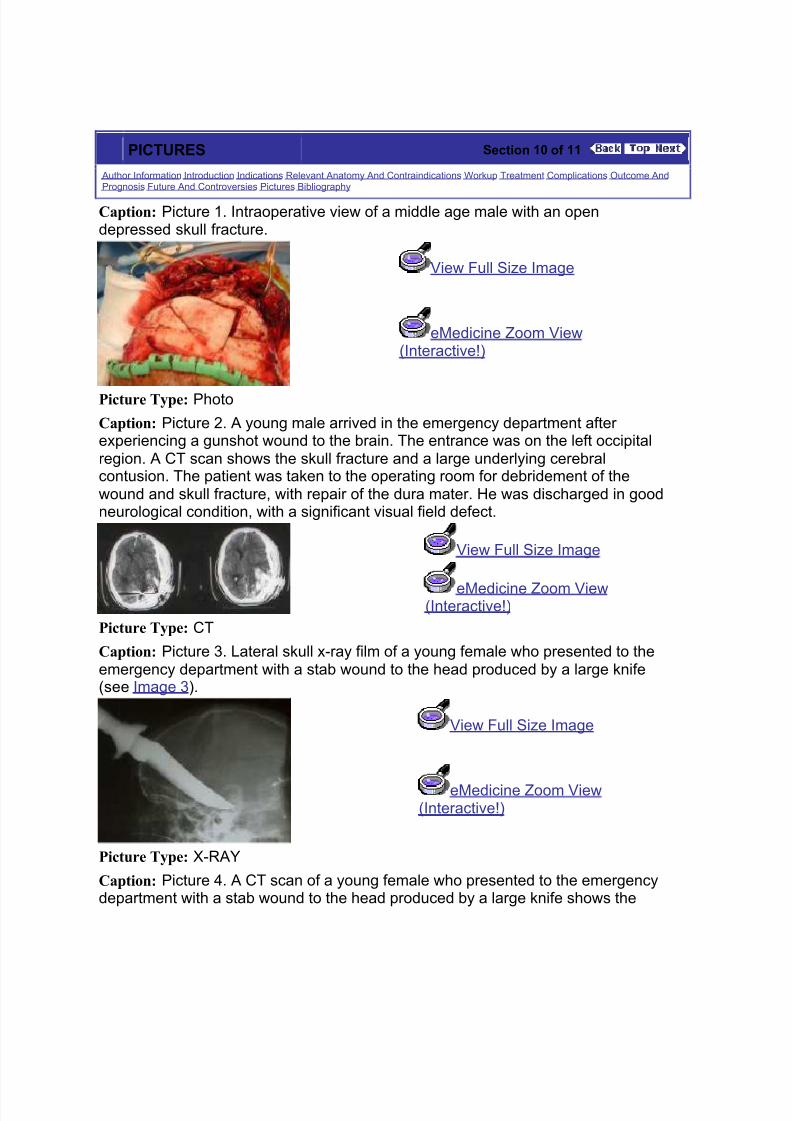
Caption: Picture 1. Intraoperative view of a middle age male with an opendepressed skull fracture.
View Full Size Image
eMedicine Zoom View(Interactive!)
Picture Type: Photo
Caption: Picture 2. A young male arrived in the emergency department after experiencing a gunshot wound to the brain. The entrance was on the left occipitalregion. A CT scan shows the skull fracture and a large underlying cerebralcontusion. The patient was taken to the operating room for debridement of thewound and skull fracture, with repair of the dura mater. He was discharged in goodneurological condition, with a significant visual field defect.
View Full Size Image
eMedicine Zoom View(Interactive!)
Picture Type: CT
Caption: Picture 3. Lateral skull x-ray film of a young female who presented to theemergency department with a stab wound to the head produced by a large knife(see Image 3).
View Full Size Image
eMedicine Zoom View(Interactive!)
Picture Type: X-RAY
Caption: Picture 4. A CT scan of a young female who presented to the emergencydepartment with a stab wound to the head produced by a large knife shows the
8/3/2019 Penetrating Head Trauma, Journal
http://slidepdf.com/reader/full/penetrating-head-trauma-journal 20/26
extent of intracranial damage, affecting midline structures (see Image 2).
View Full Size Image
eMedicine Zoom View(Interactive!)
Picture Type: CT
Caption: Picture 5. Lateral skull x-ray film of a patient who presented with a severeintracranial injury produced by a golf club (see Image 5).
View Full Size Image
eMedicine Zoom View(Interactive!)
Picture Type: X-RAY
Caption: Picture 6. The patient presented to the emergency department with a golf
club in his head. The club was removed in the operating room (see Image 4).
View Full Size Image
eMedicine Zoom View(Interactive!)
Picture Type: Photo
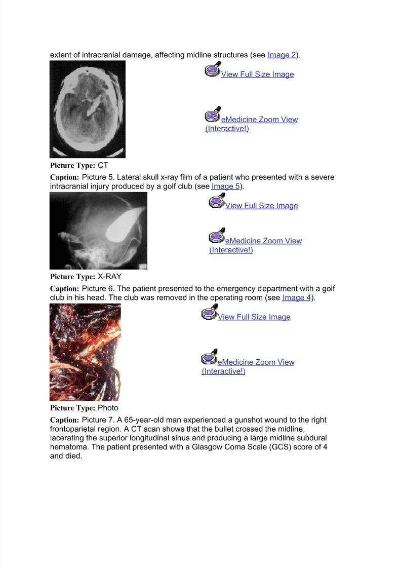
Caption: Picture 7. A 65-year-old man experienced a gunshot wound to the rightfrontoparietal region. A CT scan shows that the bullet crossed the midline,lacerating the superior longitudinal sinus and producing a large midline subduralhematoma. The patient presented with a Glasgow Coma Scale (GCS) score of 4and died.
8/3/2019 Penetrating Head Trauma, Journal
http://slidepdf.com/reader/full/penetrating-head-trauma-journal 21/26
View Full Size Image
eMedicine Zoom View(Interactive!)
Picture Type: CT
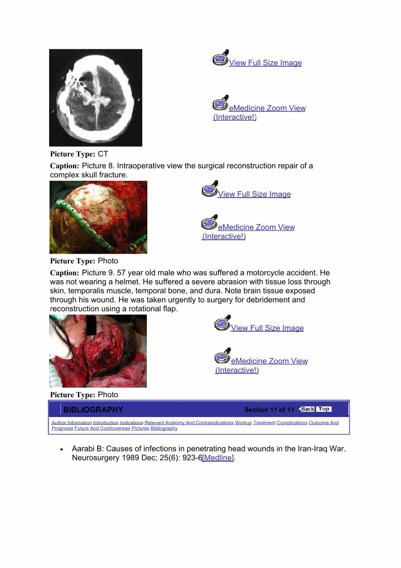
Caption: Picture 8. Intraoperative view the surgical reconstruction repair of acomplex skull fracture.
View Full Size Image
eMedicine Zoom View(Interactive!)
Picture Type: Photo
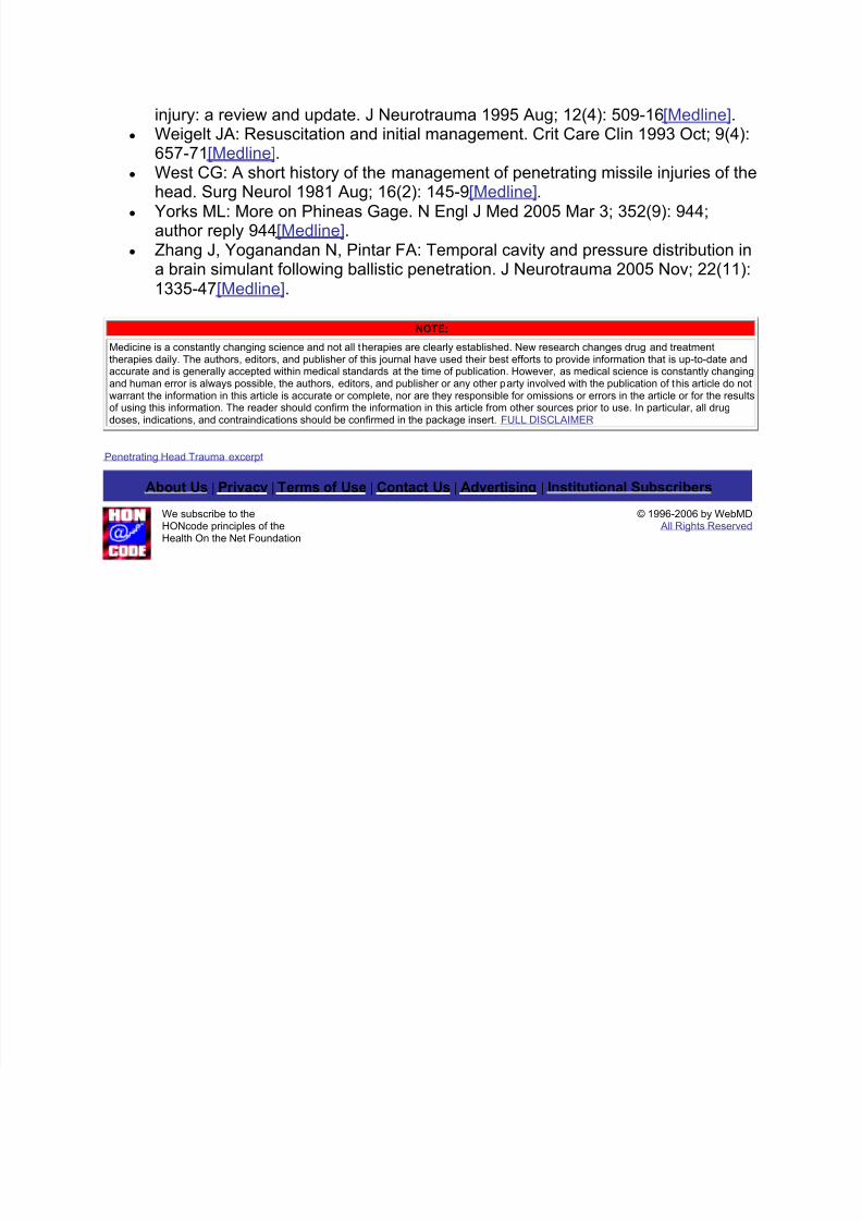
Caption: Picture 9. 57 year old male who was suffered a motorcycle accident. Hewas not wearing a helmet. He suffered a severe abrasion with tissue loss throughskin, temporalis muscle, temporal bone, and dura. Note brain tissue exposed
through his wound. He was taken urgently to surgery for debridement andreconstruction using a rotational flap.
View Full Size Image
eMedicine Zoom View(Interactive!)
Picture Type: PhotoBIBLIOGRAPHY Section 11 of 11
Author Information Introduction Indications Relevant Anatomy And Contraindications Workup Treatment Complications Outcome AndPrognosis Future And Controversies Pictures Bibliography
y Aarabi B: Causes of infections in penetrating head wounds in the Iran-Iraq War.Neurosurgery 1989 Dec; 25(6): 923-6[Medline].

8/3/2019 Penetrating Head Trauma, Journal
http://slidepdf.com/reader/full/penetrating-head-trauma-journal 22/26
y Aarabi B: Traumatic aneurysms of brain due to high velocity missile headwounds. Neurosurgery 1988 Jun; 22(6 Pt 1): 1056-63[Medline].
y Aarabi B: History of the management of craniocerebral wounds. In: Aarabi B,Kaufman HH, Dagi TF, George ED, Levy ML, eds. Missile Wounds of the Headand Neck. Vol 1 . Park Ridge, Ill: American Association of Neurological
Surgeons; 1999: 281-292.y American College of Surgeons: Advanced Trauma Life Support Guidelines.
Advanced Trauma Life Support Course. American College of Surgeons 1999.y Annegers JF, Hauser WA, Coan SP: A population-based study of seizures after
traumatic brain injuries. N Engl J Med 1998 Jan 1; 338(1): 20-4[Medline].y Arabi B: Surgical outcome in 435 patients who sustained missile head wounds
during the Iran-Iraq War. Neurosurgery 1990 Nov; 27(5): 692-5; discussion695[Medline].
y Ardill W, Gidado S: Penetrating head wound: a remarkable case. Surg Neurol2003 Aug; 60(2): 120-3; discussion 123[Medline].
y Baker CC, Oppenheimer L, Stephens B: Epidemiology of trauma deaths. Am J
Surg 1980 Jul; 140(1): 144-50[Medline].y Bartholomew BJ, Poole C, Tayag EC: Unusual transoral penetrating injury of the
foramen magnum: case report. Neurosurgery 2003 Oct; 53(4): 989-91;discussion 991[Medline].
y Benzel EC, Day WT, Kesterson L: Civilian craniocerebral gunshot wounds.Neurosurgery 1991 Jul; 29(1): 67-71; discussion 71-2[Medline].
y Berjano R, Vinas FC, Dujovny M: A review of dural substitutes used inneurosurgery. CRITICAL REVIEWS IN NEUROSURGERY 1999 Jul 28; 9(4):217-222[Medline].
y Breased JH: The Edwin Smith Surgical Papyrus. In: The Edwin Smith SurgicalPapyrus. Vol 1. Chicago, Ill: University of Chicago Press; 1930: 165-166.
y
Camuscu H, Dujovny M, Abd el-Bary T: Microanatomy of the perforators of theanterior communicating artery complex. Neurol Res 1997 Dec; 19(6): 577-87[Medline].
y Centers for Disease Control: Traumatic brain injury--Colorado, Missouri,Oklahoma, and Utah, 1990- 1993. MMWR Morb Mortal Wkly Rep 1997 Jan 10;46(1): 8-11[Medline].
y Chesnut RM, Marshall LF, Marshall SB: Medical management of intracranialpressure. In: Cooper PR, ed. Head Injury, 3rd ed. Baltimore, Md: Williams &Wilkins; 1993: 225-246.
y Coplin WM, Cullen NK, Policherla PN: Safety and feasibility of craniectomy withduraplasty as the initial surgical intervention for severe traumatic brain injury. J
Trauma 2001 Jun; 50(6): 1050-9[Medline].y CoÅar A, Gönül E, Kurt E: Craniocerebral gunshot wounds: results of less
aggressive surgery and complications. Minim Invasive Neurosurg 2005 Apr;48(2): 113-8[Medline].
y CoÅar A, Gönül E, Kurt E: Craniocerebral gunshot wounds: results of lessaggressive surgery and complications. Minim Invasive Neurosurg 2005 Apr;48(2): 113-8[Medline].
y Dagi TF, Meyer FB, Poletti CA: The incidence and prevention of meningitis after

8/3/2019 Penetrating Head Trauma, Journal
http://slidepdf.com/reader/full/penetrating-head-trauma-journal 23/26
basilar skull fracture. Am J Emerg Med 1983 Nov; 1(3): 295-8[Medline].y De Villiers JC: Stab wounds of the brain and skull. In: Vinken PJ, Bruyn GW, eds.
Handbook of clinical neurology. Vol 23. New York, NY: Elsevier SciencePublishing; 1975: 407-503.
y Erdogan E, Izci Y, Gonul E: Ventricular injury following cranial gunshot wounds:
clinical study. Mil Med 2004 Sep; 169(9): 691-5[Medline].y Feldman Z, Narayan RK, Robertson CS: Secondary insults associated with
severe closed head injury. Contemporary Neurosurgery 1992; 14: 1-8.y Giannotta SL, Gruen P: Vascular complications of head trauma. In: Barrow DL,
ed. Complications and sequelae of head injury. Park Ridge, Ill: American Association of Neurological Surgeons; 1992: 31-49.
y Goldstein M: Traumatic brain injury: a silent epidemic. Ann Neurol 1990 Mar;27(3): 327[Medline].
y Grahm TW, Williams FC Jr, Harrington T: Civilian gunshot wounds to the head: aprospective study. Neurosurgery 1990 Nov; 27(5): 696-700; discussion700[Medline].
y Gray J, Molloy D, Jenkins MG: "Glass in a scalp laceration": an unusual case of penetrating head injury presenting to the emergency department. Eur J EmergMed 2004 Apr; 11(2): 117-8[Medline].
y Harris ME, Barrow D: Traumatic carotid-cavernous fistulas. In: Barrow DL, ed.Complications and sequelae of head injury. Park Ridge, Ill: American Associationof Neurological Surgeons; 1992: 13-30.
y Hida K, Tsuda E, Sato H: Brain abscess discovered 38 years after head injury(author's transl). No Shinkei Geka 1978 Aug; 6(8): 811-3[Medline].
y Hoffmann B, Sepehrnia A: Taylored implants for alloplastic cranioplasty--clinicaland surgical considerations. Acta Neurochir Suppl 2005; 93: 127-9[Medline].
y Izci Y, Kayali H, Daneyemez M: The clinical, radiological and surgical
characteristics of supratentorial penetrating craniocerebral injuries: aretrospective clinical study. Tohoku J Exp Med 2003 Sep; 201(1): 39-46[Medline].
y Izci Y, Kayali H, Daneyemez M: Comparison of clinical outcomes betweenanteroposterior and lateral penetrating craniocerebral gunshot wounds. EmergMed J 2005 Jun; 22(6): 409-10[Medline].
y Kaufman HH, Timberlake G, Voelker J: Medical complications of head injury.Med Clin North Am 1993 Jan; 77(1): 43-60[Medline].
y Kaufman HH, Schwab K, Salazar AM: A national survey of neurosurgical care for penetrating head injury. Surg Neurol 1991 Nov; 36(5): 370-7[Medline].
y Knightly JJ, Pulliam MW: Military head injuries. In: Narayan R, Wilberger J,
Povlishock J, eds. Neurotrauma. New York, NY: McGraw Hill; 1996.y Koçak A, OZer MH: Intracranial migrating bullet. Am J Forensic Med Pathol
2004 Sep; 25(3): 246-50[Medline].y Kriet JD, Stanley RB, Grady MS: Self-inflicted submental and transoral gunshot
wounds that produce nonfatal brain injuries: management and prognosis. JNeurosurg 2005 Jun; 102(6): 1029-32[Medline].
y Levy ML, Masri LS, Lavine S: Outcome prediction after penetratingcraniocerebral injury in a civilian population: aggressive surgical management in

8/3/2019 Penetrating Head Trauma, Journal
http://slidepdf.com/reader/full/penetrating-head-trauma-journal 24/26
patients with admission Glasgow Coma Scale scores of 3, 4, or 5. Neurosurgery1994 Jul; 35(1): 77-84; discussion 84-5[Medline].
y Muizelaar JP, Marmarou A, Ward JD: Adverse effects of prolongedhyperventilation in patients with severe head injury: a randomized clinical trial. JNeurosurg 1991 Nov; 75(5): 731-9[Medline].
y Murano T, Mohr AM, Lavery RF: Civilian craniocerebral gunshot wounds: anupdate in predicting outcomes. Am Surg 2005 Dec; 71(12): 1009-14[Medline].y Nelson TJ, Wall DB, Stedje-Larsen ET: Predictors of mortality in close proximity
blast injuries during operation iraqi freedom. J Am Coll Surg 2006 Mar; 202(3):418-22[Medline].
y Nicol A: Gunshot wounds. S Afr J Surg 2005 Nov; 43(4): 150, 152[Medline].y Perez-Arjona E, Dujovny M, Viñas F: CNS child abuse: epidemiology and
prevention. Neurol Res 2002 Jan; 24(1): 29-40[Medline].y Phillips ED: Greek Medicine. London, UK: Thames & Hudsen; 1973.y Price DJ, Sleigh JD: Control of infection due to Klebsiella aerogenes in a
neurosurgical unit by withdrawal of all antibiotics. Lancet 1970 Dec 12; 2(7685):
1213-5[Medline].y Ratiu P, Talos IF, Haker S: The tale of Phineas Gage, digitally remastered. J
Neurotrauma 2004 May; 21(5): 637-43[Medline].y Rezai AR, Lee M, Kite C: Traumatic posterior cerebral artery aneurysm
secondary to an intracranial nail: case report. Surg Neurol 1994 Oct; 42(4): 312-5[Medline].
y Rosenberg WS, Harsh GR: Penetraing wounds of the head. In: Wilkins RH,Rengachary SS, eds. Neurosurgery. Vol 2. New York, NY: McGraw Hill; 1996:2813-2820.
y Rosenwasser RH, Andrews DW, Jimenez DF: Penetrating craniocerebraltrauma. Surg Clin North Am 1991 Apr; 71(2): 305-16[Medline].
y
Rosselli D: [Phineas Gage, 'Tan' and the importance of case reports]. Rev Neurol2005 Jan 16-31; 40(2): 122-4[Medline].y Salar G, Costella GB, Mottaran R: Multiple craniocerebral injuries from
penetrating nails. Case illustration. J Neurosurg 2004 May; 100(5): 963[Medline].y Salazar AM, Aarabi B, Levi L: Postraumatic epilepsy following craniocerebral
missile wounds in recent armed conflicts. In: Aarabi B, Kaufman HH, Dagi TF,George ED, Levy ML, eds. Missile Wounds of the Head and Neck. Vol 2. ParkRidge, Ill: American Association of Neurological Surgeons; 1999: 281-292.
y Siccardi D, Cavaliere R, Pau A: Penetrating craniocerebral missile injuries incivilians: a retrospective analysis of 314 cases. Surg Neurol 1991 Jun; 35(6):455-60[Medline].
y
Sinha P, Conrad GR, Williams BL: Visualization of bullet track and bullet byradionuclide brain scintigraphy. Clin Nucl Med 2005 Apr; 30(4): 249-52[Medline].y Sosin DM, Sacks JJ, Smith SM: Head injury-associated deaths in the United
States from 1979 to 1986. JAMA 1989 Oct 27; 262(16): 2251-5[Medline].y Sosin DM, Sniezek JE, Waxweiler RJ: Trends in death associated with traumatic
brain injury, 1979 through 1992. Success and failure. JAMA 1995 Jun 14;273(22): 1778-80[Medline].
y Stack BC Jr, Farrior JB: Missile injuries to the temporal bone. South Med J 1995

8/3/2019 Penetrating Head Trauma, Journal
http://slidepdf.com/reader/full/penetrating-head-trauma-journal 25/26
Jan; 88(1): 72-8[Medline].y Steinsvåg S: [Penetrating injuries in the head and neck region]. Tidsskr Nor
Laegeforen 2005 Sep 8; 125(17): 2369[Medline].y Stiernberg CM, Jahrsdoerfer RA, Gillenwater A: Gunshot wounds to the head
and neck. Arch Otolaryngol Head Neck Surg 1992 Jun; 118(6): 592-7[Medline].
y Temkin NR, Dikmen SS, Wilensky AJ: A randomized, double-blind study of phenytoin for the prevention of post-traumatic seizures. N Engl J Med 1990 Aug23; 323(8): 497-502[Medline].
y Torner JC, Choi S, Barnes TY: Epidemiology of head injuries. In: Marion DW, ed.Traumatic Brain Injury. New York, NY: Thieme; 1999: 9-25.
y Trask TW, Narayan RK: Civilian Penetrating Head Injury. In: Narayan R,Wilberger J, Povlishock, J, eds. Neurotrauma. New York, NY: McGraw Hill; 1996:868-889.
y Tsuei YS, Sun MH, Lee HD: Civilian gunshot wounds to the brain. J Chin Med Assoc 2005 Mar; 68(3): 126-30[Medline].
y Tudor M, Tudor L, Tudor KI: Complications of missile craniocerebral injuries
during the Croatian Homeland War. Mil Med 2005 May; 170(5): 422-6[Medline].y Verweij BH, Muizelaar JP, Vinas FC: Mitochondrial dysfunction after
experimental and human brain injury and its possible reversal with a selective N-type calcium channel antagonist (SNX-111). Neurol Res 1997 Jun; 19(3): 334-9[Medline].
y Verweij BH, Muizelaar JP, Vinas FC: Hyperacute measurement of intracranialpressure, cerebral perfusion pressure, jugular venous oxygen saturation, andlaser Doppler flowmetry, before and during removal of traumatic acute subduralhematoma. J Neurosurg 2001 Oct; 95(4): 569-72[Medline].
y Vetter H, Kolloch R, Appenheimer M: [Effect of a chronic alpha adrenergicreceptor blockade on basal secretion of renin in essential hypertension]. Schweiz
Med Wochenschr 1978 Dec 9; 108(49): 1978-81[Medline].y Vinas FC: Clinical Uses of Laser Doppler in the Intensive care Unit. Critical
Reviews in Neurosurgery 1999; 9: 28-33.y Vinas FC, Verweij B, Muizelaar P: Invasive monitoring of cerebral oxygenation.
Critical Reviews in Neurosurgery 1998; 8: 31-40.y Vinas FC, Fandino R, Dujovny M: Microsurgical anatomy of the supratentorial
arachnoidal trabecular membranes and cisterns. Neurol Res 1994 Dec; 16(6):417-24[Medline].
y Viñas FC: Bedside invasive monitoring techniques in severe brain-injuredpatients. Neurol Res 2001 Mar-Apr; 23(2-3): 157-66[Medline].
y Viñas FC, Ferris D, Kupsky WJ: Evaluation of expanded polytetrafluoroethylene
(ePTFE) versus polydioxanone (PDS) for the repair of dura mater defects. NeurolRes 1999 Apr; 21(3): 262-8[Medline].y Viñas FJ, Dujovny M, Barrionuevo PJ: [The craniocerebral injury. Experience
on 3,443 cases in the police health department]. Rev Fac Cienc Med Cordoba1966 Oct-Dec; 24(4): 441-57[Medline].
y Wald SL: Advances in the early management of patients with head injury. SurgClin North Am 1995 Apr; 75(2): 225-42[Medline].
y Waxweiler RJ, Thurman D, Sniezek J: Monitoring the impact of traumatic brain
8/3/2019 Penetrating Head Trauma, Journal
http://slidepdf.com/reader/full/penetrating-head-trauma-journal 26/26
injury: a review and update. J Neurotrauma 1995 Aug; 12(4): 509-16[Medline].y Weigelt JA: Resuscitation and initial management. Crit Care Clin 1993 Oct; 9(4):
657-71[Medline].y West CG: A short history of the management of penetrating missile injuries of the
head. Surg Neurol 1981 Aug; 16(2): 145-9[Medline].
y Yorks ML: More on Phineas Gage. N Engl J Med 2005 Mar 3; 352(9): 944;author reply 944[Medline].y Zhang J, Yoganandan N, Pintar FA: Temporal cavity and pressure distribution in
a brain simulant following ballistic penetration. J Neurotrauma 2005 Nov; 22(11):1335-47[Medline].
NOTE:
Medicine is a constantly changing science and not all therapies are clearly established. New research changes drug and treatmenttherapies daily. The authors, editors, and publisher of this journal have used their best efforts to provide information that is up-to-date andaccurate and is generally accepted within medical standards at the time of publication. However, as medical science is constantly changingand human error is always possible, the authors, editors, and publisher or any other party involved with the publication of this article do notwarrant the information in this article is accurate or complete, nor are they responsible for omissions or errors in the article or for the resultsof using this information. The reader should confirm the information in this article from other sources prior to use. In particular, all drugdoses, indications, and contraindications should be confirmed in the package insert. FULL DISCLAIMER
Penetrating Head Trauma excerpt
About Us | Privacy | Ter ms of Use | Contact Us | Advertising | Institutional Subscribers
We subscribe to theHONcode principles of theHealth On the Net Foundation
© 1996-2006 by WebMD All Rights Reserved


![Penetrating Trauma in Pediatric Patients(1) [Read-Only] · Penetrating Trauma in Pediatric Patients Heidi P. Cordi, MD, MPH, MS, EMTP, FACEP, FAADM EMS WEEK 2017. Introduction Trauma](https://static.fdocuments.in/doc/165x107/6004fbd56845b17933089db5/penetrating-trauma-in-pediatric-patients1-read-only-penetrating-trauma-in-pediatric.jpg)