Maxillofacial Trauma - srmuniv.ac.in · Pathophysiology • Maxillofacial fractures result from...
70
Maxillofacial Trauma DEPT OF ORAL & MAX FAX SURGERY SRM DENTAL COLLEGE KATTANKULATHUR
-
Upload
trankhuong -
Category
Documents
-
view
226 -
download
1
Transcript of Maxillofacial Trauma - srmuniv.ac.in · Pathophysiology • Maxillofacial fractures result from...
Maxillofacial Trauma
DEPT OF ORAL & MAX FAX SURGERYSRM DENTAL COLLEGEKATTANKULATHUR
Pathophysiology
• Maxillofacial fractures result from either blunt or penetrating trauma.
• Penetrating injuries are more common in city hospitals.– Midfacial and zygomatic injuries.
• Blunt injuries are more frequently seen in community hospitals.– Nose and mandibular injuries.
Presenter
Presentation Notes
Maxillofacial fractures result from either blunt or penetrating trauma. Penetrating injuries are more common in city hospitals. --------Midfacial and zygomatic injuries. Blunt injuries are more frequently seen in community hospitals. --------Nose and mandibular injuries.
Pathophysiology
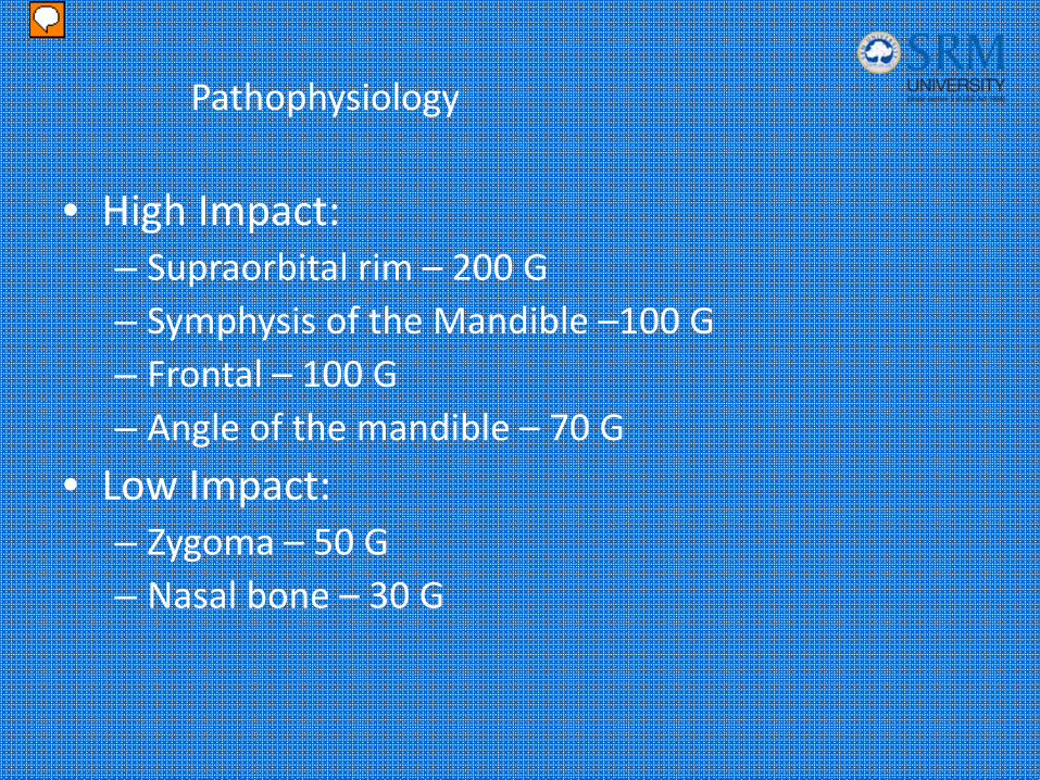
• High Impact:– Supraorbital rim – 200 G– Symphysis of the Mandible –100 G– Frontal – 100 G– Angle of the mandible – 70 G
• Low Impact:– Zygoma – 50 G– Nasal bone – 30 G
Presenter
Presentation Notes
The amount of force to fracture different facial bones have been studied and have been divided into High impact (greater then 50 times gravity) and Low Impact (less then 50 times gravity). High Impact: Supraorbital rim – 200 G Symphysis of the Mandible –100 G Frontal – 100 G Angle of the mandible – 70 G Low Impact: Zygoma – 50 G Nasal bone – 30 G
Etiology
• @60% of patients with severe facial trauma have multisystem trauma and the potential for airway compromise.– 20‐50% concurrent brain injury.
– 1‐4% cervical spine injuries.
– Blindness occurs in 0.5‐3%
Presenter
Presentation Notes
@60% of patients with severe facial trauma have multisystem trauma and the potential for airway compromise. As many as 20-50% of the patients with facial trauma sustain concurrent brain injury, especially those with upper face and midface fractures. 1-4% of these patients have cervical spine injuries. Remember to always r/o cervical spine injuries clinically and radiographically in such patients. Blindness can occur in .5-3% of these trauma pts and are mostly seen in patients with Lefort 3 (2.2%) and Lefort 2 (.64%).
Etiology
• 25% of women with facial trauma are victims of domestic violence.– Increases to 30% if an orbital wall fx is present.
• 25% of patients with severe facial trauma will develop Post Traumatic Stress Disorder
Presenter
Presentation Notes
As many as one forth of the women with facial trauma are victims of domestic violence. If a women has an orbital fracture, the likelihood of sexual assault or domestic violence increases to more then 30%. In addition to physical consequences of facial trauma, there are psychological costs as well. More then a one quarter of patients with severe facial trauma will develop Post Traumatic Stress Disorder.
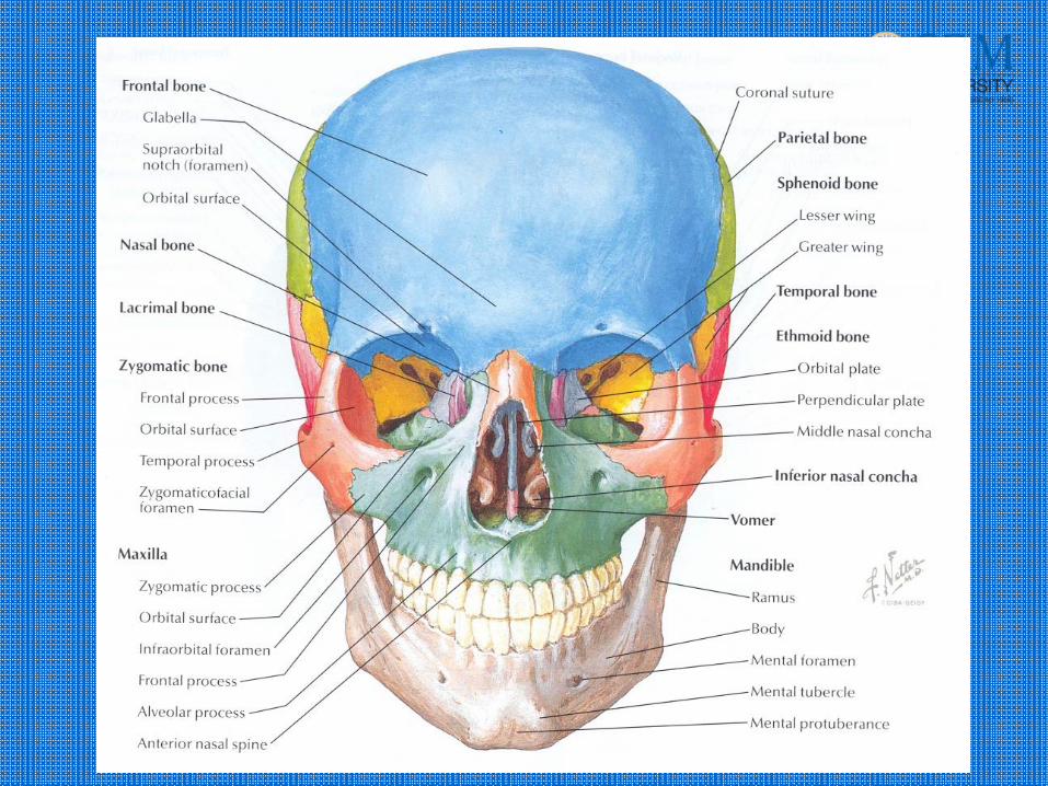
Anatomy
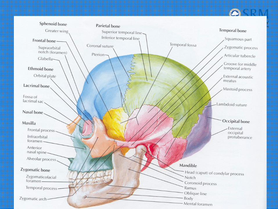
Anatomy

Emergency ManagementAirway Control
• Control airway:– Chin lift.
– Jaw thrust.
– Oropharyngeal suctioning.
– Manually move the tongue forward.
– Maintain cervical immobilization
Presenter
Presentation Notes
Emergency management consists of Controlling the airway: Chin lift. Jaw thrust. Oropharyngeal suctioning. Manually move the tongue forward. Maintain cervical immobilization
Emergency ManagementIntubation Considerations
• Avoid nasotracheal intubation:– Nasocranial intubation
– Nasal hemorrhage
• Avoid Rapid Sequence Intubation:– Failure to intubate or ventilate.
• Consider an awake intubation.
• Sedate with benzodiazepines.
Presenter
Presentation Notes
Since the cribiform plate may be disrupted, avoid nasotracheal intubation. Nasotracheal intubation can result in nasocranial intubation or dramatic nasal hemorrhage. RSI carries a particular risk in facial trauma. These include failure to intubate and subsequent failure to ventilate with a bag valve mask 2nd to facial distortion and should also be avoided. Consider awake intubation and use Benzodiazepines or another induction agent that minimizes resp. depression. If paralytics are used, be prepared for immediate backup cricothyroidotomy.
Emergency ManagementIntubation Considerations
• Consider fiberoptic intubation if available.
• Alternatives include percutaneous transtracheal ventilation and retrograde intubation.
• Be prepared for cricothyroidotomy.
Presenter
Presentation Notes
If available, fiberoptic intubation may be helpful. Other alternatives include percutaneous transtracheal ventilation and retrograde intubation. Always be prepared to perform cricothyroidotomy.
Emergency ManagementHemorrhage Control
• Maxillofacial bleeding:– Direct pressure.– Avoid blind clamping in wounds.
• Nasal bleeding:– Direct pressure.– Anterior and posterior packing.
• Pharyngeal bleeding:– Packing of the pharynx around ET tube.
Presenter
Presentation Notes
Bleeding from facial injuries typically is profuse but rarely causes hypovolemia or shock. In hypotensive patients, look for other sources of blood loss such as intrathoracic, intraabdominal, and retroperitoneal hemorrhage. Try to control bleeding with direct pressure. Blind clamping should be avoided because injury to important nonvascular structures such as the facial nerve and parotid duct can result. Anterior and posterior packing may be needed in patients with nasal bleeding that does not resolve with direct pressure alone. Pharyngeal bleeding may require packing around the ET tube. Once the airway is secured and gross hemorrhage is controlled, only then search for life threatening injuries to the chest, abd and pelvis.
History
• Obtain a history from the patient, witnesses and or EMS.
• AMPLE history
• Specific Questions:– Was there LOC? If so, how long?
– How is your vision?
– Hearing problems?
Presenter
Presentation Notes
After your ABC’s and all life threatening injuries have been addressed, Obtain a history from the pt, witnesses and or EMS. A-allergies, M-medications, P-pmedhx, L-last meal, E-events leading to the injury. Specific questions: Was there LOC? If so,how long? How is your vision? Monocular double vision-lens dislocation, corneal/retinal injury. Binocular double vision- dysfunction of the EOM or nerves.
History
• Specific Questions:– Is there pain with eye movement?
– Are there areas of numbness or tingling on your face?
– Is the patient able to bite down without any pain?
– Is there pain with moving the jaw?
Presenter
Presentation Notes
Ask specific questions.: Is there pain with eye movement?-injury to the globe, orbit Are there areas of numbness or tingling on your face?-nerve entrap. Is the patient able to bite down without any pain? Is there pain with moving the jaw?-fx, impingement temporalis m.
Physical Examination
• Inspection of the face for asymmetry.
• Inspect open wounds for foreign bodies.
• Palpate the entire face.– Supraorbital and Infraorbital rim
– Zygomatic‐frontal suture
– Zygomatic arches
Presenter
Presentation Notes
Inspection of the face for asymmetry.Best done at the head of the bed. Ask the patient to smile, frown, whistle, raise their eyebrows, close their eyes. Inspect open wounds for foreign bodies. Palpate the entire face. Supraorbital and Infraorbital rim Zygomatic-frontal suture Zygomatic arches Note ecchymosis (Battle’s sign, Raccoon eyes)
Physical Examination
• Inspect the nose for asymmetry, telecanthus, widening of the nasal bridge.
• Inspect nasal septum for septal hematoma, CSF or blood.
• Palpate nose for crepitus, deformity and subcutaneous air.
• Palpate the zygoma along its arch and its articulations with the maxilla, frontal and temporal bone.
Presenter
Presentation Notes
Inspect the nose for asymmetry, telecanthus, widening of the nasal bridge. Measure the distance between the medial canthi. In normal patients the distance is 35-40mm. If its greater then 40 mm you should suspect nasoethmoid-orbital trauma. Inspect nasal septum for septal hematoma, CSF or blood. (place a drop of blood on a paper towel and look for a halo sign, nonspecific). Palpate nose for crepitus, deformity and subcutaneous air. Palpate the zygoma along its arch and its articulations with the maxilla, frontal and temporal bone.
Physical Examination
• Check facial stability.• Inspect the teeth for malocclusions, bleeding and step‐off.
• Intraoral examination: – Manipulation of each tooth.– Check for lacerations.– Stress the mandible.– Tongue blade test.
• Palpate the mandible for tenderness, swelling and step‐off.
Presenter
Presentation Notes
Open pts mouth and grasp the maxilla arch, place the other hand on the forehead. Push back and forth, up and down and check for movement. Inspect the teeth for malocclusions, bleeding and step-off. If teeth are missing, account for to be sure they have not been aspirated. Intraoral exam: Manipulate each tooth, check for lacerations, stress the mandible, tongue blade test. (Bite down on the tongue blade, Twist the blade to try to break it. Pts with broken jaw will reflexively open their mouth.) 95% sensitive and 65% specific. Palpate the mandible for tenderness, swelling and step-off.
Physical Examination
• Check visual acuity.
• Check pupils for roundness and reactivity.
• Examine the eyelids for lacerations.
• Test extra ocular muscles.
• Palpate around the entire orbits..
Presenter
Presentation Notes
Check visual acuity. Snell chart, finger counting or presence or absence of light perception. Check pupils for roundness and reactivity. Tear drop pupil – ruptured or penetrated globe injury. Examine for exopthalmus or enopthalmus Examine the lids for lacerations. Check for injuries to the medial 3rd of the eyelids for damage to the lacrimal apparatus. Check for disruption of the levator palpebral muscles. Test extra ocular muscles.Testing for restriction. Restriction of upward gaze can be seen with zygomatic or infra orbital wall fx’s. Palpate around the entire orbits. Tenderness, subcutaneous air and deformity. Palpate the medial orbit area to r/o naso ethmoidal orbital fx. (place a Q tip inside the nose to the medial canthus, place your finger outside the medial canthus. If the bone moves NEO fx.)
Physical Examination
• Examine the cornea for abrasions and lacerations.
• Examine the anterior chamber for blood or hyphema.
• Perform fundoscopic exam and examine the posterior chamber and the retina.
Presenter
Presentation Notes
Examine the cornea for abrasions and lacerations. Fluorescein if needed. Examine the anterior chamber for blood or hyphema. When the patient is stable try to do a slit lamp examination. Perform fundoscopic exam and examine the posterior chamber and the retina. Looking for retinal detachment.
Physical Examination
• Examine and palpate the exterior ears.
• Examine the ear canals.
• Check nuero distributions of the supraorbital, infraorbital, inferior alveolar and mental nerves.
Presenter
Presentation Notes
Examine and palpate the exterior ears. Look for ecchymosis, hematomas,battle sign. Examine the ear canal. Look for lacerations, TM ruptures, Place your finger into the ear canal and have the pt open their mouth to check for condylar fx or dislocation. Check nuero distributions of the supraorbital, infraorbital, inferior alveolar and mental nerves. Supraorbital n.- forehead and vertex of scalp. Infraorbital n.- midface,maxillary incisors and premolar teeth, upper lip, lower eyelid, side of nose. Inferior alveolar n.- mandibular teeth, lower lip and chin. Mental n.- chin and lower lip.
Frontal Sinus/ Bone FracturesPathophysiology
• Results from a direct blow to the frontal bone with blunt object.
• Associated with:– Intracranial injuries
– Injuries to the orbital roof
– Dural tears
Presenter
Presentation Notes
Frontal bone and sinus injuries usually are a result from a direct blow to the frontal bone with a blunt object. Classically a lead pipe or brick. They may also result from motor vehicle trauma in which the patients head strikes the dashboard. This fracture is frequently associated with intracranial injury, secondary to disruption of the posterior table of the sinus. Dural tears are frequent and patients may have associated injuries to the orbital roof.
Frontal Sinus/ Bone FracturesClinical Findings
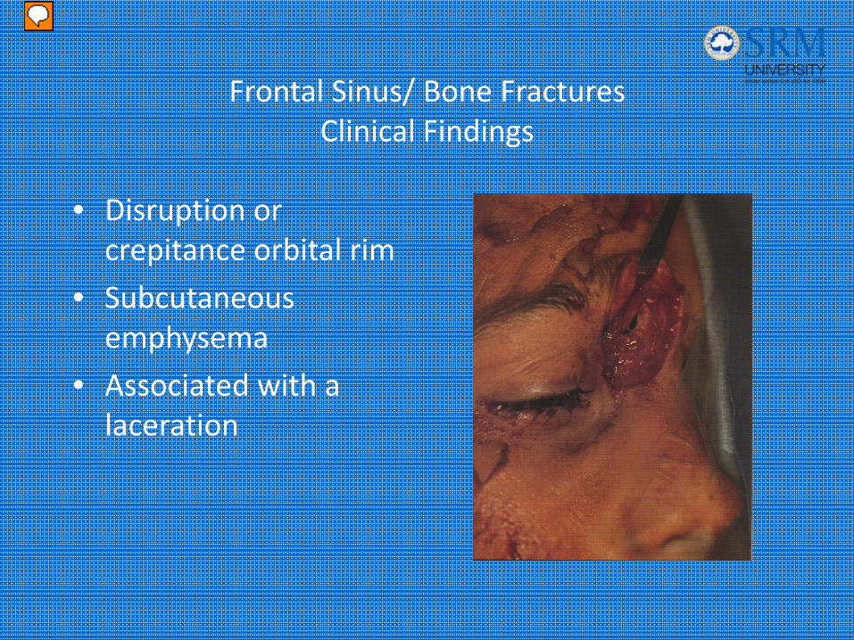
• Disruption or crepitance orbital rim
• Subcutaneous emphysema
• Associated with a laceration
Presenter
Presentation Notes
Examination usually demonstrates step off, crepitance or subcutaneous emphysema of the supraorbital rim. Often, there is an associated laceration. All open wounds should be inspected for foreign body and bony injury. Remember, patients with frontal sinus fractures require through head, neck, and neurological exam, including ophthalmologic consultation. Picture: Fracture defect seen at the base of a laceration over the frontal sinus.
Frontal Sinus/ Bone FracturesDiagnosis
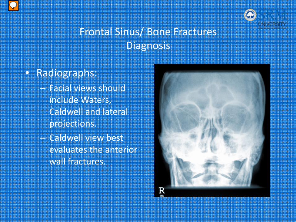
• Radiographs:– Facial views should include Waters, Caldwell and lateral projections.
– Caldwell view best evaluates the anterior wall fractures.
Presenter
Presentation Notes
Patients with a suggested mechanism or PE should get either skull films, or a caldwell view of the face. (Caldwell view is the best for anterior wall fx’s.). Picture: Caldwell view

Frontal Sinus/ Bone FracturesDiagnosis
• CT Head with bone windows:– Frontal sinus fractures.
– Orbital rim and nasoethmoidal fractures.
– R/O brain injuries or intracranial bleeds.
Presenter
Presentation Notes
CT scan of the head with bone windows should be done to r/o intracranial pathology, but also to r/o depressed or posterior fx’s. Picture: CT of a patient which demonstrates a fracture of the anterior table of the frontal sinus.
Frontal Sinus/ Bone FracturesTreatment
• Patients with depressed skull fractures or with posterior wall involvement.– ENT or nuerosurgery consultation.
– Admission.
– IV antibiotics.
– Tetanus.
• Patients with isolated anterior wall fractures, nondisplaced fractures can be treated outpatient after consultation with neurosurgery.
Presenter
Presentation Notes
Patients with depressed skull fractures or with posterior wall involvement require: ENT or nuerosurgery consultation. Admission. IV antibiotics. Tetanus. Patients with isolated anterior wall fractures, nondisplaced fractures can be treated outpatient after consultation with neurosurgery.
Frontal Sinus/ Bone FracturesComplications
• Associated with intracranial injuries:– Orbital roof fractures.
– Dural tears.
– Mucopyocoele.
– Epidural empyema.
– CSF leaks.
– Meningitis.
Presenter
Presentation Notes
Associated with with intracranial injuries: Orbital roof fractures. Dural tears. Mucopyocoele. Epidural empyema. CSF leaks. Meningitis.
Naso‐Ethmoidal‐Orbital Fracture
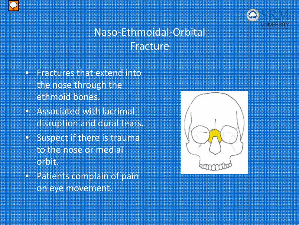
• Fractures that extend into the nose through the ethmoid bones.
• Associated with lacrimal disruption and dural tears.
• Suspect if there is trauma to the nose or medial orbit.
• Patients complain of pain on eye movement.
Presenter
Presentation Notes
NEO fracture is defined as a fracture that extends into the nose through the ethmoid bones. These fractures are associated with lacrimal disruption and dural tears. Suspect this type of a fracture if there is trauma to the nose or medial orbit. Patients typically complain of pain on eye movement.
Naso‐Ethmoidal‐Orbital Fracture
• Clinical findings:– Flattened nasal bridge or a saddle‐shaped deformity of the nose.
– Widening of the nasal bridge (telecanthus)– CSF rhinorrhea or epistaxis.– Tenderness, crepitus, and mobility of the nasal complex.
– Intranasal palpation reveals movement of the medial canthus.
Presenter
Presentation Notes
Clinical findings: Flattened nasal bridge or a saddle-shaped deformity of the nose. Widening of the nasal bridge - telecanthus CSF rhinorrhea or epistaxis. Tenderness, crepitus, and mobility of the nasal complex. Intranasal palpation reveals movement of the medial canthus.
Naso‐Ethmoidal‐Orbital Fracture
• Imaging studies:– Plain radiographs are insensitive.
– CT of the face with coronal cuts through the medial orbits.
• Treatment:– Maxillofacial consultation.
– ? Antibiotic
Presenter
Presentation Notes
Plain radiographs are insensitive. If the examination is suggestive, order a CT of the face to include coronal sections and thin axial slices through the medial orbit wall. If an NEO fracture is present, consult a maxillofacial surgeon. As with many facial fractures, AB’s are frequently prescribed for CSF leaks, but their efficacy is question.
Nasal Fractures
• Most common of all facial fractures.
• Injuries may occur to other surrounding bony structures.
• 3 types:– Depressed
– Laterally displaced
– Nondisplaced
Presenter
Presentation Notes
Most common of all facial fractures. Injuries may occur to other surrounding bony structures. Including fx’s to the orbit, frontal sinus, or cribiform plate. Suspect these injuries if the patient had LOC, Hx of a mechanism with significant force or findings of facial bone injuries. 3 types of nasal fx’s: Depressed Laterally displaced Nondisplaced
Nasal Fractures
• Ask the patient:– “Have you ever broken your nose before?”
– “How does your nose look to you?”
– “Are you having trouble breathing?”
Presenter
Presentation Notes
Ask the patient: “Have you ever broken your nose before?” “How does your nose look to you?” “Are you having trouble breathing?” You want to ask these questions because nasal trauma may be do to old or new trauma.
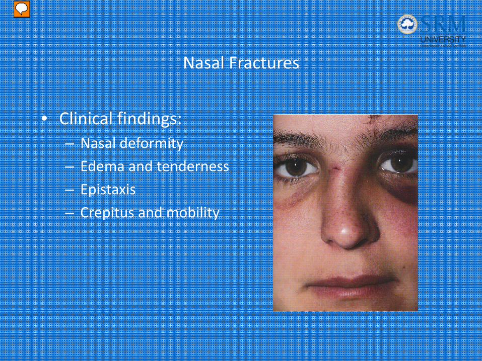
Nasal Fractures
• Clinical findings:– Nasal deformity
– Edema and tenderness
– Epistaxis
– Crepitus and mobility
Presenter
Presentation Notes
On physical exam, observe deformity, palpate for crepitus and mobility. It is important to do an intra nasal exam to inspect for bleeding and for a septal hematoma. If clear rhinorrhea is present, suspect other injuries, such as fx’s to the ethmoid bone. Picture: Deformity is evident on examination. Note peri-ocular ecchymosis indicating the possibility of other injuries.
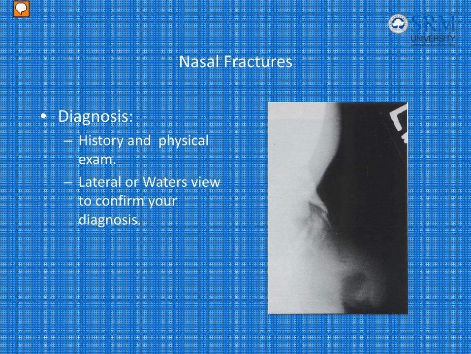
Nasal Fractures
• Diagnosis:– History and physical exam.
– Lateral or Waters view to confirm your diagnosis.
Presenter
Presentation Notes
Nasal bone fx’s can be dx clinically by hx and physical examination. Plain nasal films consisting of a lateral view or a Waters view can confirm your dx but are of little practical use. Picture: Radiograph of a displaced fracture of the nasal spine, for which no treatment other then ice and analgesics is needed.
Nasal Fractures
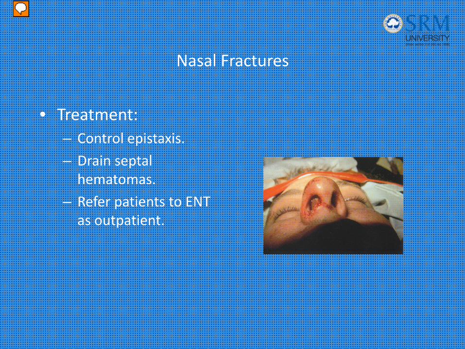
• Treatment:– Control epistaxis.
– Drain septal hematomas.
– Refer patients to ENT as outpatient.
Presenter
Presentation Notes
Treatment: Control epistaxis: Direct pressure Topical vasoconstrictor such as phenylephrine 1% or cocaine Cauterize with silver nitrate. Nasal packing ant/post packing. Draining septal hematoma’s. anesthetize the area. Using a # 11 blade incise the inferior portion of the hematoma and allow it to drain. Then pack the nose with vaseline gauze to prevent reacumulation of blood. If there is no epistaxis or deformity, treat the patient with ice and analgesics. If obvious deformity is present, including new septal deviation or deformity, treat with ice and analgesics and refer to ENT in 2-5 days for reduction. Picture: This is a picture of a septal hematoma, Notice a blueish, grapelike mass on the nasal septum. If untreated, this can result in septal necrosis and a saddle nose deformity.
Orbital Blowout Fractures
• Blow out fractures are the most common.
• Occur when the the globe sustains a direct blunt force
• 2 mechanisms of injury:– Blunt trauma to the globe
– Direct blow to the infraorbital rim
Presenter
Presentation Notes
Blowout fx’s are the most common of the orbital fx’s. They occur when the globe sustains direct blunt force. The first is a true blowout fx, where all the energy is transmitted to the globe. Since the spherical globe is stronger than the thin orbital floor, the force is then transmitted to the thin orbital floor or medially through the ethmoid bones with the resultant fx. The object causing the injury must be smaller then 5-6cm, otherwise the globe is protected by the surrounding orbit. Fists or small balls are the typical causative agents. The second mechanism occurs when the energy from the blow is transmitted to the to the infraorbital rim causing a buckling of the floor. Entrapment and globe injury is less likely with this injury.
Orbital Blowout FracturesClinical Findings
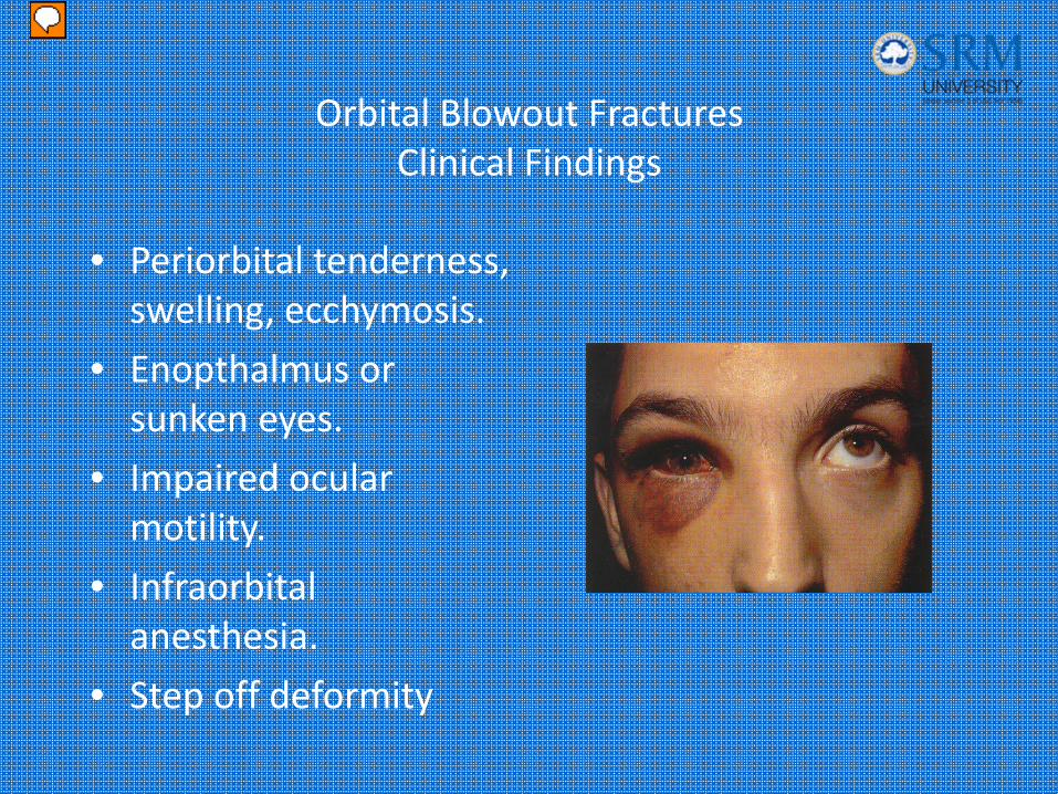
• Periorbital tenderness, swelling, ecchymosis.
• Enopthalmus or sunken eyes.
• Impaired ocular motility.
• Infraorbital anesthesia.
• Step off deformity
Presenter
Presentation Notes
Patients usually present with: 1. Periorbital tenderness, swelling, ecchymosis. 2. Enopthalmus or sunken eyes. 3. Impaired ocular motility. Usually caused by entrapment of the inferior rectus muscle. 4. Infraorbital anesthesia. This develops when the infraorbital nerve is contused by the initial trauma or when the compressed by bony fragments. Anesthesia of the maxillary teeth and upper lip is more reliable then numbness over the cheek. 5. Step off deformity can be appreciated over the infraorbital rim. Subcutaneous emphysema is pathognomonic for a fracture into a sinus or nasal antrum. Picture: The inferior rectus muscle is entrapped within the blow out fx. When the patient tries to look upward, the affected eye has limited upward gaze. The patient experiences diplopia with this maneuver.
Orbital Blowout FracturesImaging studies
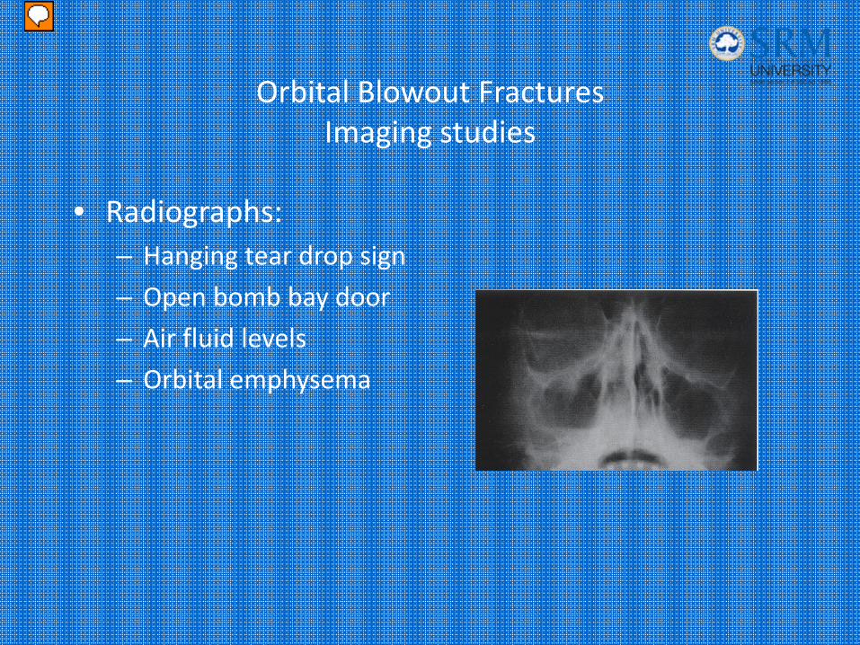
• Radiographs:– Hanging tear drop sign
– Open bomb bay door
– Air fluid levels
– Orbital emphysema
Presenter
Presentation Notes
Facial films should consist of Waters, Caldwell and lateral projections. Waters view is the best for displaying the inferior orbital rims. When reading the films look for the following: Hanging tear drop sign-herniation of the periorbital fat into the maxillary sinus. Open bomb bay door or trap door sign- Bony fragments protrude into the maxillary sinus. Air fluid levels- and maxillary clouding secondary to bleeding. Orbital emphysema X ray: Demonstrates a fx of the floor of the R orbit, with a tear drop sign due to the extruded orbital contents. There is a associated air fluid level in the maxillary sinus due to blood. Atlas pg 15.
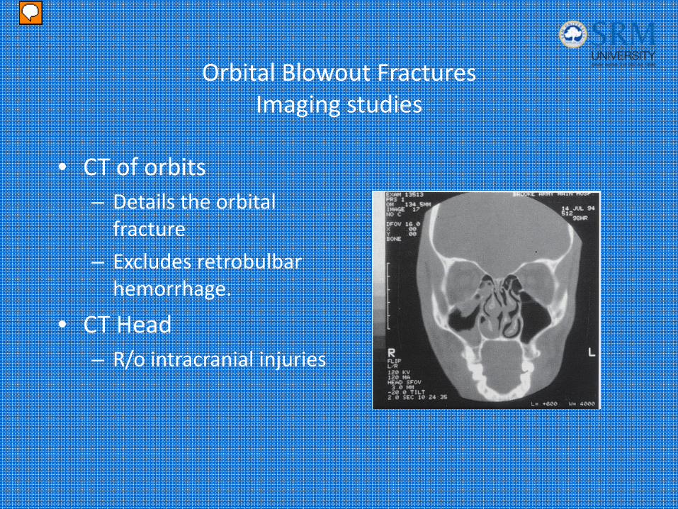
Orbital Blowout FracturesImaging studies
• CT of orbits– Details the orbital fracture
– Excludes retrobulbar hemorrhage.
• CT Head– R/o intracranial injuries
Presenter
Presentation Notes
CT of the orbits with coronal cuts is considered the DOC, Higher sensitivity then radiographs and helps with the planning operative repair. Picture: Ct of a patient demonstrating the entrapped muscle extruding into the maxillary sinus.
Orbital Blowout FracturesTreatment
• Blow out fractures without eye injury do not require admission– Maxillofacial and ophthalmology consultation– Tetanus– Decongestants for 3 days– Prophylactic antibiotics– Avoid valsalva or nose blowing
• Patients with serious eye injuries should be admitted to ophthalmology service for further care.
Presenter
Presentation Notes
Blow out fractures without eye injury do not require admission Maxillofacial and ophthalmology consultation Tetanus Decongestants for 3 days Prophylactic antibiotics Avoid valsalva or nose blowing Patients with serious eye injuries should be admitted to ophthalmology service for further care.
Zygoma Fractures
• The zygoma has 2 major components:– Zygomatic arch
– Zygomatic body
• Blunt trauma most common cause.
• Two types of fractures can occur:– Arch fracture (most common)
– Tripod fracture (most serious)
Presenter
Presentation Notes
The zygoma has 2 major components, the zygomatic arch and the body. The arch forms the the inferior and lateral orbit, and the body forms the malar eminence of the face. Fractures to the zygoma are usually the result of blunt trauma. Direct blow to the arch can result in isolated arch fractures. These are the most common. While tripod fractures are more serious and are caused by more extensive trauma.
Zygoma Arch Fractures
• Can fracture 2 to 3 places along the arch– Lateral to each end of the arch
– Fracture in the middle of the arch
• Patients usually present with pain on opening their mouth.
Presenter
Presentation Notes
Zygoma arch fractures can fracture in 2-3 places along the arch. 1. Lateral to each end of the arch. 2. Fracture in the middle of the arch causing a V fracture which could impinge on the temporalis muscle. Patients usually present with pain on opening their mouth.
Zygoma Arch FracturesClinical Findings
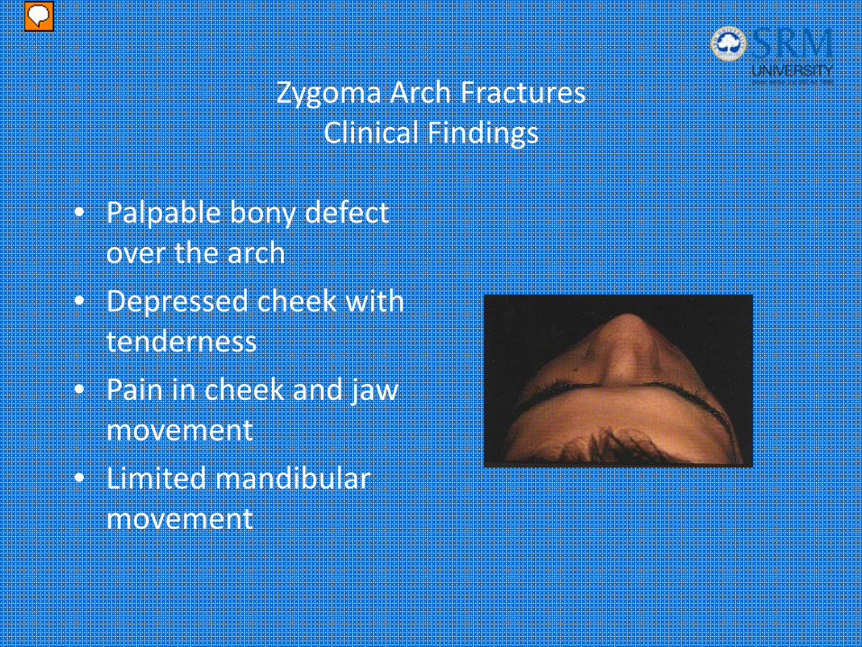
• Palpable bony defect over the arch
• Depressed cheek with tenderness
• Pain in cheek and jaw movement
• Limited mandibular movement
Presenter
Presentation Notes
Clinical findings: Palpable bony defect over the arch Depressed cheek with tenderness on palpation. Pain in cheek and jaw movement and limited mandibular movement which is due to impingement of the coronoid process of the mandible on the arch during mouth opening or impingement of the temporalis muscle. Picture: patient with blunt trauma to the zygoma. Flattening of the right malar eminence is evident.
Zygoma Arch FracturesImaging Studies & Treatment
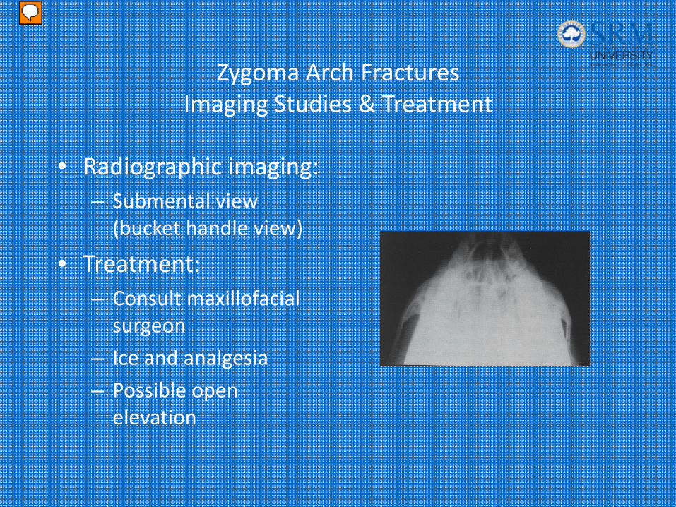
• Radiographic imaging:– Submental view (bucket handle view)
• Treatment:– Consult maxillofacial surgeon
– Ice and analgesia
– Possible open elevation
Presenter
Presentation Notes
X-ray: bucket handle view of the zygomatic arch demonstrating a depressed fracture. Treatment: Consult maxillofacial surgeon Ice and analgesia Possible open elevation if cosmetic correction is desired or if there is entrapment of the mandible persists.
Zygoma Tripod Fractures
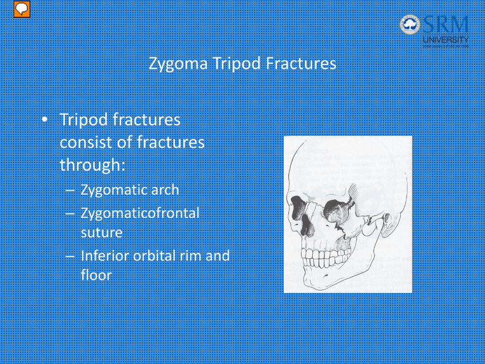
• Tripod fractures consist of fractures through:– Zygomatic arch
– Zygomaticofrontal suture
– Inferior orbital rim and floor
Presenter
Presentation Notes
Tripod fractures consist of fractures through: Zygomatic arch Zygomaticofrontal suture Inferior orbital rim and floor Picture: Diagram of a tripod fracture. Note the disruption of both the lateral orbital rim and the orbital floor, as well as the zygomatic arch.
Zygoma Tripod FracturesClinical Features
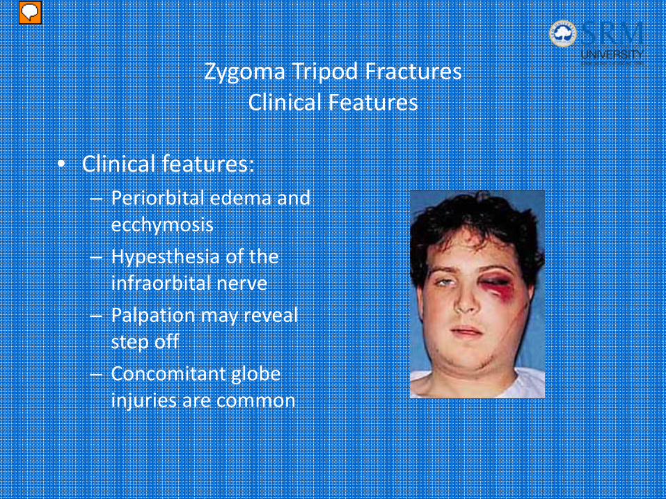
• Clinical features:– Periorbital edema and ecchymosis
– Hypesthesia of the infraorbital nerve
– Palpation may reveal step off
– Concomitant globe injuries are common
Presenter
Presentation Notes
Clinical features: Periorbital edema and ecchymosis Hypesthesia of the infraorbital nerve Palpation may reveal step off Concomitant globe injuries are common
Zygoma Tripod FracturesImaging Studies
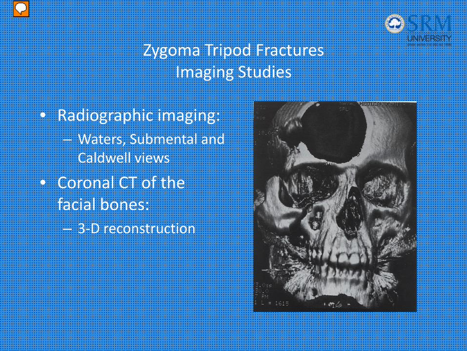
• Radiographic imaging:– Waters, Submental and Caldwell views
• Coronal CT of the facial bones:– 3‐D reconstruction
Presenter
Presentation Notes
Plain films including the waters, submental and caldwell views. Can demonstrate the fracture and evaluate the zygomaticomaxillary complex, but a Coronal CT of the facial bones will best show involvement and the degree of displacement. Picture: CT 3-D. The fracture lines involved in a tripod fracture are demonstrated in this 3-D reconstruction.
Zygoma Tripod FracturesTreatment
• Nondisplaced fractures without eye involvement– Ice and analgesics
– Delayed operative consideration 5‐7 days
– Decongestants
– Broad spectrum antibiotics
– Tetanus
• Displaced tripod fractures usually require admission for open reduction and internal fixation.
Presenter
Presentation Notes
Maxillofacial consultation Nondisplaced fractures without eye involvement Ice and analgesics Delayed operative consideration 5-7 days Decongestants Broad spectrum antibiotics since the fracture crosses into the maxillary sinus. Tetanus Displaced tripod fractures usually require admission for open reduction and internal fixation
Maxillary Fractures
• High energy injuries.
• Impact 100 times the force of gravity is required .
• Patients often have significant multisystem trauma.
• Classified as LeFort fractures.
Presenter
Presentation Notes
Fractures of the maxilla are high energy injuries. An impact 100 times the force of gravity is required to break the midface. These patients often have significant multisystem trauma. Many require resuscitation and admission. The fractures of the maxilla are classified as LeFort Fractures.

Maxillary FracturesLeFort I
• Definition:– Horizontal fracture of the maxilla at the level of the nasal fossa.
– Allows motion of the maxilla while the nasal bridge remains stable.
Presenter
Presentation Notes
LeFort I: Horizontal fracture of the maxilla at the level of the nasal fossa. Allows motion of the maxilla while the nasal bridge remains stable. The fracture is below the infraorbital nerve, so there is no hypesthesia.
Maxillary FracturesLeFort I
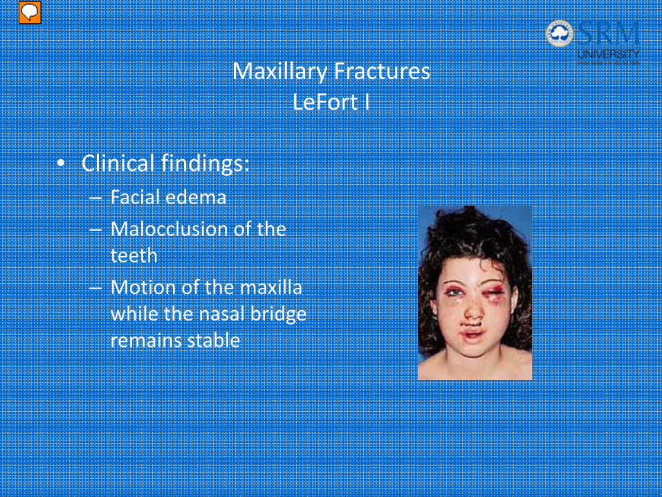
• Clinical findings:– Facial edema
– Malocclusion of the teeth
– Motion of the maxilla while the nasal bridge remains stable
Presenter
Presentation Notes
LeFort I: Physical exam: Facial edema Malocclusion of the teeth Motion of the maxilla while the nasal bridge remains stable
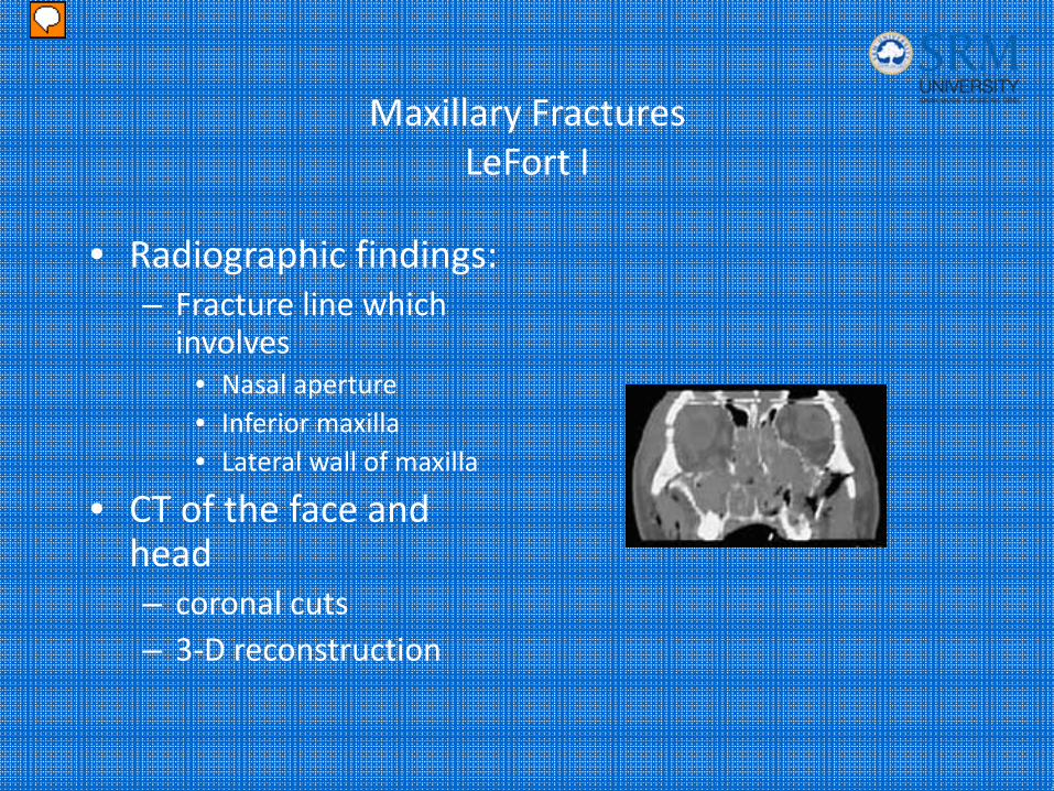
Maxillary FracturesLeFort I
• Radiographic findings:– Fracture line which involves
• Nasal aperture• Inferior maxilla• Lateral wall of maxilla
• CT of the face and head – coronal cuts– 3‐D reconstruction
Presenter
Presentation Notes
Radiographic findings: Fracture line which involves Nasal aperture Inferior maxilla Lateral wall of maxilla CT of the face with coronal cuts is superior to plain films. Head CT should also be done to r/o intracranial injury.
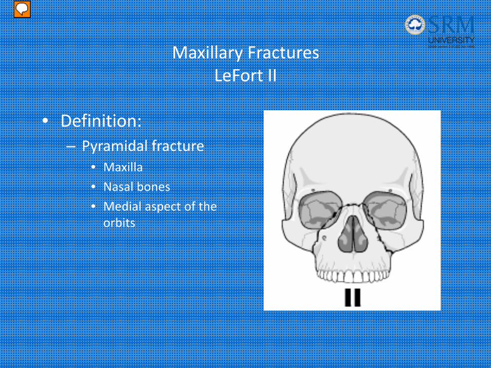
Maxillary FracturesLeFort II
• Definition:– Pyramidal fracture
• Maxilla
• Nasal bones
• Medial aspect of the orbits
Presenter
Presentation Notes
LeFort II: Pyramidal fracture which includes a fracture through: Maxilla Nasal bones Medial aspect of the orbits
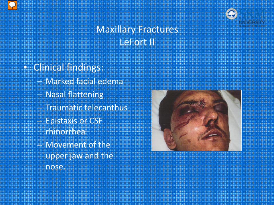
Maxillary FracturesLeFort II
• Clinical findings:– Marked facial edema
– Nasal flattening
– Traumatic telecanthus
– Epistaxis or CSF rhinorrhea
– Movement of the upper jaw and the nose.
Presenter
Presentation Notes
Clinical findings: Marked facial edema Nasal flattening Traumatic telecanthus Epistaxis or CSF rhinorrhea Movement of the upper jaw and the nose. Picture: This patient sustained a Lefort II/III fracture.
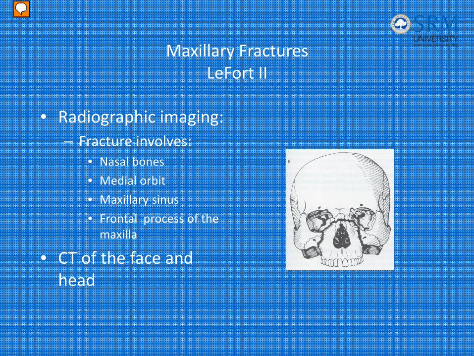
Maxillary FracturesLeFort II
• Radiographic imaging:– Fracture involves:
• Nasal bones
• Medial orbit
• Maxillary sinus
• Frontal process of the maxilla
• CT of the face and head
Presenter
Presentation Notes
Plain facial films will reveal the presence of facial fractures, but are less helpful in determining the type or extent . Head and facial CT, including three dimensional re-creations, offer much more useful information.
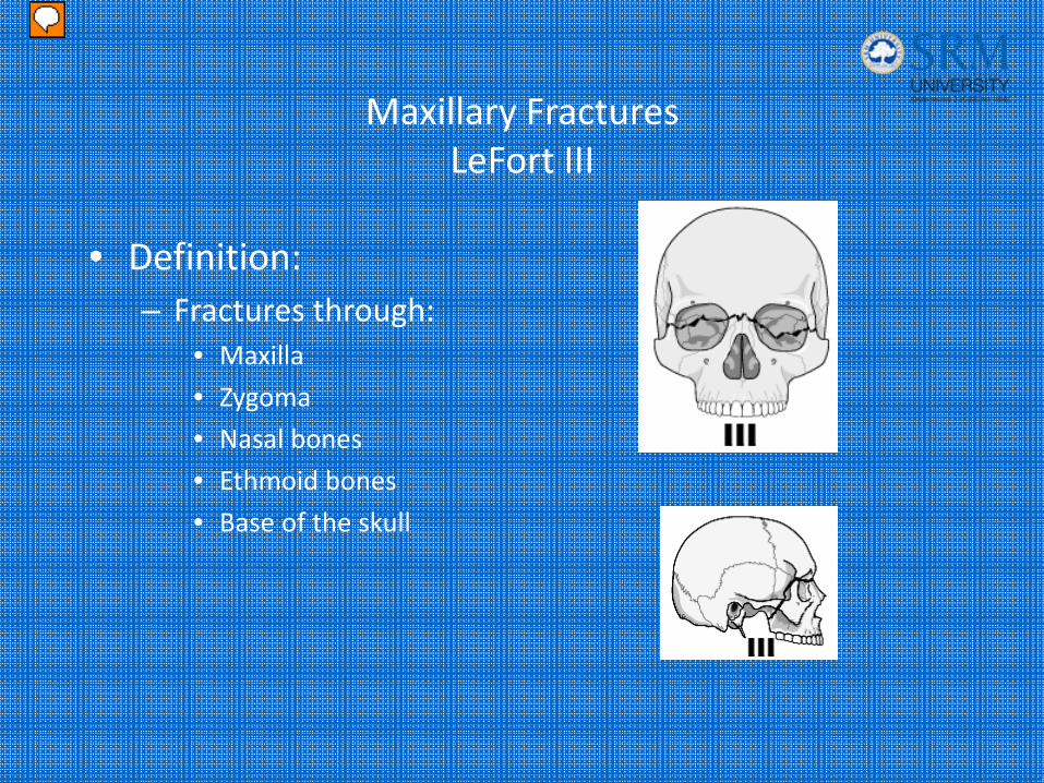
Maxillary FracturesLeFort III
• Definition:– Fractures through:
• Maxilla
• Zygoma
• Nasal bones
• Ethmoid bones
• Base of the skull
Presenter
Presentation Notes
Lefort III fractures also known as craniofacial dissociation(separates the face from the cranium) involves fractures through the maxilla, zygoma, nasal bones, ethmoid bones and the bones of the base of the skull.
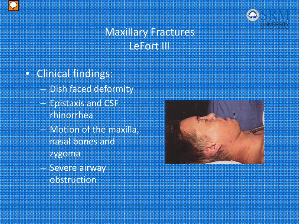
Maxillary FracturesLeFort III
• Clinical findings:– Dish faced deformity
– Epistaxis and CSF rhinorrhea
– Motion of the maxilla, nasal bones and zygoma
– Severe airway obstruction
Presenter
Presentation Notes
Clinical Findings: Patients with LeFort III fractures on physical exam have what’s known as the dish faced deformity which is facial flattening and elongation with the eyes markedly swollen shut. Since the maxilla is pushed inward, the patients mandible appears forward. Epistaxis and CSF rhinorrhea are usually present. Movement of the entire face is noted with distraction. These patients are also at high risk for airway obstruction.
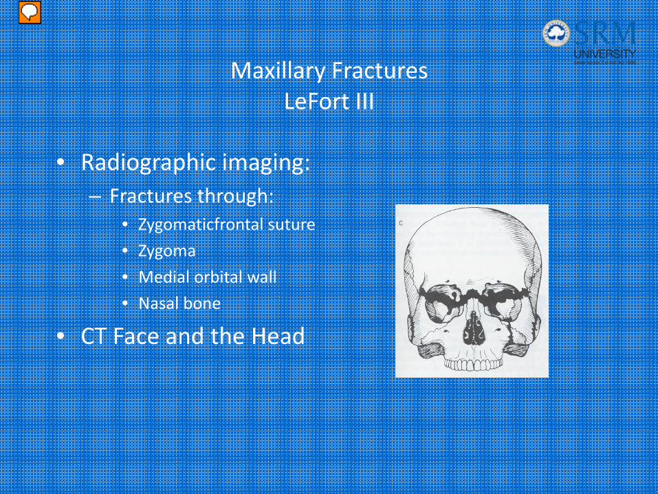
Maxillary FracturesLeFort III
• Radiographic imaging:– Fractures through:
• Zygomaticfrontal suture
• Zygoma
• Medial orbital wall
• Nasal bone
• CT Face and the Head
Presenter
Presentation Notes
Radiographic imaging: Fractures through: Zygomaticfrontal suture Zygoma Medial orbital wall Nasal bone extending posteriorly through the orbit into the spheno-palatine fossa. Again, head and facial CT will offer more information.
Maxillary FracturesTreatment
• Secure and airway
• Control Bleeding
• Head elevation 40‐60 degrees
• Consult with maxillofacial surgeon
• Consider antibiotics
• Admission
Presenter
Presentation Notes
Emergency care for all these fractures involves airway maintenance, with Intubation or cricothyrotomy if necessary. Airway compromise is possible with any of these fractures but probably more common with LeFort II and III fractures. CSF rhinorrhea is uncommon in LeFort I fracture but is often seen in LeFort II and III fractures. If CSF rhinorrhea is present or intracranial air is seen on X ray or an open skull fracture is present, the patient should be admitted and place in a head elevated position (40-60 degrees) if possible. Prophylactic antibiotics are often given in these patients (Rocephin) though it has not been shown to prevent meningitis or brain abscess. Patients with maxillary fractures also have significant epistaxis which requires nasal packing. Operative intervention may be needed if bleeding doe not resolve with packing alone. Look for associated injuries, especially intracranial, spinal, thoracic and abdominal. Incidence of blindness is high for LeFort II and III fractures so it is important to get opth. consultation. Patients with Complex maxillary fractures require admission for open reduction and internal fixation.
Mandible FracturesPathophysiology
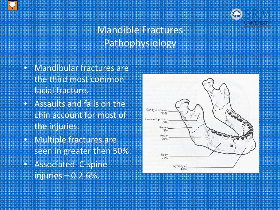
• Mandibular fractures are the third most common facial fracture.
• Assaults and falls on the chin account for most of the injuries.
• Multiple fractures are seen in greater then 50%.
• Associated C‐spine injuries – 0.2‐6%.
Presenter
Presentation Notes
Following nasal and zygomatic fractures, mandibular fractures are the third most common type of facial fracture. Assault and fall on the chin account for most of the injuries. Because of its ring shape, fractures are often multiple in up to 50% of the cases. The most common area fractured is the condyle @ 36%. Patients with a mandibular fracture, have a 0.2-6% increased risk of associated C-spine injury.
Mandible FracturesClinical findings
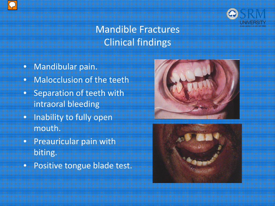
• Mandibular pain.
• Malocclusion of the teeth
• Separation of teeth with intraoral bleeding
• Inability to fully open mouth.
• Preauricular pain with biting.
• Positive tongue blade test.
Presenter
Presentation Notes
These fractures manifest clinically with mandibular pain, tenderness and malocclusion. A step off in the dental line or ecchymosis to the floor of the mouth are often present and is highly suggested of a mandibular fracture. Patients are unable to fully open their mouth. Patients may have preauricular pain with biting when there is a fracture of the condyle. Picture 1: The open fracture line is evident clinically. There is slight mal-alignment of the teeth. Picture 2: Hemorrhage or ecchymosis in the sublingual area is pathognomonic for an mandibular fracture.
Mandible Fractures
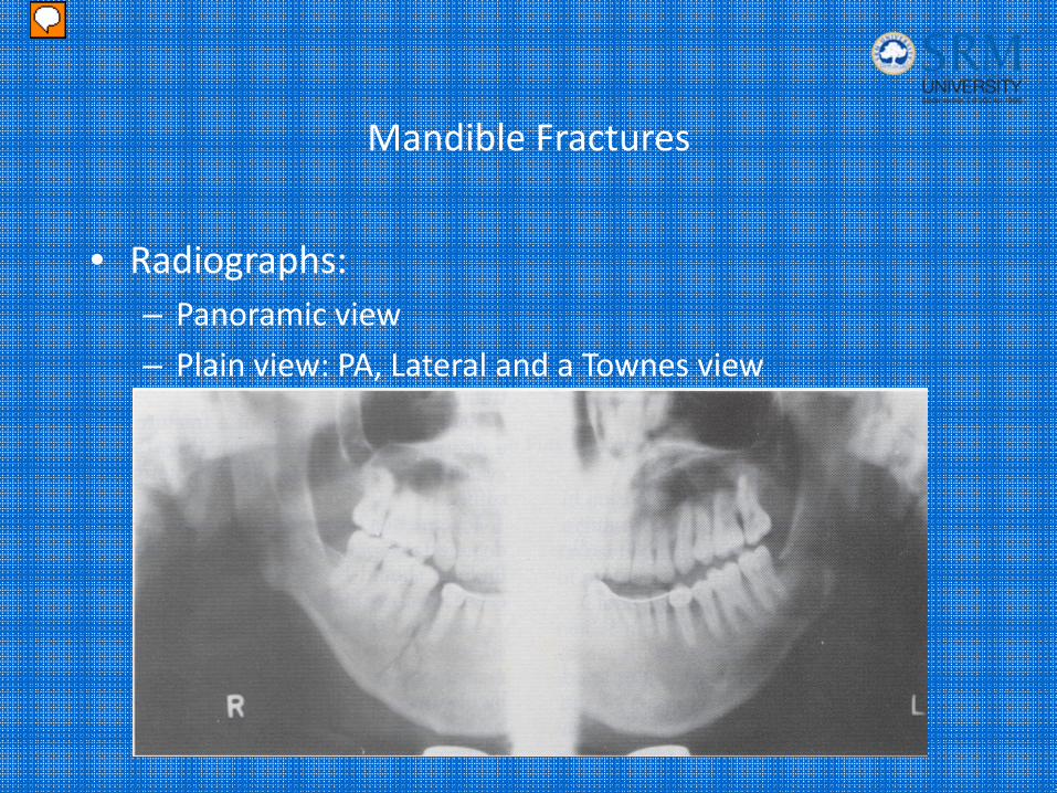
• Radiographs:– Panoramic view
– Plain view: PA, Lateral and a Townes view
Presenter
Presentation Notes
The best view for evaluating mandibular trauma is the dental panoramic view. If that is not available, plain films should include AP, bilateral oblique and a townes view to evaluate the condyles. Picture: Dental panoramic view of the mandible. Note fractures in the area of the left angle and right body.
Mandibular FracturesTreatment
• Nondisplaced fractures:– Analgesics– Soft diet– oral surgery referral in 1‐2 days
• Displaced fractures, open fractures and fractures with associated dental trauma– Urgent oral surgery consultation
• All fractures should be treated with antibiotics and tetanus prophylaxis.
Presenter
Presentation Notes
Nondisplaced fractures: Analgesics Soft diet oral surgery referral in 1-2 days Displaced fractures, open fractures and fractures with associated dental trauma Urgent oral surgery consultation, these patients are usually admitted, These patients either need closed reduction with occlusion fixation or open reduction. All patients with mandibular fractures should be treated with antibiotics and tetanus prophylaxis. Antibiotics of choice are PCN, clindamycin or a 1st generation ceph.
Mandibular Dislocation
• Causes of mandibular dislocation are:– Blunt trauma– Excessive mouth opening
• Risk factors:– Weakness of the temporal mandibular ligament– Over stretched joint capsule – Shallow articular eminence– Neurologic diseases
Presenter
Presentation Notes
Dislocation generally results from a direct blow to chin while the mouth is open, or more commonly in predisposed individuals after a vigorous yawn. Opening the mouth excessively wide while eating or laughing may also result in dislocation. It can also be seen in patients who have had a seizure, and in patients who have had a dystonic reaction from their neuroleptic medication. Weakness of the temporal mandibular ligament, overstretched joint capsule, shallow articular eminence, patients with neurologic diseases which result in increased muscular activity and extrapyramidal effects of neuroleptic medications are predisposing factors.
Mandibular Dislocation
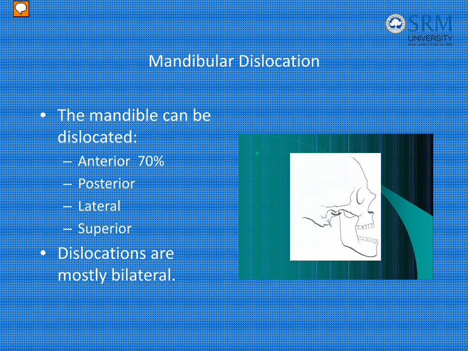
• The mandible can be dislocated:– Anterior 70%
– Posterior
– Lateral
– Superior
• Dislocations are mostly bilateral.
Presenter
Presentation Notes
The mandible can be dislocated in the anterior, posterior, lateral and superior plane. Anterior dislocation is the most common and occurs when the condyle is forced in front of the articular eminence. Anterior dislocation occurs in up to 70% of the normal individuals but can be spontaneously reduced by the patient. Once the jaw is dislocated, muscular spasm, particularly the temporalis and lateral pterygoid muscles tend to prevent reduction. Dislocations are most frequently bilateral, but they also can be unilateral.
Mandibular Dislocation
• Posterior dislocations:– Direct blow to the chin– Condylar head is pushed against the mastoid
• Lateral dislocations:– Associated with a jaw fracture– Condylar head is forced laterally and superiorly
• Superior dislocations:– Blow to a partially open mouth– Condylar head is force upward
Presenter
Presentation Notes
Posterior dislocations are rare. They occur from a direct blow to the chin that does not break the condylar neck. The condylar head is pushed against the mastoid. As a result the condylar head may prolapse into the external auditory canal. Lateral dislocations are associated with a jaw fracture. With a lateral dislocation the condylar head is forced laterally and superiorly into the temporal space. Superior dislocations occur from a blow to a partially open mouth that forces the condylar head upward. Associated injuries include cerebral contusions, facial nerve palsy and deafness.
Mandibular Dislocation
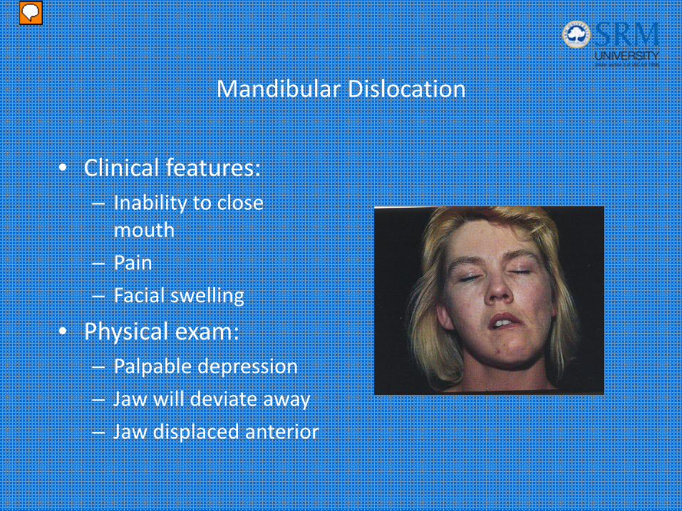
• Clinical features:– Inability to close mouth
– Pain
– Facial swelling
• Physical exam:– Palpable depression
– Jaw will deviate away
– Jaw displaced anterior
Presenter
Presentation Notes
Patients present with the inability to close an open mouth. Other associated symptoms include pain, discomfort and facial swelling near the TMJ. Unilateral dislocation results in deviation of the mandible to the unaffected side. Bilateral dislocation causes the mandible to be displace anteriorly. Picture: TMJ Dislocation Note the asymmetric jaw deviation toward the unaffected side. Always consider the possibility of an associated underlying fracture or cervical spine injury.
Mandibular Dislocation
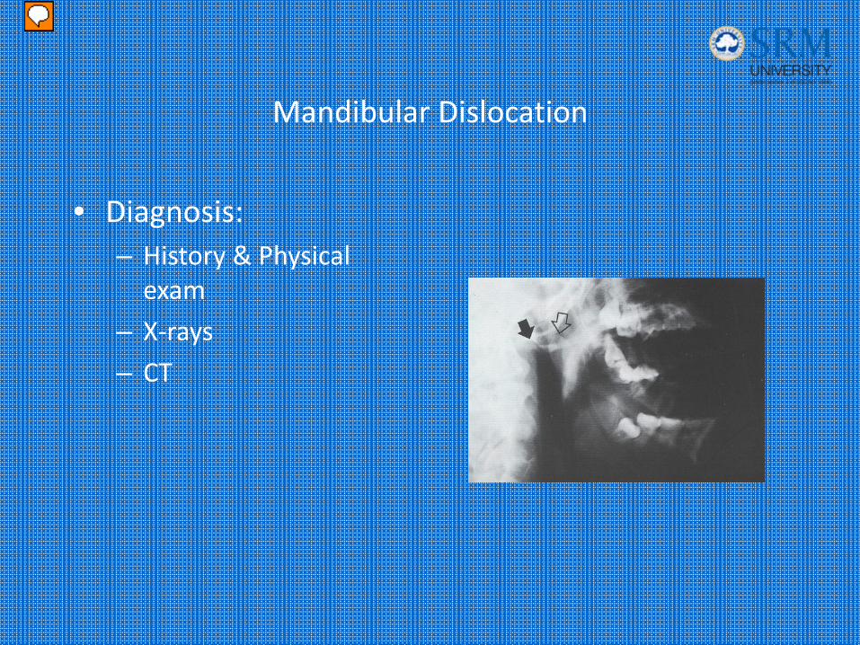
• Diagnosis:– History & Physical exam
– X‐rays
– CT
Presenter
Presentation Notes
The diagnosis of mandibular dislocation is made by H&P. X-rays should be performed if the Dx is in question or there is Hx of trauma to exclude fracture. Panoramic view usually demonstrates the pathology and excludes other mandibular injuries. If a fracture is still in questioned after X-rays, a CT can be done to provide more information. Picture: Radiograph demonstrates an anterior mandibular dislocation. The location of the condyle is indicated by the open arrow.
Mandibular Dislocation
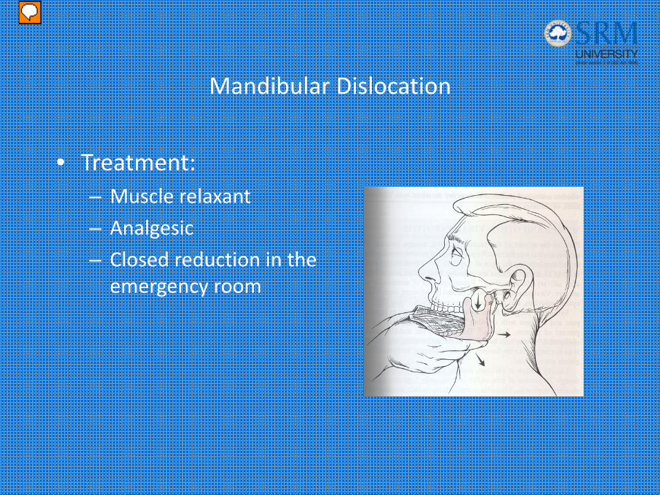
• Treatment:– Muscle relaxant
– Analgesic
– Closed reduction in the emergency room
Presenter
Presentation Notes
Reduction may be attempted in closed anterior dislocations without fracture. A short acting muscle relaxant (Versed) helps to decrease muscle spasm. An analgesic may also be considered. The patient should be seated. Facing the patient the examiner places his or hers thumbs in the patients mouth, over the mandibular molars as far back as possible. The fingers should curve beneath the angle and the body of the mandible. The examiner applies downward and backward pressure with his or hers thumbs until the condyle slides back into the articular eminence. When the dislocation is bilateral, it may be easier to relocate one side at a time. If reduction is successful, the patient should be able to close his or her mouth immediately. Post reduction films are not usually required unless the procedure was difficult or traumatic. Complications from the reduction are unusual and include iatrogenic fracture or avulsion of the articular cartilage.
Mandibular Dislocation
• Treatment:– Oral surgeon consultation:
• Open dislocations
• Superior, posterior or lateral dislocations
• Non‐reducible dislocations
• Dislocations associated with fractures
Presenter
Presentation Notes
Oral surgery should be consulted in patients who are found to have either an open dislocation, superior, posterior or lateral dislocations, non – reducible dislocation or a dislocation associated with a fracture.
Mandibular Dislocation
• Disposition:– Avoid excessive mouth opening
– Soft diet
– Analgesics
– Oral surgery follow up
Presenter
Presentation Notes
Disposition: After successful reduction, patients should be placed on a soft diet and cautioned not to open their mouths more then 2 cm for the following 2 weeks. Analgesics should be prescribed to manage discomfort. Dental follow up should be arranged. Patients with chronic dislocations may require operative intervention.