Pediatric Trauma Pediatric Trauma · Blunt trauma to the upper abdomen or lower thorax. 50 %...
15
1 Pediatric Trauma Pediatric Trauma Pediatric Trauma Pediatric Trauma Leading cause of death in children between the ages of 1 and 19 years For each death there are 32 hospitalizations 954 ED visits 1866 visits to a doctor’s office CDC, 2000 Types of Injuries Types of Injuries 1-5 years: Child Abuse, Poisonings, Burns, Foreign body aspiration, and Falls 6-12 years: Pedestrian accident injuries, and Non-vehicle bicycle accidents 13-19 years: Motor vehicle accidents, Homicide, Suicide, Drowning and Motor vehicle vs. Bicycle accidents June, 2009 Mechanism of Injury Mechanism of Injury Blunt trauma Falls Physical Abuse Motor Vehicle Accidents Penetrating trauma Other Drownings
Transcript of Pediatric Trauma Pediatric Trauma · Blunt trauma to the upper abdomen or lower thorax. 50 %...

1
Pediatric TraumaPediatric Trauma Pediatric TraumaPediatric Trauma
�Leading cause of death in children between the ages of 1 and 19 years
�For each death there are�32 hospitalizations
�954 ED visits
�1866 visits to a doctor’s officeCDC, 2000
Types of InjuriesTypes of Injuries
�1-5 years: Child Abuse, Poisonings, Burns, Foreign body aspiration, and Falls
�6-12 years: Pedestrian accident injuries, and Non-vehicle bicycle accidents
�13-19 years: Motor vehicle accidents, Homicide, Suicide, Drowning and Motor vehicle vs. Bicycle accidents
June, 2009
Mechanism of InjuryMechanism of Injury
�Blunt trauma�Falls
�Physical Abuse
�Motor Vehicle Accidents
�Penetrating trauma
�Other�Drownings

2
Physiological DifferencesPhysiological Differences
�Vital Signs
�Respiratory Reserve
�Blood Volume
�Higher Metabolic Rate
�Electrolyte imbalances
Anatomical DifferencesAnatomical Differences
�Head & Neck
�Airway
�Chest
�Abdomen
�Musculoskeletal System
Head & NeckHead & Neck
�Neck
�Occiput
�Head size
�Fontanels & Sutures
�Scalp
AirwayAirway
�Nasal passages
�Tongue, Tonsils, & Adenoids
�Larynx, Epiglottis, & Trachea
ChestChest
�Thorax
�Mediastinum
�Lungs
AbdomenAbdomen
�Liver
�Spleen
�Duodenum
�Bladder
�Kidneys

3
Musculoskeletal SystemMusculoskeletal System
�Bone properties
�Presence of growth plates
�Pseudosubluxation of C2 on C3
WeightWeight
�Fluid resuscitation and medication administration in the pediatric population is based on the patient’s body weight
�2 x age (in years) + 8
Technical InterventionsTechnical Interventions
� Interventions for infants and small children can be very time consuming, therefore, any intervention must be based on the patient’s conditions, difficulty of the procedure, and the amount of time it will take to reach definitive care.
Specialized EquipmentSpecialized Equipment
�Size appropriate airway equipment
� Intravenous catheters
�Defibrillators
�Pulse oximetry
Trauma ScoresTrauma Scores
�Glascow Coma Score
�Trauma Score
�Pediatric Trauma Score
� Injury Severity Scores
4
Developmental IssuesDevelopmental Issues
� Infants�Sucking is the major source of
gratification and tension release
�Separation anxiety begins at 5- 8 months
�Toddlers�Learn to say “no” to everything
�Older toddlers often think that they have caused the illness or injury
Developmental IssuesDevelopmental Issues
�Preschoolers�May go willingly with a stranger
�Very inquisitive - “why” or “what”questions
�Children may feel guilty for being ill or injured
�Regression may be seen
�Magical thinking
�Stalling techniques
Developmental IssuesDevelopmental Issues
�School-Age Children�Tolerate separation
�Feelings of modesty
�Comprehend simple explanations
�Adolescents�Want to be involved in decision-making
�Very modest
Cognitive & Psychological DifferencesCognitive & Psychological Differences
�Encourage parents to remain with their children
�Explain what you are going to the child in language appropriate to their development
�Be gentle and minimize painful procedures
�Be mindful of embarrassing adolescents and listen to them
Head TraumaHead TraumaChest TraumaChest TraumaAbdominal InjuryAbdominal InjuryMusculoskeletal InjuryMusculoskeletal InjurySpinal Cord InjurySpinal Cord Injury
Head TraumaHead Trauma
�Mild head injury is a common cause for Emergency Department visits
�43% of all head injuries in children are related to sports and recreational activities
�93% of all head-injured children are admitted to the hospital

5
Head InjuryHead Injury
�Scalp lacerations
�Skull fractures
�Concussion
�Contusion
�Hematomas
Scalp LacerationsScalp Lacerations
�The scalp is highly vascular and may result in profuse bleeding when injured
�A subgaleal hematoma may be the only sign of intracranial injury in an infant. 73% of infants with an Epidural Hematoma also had a significant subgaleal hematoma
Skull FracturesSkull Fractures
�Linear
�Depressed
�Compound
�Basilar
Linear Skull FracturesLinear Skull Fractures
� 75 - 90% of skull fractures in children
� Parietal bone is the most common site of skull fractures
Depressed Skull FractureDepressed Skull Fracture
� Disruption of the integrity of the skull
� Considered clinically significant if the bone fragment is depressed below the inner table of the skull to a depth greater than the full thickness of the skull

6
Basilar Skull FractureBasilar Skull Fracture
� Frequent site of fracture
� Signs & Symptoms� CSF Rhinorrhea
� Hemotympanum
� Ecchymosis behind the ear over the mastoid
� Raccoon eyes
ConcussionConcussion
�Brief alteration in consciousness (with or without Loss of Consciousness) after sustaining a closed head injury
�This alteration in consciousness is accompanied by a flaccid motor state, followed by complete recovery
�May also be associated with a period of vomiting, pallor, confusion, or amnesia
ContusionContusion
�Result of the brain parenchyma becoming bruised or crushed, resulting in hemorrhage and edema
�When brain injury occurs on the side of impact a coup lesion develops
�Damage to the opposite side of the brain from the site of impact is a contrecoup lesion
Diffuse Axonal InjuryDiffuse Axonal Injury
�Damage to the axons of the Central Nervous System can be caused by the shearing forces associated with Closed Head Injuries
�DAI may accompany concussion, contusion & subdural hematomas
�May lead to temporary alterations in consciousness, neurological deficits and in the most severe cases; coma
HematomasHematomas
�Epidural�2% of all serious head injuries
�Arterial in nature, middle meningeal artery tear
�Associated with Skull Fractures in 40-85% of cases
�Transient loss of consciousness followed by a lucid interval
7
HematomasHematomas
�Subdural�5% of all head trauma patients
�Damage to the subdural veins “bridging veins”
�Manifest hours after injury
�Underlying brain injury occurs in 50% of cases
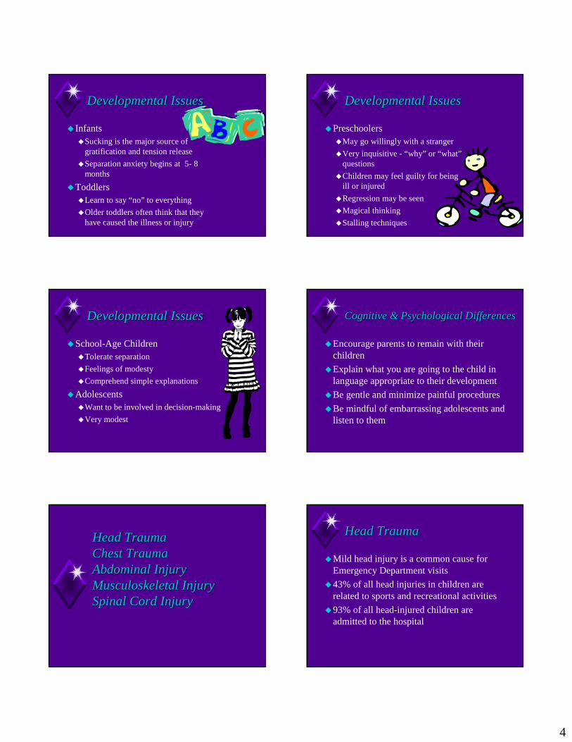
Chest TraumaChest Trauma
�Responsible for 10 - 25% of acute deaths
�Contributes to up to 50% of other deaths
�May have associated abdominal trauma
�Blunt injury is the most common cause
Chest TraumaChest Trauma
�Pneumothorax
�Hemothorax
�Flail Chest
�Traumatic asyphyxia
�Cardiac tamponade
�Myocardial contusion
�Aortic rupture
PneumothoraxPneumothorax
�Open
�Closed
�Tension

8
Open PneumothoraxOpen Pneumothorax
�Etiology: Penetrating trauma
�Pathophysiology�Loss of chest wall integrity
�Involved lung collapses on inspiration and expands slightly on expiration
�Inability to generate negative pressure
�Treatment: Dressing secured on three sides only
Tension PneumothoraxTension Pneumothorax
�Etiology�Blunt Trauma
�Penetrating Trauma
�Mechanical ventilation
� Diagnosis
�Neck veins distended
�Trachea deviated
�Shock
Tension PneumothoraxTension Pneumothorax
� Pathophysiology� Pleural pressure rises
� Lung collapses
� Mediastinum shifts
� Opposite lung compressed
� Vena Cava kinks
� Decreased venous return
� Decreased cardiac output
Needle DecompressionNeedle Decompression
� Equipment: 14 or 16G over the needle catheter
� Procedure: Patient in the supine position, place the needle in the second intercostal space, midclavicular line
HemothoraxHemothorax
�Etiology�Blunt trauma
�Moderate blood loss
�Bleeding sites usually located on the chest wall or lung parenchyma
� Penetrating trauma�Risk of massive blood loss
�Bleeding: aorta, systemic, or pulmonic vessels, heart
HemothoraxHemothorax
�Pathophysiology�Laceration to a vessel, bleeding from the lung
�Accumulation of blood in the pleural space leads to hypoxia resulting from ventilatory compromise
�Radiograph�Supine film may miss blood loss approaching
30% estimated blood loss

9
HemothoraxHemothorax Tube ThoracostomyTube Thoracostomy
�Procedure: Placement at the 4th or 5th intercostal space, mid-axially line
�Equipment: Chest Tube�Newborn 12fr
�1 - 2 years 16fr
�5 years 24fr
�10 years 32fr
�Adult 36fr
Rib FracturesRib Fractures
�Rare in healthy infants and children
�Etiology�82% caused by abuse in infants
�8% accidental (major trauma)
� 7% fragile bones
�3% birth trauma
Rib Fractures Rib Fractures
�Signs & Symptoms�Tachypnea with shallow breathing
�Pain on inspiration
�Tenderness
�Crepitus
�Swelling over fracture site
Rib FracturesRib Fractures
�Rare due to resilient rib cage
�Presence suggests massive force
�Predictive of severe organ injury
�May be complicated by flail chest, pneumothorax, hemothorax, pulmonary contusion
�May cause liver and spleen injury
Rib FracturesRib Fractures
�Treatment�Pain relief
�Bed-rest
�Restrictive bandaging is not useful
�Cough and deep breathing with splint pillow

10
Cardiac TamponadeCardiac Tamponade
�Pathophysiology�Accumulation of blood in the pericardial sac
�Heart cannot fill during diastole
�Low cardiac output
�Diagnostic Triad�Shock
�Distended neck veins
�Muffled heart sounds
Cardiac TamponadeCardiac Tamponade PericardiocentesisPericardiocentesis
�Procedure: Position the patient in reverse trendelenberg. Attach a needle to ECG lead and insert needle at a 45 degree angle one centimeter to the left of the xiphiod process.
Myocardial ContusionMyocardial Contusion
�Etiology�Blunt trauma - sharp direct blow to the sternum
�Presentation�Chest pain, Sinus tachycardia, ST -wave
changes
�Treatment�Oxygen, analgesics, cardiac monitoring
Aortic RuptureAortic Rupture
� Pathophysiology� Descending aorta is
relatively fixed
� Sudden deceleration
� Shearing forces on the aorta at the isthmus
� Radiographic signs� Increase in the width of
the superior Mediastinum

11
Abdominal InjuryAbdominal Injury
�Splenic Injury
�Liver Injury
�Lap belt complex
Splenic InjurySplenic Injury
�Etiology� Blunt trauma to the upper abdomen or lower
thorax.
�50 % related to recreational activities
�Treatment�Aimed toward splenic preservation
� ICU monitoring for 48hours
�Blood transfusions
�Splenectomy
Liver InjuryLiver Injury
� Etiology: Blunt trauma
� Classifications� Contusions
� Parenchymal lacerations
� Injuries to the hepatic vein or vena cava
� Treatment� Non-operative management
� Liver resection
Lap belt ComplexLap belt Complex
�Anatomical differences�Mechanism of injury
�Deceleration�Compression
� Injury pattern�Hip & Abdominal Contusions�Lumbar spine injuries�Intrabdominal injuries
Musculoskeletal InjuryMusculoskeletal Injury
� Extremity trauma� Fractures
� Subluxations & Dislocations
� Amputations
Spinal Cord InjuriesSpinal Cord Injuries
�SCIWORA�Spinal
�Cord
�Injury
�Without
�Radiographic
�Abnormalities

12
Spinal Cord InjurySpinal Cord Injury
�High dose Methyprednisolone�Indications
�Complete & incomplete spinal cord injury less than 8 hours old
�Dosage�30 mg/kg bolus with 4 hours of injury over 15 mins
�45 minute pause
�23 maintenance infusion = 5.4 mg/kg/hr
The Injured FamilyThe Injured Family
�The Calgary Family Assessment Model�Structural Assessment
�Developmental Assessment
�Functional Assessment

13
Structural AssessmentStructural Assessment
� Internal Structure�Who are the members of the family
�External Structure�How the family is related to an extended family
and their social system
�Context�Sociocultural background of the family
Developmental AssessmentDevelopmental Assessment
�Life Cycle
�Family Tasks
�Relational Attachments
Functional AssessmentFunctional Assessment
� Instrumental functioning
�Expressive functioning
Primary SurveyPrimary Survey
�Airway
�Breathing
�Circulation
Airway ManagementAirway Management
�100% oxygen
�Suctioning
�Jaw thrust or chin lift
�Oral airway
�Oro-tracheal intubation
�Cricothyroidotomy vs. Tracheostomy
CirculationCirculation
� Hypovolemic clues� Vital signs & blood pressure guidelines vary with age� Tachycardia can be caused by multiple factors
� Fixed stroke volume so compensate for hypovolemia by increasing heart rate
� Efficient compensation makes recognition of shock difficult – assess perfusion
� Hemorrhage control� Intravenous access� Fluid resuscitation� Maintain CPP > 40 mm Hg

14
CirculationCirculation
�Hemorrhage control
� Intravenous access
�Fluid resuscitation
�Maintain CPP > 40 mm Hg
Etiologies of ShockEtiologies of Shock
�Hypovolemic Fluid Loss
�Distributive Neurogenic
�Cardiogenic Pump Failure
�Obstructive Tamponade
DisabilityDisability
�AVPU evaluation system�Alert
�Responds to verbal commands
�Responds to pain
�Unresponsive
�Control of Intracranial Pressure�3 % Normal Saline
Expose, Examine, Emotional SupportExpose, Examine, Emotional Support
�Undress
�Avoid Hypothermia
Secondary SurveySecondary Survey
� Head to toe evaluation
� Fingers and tubes in every orifice
� Review history
� Assess and assign trauma scores
� Laboratory analysis
� Radiographic studies
MonitoringMonitoring
�Vital signs, CPP > 40 mmHg
�ECG
�Pulse oximeter
�End Tidal CO2 monitor
�Urine Output
�Arterial blood gas
�Frequent reexamination
15
TransferTransfer
� Indications
�Team composition
�Vehicle�Ground ambulance
�Fixed wing
�Helicopter
�Equipment
Ethical Principles in Pediatric Ethical Principles in Pediatric Trauma NursingTrauma Nursing
�Autonomy
�Nonmaleficence
�Benefience
�Justice
Ethical PrinciplesEthical Principles
�Autonomy�A person’s individual freedom to choose his or
her own course and make personal decisions
�Issues to consider�Age, cognition, developmental level
�Family Presence during resuscitation
Ethical PrinciplesEthical Principles
�Nonmaleficence�The obligation not to inflict intentional harm
�Withholding care�Withdrawing treatment
�Beneficence�Do Good�Protecting the rights of others, preventing harm,
helping persons with disabilities�Child Abuse
Ethical PrinciplesEthical Principles
�Justice�Distributive justice
�Fair, equitable, and appropriate distribution of resources, privileges, and opportunities
� Expensive, Highly technical interventions
� ICU Beds
Questions?????Questions?????
�Thank you for your attention


![Trauma Reach Workshop - Pediatric Trauma.pptx [Read-Only]...Pediatric Trauma Trauma REACH Workshop May 5th, 2015 Tamer A. Ahmed, MD Pediatric Trauma Medical Director Upstate’s GolisanoChildren’s](https://static.fdocuments.in/doc/165x107/5fe9ec9ba1b3915c9800251e/trauma-reach-workshop-pediatric-read-only-pediatric-trauma-trauma-reach.jpg)