Understanding and Managing Pediatric Trauma - RN.com · PDF fileDescribe management strategies...
22
Material Protected by Copyright Understanding and Managing Pediatric Trauma One (1.0) Contact Hours Expiration Date: 2/28/2020 First Published: 2/20/2014 Revised: 2/11/2017 Reproduction and distribution of these materials is prohibited without an RN.com content licensing agreement. Acknowledgments RN.com Acknowledges the valuable contributions of... ...Suzan Miller-Hoover DNP, RN, CCNS, CCRN-K Conflict of Interest RN.com strives to present content in a fair and unbiased manner at all times, and has a full and fair disclosure policy that requires course faculty to declare any real or apparent commercial affiliation related to the content of this presentation. Note: Conflict of interest is defined by ANCC as a situation in which an individual has an opportunity to affect educational content about products or services of a commercial interest with which he/she has a financial relationship. The author of this course does not have any conflict of interest to declare. The planners of the educational activity have no conflicts of interest to disclose. There is no commercial support being used for this course. Purpose and Objectives
Transcript of Understanding and Managing Pediatric Trauma - RN.com · PDF fileDescribe management strategies...
Material Protected by Copyright
Understanding and Managing Pediatric Trauma
One (1.0) Contact Hours Expiration Date: 2/28/2020 First Published: 2/20/2014 Revised: 2/11/2017 Reproduction and distribution of these materials is prohibited without an RN.com content licensing agreement.
Acknowledgments RN.com Acknowledges the valuable contributions of... ...Suzan Miller-Hoover DNP, RN, CCNS, CCRN-K
Conflict of Interest RN.com strives to present content in a fair and unbiased manner at all times, and has a full and fair disclosure policy that requires course faculty to declare any real or apparent commercial affiliation related to the content of this presentation. Note: Conflict of interest is defined by ANCC as a situation in which an individual has an opportunity to affect educational content about products or services of a commercial interest with which he/she has a financial relationship. The author of this course does not have any conflict of interest to declare. The planners of the educational activity have no conflicts of interest to disclose. There is no commercial support being used for this course. Purpose and Objectives
Material Protected by Copyright
The purpose of this course is to provide an overview of pediatric trauma, including assessment and management. After successful completion of this course, you will be able to:
1. Review anatomy and physiology of the pediatric patient 2. Identify at least three types of trauma common with the pediatric patient 3. Delineate primary and secondary assessment of pediatric trauma patients 4. Describe management strategies and nursing interventions with pediatric trauma patients 5. Review communication techniques to use with the pediatric patient and family
Introduction Trauma and injury are the most common reason for morbidity and mortality of pediatric patients. Appropriate assessment and management of pediatric trauma is essential to achieve positive patient outcomes. Trauma According to Mosby’s medical dictionary, trauma is defined as a physical injury that is caused by violent or disruptive action or toxic substance to the body, or a psychological injury from emotional shock (O’Toole, 2013). Pediatric Trauma The Centers for Disease Control and Prevention (CDC) state that a child dies from an injury every hour. In addition, approximately one in five pediatric deaths are due to an injury, and every four seconds a child is treated in an emergency department for an injury (Centers for Disease Control and Prevention (CDC), 2012). The most common types of pediatric trauma are:
• Motor vehicle accident (number one cause of pediatric deaths in the U.S.) • Suffocation • Drowning • Poisoning • Fire and/or burn • Fall
Did You Know? In 2008, the United States child death rate from injury was 8.7/100,000. This is four times the death rate of countries with the lowest rates, and is the third worst rate overall of high-income countries.
Material Protected by Copyright
Anatomy and Physiology There are some differences in anatomy and physiology with pediatric patients that are important to understand. Children are not “little adults”! Head & Neck For infants, the head is large in comparison to the body. A newborn’s head is approximately one fourth of the total body length, whereas an adult’s head is one seventh of the total body length. The disproportion in head size begins to diminish at 2-6 years of age. The large size of head increases risk for head injury, as the head often “propels forward” in young infants and toddlers. The Eustachian tube of the infant and young child is short and relatively horizontal, increasing the risk for middle ear infections. The tube starts to slant downward between ages 6-8 years. Infants are obligate nose breathers. This means they are only able to breathe through their nose, usually until 3-6 months of age. The tongue is large in comparison to the size of infant’s/young child’s oral cavity. This gradually improves as the child grows. By the age of 10 the tongue is more proportional to an adult’s oral cavity (Potts & Mandleco, 2012). Differences: Respiratory Respiration is abdominal (not thoracic) in infants and toddlers. This means that they use the abdominal muscles more to breathe, due to a lower percentage of type I skeletal muscle fibers in the diaphragm and intercostal muscles. Type I fibers are less prone to muscle fatigue. Infants also have a flatter diaphragm and more horizontal ribs. Respirations are shallower and more rapid in the infant and young child. These become deeper and slower as the child grows and moves into adolescence. The small bronchi and bronchioles have a shorter length and smaller diameter for infants and young children. This can increase the risk for respiratory infections, as well as resistance in the airway which can lead to respiratory failure (Potts & Mandleco, 2012). Did You Know? In non-emergency situations, respirations should be counted for one minute if the child is less than a year old, or if respirations are irregular. A respiratory rate is not accurate if crying.

Material Protected by Copyright
The normal range of respiratory rate per Pediatric Advanced Life Support (PALS) guidelines (American Heart Association [AHA], 2015)
Age (in years) Breaths/minute <1 30 – 60
1 – 3 24 – 40 4 – 5 22 – 34
6 – 12 18 – 30 13 – 18 12 – 16
Differences: Cardiovascular Infants and toddlers have a thin chest wall in comparison to adolescents and adults. This increases the compliancy of the chest wall, which can decrease functional residual capacity. There is also an increased amount of “referred” sounds during auscultation with the infant and toddler. Turbulent blood flow frequently results in “innocent” heart murmurs during childhood. The murmur may be congenital or develop during childhood. These murmurs are considered harmless and don’t need treatment. The heart rate of the infant and young child is much higher than an adult’s. This is due to the smaller heart size as well as the higher metabolic rate. Heart rate decreases with age, and by the age of about 10 years the heart rate equates that of an adult. Inversely, the blood pressure of the infant and young child is lower than an adult’s. This is from a decreased cardiac output and lowered peripheral resistance, which result in lower blood pressure. Blood pressure increases with age and is also affected by growth percentiles (Potts & Mandleco, 2012; American Heart Association, 2015). Did You Know? The normal range of heart rate per PALS (AHA, 2015):
Age Heart rate (awake) Heart rate (asleep) <3 months 85 – 205 80 – 160
3 months – 2 years 100 – 190 75 – 160 2 – 10 years 60 – 140 60 – 90
>10 years 60 – 100 50 - 90 Test Yourself Which of the following is true in comparing infants and children to adults?
A. Infants and children have a lower heart rate and respiratory rate

Material Protected by Copyright
B. Infants and children have a higher heart rate but lower respiratory rate C. Infants and children have a higher heart rate and respiratory rate
Rationale: Respirations are shallower and more rapid in the infant and young child. These become deeper and slower as the child grows and moves into adolescence. The heart rate of the infant and young child is much higher than an adult’s. This is due to the smaller heart size as well as the higher metabolic rate. Heart rate decreases with age, and by the age of about 10 years the heart rate equates that of an adult.
Differences: Gastrointestinal In infancy, there is an unpredictable gastric emptying time, increased peristalsis, and immature small intestine which lead to frequent soft and liquid stools. Infants and toddlers have a smaller stomach capacity. There is also a more vertical position of stomach during toddlerhood, which increases frequency of vomiting during illness. An immature esophageal sphincter with infants and toddlers result in regurgitation and reflux (Potts & Mandleco, 2012; Maryniak, 2013). Differences: Renal & Lymphatics There is a decreased ability to concentrate and filter urine during infancy, which increases the risk for dehydration. There are decreased levels of antidiuretic hormone (ADH) and a lowered response to ADH, which result in decreased ability to concentrate urine, excrete waste, or conserve fluid. The kidneys are not functionally mature until about 2 years of age. Blood flow to the kidneys is also decreased due to increased vascular resistance within the renal structures. Sodium regulation functions are immature, and the infant/toddler is unable to excrete excess sodium. Thus, electrolyte imbalances may develop and progress very quickly, and adequate fluid intake is essential to avoid severe fluid problems. Infants also have a decreased bladder capacity and control of urethral sphincter compared to adults. This improves during toddler and preschool years. Lymphatic tissue increases in size during toddler and preschool years. This results in enlarged tonsils and adenoids, as well as palpable peripheral nodes (Potts & Mandleco, 2012; Maryniak, 2013). Differences: Musculoskeletal There are immature muscle fibers in infancy/early toddlerhood. This results in an unsteady gait that usually disappears during later toddler years as muscles develop and long bones rapidly grow.

Material Protected by Copyright
Bones in infants and young children yield to pressure and muscle pull more than mature bones due to incomplete ossification. Epiphyseal plates remain open until after the pubertal growth spurt of adolescence (Potts & Mandleco, 2012; Maryniak, 2013). Differences: Metabolic Due to periods of rapid growth, an infant’s & young child’s basal metabolic rate (BMR) is higher than an adult’s. The energy needs of infants and children vary greatly due to age and biologic changes, and caloric requirements of the child are de3termined by the metabolic rate. The metabolic rate is highest from birth to age 2, and from puberty to age 15, when the child is growing the most. BMR affects heat production and, thus, body temperature. Body temperature also varies according to the time of day, with the highest body temperature normally being in the middle of the day/early evening hours. Infants produce more heat than older children and adults in relation to body weight. As a result, infants are more prone to becoming overheated (Potts & Mandleco, 2012; Maryniak, 2013). Test Yourself The kidneys become functionally mature by what age?
A. 12 months B. 2 years C. 12 years
Rationale: The kidneys are not functionally mature until about 2 years of age. Blood flow to the kidneys is also decreased due to increased vascular resistance within the renal structures. Sodium regulation functions are immature, and the infant/toddler is unable to excrete excess sodium. Thus, electrolyte imbalances may develop and progress very quickly, and adequate fluid intake is essential to avoid severe fluid problems. Fluids and Electrolytes Total Body Water (TBW)
• Newborns- TBW is 75% - 80% of body weight • During first 2 years of life- TBW slowly decreases to 60% of body weight (same as adults)
Two Major Components of Body Fluid • Intracellular fluid, or ICF (inside the cell), contains 2/3 of TBW. The main ICF electrolyte is potassium. • Extracellular fluid, or ECF (outside the cell), contains 1/3 of TBW (35% - 45% of TBW in infants;
decreases to 24% of TBW by age 2). This includes intravascular fluid (plasma) and interstitial fluid (lymph and CSF). The main ECF electrolyte is sodium. 50% of the ECF in an infant is exchanged daily, which results in a higher daily fluid requirement (Potts & Mandleco, 2012).
Material Protected by Copyright
Fluid Balance Due to the accessibility of body fluid in the extracellular space, infants are more susceptible to fluid overload and dehydration. With pediatric patients, the younger the patient, the higher insensible water losses occur which affect fluid balance. Infants have great fluid loss through evaporation related to:
• Larger total body surface area • Rapid respiratory rate • Higher metabolic rate
Fluid Requirements Fluid requirements in children are based on body weight (kg).
• <10kg o 100mL per kg of body weight
E.g.- 9kg infants requires 900mL (9 x 100) per 24 hour period • 10kg – 20kg
o 1000mL + 50mL per kg over first 10 kg of body weight E.g.- 14kg child requires 1200mL (1000 + 50x4) per 24 hour period
• >20kg o 1500mL + 20mL per kg over first 20kg of body weight
E.g. - 26kg child requires 1620mL (1500 +20x6) per 24 hour period (Potts & Mandleco, 2012) Test Yourself The majority of total body water is found in the:
A. Intracellular fluid B. Extracellular fluid C. Interstitial fluid Rationale: Intracellular fluid, or ICF (inside the cell), contains 2/3 of TBW. The main ICF electrolyte is potassium. Extracellular fluid, or ECF (outside the cell), contains 1/3 of TBW (35% - 45% of TBW in infants; decreases to 24% of TBW by age 2). This includes intravascular fluid (plasma) and interstitial fluid (lymph and CSF). The main ECF electrolyte is sodium. 50% of the ECF in an infant is exchanged daily, which results in a higher daily fluid requirement (Potts & Mandleco, 2012).
Common Pediatric Traumas Common types of pediatric trauma include:
Material Protected by Copyright
• Musculoskeletal • Cardiothoracic • Abdominal • Traumatic brain injury • Spinal cord
Trauma can also affect the patient’s fluid, electrolyte, and metabolic processes. Hypoxia, hemodynamic instability, hypothermia, and infection are other concerns that can be complications secondary to a trauma. Did You Know? Child abuse can be a cause of pediatric trauma. It is important to identify suspicious signs of abuse, or if the explanation of the child’s injury(s) does not correlate with the actual injury(s). For more information, see RN.com’s course Recognizing and Reporting Child Abuse. Primary and Secondary Assessment There are two forms of assessment that are done with a pediatric trauma patient.
• Primary assessment: This is the initial assessment done in emergency situations. The purpose is to identify potentially life-threatening injuries. The mnemonic ABCDE is used (to be discussed further). Any resuscitation that is needed is done immediately following primary assessment.
• Secondary assessment: Following any needed resuscitation, this assessment is to identify and treat conditions that are not life-threatening. A complete head-to-toe assessment is performed, a history is taken, re-evaluation of the primary assessment is done for signs of deterioration, and further laboratory and diagnostic tests. Decisions are also made concerning the prioritization of treatment of the injuries that exist. The mnemonic FGHI may be used (to be discussed further) (AHA, 2015).
Primary Assessment During primary assessment and resuscitation, the mnemonic ABCDE is most commonly used.
• A = Airway and cervical spine stabilization • B = Breathing • C = Circulation • D = Disability (brief neurological exam) • E = Exposure
Airway and Cervical Spine Stabilization
• Airway and Cervical Spine Stabilization o The first priority in primary assessment and resuscitation is protecting the airway, which can be
compromised quickly with the pediatric patient. The cervical spine must also be protected while
Material Protected by Copyright
establishing the airway, which continued protection until injury has been ruled out through assessment and cervical spine x-rays.
o The modified jaw thrust maneuver is the best way to open the airway and protect the cervical spine. The oral cavity can be suctioned with a Yankauer catheter to remove secretions, but caution must be used to avoid stimulating the gag reflex.
• Artificial Airways o An oropharyngeal (oral) airway should only be inserted if the child is unconscious, and should
not be used if a gag reflex is present. The size of the oral airway usually ranges from 00-5, and is determined by measuring from the corner of the mouth to the angle of the jaw. An incorrect size can obstruct the airway.
o Nasopharyngeal (nasal) airways can be used with caution in children who have a gag reflex present. The diameter and length are important, and the catheters usually range from 16-32 Fr. A small-diameter nasopharyngeal airway may be obstructed easily by secretions, and may need frequent suctioning (AHA, 2015).
• Intubation o Some trauma patients may need endotracheal intubation. Indications for intubation include:
Need for prolonged patency of the airway and/or prevention of aspiration (such as an unconscious child)
Need for ventilation and/or inability to adequately ventilate using a bag and mask Shock unresponsive to fluid resuscitation Neurological problems, including seizures and head injury
o Only experienced health care providers can intubate. The patient should be provided with 100% oxygen prior to an intubation attempt. The alignment of cervical spine should be maintained, and the patient’s heart rate and oxygen saturation should be monitored during the intubation attempt (AHA, 2015).
• ET Tubes o Uncuffed endotracheal (ET) tubes are used in children less than eight years of age. Length-
based tapes, such as the Broselow® Pediatric Tape, provide a guide to selecting appropriate ET tube sizes. Pediatric ET tube sizes usually range from 3.0 – 7.0 mm. Formulas for calculating ET tube sizes include: Uncuffed endotracheal tube internal diameter (mm) = 4 + (age/4) Cuffed endotracheal tube internal diameter (mm) = 3.5 + (age/4)
• Post Intubation o Assessment for correct tube placement includes:
Symmetrical chest movement Equal bilateral breath sounds Condensation in the endotracheal tube Use of an exhaled carbon dioxide (CO2) detector for confirmation (yellow with correct
placement, and purple with incorrect tube placement). Poor pulmonary blood flow, decreased cardiac output, and/or acidosis can cause absence of color change with the CO2 detector.

Material Protected by Copyright
X-ray confirmation: the tip of the ET tube should be located 1 to 2 cm above the carina, seen at approximately the third to fourth rib on the x-ray.
• Other Considerations o End-title carbon dioxide (ETCO2) monitoring is recommended for all intubated patients. To
prevent gastric distention and enhance ventilation, a nasogastric or orogastric tube should be placed as soon as possible following intubation.
o When there is direct injury to the larynx and/or trachea, a cricothyrotomy or emergency tracheostomy may need to be performed by a surgeon ((AHA, 2015).
Did You Know? Rapid sequence intubation medications should be considered to decrease the adverse effects of intubation. If medications are used, sedation is always given prior to a paralytic agent. Breathing
• Breathing o Chest injury with a pediatric patient may be subtle and hard to detect. Assessment includes
respiratory rate, respiratory depth and effort, auscultation of breath sounds, and visualization of the chest for asymmetry in chest wall movement and expansion. Because of a thin chest wall, breath sounds are easily transmitted through the chest wall, which can make it difficult to determine adequacy of ventilation with the pediatric patient. The chest should be exposed to inspect for signs of trauma, and palpated for tenderness and pain.
o Important findings that indicate a need for respiratory support include nasal flaring, grunting, retractions, breath sounds that are unequal/diminished/absent, unequal chest rise, and an oxygen saturation of <90%. A chest x-ray can confirm complications, and life-threatening thoracic injuries such as pneumothorax, hemothorax, and pericardial tamponade must be immediately treated (AHA, 2015).
• Oxygen o Oxygen should be placed on all trauma patients via nasal cannula or facemask, even if there are
no signs of airway or respiratory compromise. A pulse oximeter should be placed to measure oxygen saturation continuously and to maintain oxygen saturation greater than 96%. For the apneic or bradypneic child, rescue breathing should be initiated with a bag-valve-mask using 100% oxygen. Intubation and mechanical ventilation may be required in some patients as previously discussed.
Circulation
• Circulation o Circulation includes assessing the heart rate and rhythm, peripheral pulses compared with
central pulses (such as brachial versus apical), blood pressure, capillary refill, color, skin
Material Protected by Copyright
temperature, and identification of any hemorrhage sites. Heart sounds should be auscultated to determine quality. A cardiac tamponade must be suspected if muffled heart should, distended neck veins, and signs of shock are present in the patient.
o Late signs of circulatory insufficiency requiring the need for circulatory support include tachycardia and bradycardia, decreased or differences in peripheral pulses, capillary refill >3 seconds, pallor, cyanosis, cool and clammy skin, hypotension, dysrhythmias, and hemorrhage (AHA, 2015).
• Resuscitation o The first priority of circulation is to confirm the presence of a pulse by palpating a central pulse.
If no pulse is present, begin cardiopulmonary resuscitation (CPR) immediately with chest compressions.
o External hemorrhages are the next treatment priority, which can be treated by applying direct pressure using a sterile pressure dressing, being careful not to occlude distal pulses. Unless it is contraindicated, a bleeding extremity should then be elevated.
o Intravenous access is a priority. Two large-bore catheters, preferable in the upper extremities, should be attempted. Intraosseous or central venous cannulation may be necessary is peripheral access attempts are unsuccessful. Volume expanders, such as Lactated Ringer’s or normal saline, are administered as indicated. If the child shows signs of severe hypovolemia or shock, fluid boluses of volume expanders at 20mL/kg are given and repeated up to three times. O-negative packed red cells or whole blood may also be used, infused at 10 mL/kg. Boluses are repeated until perfusion and peripheral pulses improve. Surgical assessment and intervention may be necessary to control bleeding (AHA, 2015).
Disability
• Disability (brief neurological exam) o A brief neurological assessment is done to determine changes in level of consciousness. The
patient’s response to stimuli is assessed as either awake and alert, responds to verbal stimuli, responds to painful stimuli, or unresponsive. A baseline pupillary assessment is also important. A more detailed neurological assessment is completed during a secondary assessment.
o Neurological status should be monitored frequently throughout the care of the patient. Changes in level of consciousness are an early indication of decreased oxygenation and perfusion, or significant head injury. A child with signs of increased intracranial pressure (ICP) should be intubated and oxygenated on 100%. Signs of increased ICP in the child include severe headache, emesis, irritability, rapidly deteriorating mental status, abnormal posturing, neurological deficits, pupillary abnormalities, and seizures. Infants can also demonstrate bulging fontanels and split sutures (Potts & Mandleco, 2012).
Exposure

Material Protected by Copyright
• Exposure o A detailed inspection and examination of the body for injuries is required. Obvious signs of
injury such as bruising, bleeding, and abrasions should be assessed. The assessment should be thorough but done quickly, to reduce the chance of lowering the child’s temperature. Pediatric patients are prone to hypothermia due to increased heat loss from their larger body surface area, and trauma can also increase susceptibility to hypothermia.
o The pediatric patient should be covered as much as possible, exposing only needed body surface areas during any assessment or procedure. To prevent hypothermia, all wet clothing or sheets should be removed, and a warming blanket, and/or a radiant heat source can be used. Active warming may be initiated by using warmed IV fluids and blood to increase the core temperature (McFadyen, Ramajah, & Bhananker, 2012; Potts & Mandleco, 2012).
Test Yourself Which of the following is priority during primary assessment and resuscitation?
A. Establishing an airway B. Applying pressure to a bleeding wound C. Starting cardiac compressions
Rationale: The first priority in primary assessment and resuscitation is protecting the airway, which can be compromised quickly with the pediatric patient. Secondary Assessment During secondary assessment, the mnemonic FGHI is most commonly used.
• F = Full set of vital signs; Family presence • G = Give comfort • H = Head-to-toe assessment; History • I = Inspect posterior surfaces
Full set of vital signs; Family presence
• Full Set of Vital Signs o A full set of vital signs should be obtained, including heart rate, respiratory rate, blood pressure,
temperature, oxygen saturation, and pain assessments. Repeat vital signs are performed frequently until the child is stable. The frequency of vital signs is dependent on the extent of the patient’s injuries. Invasive lines, catheters, and/or tubes may be inserted for additional monitoring. Weight should also be obtained as soon as possible (McFadyen et al., 2012; Potts & Mandleco, 2012).
• Family Presence
Material Protected by Copyright
o Family presence should be supported with the child during procedures and treatment. One health care provider should be assigned as a primary communicator to the family, and must explain interventions and provide support. Support services, such as Spiritual Care, Social Services, or Resource Management should be available to support the family’s spiritual and emotional needs (McFadyen et al., 2012; Potts & Mandleco, 2012).
Give Comfort
• Give Comfort o Pain and comfort are important, as unmanaged pain can increase oxygen consumption. The
child’s pain should be assessed using an appropriate pain scale. Pain scales include: Face, Legs, Activity, Cry and Consolability (FLACC) scale, is based on observed behaviors.
This is most commonly used with pediatric patients less than three years of age. The behaviors that are described are associated with a number; each component is totaled for a number ranging from 1 to 10.
The Wong-Baker FACES Scale uses drawn faces for patients to express their level of pain. The faces are associated with numbers on a scale ranging from 0 to 10. This scale is most commonly used with children, and is appropriate to use with patients ages three and older.
The numeric scale is used to rate pain on a scale of 0 -10, through verbal or visual responses. This scale is appropriate with patients aged nine and older that are able to use numbers to rate their pain intensity.
o Pain medication should be administered immediately, and non-pharmacological techniques can also be used. For more information on pain, see also RN.com’s course Acute Pain: Evaluation and Management
Head-to-toe assessment; History
• Head-to-Toe Assessment o During secondary assessment, the data collected during primary assessment should be re-
evaluated for any signs of further deterioration. The assessment should include inspection, auscultation, and palpation techniques.
• Head, Face, & Neck o The head and face should be inspected for lacerations, depressions, or foreign bodies, and
palpated for pain and tenderness. The ears should be checked for blood or cerebral spinal fluid. Pupillary reaction should be assessed. The face should be observed for asymmetrical movements. The nose should be examined for any displacement, blood, and cerebral spinal fluid. The mouth is assessed for injury, including lost teeth, and broken dental apparatus (like braces). The neck should be assessed for lacerations, swelling, deformities, and jugular vein distention. Tracheal position should be inspected for any deviations from midline, and the larynx should be palpated to rule out a fracture. A hoarse voice or cough may be a sign on
Material Protected by Copyright
tracheal trauma or damage to cardiovascular vessels (CMcFadyen et al., 2012; Potts & Mandleco, 2012).
• Chest o The chest should be inspected for symmetry of chest wall movement and expansion, and assess
for pain during respirations. Signs of increased respiratory effort should be assessed, such as retractions or nasal flaring. The chest should be inspected for wounds. Lung and heart sounds should be auscultated. The chest should also be palpated for pain and tenderness.
• Abdomen o Abdominal injuries may not be easy to detect, so frequent assessments of the abdomen are
needed. The abdomen should be inspected for distention, bruising, and lacerations. The abdomen should be auscultated for bowel sounds. Palpation of the abdomen should be done for tenderness and pain. The abdominal girth should be measured and marked, with repeated measurement of the abdominal girth to monitor for distention (McFadyen et al., 2012; Potts & Mandleco, 2012).
• Pelvis & Genitourinary o The pelvis should be inspected for bruising, lacerations, or blood. The bony prominences of the
pelvis should be palpated for pain and instability, which may indicate a fracture. The genitalia, rectum, and perineum should be inspected for signs of trauma and presence of blood. A rectal sphincter that is flaccid may indicate spinal cord trauma. A urine specimen should also be sent for examination on all trauma patients.
• Extremities & Neurological o The extremities should be inspected and palpated for pain, tenderness, deformities, or signs of
soft tissue damage. Evaluate neurological status by evaluating the child’s ability to move fingers and toes, strength of bilateral hand grasps and foot flexion.
o The Glasgow Coma Scale should be used to determine the severity of any head injury. A thorough neurological exam that includes an assessment of motor, sensory and cranial nerves should be completed (McFadyen et al., 2012; Potts & Mandleco, 2012).
• History o The past medical history should be obtained as soon as possible, including allergies,
immunization, pregnancy and birth history, and family history. A history of the injury is vital, including the mechanism of injury and any treatment received prior to arriving at the hospital.
Inspect posterior surfaces
• Inspect Posterior Surfaces o It is important to assess all surfaces of the patient, including posterior surfaces. The spinal cord
can be maintained in a neutral position by logrolling. The back, flanks, buttocks, and posterior legs should be inspected for injury. The vertebra should be palpated for pain or tenderness and instability (McFadyen et al., 2012; Potts & Mandleco, 2012).
Material Protected by Copyright
Test Yourself The head-to-toe assessment during the secondary assessment phase is to:
A. Complete the information needed for the chart B. Re-evaluate the primary assessment C. Determine resuscitation measures needed Rationale: During secondary assessment, the data collected during primary assessment should be re-evaluated for any signs of further deterioration. The assessment should include inspection, auscultation, and palpation techniques.
Deterioration Pediatric patients can initially compensate to trauma quite well. They can quickly deteriorate as their compensatory mechanisms fail, resulting in an emergent and potentially life-threatening situation (Potts & Mandleco, 2012). Musculoskeletal: Fractures Types of fractures include:
• Transverse o This fracture is vertical to the long axis of the bone; this is common in infants and small
children. • Spiral
o This type of fracture is caused by a twisting and/or circular motion, as a result of an extremity rotating while being firmly fixed. This is frequently seen in cases of child abuse.
• Oblique o This fracture has a variety of angles to the long axis of the bone.
• Simple o Also known as a closed fracture, where there is no break in the skin.
• Compound o Also known as an open fracture, where the skin is broken with bone protruding.
• Greenstick o This is an incomplete fracture of the bone, where the periosteum continues to be intact.
Musculoskeletal Assessment An orthopedic injury begins with the primary assessment as with any other traumas, with those findings identified and treated first. During secondary assessment, an extensive orthopedic exam is performed. All extremities should be inspected and palpated for bleeding, deformity, crepitus, circulation, sensory, and motor function. Signs and symptoms of injury also include swelling, pain, bruising, rigidity, and diminished use of the affected limb. Indications of neurovascular compromise are closely monitored.
Material Protected by Copyright
Musculoskeletal Trauma Management Emergency management of fractures involves assessing damage, avoiding further injury and providing comfort. Immobilization of the fracture is needed, and realignment may be required. Realignment of the fracture can be done through closed reduction (external manipulation) or open reduction (a surgical internal fixation). Splinting, casting, and traction may be performed (Potts & Mandleco, 2012). Cardiothoracic Injuries Types of cardiothoracic injury include:
• Pulmonary contusion o This may be present with or without rib fractures and is a common cause of fatal chest injury.
Pulmonary contusion is a result of direct lung compression, causing bleeding and edema into the lung tissue. Increased capillary permeability causes both alveolar and interstitial edema resulting in impaired gas exchange and decreased lung compliance.
• Traumatic asphyxia o This occurs as a result of sudden massive compression of the chest, and is unique in children
due to their compliant chest wall. Direct compression to a closed glottis causes pressure to be transferred to the heart, vena cava, lungs, neck and head. This increased pressure causes bleeding to the brain and other organs.
• Rib fractures o This is less common with pediatric patients, but may be the result of severe blunt trauma. The
fractures may puncture the lungs, liver, spleen, and/or kidney. • Pneumothorax
o This is the most common pediatric cardiothoracic injury, and can be caused by either blunt or penetrating trauma. A pneumothorax occurs when there is a tear in the chest wall, esophagus, or tracheobronchial tree, which allows air to escape into the pleural space, increasing intrathoracic pressure and producing a lung collapse.
• Tension pneumothorax o The pneumothorax occurs when injured lung tissue is mechanically ventilated, causing air to be
trapped in the pleural space. There is a collapse of the lung along with a shift of the mediastinum, causing pressure on the other ling and the heart. This can progress rapidly.
• Hemothorax o Blunt or penetrating trauma injuries to the heart or other structures, producing accumulation in
the pleural space. This, in turn, causes lung collapse. • Ruptured diaphragm
Material Protected by Copyright
o This often occurs after a blunt trauma that increases the intra-abdominal or intrathoracic pressure against the diaphragm. Abdominal organs may pass into the thoracic cavity, which can compress the lungs and displace the mediastinum.
• Cardiac tamponade o Blunt or penetrating trauma can cause blood or fluids to build up in the pericardial sac. This
reduces cardiac output as the heart chambers cannot completely fill. Venous congestion, increased venous pressure, and shock can occur.
• Myocardial contusion o This is caused from blunt trauma, resulting in bruising of the heart muscle and tissue damage.
• Ventricular rupture o This is not a common injury, but is a severe one. The rupture occurs with a direct force to the
anterior chest wall, most likely when the ventricles are at end-diastole or filled with blood. • Aortic rupture
o This can be a complete or partial tear from blunt or penetrating trauma, usually in the descending aorta (McFadyen et al., 2012; Potts & Mandleco, 2012).
Cardiothoracic Assessment Blunt trauma is the most common cause of chest injury in children, as a result of motor vehicle accidents, sports-related injuries, falls, and child abuse. Identification of cardiothoracic injury with pediatric patients may be difficult as a result of absence of external signs of injury. Thus, close monitoring and assessment of these patients is vital, particularly respiratory and cardiac systems. Cardiothoracic Trauma Management Mild pulmonary contusions will require frequent observation and assessment, oxygen, pain management, and restricted fluids. Diuretics, intubation, and mechanical ventilation may be required for severe pulmonary contusions. Treatment of traumatic asphyxia, pneumothorax, and hemothorax may include oxygen, thoracentesis, chest tubes, mechanical ventilation, restricted fluids, and management of intracranial pressure. Surgery is required to repair a ruptured diaphragm, and ventricular or aortic ruptures. Cardiac tamponade is treated with needle aspiration (McFadyen et al., 2012; Potts & Mandleco, 2012). Abdominal Trauma Types of abdominal traumas can include:
• Spleen injury o Spleen lacerations and hematomas can commonly occur with blunt trauma.
• Liver injury

Material Protected by Copyright
o Lacerations and hematomas of the liver can also occur commonly with blunt trauma. Hemorrhage is a high potential with liver injury, due to the increased vascularity of the organ.
• Pancreatic injury o This is not a common injury, and is usually associated with other abdominal injuries.
• Stomach and intestinal injury o This type of injury can be blunt or penetrating trauma, and can include contusions, hematomas,
lacerations, or perforation. Abdominal Assessment Abdominal trauma is frequently unrecognized in pediatric patients, which can be fatal. Increased abdominal girth, distension/rigidity, pain and tenderness, abnormal bowel sounds, bruising, ecchymosis, abdominal mass, nausea, vomiting, abnormal vital signs, abdominal pallor/mottling, and abrasions can indicate abdominal trauma. An x-ray may also show free air in the abdomen or areas of bleeding. Abdominal Trauma Treatment Treatment of abdominal trauma is dependent on the location and extent of the injury. Medical management may include insertion of a nasogastric tube, placing the patient on NPO, bed rest, fluid management, and total parenteral nutrition. Blood transfusions may also be needed if the patient has hemorrhaging or hemodynamic instability. Surgery may be needed for liver, spleen, or pancreatic lacerations, or intestinal perforations (McFadyen et al., 2012; Potts & Mandleco, 2012). Traumatic Brain Injury Types of traumatic brain injury (TBI) include:
• Concussion o This is caused by a direct blow that produces brain injury. The patient may or may not lose
consciousness. • Skull fractures
o A linear skull fracture is most common with pediatric patients. A basilar skull fracture is a linear fracture at the base of the skull, which can cause a tear in the dura. A comminuted fracture occurs when the bone of the skull breaks into fragments. A depressed skull fracture is one with a noticeable depression, and may be associated with cortical or dura lacerations.
• Cerebral contusion o This can be from blunt or penetrating trauma, or acceleration/deceleration injuries. The brain
tissue can be bruised or torn. Contusions can result in local cerebral edema, infarcts, and/or late-developing hematomas, which cause progressive neurological deterioration.
• Epidural hematoma
Material Protected by Copyright
o This occurs between the dura and the skull following a laceration to a vein or artery. • Subdural hematoma
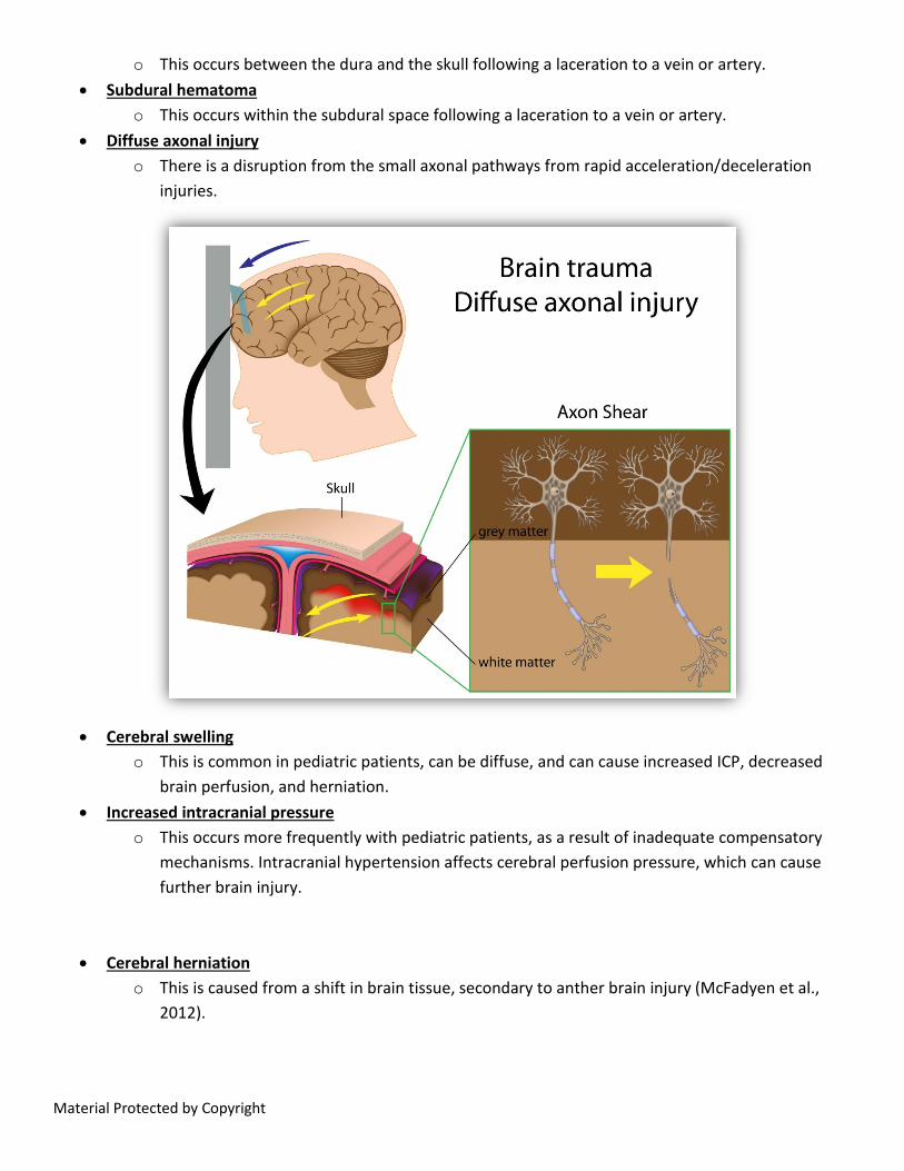
o This occurs within the subdural space following a laceration to a vein or artery. • Diffuse axonal injury
o There is a disruption from the small axonal pathways from rapid acceleration/deceleration injuries.
• Cerebral swelling
o This is common in pediatric patients, can be diffuse, and can cause increased ICP, decreased brain perfusion, and herniation.
• Increased intracranial pressure o This occurs more frequently with pediatric patients, as a result of inadequate compensatory
mechanisms. Intracranial hypertension affects cerebral perfusion pressure, which can cause further brain injury.
• Cerebral herniation o This is caused from a shift in brain tissue, secondary to anther brain injury (McFadyen et al.,
2012).
Material Protected by Copyright
TBI Assessment Swelling and tenderness can indicate fractures. A basilar skull fracture may also have accompanying clear or pink-tinged fluid from the nose and ears, or a bruising around the ears or eyes. Changes in level of consciousness, lethargy, nausea, pallor, diaphoresis, headaches, dizziness, disorientation, and amnesia can indicate TBI. Loss of consciousness, seizures, shock, bulging fontanels (infants), hypoxia, changes in vital signs (particularly hypertension and bradycardia), vomiting, posturing, changes in pupil size/responsiveness, and apnea can be late signs with TBI (McFadyen et al., 2012). TBI Management Treatment includes goals for preventing complications, promoting healing, and managing pain. Implantation of drains, medical management of symptoms (including medications for reducing ICP), fluid management, and/or surgical interventions may be needed, depending on the severity of the TBI. Spinal Cord Types of spinal cord injuries include:
• Complete o This includes the complete loss of sensory and motor function from a cut in the nerve pathways
below the traumatized area. The patient may be quadriplegic or paraplegic, and the rectal sphincter tone is absent.
• Incomplete o The injury has a partial loss of motor and sensory function below the traumatized area. The
patient may experience intermittent weakness or paresthesia. • Contusion
o This is from edema and bruising of the spinal cord, which can cause a temporary or permanent loss of function.
• Transection o This is a complete cut through the spinal cord, resulting in permanent loss of function.
Spinal Cord Assessment Signs and symptoms of spinal cord trauma vary, and can include an abnormal neurological exam, limited neck or spine mobility, tenderness along the spine, pain with movement, or hypotension. Edema, ecchymosis, observable deformity, and muscle spasms can also occur. Assessment of spinal cord injury may be difficult if the patient has also had a head injury. Children can have injury to the spinal cord without obvious neurological signs and symptoms, or without abnormalities noted on radiological studies (McFadyen et al., 2012).
Material Protected by Copyright
Spinal Cord Trauma Management Traction, stabilization, fluid balance, and medical management of secondary effects such as neurogenic shock are potential treatments of spinal cord injury. Surgery may be required for decompression, fractures, and dislocations. Communication with Patients and Families Communications and interventions with the pediatric patient should be individualized, based on behavior and developmental stage. A family-focused approach should be used with pediatric patients, and trust/rapport must be developed with the patient and family. Some considerations include therapeutic presence, active listening, empathy, and sensitivity to nonverbal communication. Use of a multidisciplinary team is beneficial to address the needs of the patient and family (McFadyen et al., 2012). Conclusion Pediatric trauma patients are unique in the assessment and management required. Differences in anatomy, physiology, growth and development are important to understand. Education and awareness in the community needs to continue in an attempt to reduce the incidence of pediatric injury. References American Heart Association. (2015). Pediatric advanced life support: Provider manual. Dallas, TX: American Heart Association. Bowden, V., & Greenburg, C. (2012) Pediatric nursing procedures (3rd ed.). Philadelphia, PA: Lippincott Williams and Wilkins. Centers for Disease Control and Prevention: National Center for Injury Prevention and Control, Division of Unintentional Injury Prevention. (2012). Child injury. Retrieved from: http://www.cdc.gov/vitalsigns/ChildInjury/index.html Maryniak, K. (2013). Pediatric history and physical assessment (unpublished presentation). McFadyen, J.G., Ramaiah, R., & Bhananker, S.M. (2012). Initial assessment and management of pediatric trauma patients. International Journal of Critical Illness and Injury Science, 2(3), 121-127. O’Toole, M (ed.). (2013). Mosby’s medication dictionary (9th ed.). St. Louis, MO: Elsevier. Potts, N., & Mandleco, B. (2012). Pediatric nursing: Caring for children and their families (3rd ed.). Clifton Park, NY: Delmar.
Material Protected by Copyright
Disclaimer This publication is intended solely for the educational use of health care professionals taking this course, for credit, from RN.com, in accordance with RN.com terms of use. It is designed to assist health care professionals, including nurses, in addressing many issues associated with health care. The guidance provided in this publication is general in nature, and is not designed to address any specific situation. As always, in assessing and responding to specific patient care situations, health care professionals must use their judgment, as well as follow the policies of their organization and any applicable law. This publication in no way absolves facilities of their responsibility for the appropriate orientation of health care professionals. Health care organizations using this publication as a part of their own orientation processes should review the contents of this publication to ensure accuracy and compliance before using this publication. Health care providers, hospitals and facilities that use this publication agree to defend and indemnify, and shall hold RN.com, including its parent(s), subsidiaries, affiliates, officers/directors, and employees from liability resulting from the use of this publication. The contents of this publication may not be reproduced without written permission from RN.com Participants are advised that the accredited status of RN.com does not imply endorsement by the provider of ANCC of any products/therapeutics mentioned in this course. The information in the course is for educational purposes only. There is no “off label” usage of drugs of products discussed in this course. You may find that both generic and trade names are used in courses produced by RN.com. The use of trade names does not indicate any preference of one trade named agent or company over another. Trade names are provided to enhance recognition of agents described in the course. Note: all dosages given are for adults unless otherwise stated. The information on medications contained in this course is not meant to be prescriptive or all-encompassing. You are encouraged to consult with physicians and pharmacists about all medications issued for your patients.