Patterns and dynamics of 32P-phosphate and labelled 2-aminoisobutyric acid (14C-AIB) translocation...
12
Patterns and dynamics of 32 P-phosphate and labelled 2-aminoisobutyric acid ( 14 C-AIB) translocation in intact basidiomycete mycelia Stefan Olsson a; *, Simon N. Gray b a Section of Genetics and Microbiology, Department of Ecology and Molecular Biology, Royal Veterinary and Agricultural University, Thorvaldsensvej 40, DK-1871 Frederiksberg C, Copenhagen, Denmark b Faculty of Science and Technology, University of Luton, Luton, UK Accepted 20 March 1998 Abstract Following uptake of 32 P-orthophosphate and 14 C-aminoisobutyric acid ( 14 C-AIB) the patterns of distribution of the isotopes through intact basidiomycete mycelia were non-destructively mapped at regular intervals using a L-scanner. Analysis of the results suggests that translocation of 32 P and 14 C-AIB through mycelia of Pleurotus ostreatus and Schizophyllum commune occurred along a restricted number of clearly defined, but macroscopically invisible, routes through the mycelium. In contrast to this, 32 P added to mycelia of Coprinus cinereus remained immobilised at the addition point. Simultaneous acropetal and basipetal translocation of 32 P and 14 C-AIB was observed in different regions of colonies of P. ostreatus and S. commune. Translocation of label around the periphery of colonies strongly suggested the existence of anastomoses around the colony margin. Both 32 P and 14 C-AIB were initially immobilised at the addition point, from which each was subsequently translocated to other parts of the mycelium. The observed translocation of nutrients could not be explained by simple diffusion alone. The velocity of translocation and the complexity of the translocation pattern of 32 P were greatest in mycelia of P. ostreatus,a hardwood decomposer, followed by S. commune, a wood and litter decomposer and parasite. Translocation through mycelia of C. cinereus, a coprophilus saprophyte, was very slow. This study provides the first detailed description of nutrient translocation through intact, entire fungal mycelia over time. z 1998 Federation of European Microbiological Societies. Published by Elsevier Science B.V. All rights reserved. Keywords : Nutrient translocation ; Nutrient relocation ; Fungal mycelium 1. Introduction The nature and concentration of nutrients and other physicochemical factors vary greatly in space within the natural habitat of many fungi. This is especially true for fungi inhabiting soil. These fungi often produce large mycelia spanning a variety of microsites, each with di¡erent environmental condi- tions. Such fungi might be expected to be especially adapted for growth in heterogeneous environments. It is evident from a number of studies that wood and litter decomposing fungi are able to reallocate 0168-6496 / 98 / $19.00 ß 1998 Federation of European Microbiological Societies. Published by Elsevier Science B.V. All rights reserved. PII:S0168-6496(98)00026-9 * Corresponding author. Tel.: +45 35 28 26 46; Fax: +45 35 28 26 06; E-mail: [email protected] FEMS Microbiology Ecology 26 (1998) 109^120
-
Upload
stefan-olsson -
Category
Documents
-
view
212 -
download
0
Transcript of Patterns and dynamics of 32P-phosphate and labelled 2-aminoisobutyric acid (14C-AIB) translocation...

Patterns and dynamics of 32P-phosphate and labelled2-aminoisobutyric acid (14C-AIB) translocation in intact
basidiomycete mycelia
Stefan Olsson a;*, Simon N. Gray b
a Section of Genetics and Microbiology, Department of Ecology and Molecular Biology, Royal Veterinary and Agricultural University,Thorvaldsensvej 40, DK-1871 Frederiksberg C, Copenhagen, Denmarkb Faculty of Science and Technology, University of Luton, Luton, UK
Accepted 20 March 1998
Abstract
Following uptake of 32P-orthophosphate and 14C-aminoisobutyric acid (14C-AIB) the patterns of distribution of the isotopesthrough intact basidiomycete mycelia were non-destructively mapped at regular intervals using a L-scanner. Analysis of theresults suggests that translocation of 32P and 14C-AIB through mycelia of Pleurotus ostreatus and Schizophyllum communeoccurred along a restricted number of clearly defined, but macroscopically invisible, routes through the mycelium. In contrastto this, 32P added to mycelia of Coprinus cinereus remained immobilised at the addition point. Simultaneous acropetal andbasipetal translocation of 32P and 14C-AIB was observed in different regions of colonies of P. ostreatus and S. commune.Translocation of label around the periphery of colonies strongly suggested the existence of anastomoses around the colonymargin. Both 32P and 14C-AIB were initially immobilised at the addition point, from which each was subsequently translocatedto other parts of the mycelium. The observed translocation of nutrients could not be explained by simple diffusion alone. Thevelocity of translocation and the complexity of the translocation pattern of 32P were greatest in mycelia of P. ostreatus, ahardwood decomposer, followed by S. commune, a wood and litter decomposer and parasite. Translocation through mycelia ofC. cinereus, a coprophilus saprophyte, was very slow. This study provides the first detailed description of nutrient translocationthrough intact, entire fungal mycelia over time. z 1998 Federation of European Microbiological Societies. Published byElsevier Science B.V. All rights reserved.
Keywords: Nutrient translocation; Nutrient relocation; Fungal mycelium
1. Introduction
The nature and concentration of nutrients andother physicochemical factors vary greatly in space
within the natural habitat of many fungi. This isespecially true for fungi inhabiting soil. These fungioften produce large mycelia spanning a variety ofmicrosites, each with di¡erent environmental condi-tions. Such fungi might be expected to be especiallyadapted for growth in heterogeneous environments.It is evident from a number of studies that woodand litter decomposing fungi are able to reallocate
0168-6496 / 98 / $19.00 ß 1998 Federation of European Microbiological Societies. Published by Elsevier Science B.V. All rights reserved.PII: S 0 1 6 8 - 6 4 9 6 ( 9 8 ) 0 0 0 2 6 - 9
FEMSEC 912 11-6-98
* Corresponding author. Tel. : +45 35 28 26 46;Fax: +45 35 28 26 06; E-mail: [email protected]
FEMS Microbiology Ecology 26 (1998) 109^120
nutrients between di¡erent parts of their mycelium[1^4]. Extensive translocation of phosphorus hasbeen shown to take place through mycelial cord sys-tems of Phanerochaete velutina, Phallus impudicusand Hypholoma fasciculare grown in laboratory mi-crocosms and in the ¢eld. In such systems, 32P-la-belled phosphate was translocated 10 times morequickly through connective than through non-con-nective mycelium. Reallocation of phosphate withincord systems was dependent upon the potential ofnewly colonised, distant resource units as carbonsources, the decay state of those resource units andthe temperature. The amount of phosphorus trans-located between resource units was species-depend-ent, and 32P added to cord systems in the ¢eld wastranslocated for distances of up to 0.75 m, and waslater detected in nearby decaying wood, leaf litter,and plants [2,5^7]. Four main mechanisms havebeen suggested for the translocation of nutrientsthrough fungal mycelia: di¡usion [4]; di¡usion aidedby uptake in excess of local needs [8]; involvement ofa contractile system [9] ; and pressure-driven bulk£ow [10,11]. The last two mechanisms are activeprocesses, requiring energy expenditure to drive theactual translocation process. It is important to notethat the mechanisms are not mutually exclusive. Ithas been known for some time that acropetal andbasipetal translocation of di¡erent nutrients can oc-cur simultaneously in individual rhizomorphs of Ar-millaria mellea [12]. In a previous study we showedthat a nutrient, phosphorus, was translocatedthrough a mycelium of Schizophyllum commune ina di¡erent and more active way than the non-nu-trient caesium [13]. This result supports the viewthat more than one of the translocation mechanismsdescribed above may simultaneously be active in amycelium.
In many previous studies, the translocation of nu-trients through ¢lamentous fungi has been measuredalong a one-dimensional structure [8,12,14]. In otherstudies, the rate of removal of a nutrient from oraccumulation within a particular point or pointswithin a mycelium was measured quantitatively[2,15], or else translocation across a whole myceliumwas measured qualitatively [15^17]. This inevitablygives an incomplete picture of the translocationprocesses occurring within a mycelium. Recent ad-vances in the conceptualisation of fungal develop-
ment at the organism level have resulted in myceliabeing described as indeterminate, potentially in¢n-itely expandable living systems. According to thisparadigm, patterns of mycelial development are crit-ically dependent upon the breakdown, maintenanceand generation of connections allowing the realloca-tion of resources across the mycelium [18]. It is,therefore, desirable to gain an improved understand-ing of nutrient translocation through ¢lamentousfungi at the organism level, as translocation proc-esses observed within isolated hyphae or cords, orat particular points within a mycelium, may not berepresentative of the behaviour of the mycelium as awhole.
The aim of this work was to study the transloca-tion of phosphorus and amino acids through intact,whole mycelia of saprotrophic basidiomycete fungi.This was achieved by non-destructively recording thedistribution of labelled phosphorus and [14C]2-ami-noisobutyric acid (14C-AIB) through largely 2-di-mensional agar cultures of wood and litter decom-posing basidiomycetes through time. Three specieswere studied, in order to investigate whether fungalspecies di¡er in their ability to translocate nutrients.Similar experiments have been performed previouslyfor 137Cs [13], a pollutant, but the present paper isthe ¢rst to describe this for nutrients, in this casephosphorus and the amino acid analogue AIB.AIB was chosen as a marker for the amino acidpool because it is taken up by fungi and is trans-located like other amino acids, without being metab-olised [19].
2. Materials and methods
2.1. Origin and characteristics of fungal isolates
S. commune Fr.:Fr. `SC' dikaryon and Pleurotusostreatus (Jacquin: Fr.) Kummer `PO2' dikaryonwere provided by Dr. Paul Markham, King'sCollege, University of London. Coprinus cine-reus (Schae¡er: Fr.) S.F. Gray `Meathop' dikary-on was provided by Dr. David Moore, School ofBiological Sciences, University of Manchester. Thefungi were maintained on malt extract agar (2%w/v malt extract broth+1.5% w/v agar No. 1, Ox-oid).
FEMSEC 912 11-6-98
S. Olsson, S.N. Gray / FEMS Microbiology Ecology 26 (1998) 109^120110

2.2. Plate cultures
Glass plates (3 mm thick standard glass used forwindow panes) were cut into 200U100 mm rectan-gles, sterilised by autoclaving and allowed to cool.Plates were placed in pairs into 240U240 mm sterilepolystyrene plastic screening dishes (Nunc), and110 ml malt extract agar was poured into eachdish. This was su¤cient agar to cover the glass platesto a depth of 1 mm. Each plate was inoculated cen-trally with an inverted single plugs (8 mm in diame-ter) cut from the edge of a stock cultures. Excessagar was shaved o¡ each inoculum plug with a scal-pel leaving only a mat of mycelium as the inoculum.This was done to prevent the agar from interferingwith subsequent scanning in the L-scanner. There-after the plates were incubated at 20³C until the col-ony diameter had reached 90^100 mm.
2.3. Labelling with 32P-orthophosphate and 14C-AIB
The glass plates carrying the fungal colonies werecut out from the agar. Label was then added into theagar at either the centre of the colonies, or at thecolony edge, using a micro-pipette. Labelled phos-phorus was added as 0.5 Wl of an aqueous 32P sol-ution (18.5 kBq; 55 fmol total P). Labelled AIB wasadded as 4 Wl of a solution of 14C-AIB (29.6 kBq, 13nmol total AIB) in 0.01 M HCl. The glass plateswere then wrapped in polyethylene foil (Glad wrap)to prevent desiccation. The shape of the colony andthe point of addition of radiolabel were carefullytraced directly onto the foil by using a soft pen.This trace was also used to control that the radialgrowth rate during the incubation with the labelledcompounds did not change compared to before la-belling.
2.4. Labelling with CaH32PO4
Labelled calcium phosphate was prepared by heat-ing 400 Wl phosphate solution (K2HPO4, 1.0 g l31,pH 5.4), to which 20 Wl H3
32PO4 solution (740 kBq,2.2 pmol) had been added, together with 400 Wl cal-cium solution (CaCl2W2H2O, 0.85 g l31) at 50³C on awater bath until a precipitate of CaH32PO4 wasformed. The precipitate was collected by dropwiseaddition of 80-Wl aliquots of suspension onto discs
of glass ¢bre ¢lter paper (GF/A Whatman) 5.5 mmin diameter, placed on plain ¢lter paper. The discswere then moved to new sheets of plain ¢lter paper,and each was washed 10 times with 10 drops of dis-tilled water. The resulting discs, loaded withCaH32PO4 crystals, were used for labelling fungalcolonies. A single disc was added to each colony,either centrally or at the colony edge.
2.5. Controls
Plates which had not been inoculated with fungiwere labelled in the same manner as the experimentalplates described above. Where plates were labelledwith 32P-orthophosphate or 14C-AIB, these controlsprovided a means of distinguishing between di¡usivemovement of label through the agar and transloca-tion through fungal mycelia. Where plates were la-belled with calcium 32P-phosphate, the controls wereused to determine the degree of solubility of the cal-cium phosphate precipitate. Finally but most impor-tantly, the controls were used to verify that the ob-served translocation was not an artefact of theexperimental system.
2.6. Determination of radiolabel distribution throughmycelia
The distribution of radiolabel across experimentalplates was carried out non-destructively using a L-scanner (Bioscan Imaging Scanner System 2000equipped with Autochanger 1000). The scanner de-tects ionisation events resulting from radioactive de-cay by means of a 200 mm long position-sensitiveanode wire. To improve resolution and to reducescatter in the direction along the anode wire a metalwindow with a metal grid, a mechanical collimator,is ¢tted in front of the anode wire. The mechanicalcollimators can have di¡erent slit widths or di¡erentdensity of the metal grids. A high grid density giveshigher resolution but lower sensitivity. Two-dimen-sional scanning is obtained by moving the Autoch-anger table in steps in a direction perpendicular tothe anode wire. For the experiments described here,the glass plates were always oriented on the Autoch-anger table to move in steps along the shortest di-mension (100 mm) of the glass plates. The positionof decay events, and hence the distribution of radio-
FEMSEC 912 11-6-98
S. Olsson, S.N. Gray / FEMS Microbiology Ecology 26 (1998) 109^120 111
label, was determined at a resolution of 0.78 mmalong the length of the glass plates, and at a resolu-tion of either 3 mm or 5 mm across the width of theglass plates depending upon the isotope used (seebelow).
The isotope 14C emits low-energy L-radiationwhich allows good spatial resolution of the originof a decay event, but is prone to self-absorption ofradiation by the sample. In contrast, 32P emits high-energy L-radiation which is less prone to self-absorp-tion. This results in poorer spatial resolution thanthat obtained with a low energy isotope, but givesimproved accuracy when quantifying the distribution
of label below the surface of the mycelium or agar.Therefore, when measuring 14C-AIB, 30 Autoch-anger steps 3 mm apart were used together with alow resolution, high sensitivity mechanical collimatorof 6 mm slit width for a counting time of 2 min foreach step. For 32P, 19 Autochanger steps 5 mm apartwere used together with a high resolution, low sensi-tivity mechanical collimator of 10 mm slit width fora counting time of 1 min for each step. Plates weremaintained at a temperature 4^5³C below the 23^28³C in the scanner during scanning to prevent theformation of condensation on the inside of the foil[20]. Plates were returned to the 20³C incubator im-
FEMSEC 912 11-6-98
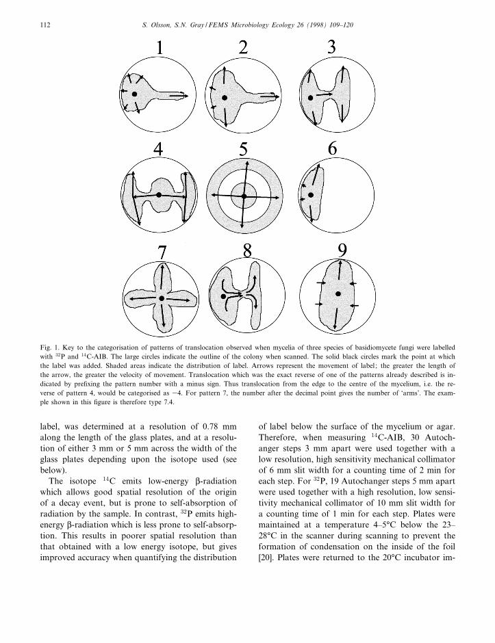
Fig. 1. Key to the categorisation of patterns of translocation observed when mycelia of three species of basidiomycete fungi were labelledwith 32P and 14C-AIB. The large circles indicate the outline of the colony when scanned. The solid black circles mark the point at whichthe label was added. Shaded areas indicate the distribution of label. Arrows represent the movement of label ; the greater the length ofthe arrow, the greater the velocity of movement. Translocation which was the exact reverse of one of the patterns already described is in-dicated by pre¢xing the pattern number with a minus sign. Thus translocation from the edge to the centre of the mycelium, i.e. the re-verse of pattern 4, would be categorised as 34. For pattern 7, the number after the decimal point gives the number of `arms'. The exam-ple shown in this ¢gure is therefore type 7.4.
S. Olsson, S.N. Gray / FEMS Microbiology Ecology 26 (1998) 109^120112
mediately after scanning. The lower limits for detec-tion of 32P and 14C-AIB by the L-scanner were1.33U1032 Bq mmÿ2 (3.84U1035 fmol P mm32)and 2.61U1031 Bq mm32 (114.7 fmol AIB mm32)respectively.
2.7. Replication and data processing
All of the experiments described above were car-ried out in duplicate. The data generated by the Bio-scan system was exported as ascii ¢les using a spe-cially supplied ascii export program (Bioscan). Datamanipulation and construction of two- and three-di-mensional plots and subtraction plots was carriedout using Excel (Microsoft). Di¡usion coe¤cientsof 32P and 14C-AIB were estimated from the controlplates by two dimensional curve ¢tting of the stand-ard equation for the distribution of substances in atwo-dimensional di¡usion to the experimental data,using the least squares method and employing Ex-cel's Problem Solver.
Di¡usion coe¤cients measured from the uninocu-lated control plates were used to calculate the distri-butions of radiolabel through the plates inoculatedwith fungi which would have arisen, had di¡usionbeen the mechanism of translocation. The measuredand calculated distributions of radiolabel were com-pared by construction of di¡erence plots, where thedistribution of label predicted by the di¡usion model
was subtracted from the measured distribution oflabel through fungal mycelia. A perfect ¢t of thelaboratory data to the di¡usion model would havegiven no di¡erence in cpm for all points in X and Y.Positive values on such a di¡erence plot indicate thepresence of more label in a particular region of acolony than would be predicted on the basis of dif-fusion; negative values demonstrate the presence ofless label in that part of the colony than would bepredicted by the di¡usion model.
The angle or direction of translocation `arms' (seebelow) was determined from contour maps showingthe distribution of radiolabel across each platein plan view. Arms were located and markedon the map, and the angle of each arm from thepositive y-axis measured to nearest 5³ with protrac-tor.
3. Results
No macroscopic di¡erentiation of fungal mycelia,such as cord formation or variation in hyphal den-sity between regions of the mycelium of the sameage, was observed in any of the experiments carriedout. However, the three fungi reallocated the addednutrients in di¡erent ways. The di¡erent types ofrelocation pattern observed for the di¡erent fungiare summarised in Table 1 and Fig. 1.
FEMSEC 912 11-6-98
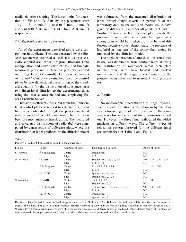
Table 1Patterns of nutrient translocation found in the experiments
Fungus Label Addition of label Translocation pattern Angle of `arms'
C. cinereus 32P-phosphate Centre Symmetrical NDEdge 1, 2 ND
P. ostreatus 14C-AIB Centre Symmetrical, 7.1, 7.2, 7.4 105 185 250 350Edge 2, 3, 7.1, 8 ND
32P-phosphate Centre 7.1, 37.2, 7.3, 37.3 5 120 240Edge 2, 6, 8 ND
CaH32PO4 Centre Symmetrical, 4, 34 NDEdge Symmetrical, 2, 4, 6 ND
S. commune 14C-AIB Centre 37.1, 7.1, 7.2 5 185Edge Symmetrical, 2, 6 ND
32P-phosphate Centre Symmetrical, 37.1, 7.2, 37.3, 7.3 90 145 225Edge 2, 6, 9 ND
CaH32PO4 Centre Symmetrical NDEdge Symmetrical, 6 ND
Duplicate plates (A and B) were scanned at approximately 0, 6, 24, 48 and 120^144 h after the addition of label to either the centre or theedge of the colony. The pattern of translocation between consecutive time intervals was categorised according to the key shown in Fig. 1.Where di¡erent translocation patterns were observed on the same plate at di¡erent times, all are listed. Where de¢ned `arms' of translocationwere observed, the angle between each `arm' and the positive y-axis was measured in a clockwise direction.
S. Olsson, S.N. Gray / FEMS Microbiology Ecology 26 (1998) 109^120 113

FEMSEC 912 11-6-98
Fig. 2. Distribution of added 32P-orthophosphate (A and B) and 14C-AIB (C and D) through colonies of P. ostreatus at various intervalsafter the addition of label. In A and C, the label was added at the centre of the colony, and in B and D at the edge of the colony.
S. Olsson, S.N. Gray / FEMS Microbiology Ecology 26 (1998) 109^120114

3.1. Translocation of 32P and 14C-AIB throughcolonies labelled centrally
In mycelia of P. ostreatus both phosphorus and
AIB were translocated out from the point of addi-tion at the centre of the colony towards the edge ofthe colony along de¢ned translocation `arms' (Table1; Figs. 1 and 2A,C). Both 32P and 14C-AIB were
FEMSEC 912 11-6-98
Fig. 4. Distribution of added 32P-orthophosphate (A and B) and 14C-AIB (C and D) in a mycelium of S. commune 120 h after additionof label. In A and C, the label was added at the centre of the colony, and in B and D at the edge of the colony.
Fig. 3. Di¡erence plots showing the change in distribution of added 32P-orthophosphate in a colony of P. ostreatus between 24 h and 48h after labelling (A and B), and the change in distribution of 14C-AIB between 48 h and 120 h after labelling (C and D). In A and C,the label was added at the centre of the colony, and in B and D at the edge of the colony.
S. Olsson, S.N. Gray / FEMS Microbiology Ecology 26 (1998) 109^120 115

clearly detectable at the colony edge 24 h after addi-tion of label. Di¡erence plots, highlighting thechange in distribution of radiotracer between 24and 48 h (32P) or 48 h and 120 h (14C-AIB), showthat label was lost from the central point of addition,and accumulated close to the edge of the mycelium(Fig. 3A,C). The pattern of translocation through S.commune was similar, but the rate of accumulationof label at the edge of the colony was slower and the`arms' were less pronounced (Table 1; Figs. 1 and4A,C). The velocity of the front of 32P through my-celia of C. cinereus was considerably slower than ineither of the two other species studied. Translocation`arms' were not apparent in C. cinereus, and radialsymmetry in the distribution of radiolabel across the
mycelium around the central point of addition wasmaintained (Table 1; Fig. 5A). In all fungi the ve-locity of the front of AIB was lower than that ofphosphorus. It is not possible to be certain of theextent to which the di¡erence in lower limits fordetection of the two isotopes contributed to this dif-ference.
Where translocation `arms' were observed, the`arms' on any individual plate were consistentlyevenly spaced (Table 1). Thus, on a plate with three`arms' such as duplicate A of P. ostreatus labelledcentrally with 14C-AIB, the angles of the arms clock-wise from the positive y-axis were 5³, 120³ and 240³.The angles between adjacent arms on this plate weretherefore 115³, 120³ and 125³. Similarly, where four
FEMSEC 912 11-6-98
Fig. 5. Distribution pattern of added 32P-orthophosphate through a colony of C. cinereus 144 h after addition of label to either the centre(A) or the edge (B) of the colony.
Fig. 6. Comparison of the distribution of label after 24 h di¡usion of 32P-orthophosphate (A) and 26.5 h di¡usion of 14C-AIB (B) oncontrol agar plates (F) with that predicted for two-dimensional di¡usion (R). The predicted values were calculated for the measuredpoints on the plate therefore the cross sections shown are close to, but not through, the exact centre of the distribution.
S. Olsson, S.N. Gray / FEMS Microbiology Ecology 26 (1998) 109^120116
`arms' were observed they tended to be spaced at90 þ 10³. Where the angles between `arms' were noteven, they were consistent with those observed ona plate with one or two more `arms'. For exam-ple, duplicate B of S. commune labelled centrallywith 32P had two `arms' separated by 90³, whichwould give an even spacing on a plate with four`arms'.
3.2. Translocation of 32P and 14C-AIB throughcolonies labelled at the edge
Labelled phosphorus or AIB added to the edge ofmycelia of P. ostreatus was moved principally eithertowards the centre of the colony or around the pe-riphery of the colony. There was also some trans-location in the direction of growth of the leadingedge of the mycelium (Table 1; Figs. 1 and 2B).The label translocated to the centre of the colonysubsequently spread out from the centre, along`arms' apparently similar to those observed whenthe label had been added centrally (Fig. 2B). Myceliaof S. commune reallocated label added to the edge ofthe colony in a pattern similar to that seen inP. ostreatus, but the velocity of the label front waslower and the patterns developed were less pro-nounced (Table 1; Fig. 4B,D). Label added to theedge of mycelia of C. cinereus was also reallocatedtowards the centre, around the periphery and to-wards the edge of the colony, but at a much lowervelocity than in either of the other two fungi studied(Table 1; Figs. 1 and 5B). No label reached thecentre of the colony before the end of the experi-ment. It is consequently not possible to be certain
whether `arms' of translocation would subsequentlyhave developed.
3.3. Comparison with a di¡usion model
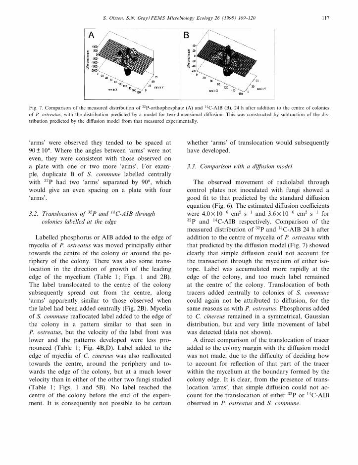
The observed movement of radiolabel throughcontrol plates not inoculated with fungi showed agood ¢t to that predicted by the standard di¡usionequation (Fig. 6). The estimated di¡usion coe¤cientswere 4.0U1036 cm2 s31 and 3.6U1036 cm2 s31 for32P and 14C-AIB respectively. Comparison of themeasured distribution of 32P and 14C-AIB 24 h afteraddition to the centre of mycelia of P. ostreatus withthat predicted by the di¡usion model (Fig. 7) showedclearly that simple di¡usion could not account forthe transaction through the mycelium of either iso-tope. Label was accumulated more rapidly at theedge of the colony, and too much label remainedat the centre of the colony. Translocation of bothtracers added centrally to colonies of S. communecould again not be attributed to di¡usion, for thesame reasons as with P. ostreatus. Phosphorus addedto C. cinereus remained in a symmetrical, Gaussiandistribution, but and very little movement of labelwas detected (data not shown).
A direct comparison of the translocation of traceradded to the colony margin with the di¡usion modelwas not made, due to the di¤culty of deciding howto account for re£ection of that part of the tracerwithin the mycelium at the boundary formed by thecolony edge. It is clear, from the presence of trans-location `arms', that simple di¡usion could not ac-count for the translocation of either 32P or 14C-AIBobserved in P. ostreatus and S. commune.
FEMSEC 912 11-6-98
Fig. 7. Comparison of the measured distribution of 32P-orthophosphate (A) and 14C-AIB (B), 24 h after addition to the centre of coloniesof P. ostreatus, with the distribution predicted by a model for two-dimensional di¡usion. This was constructed by subtraction of the dis-tribution predicted by the di¡usion model from that measured experimentally.
S. Olsson, S.N. Gray / FEMS Microbiology Ecology 26 (1998) 109^120 117

3.4. Translocation of 32P added as insoluble calciumphosphate
Translocation of labelled insoluble calcium phos-phate di¡ered from that of labelled soluble phos-phate in two ways. Firstly, reallocation of insolublelabelled phosphate was delayed by 24^48 h in com-parison to soluble phosphate (data not shown). Sec-ondly, the distribution of label through fungal my-celia after translocation had occurred deviated lessfrom a symmetrical or Gaussian distribution wherephosphate was added as an insoluble calcium salt,than where it was added as soluble orthophosphate(Table 1, Fig. 1). In control plates without fungi weunexpectedly found di¡usion of the calcium phos-phate label through the medium. The di¡usion coef-¢cient was approximately half of that estimated for32P-orthophosphate.
4. Discussion
The detection of both 32P and 14C-AIB at the edgeof colonies of P. ostreatus and S. commune within 24h of addition at the centre of the colony, 45 mmaway, implies a velocity of the label front of at least1.8 mm h31. This is an order of magnitude fasterthan the reported velocity of translocation of 137Csthrough intact mycelia of S. commune, Armillariagallica and A. bulbosa [13,21], and four times fasterthan the velocity of translocation of 86Rb, added as atracer for potassium, through intact mycelia of Neu-rospora crassa [11]. The contrast between the veloc-ities of translocation of the tracers for nutrients re-ported here and the non-nutrient caesium con¢rmsthat fungi possess some mechanism for acceleratedtranslocation of nutrients. It may be that, in a wood-land soil environment, potassium is far less oftenlimiting for growth of a fungus than phosphorus oramino acids. Consequently soil fungi may not haveevolved such e¤cient mechanisms for translocationof potassium as for phosphorus and amino acids.
Velocities of translocation of 14C-aspartic acid and32P-phosphate through cords of Serpula lacrimans inexcess of 200 mm h31, and of 32P-phosphate throughrhizomorphs of A. mellea in excess of 20 mm h31,have previously been reported [12,15]. These studieswere performed with specialised translocation struc-
tures of the di¡erentiated fungi and were carried outin linear systems likely to enhance the rate of trans-location observed through their design. This con-¢rms the importance of carrying out studies of trans-location on entire, intact mycelia.
The labelled nutrients were added in trace amounts(femtomolar and picomolar quantities), in a mannerwhich does not allow the accurate calculation of thespeci¢c activities of either 32P or 14C-AIB relative tothe total concentrations of phosphorus and aminoacids present. Therefore the movements of tracer ob-served will largely re£ect the translocation of aminoacids and phosphorus already present in the mycelia,rather than translocation in response to the additionof the radiotracers. It must also be borne in mindwhen interpreting the results that the speci¢c activityis unknown. Nevertheless it is clear that, particularlyin P. ostreatus but also in S. commune, translocationof both phosphorus and amino acids took place alonga restricted number of clearly de¢ned but macroscopi-cally invisible routes through the mycelium (Figs. 2^5). This was not the case with C. cinereus. Further-more, simultaneous acropetal and basipetal move-ment of both 32P and 14C-AIB was observed in di¡er-ent regions of colonies of P. ostreatus and S. commune(Table 1). For example, in the case of replicate A of P.ostreatus labelled centrally with 32P, label was trans-located acropetally along three `arms' for the ¢rst 48 hafter labelling. However, between 48 and 120 h afterlabelling, the direction of translocation in one ofthe `arms' reversed, becoming basipetal, whilst trans-location along the other two `arms' remained acro-petal. The translocation of nutrients observed couldnot be explained by simple di¡usion alone, whichmay imply the presence of an active mechanism fortranslocation. Active translocation has most fre-quently been postulated as a method of nutrienttranslocation in wood-decomposing [1^4] and mycor-rhizal basidiomycetes [3,16,17,20], and arbuscularmycorrhizal fungi [21^23]. Evidence for active trans-location in other fungi has also been published[4,11,24].
Comparison of the experimental data with the dif-fusion model suggested that label was immobilised atthe point of addition to the mycelium. Similar ob-servations were made by Clipson et al. [25] for theuptake of phosphorus by hyphal cords in the ¢eld.They suggested that the major fraction of phosphate
FEMSEC 912 11-6-98
S. Olsson, S.N. Gray / FEMS Microbiology Ecology 26 (1998) 109^120118
absorbed by the cords was converted into an immo-bile form, not available for translocation. However,other studies have shown extensive translocation ofphosphorus through mycelial cord systems in the¢eld and in microcosms [2,5^7]. In the present ex-periments, the immobilised label subsequently servedas a pool of nutrient that decreased as the labelaccumulated in other parts of the mycelium (seeFig. 2).
The absence of macroscopic di¡erentiation in themycelia studied clearly does not preclude functionaldi¡erentiation between hyphae, or di¡erences at themicroscopic level. The patterns of label distributionthrough mycelia of P. ostreatus and S. communeshow striking similarities with those generated by areaction-di¡usion model proposed by Davidson et al.[18] to account for the large-scale properties of fun-gal mycelia. For this particular model, formation ofpeaks in the concentration of an activator (in thatcase biomass) near the colony margin was condition-al upon the rate of replenishment of substrate ex-ceeding the rate of decay of activator. The formationof clearly bounded, stable radial patterns, as a con-sequence of the ampli¢cation of small concentrationgradients, has been documented in other biologicalsystems [26]. In the context of nutrient reallocationas described here, self-organisation of the myceliumto enable e¤cient translocation of nutrients could beachieved through modi¢cation of the degree of insu-lation of the boundaries to nutrient uptake andtranslocation (hyphal walls and septa), and throughmodi¢cation of the degree of overall resistance to£ow through the mycelium [27]. The regular spacingbetween clearly de¢ned translocation `arms', whichwas observed in mycelia of P. ostreatus and S. com-mune, may be due to self-organisation within themycelia. The overall resistance to £ow through themycelium is increased by septation but reduced byanastomosis [27]. The translocation of both 32P and14C-AIB around the periphery of mycelia of P. os-treatus and S. commune, clearly seen when colonieswere labelled at the edge (Table 1, Fig. 2B,D, Fig.4B,D), strongly suggests the existence of anastomo-ses around the colony margin. Thus the patterns ofnutrient reallocation observed here are consistentwith the view of a mycelium as a self-organising,`communication network' proposed by Rayner [28].Such a network will exhibit context-dependant mac-
roscopic patterns of growth [18], which will includepatterns of nutrient reallocation.
The pattern of nutrient translocation through S.commune was less rigidly bounded than that observedin P. ostreatus. Very little translocation was observedwhen label was added to mycelia of C. cinereus. Thedi¡erence in translocation velocity and the complex-ity of the translocation pattern in the di¡erent speciesmight re£ect their adaptation to ecological nichesvarying in the heterogeneity with which carbon, ni-trogen and phosphorus sources are distributed. Spe-ci¢cally, P. ostreatus is a primary decomposer ofhardwood tree species; S. commune grows sapro-phytically on pine litter, and saprophytically and par-asitically on woody substrates; and C. cinereus is acoprophilus saprophyte. Inter- and intra-speci¢c dif-ferences in the ability of fungi to translocate carbonand mineral nutrients have been reported previously[4]. Although caution must be exercised when apply-ing data obtained in vitro to the ¢eld situation, theresults reported above would support the hypothesisthat inter-speci¢c di¡erences in the ability of fungi totranslocate nutrients relate to the adaptation of par-ticular fungal species or isolates to exploit a particu-lar niche in soil ecosystems [4].
The most likely explanation for the unexpecteddi¡usion of 32P added as calcium phosphate in thecontrol plates is that the calcium phosphate wassparingly soluble in malt agar, which has a pH of5.4 þ 0.2. Even so, the rate of reallocation of labelledphosphate added to mycelia of P. ostreatus and S.commune was slower when phosphate was added in asparingly soluble form than when it was added in afreely soluble form. Whether the observed di¡erencesin the pattern of reallocation of insoluble phosphatecompared to soluble phosphate represent a majorshift in the translocatory activity of the mycelium,or are simply an artefact of the slower rate of real-location of insoluble phosphate, is uncertain. Thismerits further investigation, as there is little informa-tion on how fungi respond to the variety of chemicalforms in which they encounter phosphorus and othernutrients in the ¢eld.
Acknowledgments
This work was supported by grants to S.O. from
FEMSEC 912 11-6-98
S. Olsson, S.N. Gray / FEMS Microbiology Ecology 26 (1998) 109^120 119

the Swedish Natural Research Council and TheSwedish Agricultural and Forestry Research Counciland was carried out at the Department of MicrobialEcology, Lund, Sweden. The work was also sup-ported by a FEMS Fellowship for Young Scientiststo S.N.G.
References
[1] Jennings, D.H. (1990) The ability of basidiomycete myceliumto move nutrients through the soil ecosystem. In: NutrientCycling in Terrestrial Ecosystems: Field Methods, Applica-tion and Interpretation (Harrison, A.F., Ineson, P. andHeal, O.W., Eds), pp. 233^245. Academic Press, New York.
[2] Wells, J.M., Hughes, C. and Boddy, L. (1990) The fate of soilderived phosphorus in mycelial cord systems of Phanerochaetevelutina and Phallus impudicus. New Phytol. 114, 595^606.
[3] Cairney, J.W.G. (1992) Translocation of solutes in ectomycor-rhizal and saprotrophic rhizomorphs. Mycol. Res. 96, 135^141.
[4] Olsson, S. (1995) Mycelial density pro¢les of fungi on hetero-geneous media and their interpretation in terms of nutrientreallocation patterns. Mycol. Res. 99, 143^153.
[5] Wells, J.M. and Boddy, L. (1990) Wood decay, and phospho-rus and fungal biomass allocation, in mycelial cord systems.New Phytol. 116, 285^295.
[6] Wells, J.M. and Boddy, L. (1995) Phosphorus translocationby saprophytic basidiomycete mycelial cord systems on the£oor of a mixed deciduous woodland. Mycol. Res. 99, 977^980.
[7] Wells, J.M. and Boddy, L. (1995) E¡ect of temperature onwood decay and translocation of soil-derived phosphorus inmycelial cord systems. New Phytol. 129, 289^297
[8] Olsson, S. and Jennings, D.H. (1991) Evidence for di¡usionbeing the mechanism of translocation in the hyphae of threemolds. Exp. Mycol. 15, 302^309.
[9] Jennings, D.H. (1994) Translocation in mycelia. In: The My-cota I: Growth, Di¡erentiation and Sexuality (Wessels,J.G.H. and Meinhardt, H., Eds.), pp. 163^173. Springer-Ver-lag, Berlin.
[10] Jennings, D.H. (1976) Transport and translocation in ¢lamen-tous fungi. In: The Filamentous Fungi, Vol. 2. Biosynthesisand Metabolism (Smith, J.E. and Berry, D.R., Eds.), pp. 32^64. Edward Arnold, London.
[11] Girvin, D. and Thain, J.F. (1987) Growth of and transloca-tion in mycelium of Neurospora crassa on a nutrient de¢cientmedium. Trans. Br. Mycol. Soc. 88, 237^246.
[12] Granlund, H.I., Jennings, D.H. and Thompson, W. (1985)Translocation of solutes along rhizomorphs of Armillaria mel-lea. Trans. Br. Mycol. Soc. 84, 111^119.
[13] Gray, S.N., Dighton, J., Olsson, S. and Jennings, D.H. (1995)Real-time measurement of uptake and translocation of 137Cswithin mycelium of Schizophyllum commune Fr. by autora-
diography followed by quantitative image analysis. New Phy-tol. 129, 449^465.
[14] Olsson, S. and Jennings, D.H. (1991) A glass ¢ber ¢lter tech-nique for studying nutrient uptake by fungi: The techniqueused on colonies grown on nutrient gradients of carbon andphosphorus. Exp. Mycol. 15, 292^301.
[15] Brownlee, C. and Jennings, D.H. (1982) Long distance trans-location in Serpula lacrimans. Velocity estimates and the con-tinuous monitoring of induced perturbations. Trans. Br. My-col. Soc. 79, 143^148
[16] Finlay, R.D. and Read, D.J. (1986) The structure and func-tion of vegetative mycelium of ectomycorrhizal plants. I.Translocation of 14C-labelled carbon between plants intercon-nected by a common mycelium. New Phytol. 103, 143^156.
[17] Finlay, R.D. and Read, D.J. (1986) The structure and func-tion of vegetative mycelium of ectomycorrhizal plants. II. Theuptake and distribution of phosphorus by mycelial strandsinterconnecting host plants. New Phytol. 103, 157^165.
[18] Davidson, F.A., Sleeman, B.D., Rayner, A.D.M., Crawford,J.W. and Ritz, K. (1996) Context-dependent macroscopic pat-terns in growing and interacting mycelial networks. Proc. R.Soc. Lond. B Biol. Sci. 263, 873^880.
[19] Lilly, W.W., Higgins, S.M. and Wallweber, G.J. (1990) Up-take and translocation of 2-aminoisobutyric acid by Schizo-phyllum commune. Exp. Mycol. 14, 169^177.
[20] Timonen, S., Finlay, R.D., Olsson, S. and Soëderstroëm, B.(1996) Dynamics of phosphorus translocation in intact ecto-mycorrhizal systems: non-destructive monitoring using a L-scanner. FEMS Microbiol. Ecol. 19, 171^180.
[21] Gray, S.N., Dighton, J. and Jennings, D.H. (1996) The phys-iology of basidiomycete linear organs. III. Uptake and trans-location of radiocaesium within di¡erentiated mycelia of Ar-millaria spp. growing in microcosms and in the ¢eld. NewPhytol. 132, 471^482.
[22] Cooper, K.M. and Tinker, P.B. (1978) Translocation andtransfer of nutrients in vesicular arbuscular mycorrhizas.Part 2. Uptake and translocation of phosphorus, zinc andsulfur. New Phytol. 81, 43^52.
[23] George, E., Haeussler, K.U., Vetterlein, D., Gorgus, E. andMarschner, H. (1992) Water and nutrient translocation byhyphae of Glomus mosseae. Can. J. Bot. 70, 2130^2137.
[24] Amir, R., Steudle, E., Levanon, D., Hadar, Y. and Chet, I.(1995) Turgor changes in Morchella esculenta during translo-cation and sclerotial formation. Exp. Mycol. 19, 129^136.
[25] Clipson, N.J.W., Cairney, J.W.G. and Jennings, D.H. (1987)The physiology of basidiomycete linear organs. I. Phosphateuptake by cords and mycelium in the laboratory and the ¢eld.New Phytol. 105, 449^457.
[26] Jorgensen, R.A. (1995) Cosuppression, £ower color patternsand metastable gene expression states. Science 268, 686^691.
[27] Rayner, A.D.M., Gri¤th, G.S. and Ainsworth, A.M. (1994)Mycelial interconnectedness. In: The Growing Fungus (Gow,N.A.R. and Gadd, G.M., Eds.), pp. 21^40. Chapman andHall, London.
[28] Rayner, A.D.M. (1991) The challenge of the individualisticmycelium. Mycologia 83, 48^71.
FEMSEC 912 11-6-98
S. Olsson, S.N. Gray / FEMS Microbiology Ecology 26 (1998) 109^120120