Web Blight - Recinto Universitario de...
24
1 Web Blight [Updated by: Graciela Godoy-Lutz , Plant Pathologist, Instituto Dominicano de Investigaciones Agropecuarias y Forestales (IDIAF), Dominican Republic James S. Beaver , Breeder, University of Puerto Rico, Juan. C. Rosas , Breeder, Escuela Agrícola Panamericana, Honduras James R. Steadman , Plant Pathologist, University of Nebraska-Lincoln, Nebraska; August, 2010] Fig 1. Advanced symptoms produced by mycelial infection of Thanatephorus cucumeris (anamorph:Rhizoctonia solani subgroup AG-1-IE). Isabela, Puerto Rico. (J. Beaver)
Transcript of Web Blight - Recinto Universitario de...

1
Web Blight
[Updated by: Graciela Godoy-Lutz, Plant Pathologist, Instituto Dominicano de Investigaciones Agropecuarias y Forestales (IDIAF), Dominican Republic James S. Beaver, Breeder, University of Puerto Rico, Juan. C. Rosas, Breeder, Escuela Agrícola Panamericana, Honduras James R. Steadman, Plant Pathologist, University of Nebraska-Lincoln, Nebraska; August, 2010]
Fig 1. Advanced symptoms produced by mycelial infection of Thanatephorus cucumeris (anamorph:Rhizoctonia solani subgroup AG-1-IE). Isabela, Puerto Rico. (J. Beaver)
2
INTRODUCTION Web blight (WB), caused by a basidiomycete fungus Thanatephorus cucumeris (Frank) Donk (anamorph: Rhizoctonia solani Kühn), is an important bean disease in the hot and humid regions of the tropics causing reductions in seed yield and seed quality (Godoy-Lutz et al., 1996; 1998). The disease is endemic in the Central America and the Caribbean region but is also problematic in East Africa and South America. Efforts to manage WB have proven to be costly and often ineffective. The pathogen spreads by airborne basidiospores, mycelial bridges between plants, rain-splashed sclerotia, infested soil debris, and infected seeds (Galvez, et al. 1989; Godoy -Lutz, et al. 1996). The WB pathogen, R. solani (Rs), is a complex species composed of subgroups within Anastomosis Groups. At least six subgroups of Rs cause symptoms of web blight (Godoy-Lutz et al. 2008; Godoy-Lutz, 2003). The subgroups are: AG-1-IA, AG-1-IB, AG-2-2IV and three new subgroups of designated as AG-1-IE, AG-1-IF and AG-2-2WB. Variability among these subgroups includes different virulence patterns, fungicide resistance, optimal growth temperature and epidemiology (disease development rate, fungal propagule type, dissemination and survival). Godoy-Lutz et al. (2003) studied the variability among 45 isolates of R. solani from bean plants with web blight symptoms. The isolates were obtained from Argentina, Costa Rica, Cuba, the Dominican Republic, Honduras, Panama and Puerto Rico. They found morphological differences and variations in virulence among the isolates when the black bean line HT 7719 was inoculated in the greenhouse using the detached-leaf technique. Progress in breeding for WB resistance in common bean has been limited due to considerable variability in varietal reaction and the need for more understanding of pathogen variation and disease resistance mechanisms. At present, high levels of WB resistance are unavailable in commercial varieties. Researchers at the University of Nebraska, the University of Puerto Rico, the Escuela Agricola Panamericana and Dominican Republic collaborate in an effort to understand pathogen variability and identify and incorporate novel sources of resistance. The following is compilation of research protocols and other information relevant to the investigation of web blight of common beans.
3
MOLECULAR DIAGNOSIS OF ISOLATES OF SUBGROUPS OF R. solani ASSOCIATED WITH WB OF DRY BEANS
Recommended protocol for DNA extraction using UltraClean Plant DNA isolation kit by MO BIO Laboratories, Inc., Solana Beach, CA, USA. A protocol adapted for extracting genomic DNA from mycelia from pure cultures of R. solani infected tissue, small roots or seed. For DNA extraction from mycelia grown on PDA plates: Cut 5-6 mycelial discs with cork borer #2. Use top layer with mycelia and remove the bottom of agar. For DNA extraction from infected leaves: Follow manufacture’s procedure. For DNA extraction from roots, stems or seeds: Grind tissue with liquid nitrogen and weigh out up to 50 mg of ground tissue. MO BIOs’ protocol: 1. To the 2 ml Bead Solution tubes provided, add 5-50 mg of plant tissue. 2. (Check Solution P1. If precipitated, heat to dissolve.) 3. Add 60 µl of Solution P1 and vortex once to mix. A white precipitate may form initially, but will dissolve upon heating. 4. Place the Bead Solution Tubes in a water bath at 65°C for 15 minutes. 5. Secure Bead Solution Tubes horizontally using the MO BIO Vortex Adapter tube holder for the vortex (MO BIO Catalog# 13000-V1) or onto a flat-bed pad with tape. 6. Vortex at maximum speed for 20 minutes. 7. Make sure the Bead Solution Tubes rotate freely in your centrifuge without rubbing. 8. Centrifuge tubes at 10,000 x g for 30 seconds. CAUTION: Be sure not to exceed 10,000 x g or tubes may break. 9. Transfer the supernatant to a clean 2 ml Collection Tube (provided). Note: With 50 mg of plant tissue and depending upon plant type, expect 400 – 450 µl l of supernatant, which may contain some particles (same amount of supernatant for other tissues). 10. Add 250 µl of Solution P2. 11. Vortex 5 seconds. 12. Incubate at 4°C for 5 minutes (can be kept overnight or for more than 48 hrs in the refrigerator). 13. Centrifuge sample tubes for 1 minute at 10,000 x g. (Repeat this step twice for good precipitation). 14. Avoiding the pellet, transfer 500 µl of supernatant to a clean 2 ml Collection Tube (provided). 15. Shake to mix Solution P3. Add 1 ml of Solution P3 to the supernatant. 16. Vortex 5 seconds. 17. Note: A total of three loads for each sample processed are required Load approximately 650 µl onto Spin Filter and centrifuge at 10,000 x g for 1 min. Discard the flow through and add the remaining supernatant to the Spin Filter and centrifuge at 10,000 x g for 1 min. 18. Add 300 µl of Solution P4. 19. Centrifuge for 30 seconds at 10,000 x g.

4
20. Discard the flow through. 21. Centrifuge again for 1 minute to remove residual Solution P4. (Repeat up to three times to remove any residual solution). 22. Carefully place Spin Filter in a new clean 2 ml Collection Tube (provided). Avoid splashing any Solution P4 onto the Spin Filter. 23. Add 50 µl of Solution P5 to the center of the white filter membrane. 24. Centrifuge 30 seconds. 25. Discard the Spin Filter. DNA in the tube is now application ready. No further steps are required. We recommend storing DNA frozen (-20°C). Solution P5 contains no EDTA. Warning: With this protocol extracted DNA will be diluted so you may not detect any if you run a check gel prior to PCR amplification Table 1. Primer sequences, annealing temperature and product sizes of PCR specific to subgroups of Rhizoctonia solani AG-1 and AG-2 causing Web Blight of common beans AG-1 Specificity
Primer pair
Sequences( 5’->3’) Annealing Temp º C
Product Size (bp)
IE* AG-1-IE
IE-AF IE-AR
CCTTAATTTGGCGGGAGGCA GACTATTAGAAGCGGTTA
58 540
IF* AG-1-IF
IF-BF IF-BR
GTTGGTTTGGAGTCGGTGTG GGACTATTAGAAGCGGTTCG
58 510
IA AG-1-IA
IA-F IA-R
CCTTAATTTGGCAGGAGGGG GACTATTAGAAGCGGTTCA
58 540
IB AG-1-IB
IB-F IB-R
TGTAGCTGGCCTTTTAAC GGACTATTAGAAGCGGTTCG
58 580
AG-2-2 Specificity
P22-WB* AG-2-2 WB
2-2WB GAGCATGCAC( R=A/G)CCTTG GGAACCAAGCA(Y=C/T)AACACC
60 500
P22-IIIB AG-2-2 IIIB
IIIB-F IIIB-R
AGGCAGAG(A/G)CATGGATGGGAG ACCTTGGCCA(A/C)CCTTTTTATC
62 500
P22-IV AG-2-2 IV
IV-F IV-R
AGGCAGAGACATGGATGGGAA CTTGGCCACCC(A/C)TTTTTTAC
62 500
P22-LP AG-2-2 LP
LP-F LP-R
AGGCAGAGAAACATGGATGGGC CCTCCAATACCAAAGTGAAACCAAATC
62 400
AG-4 Specificity
AG-4 4-F 4-R
TGGGGGGGAAGGAACTTTATTGGAC CAGCTAATGCACAAAGAGGAGCAGG
58 370
* New subgroups associated with WB of common beans (Godoy-Lutz et al., 2008) Other primers sequences for WB isolates were provided by S. Kuninaga. Health Sciences, University of Hokkaido, Ishikari-Tobetsu, Hokkaido 061-0293, Japan

5
The PCR amplification reactions were performed by adding 1.0 μl DNA (10-20 ng) to a 50 μl reaction mixture prepared with nuclease-free doubled distilled water, 1X Taq DNA polymerase buffer with MgCl2, 1.25 mM dNTPs, 20μM primers (forward and reverse) (Invitrogen, Carlsbad, CA, USA) and 2.5 units Taq polymerase (Promega, Madison, WI, USA). The amplification conditions were 1 cycle of 94ºC for 2 mins, followed by 30 cycles of 94ºC for 40 secs, 53ºC or 55 ºC for 1 min and 72ºC for 1 min, completed by a final extension at 72ºC for 5 min for primers WB-A and WB-B. For amplification with primers 2-2 WB, 2-2 IIIb, 2-2 LP and 2-2 IV the annealing temperature should be changed to 62 º C. The PCR reaction mixture can be modified according to manufactures specifications if a commercial Master Mix 2X that already contains dNTP’s, Buffer, MgCl2 , and Taq Polymerase is used.
Fig 2. Amplification with primers WB-A (2-4), WB-B (5-7), 2-2 WB (8-10) and AG-4 (11-13). R. solani causing WB (2-10) or stem and root rot (11-13).
6
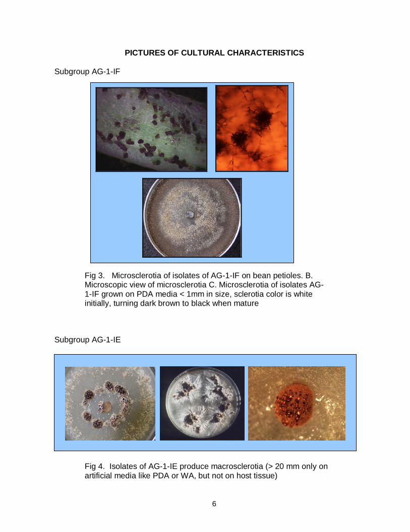
PICTURES OF CULTURAL CHARACTERISTICS Subgroup AG-1-IF
Fig 3. Microsclerotia of isolates of AG-1-IF on bean petioles. B. Microscopic view of microsclerotia C. Microsclerotia of isolates AG-1-IF grown on PDA media < 1mm in size, sclerotia color is white initially, turning dark brown to black when mature
Subgroup AG-1-IE
Fig 4. Isolates of AG-1-IE produce macrosclerotia (> 20 mm only on artificial media like PDA or WA, but not on host tissue)

7
Subgroup AG-2-2 WB
Fig 5. Isolates of subgroups AG-2-2 WB develop a crusty or loose mass of interwoven of mycelia which turns light to dark brown with no apparent rind. It does not produce sclerotia on plant tissue
Subgroup AG-1-IA
Fig 6. Isolates of AG-1-IA develop small sclerotia on PDA known as the “sasaki” type. It can produce sclerotia on plant tissue.
8
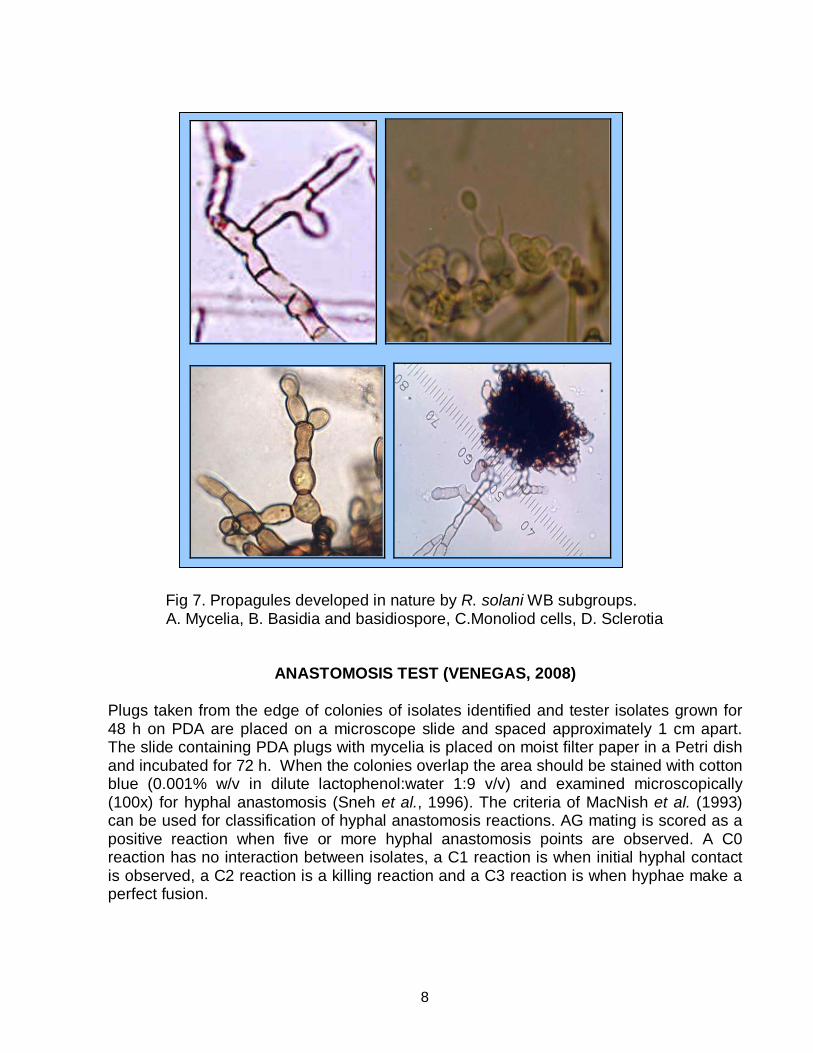
Fig 7. Propagules developed in nature by R. solani WB subgroups. A. Mycelia, B. Basidia and basidiospore, C.Monoliod cells, D. Sclerotia
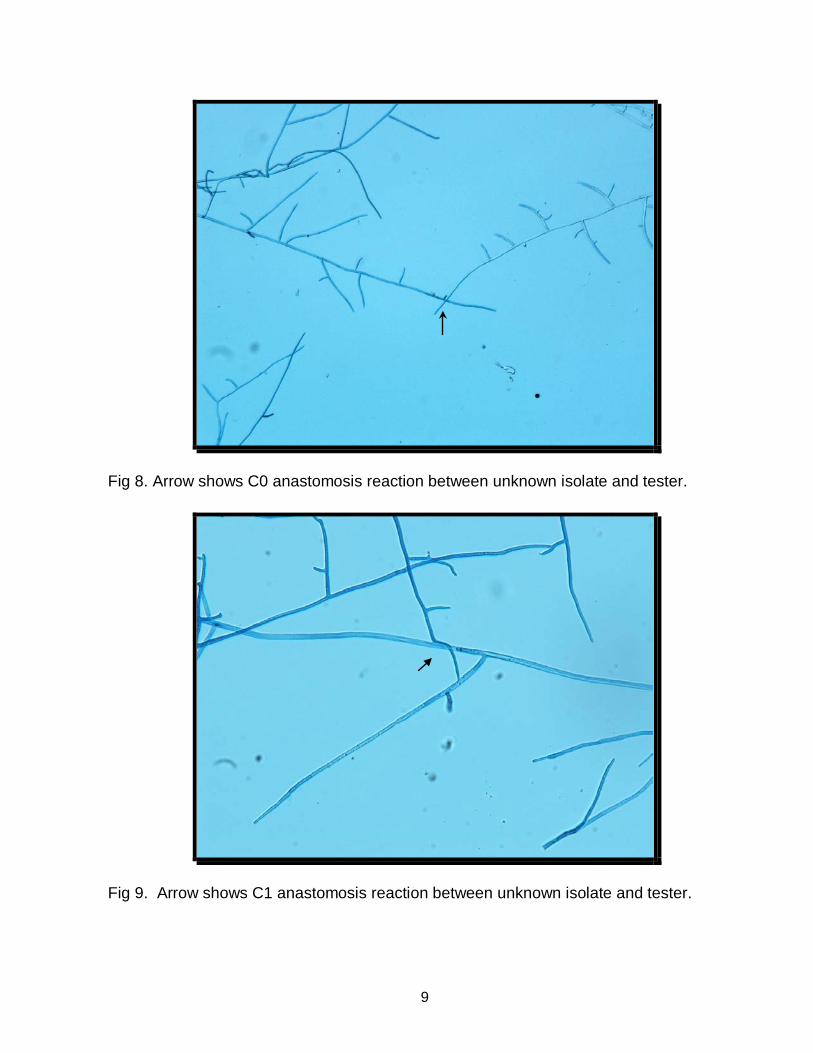
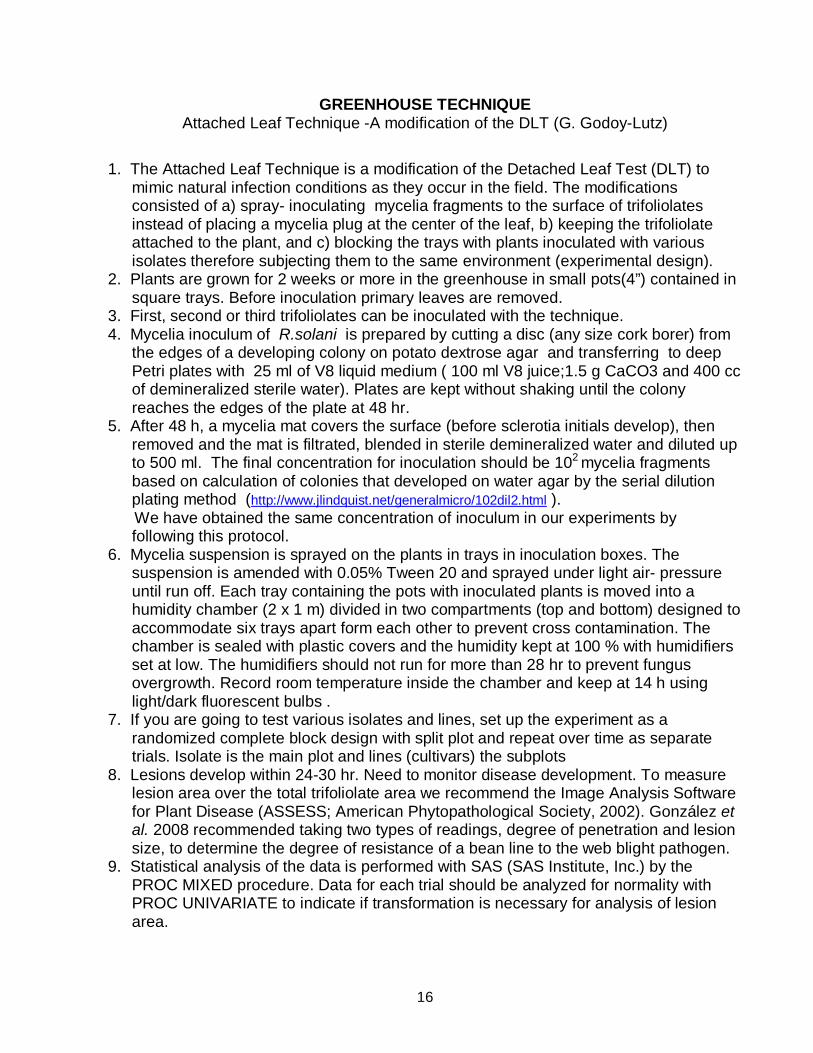
ANASTOMOSIS TEST (VENEGAS, 2008) Plugs taken from the edge of colonies of isolates identified and tester isolates grown for 48 h on PDA are placed on a microscope slide and spaced approximately 1 cm apart. The slide containing PDA plugs with mycelia is placed on moist filter paper in a Petri dish and incubated for 72 h. When the colonies overlap the area should be stained with cotton blue (0.001% w/v in dilute lactophenol:water 1:9 v/v) and examined microscopically (100x) for hyphal anastomosis (Sneh et al., 1996). The criteria of MacNish et al. (1993) can be used for classification of hyphal anastomosis reactions. AG mating is scored as a positive reaction when five or more hyphal anastomosis points are observed. A C0 reaction has no interaction between isolates, a C1 reaction is when initial hyphal contact is observed, a C2 reaction is a killing reaction and a C3 reaction is when hyphae make a perfect fusion.
9
Fig 8. Arrow shows C0 anastomosis reaction between unknown isolate and tester.
Fig 9. Arrow shows C1 anastomosis reaction between unknown isolate and tester.

10
Fig 10. C2 anastomosis reaction between unknown isolate and tester. Arrows show the fusion area between hyphae and arrowheads show dead cells after the fusion.
Fig 11. Arrow shows C3 anastomosis reaction between unknown isolate and tester.

11
MYCELIAL COMPATIBILITY TEST (GONZALES, 2008)
This test is conducted within subgroups to determine clonality. Each isolate is grown on PDA for 2-3 days. At that time one plug from isolate “A” and one plug from isolate “B” are placed approximately 2 inches (5 cm) apart on PDA. Plates are incubated at ambient temperature (22-24° C) until the mycelia from each isolate meet or form sclerotia (approximately 2 weeks). Two plates of each combination should be prepared. Compatibility is rated as: (–) = no compatibility based on formation of sclerotia at the area of contact between the two paired isolates; (0) = formation of a clear zone where no apparent mycelial growth is visible; (+) = mycelium from each isolate merge into one another and no apparent line of contact is visible
Fig 12. Reactions were rated as: incompatible – formation of sclerotia at the area of contact between the two paired isolates (a, b); incompatible 0 formation of a clear zone where no apparent mycelial growth is visible (c, d); compatible + mycelium from each isolate merged together and no apparent barrage line is visible (e,f).
a b
d
e f
c

12
FIELD SCREENING FOR WEB BLIGHT RESISTANCE Field screening for web blight resistance is limited to growing seasons that are favorable for the development of the disease. Inoculation of field trials with a suspension of mycelia of a virulent isolate of the web blight pathogen can be used to promote infection (Takegami et al., 2004). Short periods of overhead irrigation early in the morning for two weeks following inoculation also favor disease development. Plants were inoculated in the field approximately 35 days after planting. Leaves were evaluated at 8 and 15 days after inoculation using the CIAT 1-9 scale. The percentage of damaged (not commercially acceptable) seed was evaluated after the harvest. Godoy-Lutz et al. (1996) demonstrated that isolates of the web blight pathogen from different regions vary in virulence patterns. Isolates from the target area should be used to screen beans for resistance. Recurrent selection with screening for resistance in Honduras and Puerto Rico is currently being conducted to determine if bean lines with resistance over a broad geographical region can be selected (Beaver et al., 2008).
In Puerto Rico, replicated field trials were effective for screening for web blight resistance of advanced generation (F5 and F6) bean lines (Beaver et al. 2002). One-meter hill plots replicated six times provided sufficient precision to detect differences among lines for web blight reaction (Takegami et al., 2004).
Fig 13. Inoculation of field trials using a backpack sprayer.
Protocol used for the preparation of inoculum, field inoculation and the evaluation of bean plants for web blight reaction (Takegami et al., 2004)
1. A pure culture of the mycelial state of Rhizoctonia solani (anastomosis group AG-1-E)
and a potato dextrose (PDA) medium were used to prepare the inoculum. 2. Eighteen 4 mm diameter disks from the margin of the fungal colonies were placed in a
liquid medium containing 10 g of peptose, 15 g of dextrose, 0.5 g of KH2PO4 and 0.25 g of MgSO4 per liter of water. The solution was agitated in a shaker-incubator at 27º C for a period of 14 days.

13
3. The resultant mycelial growth was blended for 30 seconds at low speed, filtered and washed in distilled water. The filtrate was dried 24 h before water was added.
4. The final concentration of the inoculum was obtained using a spectrophotometer. Water was added until 25% transmittance was obtained at a wavelength of 640 nm. Tween 80 was added at a concentration of 1 ml/L to increase the adhesion of the inoculum to the surface of the leaves.
5. At 35 days after planting, the leaves of the bean plants were inoculated in the field with a backpack sprayer using a pressure of 15 lb/in2. The inoculum was applied late in the afternoon and the trial received 15 minutes of irrigation each morning during the first week after inoculation.
6. Leaf damage was evaluated at 8 and 15 days after inoculation using the CIAT (1-9) scale. After the harvest, seed was separated into commercially acceptable and blemished seed categories to calculate percentage of damaged seed.
Fig 14. Segregation for web blight reaction in a field trial planted at Isabela, Puerto Rico.
Table 2 . CIAT 1-9 scale for field evaluation of bean lines for web blight reaction.
Score Symptoms 1 No visible symptoms of the disease 3 5-10% of the leaf area with symptoms 5 20-30% of the leaf area with symptoms 7 40-60% of the leaf area with symptoms 9 > 80% of the leaf area with symptoms
Source: CIAT (1987). - http://www.google.com/books?id=e7144M7teYcC

14
Lower levels of web blight infection in the field can be due to physiological resistance or avoidance of the disease due to an erect plant architecture and/or an open canopy arrangement of leaves. A detached leaf technique has been used to screen bean lines for physiological resistance to web blight (González et al. 2008, Bautista-Pérez and Echávez-Badel, 2000; Galindo et al., 1982).
Fig 15. A detached-leaf technique to screen bean lines for web blight reaction.
Protocol used for the preparation of inoculum, inoculation and the evaluation of bean plants for web blight using the detached-leaf technique (Takegami et al., 2004).
1. A pure culture of the mycelial state of Rhizoctonia solani (anastomosis group AG-1-
1E) and a potato dextrose agar (PDA) medium was the source of the infected disks used as inoculum.
2. Fully expanded trifoliolate leaves were detached from the bean plants and immediately placed in orchid tubes filled with water.
3. The leaflets were positioned in 42 x 30 x 6 cm aluminum” turkey” trays. Because the base of the trays contained moistened paper towels, the leaflets were placed on top of Petri plates to avoid immersion in water. One of the leaflets in each tray should be from a line susceptible to web blight to serve as a check.
4. One 4 mm diameter disk of agar colonized with R. solani was placed on the ad axial side and centered on each leaflet. A disk not colonized with the fungus should be placed on one of the leaflets to serve as a control. The control and infected treatments should be applied at random
5. In addition, each tray should contain a leaflet of a susceptible bean line as a means to confirm that conditions were favorable for the development of infection.

15
6. The aluminum trays were placed inside plastic bags after inoculation to create a high humidity environment favorable for the development of the fungus. The trays were incubated in a laboratory at 27±1° C.
7. Mean lesion size was measured at 24, 48 and 72 h after inoculation. The measurement of the lesion size can be facilitated by the use of a digital image system such as ‘Scion Image Beta 4.02 Win’
16
GREENHOUSE TECHNIQUE Attached Leaf Technique -A modification of the DLT (G. Godoy-Lutz)
1. The Attached Leaf Technique is a modification of the Detached Leaf Test (DLT) to mimic natural infection conditions as they occur in the field. The modifications consisted of a) spray- inoculating mycelia fragments to the surface of trifoliolates instead of placing a mycelia plug at the center of the leaf, b) keeping the trifoliolate attached to the plant, and c) blocking the trays with plants inoculated with various isolates therefore subjecting them to the same environment (experimental design).
2. Plants are grown for 2 weeks or more in the greenhouse in small pots(4”) contained in square trays. Before inoculation primary leaves are removed.
3. First, second or third trifoliolates can be inoculated with the technique. 4. Mycelia inoculum of R.solani is prepared by cutting a disc (any size cork borer) from
the edges of a developing colony on potato dextrose agar and transferring to deep Petri plates with 25 ml of V8 liquid medium ( 100 ml V8 juice;1.5 g CaCO3 and 400 cc of demineralized sterile water). Plates are kept without shaking until the colony reaches the edges of the plate at 48 hr.
5. After 48 h, a mycelia mat covers the surface (before sclerotia initials develop), then removed and the mat is filtrated, blended in sterile demineralized water and diluted up to 500 ml. The final concentration for inoculation should be 102 mycelia fragments based on calculation of colonies that developed on water agar by the serial dilution plating method (http://www.jlindquist.net/generalmicro/102dil2.html ).
We have obtained the same concentration of inoculum in our experiments by following this protocol.
6. Mycelia suspension is sprayed on the plants in trays in inoculation boxes. The suspension is amended with 0.05% Tween 20 and sprayed under light air- pressure until run off. Each tray containing the pots with inoculated plants is moved into a humidity chamber (2 x 1 m) divided in two compartments (top and bottom) designed to accommodate six trays apart form each other to prevent cross contamination. The chamber is sealed with plastic covers and the humidity kept at 100 % with humidifiers set at low. The humidifiers should not run for more than 28 hr to prevent fungus overgrowth. Record room temperature inside the chamber and keep at 14 h using light/dark fluorescent bulbs .
7. If you are going to test various isolates and lines, set up the experiment as a randomized complete block design with split plot and repeat over time as separate trials. Isolate is the main plot and lines (cultivars) the subplots
8. Lesions develop within 24-30 hr. Need to monitor disease development. To measure lesion area over the total trifoliolate area we recommend the Image Analysis Software for Plant Disease (ASSESS; American Phytopathological Society, 2002). González et al. 2008 recommended taking two types of readings, degree of penetration and lesion size, to determine the degree of resistance of a bean line to the web blight pathogen.
9. Statistical analysis of the data is performed with SAS (SAS Institute, Inc.) by the PROC MIXED procedure. Data for each trial should be analyzed for normality with PROC UNIVARIATE to indicate if transformation is necessary for analysis of lesion area.

17
A. Score = 1 B. Score = 3 C. Score = 5
D. Score = 7 E. Score = 9
Fig 18. Disease severity scale from CIAT used for the evaluation of the candidate bean lines inoculated with different isolates of Rhizoctonia solani. (From González Martínez,
2004)
Fig 16. Plants inoculated with mycelia suspension (G. Godoy-Lutz)
Fig 17. Humidity box where conditions are developed for infection by mycelia inoculum. (G. Godoy-Lutz)

18
FUNGAL PRESERVATION
Rhizoctonia solani can be preserved in beet (Beta vulgaris) seed. Before placing the seed in a Petri plate containing PDA media, the beet seed should be double sterilized in an autoclave. A 4 mm diameter disk of agar colonized with Rhizoctonia solani is placed in the center of the Petri plate. After the fungus has covered the Petri plate, the infected seed should be picked out with forceps, placed on sterile filter paper (Whatman International Ltd) and let air dry in a transfer hood overnight. After infected seed (mycelia or sclerotia cover its surface) is dry, transfer them to small glass container or cryogenic vial for storage. If glass container is used, silica gel desiccant should be placed in the base of the container and a cotton plug should separate the seed and the desiccant. The infected beet seed can be stored for several months or years in a refrigerator (4º C) inside a desiccant chamber. (Note: G. Godoy-Lutz has recovered R.solani isolates 10 years later after being stored this way.)
Fig 19. Sugarbeet seed embedded on culture, after infection by isolates of WB subgroups, seed is collected, airdried and put in vials and are ready for long term storage
in a desiccant chamber in the refrigerator
SOURCES OF RESISTANCE
Only moderate levels of resistance to web blight have been identified in common bean (Beaver et al., 2008; Takegami et al., 2004; Arnaud-Santana et al., 1998; Montoya et al., 1997). Although no specific genes for resistance for resistance have been identified, Jung et al. (1996) reported QTL’s for web blight resistance. Interspecific crosses are being used to attempt to transfer web blight resistance from scarlet runner bean (Phaseolus coccineus L.) to common bean (González et al., 2005; Beaver et al., 2007).

19
Table 1. Sources of resistance to web blight in different seed classes.
Name or number Seed color / type Reference Arroyo Loro Negro
Talamanca, HT 7719 PR0650-31, PR0650-32
9 / Black Arnaud et al. (2000) Gálvez et al. (1989) Beaver et al. (2008)
1 / Navy 2M / Pinto 1 / Great Northern
VAX 6 7 / Purple Singh and Muñoz (1999) Bribri
PR0401-277 6 / Small red Rosas et al. (2003)
Beaver et al. (2008) PR0401-257, PR0401-259 5/ Pink Beaver et al. (2008
2R / Cranberry A 193 6M / Red mottled Araya y Araya (2000)
6K / Dark red kidney
1 / Snap G 35163 P. coccineus González et al. (2005)
GALLERY OF WEB BLIGHT SYMPTOMS
Fig 20. Web blight symptoms on dry bean leaves and pods caused by basidiospores of Thanatephorus cucumeris (anamorph:Rhizoctonia solani ) subgroup AG-2-2WB. La Vega, Dominican Republic. (G.Godoy-Lutz)

20
Fig 21. Advanced symptoms on dry bean leaves caused by basidiospores of Thanatephorus cucumeris (anamorph:Rhizoctonia solani ) subgroup AG-2-2WB.
La Vega, Dominican Republic. (G. Godoy-Lutz)
Fig 22. Symptoms of defoliation and pod blight caused by isolates of the WB pathogen AG-1-IF subgroup. Buena Vista, San Juan Valley, Dominican Republic. (G. Godoy-Lutz)

21
Fig 23. Infected pods and decayed seeds of Pompadour PC-50 and B. blemished vs. symptomless seed caused by Thanatephorus cucumeris
(anamorph: Rhizoctonia solani subgroup AG-1-IF). San Juan Valley, Dominican Republic. (G. Godoy-Lutz)

22
Fig 24. Unusual occurrence of WB outbreak by AG-2-2 WB and AG-1-IF (due to seedborne infection) during dry planting season. Manoguayabo, San Juan. Dominican Rep. Dec, 2007 (Godoy-Lutz)
REFERENCES Araya, C.M. y R. Araya. 2000. Avances en la selección de fuentes de resistencia a las principales enfermedades de frijol común (Phaseolus vulgaris L.) en Costa Rica. Agronomía Mesoamericana 11(2):25-29. Arnaud-Santana, E., D.P. Coyne and J.R. Steadman. 1998. Inheritance and heritabilities of the reaction to web blight. Ann. Rep. Bean Improv. Coop. 41:29-30. Bautista-Pérez, M. and R. Echávez-Badel. 2000. Methodology for screening common bean for resistance to web blight. J. Agric. Univ. of Puerto Rico 84:91-94. Beaver, J.S., M. Alameda and J.C. Rosas. 2008. Breeding beans for resistance to web blight. Ann. Rep. Bean Improv. Coop. 51:30-31. Beaver, J.S., G. Godoy-Lutz, J.C. Rosas y J.R. Steadman. 2002. Estrategias para seleccionar frijol común con mayor resistancia a mustia hilachosa. Agronomia Mesoamericana 13:67-72. CIAT (Centro Internacional de Agricultura Tropical). 1987. Standard system for the evaluation of bean germplasm. Van Schoonhoven, A. and Pastor-Corrales, M.A. (compilers). Cali, Colombia 54 p. - http://www.google.com/books?id=e7144M7teYcC
23
Galindo, J.J., G.S. Abawi, H.D. Thurston and G. Galvez. 1982. Characterization of Thanatophorus cucumeris isolates causing web blight of beans in Costa Rica. Turrialba 32:447-455. Galvez,G.E.,Mora,B.,and Pastor Corrales, M.A.1989.Web blight. Pages 195-259 in: Bean Production Problems in the Tropics. H.F.Schwartz and M.A.Pastor Corrales,eds.CIAT,Cali, Colombia. Godoy-Lutz, G., S. Kuninaga, J.R. Steadman and K. Powers. 2008. Phylogenetic analysis of Rhizoctonia solani subgroups associated with web blight symptoms on common bean based on ITS-5.8s rDNA. J. Gen. Plant Path. 74:32-40. Godoy-Lutz, G., S. Kuninaga, J.R. Steadman, J.S. Beaver and J.C. Rosas. 2006. DNA sequencing and analysis: A tool for improving web blight management and resistance breeding. Ann. Rep. of the Bean Improv. Coop. 49:87-88. Godoy-Lutz, G., J. R. Steadman, B. Higgins, and K. Powers. 2003. Genetic variation among isolates of the web blight pathogen of common bean based on PCR-RFLP of the ITS-rDNA Region. Plant Dis. 87:766:771. Godoy-Lutz, G., J. Arias, E. Arnaud-Santana and J.R. Steadman. 1998. Web blight affects seed yield and quality of red mottled bean lines and cultivars in the Dominican Republic. Ann. Rep. Bean Improv. Coop. 41:72-73. Godoy-Lutz,G.,Arias, J., J.R.Steadman and K.M Eskridge.1996. Role of natural seed infection by the web blight pathogen in common bean seed damage, seedling emergence, and early disease development. Plant Dis.80:887-890. Godoy-Lutz, G, J. Arias, F. Saladin, J.R. Steadman and D.E. Carling. 1996b. Characterization of isolates of Rhizoctonia solani that can cause web blight on common bean in Central America and the Caribbean with implications for disease management. Ann. Rep. Bean Improv. Coop. 39:154-155. Godoy-Lutz, G., J.R. Steadman, J. Arias, Y. Segura and F. Saladin. 1994. Seed transmission of web blight pathogen Thanatephorus cucumeris in dry beans in the Dominican Republic. Ann. Rep. Bean Improv. Coop. 37:69-70. González, N., J. Beaver, J.C. Rosas, G. Godoy-Lutz and J.R. Steadman. 2008. Development of a differential set of common bean lines to screen for web blight pathogen virulence. Ann. Rep. Bean Improv. Coop. 51:32-33. González, N. 2008 Genetic diversity of Rhizoctonia solani causing web blight and sources of resistance in Phaseolus spp. Ph.D Thesis. University of Nebraska-Lincoln. 213 pp.

24
González Martínez, N.A., F.H. Ferwerda, M. Alameda, J.C. Rosas and J.S. Beaver. 2005. Identification of new sources of resistance to web blight of common bean. Ann. Rep. of the Bean Improv. Coop. 48:130-131 González Martínez, N.A. 2004. Identification of new sources of resistance to web blight of common bean (Phaseolus vulgaris L). Masters thesis: University of Puerto Rico. 46 pp. Jung, G., D.P. Coyne, P.W. Skroch, J. Nienhuis, E. Arnaud-Santana, J. Bokosi, H.M. Ariyarathne, J.R. Steadman, J.S. Beaver, S. Kaeppler. 1996. Molecular markers associated with plant architecture and resistance to common blight. J. Amer. Soc. Hort. Sci. 121:794:-803. MacNish, G. C., D. E. Carling, K. A. Brainard. 1993. Characterization of Rhizoctonia solani AG-8 from bare patches by pectic isozyme (zymogram) and anastomosis techniques. Phytopathology 83:922-927 Montoya, C.A., J.S. Beaver, R. Rodríguez, P.N. Miklas and G. Godoy-Lutz. 1997. Heritability of resistance to web blight in five common bean populations. Crop Sci. 37:780-783. Singh, S.P. and C.G. Muñoz. 1999. Resistance to common bacterial blight among Phaseolus species and common bean improvement. Crop Sci. 39:80-89. Sneh, B., S. Jabaji-Hare, S. Neate and G. Dijst. 1996. Rhizoctonia Species: Taxonomy, molecular biology, ecology, pathology and disease control. Kluwer Academic Publishers. Netherlands. 578 pp. Takegami, J.C., J.S. Beaver, G. Godoy-Lutz, R. Echávez-Badel and J.R. Steadman. 2004. Inheritance of web blight resistance in common bean. J. Agric. Univ. of Puerto Rico 88:45-54. Venegas, J.P. 2008. Identification of rust resistance and a molecular marker in a cross within tertiary gene pool of common bean: and characterization of Rhizoctonia spp. isolates from Western Nebraska. MS Thesis. University of Nebraska-Lincoln. 141 pp.