Extensive sampling of basidiomycete genomes demonstrates ...
16
Extensive sampling of basidiomycete genomes demonstrates inadequacy of the white-rot/brown-rot paradigm for wood decay fungi Robert Riley a , Asaf A. Salamov a , Daren W. Brown b , Laszlo G. Nagy c , Dimitrios Floudas c , Benjamin W. Held d , Anthony Levasseur e , Vincent Lombard f , Emmanuelle Morin g , Robert Otillar a , Erika A. Lindquist a , Hui Sun a , Kurt M. LaButti a , Jeremy Schmutz a,h , Dina Jabbour i , Hong Luo i , Scott E. Baker j , Antonio G. Pisabarro k , Jonathan D. Walton i , Robert A. Blanchette d , Bernard Henrissat f , Francis Martin g , Dan Cullen l , David S. Hibbett c,1 , and Igor V. Grigoriev a,1 a US Department of Energy (DOE) Joint Genome Institute, Walnut Creek, CA 94598; b US Department of Agriculture (USDA), Peoria, IL 61604; c Department of Biology, Clark University, Worcester, MA 01610; d University of Minnesota, St. Paul, MN 55108; e Institut National de la Recherche Agronomique, Unité Mixte de Recherche 1163, Aix-Marseille Université, 13288 Marseille, France; f Centre National de la Recherche Scientifique, Unité Mixte de Recherche 7257, Aix-Marseille Université, 13288 Marseille, France; g Institut National de la Recherche Agronomique, Unité Mixte de Recherche 1136, Institut National de la Recherche Agronomique-Université de Lorraine, Interactions Arbres/Micro-organismes, 54280 Champenoux, France; h HudsonAlpha Institute of Biotechnology, Huntsville, AL 35806; i DOE Great Lakes Bioenergy Research Center, Michigan State University, East Lansing, MI 48824; j Environmental Molecular Sciences Laboratory, Pacific Northwest National Laboratory, Richland, WA 99354; k Departamento de Producción Agraria, Universidad Pública de Navarra, 31006 Pamplona, Spain; and l USDA Forest Products Laboratory, Madison, WI 53726 Edited* by Thomas N. Taylor, University of Kansas, Lawrence, KS, and approved May 16, 2014 (received for review January 12, 2014) Basidiomycota (basidiomycetes) make up 32% of the described fungi and include most wood-decaying species, as well as patho- gens and mutualistic symbionts. Wood-decaying basidiomycetes have typically been classified as either white rot or brown rot, based on the ability (in white rot only) to degrade lignin along with cellulose and hemicellulose. Prior genomic comparisons sug- gested that the two decay modes can be distinguished based on the presence or absence of ligninolytic class II peroxidases (PODs), as well as the abundance of enzymes acting directly on crystal- line cellulose (reduced in brown rot). To assess the generality of the white-rot/brown-rot classification paradigm, we compared the ge- nomes of 33 basidiomycetes, including four newly sequenced wood decayers, and performed phylogenetically informed principal-com- ponents analysis (PCA) of a broad range of gene families encoding plant biomass-degrading enzymes. The newly sequenced Botryoba- sidium botryosum and Jaapia argillacea genomes lack PODs but possess diverse enzymes acting on crystalline cellulose, and they group close to the model white-rot species Phanerochaete chryso- sporium in the PCA. Furthermore, laboratory assays showed that both B. botryosum and J. argillacea can degrade all polymeric com- ponents of woody plant cell walls, a characteristic of white rot. We also found expansions in reducing polyketide synthase genes spe- cific to the brown-rot fungi. Our results suggest a continuum rather than a dichotomy between the white-rot and brown-rot modes of wood decay. A more nuanced categorization of rot types is needed, based on an improved understanding of the genomics and biochem- istry of wood decay. lignocellulose | phylogenomics | bioenergy F ungi of the phylum Basidiomycota (basidiomycetes) comprise 32% of the described fungi (1) and are important to forestry (2–4), agriculture (5–7), and medicine (8–11). This diverse phy- lum includes the mushrooms (12–14); pathogens of plants (2), animals (9–11), and other fungi (15); osmotically tolerant molds (16); ectomycorrhizal symbionts like Laccaria bicolor, which are critical for plant growth; plant pathogens, such as rusts and smuts (7); and saprotrophs, including wood-decaying fungi (17). The 26,000 basidiomycete taxa in the National Center for Biotechnology Information (NCBI) database (18, 19) are divided into three subphyla: Agaricomycotina (∼22,000 taxa), Puccinio- mycotina (∼2,300 taxa), and Ustilaginomycotina (∼1,000 taxa). Agaricomycotina includes many decomposers of wood and leaf litter (12, 17, 20–23) that produce lignocellulolytic enzymes that have potential to be used in bioenergy production (24–27). Thus, much of sequencing effort at the US Department of Energy (DOE) Joint Genome Institute (JGI) (http://jgi.doe.gov/fungi) has targeted Agaricomycotina, particularly the Agaricomycetes (mushroom-forming fungi), with the large orders Agaricales (predominantly gilled mushrooms), Polyporales (wood-decaying polypores and others), and Boletales (porcini mushrooms and others) being especially deeply sampled. A keen focus in the comparative genomics of Basidiomycota has concerned lineages of wood decay fungi (13, 17, 20–23, 28). For decades, two broad categories have been recognized: white rot and brown rot (29–31). During brown rot, cellulose is rapidly Significance Wood decay fungi have historically been characterized as ei- ther white rot, which degrade all components of plant cell walls, including lignin, or brown rot, which leave lignin largely intact. Genomic analyses have shown that white-rot species possess multiple lignin-degrading peroxidases (PODs) and ex- panded suites of enzymes attacking crystalline cellulose. To test the adequacy of the white/brown-rot categories, we an- alyzed 33 fungal genomes. Some species lack PODs, and thus resemble brown-rot fungi, but possess the cellulose-degrading apparatus typical of white-rot fungi. Moreover, they appear to degrade lignin, based on decay analyses on wood wafers. Our results indicate that the prevailing paradigm of white rot vs. brown rot does not capture the diversity of fungal wood decay mechanisms. Author contributions: R.R., S.E.B., A.G.P., J.D.W., R.A.B., B.H., F.M., D.C., D.S.H., and I.V.G. designed research; R.R., D.W.B., L.G.N., D.F., B.W.H., A.L., E.M., and R.A.B. performed research; A.G.P., J.D.W., B.H., and D.S.H. contributed new reagents/analytic tools; R.R., A.A.S., D.W.B., L.G.N., D.F., A.L., V.L., E.M., R.O., E.A.L., H.S., K.M.L., J.S., D.J., and H.L. analyzed data; and R.R., D.W.B., J.D.W., R.A.B., B.H., D.C., D.S.H., and I.V.G. wrote the paper. The authors declare no conflict of interest. *This Direct Submission article had a prearranged editor. Freely available online through the PNAS open access option. Data deposition: The sequences reported in this paper have been deposited in the Gen- Bank database [AYEP00000000 (Botryobasidium botryosum), AYUM00000000 (Galerina marginata), AYUL00000000 (Jaapia argillacea), and AYUK00000000 (Pleurotus ostreatus)]. 1 To whom correspondence may be addressed. E-mail: [email protected] or dhibbett@ clarku.edu. This article contains supporting information online at www.pnas.org/lookup/suppl/doi:10. 1073/pnas.1400592111/-/DCSupplemental. www.pnas.org/cgi/doi/10.1073/pnas.1400592111 PNAS Early Edition | 1 of 6 MICROBIOLOGY
Transcript of Extensive sampling of basidiomycete genomes demonstrates ...

Extensive sampling of basidiomycete genomesdemonstrates inadequacy of the white-rot/brown-rotparadigm for wood decay fungiRobert Rileya, Asaf A. Salamova, Daren W. Brownb, Laszlo G. Nagyc, Dimitrios Floudasc, Benjamin W. Heldd,Anthony Levasseure, Vincent Lombardf, Emmanuelle Moring, Robert Otillara, Erika A. Lindquista, Hui Suna,Kurt M. LaButtia, Jeremy Schmutza,h, Dina Jabbouri, Hong Luoi, Scott E. Bakerj, Antonio G. Pisabarrok,Jonathan D. Waltoni, Robert A. Blanchetted, Bernard Henrissatf, Francis Marting, Dan Cullenl, David S. Hibbettc,1,and Igor V. Grigorieva,1
aUS Department of Energy (DOE) Joint Genome Institute, Walnut Creek, CA 94598; bUS Department of Agriculture (USDA), Peoria, IL 61604; cDepartment ofBiology, Clark University, Worcester, MA 01610; dUniversity of Minnesota, St. Paul, MN 55108; eInstitut National de la Recherche Agronomique, Unité Mixte deRecherche 1163, Aix-Marseille Université, 13288 Marseille, France; fCentre National de la Recherche Scientifique, Unité Mixte de Recherche 7257, Aix-MarseilleUniversité, 13288 Marseille, France; gInstitut National de la Recherche Agronomique, Unité Mixte de Recherche 1136, Institut National de la RechercheAgronomique-Université de Lorraine, Interactions Arbres/Micro-organismes, 54280 Champenoux, France; hHudsonAlpha Institute of Biotechnology,Huntsville, AL 35806; iDOE Great Lakes Bioenergy Research Center, Michigan State University, East Lansing, MI 48824; jEnvironmental Molecular SciencesLaboratory, Pacific Northwest National Laboratory, Richland, WA 99354; kDepartamento de Producción Agraria, Universidad Pública de Navarra, 31006Pamplona, Spain; and lUSDA Forest Products Laboratory, Madison, WI 53726
Edited* by Thomas N. Taylor, University of Kansas, Lawrence, KS, and approved May 16, 2014 (received for review January 12, 2014)
Basidiomycota (basidiomycetes) make up 32% of the describedfungi and include most wood-decaying species, as well as patho-gens and mutualistic symbionts. Wood-decaying basidiomyceteshave typically been classified as either white rot or brown rot,based on the ability (in white rot only) to degrade lignin alongwith cellulose and hemicellulose. Prior genomic comparisons sug-gested that the two decay modes can be distinguished based onthe presence or absence of ligninolytic class II peroxidases (PODs),as well as the abundance of enzymes acting directly on crystal-line cellulose (reduced in brown rot). To assess the generality of thewhite-rot/brown-rot classification paradigm, we compared the ge-nomes of 33 basidiomycetes, including four newly sequenced wooddecayers, and performed phylogenetically informed principal-com-ponents analysis (PCA) of a broad range of gene families encodingplant biomass-degrading enzymes. The newly sequenced Botryoba-sidium botryosum and Jaapia argillacea genomes lack PODs butpossess diverse enzymes acting on crystalline cellulose, and theygroup close to the model white-rot species Phanerochaete chryso-sporium in the PCA. Furthermore, laboratory assays showed thatboth B. botryosum and J. argillacea can degrade all polymeric com-ponents of woody plant cell walls, a characteristic of white rot. Wealso found expansions in reducing polyketide synthase genes spe-cific to the brown-rot fungi. Our results suggest a continuum ratherthan a dichotomy between the white-rot and brown-rot modes ofwood decay. A more nuanced categorization of rot types is needed,based on an improved understanding of the genomics and biochem-istry of wood decay.
lignocellulose | phylogenomics | bioenergy
Fungi of the phylum Basidiomycota (basidiomycetes) comprise32% of the described fungi (1) and are important to forestry
(2–4), agriculture (5–7), and medicine (8–11). This diverse phy-lum includes the mushrooms (12–14); pathogens of plants (2),animals (9–11), and other fungi (15); osmotically tolerant molds(16); ectomycorrhizal symbionts like Laccaria bicolor, which arecritical for plant growth; plant pathogens, such as rusts and smuts(7); and saprotrophs, including wood-decaying fungi (17).The 26,000 basidiomycete taxa in the National Center for
Biotechnology Information (NCBI) database (18, 19) are dividedinto three subphyla: Agaricomycotina (∼22,000 taxa), Puccinio-mycotina (∼2,300 taxa), and Ustilaginomycotina (∼1,000 taxa).Agaricomycotina includes many decomposers of wood and leaflitter (12, 17, 20–23) that produce lignocellulolytic enzymes that
have potential to be used in bioenergy production (24–27). Thus,much of sequencing effort at the US Department of Energy(DOE) Joint Genome Institute (JGI) (http://jgi.doe.gov/fungi)has targeted Agaricomycotina, particularly the Agaricomycetes(mushroom-forming fungi), with the large orders Agaricales(predominantly gilled mushrooms), Polyporales (wood-decayingpolypores and others), and Boletales (porcini mushrooms andothers) being especially deeply sampled.A keen focus in the comparative genomics of Basidiomycota
has concerned lineages of wood decay fungi (13, 17, 20–23, 28).For decades, two broad categories have been recognized: whiterot and brown rot (29–31). During brown rot, cellulose is rapidly
Significance
Wood decay fungi have historically been characterized as ei-ther white rot, which degrade all components of plant cellwalls, including lignin, or brown rot, which leave lignin largelyintact. Genomic analyses have shown that white-rot speciespossess multiple lignin-degrading peroxidases (PODs) and ex-panded suites of enzymes attacking crystalline cellulose. Totest the adequacy of the white/brown-rot categories, we an-alyzed 33 fungal genomes. Some species lack PODs, and thusresemble brown-rot fungi, but possess the cellulose-degradingapparatus typical of white-rot fungi. Moreover, they appear todegrade lignin, based on decay analyses on wood wafers. Ourresults indicate that the prevailing paradigm of white rot vs.brown rot does not capture the diversity of fungal wooddecay mechanisms.
Author contributions: R.R., S.E.B., A.G.P., J.D.W., R.A.B., B.H., F.M., D.C., D.S.H., and I.V.G.designed research; R.R., D.W.B., L.G.N., D.F., B.W.H., A.L., E.M., and R.A.B. performedresearch; A.G.P., J.D.W., B.H., and D.S.H. contributed new reagents/analytic tools; R.R.,A.A.S., D.W.B., L.G.N., D.F., A.L., V.L., E.M., R.O., E.A.L., H.S., K.M.L., J.S., D.J., and H.L.analyzed data; and R.R., D.W.B., J.D.W., R.A.B., B.H., D.C., D.S.H., and I.V.G. wrotethe paper.
The authors declare no conflict of interest.
*This Direct Submission article had a prearranged editor.
Freely available online through the PNAS open access option.
Data deposition: The sequences reported in this paper have been deposited in the Gen-Bank database [AYEP00000000 (Botryobasidium botryosum), AYUM00000000 (Galerinamarginata), AYUL00000000 (Jaapia argillacea), and AYUK00000000 (Pleurotus ostreatus)].1To whom correspondence may be addressed. E-mail: [email protected] or [email protected].
This article contains supporting information online at www.pnas.org/lookup/suppl/doi:10.1073/pnas.1400592111/-/DCSupplemental.
www.pnas.org/cgi/doi/10.1073/pnas.1400592111 PNAS Early Edition | 1 of 6
MICRO
BIOLO
GY

depolymerized via oxidative mechanisms, whereas modified lig-nin remains as a polymeric residue (32–35). In contrast, white-rotfungi use hydrolases that gradually degrade cellulose while ligninis completely mineralized. Lignin degradation involves high-oxidation potential class II peroxidases (PODs) that, on the basis ofconserved catalytic and Mn-binding sites, are classified as ligninperoxidase (LiP), manganese peroxidase (MnP), or versatile per-oxidase (VP) (36–38). The first genomes of the white-rot fungusPhanerochaete chrysosporium and brown-rot fungus Postia placentarevealed a gene complement consistent with their respectivemodes of wood decay (22, 23). Further comparative genomicsstudies of larger sets of wood decay fungi supported a consistentrelationship between decay patterns and several enzyme families.Specifically, white-rot fungi had high-oxidation potential PODsfor lignin degradation as well as cellobiohydrolases for degradingcrystalline cellulose. Classified in glycoside hydrolase (GH) fami-lies (39) GH6 and GH7, cellobiohydrolases attack cellulose in asynergistic manner and often carry a cellulose binding module(CBM1). In contrast, the genomes of brown-rot fungi didnot encode PODs and the predicted cellobiohydrolase-encodinggenes were generally absent or lacking a CBM1 domain.Here, we present comparative analyses of 33 sequenced ba-
sidiomycete genomes (Table S1). Included are 22 wood decayers,of which Galerina marginata, Pleurotus ostreatus, Botryobasidiumbotryosum, and Jaapia argillacea are newly sequenced. The resultscall into question the prevailing white-rot/brown-rot dichotomy.
Results and DiscussionPhylogeny and Protein Conservation. A maximum-likelihood (ML)phylogeny (40) was inferred from protein sequence alignments of183 conserved gene families (Fig. S1A). Overall, the tree topol-ogy is consistent with prior studies using genome-scale datasets(17) as well as phylogenetic analyses using small numbers ofgenes (41), such as the sister group relationship between Usti-laginomycotina and Agaricomycotina, and the placement ofWallemia sebi (Wallemiomycetes) as the sister group of therest of the Agaricomycotina (16). Brown-rot fungi are poly-phyletic, as shown previously (17), and include species inPolyporales, Boletales, Gloeophyllales, and Dacrymycetales.Some aspects of the phylogeny remain uncertain: our placementof Jaapia argillacea as the sister group of the Gloeophyllalesconflicts with a previous study using six genes (42); and Auricu-laria delicata was inferred as the sister group of Piriformosporaindica, differing from multigene phylogenies (41, 43–45), whichsuggested that these lineages form a paraphyletic assemblage.Gene families in 33 basidiomycetes and 30 other fungi (Table
S2) with sequenced genomes were inferred by Markov chain(MCL) clustering (46) of all-vs.-all protein BLAST (47) align-ments. Analysis of protein conservation suggests a conservedcore fungal genome of ∼5,000 genes (Fig. S1C). Roughly one-half of the proteins (49%) in Basidiomycota lack homologs inother groups of fungi, and 23% of Basidiomycota proteins areunique to a single organism (i.e., each of the 33 basidiomycetesanalyzed harbored proteins not found in any other sampledspecies, ranging from 4% to 51%). In contrast, in Ascomycota,30% of the proteins are phylum specific and 13% are organismspecific (using a comparative set of 27 Ascomycota fungi; TableS2). We were able to assign functions from the EukaryoticClusters of Orthologs (KOG) (48) to 38% of Basidiomycotaproteins (compared with 43% in Ascomycota); 74% of coreproteins in both Basidiomycota and Ascomycota; and 17% ofBasidiomycota-specific proteins (23% in Ascomycota) (Fig. S2).
Diversity of CAZymes and Associated Activities in Basidiomycota.Thirty-three basidiomycete genomes were searched for geneswhose protein products are implicated in the breakdown of thepolysaccharide portion of plant cell walls (cellulose, hemi-cellulose, pectin) using the CAZy database pipeline (39, 49). The
number of genes in each CAZy family, in each organism, is pre-sented in Dataset S1.Cellulolytic families. During white-rot decay, cellulose is targetedprimarily by hydrolytic enzymes of multiple GH families. Lyticpolysaccharide monooxygenases (LPMOs) of the AA9 family(formerly GH61) also participate via oxidative mechanisms.White-rot fungi generally have more cellulolytic genes (bothhydrolytic and oxidative) and cellulose binding domains (CBM1)relative to brown-rot fungi (Fig. 1). Cellobiohydrolases of fami-lies GH6 and GH7 (which cleave cellulose to the disaccharidecellobiose) are correlated with white rot [r = 0.3 and 0.6, re-spectively, by the independent contrasts method (50)]. Likelyinvolved in boosting cellulase activity (51–54), LPMOs areabundant in white-rot fungi (r = 0.3) but reduced in brown-rotfungi (r = −0.5). Cellobiose dehydrogenases (family AA3_1),which enhance cellulose degradation (52), are uniformly presentin a single copy in all white-rot fungi and absent from the ma-jority of brown-rot fungi (although Gloeophyllum trabeum hasone copy, and Boletales Coniophora puteana and Serpula lacry-mans each have two copies). Additional cellulolytic families werefound in basidiomycetes. For examples, GH5 (containing endo-acting cellulases as well as many other enzymes of differingsubstrate specificity; Dataset S2), GH12, GH44, and GH45 areexpanded in white-rot fungi (r = 0.2, 0.1, 0.4, 0.5, respectively)and diminished in brown-rot fungi (r < −0.2; for all, see alsoDataset S1).Among the newly sequenced genomes,G. marginata, P. ostreatus,
B. botryosum, and J. argillacea all possess diverse CAZymes typicalof white-rot fungi. B. botryosum has 32 genes encoding LPMOs, andthree encoding cellobiose dehydrogenases, both more than anyother wood decay fungus in our set. J. argillacea has 15 genesencoding LPMOs, and one encoding a cellobiose dehydrogenase,both numbers typical of white-rot fungi. A similar pattern is evidentwhen considering families GH6, GH7, and CBM1; B. botryosumand J. argillacea show a genetic complement similar to white-rot fungi.Hemicellulolytic and pectinolytic families. Hemicellulose and pectincomprise a variety of linear and branched complex polysaccharides.Hemicellulose includes xylans, xyloglucans, glucuronoxylans, ara-binoxylans, mannans, glucomannans, and galactoglucomannans.Pectins include polygalacturonic acid, linear and branched rham-nogalacturonans, and arabinogalactans. Basidiomycetes containsome seven CAZy families that target hemicelluloses, and 11 thattarget pectins (Table S3). In contrast to the cellulolytic families,there is not a clear dichotomy in which white-rot fungi have moregenes encoding hemicellulases and pectinases, and brown-rot fungihave fewer (Dataset S1).
Distribution of Lignin-Degrading Enzymes Blurs the Distinction BetweenWhite- and Brown-Rot Fungi.With respect to PODs, there was a cleardichotomy between white-rot fungi, which have various combina-tions of MnP, LiP, and VP (r = 0.8), and brown-rot fungi, whichlack PODs (r = −0.5). By this measure, the newly sequencedG. marginata and P. ostreatus resemble typical white-rot fungi with10 and 9 PODs, respectively. In contrast, B. botryosum andJ. argillacea resemble typical brown-rot fungi in their lack of PODs.Schizophyllum commune had previously been characterized as awhite-rot fungus (55), but it also lacks PODs. The one class IIperoxidase in J. argillacea lacks the catalytic Trp and Mn-bindingsites, and is therefore classified as a generic peroxidase (56, 57) thatis unlikely to directly modify lignin. Affirming the observed geneticcomplement, extracellular filtrates from J. argillacea and B. botryosumcultures were unable to oxidize veratryl alcohol or 2,6-dimethox-yphenol, standard substrates for high-oxidation potential PODs(58, 59). According to our phylogeny (Fig. S1A), the contraction ofligninolytic PODs in J. argillacea may have arisen before its evolu-tionary separation from G. trabeum because both species lack theseproteins. However, G. trabeum also has a reduced complement of
2 of 6 | www.pnas.org/cgi/doi/10.1073/pnas.1400592111 Riley et al.

enzymes attacking crystalline cellulose and is a model system forbrown rot (60, 61).Although PODs have been shown to be important for lignin
degradation, numerous other classes of enzymes are capable ofdegrading, or at least modifying, lignin and about a dozen kindsof enzymes potentially participating in ligninolysis are found inbasidiomycetes (Fig. 1). The multicopper oxidases of family AA1are such an example. Laccases (subfamily AA1_1) may be ca-pable of cleaving lignin bonds in the presence of certain medi-ators (62) and are more abundant in white-rot fungi than brown-rot fungi (Fig. 1; r = 0.4 for white rot; r = −0.3 for brown rot). Inthe cases of J. argillacea and B. botryosum, no laccase activity wasdetected in concentrated culture filtrates (63). Nevertheless, thewhite-rot fungi P. chrysosporium, Phanerochaete carnosa, and A.delicata have no basidiomycete-type laccases sensu stricto (17).These three fungi instead have four to eight genes each of therelated AA1_dist subfamily of multicopper oxidases (which,predictably, are reduced in brown-rot fungi at 0 to two genes),reflecting the diversity of chemistries used in lignin degradation.
Correlations Between Decay Mode and Secondary Metabolism. Sec-ondary metabolites include polyketides, nonribosomal peptides,and terpenoids, among many other substances that are notconsidered to be critically involved in an organism’s growth,development, and reproduction, but that typically confer someadvantage in the never-ending competition with other organisms(64). To assess the diversity of secondary metabolism in basi-diomycetes, we characterized fatty acid synthases (FASs), non-ribosomal peptide synthases (NRPSs), polyketide synthases(PKSs), and terpene synthases (TSs). PKSs were additionallysubdivided into reducing and nonreducing (R-PKS and NR-PKS),each of which was further divided into subfamilies (Fig. S3).Secondary metabolism enzymes are summarized in the lowerpanel of Fig. 1.Wood-decaying basidiomycetes tend to have similar numbers
of FAS and NR-PKS genes (approximately one gene each), withthe exception of B. botryosum, which has five NR-PKS genes.NRPSs vary in gene number (0 to nine genes), with no apparentcorrelation with lifestyle. TS genes, as well, vary in number (1–13genes), with no apparent lifestyle correlation.R-PKSs are expanded in the brown-rot fungi, with all but the
phylogenetically basal Dacryopinax sp. possessing 4–14 R-PKSgenes. In contrast, the white-rot fungi have generally fewerR-PKS genes and many taxa lack them entirely. R-PKS genenumber correlates with brown rot (independent-contrastsr = 0.8). Although it was previously suggested that secondary
metabolism could play a role in wood decay by the brown-rotfungus Serpula lacrymans (20), a widespread pattern of sec-ondary metabolism expansion has not yet been reported forbrown-rot fungi in general. The observation that the number ofR-PKS genes is expanded in three phylogenetically distinctclades of brown-rot fungi: the Boletales (S. lacrymans and C.puteana), the Polyporales (Wolfiporia cocos, P. placenta, andFomitopsis pinicola), and Gloeophyllales (G. trabeum), suggeststhat R-PKSs have a possible role in the brown-rot lifestyle, andthat the apparent correlation is not an artifact of phylogeneticsampling. The exact biochemical role of R-PKSs in wood rot ingeneral is unknown. However, it has been suggested thatsecondary metabolites including certain catechols and quinonescan serve as extracellular Fe3+ reductants in G. trabeum, S.lacrymans, P. placenta, and W. cocos. Together with H2O2, thereduced iron is thought to drive a nonenzymatic Fenton reactionin which highly reactive hydroxyl radicals depolymerize cellulose(20, 65–68).Secondary metabolism plays a critical role for fungal survival
and success, including Basidiomycota (69), and a rich array ofsecondary metabolism genes has been reported in Serpulalacrymans and Postia placenta (20), and Omphalotus olearius (70).It may be that secondary metabolites, widely considered to conferadvantage by inhibiting competing microorganisms, are more im-portant in the ecological niches occupied by brown-rot fungi,compared with white-rot fungi. Indeed, our observation that ex-pansions of R-PKSs are correlated with brown rot in multiplepolyphyletic lineages highlights gaps in our knowledge of themechanisms of wood decay even in well-studied species of Bole-tales, Polyporales, and Gloeophyllales.
Insights into Diversity of Decay Mechanisms from PhylogeneticallyInformed Principal-Components Analyses of Decay-Related GeneFamilies. The expanded sampling of basidiomycete genomes pre-sented here includes species (B. botryosum, J. argillacea, andS. commune) that deviate from the classical models of white rotvs. brown rot, in that they lack PODs (like brown-rot fungi) butpossess diverse arrays of enzymes acting on crystalline cellulose(like white-rot fungi). To visualize the diversity of decay chem-istries in basidiomycetes, we applied phylogenetically informedprincipal-components analysis (PCA) (71) to the set of predictedcarbohydrate- and lignin-active enzymes (Dataset S1). We hy-pothesized that organisms should cluster with others of similarnutritional mode, perhaps implying nutritional mode for B.botryosum and J. argillacea. Fig. 2 shows the wood-decaying fungiplotted on the first two principal components, which together
Fig. 1. Lignocellulose-degrading and secondary metabo-lism in wood-decaying fungi. Organisms use the followingabbreviations: Aurde, Auricularia delicata; Botbo, Botryo-basidium botryosum; Cersu, Ceriporiopsis subvermispora;Conpu, Coniophora puteana; Dacsp, Dacryopinax sp.; Dicsq,Dichomitus squalens; Fomme, Fomitiporia mediterranea;Fompi, Fomitopsis pinicola; Galma, Galerina marginata; Glotr,Gloeophyllum trabeum; Hetan, Heterobasidion annosum;Jaaar, Jaapia argillacea; Phaca, Phanerochaete carnosa; Phchr,Phanerochaete chrysosporium; Pleos, Pleurotus ostreatus;Pospl, Postia placenta; Punst, Punctularia strigosozonata;Schco, Schizophyllum commune; Serla, Serpula lacrymans;Stehi, Stereum hirsutum; Trave, Trametes versicolor; Wolco,Wolfiporia cocos. Gene number is shaded red/white, andindependent-contrasts correlation of enzyme with rot typeis shaded orange/blue. Notice a strict white/brown-rot di-chotomy with respect to the lignin-attacking PODs and theCAZymes that target crystalline cellulose (CBM1, GH6, andGH7), and a continuum with other lignin-targeting enzymes.
Riley et al. PNAS Early Edition | 3 of 6
MICRO
BIOLO
GY

explain 50% of the variability in the data. For this analysis, weagain relied on categorization of fungi as producing either white orbrown rot whenever possible. Clear separation of known white- andbrown-rotting fungi is seen along PC1. B. botryosum and J. argillaceagroup with the white-rot fungi on PC1 and are closest to the modelwhite-rot fungus, P. chrysosporium. In contrast, S. commune is in-termediate between white-rot and brown-rot species along PC1.Both white-rot and brown-rot species are widely distributed alongPC2, which suggests heterogeneity in modes of wood degradationwithin these functional classes. Despite the lack of PODs (Fig. 1),PCA analysis suggests that B. botryosum and J. argillacea have wooddecay properties that are similar to certain white-rot fungi.
Decay Assays in Wood Substrates. To assess the extent to which B.botryosum and J. argillacea are able to degrade all components ofwood, we performed experiments using single-spore isolates ofeach fungus, on both pine and aspen wood. Both species readilycolonized both types of wood, but caused only superficial deg-radation of the wood surfaces. Nevertheless, in localized areas B.botryosum eroded all cell wall layers, indicating degradation anddisruption of all cell wall polymers. Hyphae of B. botryosum werevisible growing in the voids where cell wall degradation hadtaken place (Fig. 3). The removal of cellulose, hemicellulose, andlignin in these areas is consistent with white rot, whereas in brownrot we would observe residual lignin remaining after a diffuse de-polymerization of the cellulose. Similarly, J. argillacea degraded a lo-calized area of the wood cells in which all of the cell wall layers weredegraded and replaced with the mycelia of the fungus. Once again,complete degradation of all cell wall layers implies all of the wallcomponents have been degraded, as is characteristic of white rot.We did not include S. commune in decay assays as previous
studies have shown that this species, although classified as a white-rot species, lacks the ability to degrade lignin appreciably in vitro(55). The S. commune genome lacks PODs and has a reducedcomplement of enzymes bearing cellulose-binding modules (CBM1),but it retains 22 genes encoding LPMOs. Thus, S. commune likelyhas a very different mode of decay than B. botryosum and J. argillacea(with which it shares the absence of PODs), which is consistent withits isolated position, intermediate between white- and brown-rotspecies, in the PCA (Fig. 2).
Need for a New Classification of Wood Decay Modes. Discussions ofthe genomic correlates of the white- vs. brown-rot modes ofwood decay have typically focused on the lignin-degrading PODsand the hydrolytic and oxidative enzymes involved in attack ofcrystalline cellulose (17, 23, 25, 72). However, it is also acceptedthat other enzymes, such as laccases, cellobiose dehydrogenases,and potentially many others, can contribute to white rot (62, 73,74); accordingly, a database of functional annotations has beendedicated to cataloging lignin-degrading enzymes (49) (Figs. S4and S5). Our results indicate that the simple dichotomy of whiterot vs. brown rot does not adequately reflect the diversity ofmechanisms by which wood-rotting fungi obtain nutrition. Specifi-cally, B. botryosum and J. argillacea show similarities to white-rotfungi in PCA of all predicted carbohydrate- and lignin-activeenzymes and can degrade all components of wood, but they do sowithout the PODs that are a hallmark of white rot. S. commune isanother putative white-rot fungus (55) that lacks PODs, but it isquite distinct from B. botryosum and J. argillacea in the PCA (andpresumably also in its mode of wood decay). Therefore, wesuggest that a more nuanced categorization scheme is needed todescribe wood decay by species that degrade all cell wall poly-mers, including lignin, but lack PODs. As our discovery of thepotential role of R-PKSs in brown rot indicates, other functionalconsiderations beyond lignin-degrading PODs and CAZymesmay be important for a biologically descriptive classification ofdecay modes. Finally, we suggest restricting the term “white rot”to only those fungi that degrade all cell wall polymers throughthe action of the lignin-degrading PODs in concert with enzymesthat attack crystalline cellulose.
MethodsData Access. Genome assemblies and annotations for the organisms used inthis study are available via the JGI Genome Portal MycoCosm (http://jgi.doe.gov/fungi; see also Table S1). In addition, the newly sequenced genomeassemblies and annotations have been deposited to GenBank under thefollowing accession numbers: B. botryosum: AYEP00000000; G. marginata:AYUM00000000; J. argillacea: AYUL00000000; P. ostreatus: AYUK00000000.The versions described in this paper are AYEP01000000, AYUM01000000,AYUL01000000, and AYUK01000000, respectively.
Sequencing, Assembly, and Annotation. The B. botryosum, G. marginata, andJ. argillacea genome assemblies were produced using shredded consensifrom Velvet-assembled (75) Illumina paired-end data combined with Roche(454) data assembled with Newbler (www.454.com). P. ostreatus was se-quenced with the Sanger whole-genome shotgun approach and assembledwith Arachne (76). All genomes were annotated using the JGI AnnotationPipeline (77), which combines several gene prediction and annotationmethods with transcriptomics data, and integrates the annotated genomesinto MycoCosm (78), a Web-based fungal resource for comparative analysis.
Fig. 2. Wood-decaying basidiomycetes plotted on the first two principalcomponents from phylogenetic PCA of CAZymes (including lignin-relatedauxiliary activities) of the organisms.
Fig. 3. Wood decay experiments indicating mode of decay by Botryobasi-dium botryosum and Jaapia argillacea. (A) Micrograph of B. botryosum onaspen wood with vessel, fiber, and parenchyma cell walls degraded. Myceliaare visible growing through the voids. (B) Micrograph of J. argillacea on pineshowing an area within the wood where the fungus has caused a localizedsimultaneous decay of the cells. Residual cell wall material and mycelia fillthe degraded zone.
4 of 6 | www.pnas.org/cgi/doi/10.1073/pnas.1400592111 Riley et al.

Protein Sequence Clustering. Predicted protein sequences were clusteredusing the MCL clustering algorithm (46), with an inflation parameter of 2.0.Two clustering runs were performed: a first one using 63 organisms (33basidiomycetes, 27 ascomycetes, and 3 from other fungal phyla), which wasused for core genes and protein conservation analysis; and a second using39 organisms (33 basidiomycetes and 4 ascomycetes: Aspergillus nidulans,Pichia stipitis, Stagonospora nodorum, and Trichoderma reesei ) as out-groups, which was used for generating the phylogenetic tree (see SI Textand Dataset S3 for further details).
Phylogeny. Protein sequences from 183 single-copy clusters were concate-nated and multiple-aligned using MAFFT (79) with default parameters,resulting in an alignment with 163,105 amino acid positions. Gblocks (80)was run with the options t = p and −b4 = 5 to remove poorly alignedregions, resulting in 38,423 positions. A ML tree was then inferred usingRAxML (40) on the Gblocks-trimmed alignment with the PROTGAMMAWAGmodel, 100 rounds of bootstrapping, and the four Ascomycete organismsA. nidulans, P. stipitis, S. nodorum, and T. reesei specified to the program asoutgroups. The resulting tree was used for subsequent analyses.
Annotation of Carbohydrate-Active and Auxiliary Redox Enzymes. The carbo-hydrate-active enzymes (CAZy) and auxiliary redox enzymes (AA) were an-notated as in refs. 39 and 49. A manually curated subset of the AA2 familywas created, limited to genes predicted to encode high-oxidation potentialperoxidases: LiP, MnP, and VP. These are referred to in this work as “POD.”
Annotation of Secondary Metabolism Enzymes. Proteins with an AMP-bindingdomain (PF00501), a phosphopantetheinyl-binding domain (PF00550), and acondensation domain (PF00668) were designated as NRPS. Proteins witha terpene synthase domain (PF03936) were designated as TS. Proteins witha ketoacyl-synthase (KS) domain (PF00109 and PF02801) were divided intothree categories: FAS, NR-PKS, and R-PKS based on their phylogenetic rela-tionships. A gene genealogywas constructed bymaximumparsimony analysisin PAUP* 4.0b10 (81) using a ClustalW+ (82) alignment of deduced aminoacid sequences. Statistical support for branches was generated by bootstrapanalysis with 1,000 pseudoreplications. Putative FASs were grouped ina clade (100% support) that included an Ascomycete KS with significantidentity to a FAS from Aspergillus nidulans (83). Putative NR-PKSs weregrouped in a clade (90% support) that included an Ascomycete NR-PKS in-volved in the synthesis of a nonreduced polyketide (84). Putative R-PKSswere grouped in three clades (83%, 100%, and 64% support), of whicha majority included a ketoreductase (KR) domain (PF08659) (85).
Correlation of Genomic Features and Lifestyle. To infer correlations betweengenomic features and phenotypes (e.g., repeat content with genome size, orCAZymeswithwhite or brown rot), we used Felsenstein’smethodof independentcontrasts (50) as implemented in the contrast program from Phylip, version 3.62downloaded from http://evolution.genetics.washington.edu/phylip (86).
Phylogenetic PCA. Phylogenetic PCA was implemented using the functionphyl.pca from the R package phytools (www.phytools.org/). The algorithmcorrects the covariance/correlation matrices for nonindependence amongthe observations for species, by additionally taking into account theexpected covariances/correlations due to pure phylogenetic relatedness ofspecies (71). A matrix of CAZyme gene number in each organism (DatasetS1), restricted to the wood decay fungi (Aurde, Botbo, Cersu, Conpu, Dacsp,Dicsq, Fomme, Fompi, Galma, Glotr, Hetan, Jaaar, Phaca, Phchr, Pleos, Pospl,Punst, Schco, Serla, Stehi, Trave, and Wolco), and the ML tree, were usedas input.
Wood Decay Assays. Micromorphological observations of degraded aspen(Populus sp.) and pine (Pinus sp.) wood were used to elucidate the type ofdecay resulting after inoculation with B. botryosum or J. argillacea. Physicalaspects of decay were investigated using scanning electron microscopy onwood wafers after decay using the same single spore isolate of each fungusthat was used for genome sequencing. Dried wood wafers from the sap-wood of aspen and pine were cut to 10 × 10 × 1 mm, hydrated to 80–100%moisture, and autoclaved for 60 min at 120 °C. Wood wafers were placed oninoculated Petri plates with malt extract agar (15 g of malt extract, 15 g ofagar, 2 g of yeast extract, and 1,000 mL of water). Six plates of each woodtype were inoculated with each fungus by placing a 2-mm plug of the assayfungus at the center of a Petri plate 14 d before wafers were added. Waferswere removed 90 d after inoculation and prepared for scanning electronmicroscopy as previously described (87). Samples were examined and pho-tographed using a Hitachi S3500 N (Hitachi) scanning electron microscope.
Culture Conditions and Enzyme Assays. Standard media included carbon- andnitrogen-starved B3 (88, 89) as well as a more complex medium containing0.5% (wt/vol) Wiley-mill ground aspen as sole carbon source (90). Culturefiltrates were concentrated 6- to 10-fold with 10-kDa spin concentrators(Pall). Protein concentration was determined by the Bradford assay (Sigma-Aldrich). Measurement of LiP, MnP, laccase, and glyoxal oxidase involvedoxidation of veratryl alcohol (Sigma-Aldrich) (58), 2,6-dimethoxyphenol (59),2,2-azonodi-3-ethylbenzothiazoline-6-sulfuric acid (Boehringer), and methylglyoxal as substrate (91), respectively (see SI Text and Table S4 for further details).
ACKNOWLEDGMENTS.We thank Jill Gaskell (Forest Products Laboratory) forassistance with cultures and enzyme assays. The work conducted by the USDepartment of Energy (DOE) Joint Genome Institute is supported by theOffice of Science of the DOE under Contract DE-AC02-05CH11231. J.D.W.and D.J. were supported by the DOE Great Lakes Bioenergy Research Center(DOE Office of Science Biological and Environmental Research ContractDE-FC02-07ER64494). B.H. is Honorary Professor at the Faculty of Health andMedical Sciences, University of Copenhagen. F.M.’s research group is part ofthe Laboratory of Excellence for Advanced Research on the Biology of ForestEcosystems (ANR-11-LABX-0002-01).
1. Kirk PMCP, Minter DW, Stalpers JA (2008) Dictionary of the Fungi (CAB International,Wallingford, UK), 10th Ed, p 72.
2. Johansson M, Stenlid J (1985) Infection of roots of Norway spruce (Picea abies) byHeterobasidion Annosum: Initial reactions in sapwood by wounding and infection.Eur J Forest Pathol 15(1):32–45.
3. Otrosina WJ, Chase TE, Cobb FW, Korhonen K (1993) Population structure of Heter-obasidion annosum from North America and Europe. Can J Bot 71(8):1064–1071.
4. Martin F, et al. (2008) The genome of Laccaria bicolor provides insights into mycorrhizalsymbiosis. Nature 452(7183):88–92.
5. Jin Y, et al. (2007) Characterization of seedling infection types and adult plant in-fection responses of monogenic Sr gene lines to race TTKS of Puccinia graminis f. sptritici. Plant Dis 91(9):1096–1099.
6. Jin Y, Wanyera R, Kinyua M, Jin Y, Singh R (2005) The spread of Puccinia graminis f. sptritici with broad virulence in eastern Africa. Phytopathology 95(6):S49.
7. Duplessis S, et al. (2011) Obligate biotrophy features unraveled by the genomicanalysis of rust fungi. Proc Natl Acad Sci USA 108(22):9166–9171.
8. Rihs JD, Padhye AA, Good CB (1996) Brain abscess caused by Schizophyllum commune:An emerging basidiomycete pathogen. J Clin Microbiol 34(7):1628–1632.
9. Lacaz CdaS, Heins-Vaccari EM, De Melo NT, Hernandez-Arriagada GL (1996) Basidio-mycosis: A review of the literature. Rev Inst Med Trop Sao Paulo 38(5):379–390.
10. Brown SM, Campbell LT, Lodge JK (2007) Cryptococcus neoformans, a fungus understress. Curr Opin Microbiol 10(4):320–325.
11. Dawson TL, Jr. (2007) Malassezia globosa and restricta: Breakthrough understandingof the etiology and treatment of dandruff and seborrheic dermatitis through whole-genome analysis. J Investig Dermatol Symp Proc 12(2):15–19.
12. Morin E, et al. (2012) Genome sequence of the button mushroom Agaricus bisporusreveals mechanisms governing adaptation to a humic-rich ecological niche. Proc NatlAcad Sci USA 109(43):17501–17506.
13. Ohm RA, et al. (2010) Genome sequence of the model mushroom Schizophyllumcommune. Nat Biotechnol 28(9):957–963.
14. Stajich JE, et al. (2010) Insights into evolution of multicellular fungi from the as-sembled chromosomes of the mushroom Coprinopsis cinerea (Coprinus cinereus). ProcNatl Acad Sci USA 107(26):11889–11894.
15. Griffith NT, Barnett HL (1967) Mycoparasitism by Basidiomycetes in culture. Mycolo-gia 59(1):149–154.
16. Padamsee M, et al. (2012) The genome of the xerotolerant mold Wallemia sebi re-veals adaptations to osmotic stress and suggests cryptic sexual reproduction. FungalGenet Biol 49(3):217–226.
17. Floudas D, et al. (2012) The Paleozoic origin of enzymatic lignin decomposition re-constructed from 31 fungal genomes. Science 336(6089):1715–1719.
18. Benson DA, Karsch-Mizrachi I, Lipman DJ, Ostell J, Sayers EW (2009) GenBank. NucleicAcids Res 37(Database issue):D26–D31.
19. Sayers EW, et al. (2009) Database resources of the National Center for BiotechnologyInformation. Nucleic Acids Res 37(Database issue):D5–D15.
20. Eastwood DC, et al. (2011) The plant cell wall-decomposing machinery underlies thefunctional diversity of forest fungi. Science 333(6043):762–765.
21. Fernandez-Fueyo E, et al. (2012) Comparative genomics of Ceriporiopsis subvermisporaand Phanerochaete chrysosporium provide insight into selective ligninolysis. Proc NatlAcad Sci USA 109(14):5458–5463, and correction (2012) 109(21):8352.
22. Martinez D, et al. (2009) Genome, transcriptome, and secretome analysis of wooddecay fungus Postia placenta supports unique mechanisms of lignocellulose conver-sion. Proc Natl Acad Sci USA 106(6):1954–1959.
23. Martinez D, et al. (2004) Genome sequence of the lignocellulose degrading fungusPhanerochaete chrysosporium strain RP78. Nat Biotechnol 22(6):695–700.
24. Baldrian P, Valásková V (2008) Degradation of cellulose by basidiomycetous fungi.FEMS Microbiol Rev 32(3):501–521.
Riley et al. PNAS Early Edition | 5 of 6
MICRO
BIOLO
GY

25. Lundell TK, Mäkelä MR, Hildén K (2010) Lignin-modifying enzymes in filamentousbasidiomycetes—ecological, functional and phylogenetic review. J Basic Microbiol50(1):5–20.
26. Morita T, Fukuoka T, Imura T, Kitamoto D (2009) Production of glycolipid bio-surfactants by basidiomycetous yeasts. Biotechnol Appl Biochem 53(Pt 1):39–49.
27. Rasmussen ML, Shrestha P, Khanal SK, Pometto AL, 3rd, Hans van Leeuwen J (2010)Sequential saccharification of corn fiber and ethanol production by the brown rotfungus Gloeophyllum trabeum. Bioresour Technol 101(10):3526–3533.
28. Grigoriev IV, et al. (2011) Fueling the future with fungal genomics. Mycology 2(3):192–209.
29. Daniel G (1994) Use of electron microscopy for aiding our understanding of woodbiodegradation. FEMS Microbiol Rev 13:199–233.
30. Blanchette R (1991) Delignification by wood-decay fungi. Annu Rev Phytopathol29:381–398.
31. Eriksson K-EL, Blanchette RA, Ander P (1990)Microbial and Enzymatic Degradation ofWood and Wood Components (Springer, Berlin).
32. Blanchette R (1995) Degradation of the lignocellulose complex in wood. Can J Bot73(Suppl 1):999–1010.
33. Worrall JJ, Anagnost SE, Zabel RA (1997) Comparison of wood decay among diverselignicolous fungi. Mycologia 89:199–219.
34. Yelle DJ, Ralph J, Lu F, Hammel KE (2008) Evidence for cleavage of lignin by a brownrot basidiomycete. Environ Microbiol 10(7):1844–1849.
35. Niemenmaa O, Uusi-Rauva A, Hatakka A (2008) Demethoxylation of [O14CH3]-labelledlignin model compounds by the brown-rot fungi Gloeophyllum trabeum and Poria(Postia) placenta. Biodegradation 19(4):555–565.
36. Kirk TK, Farrell RL (1987) Enzymatic “combustion”: The microbial degradation oflignin. Annu Rev Microbiol 41:465–505.
37. Hammel KE, Cullen D (2008) Role of fungal peroxidases in biological ligninolysis. CurrOpin Plant Biol 11(3):349–355.
38. Martinez AT (2002) Molecular biology and structure-function of lignin-degradingheme peroxidases. Enzyme Microb Technol 30:425–444.
39. Lombard V, Golaconda Ramulu H, Drula E, Coutinho PM, Henrissat B (2014) Thecarbohydrate-active enzymes database (CAZy) in 2013. Nucleic Acids Res 42(Databaseissue):D490–D495.
40. Stamatakis A (2006) RAxML-VI-HPC: Maximum likelihood-based phylogenetic analy-ses with thousands of taxa and mixed models. Bioinformatics 22(21):2688–2690.
41. Matheny PB, et al. (2007) Contributions of rpb2 and tef1 to the phylogeny ofmushrooms and allies (Basidiomycota, Fungi). Mol Phylogenet Evol 43(2):430–451.
42. Binder M, Larsson KH, Matheny PB, Hibbett DS (2010) Amylocorticiales ord. nov. andJaapiales ord. nov.: Early diverging clades of agaricomycetidae dominated by corti-cioid forms. Mycologia 102(4):865–880.
43. Hibbett DS (2006) A phylogenetic overview of the Agaricomycotina. Mycologia 98(6):917–925.
44. Hibbett DS, et al. (2007) A higher-level phylogenetic classification of the Fungi. MycolRes 111(Pt 5):509–547.
45. Matheny PB, Gossmann JA, Zalar P, Kumar TKA, Hibbett DS (2006) Resolving thephylogenetic position of the Wallemiomycetes: An enigmatic major lineage of Basi-diomycota. Can J Bot 84(12):1794–1805.
46. Enright AJ, Van Dongen S, Ouzounis CA (2002) An efficient algorithm for large-scaledetection of protein families. Nucleic Acids Res 30(7):1575–1584.
47. Altschul SF, Gish W, Miller W, Myers EW, Lipman DJ (1990) Basic local alignmentsearch tool. J Mol Biol 215(3):403–410.
48. Koonin EV, et al. (2004) A comprehensive evolutionary classification of proteins en-coded in complete eukaryotic genomes. Genome Biol 5(2):R7.
49. Levasseur A, Drula E, Lombard V, Coutinho PM, Henrissat B (2013) Expansion of theenzymatic repertoire of the CAZy database to integrate auxiliary redox enzymes.Biotechnol Biofuels 6(1):41.
50. Felsenstein J (1985) Phylogenies and the comparative method. Am Nat 125(1):1–15.51. Harris PV, et al. (2010) Stimulation of lignocellulosic biomass hydrolysis by proteins of
glycoside hydrolase family 61: Structure and function of a large, enigmatic family.Biochemistry 49(15):3305–3316.
52. Langston JA, et al. (2011) Oxidoreductive cellulose depolymerization by the enzymescellobiose dehydrogenase and glycoside hydrolase 61. Appl Environ Microbiol 77(19):7007–7015.
53. Quinlan RJ, et al. (2011) Insights into the oxidative degradation of cellulose bya copper metalloenzyme that exploits biomass components. Proc Natl Acad Sci USA108(37):15079–15084.
54. Vaaje-Kolstad G, et al. (2010) An oxidative enzyme boosting the enzymatic conversionof recalcitrant polysaccharides. Science 330(6001):219–222.
55. Schmidt O, Liese W (1980) Variability of wood degrading enzymes of Schizophyllumcommune. Holzforschung 34(2):67–72.
56. Blodig W, et al. (1998) Autocatalytic formation of a hydroxy group at C beta of trp171in lignin peroxidase. Biochemistry 37(25):8832–8838.
57. Poulos TL, Edwards SL, Wariishi H, Gold MH (1993) Crystallographic refinement oflignin peroxidase at 2 Å. J Biol Chem 268(6):4429–4440.
58. Tien M, Kirk TK (1984) Lignin-degrading enzyme from Phanerochaete chrysosporium:Purification, characterization, and catalytic properties of a unique H2O2-requiringoxygenase. Proc Natl Acad Sci USA 81(8):2280–2284.
59. Wariishi H, Valli K, Gold MH (1992) Manganese(II) oxidation by manganese peroxi-dase from the basidiomycete Phanerochaete chrysosporium. Kinetic mechanism androle of chelators. J Biol Chem 267(33):23688–23695.
60. Kerem Z, hammel, Hammel KE (1999) Biodegradative mechanism of the brown rotbasidiomycete Gloeophyllum trabeum: Evidence for an extracellular hydroquinone-driven fenton reaction. FEBS Lett 446(1):49–54.
61. Jensen KA, Jr., Houtman CJ, Ryan ZC, Hammel KE (2001) Pathways for extracellularFenton chemistry in the brown rot basidiomycete Gloeophyllum trabeum. Appl En-viron Microbiol 67(6):2705–2711.
62. Eggert C, Temp U, Eriksson KEL (1996) The ligninolytic system of the white rot fungusPycnoporus cinnabarinus: Purification and characterization of the laccase. Appl En-viron Microbiol 62(4):1151–1158.
63. Niku-Paavola ML, Karhunen E, Salola P, Raunio V (1988) Ligninolytic enzymes of thewhite-rot fungus Phlebia radiata. Biochem J 254(3):877–883.
64. Keller NP, Turner G, Bennett JW (2005) Fungal secondary metabolism—from bio-chemistry to genomics. Nat Rev Microbiol 3(12):937–947.
65. Arantes V, Milagres AM, Filley TR, Goodell B (2011) Lignocellulosic polysaccharidesand lignin degradation by wood decay fungi: The relevance of nonenzymatic Fenton-based reactions. J Ind Microbiol Biotechnol 38(4):541–555.
66. Cohen R, Jensen KA, Houtman CJ, Hammel KE (2002) Significant levels of extracellularreactive oxygen species produced by brown rot basidiomycetes on cellulose. FEBS Lett531(3):483–488.
67. Korripally P, Timokhin VI, Houtman CJ, Mozuch MD, Hammel KE (2013) Evidence fromSerpula lacrymans that 2,5-dimethoxyhydroquinone is a lignocellulolytic agent ofdivergent brown rot basidiomycetes. Appl Environ Microbiol 79(7):2377–2383.
68. Shimokawa T, Nakamura M, Hayashi N, Ishihara M (2004) Production of 2,5-dime-thoxyhydroquinone by the brown-rot fungus Serpuyla lacrymans to drive extracel-lular Fenton reaction. Holzforschung 58:305–310.
69. Lackner G, Misiek M, Braesel J, Hoffmeister D (2012) Genome mining reveals theevolutionary origin and biosynthetic potential of basidiomycete polyketide synthases.Fungal Genet Biol 49(12):996–1003.
70. Wawrzyn GT, Quin MB, Choudhary S, López-Gallego F, Schmidt-Dannert C (2012)Draft genome of Omphalotus olearius provides a predictive framework for sesqui-terpenoid natural product biosynthesis in Basidiomycota. Chem Biol 19(6):772–783.
71. Revell LJ (2009) Size-correction and principal components for interspecific compara-tive studies. Evolution 63(12):3258–3268.
72. Gold MH, Alic M (1993) Molecular biology of the lignin-degrading basidiomycetePhanerochaete chrysosporium. Microbiol Rev 57(3):605–622.
73. Ander P, Eriksson KE (1976) Importance of phenol oxidase activity in lignin degra-dation by white-rot fungus Sporotrichum pulverulentum. Arch Microbiol 109(1-2):1–8.
74. Hiroi T, Eriksson KE (1976) Microbiological degradation of lignin: Influence of cellu-lose on degradation of lignins by white-rot fungus Pleurotus ostreatus. Sven Pap-perstidn 79(5):157–161.
75. Zerbino DR, Birney E (2008) Velvet: Algorithms for de novo short read assembly usingde Bruijn graphs. Genome Res 18(5):821–829.
76. Jaffe DB, et al. (2003) Whole-genome sequence assembly for mammalian genomes:Arachne 2. Genome Res 13(1):91–96.
77. Grigoriev IV, Martinez DA, Salamov AA (2006) Fungal genomic annotation. ApplMycol Biotechnol 6:123–142.
78. Grigoriev IV, et al. (2014) MycoCosm portal: Gearing up for 1000 fungal genomes.Nucleic Acids Res 42(Database issue):D699–704.
79. Katoh K, Misawa K, Kuma K, Miyata T (2002) MAFFT: A novel method for rapidmultiple sequence alignment based on fast Fourier transform. Nucleic Acids Res30(14):3059–3066.
80. Castresana J (2000) Selection of conserved blocks from multiple alignments for theiruse in phylogenetic analysis. Mol Biol Evol 17(4):540–552.
81. Swofford DL (2003) PAUP*. Phylogenetic Analysis Using Parsimony (*and OtherMethods). Version 4 (Sinauer Associates, Sunderland, MA).
82. Larkin MA, et al. (2007) Clustal W and Clustal X version 2.0. Bioinformatics 23(21):2947–2948.
83. Brown DW, Adams TH, Keller NP (1996) Aspergillus has distinct fatty acid synthasesfor primary and secondary metabolism. Proc Natl Acad Sci USA 93(25):14873–14877.
84. Li Y, Chooi YH, Sheng Y, Valentine JS, Tang Y (2011) Comparative characterization offungal anthracenone and naphthacenedione biosynthetic pathways reveals anα-hydroxylation-dependent Claisen-like cyclization catalyzed by a dimanganese thi-oesterase. J Am Chem Soc 133(39):15773–15785.
85. Hopwood DA, Sherman DH (1990) Molecular genetics of polyketides and its com-parison to fatty acid biosynthesis. Annu Rev Genet 24:37–66.
86. Felsenstein J (2005) PHYLIP (Phylogeny Inference Package) Distributed by the author.(Department of Genome Sciences, University of Washington, Seattle), Version 3.6.
87. Blanchette RA, et al. (2010) An Antarctic hot spot for fungi at Shackleton’s historic huton Cape Royds. Microb Ecol 60(1):29–38.
88. Brown A, Sims PFG, Raeder U, Broda P (1988) Multiple ligninase-related genes fromPhanerochaete chrysosporium. Gene 73(1):77–85.
89. Kirk TK, Schultz E, Conners WJ, Lorentz LF, Zeikus JG (1978) Influence of culture pa-rameters on lignin metabolism by Phanerochaete chrysosporium. Arch Microbiol 117:277–285.
90. Hori C, et al. (2013) Genomewide analysis of polysaccharides degrading enzymes in 11white- and brown-rot Polyporales provides insight into mechanisms of wood decay.Mycologia 105(6):1412–1427.
91. Kersten PJ, Kirk TK (1987) Involvement of a new enzyme, glyoxal oxidase, in extracellularH2O2 production by Phanerochaete chrysosporium. J Bacteriol 169(5):2195–2201.
6 of 6 | www.pnas.org/cgi/doi/10.1073/pnas.1400592111 Riley et al.

Supporting InformationRiley et al. 10.1073/pnas.1400592111SI Text
Genome AnnotationBefore gene prediction, assembly scaffolds were masked usingRepeatMasker (1), RepBase library (2), and most frequent (>150times) repeats recognized by RepeatScout (3). The followingcombination of gene predictors was run on the masked assembly:ab initio Fgenesh (4) and GeneMark (5), homology-based Fge-nesh+ (4) and Genewise (6) seeded by BLASTx (7) alignmentsagainst National Center for Biotechnology Information (NCBI)NR database, and transcriptome-based assemblies. In addition toprotein-coding genes, tRNAs were predicted using tRNAscan-SE(8). All predicted proteins are functionally annotated using Sig-nalP (9) for signal sequences, TMHMM (10) for transmembranedomains, InterProScan (11) for integrated collection of functionaland structure protein domains, and protein alignments to NCBInr, SwissProt (www.expasy.org/sprot/), KEGG (12) for metabolicpathways, and KOG (13) for eukaryotic clusters of orthologs.Interpro and SwissProt hits are used to map Gene Ontology (GO)terms (14). For each genomic locus, the best representative genemodel was selected based on a combination of protein homologyand EST support, which resulted in the final set of 6,903 genemodels used for further analysis in this work.
Genomes OverviewGenome sizes vary over an order of magnitude in Basidiomycota(Fig. S1 and Table S1). The plant-pathogenic rusts (15) Pucciniagraminis (88.6 Mb) and Melampsora laricis-populina (101.1 Mb),along with the early-diverging Agaricomycete Auricularia delicata(16) (74.9 Mb) feature the largest genomes, whereas the humanpathogen Malassezia globosa (17) (9.0 Mb) and xerophilic moldWallemia sebi (18) (9.8 Mb) have the smallest genomes.
Protein Clusters and PhylogenyGene families in 33 basidiomycetes and 30 other fungi (Table S2)with sequenced genomes were inferred by Markov chain (MCL)clustering (19) of all-vs.-all protein BLAST (7) alignments. Twoclustering runs were performed. The first clustering run, used forcore genes and conservation analysis, used 765,862 protein se-quences resulting in 121,327 clusters and is visible at the follow-ing: http://genome.jgi-psf.org/clustering/pages/cluster/clusters.jsf?runId=2655. The second run, used for building the phylogenetictree, used 472,010 protein sequences resulting in 73,519 clusters,and is visible at the following: http://genome.jgi-psf.org/clustering/pages/cluster/clusters.jsf?runId=2656. From this second clusterrun, 183 clusters, in which each organism contributed a singleprotein sequence, were extracted for subsequent use in inferringthe phylogeny. (Dataset S3 contains one cluster per line, with eachprotein in a cluster denoted by its Joint Genome Institute (JGI)protein id and JGI portal id separated by the “j” symbol).The maximum-likelihood phylogeny (20), inferred from the
protein sequences of 183 conserved gene families, along with anoverview of genome size, repeat content, gene number, and geneconservation is shown in Fig. S1. The earliest-diverging nodes ofthe Agaricomycetes remain unclear, in particular the position ofPiriformospora indica (Sebacinales) and the position of Jaapiaargillacea (Jaapiales). The former species was inferred as mono-phyletic with Auricularia delicata, whereas previous multigene phy-logenies (21–24) placed them in different clades along the backboneof the Agaricomycetes. The position of the Jaapiales is likewisesomewhat uncertain; it forms a well-supported clade with Gloeo-phyllum trabeum and Punctularia strigosozonata, but that clade’s
position on the tree, either as a sister clade of the Polyporales, or ofthe clade containing the Russulales, Boletales, and Agaricales, isuncertain.The protein clusters’ KOG (13) annotations suggest that one-
half of the proteins in Basidiomycota have no predicted function(Fig. S2). However, only 8% of the proteins in the “core pro-teome” (i.e., MCL clusters that have at least one member in allBasidiomycota) have no KOG annotation, suggesting we canpredict functions for some 92% of the core proteins. In contrast,78% of “noncore” proteins (those present in some, but notall basidiomycetes) have no KOG annotation. Protein familiessporadically present in basidiomycetes are therefore mostly ofunknown function and may provide clues to the unique adap-tations of basidiomycete lineages.
Secondary MetabolismPhylogenetic relationships of Basidiomycete and Ascomycete poly-ketide synthases (PKSs) within and among species were examinedby maximum parsimony analysis of deduced amino acid sequencesof 225 keto synthase (KS) domains (PF00109.17 and PF02801.13)identified in 35Basidiomycete (the 33 used in the rest of this analysisplus Rhodotorula graminis and Sporobolomyces roseus downloadedfrom MycoCosm: http://jgi.doe.gov/fungi) and four Ascomycetegenomes (Aspergillus niger, Pichia stipitis, Stagonospora nodorum,and Trichoderma reesei). The KS from the Gallus gallus fatty acidsynthase (FAS) served as an outgroup. AA sequence alignmentswere generated with the ClustalW (25) using the Blosum multiplesequence alignment scoring matrix (26). The aligned sequenceswere then used to construct a gene genealogy using parsimony inPAUP* 4.0b10 (27). Statistical support for branches was generatedby bootstrap analysis with 1,000 pseudoreplications.To investigate the secondary metabolite biosynthetic potential
of the Basidiomycetes, we examined the evolution of the KSdomain, a key domain involved in polyketide and fatty acidsynthesis, from the predicted protein sequence of 144 KS domainsfrom 35 Basidiomycetes and 81 KS domains from four Asco-mycetes. Maximum parsimony analysis of the amino acid align-ment resolved the predicted peptides into five major groups (Fig.S3) corresponding to FASs, nonreducing type PKSs (NR-PKSs),and reducing type PKSs (R-PKSs) and is consistent with previouswork (28–30).BLASTP analysis against the NCBI NR database with KSs,
represented by the dark blue and light blue triangles in Fig. S3,match numerous yeast and fungal FASs (E < 1 × 10−100), sug-gesting that the query proteins are involved in the synthesis ofa fully reduced carbon chain typical of constitutive fatty acidbiosynthesis. The largest clade (large dark blue triangle), with79% bootstrap support, includes 38 KS domains from 34 dif-ferent Basidiomycetes (Fig. S3, large blue triangle). Most Basid-iomycetes possess one FAS gene; four Basidiomycetes have twoFASs, whereas one, Malassezia globosa, lacks a FAS. M. globosais associated with most skin disease in humans, including dan-druff, and presumably does not need a FAS as it uses fatty acidspresent in sebaceous gland secretions (31). The clade with one KSfrom each Ascomycete examined in this study (small dark bluetriangle), likely represents constitutive FASs. Three branches, rep-resented by light blue triangles, include five KSs from two Asco-mycetes; four from A. niger and one from S. nodorum. Productsfrom these FAS-like proteins have not yet been determined. Adja-cent to the FAS branches is a branch with three Ascomycete KSs(dark pink triangle), with 99% support, that by BLASTP, are similar
Riley et al. www.pnas.org/cgi/content/short/1400592111 1 of 10

to the keto acyl synthases or type III PKSs involved in secondarymetabolite synthesis in Aspergillus species (32).Domains adjacent to the KS domain present in the “PKS and
PKS-like” group are typically found in nonribosomal peptidesynthetases (NRPSs). In contrast to most previously describedPKSs with an NRPS module in Ascomycetes, the NRPS domain(s) here are located at the amino terminus rather than the car-boxyl terminus. The first group (green) includes three Basidiomy-cete KS domains, which, in each case, is adjacent to a condensation(C) (PF00668.11), AMP-binding (PF00501.19), and phosphopante-theine-binding (PP-binding) (PF00550.16) domain. The next group(gray), with 83% bootstrap support, includes 14 BasidiomyceteKSs present in three sister clades, each with 100% bootstrapsupport. The domain organization of the predicted proteins as-sociated with each clade is significantly different but consistentwithin each clade. For example, the predicted proteins in thefirst clade include an AMP-binding domain adjacent to the KSfollowed by an acyl transferase (AT) (PF00698.12) and a keto re-ductase (KR) (PF08659.1) domain. In contrast, most of the pre-dicted proteins in the second clade include an AMP-bindingdomain adjacent to the KS but lack AT and KR domains,whereas the predicted proteins in the third clade lack the AMP-binding domain but have an epimerase domain (PF01370.12) be-fore the AT domain and lack a KR domain. The observation thatfive of the six fungi with KSs in this clade are white-rot fungi maysuggest that the chemical product(s) generated may be importantfor a subset of fungi with this lifestyle, despite their distant re-latedness. Adjacent to the “PKS and PKS-like group” is a branch(light pink) with two Ascomycete KSs, which BLASTP analysissuggest are homologs of 6-methylsalicylic acid synthase fromPenicillium griseofulvum (E = 1 × 10−154).The NR-PKS group consists of 48 KSs from both Basidio-
mycetes and Ascomycetes in a single clade with 90% bootstrapsupport. A majority of the Basidiomycetes KSs (26) forms a cladethat is sister to a clade, with 93% support, composed of bothBasidiomycete (4) and Ascomycete (10) KSs. The remainingseven Ascomycete KSs belong to a more basal clade, with 82%support. Of the 27 KSs in the Basidiomycete clade, two pairs ofKSs have 100% bootstrap support, which may reflect recent geneduplication events. The 27 KSs are very well distributed with87% of white-rot and 71% brown-rot fungi having at least onemember. This group also appears to be ancient as only a fewbranches have significant bootstrap support.The KSs corresponding to R-PKSs separate into two major
groups corresponding to their phylum. For example, all 49 Asco-mycetes KSs, representing the three previously described R-PKSclades I, II, and III (28–30), form one group, whereas the 58 Ba-sidiomycete KSs form the other group. The limited number oforthologous gene pairs in the Ascomycete set was previously notedfor a much larger KS set and it was suggested that recent geneduplication events have not contributed significantly to the ex-pansion of this gene family (28, 29). The 58 KSs from 19 Basi-diomycetes form two sister clades with 100% (42 KSs) and 64%(16 KSs) bootstrap support. Most of the KSs of this group (96%)are from wood-decaying fungi. In contrast to the AscomyceteR-PKSs noted above, nine pairs of KSs from the same fungus sharebetween 64% and 100% bootstrap support (average, 91%), sug-gesting that gene duplication events contributed more significantlyto PKS the expansion of this gene family. Analysis of additional,more closely related Basidiomycete genomes may suggest how longin the evolutionary past these events occurred.
Clustering of Auxiliary Redox EnzymesThe AA families of auxiliary redox enzymes are thought to breakdown cell wall components, including lignin (33). To better un-derstand the role of these enzymes in wood-decaying basidiomy-cetes, we used the CAFE program (34) to explore their evolutionaccording to a stochastic model of gene death and birth. Additionally,
we performed hierarchical clustering of both AA families and or-ganisms, using a table of the number of genes in each family. Asdone elsewhere in this study, we used a manually curated subset ofthe AA2 family, limited to the predicted high-oxidation potentialperoxidases (PODs): lignin peroxidase (LiP), manganese peroxi-dase (MnP), and versatile peroxidase (VP). We refer to this subsetas “POD.”The CAFE analysis revealed POD, AA3_2 (GMC oxidore-
ductase), and AA9 [lytic polysaccharide monooxygenase (LPMO)]as significantly departing from a random model of gene birth anddeath, using a family-wide significance threshold of 0.05, implyinglineage-specific shifts of the duplication and loss of these genes,presumably in accordance with lifestyle. The CAFE analysis alsoallowed the inference of statistically significant gene family gainsand losses in the various lineages. Fig. S4 shows a heat map of thenumber of genes in each AA family for each organism, along withthe number of genes inferred to be gained or lost since thatorganism’s split with its nearest neighbor. It is clear that the POD,AA3_2, and AA9 families have generally undergone gains in thewhite-rot lineages and losses in the brown-rot, mycorrhizal, andsoil saprotrophic lineages, consistent with an earlier report (16).Interestingly, Botryobasidium botryosum, J. argillacea have sub-stantial gains in the AA9 family, and Schizophyllum commune isalso rich in these enzymes, suggesting a possible heightened im-portance of oxidative attack on cellulose in fungi that lack lig-ninolytic PODs, but that nevertheless are capable of degrading allcell wall components. S. commune also has undergone reductionsin the AA1_1 (laccase) and AA5_1 (copper radical oxidase)families, which are abundant in white-rot lineages, and whichtherefore highlight S. commune’s break from the white-rot/brown-rot dichotomy.The results of the double-hierarchical clustering based on AA
families shown in Fig. S5 is in full agreement with previous resultsbased on the analysis of a different set of fungal genomes (33).Thus, families AA1_1, AA3_2, AA3_3, POD, and AA5_1 clustertogether in Fig. S5 in accord with the known cooperation ofthese enzymes (33). A difference between the results of Le-vasseur et al. (33) and the analysis reported here is that ourclustering also includes LPMOs, whereas Levasseur et al. fo-cused exclusively on ligninolytic enzymes. Interestingly, familyAA9 LPMOs clustered with families AA1_1, AA3_2, AA3_3,POD, and AA5_1, suggesting a certain degree of cooperation ofthe set of enzyme families during the breakdown of plant cell walls.In an analysis based exclusively on the ligninolytic machinery,
Levasseur et al. unexpectedly found S. commune in the BRgroup. Here, we show that the addition of family AA9 LPMOs tothe oxidoreductase gene sets used for the clustering is sufficientto take S. commune out of the BR group and to place this fungusnear B. botryosum. J. argillacea is most similar to white-rot fungusA. delicata.
Assays for Oxidoreductases Related to Degradation ofLigninThree media were evaluated. Carbon-limited B3 and nitrogen-limited B3 medium (35, 36) were incubated statically for 5, 9, and11 d. Cultures were flushed with O2 after 2 d. Nutrient-starvedB3 media have been widely used for production of LiP, MnP,and glyoxal oxidase. A more complex medium contained 0.5%(wt/vol) Wiley-mill ground Populus grandidentata (aspen) as solecarbon source in Highley’s basal salts (37). These cultures wereincubated on a rotary shaker at 150 rpm. In all cases, 20 mL ofmedium was inoculated with mycelium-covered agar plugs.Phanerochaete chrysosporium was incubated at 37 °C, and allother cultures remained at room temperature (∼20 °C). Basedon appearance and total extracellular protein, white-rot fungiP. chrysosporium, Ceriporiopsis subvermispora and brown-rotfungi Wolfiporia cocos and Postia placenta grew at the expectedrates and were therefore harvested at day 5. The sequenced Jaapia
Riley et al. www.pnas.org/cgi/content/short/1400592111 2 of 10

argillacea MUCL-33604 SS and Botryobasidium botryosum FD172SS-1 monokaryons exhibited relatively slower growth, so additionalcultures were harvested after 9- and 11-d incubation.Cultures were harvested by filtration through Miracloth
(Calbiochem). The B3 culture filtrates were then passed througha 0.45-μm filter before concentration 6- to 10-fold with a 10-kDacutoff Microsep spin concentrator (Pall). Filtrates from culturescontaining ground Populus required a low-speed centrifugationbefore the 0.45-μm filter and Microsep concentration.Protein concentration was determined by the Bradford assay
(Sigma-Aldrich) according manufacturer’s instructions. Measure-ment of MnP activity was based on the oxidative dimerization of2,6-dimethoxyphenol (2,6-DMP) (38). The reaction mixture con-tained 100 μM 2,6-DMP, 100 μM MnSO4, 50 mM sodium tartrate(pH 4.5), 50 μMH2O2, and culture filtrate in 1,000 μL. LiP activitywas determined by conversion of veratryl alcohol (Sigma-Aldrich)to veratraldehyde in the presence of H2O2 (39). Fifty millimolarsodium tartrate (pH 3.0) served as buffer and the culture filtrate
was mixed with 2 mM veratryl alcohol and 0.4 mMH2O2 in 1,000 μLat room temperature. Laccase (Lac) activity was assayed with 2,2′-azonodi-3-ethylbenzothiazoline-6-sulfuric acid (ABTS) (Boehringer)as a substrate in 30 mM glycine/HCl buffer (pH 3.0) at room tem-perature (40). The reaction contained 14 μM ABTS and culturefiltrate in 1,000 μL. Copper radical oxidase activity was measured asoptimized for glyoxal oxidase (GLOX) using methylglyoxal as sub-strate (41).As expected, we observed MnP and LiP activity after 5 d in B3
cultures inoculated with the white-rot fungi P. chrysosporium andC. subvermispora (Table S4). Also consistent with many earlierstudies, methylglyoxal oxidation was highest in P. chrysosporiumcultures, likely due to GLOX. ABTS oxidation indicated laccaseactivity in C. subvermispora in B3 media. Consistent with theabsence of PODs, veratryl alcohol and DMP oxidation could notbe detected in the J. argillacea or B. botryosum culture filtrates.ABTS oxidation, typical of laccase activity, was also absent despitethe presence of a laccase-encoding gene in the B. botryosum genome.
1. Smit AFA, Hubley R, Green P (1996–2010) RepeatMasker Open-3.0. Available at www.repeatmasker.org. Accessed November 2002.
2. Jurka J, et al. (2005) Repbase Update, a database of eukaryotic repetitive elements.Cytogenet Genome Res 110(1-4):462–467.
3. Price AL, Jones NC, Pevzner PA (2005) De novo identification of repeat families inlarge genomes. Bioinformatics 21(Suppl 1):i351–i358.
4. Salamov AA, Solovyev VV (2000) Ab initio gene finding in Drosophila genomic DNA.Genome Res 10(4):516–522.
5. Ter-Hovhannisyan V, Lomsadze A, Chernoff YO, BorodovskyM (2008) Gene predictionin novel fungal genomes using an ab initio algorithm with unsupervised training.Genome Res 18(12):1979–1990.
6. Birney E, Clamp M, Durbin R (2004) GeneWise and Genomewise. Genome Res 14(5):988–995.
7. Altschul SF, Gish W, Miller W, Myers EW, Lipman DJ (1990) Basic local alignmentsearch tool. J Mol Biol 215(3):403–410.
8. Lowe TM, Eddy SR (1997) tRNAscan-SE: A program for improved detection of transferRNA genes in genomic sequence. Nucleic Acids Res 25(5):955–964.
9. Nielsen H, Engelbrecht J, Brunak S, von Heijne G (1997) Identification of prokaryotic andeukaryotic signal peptides and prediction of their cleavage sites. Protein Eng 10(1):1–6.
10. Melén K, Krogh A, von Heijne G (2003) Reliability measures for membrane proteintopology prediction algorithms. J Mol Biol 327(3):735–744.
11. Quevillon E, et al. (2005) InterProScan: Protein domains identifier. Nucleic Acids Res33(Web Server issue):W116–W120.
12. Kanehisa M, et al. (2006) From genomics to chemical genomics: New developments inKEGG. Nucleic Acids Res 34(Database issue):D354–D357.
13. Koonin EV, et al. (2004) A comprehensive evolutionary classification of proteinsencoded in complete eukaryotic genomes. Genome Biol 5(2):R7.
14. Ashburner M, et al.; The Gene Ontology Consortium (2000) Gene ontology: Tool forthe unification of biology. Nat Genet 25(1):25–29.
15. Duplessis S, et al. (2011) Obligate biotrophy features unraveled by the genomicanalysis of rust fungi. Proc Natl Acad Sci USA 108(22):9166–9171.
16. Floudas D, et al. (2012) The Paleozoic origin of enzymatic lignin decompositionreconstructed from 31 fungal genomes. Science 336(6089):1715–1719.
17. Xu J, et al. (2007) Dandruff-associated Malassezia genomes reveal convergent anddivergent virulence traits shared with plant and human fungal pathogens. Proc NatlAcad Sci USA 104(47):18730–18735.
18. Padamsee M, et al. (2012) The genome of the xerotolerant mold Wallemia sebi revealsadaptations to osmotic stress and suggests cryptic sexual reproduction. Fungal GenetBiol 49(3):217–226.
19. Enright AJ, Van Dongen S, Ouzounis CA (2002) An efficient algorithm for large-scaledetection of protein families. Nucleic Acids Res 30(7):1575–1584.
20. Stamatakis A (2006) RAxML-VI-HPC: Maximum likelihood-based phylogenetic analyseswith thousands of taxa and mixed models. Bioinformatics 22(21):2688–2690.
21. Hibbett DS (2006) A phylogenetic overview of the Agaricomycotina. Mycologia 98(6):917–925.
22. Matheny PB, Gossmann JA, Zalar P, Kumar TKA, Hibbett DS (2006) Resolving thephylogenetic position of the Wallemiomycetes: An enigmatic major lineage ofBasidiomycota. Can J Bot 84(12):1794–1805.
23. Matheny PB, et al. (2007) Contributions of rpb2 and tef1 to the phylogeny ofmushrooms and allies (Basidiomycota, Fungi). Mol Phylogenet Evol 43(2):430–451.
24. Hibbett DS, et al. (2007) A higher-level phylogenetic classification of the Fungi. MycolRes 111(Pt 5):509–547.
25. Larkin MA, et al. (2007) Clustal W and Clustal X version 2.0. Bioinformatics 23(21):2947–2948.
26. Henikoff S, Henikoff JG (1992) Amino acid substitution matrices from protein blocks.Proc Natl Acad Sci USA 89(22):10915–10919.
27. Swofford DL (2003) PAUP*. Phylogenetic Analysis Using Parsimony (*and OtherMethods). Version 4 (Sinauer Associates, Sunderland, MA).
28. Kroken S, Glass NL, Taylor JW, Yoder OC, Turgeon BG (2003) Phylogenomic analysis oftype I polyketide synthase genes in pathogenic and saprobic ascomycetes. Proc NatlAcad Sci USA 100(26):15670–15675.
29. Baker SE, et al. (2006) Two polyketide synthase-encoding genes are required forbiosynthesis of the polyketide virulence factor, T-toxin, by Cochliobolus heterostrophus.Mol Plant Microbe Interact 19(2):139–149.
30. Brown DW, Butchko RA, Baker SE, Proctor RH (2012) Phylogenomic and functionaldomain analysis of polyketide synthases in Fusarium. Fungal Biol 116(2):318–331.
31. Dawson TL, Jr. (2007) Malassezia globosa and restricta: Breakthrough understandingof the etiology and treatment of dandruff and seborrheic dermatitis through whole-genome analysis. J Investig Dermatol Symp Proc 12(2):15–19.
32. Seshime Y, Juvvadi PR, Kitamoto K, Ebizuka Y, Fujii I (2010) Identification of csypyroneB1 as the novel product of Aspergillus oryzae type III polyketide synthase CsyB. BioorgMed Chem 18(12):4542–4546.
33. Levasseur A, Drula E, Lombard V, Coutinho PM, Henrissat B (2013) Expansion of theenzymatic repertoire of the CAZy database to integrate auxiliary redox enzymes.Biotechnol Biofuels 6(1):41.
34. De Bie T, Cristianini N, Demuth JP, Hahn MW (2006) CAFE: A computational tool forthe study of gene family evolution. Bioinformatics 22(10):1269–1271.
35. Brown A, Sims PFG, Raeder U, Broda P (1988) Multiple ligninase-related genes fromPhanerochaete chrysosporium. Gene 73(1):77–85.
36. Kirk TK, Schultz E, Conners WJ, Lorentz LF, Zeikus JG (1978) Influence of cultureparameters on lignin metabolism by Phanerochaete chrysosporium. Arch Microbiol117:277–285.
37. Hori C, et al. (2013) Genomewide analysis of polysaccharides degrading enzymes in 11white- and brown-rot Polyporales provides insight into mechanisms of wood decay.Mycologia 105(6):1412–1427.
38. Wariishi H, Valli K, Gold MH (1992) Manganese(II) oxidation by manganese peroxidasefrom the basidiomycete Phanerochaete chrysosporium. Kinetic mechanism and role ofchelators. J Biol Chem 267(33):23688–23695.
39. Tien M, Kirk TK (1984) Lignin-degrading enzyme from Phanerochaete chrysosporium:Purification, characterization, and catalytic properties of a unique H2O2-requiringoxygenase. Proc Natl Acad Sci USA 81(8):2280–2284.
40. Niku-Paavola ML, Karhunen E, Salola P, Raunio V (1988) Ligninolytic enzymes of thewhite-rot fungus Phlebia radiata. Biochem J 254(3):877–883.
41. Kersten PJ, Kirk TK (1987) Involvement of a new enzyme, glyoxal oxidase, inextracellular H2O2 production by Phanerochaete chrysosporium. J Bacteriol 169(5):2195–2201.
Riley et al. www.pnas.org/cgi/content/short/1400592111 3 of 10
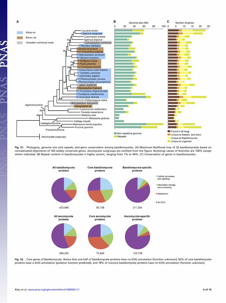
Fig. S1. Phylogeny, genome size and repeats, and gene conservation among basidiomycetes. (A) Maximum-likelihood tree of 33 basidiomycetes based onconcatenated alignments of 183 widely conserved genes. Ascomycete outgroups are omitted from the figure. Bootstrap values of branches are 100% exceptwhere indicated. (B) Repeat content in basidiomycetes is highly variant, ranging from 1% to 44%. (C) Conservation of genes in basidiomycetes.
Fig. S2. Core genes of Basidiomycota. Notice that one-half of basidiomycete proteins have no KOG annotation (function unknown); 92% of core basidiomyceteproteins have a KOG annotation (putative function predicted); and 78% of noncore basidiomycete proteins have no KOG annotation (function unknown).
Riley et al. www.pnas.org/cgi/content/short/1400592111 4 of 10

Fig. S3. Phylogenetic tree of secondary metabolism enzymes. Proteins with a ketoacyl synthase (KS) domain (PF00109 and PF02801) were divided into threecategories: fatty acid synthase (FAS), nonreducing polyketide synthase (NR-PKS), and reducing PKS (R-PKS) based on their phylogenetic relationships.
Riley et al. www.pnas.org/cgi/content/short/1400592111 5 of 10

Fig. S4. Heat map of the number of genes in each auxiliary redox enzyme (AA) family for each organism, along with the number of genes inferred, using theCAFE program, to be gained or lost since that organism’s divergence with its nearest neighbor. The gene families are hierarchically clustered.
Fig. S5. Double-hierarchical clustering of auxiliary redox enzyme (AA) families. Gene families and organisms are hierarchically clustered.
Riley et al. www.pnas.org/cgi/content/short/1400592111 6 of 10

TableS1
.Statistics
onge
nomes
andge
nean
notation
sforba
sidiom
ycetefung
i
Subp
hylum
Order
Organ
ism
Ref.
JGI
portal
idGen
ome
size
,Mb
Gen
eno
.Med
ian
gene
leng
thMed
ian
intron
leng
th%
GC
geno
me
Med
ianex
ons
perge
neGen
ede
nsity,
gene
s/Mb
Aga
rico
mycotina
Aga
ricales
Aga
ricu
sbisporus
1Aga
bi_varbisH
97_2
30.2
10,438
1,49
755
465
345
Coprinop
siscine
rea
2Co
pci1
36.3
13,393
1,47
757
525
369
Galerinamargina
taGalma1
59.4
21,461
1,34
958
484
361
Laccaria
bico
lor
3La
cbi2
64.9
19,036
1,20
956
474
293
Pleu
rotusostrea
tus
Pleo
sPC1
5_2
34.3
12,330
1,39
456
515
359
Schizo
phyllum
commun
e4
Schc
o238
.513
,210
1,47
554
575
343
Auriculariales
Auricularia
delicata
5Aurde
174
.923
,577
1,30
955
594
315
Boletales
Serpulalacrym
ans
6Se
rlaS
7_9_
242
.812
,917
1,31
758
464
302
Coniop
hora
putean
a5
Conp
u143
.013
,761
1,63
658
535
320
Cantha
rella
les
Botryo
basidion
botryo
sum
Botbo1
46.7
16,526
1,40
764
524
354
Corticiales
Punc
tularia
strigo
sozo
nata
5Pu
nst1
34.2
11,538
1,54
257
555
338
Dacrymycetales
Dacryop
inax
sp.
5Dacsp1
29.5
10,242
1,37
953
524
347
Gloeo
phyllales
Gloeo
phyllum
trab
eum
5Glotr1_
137
.211
,846
1,50
156
545
319
Hym
enoc
haetales
Fomitiporia
med
iterrane
a5
Fomme1
63.4
11,333
1,54
060
465
179
Jaap
iales
Jaap
iaargilla
cea
Jaaa
r145
.116
,419
1,40
259
504
364
Polypo
rales
Dicho
mitus
squa
lens
5Dicsq1
42.7
12,290
1,56
259
565
287
Ceripo
riop
sis
subv
ermispo
ra7
Cersu1
39.0
12,125
1,67
856
545
311
Fomitop
sispinico
la5
Fompi1
46.3
14,724
1,37
256
564
318
Phan
eroc
haete
carnosa
8Ph
aca1
46.3
13,937
1,44
857
534
301
Phan
eroc
haete
chrysosporium
9Ph
chr1
35.1
10,048
1,36
354
575
286
Postia
placen
ta10
PosplRSB
12_1
42.5
12,541
1,49
258
505
295
Tram
etes
versicolor
5Trav
e144
.814
,296
1,48
958
584
319
Wolfipo
riaco
cos
5Wolco
150
.512
,746
1,63
356
535
252
Russulales
Heterob
asidion
anno
sum
11Hetan
233
.613
,405
1,28
359
524
398
Stereu
mhirsutum
5Steh
i146
.514
,072
1,71
865
515
303
Seba
cina
les
Piriform
ospo
raindica
12Pirin1
25.0
11,767
1,28
350
514
471
Trem
ellales
Cryp
toco
ccus
neoforman
s13
Cryn
e_H99
_118
.96,96
71,68
556
485
369
Trem
ella
mesen
terica
5Trem
e128
.68,31
31,74
177
475
290
Wallemiales
Wallemia
sebi
14Walse1
9.8
5,28
41,50
347
403
538
Pucciniomycotina
Pucciniales
Puccinia
gram
inis
15Pu
cgr1
88.6
20,534
1,32
991
434
232
Melam
psora
laricis-po
pulin
a15
Mellp1
101.1
16,831
1,37
680
414
166
Ustila
gino
mycotina
Ustila
gina
les
Ustila
gomay
dis
16Ustma1
19.7
6,52
21,57
897
541
331
Malasseziales
Malasseziaglob
osa
17Malgl1
9.0
4,28
61,21
850
521
478
Gen
emod
elsforallg
enom
es(excep
tC.
cine
rea,
C.ne
oforman
s,M.g
lobo
sa,P
.ind
icus
,P.g
raminis,a
ndU.m
aydis;seereferenc
es)w
erepred
ictedusingtheJG
IAnn
otationPipe
line,
which
employ
sava
rietyof
gene
-mod
elingalgo
rithmsan
dqu
alityfilteringto
select
thebe
stmod
els.
Riley et al. www.pnas.org/cgi/content/short/1400592111 7 of 10

1.Morin
E,et
al.(20
12)Gen
omesequ
ence
ofthebu
tton
mushroo
mAga
ricu
sbisporus
reve
alsmecha
nism
sgo
verningad
aptation
toahu
mic-richecolog
ical
nich
e.Proc
NatlA
cadSciU
SA10
9(43
):175
01–17
506.
2.StajichJE,e
tal.(20
10)Insigh
tsinto
evolutionof
multicellu
larfung
ifrom
theassembled
chromosom
esof
themushroo
mCo
prinop
siscine
rea(Cop
rinu
scine
reus).Proc
NatlA
cadSciU
SA10
7(26
):118
89–11
894.
3.MartinF,
etal.(20
08)Th
ege
nomeof
Laccaria
bico
lorprov
ides
insigh
tsinto
mycorrhizal
symbiosis.N
ature45
2(71
83):8
8–92
.4.
Ohm
RA,e
tal.(20
10)Gen
omesequ
ence
ofthemod
elmushroo
mSchizo
phyllum
commun
e.Nat
Biotechn
ol28
(9):9
57–96
3.5.
Flou
dasD,e
tal.(20
12)Th
ePa
leoz
oicorigin
ofen
zymatic
ligninde
compo
sition
reco
nstruc
tedfrom
31fung
alge
nomes.S
cien
ce33
6(60
89):1
715–
1719
.6.
Eastwoo
dDC,
etal.(20
11)Th
eplan
tcellwall-d
ecom
posing
machine
ryun
derliesthefunc
tion
aldive
rsityof
forest
fung
i.Scienc
e33
3(60
43):7
62–76
5.7.
Fernan
dez-Fu
eyoE,
etal.(20
12)C
ompa
rative
geno
micsof
Ceripo
riop
sissubv
ermispo
raan
dPh
aneroc
haetech
rysosporium
prov
ideinsigh
tinto
selectivelig
nino
lysis.Proc
NatlA
cadSciU
SA10
9(14
):545
8–54
63,a
ndco
rrection
(201
2)10
9(21
):835
2.8.
Suzu
kiH,e
tal.(20
12)Co
mpa
rative
geno
micsof
thewhite-rot
fung
i,Ph
aneroc
haetecarnosaan
dP.
chrysosporium,to
eluc
idatethege
neticba
sisof
thedistinct
woo
dtype
sthey
colonize
.BMCGen
omics13
:444
.9.
Martine
zD,e
tal.(20
04)Gen
omesequ
ence
ofthelig
nocellu
lose
degrad
ingfung
usPh
aneroc
haetech
rysosporium
strain
RP78
.Nat
Biotechn
ol22
(6):6
95–70
0.10
.Martine
zD,e
tal.(20
09)Gen
ome,
tran
scriptom
e,an
dsecretom
ean
alysisof
woo
dde
cayfung
usPo
stia
placen
tasupp
orts
unique
mecha
nism
sof
ligno
cellu
lose
conv
ersion
.ProcNatlA
cadSciU
SA10
6(6):195
4–19
59.
11.O
lson
A,e
tal.(20
12)Insigh
tinto
trad
e-offbe
twee
nwoo
dde
cayan
dpa
rasitism
from
thege
nomeof
afung
alforest
pathog
en.N
ewPh
ytol
194(4):100
1–10
13.
12.Z
uccaro
A,e
tal.(20
11)En
doph
ytic
lifestrategies
deco
dedby
geno
mean
dtran
scriptom
ean
alyses
ofthemutua
listicroot
symbion
tPiriform
ospo
raindica.P
LoSPa
thog
7(10
):e10
0229
0.13
.LoftusBJ,e
tal.(20
05)Th
ege
nomeof
theba
sidiom
ycetou
sye
astan
dhu
man
pathog
enCryp
toco
ccus
neoforman
s.Scienc
e30
7(57
13):1
321–
1324
.14
.Pad
amseeM,e
tal.(20
12)Th
ege
nomeof
thexe
rotolerant
moldW
allemia
sebi
reve
alsad
aptation
sto
osmotic
stress
andsugg
ests
cryp
ticsexu
alreprod
uction
.Fu
ngal
Gen
etBiol
49(3):2
17–22
6.15
.Dup
lessisS,
etal.(20
11)Obligatebiotroph
yfeatures
unrave
ledby
thege
nomic
analysisof
rust
fung
i.Proc
NatlA
cadSciU
SA10
8(22
):916
6–91
71.
16.K
ämpe
rJ,et
al.(20
06)Insigh
tsfrom
thege
nomeof
thebiotroph
icfung
alplan
tpa
thog
enUstila
gomay
dis.Nature44
4(71
15):9
7–10
1.17
.XuJ,et
al.(20
07)Dan
druff-associated
Malasseziage
nomes
reve
alco
nverge
ntan
ddive
rgen
tvirulenc
etraits
shared
withplan
tan
dhu
man
fung
alpa
thog
ens.Proc
NatlA
cadSciU
SA10
4(47
):187
30–18
735.
Riley et al. www.pnas.org/cgi/content/short/1400592111 8 of 10

Table S2. Nonbasidiomycete fungi used for comparative purposes
Subkingdom Phylum Subphylum Organism JGI portal ID
Dikarya Ascomycota Pezizomycotina Stagonospora nodorum SN15 Stano2Aspergillus niger ATCC 1015 Aspni5Aspergillus nidulans Aspnid1Botrytis cinerea Botci1Fusarium graminearum Fusgr1Fusarium oxysporum Fusox1Leptosphaeria maculans Lepmu1Magnaporthe grisea Maggr1Mycosphaerella graminicola Mycgr3Nectria hematococca Necha2Neurospora crassa OR74A Neucr1Neurospora tetrasperma
FGSC 2508 mat ANeute_matA2
Neurospora tetraspermaFGSC 2509 mat a
Neute_mat_a1
Pyrenophora tritici-repentis Pyrtr1Pyrenophora teres f. teres Pyrtt1Sclerotinia sclerotiorum Sclsc1Sporotrichum thermophile Spoth2Thielavia terrestris Thite2Trichoderma atroviride Triat2Trichoderma reesei Trire2Tuber melanosporum Tubme1Verticillium dahliae Verda1
Saccharomycotina Candida tenuis NRRL Y-1498 Cante1Dekkera bruxellensis CBS 2499 Dekbr2Pichia stipitis Picst3Saccharomyces cerevisiae S288C Sacce1Spathaspora passalidarum NRRL Y-27907 Spapa3
Fungi incertaesedis
Chytridiomycota N/A Batrachochytrium dendrobatidis JAM81 Batde5Mucoromycotina Mucoromycotina Phycomyces blakesleeanus NRRL1555 Phybl2
Rhizopus oryzae 99–880 Rhior3
Riley et al. www.pnas.org/cgi/content/short/1400592111 9 of 10

Table S3. CAZymes grouped by three major substrates
Substrate preferenceCAZymes and auxiliary
redox enzymes
Cellulose GH6GH7GH12GH44GH45
AA9 (formerly GH61)AA10 (formerly CBM33)
CBM1Hemicelluloses GH10
GH11GH26GH30GH51GH74GH43
Pectin GH43GH28GH53GH78GH88GH105PL1PL3PL4CE8CE12
Table S4. Enzyme activities after 5 d
B3 C-limited B3 N-limited Wiley-milled aspen
Organism LiP MnP GLOX Lac LiP MnP GLOX Lac LiP MnP GLOX Lac
Phchr ND 9.44 0.087 0.74 6.74 11.8 9.19 ND 5.57 ND 0.708 NDCersu ND 4.05 0.082* 7.95 ND 3.83 0.01* 6.15 ND ND ND NDPospl ND ND 0.587 0.056 ND ND 0.713 0.056 ND ND 0.694 0.341Wolco ND ND 0.634 ND ND ND 0.796 ND ND ND 0.541 1.00Jaaar ND ND 0.027* ND ND ND 0.056* ND ND ND 0.014* NDBotbo ND ND ND ND ND ND 0.056* ND ND ND 0.014* ND
In nanomoles per milliliter per minute. ND, not detected.*Trace levels below reliable measurement. Assessment of J. argillacea and B. botryosum culturesafter days 9 and 11 d were essentially identical, i.e., no reliable activity.
Other Supporting Information Files
Dataset S1 (XLSX)Dataset S2 (XLSX)Dataset S3 (TXT)
Riley et al. www.pnas.org/cgi/content/short/1400592111 10 of 10
![Hormographiella aspergillata: an emerging basidiomycete in ...filamentous basidiomycete found in numerous substrates including soils, leaves, pressmud compost and in the air [ 3, 4].](https://static.fdocuments.in/doc/165x107/6123c989ca7b815f6634ae2f/hormographiella-aspergillata-an-emerging-basidiomycete-in-filamentous-basidiomycete.jpg)