Pathology of Peritoneal Metastases
29
123 Olivier Glehen Aditi Bhatt Editors Pathology of Peritoneal Metastases The Unchartered Fields
Transcript of Pathology of Peritoneal Metastases

123
Olivier Glehen Aditi BhattEditors
Pathology of Peritoneal MetastasesThe Unchartered Fields

Pathology of Peritoneal Metastases

Olivier Glehen • Aditi BhattEditors
Pathology of Peritoneal Metastases
The Unchartered Fields
ISBN 978-981-15-3772-1 ISBN 978-981-15-3773-8 (eBook)https://doi.org/10.1007/978-981-15-3773-8
© Springer Nature Singapore Pte Ltd. 2020This work is subject to copyright. All rights are reserved by the Publisher, whether the whole or part of the material is concerned, specifically the rights of translation, reprinting, reuse of illustrations, recitation, broadcasting, reproduction on microfilms or in any other physical way, and transmission or information storage and retrieval, electronic adaptation, computer software, or by similar or dissimilar methodology now known or hereafter developed.The use of general descriptive names, registered names, trademarks, service marks, etc. in this publication does not imply, even in the absence of a specific statement, that such names are exempt from the relevant protective laws and regulations and therefore free for general use.The publisher, the authors and the editors are safe to assume that the advice and information in this book are believed to be true and accurate at the date of publication. Neither the publisher nor the authors or the editors give a warranty, expressed or implied, with respect to the material contained herein or for any errors or omissions that may have been made. The publisher remains neutral with regard to jurisdictional claims in published maps and institutional affiliations.
This Springer imprint is published by the registered company Springer Nature Singapore Pte Ltd.The registered company address is: 152 Beach Road, #21-01/04 Gateway East, Singapore 189721, Singapore
EditorsOlivier GlehenGeneral and Oncologique SurgeryCentre Hospitalier Lyon-SudLyonFrance
Aditi BhattDepartment of Surgical OncologyZydus HospitalAhmedabadIndia

v
This is a book on pathology of peritoneal metastases that has been edited and largely authored by surgeons which is unusual. Peritoneal surface oncology is a field that has always been at the cross roads—in the early years of evolu-tion of surgical treatment, hyperthermia was being increasingly used to potentiate cancer therapy and thus was combined with the surgical treatment of peritoneal metastases, that is, cytoreductive surgery. The surgery itself had to prove its merit over systemic therapies and was burdened with proving the merit of another treatment that added to the morbidity. Similarly, disease biology was only partially understood and remains a major challenge for future progresses. While the prognostic factors were still being identified, and validated, oncology ushered into the era of genetics and molecular biology. And the gaps in understanding the pathophysiology of peritoneal metastases persisted. Pathological expertise has largely been directed at the diagnosis and classification of uncommon tumors.
During cytoreductive surgery that comprises of peritonectomy procedures and visceral resections, a large amount of tissue is submitted for histopatho-logical evaluation. This remains a potential source of prognostic information regarding tumor biology. It provides a good opportunity to also study the patterns and pathways of peritoneal dissemination from various tumors.
In this book, we use these pathological findings to better explain the pat-terns and pathways of peritoneal cancer dissemination and their potential implications on clinical practice. We provide a rationale and recommenda-tions for standardizing CRS procedures and evaluation of surgical specimens. In turn, we raise research question that can be addressed in future studies.
Some of the other aspects of pathological evaluation like pathological response to chemotherapy, diagnosis and classification of rare peritoneal tumors have also been covered in different chapters. Keeping in sync with the progress in molecular oncology, we look at the role of molecular oncology in the current and future management of peritoneal metastases.
We are grateful to all the contributors for lending their time and expertise to this book. We are also grateful to our pathology colleagues for their invalu-able contribution to this work.
3 December 2019 Olivier Glehen Aditi Bhatt
Preface
vii
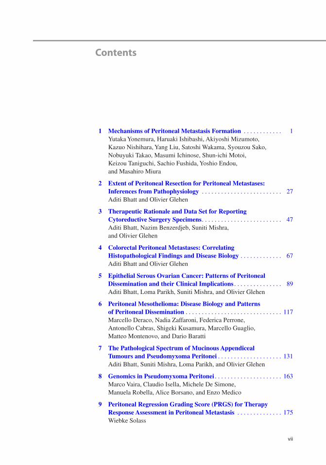
1 Mechanisms of Peritoneal Metastasis Formation . . . . . . . . . . . . 1Yutaka Yonemura, Haruaki Ishibashi, Akiyoshi Mizumoto, Kazuo Nishihara, Yang Liu, Satoshi Wakama, Syouzou Sako, Nobuyuki Takao, Masumi Ichinose, Shun-ichi Motoi, Keizou Taniguchi, Sachio Fushida, Yoshio Endou, and Masahiro Miura
2 Extent of Peritoneal Resection for Peritoneal Metastases: Inferences from Pathophysiology . . . . . . . . . . . . . . . . . . . . . . . . . 27Aditi Bhatt and Olivier Glehen
3 Therapeutic Rationale and Data Set for Reporting Cytoreductive Surgery Specimens . . . . . . . . . . . . . . . . . . . . . . . . . 47Aditi Bhatt, Nazim Benzerdjeb, Suniti Mishra, and Olivier Glehen
4 Colorectal Peritoneal Metastases: Correlating Histopathological Findings and Disease Biology . . . . . . . . . . . . . 67Aditi Bhatt and Olivier Glehen
5 Epithelial Serous Ovarian Cancer: Patterns of Peritoneal Dissemination and their Clinical Implications . . . . . . . . . . . . . . . 89Aditi Bhatt, Loma Parikh, Suniti Mishra, and Olivier Glehen
6 Peritoneal Mesothelioma: Disease Biology and Patterns of Peritoneal Dissemination . . . . . . . . . . . . . . . . . . . . . . . . . . . . . . 117Marcello Deraco, Nadia Zaffaroni, Federica Perrone, Antonello Cabras, Shigeki Kusamura, Marcello Guaglio, Matteo Montenovo, and Dario Baratti
7 The Pathological Spectrum of Mucinous Appendiceal Tumours and Pseudomyxoma Peritonei . . . . . . . . . . . . . . . . . . . . 131Aditi Bhatt, Suniti Mishra, Loma Parikh, and Olivier Glehen
8 Genomics in Pseudomyxoma Peritonei . . . . . . . . . . . . . . . . . . . . . 163Marco Vaira, Claudio Isella, Michele De Simone, Manuela Robella, Alice Borsano, and Enzo Medico
9 Peritoneal Regression Grading Score (PRGS) for Therapy Response Assessment in Peritoneal Metastasis . . . . . . . . . . . . . . 175Wiebke Solass
Contents

viii
10 Rare Peritoneal Tumours: Histopathological Diagnosis and Patterns of Peritoneal Dissemination . . . . . . . . . . . . . . . . . . . 181Suniti Mishra, Snita Sinukumar, Nutan Jumale, Loma Parikh, Aditi Bhatt, and Olivier Glehen
11 Approach to a Patient with Peritoneal Metastases with Unknown Primary Site: Focus on Histopathological Evaluation . . . . . . . . . . . . . . . . . . . . . . . . . . . . . . . . . . . . . . . . . . . . 229Aditi Bhatt, Loma Parikh, Suniti Mishra, and Olivier Glehen
12 Biomarkers in the Management of Peritoneal Metastases . . . . . 251Ninad Katdare, Aditi Bhatt, and Olivier Glehen
Contents
ix
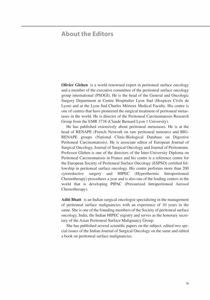
Olivier Glehen is a world renowned expert in peritoneal surface oncology and a member of the executive committee of the peritoneal surface oncology group international (PSOGI). He is the head of the General and Oncologic Surgery Department at Centre Hospitalier Lyon Sud (Hospices Civils de Lyon) and at the Lyon Sud Charles Mérieux Medical Faculty. His centre is one of centres that have pioneered the surgical treatment of peritoneal metas-tases in the world. He is director of the Peritoneal Carcinomatosis Research Group from the EMR 3738 (Claude Bernard Lyon 1 University).
He has published extensively about peritoneal metastases. He is at the head of RENAPE (French Network on rare peritoneal tumours) and BIG-RENAPE groups (National Clinic-Biological Database on Digestive Peritoneal Carcinomatosis). He is associate editor of European Journal of Surgical Oncology, Journal of Surgical Oncology and Journal of Peritoneum. Professor Glehen is one of the directors of the Inter-University Diploma on Peritoneal Carcinomatosis in France and his centre is a reference centre for the European Society of Peritoneal Surface Oncology (ESPSO) certified fel-lowship in peritoneal surface oncology. His centre performs more than 200 cytoreductive surgery and HIPEC (Hyperthermic Intraperitoneal Chemotherapy) procedures a year and is also one of the leading centers in the world that is developing PIPAC (Pressurized Intraperitoneal Aerosol Chemotherapy).
Aditi Bhatt is an Indian surgical oncologist specializing in the management of peritoneal surface malignancies with an experience of 10 years in the same. She is one of the founding members of the Society of peritoneal surface oncology, India, the Indian HIPEC registry and serves as the honorary secre-tary of the Asian Peritoneal Surface Malignancy Group.
She has published several scientific papers on the subject, edited two spe-cial issues of the Indian Journal of Surgical Oncology on the same and edited a book on peritoneal surface malignancies.
About the Editors

1© Springer Nature Singapore Pte Ltd. 2020 O. Glehen, A. Bhatt (eds.), Pathology of Peritoneal Metastases, https://doi.org/10.1007/978-981-15-3773-8_1
Mechanisms of Peritoneal Metastasis Formation
Yutaka Yonemura, Haruaki Ishibashi, Akiyoshi Mizumoto, Kazuo Nishihara, Yang Liu, Satoshi Wakama, Syouzou Sako, Nobuyuki Takao, Masumi Ichinose, Shun-ichi Motoi, Keizou Taniguchi, Sachio Fushida, Yoshio Endou, and Masahiro Miura
1.1 Introduction
It has long been considered that the establishment of peritoneal metastasis (PM) is a multi- step pro-cess, consisting of (1) detachment of cancer cells from the primary tumor, (2) adhesion of perito-neal free cancer cells (PFCCs) on the distant peri-toneal surface, (3) invasion into the submesothelial tissue, and (4) proliferation accompanying with the angiogenesis and the induction of stromal tis-sue [1]. The process is called “trans-mesothelial metastasis” by Yonemura et al. [2] or “randomly
proximal distribution” by Sugarbaker [3] (Fig. 1.1). Through the process, cancer cells with high malignant potential can metastasize on the peritoneum by concerted expression of metasta-sis-related genes [1–3]. Recently, new concepts of the formation of PM were proposed: i.e., (1) Trans-lymphatic metastasis and (2) superficial growing metastasis (Table 1.1) [3].
In this chapter, mechanisms of the formation of PM will be described in terms of the morpho-logical, histological, and molecular biological aspects.
Y. Yonemura (*) Asian School of Peritoneal Surface Malignancy Treatment, Kyoto, Japan
Department of Regional Cancer Therapy, Peritoneal Dissemination Center, Kishiwada Tokushukai Hospital, Kishiwada, Osaka, Japan
Department of Regional Cancer Therapy, Peritoneal Dissemination Center, Kusatsu General Hospital, Kusatsu, Shiga, Japane-mail: [email protected]
H. Ishibashi · K. Nishihara · Y. Liu · S. Wakama S. Sako Department of Regional Cancer Therapy, Peritoneal Dissemination Center, Kishiwada Tokushukai Hospital, Kishiwada, Osaka, Japan
A. Mizumoto · N. Takao · M. Ichinose · S.-i. Motoi Department of Regional Cancer Therapy, Peritoneal Dissemination Center, Kusatsu General Hospital, Kusatsu, Shiga, Japan
K. Taniguchi Department of Surgery, Mizonokuchi Hospital, Teikyo University, School of Medicine, Kawasaki, Kanagawa, Japan
S. Fushida Department of Surgery, Kanazawa University, Kanazawa, Japan
Y. Endou Central Research Resource Center, Cancer Research Institute, Kanazawa University, Kanazawa, Japan
M. Miura Department of Anatomy, Oita Medical University, Oita, Japan
1

2
Gene products
fromcancer cells
Rolling, cross talk
with mesothelial cell (MC)
Invasion into the gap of MC cells
Adhesion tobasementmembrane
Invasion into
submesothelialtissue
angiogenesis Establishment of peritoneal metastasis
Cancer cells
CD-44, CA19-9CEA,
P-cadherinIntegrin,
Adhesion mol.
EGF/EGFR,MET
REG IV/Reg receptor
Growth factors and their receptors
HGF, EGFTGF-β
Contraction of MC
MC injuring factors
Gene products
from stromal
cells
AMF/AMFR,MET
Rho, S1004AMotility factors
VEGFR-1,2, Tie-2, TGF-β
CD144, CD31 PIGF, plasmin
Ephrin, VE-cadherin
Proliferation of vascular endothelial cells
MacrophageMMP-8 , -9
FibroblastMMP-1,-2,-3
UPA/UPARMMPs, MT-
MMPsMotility factors
Matrix digestingenzymes
HGF, EGFTGF-β
Fibroblasts
Integrinα2,3,5β1,2
Heterophilicadhesionmolecules
ICAMVCAM
PECAM-1E-selectin
Hyaluronic acid
Mesothelial cells(MC)
HIF, VEGFTGF-β, PIGF
Angiogenesisfactors
Peritoneal cavity
Blood vessel
Blood vessel
PFCC
90µmBlood peritoneal barrier
Macrophage
FibroblastCD34
Mesothelialcell
Basement membraneMacula cribriformis
Fig. 1.1 Trans-mesothelial metastasis as named by Yonemura et al. or randomly proximal distribution by Sugarbaker. Peritoneal free cancer cells (PFCCs) exfoli-ated from the serosal surface of primary tumors migrate into the peritoneal cavity, and adhere on the peritoneal surface (Process 1). During the rolling of PFCCs on the peritoneal surface, peritoneal mesothelial cells shrink by cytokines produced by PFCCs, resulting in the exposure
of basement membrane and macula cribriformis (Process 2). PFCCs then migrate into the inter mesothelial space, and invade into the submesothelial tissue by degradation of extracellular matrix by matrix-digesting enzymes and by locomotive activity using motility factors (Process 3). Finally, cancer cells proliferate near the submesothelial blood vessels with introduction of tumor stromal tissues and neogenesis of tumor blood vessels (Process 4)
Table 1.1 Three patterns of peritoneal metastasis according to the biological malignant potentials and the morphologi-cal feature of peritoneal free cancer cells
Pattern of metastasisBiologic malignant behavior Cancer
Morphological features of PFCCs
Trans-mesothelial metastasis High Gastric, colorectal, pancreatic, biliary ovarian cancer
Single or small clusters
Trans-lymphatic metastasis Omental milky spots (OMS) Initial lymphatics outside OMS
High, moderate Gastric, colorectal, ovarian cancer
Single or small clusters
Superficial growing metastasis Moderate, low AMNa, GCSb, mesothelioma, MCMc, hepatoma
Large with or without mucinous material
aAMN low-grade appendiceal mucinous neoplasmbGCS granulosa cell tumorcMCM multicystic mesothelioma
Y. Yonemura et al.

3
1.2 Mechanisms of Trans- mesothelial Metastasis
1.2.1 Mechanisms of Cancer Cell Spillage into the Peritoneal Cavity
The first step of PM is detachment of cancer cells from the serosal surface of the primary tumor in highly malignant tumors and the rupture of appendix vermiformis or ovary by the increased intrinsic pressure due to the proliferation on mucinous neoplasm. Additionally, during sur-gery, blood or lymphatic fluid contaminated with cancer cells may spill into the peritoneal cavity from damaged blood and lymphatic vessels. The exfoliated cancer cells in the peritoneal cavity are called peritoneal free cancer cells (PFCCs). PFCCs have high proliferative activities and can grow in the distant peritoneum.
In the process of detachment of cancer cells from the primary tumor, homophilic cell-cell adhesion molecules play important roles. Epithelial cells tightly interconnect with a tight junction on the apical membrane and an adher-
ence junction on the basolateral membrane side (Fig. 1.2). Tight junction components are trans-membrane proteins, claudin and occludin and the cytoplasmic scaffolding protein, ZO-1, -2, and -3, which bind the actin bundles of the cytoplas-mic protein. Tight junction is mainly composed of claudin, which is observed as continuous bead- like particles expressed on the lateral membrane of cells [4]. The partition of the basilar membrane by the tight junction functions as a barrier to con-trol the movement of materials and as a selective permeation channel [4]. However, dysfunction of the tight junction causes the loosening of cell-cell adhesion, resulting in edema. Disappearance of tight junction causes cells to disperse by the loss of polarity. Vascular endothelial growth factor (VEGF) and some cytokines reduce the function of tight junction, and cause edema [5].
Serial analyses of gene expression (SAGE) clarified the reduced expression of claudin in poorly differentiated adenocarcinoma of the stomach [6], in which reduced expression of claudin 4, 7 is found and the patients with gastric cancer showing low expression of claudin have a poor prognosis. In addition, ZO expression is
Tight junction
Adherencejunction
JAM
Occludin
Claudin
ZO-1, -2, -3
Afadin
Nectin
Cadherin
ZO-1, -2, -3
ZO-1, -2, -3
Basement membrane
Actin bundle
ADIP Ponsin
α-Actinin Vinculin
β-catenin α-catenin
P120ctn
Fig. 1.2 Molecules associated with homophilic cell-cell adhesion of epithelial cells. Epithelial cells interconnect with tight junction, located at apical membrane of cells, and with adherence junction on the basolateral membrane. Cancer cells showing the functional abnormalities of tight junction and adherence junction tend to disperse and detach from the serosal surface, spill from the primary tumor and migrate into the peritoneal cavity
1 Mechanisms of Peritoneal Metastasis Formation

4
also reduced in poorly differentiated adenocarci-noma which tends to establish PM [6, 7].
In the components of the adherence junction, E-cadherin expression is important. E-cadherin is a transmembrane glycoprotein, and cells tightly connect with the extracellular domain of the mol-ecule on the basolateral membrane (homophilic adhesion). The intracellular domain of E-cadherin connects with α-, β-, γ-catenin and the complex controls the function of E-cadherin [8–10]. In gastric cancer, poorly differentiated adenocarci-noma especially macroscopic type-4 has a high potential of PM and the downregulation of E-cadherin expression is an important feature [11, 12]. The causes of reduced expression of E-cadherin are loss of the E-cadherin gene, point mutation, and the methylation of the promoter region [12]. In addition, the function of E-cadherin is reduced via the inhibition of the function of catenin molecules by the loss of the α-catenin gene, and phosphorylation of the tyro-sine residue of β-catenin [13]. In colorectal can-cer, downregulation of E-cadherin level is associated with poorly differentiated type, higher potential of metastasis, and progression [14]. The lower expression of E-cadherin in poorly differ-entiated colorectal cancer may explain the aggressive nature, and poorly differentiated ade-nocarcinoma is known as a subtype that fre-quently metastasizes on peritoneum [14, 15].
Accordingly, cancer cells with the functional abnormalities of tight junction and adherence junction tend to disperse and detach from the serosal surface, spill from the primary tumor and migrate into the peritoneal cavity.
1.2.2 Adhesion of PFCCs and Mesothelial Cells (Fig. 1.1, Process 1)
PFCCs migrate on the distant peritoneal surface and adhere to mesothelial cells during rolling on the mesothelial cell surface. PFCCs express sev-eral kinds of integrin molecules and adhere with their ligands expressed on mesothelial cells (Fig. 1.1, Process 1, 2). Peritoneal mesothelial cells express immunoglobulin-like intercellular
adhesion molecules (ICAM-1, CD54) [16] and vascular cell adhesion molecules (VCAM-1, CD106) [17], and interact with integrin αLβ2 (LFA-1α, CD11a) expressed on PFCCs and inflammatory cells (heterotypic cell-cell adhesion).
Platelet-endothelial cell adhesion molecules (PECAM-1, CD31) play a role in the transmigra-tion of white blood cells and PFCCs. After the process of slowing down the passage of leuko-cytes and PFCCs over activated vascular endo-thelium and peritoneal mesothelial cells (“rolling”), integrins are crucial for stopping the cells at the extravasation site and migration into submesothelial tissue [18].
Stopping is the result of the interaction of inte-grins on the leukocytes (β2 integrin, α4β1 (VLA- 4), or α4β7) and immunoglobulin-like adhesion molecules (ICAM-1 and VCAM-1) on endothe-lial and mesothelial cells [19]. Next, for migra-tion based on the interaction between β2 integrins on the leukocytes and ICAM-1, VCAM-1, and PECAM-1 on the endothelial cells, mesothelial cells adhere with inflammatory cells or PFCCs via homophilic adhesion of PECAM-1. The expression of VCAM-1, and E-selectin, is upreg-ulated by IL-1β, TNF-1α, and IFN-γ, so the mesothelial cell adhesion can be facilitated by local inflammation [16, 20].
As shown in Fig. 1.3, several adhesion mole-cules are associated with the adhesion between mesothelial cells and PFCCs. The only classical cadherin expressed by mesothelial cells and PFCCs is P-cadherin, and this molecule could serve homophilic heterotypic adhesion (hetero-philic adhesion by the same adhesion molecule) between PFCCs and mesothelial cells (Fig. 1.3) [21]. P-cadherin promotes intraperitoneal dis-semination of ovarian cancer cells by facilitating tumor cell aggregation and tumor peritoneum interaction in addition to promoting tumor cell migration [22].
Hyaluronic acid (hyaluronate) is a mucopro-tein with a molecular weight of 200,000–400,000, consisting of alternate binding of N-acetylglucosamine and glucuronic acid. This molecule is one of the components of extracellu-lar matrices and acts as a cushion to support the
Y. Yonemura et al.

5
cells by binding a considerable amount of water. Pericellular hyaluronate produced by mesothelial cells has a function as a lubricant (Fig. 1.3). CD44 is a transmembrane glycoprotein and acts as a receptor for hyaluronate. This molecule has four functioning domains, and hyaluronate binds with the extracellular domain of CD44. PFCCs expressing CD44 bind to the pericellular hyal-uronate of the mesothelial cells. The CD44 gene has 20 exons and many translational products (v1–v10) are produced from alternative RNA splicing and post-translational modifications. Certain CD44 isoforms that regulate activation and migration of lymphocytes and macrophages may also enhance local growth and metastatic spread of tumor cells [23]. CD44v, v7 splice vari-ants are expressed by some gastro-intestinal can-cers, and considered as markers for their metastatic capability [24]. CD44v6 expressed on PFCCs accounts for the binding with mesothelial cells [24]. The TGF-β produced from the fibro-blasts in the stroma of cancer tissue upregulates the CD44 expression from cancer cells [25]. Expression of CD44 by peritoneal mesothelial cells also seems to contribute to heterotypic cell adhesion by pancreatic cancer cells, and is upreg-ulated by TNF-α/IL-1β from the peritoneal mac-rophages [26].
The sialyl Lewis-a (sLea) antigen is a carbohy-drate structure present on cancer cells and has a structure of sialic form of Lea antigen with sialyl acid. The monoclonal antibody CA19-9 is a use-ful and popular tool to assess circulating sialyl Lewis-a epitopes in the blood of cancer patients. sLea expressed on leukocytes binds to P-cadherin on the activated vascular endothelial cells, and the weak affinity interaction of sLea and P-cadherin is considered as the initial force implicated in the “rolling” of extravasating leu-kocytes. E-selectin also can bind to sLea, and sLea on cancer cells produces a similar interaction with E-selectin on the endothelial cells [27]. As shown in Fig. 1.3, E-selectin is also found on peritoneal mesothelial cells. The interaction of E-selectin and P-cadherin on mesothelial cells and sLea on PFCCs has a role on the het-erotypic adhesion in early pathophysiological event in PM.
CA125 is a cell surface mucin-like glycopro-tein expressed in mesothelial cells, and is upregu-lated in malignant ovarian tumors [28, 29]. It is considered as a relatively specific circulating tumor marker in ovarian cancer patients. Mesothelin is expressed by the normal mesothe-lial cells, and soluble mesothelin is used to detect the overexpressed protein as a circulating tumor
VCAM-1CD56
α4β1α4β7 αLβ2
α4β7
ICAM-1CD106
CD44
hyaluronate
Sialyl Lex
E-Selectin
CA125
mesothelin
L1CAM
Neuropirin-1
TNFα
IL-1β
Peritoneal free cancer cell
Mesothelial cell
TNFα
IL-1β
macrophage
Inflammatory cellfibroblast
P-cadherin
P-cadherin
actin
Fig. 1.3 Heterotypic adhesion molecules associated with adhesion between cancer cells and mesothelial cells
1 Mechanisms of Peritoneal Metastasis Formation
6
marker for mesothelioma. Mesothelin binds with CA125 expressed on PFCCs and has a role as an adhesion molecule between ovarian cancer cells and mesothelial cells [30].
L1CAM (CD171), an adhesion molecule expresses on pancreas cancer and colorectal can-cer cells and binds with neuropilin-1 (Fig. 1.3). It is upregulated by TGF-β1 [31]. Soluble L1CAM (sL1) binds to VEGF-A (165), and activates VEGFR-2, resulting in angiogenesis [32].
1.2.3 Morphological Changes of Mesothelial Cells (Fig. 1.1, Process 1), Submesothelial Invasion of PFCCs, Attachment of PFCCs to the Basement Membrane (Fig. 1.1, Process 2)
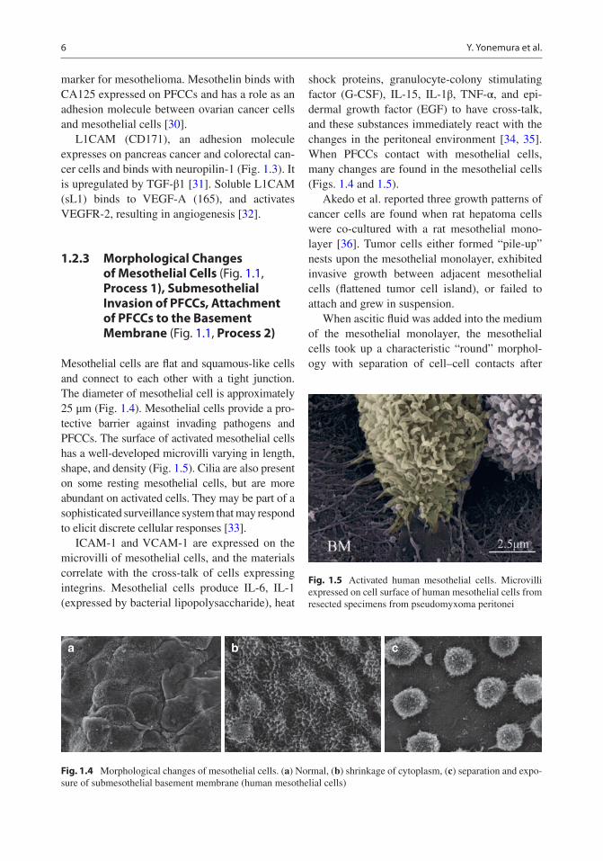
Mesothelial cells are flat and squamous-like cells and connect to each other with a tight junction. The diameter of mesothelial cell is approximately 25 μm (Fig. 1.4). Mesothelial cells provide a pro-tective barrier against invading pathogens and PFCCs. The surface of activated mesothelial cells has a well-developed microvilli varying in length, shape, and density (Fig. 1.5). Cilia are also present on some resting mesothelial cells, but are more abundant on activated cells. They may be part of a sophisticated surveillance system that may respond to elicit discrete cellular responses [33].
ICAM-1 and VCAM-1 are expressed on the microvilli of mesothelial cells, and the materials correlate with the cross-talk of cells expressing integrins. Mesothelial cells produce IL-6, IL-1 (expressed by bacterial lipopolysaccharide), heat
shock proteins, granulocyte-colony stimulating factor (G-CSF), IL-15, IL-1β, TNF-α, and epi-dermal growth factor (EGF) to have cross-talk, and these substances immediately react with the changes in the peritoneal environment [34, 35]. When PFCCs contact with mesothelial cells, many changes are found in the mesothelial cells (Figs. 1.4 and 1.5).
Akedo et al. reported three growth patterns of cancer cells are found when rat hepatoma cells were co-cultured with a rat mesothelial mono-layer [36]. Tumor cells either formed “pile-up” nests upon the mesothelial monolayer, exhibited invasive growth between adjacent mesothelial cells (flattened tumor cell island), or failed to attach and grew in suspension.
When ascitic fluid was added into the medium of the mesothelial monolayer, the mesothelial cells took up a characteristic “round” morphol-ogy with separation of cell–cell contacts after
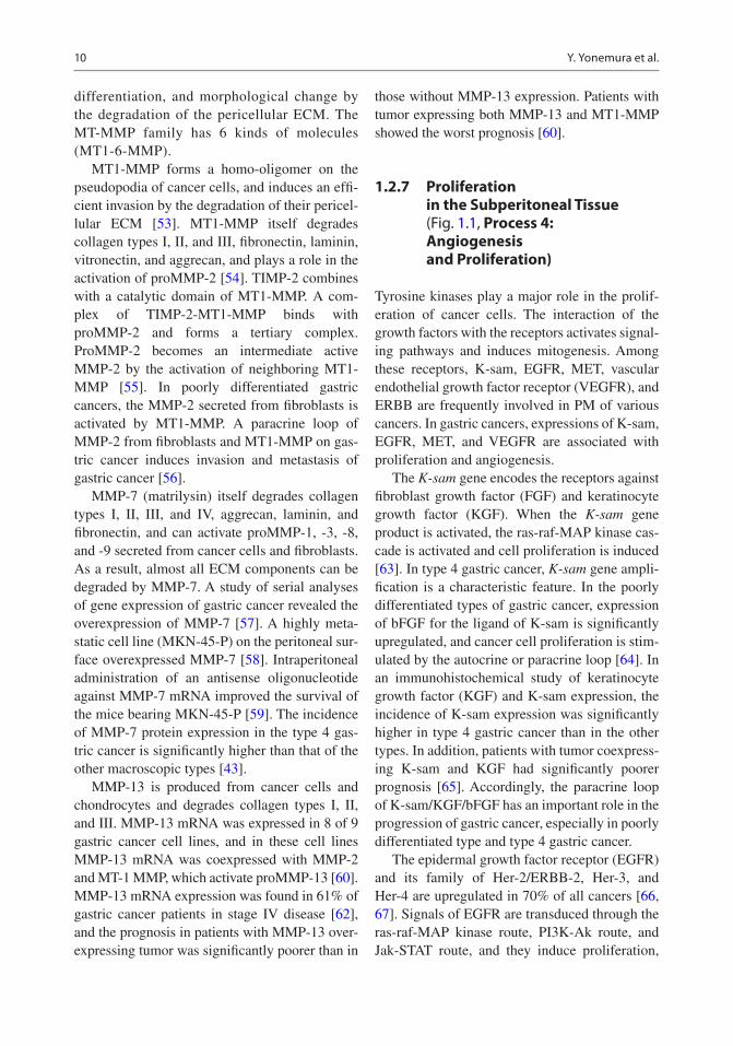
a b c
Fig. 1.4 Morphological changes of mesothelial cells. (a) Normal, (b) shrinkage of cytoplasm, (c) separation and expo-sure of submesothelial basement membrane (human mesothelial cells)
Fig. 1.5 Activated human mesothelial cells. Microvilli expressed on cell surface of human mesothelial cells from resected specimens from pseudomyxoma peritonei
Y. Yonemura et al.

7
20 h [37]. These results denote that the malignant ascitic fluid contains factors that induce the changes of mesothelial cell morphology (meso-thelial cell injury factors). These factors are produced from cancer cells, peritoneal macro-phages, and mesothelial cells [38].
1.2.4 Adhesion of PFCCS to the Submesothelial Basement Membrane (Fig. 1.1, Process 2 and Fig. 1.6)
After mesothelial cell contraction by cytokines, the submesothelial basement membrane is exposed (Fig. 1.4). The basement membrane con-sists of laminin, type IV collagen, heparin sulfate proteoglycan, entactin, and perlecan. Mesothelial cells and fibroblasts produce these elements. Current evidence suggests that adherence to the basement membrane of PFCCs is mediated via an integrin-ligand interaction.
The integrin molecule is a heterodimer consist-ing of an α and a β subunit and is expressed on the cell membrane. Integrins are the important mole-cules for cell-cell and cell-ECM adhesion. According to the combination of 17α subunits and
8β subunits, 24 kinds of integrins exist [39]. Many kinds of integrins are expressed from PFCCs, and the overexpression of integrins correlates with metastatic potential [40–42]. Integrin α2 and α3 expressions were significantly elevated in the peritoneal dissemination of gastric cancer [40, 41]. These α-integrins dimerize with β-subunits to form adhesion molecules for basement membrane proteins, including fibronectin, laminin, and col-lagen IV. Treatment with anti-β1 integrin antibody significantly inhibited the adherence of highly metastatic cell line on the peritoneum in an ex-vivo peritoneal model, suggesting a role for β1-mediated integrin adhesion to the submesothe-lial basement membrane [41]. In ovarian cancers, integrin α5β1 and α6β1 correlate with PM.
1.2.5 Invasion into the Submesothelial Tissue (Fig. 1.1, Process 3)
Factors associated with invasion into the subme-sothelial tissue are the autocrine motility factor (AMF)/AMF receptor, Rho/ROCK, S100A-4, and hepatocyte growth factor (HGF)/MET (receptor for HGF) [43–48].
Fig. 1.6 Highly metastatic cell line (MKN-45) from gastric cancer express filopodia, and attach to the basement mem-brane of human greater omentum
1 Mechanisms of Peritoneal Metastasis Formation

8
AMF is a 55 kDa protein, which stimulates chemotaxis and chemokinetics [49]. AMFR is a member of the tyrosine kinases, which are located on the cell membrane. Binding of AMF with AMFR stimulates changes in the cytoskeleton and formation of invadopodia, resulting in the induction of amoebic movement [43]. Type 4 gastric cancer is more significantly associated with PM than the other macroscopic types. In type 4 gastric cancer, expression of the AMFR protein was significantly higher than that in type 3 tumors [50]. Accordingly, poorly differentiated adenocarcinoma of the stomach has high motility by the activation of the AMF/AMFR cascade combined with downregulation of E-cadherin and claudin [7, 12, 50].
Rho is a G protein, which induces ruffling of the cell membrane in cooperation with effectors of its downstream, like mDia, Crk, Rac and ROCK, and FAK/paxillin [49]. Rho upregulates actin filaments by activation of mDia, and ROCK increases the contractile strength of myosin, which bridges actin filaments. Rho and Rac expressions were upregulated in poorly differen-tiated adenocarcinoma and advanced cancers in the late stage [43].
S100A4, a member of the S100 protein fam-ily, is known as a calcium-binding protein, and increases cell motility by activating myosin [44]. S100A4 activates myosin in lamellipodia expressed on the invasion front of cancer cells (Fig. 1.6) [45]. The actin filament that bridges myosin, combined with the vinculin connected with talin and the intracellular domain of integrin [45, 46]. In gastric cancer, S100A4 upregulation is significantly associated with poorly differenti-ated adenocarcinoma, lymph node metastasis, peritoneal dissemination, and a poor prognosis [46]. In addition, downregulation of E-cadherin and upregulation of S100A4 were found in type 4 gastric cancer [46]. Moriyama et al. reported that S100A4 gene was transfected into a non-invasive oral cancer cell line of OSC-19, and that the new cell line overexpressed S100 A4, showed signifi-cant invasive activity, and downregulated E-cadherin and β-catenin [47].
The scatter factor (SF) called hepatocyte growth factor (HGF) and its receptor of MET (a
tyrosine kinase type receptor) are important mol-ecules for cell motility and proliferation. When HGF binds with MET, MET is activated by the autophosphorylation of tyrosine residue on the intracellular domain and induces cell motility by activation of F-actin and microtubules. In the peritoneal cavity, HGF is produced from acti-vated mesothelial cells, and fibroblasts, and induces mesothelial cell contraction and invasion of PFCCs through the intercellular space of mesothelial cells [48].
IL-1β, and TNF-α from peritoneal macro-phages, fibroblasts, and inflammatory cells induce HGF production from mesothelial cells [51]. The HGF/MET paracrine cascade corre-lates with not only cancer cell motility but also proliferation and angiogenesis. Recently, molec-ular targeting therapy to control the cascade has been developed [52].
1.2.6 Destruction of Submesothelial Basement Membrane and Extracellular Matrix (ECM) and Invasion into Submesothelial Tissue (Fig. 1.1, Process 3)
The tissue between mesothelial cells and subme-sothelial arterial blood capillaries is named the peritoneal-blood barrier, and the average width is 90 μm (Figs. 1.1, 1.17 and 1.29) [53]. This barrier prohibits the diffusion of drugs administered by systemic chemotherapy. The diffusion length of oxygen from arterial blood capillaries is 100 μm, and PFCCs attached to the submesothelial base-ment membrane can survive by the oxygen nutri-tional supplement from blood vessels [54]. PFCCs with high invasive capacity destroy the ECM in peritoneal-blood barrier, invade near the arterial blood capillaries, and proliferate with angiogenesis (Fig. 1.1, Process 4).
The subperitoneal basement membrane between mesothelial cells and submesothelial stromal tissue is a thin membrane of 50–100 nm in width, and is composed of collagen type IV, laminin, entactin, heparin sulfate proteoglycan, and perlecan [55, 56]. Molecules associated
Y. Yonemura et al.
9
with the destruction of basement membrane are matrix metalloproteinases [MMPs: MMP-2, MMP-7, MMP-14 (MT1-MMP)] and plasmin. These molecules are produced from cancer cells, mesothelial cells, fibroblasts, inflamma-tory cells, and macrophages. Subperitoneal tis-sues are composed of dense network of ECM, which prohibits the movement of materials with molecular weight higher than 100,000. Cancer cells produce several kinds of matrix-digesting enzymes to destroy and invade into subperito-neal stromal tissue. An immunohistochemical study of gastric cancers revealed that urokinase-type plasminogen activator (UPA) is detected in the cytoplasm in 66% of gastric cancers [57]. UPA from gastric cancer and fibroblasts binds with its receptor (UPAR) on the cell membrane and is activated with plasmin and kallikrein. Activated UPA on the cell membrane activates plasminogen to plasmin [58]. Plasmin then degrades the ECM and further activates plas-minogen and latent MMPs. UPA is specifically inactivated by plasminogen activator inhibitor- 2 (PAI-2), and PAI-2 can inhibit the formation of experimental peritoneal carcinomatosis [59, 60]. UPAR expression in type 4 gastric cancers is significantly higher than that in other macro-scopic types [57]. Fibroblasts accumulate in the stroma of the invasive front of type 4 gastric cancer. UPA secreted from fibroblasts is com-bined with UPAR on the cancer cells via the paracrine loop, leading to activation of plasmin in the cancer cells which help them to invade the stomach wall [60].
MMP family includes collagenases (MMP-1, MMP-8, and -13), gelatinases (MMP-2 and MMP-9), stromelysin-1, -2 (MMP-3, and MMP- 10), transmembrane MMPs (MT-MMP families), and others: matrilysin, MMP-7; stromelysin-3, MMP-11; metalloesterase, MMP- 12; and enam-elysin, MMP-20. Activities of MMPs are con-trolled by the activation of proMMPs and inhibition by TIMPs (tissue inhibitor metallopro-teinases), and MMPs are mutually activated by plasmin and the other MMPs. Four types of TIMPs have been reported and control the activ-ity of MMPs, resulting in the degradation of col-lagen and the induction of fibrosis.
MMP genes are upregulated by IL-1, TNF-α, EGF, PDGF, and FGF. MMP-1, -2, -7, -13, and -14 (MT1-MMP) play roles in the stromal inva-sion of gastric cancer.
MMP-1 specifically cuts the helix structure of collagen types I, II, and III. In gastric cancer tis-sue, MMP-1 is secreted from the stromal fibro-blasts. TGF-β produced from poorly differentiated adenocarcinomas of the stomach stimulates the proliferation of fibroblast in the invasive front [61]. Cancer cells invade the stroma utilizing MMP-1, and MMP-2 produced from the fibro-blasts. TGF-β inhibits the proliferation of the epi-thelial cells. In contrast, TGF-β II receptor expression is downregulated in type 4 gastric cancer, which evades the inhibition of prolifera-tion by TGF-β from fibroblasts [61].
MMP-2 (gelatinase A) degrades gelatin, col-lagen types IV, V, VII, X, and XI, fibronectin, elastin, and proteoglycan, which are components of the ECM [52]. TIMP-2 combines with acti-vated MMPs and proMMP-2, and controls the activity and degradation of MMP-2. ProMMP-2 (72 kDa) when activated by MT1-MMP becomes active MMP-2 (62 kDa), which activates MMP-9 and MMP-13, resulting in the degradation of many kinds of ECM components.
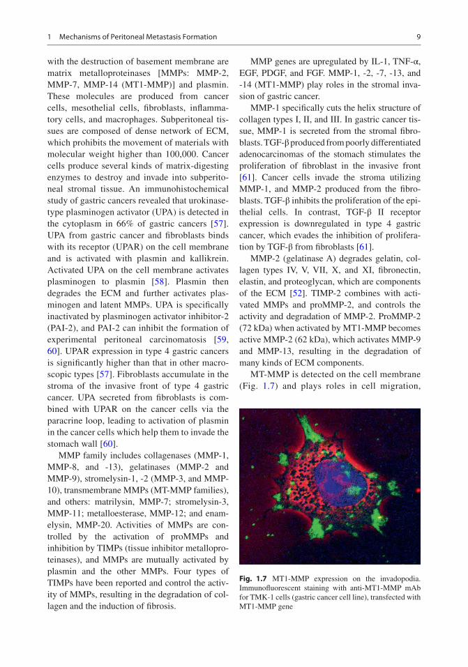
MT-MMP is detected on the cell membrane (Fig. 1.7) and plays roles in cell migration,
Fig. 1.7 MT1-MMP expression on the invadopodia. Immunofluorescent staining with anti-MT1-MMP mAb for TMK-1 cells (gastric cancer cell line), transfected with MT1-MMP gene
1 Mechanisms of Peritoneal Metastasis Formation
10
differentiation, and morphological change by the degradation of the pericellular ECM. The MT-MMP family has 6 kinds of molecules (MT1-6-MMP).
MT1-MMP forms a homo-oligomer on the pseudopodia of cancer cells, and induces an effi-cient invasion by the degradation of their pericel-lular ECM [53]. MT1-MMP itself degrades collagen types I, II, and III, fibronectin, laminin, vitronectin, and aggrecan, and plays a role in the activation of proMMP-2 [54]. TIMP-2 combines with a catalytic domain of MT1-MMP. A com-plex of TIMP-2-MT1-MMP binds with proMMP-2 and forms a tertiary complex. ProMMP-2 becomes an intermediate active MMP-2 by the activation of neighboring MT1- MMP [55]. In poorly differentiated gastric cancers, the MMP-2 secreted from fibroblasts is activated by MT1-MMP. A paracrine loop of MMP-2 from fibroblasts and MT1-MMP on gas-tric cancer induces invasion and metastasis of gastric cancer [56].
MMP-7 (matrilysin) itself degrades collagen types I, II, III, and IV, aggrecan, laminin, and fibronectin, and can activate proMMP-1, -3, -8, and -9 secreted from cancer cells and fibroblasts. As a result, almost all ECM components can be degraded by MMP-7. A study of serial analyses of gene expression of gastric cancer revealed the overexpression of MMP-7 [57]. A highly meta-static cell line (MKN-45-P) on the peritoneal sur-face overexpressed MMP-7 [58]. Intraperitoneal administration of an antisense oligonucleotide against MMP-7 mRNA improved the survival of the mice bearing MKN-45-P [59]. The incidence of MMP-7 protein expression in the type 4 gas-tric cancer is significantly higher than that of the other macroscopic types [43].
MMP-13 is produced from cancer cells and chondrocytes and degrades collagen types I, II, and III. MMP-13 mRNA was expressed in 8 of 9 gastric cancer cell lines, and in these cell lines MMP-13 mRNA was coexpressed with MMP-2 and MT-1 MMP, which activate proMMP-13 [60]. MMP-13 mRNA expression was found in 61% of gastric cancer patients in stage IV disease [62], and the prognosis in patients with MMP-13 over-expressing tumor was significantly poorer than in
those without MMP-13 expression. Patients with tumor expressing both MMP-13 and MT1-MMP showed the worst prognosis [60].
1.2.7 Proliferation in the Subperitoneal Tissue (Fig. 1.1, Process 4: Angiogenesis and Proliferation)
Tyrosine kinases play a major role in the prolif-eration of cancer cells. The interaction of the growth factors with the receptors activates signal-ing pathways and induces mitogenesis. Among these receptors, K-sam, EGFR, MET, vascular endothelial growth factor receptor (VEGFR), and ERBB are frequently involved in PM of various cancers. In gastric cancers, expressions of K-sam, EGFR, MET, and VEGFR are associated with proliferation and angiogenesis.
The K-sam gene encodes the receptors against fibroblast growth factor (FGF) and keratinocyte growth factor (KGF). When the K-sam gene product is activated, the ras-raf-MAP kinase cas-cade is activated and cell proliferation is induced [63]. In type 4 gastric cancer, K-sam gene ampli-fication is a characteristic feature. In the poorly differentiated types of gastric cancer, expression of bFGF for the ligand of K-sam is significantly upregulated, and cancer cell proliferation is stim-ulated by the autocrine or paracrine loop [64]. In an immunohistochemical study of keratinocyte growth factor (KGF) and K-sam expression, the incidence of K-sam expression was significantly higher in type 4 gastric cancer than in the other types. In addition, patients with tumor coexpress-ing K-sam and KGF had significantly poorer prognosis [65]. Accordingly, the paracrine loop of K-sam/KGF/bFGF has an important role in the progression of gastric cancer, especially in poorly differentiated type and type 4 gastric cancer.
The epidermal growth factor receptor (EGFR) and its family of Her-2/ERBB-2, Her-3, and Her-4 are upregulated in 70% of all cancers [66, 67]. Signals of EGFR are transduced through the ras-raf-MAP kinase route, PI3K-Ak route, and Jak-STAT route, and they induce proliferation,
Y. Yonemura et al.

11
growth, and apoptosis. Type 4 gastric cancer co- expresses EGF and EGFR [68].
MET is associated with not only cell scatter-ing, but also proliferation [69]. In gastric cancer, alteration of the domain induces a constant acti-vation of the downstream components [70]. In addition, c-met gene amplification is detected in gastric cancer, and upregulates the signal trans-duction downstream of MET activated in a ligand-dependent or non-ligand-dependent man-ner [71]. Many molecular targeting strategies to decease MET function by controlling Try 1003 have been studied [72, 73].
When the diameter of a cancer nest is greater than 100 μm, oxygen and nutritional supplemen-tation from preexisting blood vessels are not sufficient for survival of cancer cells. Accordingly, cancer cells that are located more than 100 μm apart from blood vessels will die off.
In such a situation, angiogenesis is induced by angiogenic factors secreted from cancer cells, and cancer tissue with newly formed vessels can be established. Proliferating cancer cells upregu-late hypoxia inducible factor (HIF)-1α to induce
angiogenesis [74], and the expression of vascular endothelial growth factor (VEGF) is stimulated [75, 76]. VEGF binds with VEGF receptor-1 and stimulates endothelial cell proliferation. Almost all cancer cells produce VEGF, which has a major role in the establishment of PM [77]. VEGF-C, which is a specific molecule for lymphangiogen-esis, activates VEGFR-3 (flt-4) [64].
1.3 Trans-lymphatic Metastasis
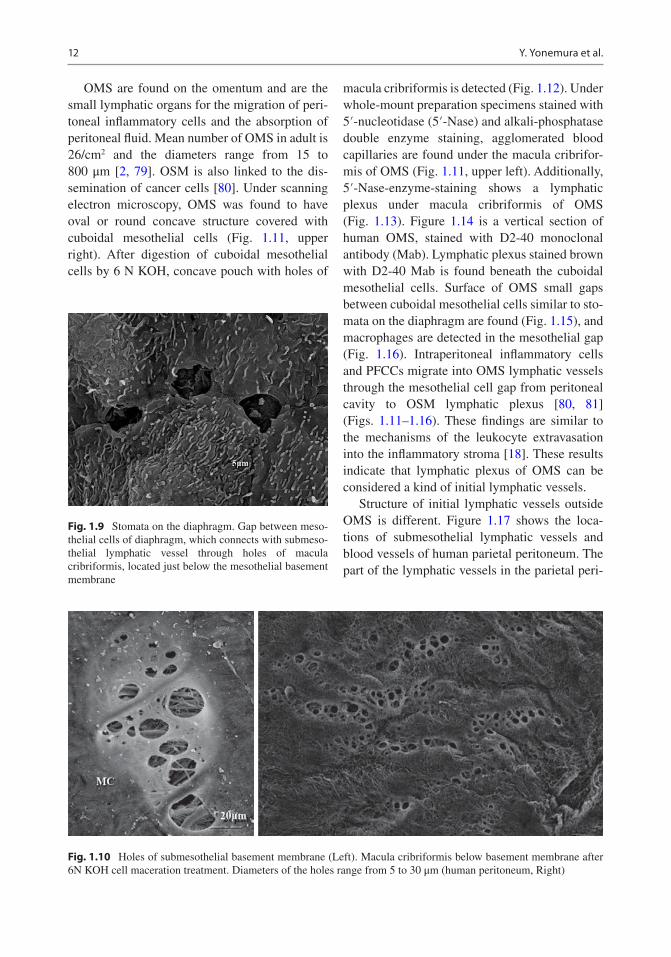
In 2012, Yonemura reported a new concept of PM formation, called trans-lymphatic metastasis [78] (Fig. 1.8). Trans-lymphatic metastasis is the met-astatic pathway by which the PFCCs migrate into the submesothelial initial lymphatic vessels through mesothelial stomata (Fig. 1.9), and holes of macula cribriformis (Fig. 1.10) [78]. Subperitoneal lymphatic vessels associated with trans-lymphatic metastasis are found in omental milky spots (OMS) and initial lymphatic vessels in parietal peritoneum, and small bowel mesentery.
Cribriformplate
Basementmembrane
Initiallymphatic
vessel
Macura cribriformis
stomata
Submesothelial lymphatic vessel
Mesothelial gap, stomata
Bloodvessel
Flat type Protrudedtype
Fig. 1.8 Initial lymphatic vessels, which directly connect with peritoneal cavity via mesothelial stomata and hole of macula cribriformis. Two types of initial lymphatic ves-sels on the parietal peritoneum. Flat type (Left) and pro-truded type (Right). The former is found on the Morrison’s
pouch, paracolic gutter, and small bowel mesentery. The latter is detected on the pelvic peritoneum. PFCCs migrated into initial lymphatic vessels through mesothe-lial stomata and holes of macula cribriformis
1 Mechanisms of Peritoneal Metastasis Formation
12
OMS are found on the omentum and are the small lymphatic organs for the migration of peri-toneal inflammatory cells and the absorption of peritoneal fluid. Mean number of OMS in adult is 26/cm2 and the diameters range from 15 to 800 μm [2, 79]. OSM is also linked to the dis-semination of cancer cells [80]. Under scanning electron microscopy, OMS was found to have oval or round concave structure covered with cuboidal mesothelial cells (Fig. 1.11, upper right). After digestion of cuboidal mesothelial cells by 6 N KOH, concave pouch with holes of
macula cribriformis is detected (Fig. 1.12). Under whole-mount preparation specimens stained with 5′-nucleotidase (5′-Nase) and alkali-phosphatase double enzyme staining, agglomerated blood capillaries are found under the macula cribrifor-mis of OMS (Fig. 1.11, upper left). Additionally, 5′-Nase-enzyme-staining shows a lymphatic plexus under macula cribriformis of OMS (Fig. 1.13). Figure 1.14 is a vertical section of human OMS, stained with D2-40 monoclonal antibody (Mab). Lymphatic plexus stained brown with D2-40 Mab is found beneath the cuboidal mesothelial cells. Surface of OMS small gaps between cuboidal mesothelial cells similar to sto-mata on the diaphragm are found (Fig. 1.15), and macrophages are detected in the mesothelial gap (Fig. 1.16). Intraperitoneal inflammatory cells and PFCCs migrate into OMS lymphatic vessels through the mesothelial cell gap from peritoneal cavity to OSM lymphatic plexus [80, 81] (Figs. 1.11–1.16). These findings are similar to the mechanisms of the leukocyte extravasation into the inflammatory stroma [18]. These results indicate that lymphatic plexus of OMS can be considered a kind of initial lymphatic vessels.
Structure of initial lymphatic vessels outside OMS is different. Figure 1.17 shows the loca-tions of submesothelial lymphatic vessels and blood vessels of human parietal peritoneum. The part of the lymphatic vessels in the parietal peri-
Fig. 1.9 Stomata on the diaphragm. Gap between meso-thelial cells of diaphragm, which connects with submeso-thelial lymphatic vessel through holes of macula cribriformis, located just below the mesothelial basement membrane
Fig. 1.10 Holes of submesothelial basement membrane (Left). Macula cribriformis below basement membrane after 6N KOH cell maceration treatment. Diameters of the holes range from 5 to 30 μm (human peritoneum, Right)
Y. Yonemura et al.

13
toneum that are attached to macula cribriformis and mesothelial stomata are called initial lym-phatic vessels (Fig. 1.8). There are two types of initial lymphatic vessels, i.e., flat type (Fig. 1.8, left, and Fig. 1.18) and protruded type (Fig. 1.8, right, and Fig. 1.19). The former type is found on the Morrison’s pouch, paracolic gutter, and small bowel mesentery. After intraperitoneal injection of activated carbon CH40 [82], the tip of flat type of initial lymphatic vessel alone is stained with CH40. The blind-looped lymphatic vessels extending from the submesothelial lymphatic vessels are the protruded type, and their blind tips attach to the holes of macula cribriformis and sto-mata (Fig. 1.8, right, Fig. 1.19). Since there is no adhesion of CH40 except at the tip of initial lymphatic vessel, the tips of initial lymphatic
vessels alone are considered to communicate with peritoneal cavity. The size of the holes of macula cribriformis ranges from 5 to 30 μm. PFCCs migrate into the initial lymphatic vessels through the stomata on the mesothelial surface without destruction of macula cribriformis (Fig. 1.20) and then proliferate in the lymphatic vessels (Fig. 1.21).
The triplet structure consisting of mesothelial stomata, hole of macula cribriformis, and initial lymphatic vessels is essential for the migration of PFCCs into the submesothelial lymphatic vessels (Fig. 1.22). When the stomata, hole of macula cribriformis, and the tip of initial lymphatic ves-sels are aligned in a row, direct communication between peritoneal cavity and submesothelial lymphatic vessels is established, resulting in the
5’-Nase-ALPaseALPase-positive arterial blood
capillaries (blue stain)OMS
Peritoneal cavity
MS: Mesothelial stomata
Initial lymphatics (IL)
Omental foramen
OMS
OMS
Cuboidalmesothelial
cells
Submesothelialcollagen
plate (SMCP)
CP: glomerulararterial capillary
Flattermesothelial
cells
Cuboidalmesothelial
cells
On removalof OMS cellelements
Maculacribriformis-like
foramens
Submesothelialcollagen
plate
SMCP
LyLyLyLy
Ly
BVBV
MS
BV
CP
ILILIL
Fig. 1.11 Omental milky spot stained with 5′-nucleotid-ase and alkali-phosphatase double enzyme staining (Upper left), electron microscopic finding (Upper right), and the schema of the structure. OMS is oval or round
concave, and the cuboidal mesothelial cells cover the basement membrane of the bottom. Lymphatic plexus (red) is found below the macula cribriformis
1 Mechanisms of Peritoneal Metastasis Formation
14
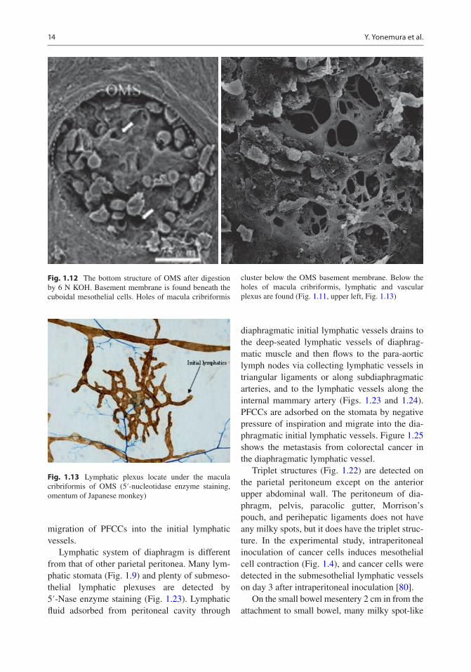
Fig. 1.12 The bottom structure of OMS after digestion by 6 N KOH. Basement membrane is found beneath the cuboidal mesothelial cells. Holes of macula cribriformis
cluster below the OMS basement membrane. Below the holes of macula cribriformis, lymphatic and vascular plexus are found (Fig. 1.11, upper left, Fig. 1.13)
Fig. 1.13 Lymphatic plexus locate under the macula cribriformis of OMS (5′-nucleotidase enzyme staining, omentum of Japanese monkey)
migration of PFCCs into the initial lymphatic vessels.
Lymphatic system of diaphragm is different from that of other parietal peritonea. Many lym-phatic stomata (Fig. 1.9) and plenty of submeso-thelial lymphatic plexuses are detected by 5′-Nase enzyme staining (Fig. 1.23). Lymphatic fluid adsorbed from peritoneal cavity through
diaphragmatic initial lymphatic vessels drains to the deep-seated lymphatic vessels of diaphrag-matic muscle and then flows to the para-aortic lymph nodes via collecting lymphatic vessels in triangular ligaments or along subdiaphragmatic arteries, and to the lymphatic vessels along the internal mammary artery (Figs. 1.23 and 1.24). PFCCs are adsorbed on the stomata by negative pressure of inspiration and migrate into the dia-phragmatic initial lymphatic vessels. Figure 1.25 shows the metastasis from colorectal cancer in the diaphragmatic lymphatic vessel.
Triplet structures (Fig. 1.22) are detected on the parietal peritoneum except on the anterior upper abdominal wall. The peritoneum of dia-phragm, pelvis, paracolic gutter, Morrison’s pouch, and perihepatic ligaments does not have any milky spots, but it does have the triplet struc-ture. In the experimental study, intraperitoneal inoculation of cancer cells induces mesothelial cell contraction (Fig. 1.4), and cancer cells were detected in the submesothelial lymphatic vessels on day 3 after intraperitoneal inoculation [80].
On the small bowel mesentery 2 cm in from the attachment to small bowel, many milky spot- like
Y. Yonemura et al.

15
Macrophagex with activated carbon andcuboidal mesothelial cells
Lymphaticplexus ofOMS
Flat mesothelial cells
Fig. 1.14 Human OMS stained with D2-40 monoclonal antibody. Lymphatic plexus (brown) are found beneath the cuboidal mesothelial cells
Fig. 1.15 SEM findings of the surface of human OMS. Gaps (stars) between cuboidal mesothelial cells, and the gaps connect with macula cribriformis and initial lymphatic vessels
Fig. 1.16 Macrophage is found in the gap (mesothelial stoma) between cuboidal mesothelial cells
Blood vessels
Initial lymphatic vessel (brown)Mesothelial cells
BPB#
Fig. 1.17 Normal structure of human Morrison’s pouch stained by D2-40 monoclonal antibody. Initial lymphatic vessels attached to mesothelial cell gap. Blood vessels locate in the deeper subperitoneal tissue than lymphatic vessels. BPB blood peritoneal barrier
1 Mechanisms of Peritoneal Metastasis Formation

16
ジMacrophage (CH40)
Fig. 1.18 Flat type of initial lymphatic vessel of Morrison’s pouch. Initial lymphatic vessel is stained black with CH40 (activated carbon), introduced intraperitone-ally before sampling. There is no CH40 attachment on the
subperitoneal lymphatic plexus except for flat type initial lymphatic vessel. Accordingly, the tip of lymphatic vessel stained with CH-40 is considered to contact with perito-neal cavity
carbon particlesFig. 1.19 Protruded type of initial lymphatic vessel found in pelvic peritoneum. Tip of initial lymphatic vessel is stained with CH40, introduced intraperitoneally before sampling. There is no adsorption of CH40 except at the tip of initial lymphatic vessel (Left). CH40 particles adhere on the junction between lymphatic mesothelial cells
Fig. 1.20 SEM findings of peritoneal free cancer cells from gastric cancer migrate into initial lymphatic vessels through the holes of macula cribriformis
Y. Yonemura et al.

17
Initial lymphatic vessel
リンパ管
Initial lymphatic vessel
Fig. 1.21 Findings of immunohistochemical staining using D2-40 monoclonal antibody for pelvic peritoneum from patients with gastric cancer. Gastric cancer cells pro-
liferate in the flat type (Left) and protruded type of initial lymphatic vessels (Right)
Lymphatic vessel
Mesothelial layer
Mesothelial stomata
Macula cribriformis
Colagen fiber plate
Lymphatic anchorringfilament
Fig. 1.22 Schema of stomata, macula cribriformis, and initial lymphatic vessels. When the stomata, hole of macula cribriformis, and the tip of initial lymphatic vessels are aligned in a row, direct communication between peritoneal cavity and submesothelial lymphatic vessels is established, resulting in the migration of PFCCs into the initial lymphatic vessels
Anterior abdominalWall*
Right diaphragm
Morrison’s pouch
Right diaphragm
Anterior abdominal wall
Fig. 1.23 Metastatic nodules are found on the right dia-phragm and Morrison’s pouch, but are not detected on the anterior abdominal wall (Left, star). Diaphragmatic lym-phatic network stained with 5′-Nase enzyme staining
(Right). Lymphatic plexus is scarce on anterior abdominal wall, but plenty of lymphatic plexuses are detected on the subdiaphragmatic surface
1 Mechanisms of Peritoneal Metastasis Formation

18
Abdominal cavity
Subperitonealinitial lymphaticvessel
Diaphragmatic muscle
Lymphaticstomata
D2-40 staining
Subperitoneal initiallymphatic vessel
Holes of maculacribriformis
Subperitonealinitial lymphatics
Lymphatic/mesothelial stoma Mesothelial layer
Mesothelial gap
Subperitonealcollagen plateLVLV
Diaphragmaticmuscular layer
Subpleural collagenplate
Pleural esotheliallayer
Fig. 1.24 Diaphragmatic lymphatic system. Left: 5′-Nase enzyme staining shows the connection of holes of macula cribriformis and diaphragmatic initial lymphatic vessels. Middle: Schema of diaphragmatic lymphatic sys-tem. Mesothelial gaps that do not connect with initial lym-phatic vessel are not called stomata, but those that communicate with initial lymphatic vessels are named
mesothelial stomata. Right: Immunohistochemical stain-ing using D2-40 monoclonal antibody shows the lym-phatic stomata and diaphragmatic initial lymphatic vessels. PFCCs are adsorbed through the stomata by nega-tive pressure of inspiration and migrate into the diaphrag-matic initial lymphatics
Lymphaticvessels
Fig. 1.25 Lymphatic metastasis from colorectal cancer in diaphragmatic lymphatic vessel (immunohistochemical staining using D2-40 monoclonal antibody)
structures are found. On this special peritoneal area, round or oval shaped structures covered with cuboidal mesothelial cells are detected by SEM (Fig. 1.26). Below the cuboidal mesothelial cells, macula cribriformis is detected (Fig. 1.26, mid-dle). Since the area is frequently involved in PM, and CH40 injected into peritoneal cavity adheres on the area, absorption of CH40 by initial lym-phatic vessels is suggested. These results indicate
that the metastasis in the area must be involved by trans-lymphatic metastasis (Fig. 1.26). Trans-lymphatic metastasis is found in gastric, colorec-tal, and pancreas cancer.
However, lymphatic system in peritoneum covering the rectus abdominis muscle between hypochondrium and semilunar arc is quite differ-ent from that of other parts of peritoneal surface. In this area, no initial lymphatic vessels or submesothelial lymphatic plexuses are detected. Lymphatic vessels locate in deep subperitoneal tissue 200 μm from the peritoneal surface (Fig. 1.27), and the blood vessels are also scarce. Accordingly, trans-lymphatic metastasis does not develop in the area. The peritoneal area must be involved at the late stage of PM and should be preserved when there is no macroscopic involve-ment on the sector.
1.4 Mechanisms of Superficial Growing Metastasis
PFCCs from appendiceal mucinous neoplasm (AMN) cannot metastasize through trans- mesenteric or trans-lymphatic metastasis, because
Y. Yonemura et al.

19
CH44
Fig. 1.26 Milky spot-like structure detected on the small bowel mesentery in 2 cm from the attachment to small bowel (Left). Below the cuboidal mesothelial cells, holes of macula cribriformis are detected by SEM (Middle).
CH40 injected into peritoneal cavity adheres on the peri-toneal area, suggesting absorption of CH40 by initial lym-phatic vessels (Right)
200mm
Fig. 1.27 Lymphatic vessels of anterior abdominal wall between hypochondrium and semilunar arc. Lymphatic vessels are located 200 μm from the peritoneal surface (Left). Lymphatic vessels and blood vessels in falciform ligament located just below the mesothelial cells (Right). Upper left: Lymphatic vessels of anterior abdominal wall
stained with D2-40 monoclonal antibody. Upper right: Lymphatic vessels of falciform ligament, stained with D2-40 monoclonal antibody. Lower left: Blood vessels of anterior abdominal wall, stained with CD31 monoclonal antibody. Lower right: Blood vessels of falciform liga-ment, stained with CD31 monoclonal antibody
PFCCs of AMN are large and covered with muci-nous material (Fig. 1.28). They cannot migrate into the submesothelial tissue or initial lymphatics (Fig. 1.29).
However, AMN can establish PM in depen-dent areas such as on the pelvis, subdiaphrag-matic surface, and greater omentum. They also grow in the pocket-like structure of omental
1 Mechanisms of Peritoneal Metastasis Formation

20
bursa (inferior and superior recess of omental bursa), intersigmoid recesses, recesses in duodeno- jejunal folds, and ileocecal fossa.
A large volume of mucinous materials with tumor cells accumulates on the dependent perito-neal parts as a result of peritoneal fluid resorption by the negative pressure of initial lymphatic ves-sels and/or gravity [79, 83].
PFCCs of AMN attach on the pelvic perito-neal surface by gravity or by the interaction of adhesion molecules on the mesothelial cells and mucinous materials. As shown in Fig. 1.30, muci-nous material attaches on the paravesical fossa, and immunohistochemical staining with CD31
shows newly formed vasculature in the mucinous material without epithelial cells. These results strongly suggest that angiogenesis factors released from mucinous materials induce angio-genesis in the mucinous stroma.
Figure 1.31 shows the metastasis on the sur-face of ovary by AMN. HE staining shows low- grade mucinous neoplasm growing on the surface of ovary (Fig. 1.31, upper left). Angiogenesis from the preexisting ovarian vasculature and epi-thelial cells in the proliferating phase (positive stain by MIB-1 antibody) are found.
On the omental surface, PFCCs with muci-nous materials from AMN are adsorbed on OMS,
Subperitoneallymphatic vessel
Blood vessel
Initiallymphaticvessel
Blood-peritonealbarrier(90 mm)
Adsorption of peritoneal free cancer cellsfrom appendiceal mucinous neoplasm
Fig. 1.29 Mechanism of superficial growing metastasis. Peritoneal free cancer cells from appendiceal mucinous neoplasm attach on the pelvic peritoneum by the interaction of mucinous material and adhesion molecules (CD44) expressed on mesothelial cells and/or by gravity
Fig. 1.28 Peritoneal free cancer cells of appendiceal mucinous neoplasm. Neoplastic cells are covered with mucinous material and the diameter is several hundred micrometers (Left, Alcian blue staining). Neoplastic
cells show high proliferative activity (Right, Immunohistochemical staining using MIB-1 monoclo-nal antibody)
Y. Yonemura et al.

21
Fig. 1.30 Mucinous material without epithelial cells accumulates on paravesical fossa (Left). HE staining of the vertical section of the bar in left photograph (Middle).
Immunohistochemical staining using anti-CD31 mono-clonal antibody shows newly formed vasculature (Right)
Fig. 1.31 Superficial growing metastasis on ovarium from appendiceal mucinous neoplasm (AMN) (Upper left). AMN growing on the surface of ovary with production of mucinous material, and the neoplastic cells grow on the ovarian surface showing pushing invasion into the corpus of
ovary (Upper right). Newly formed vasculatures from pre-existing ovarian blood vessel are found (Immunohistological staining (HIS) using anti-CD31 Mab) (Left lower). Right lower photograph shows proliferative activities of tumor cells (IHS using MIB-1 mAb)
and many flat mucinous spots are found on the OMS (Fig. 1.32, left). HE staining shows three layers, consisting of a metastatic layer, inflamma-tory layer between metastatic layer and omen-
tum, and normal omentum (Fig. 1.32, middle, and Fig. 1.33). Inflammatory layer shows CD34- positive interstitial tissues. CD34 is a glycopro-tein expressed on interstitial stem cells and
1 Mechanisms of Peritoneal Metastasis Formation