Encapsulating Peritoneal Sclerosis...
26
Encapsulating Peritoneal Sclerosis (EPS) Joni H. Hansson 1 Scott F. Cameron 1 Zenon Protopapas 1 Rajnish Mehrotra 2 1 Hospital of Saint Raphael/Yale University, New Haven, CT 2 Harbor-UCLA Medical Center, Torrance, CA
Transcript of Encapsulating Peritoneal Sclerosis...

Encapsulating Peritoneal Sclerosis
(EPS)
Joni H. Hansson1
Scott F. Cameron1
Zenon Protopapas1
Rajnish Mehrotra2
1Hospital of Saint Raphael/Yale University, New Haven, CT2Harbor-UCLA Medical Center, Torrance, CA

EPS: Outline
• Definition and epidemiology• Clinical presentation• Imaging • Pathology• Treatment• Outcomes• Summary

Encapsulating Peritoneal Sclerosis (EPS)
• Rare complication but serious complication of peritoneal dialysis (PD):
– First described in 1980 (1)– Incidence varies between 0.5 to 4.4% (2-8)– Associated with significant morbidity and mortality– Many present after PD has been discontinued– The diagnosis requires both the:
• Clinical features of intestinal obstruction or disturbed gastrointestinal function
• Evidence of bowel encapsulation either radiologically or pathologically

Incidence of EPS
SCOTLAND1238 pts (1/1/2000-12/31/2007)
46 cases8-year cumulative incidence, 8.1%
Australia and New Zealand7618 pts (1/1/1995-12/31/2007)
33 cases8-year cumulative incidence, 3.9%Brown et al, Clin J Am Soc Nephrol 2009; 4: 1222-9
Johnson et al, Kidney Int 2010; 77: 904-12

EPS: Risk Factors• The only consistent risk factor is duration on
peritoneal dialysis
• Other potential factors– Glucose exposure– Inflammation – peritonitis– Chemical exposure
• chlorhexidine
– Genetic factors– Various other factors unrelated to peritoneal dialysis

EPS: Clinical Manifestations• Symptoms of bowel obstruction
– Persistent/intermittent– Partial/complete – Abdominal pain/distention/nausea/vomiting
• Abdominal mass
• Hemoperitoneum
• Malnutrition and failure to thrive
• Ultrafiltration problems
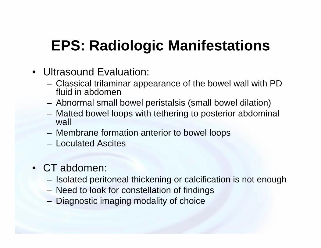
EPS: Radiologic Manifestations• Ultrasound Evaluation:
– Classical trilaminar appearance of the bowel wall with PD fluid in abdomen
– Abnormal small bowel peristalsis (small bowel dilation)– Matted bowel loops with tethering to posterior abdominal
wall– Membrane formation anterior to bowel loops– Loculated Ascites
• CT abdomen:– Isolated peritoneal thickening or calcification is not enough– Need to look for constellation of findings– Diagnostic imaging modality of choice

CT Scan Findings in EPSTarzi et al Vlijm et al
Peritoneal thickeningPeritoneal calcification
Bowel wall thickeningBowel tetheringBowel dilatation
Loculation (ascites)
Peritoneal thickeningPeritoneal calcificationPeritoneal enhancement
Adhesions of bowel loops
Signs of bowel obstructionFluid loculation/septation
CT score 0-4 for each except bowel tethering and loculation (scored 0-3); range 0-22
Presence of three of above six items
Median score: pts, 9; PD controls, 1; HD, 0
Sensitivity, 79-100%; Specificity, 88-94%
Tarzi et al, Clin J Am Soc Nephrol 2008, 3: 1702-10Vlijm et al, Perit Dial Int 2009; 29: 517-22

CT Findings of EPS
Thickened EnhancingPeritoneum (arrows)
Loculated FluidCollection (arrows)
Tethered small bowelLoops (arrows)
Cameron et al: American Roentgen Ray Society Annual Meeting, 2010

CT Findings of EPS
Dilated and thick-walled loops of small bowel filled with fluid and oral contrast (arrows).
Peritoneal thickening (arrow).
Loculated fluid collections (arrows).
Cameron et al: American Roentgen Ray Society Annual Meeting, 2010

CT Findings of EPS
Bowel Tethering (arrow)
Peritoneal calcification (arrow)
Cameron et al: American Roentgen Ray Society Annual Meeting, 2010

CT Findings of EPS
Vlijm et al, Perit Dial Int 2009; 29: 517-22
Tethered bowelLoops (arrows)
Peritoneal Enhancement (arrow)

Gross Pathologic Findings of EPS
Peritoneal thickening (arrow)
Peritoneal inflammatory changes (arrow)
Peritoneal calcifications (arrow)
Omental fat (arrow)
Cameron et al: American Roentgen Ray Society Annual Meeting, 2010

Histology of EPSPeritoneal thickening and fibrosis (arrows)Peritoneal calcification (arrow)Inflammatory cells -neutrophils/lymphocytes (arrow)
Cameron et al: American Roentgen Ray Society Annual Meeting, 2010
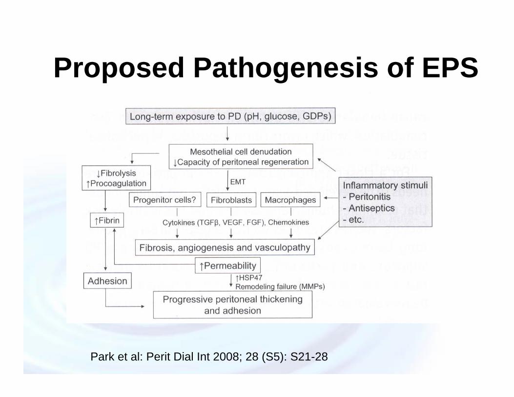
Proposed Pathogenesis of EPS
Park et al: Perit Dial Int 2008; 28 (S5): S21-28

EPS: Management
• Supportive therapy (ileus):– Aggressive nutritional support
• Medical Therapy:– Tamoxifen (anti-fibrotic)– Immunosuppressive (anti-inflammatory)
• Surgical Intervention (encapsulating):– Partial or complete enterolysis,– Avoid enterotomy
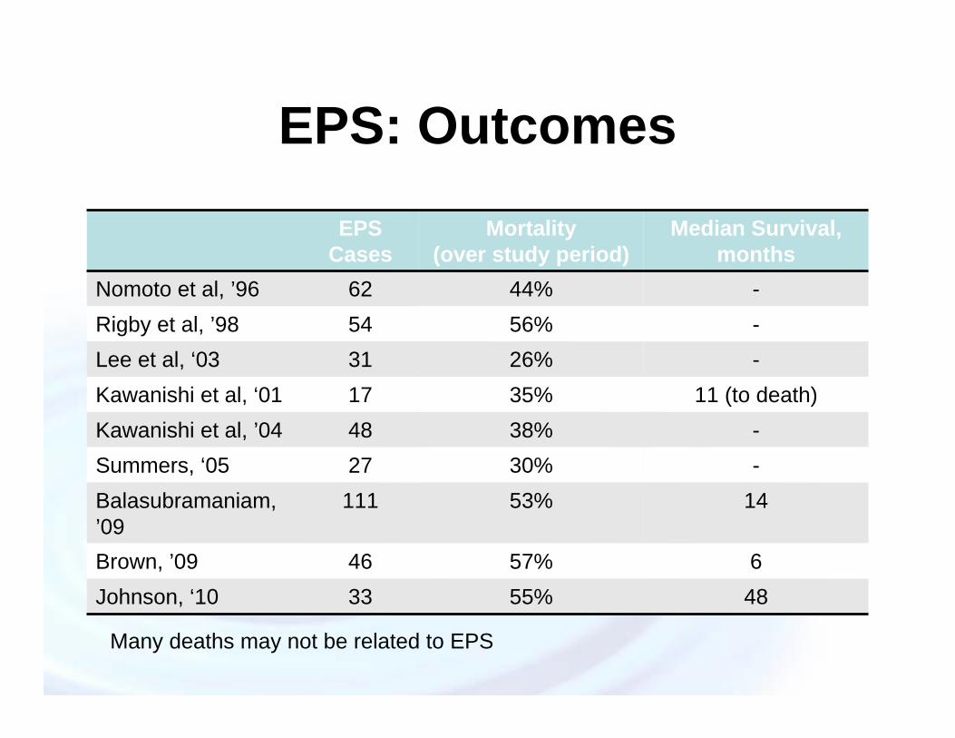
EPS: OutcomesEPS
CasesMortality
(over study period)Median Survival,
monthsNomoto et al, ’96 62 44% -Rigby et al, ’98 54 56% -Lee et al, ‘03 31 26% -Kawanishi et al, ‘01 17 35% 11 (to death)Kawanishi et al, ’04 48 38% -Summers, ‘05 27 30% -Balasubramaniam, ’09
111 53% 14
Brown, ’09 46 57% 6Johnson, ‘10 33 55% 48
Many deaths may not be related to EPS

EPS: Prevention• No prospective data supporting a benefit of
preemptively transferring long-term PD patients to HD
• Preserve residual renal function
• Try to minimize the use of high-glucose PD solutions
• Minimize episodes of peritonitis
• Await experience with “biocompatible” PD solutions

EPS: Summary• Rare:
– Most long-term patients will not develop the complication– Mandatory transition off PD after a pre-determined interval does
not seem justified
• Diagnosis requires:– high index of clinical suspicion from clinical manifestations– constellation of CT or pathologic findings consistent with EPS
• Aggressive nutritional support, trial of medical therapy, and surgery in selected cases
• Outcomes better in contemporary cohorts?

EPS: References1. Gandhi et al: Sclerotic thickening of the peritoneal membrane in Maintenance
peritoneal dialysis patients. Arch Int Med 1980; 140:1201-1203.2. Kawaguchi et al: Encapsulating Peritoneal Sclerosis: Definition, Etiology,
Diagnosis, and Treatment. Perit Dial Int 2000; 20 (suppl 4): S43-55.3. Rigby et al: Sclerosing peritonitis: the experience in Australia. Nephrol Dial
Transplant 1998; 13: 154-159.4. Johnson et al: Encapsulating peritoneal sclerosis: incidence, predictors, and
outcomes. Kidney Int 2010; 77: 901-912.5. Kawanishi H: Encapsulating Peritoneal Sclerosis in Japan: Prospective Multicenter
Controlled Study. Perit Dial Int 2001; 21 (suppl 3): S67-S71.6. Kawanishi et al: Encapsulating Peritoneal Sclerosis in Japan: A Prospective,
Controlled, Multicenter Study. Am J Kidney Dis 2004; 44: 729-737.7. Lee et al: Sclerosing encapsulating peritonitis as a complication of long-term
continuous ambulatory peritoneal dialysis in Korea. Nephrology 2003; 8 (suppl) S33-S39.
8. Brown et al: Length of Time on Peritoneal Dialysis and Encapsulating Peritoneal Sclerosis: Position Paper for ISPD. Perit Dial Int 2009; 29: 595-600
9. Schmidt et al: Pathogenesis and Treatment of Encapsulating Peritoneal Sclerosis: Basic and Translational Research. Perit Dial Int 2008; 28 (suppl 5) S10-S15.
10. Brown et al: Encapsulating Peritoneal Sclerosis in the New Millennium: A National Cohort Study. Clin J Am Soc Nephrol 2009; 4: 1222-1229.

11. Kawanishi et al: Treatment options for encapsulating peritoneal sclerosis based on progressive stage. Adv Perit Dial 2001; 17: 200-4.
12. Tarzi et al: Assessing the Validity of an Abdominal CT Scoring System in the Diagnosis of Encapsulating Peritoneal Sclerosis. Clin J Am Soc Nephrol 2008; 3: 1702-1710.
13. Vlijm et al: Computed Tomographic Findings Characteristic for Encapsulating Peritoneal Sclerosis: A Case-Control Study. Perit Dial Int 2009; 29: 517-522
14. Gayomali et al: Incidence of Encapsulating Peritoneal Sclerosis: A Single-Center Experience with Long Term Peritoneal Dialysis in the United States. Perit Dial Int in press.
15. Cameron et al: Encapsulating Peritoneal Sclerosis – Imaging findings behind an under recognized complication of long-term peritoneal dialysis. American Roentgen Ray Society Annual Meeting 2010.
16. Honda et al: Pathology of Encapsulating Peritoneal Sclerosis. Perit Dial Int 2005; 25 (suplp 4) S19-S29.
17. Dobbie et al: Long-term effects of peritoneal dialysis on peritoneal morphology. Perit Dial Int 1994; 14 (suppl 3): S16-S20.
18. Park et al: Experimental Encapsulating Peritoneal Sclerosis Models: Pathogenesis and Treatment. Perit Dial Int 2008. 28 (suppl 5:) S21-S28.
19. Kawanishi et al: Recommendation of the Surgical Option for Treatment of Encapsulating Peritoneal Sclerosis. Perit Dial Int 2008; 28(suppl 3): S205-S210.
20. Kawanishi et al: Successful Surgical Management of Encapsulating Peritoneal Sclerosis. Perit Dial Int 2005; 25 (suppl 4): S39-S47.

21.Nomoto et al: Sclerosing Encapsulating Peritonitis is Patients Undergoing Continuous Ambulatory Peritoneal Dialysis: A Report of the Japanese Sclerosing Encapsulating Peritonitis Study Group. Am J Kidney Dis 1996; 28: 420-427.
22.Summers et al: Single-center experience of encapsulating peritoneal sclerosis in patients on peritoneal dialysis for end-stage renal failure. Kidney Int 2005; 68: 2381-2388.
23.Balasubramaniam et al: The Pan-Thames EPS study: treatment and outcomes of encapsulating peritoneal sclerosis. Nephrol Dial Transplant 2009; 24: 3209-3215.
24.Nakamoto H: Encapsulating Peritoneal Sclerosis – A Clinician’s Approach to Diagnosis and Medical Treatment. Perit Dial Int 2005; 25 (suppl 4): S30-S38.
25.Kawaguchi et al: Recommendations of the Management of Encapsulating Peritoneal Sclerosis in Japan, 2005: Diagnosis, Predictive Mardkers, Treatment, and Preventive Measures. Perit Dial Int 2005; 25 (suppl 4): S83-S95.
26.Woodrow et al: UK Encapsulating Peritoneal Sclerosis Clinical Practice Guidelines – July 2009. Produced by UK EPS Clinical Guidelines Group.
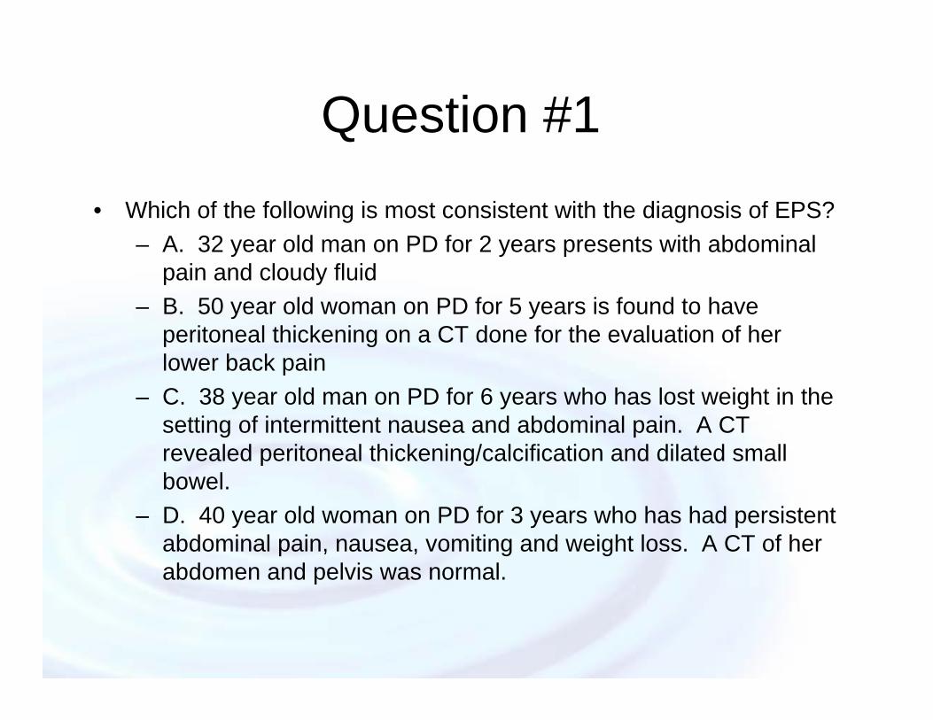
Question #1• Which of the following is most consistent with the diagnosis of EPS?
– A. 32 year old man on PD for 2 years presents with abdominal pain and cloudy fluid
– B. 50 year old woman on PD for 5 years is found to have peritoneal thickening on a CT done for the evaluation of her lower back pain
– C. 38 year old man on PD for 6 years who has lost weight in thesetting of intermittent nausea and abdominal pain. A CT revealed peritoneal thickening/calcification and dilated small bowel.
– D. 40 year old woman on PD for 3 years who has had persistent abdominal pain, nausea, vomiting and weight loss. A CT of her abdomen and pelvis was normal.

Question #1: Answer
• The correct answer is C. The diagnosis of EPS requires both the clinical features of the disorder and evidence of bowel encapsulation either radiographically or pathologically.
Question #2
• Which is the most consistent risk factor for the development of EPS?– A. Rapid transport status– B. Recurrent peritonitis– C. Ultrafiltration problems– D. Length of time on PD
Question #2: Answer
• The correct answer is D. The only consistent risk factor for EPS is duration on PD.