Pathology of Endosulfan - InTech -...
20
15 Pathology of Endosulfan Ozlem Ozmen University of Mehmet Akif Ersoy Turkey 1. Introduction Endosulfan (6,7,8,9,10,10-hexachloro-1,5,5a,6,9,9a-hexahydro- 6,9-methano-2,4,3-benzo-o- dioxa-thiepin-3-oxide) is a chlorinated cyclodiene insecticide which acts as a contact poison in a wide variety of insects and mites (Naqvi and Vaishnavi, 1993). Endosulfan was first registered for use in the USA in 1954 to control agricultural insects and mite pests. Due to its toxic effect, the World Health Organization (WHO) has classified Endosulfan as a moderately hazardous Class II pesticide (WHO, 2002). Endosulfan is a persistent organic pollutant. The half-life of endosulfan in water varies from 3 to 7 days to about 5 months, depending on the dissolved oxygen, turbidity, pH and other contaminants in the water. This insecticide is a mixture of two stereoisomers, namely ǂ- and ǃ- endosulfan (Hayes and Laws, 1991), in a ratio of 7:3. It has been used worldwide in agriculture, viticulture and horticulture (Hack et al., 1995; Oktay et al., 2003; Mor and Ozmen, 2003; Yavuz et al., 2007). Endosulfan can cause toxic effects in almost all tissues of both humans and animals, including the liver, lung, central nervous system, genital system, pancreas etc. (Howard, 1991, Mor and Ozmen, 2003; Kalender et al., 2004b; Hatipoglu et al., 2009). It is also effect blood biochemistry and hematological values (Hatipoglu et al., 2009). Endosulfan is a contact hepatotoxin that is readily absorbed into an organism through its stomach, lung and even through the skin (Howard, 1991). The primary purpose of this chapter is to provide pathological findings in endosulfan toxicity in animals and human. It contains descriptions and evaluations of pathological studies about endosulfan. Gross and histopathological lesions are described in different kind of animals and human in experimental and natural toxication cases. 2. Nervous system toxicity Clinical signs, such as depression, inappatence and slight nervous symptoms such as teeth grinding and hyperexcitability reported in the rabbits suffer from subacute endosulfan toxication (Mor and Ozmen, 2010a; Mor and Ozmen, 2010b). In acute toxication by endosulfan in cattle cause rapid and difficult breathing, foamy exudates in mouth, tremors, exophtalmos, coma and death (Mor and Ozmen, 2003). At the gross examination of the brains, marked hyperemia at the meningeal vessels and slight hemorrhages in brains and cerebellums in rabbits suffer from endosulfan poisoning were reported. The occurring of findings is prominent in rabbits that had shown clinical nervous symptoms (Mor and Ozmen, 2010b). www.intechopen.com
Transcript of Pathology of Endosulfan - InTech -...

15
Pathology of Endosulfan
Ozlem Ozmen University of Mehmet Akif Ersoy
Turkey
1. Introduction
Endosulfan (6,7,8,9,10,10-hexachloro-1,5,5a,6,9,9a-hexahydro- 6,9-methano-2,4,3-benzo-o-
dioxa-thiepin-3-oxide) is a chlorinated cyclodiene insecticide which acts as a contact poison
in a wide variety of insects and mites (Naqvi and Vaishnavi, 1993). Endosulfan was first
registered for use in the USA in 1954 to control agricultural insects and mite pests. Due to its
toxic effect, the World Health Organization (WHO) has classified Endosulfan as a
moderately hazardous Class II pesticide (WHO, 2002). Endosulfan is a persistent organic
pollutant. The half-life of endosulfan in water varies from 3 to 7 days to about 5 months,
depending on the dissolved oxygen, turbidity, pH and other contaminants in the water. This
insecticide is a mixture of two stereoisomers, namely ┙- and ┚- endosulfan (Hayes and
Laws, 1991), in a ratio of 7:3. It has been used worldwide in agriculture, viticulture and
horticulture (Hack et al., 1995; Oktay et al., 2003; Mor and Ozmen, 2003; Yavuz et al., 2007).
Endosulfan can cause toxic effects in almost all tissues of both humans and animals,
including the liver, lung, central nervous system, genital system, pancreas etc. (Howard,
1991, Mor and Ozmen, 2003; Kalender et al., 2004b; Hatipoglu et al., 2009). It is also effect
blood biochemistry and hematological values (Hatipoglu et al., 2009). Endosulfan is a
contact hepatotoxin that is readily absorbed into an organism through its stomach, lung and
even through the skin (Howard, 1991).
The primary purpose of this chapter is to provide pathological findings in endosulfan
toxicity in animals and human. It contains descriptions and evaluations of pathological
studies about endosulfan. Gross and histopathological lesions are described in different
kind of animals and human in experimental and natural toxication cases.
2. Nervous system toxicity
Clinical signs, such as depression, inappatence and slight nervous symptoms such as teeth
grinding and hyperexcitability reported in the rabbits suffer from subacute endosulfan
toxication (Mor and Ozmen, 2010a; Mor and Ozmen, 2010b). In acute toxication by
endosulfan in cattle cause rapid and difficult breathing, foamy exudates in mouth, tremors,
exophtalmos, coma and death (Mor and Ozmen, 2003). At the gross examination of the
brains, marked hyperemia at the meningeal vessels and slight hemorrhages in brains and
cerebellums in rabbits suffer from endosulfan poisoning were reported. The occurring of
findings is prominent in rabbits that had shown clinical nervous symptoms (Mor and
Ozmen, 2010b).
www.intechopen.com
Pesticides in the Modern World – Effects of Pesticides Exposure 290
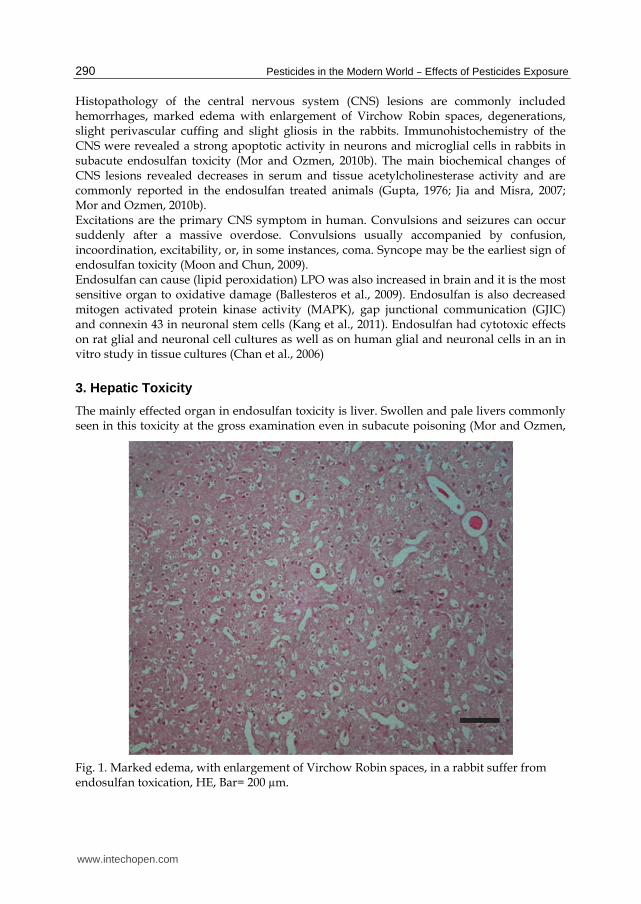
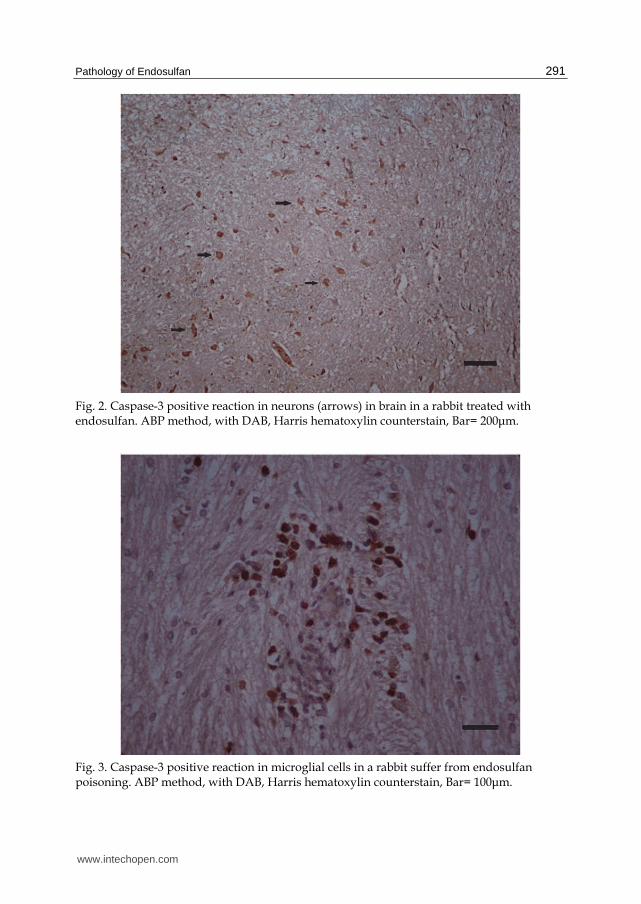
Histopathology of the central nervous system (CNS) lesions are commonly included hemorrhages, marked edema with enlargement of Virchow Robin spaces, degenerations, slight perivascular cuffing and slight gliosis in the rabbits. Immunohistochemistry of the CNS were revealed a strong apoptotic activity in neurons and microglial cells in rabbits in subacute endosulfan toxicity (Mor and Ozmen, 2010b). The main biochemical changes of CNS lesions revealed decreases in serum and tissue acetylcholinesterase activity and are commonly reported in the endosulfan treated animals (Gupta, 1976; Jia and Misra, 2007; Mor and Ozmen, 2010b). Excitations are the primary CNS symptom in human. Convulsions and seizures can occur suddenly after a massive overdose. Convulsions usually accompanied by confusion, incoordination, excitability, or, in some instances, coma. Syncope may be the earliest sign of endosulfan toxicity (Moon and Chun, 2009). Endosulfan can cause (lipid peroxidation) LPO was also increased in brain and it is the most sensitive organ to oxidative damage (Ballesteros et al., 2009). Endosulfan is also decreased mitogen activated protein kinase activity (MAPK), gap junctional communication (GJIC) and connexin 43 in neuronal stem cells (Kang et al., 2011). Endosulfan had cytotoxic effects on rat glial and neuronal cell cultures as well as on human glial and neuronal cells in an in vitro study in tissue cultures (Chan et al., 2006)
3. Hepatic Toxicity
The mainly effected organ in endosulfan toxicity is liver. Swollen and pale livers commonly seen in this toxicity at the gross examination even in subacute poisoning (Mor and Ozmen,
Fig. 1. Marked edema, with enlargement of Virchow Robin spaces, in a rabbit suffer from endosulfan toxication, HE, Bar= 200 µm.
www.intechopen.com
Pathology of Endosulfan 291
Fig. 2. Caspase-3 positive reaction in neurons (arrows) in brain in a rabbit treated with endosulfan. ABP method, with DAB, Harris hematoxylin counterstain, Bar= 200μm.
Fig. 3. Caspase-3 positive reaction in microglial cells in a rabbit suffer from endosulfan poisoning. ABP method, with DAB, Harris hematoxylin counterstain, Bar= 100μm.
www.intechopen.com
Pesticides in the Modern World – Effects of Pesticides Exposure 292
2003; Mor and Ozmen, 2010a). At necropsy, hemorrhages can be seen in livers in acute
poisoning in cattle (Mor and Ozmen, 2003). Liver histology of rabbits suffers from
endosulfan toxication characterized by loss of radial cellular arrangement, hypertrophy of
hepatocytes, significant increase of Kupffer cells, circulatory disturbances, focal necrosis,
fatty degeneration, nuclear pyknosis, narrowing of sinusoids and bile duct hyperplasia.
Hemorrhages and infiltration of inflammatory cells that localized around the central vein
and portal space can be seen. Interlobular mononuclear inflammatory cells among
vacuolated hepatic cells and dilated congested sinusoids are reported. Apoptotic activity in
liver cells increased in livers by endosufan exposure (Mor and Ozmen, 2010a). Liver enzyme
levels are elevated in endosulfan toxicity (Khan et al., 2010). Endosulfan can also cause
catalase (CAT) inhibition and increase of LPO levels in liver (Ballesteros et al., 2009).
Histopathological examinations of liver tissues of long term (180 days) exposure of
endosulfan shows chronic toxic hepatitis in liver in mice. There is portal mononuclear
inflammatory infiltration and some eosinophyl leucocytes, lobulary inflammation and liver
cell necrosis. Generally, any neoplastic and dysplastic changes have not been observed in
liver. Histopathological examinations of liver tissues of short term (90 days) exposure show
some regenerative findings with mild hepatitis. Hepatocytes had more than one nucleus,
nuclear hyperchromasy and minimal microvesiculary fatty degeneration. In addition, crude
glycogen granules in hepatocytes also are reported (Kurutas and Doran, 2001).
Microscopical hepatic lesions of endosulfan poisoning are more severe in diabetic or protein
malnourished rats (Benjamin et al., 2006).
4. Nephrotoxicity
Kidney changes in endosulfan poisoning are dose dependent. Tubular dilation, hydropic
degeneration in tubular epithelium, hemorrhage in the cortical and medulla part of the
kidney were reported (Kayhan et al., 2009). The effect of the endosulfan is mainly on the
proximal convoluted tubule cells (Powers et al 1978; Caglar et al., 2003; Benjamin et al.,
2006). Mitochondrial degeneration, lipofuscin granules and membranous structures in
cytoplasm of proximal convoluted tubule cells were reported in mice suffer from endosulfan
toxication (Caglar et al., 2003). While degenerative changes have been observed in proximal
or distal convoluted tubules; glomerular tuft and Bowman’s capsule are generally normal in
mild endosulfan poisoning in rats. Lesions occur more severe in diabetic and malnourished
rats and became worse related the duration of the toxication. In severely poisoned rats
complete necrosis of tubular epithelium and hemorrhages in glomeruli are prominent.
Increased Bowman’s spaces commonly have been seen in severely affected rats (Benjamin et
al., 2006). There is an increase in the cytoplasmic density of some of the distal convoluted
tubule cells are generally observed. Extenstion in the length of some cells, and cytoplasmic
bulges toward the lumen from the apical cytoplasm are reported. Ultrastructurally fusion in
pedicels and focal thickening at glomerular basal membrane were also reported in some
glomeruli (Caglar et al., 2003).
Renal calcium deposits may be seen in endosulfan toxication in males. The toxic
nephropathy observed in animals was characterized as degenerative changes in the
proximal convoluted tubules at the junction of the cortex and medulla, and associated
cloudy swelling, fatty degeneration, and necrosis of the tubular epithelium (Powers et al
1978). Glucose-6-phosphate dehydrogenase (G6PD), Catalase (CAT), Superoxide Dismutase
www.intechopen.com
Pathology of Endosulfan 293
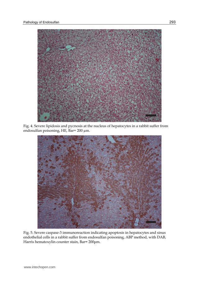
Fig. 4. Severe lipidosis and pycnosis at the nucleus of hepatocytes in a rabbit suffer from endosulfan poisoning, HE, Bar= 200 µm.
Fig. 5. Severe caspase-3 immunoreaction indicating apoptosis in hepatocytes and sinus endothelial cells in a rabbit suffer from endosulfan poisoning, ABP method, with DAB, Harris hematoxylin counter stain, Bar= 200μm.
www.intechopen.com
Pesticides in the Modern World – Effects of Pesticides Exposure 294
(SOD), GSH (gulutatione) and malondi-aldehid (MDA) activities increased in the
endosulfan - treated group kidney tissues. Degeneration and necrosis in kidneys may be
thought that oxidative stress may play a role to the mediator in changing configuration of
cell membrane and seem to account for the morphologic alteration of kidney (Caglar et al.,
2003).
5. Reproductive toxicity
Endosulfan toxicity commonly studied especially in males. Numerous studies have
consistently demonstrated that endosulfan behaves physiologically as an anti-androgen
(Wilson and LeBlanc, 1998). The effects of endosulfan are most pronounced in immature
animals whose reproductive systems and brains are still developing (Sinha et al.1995; Sinha
et al.1997). Studies showed that toxicity can cause morphological and functional changes in
male reproductive system. The main problems are decreased spermatozoon count and
testosterone inhibition (Khan and Sinha, 1996; Esin, 2008; Hatipoglu et al., 2009). In mice,
endosulfan reduces overall sperm count and increases the prevalence of malformed sperm
(Khan and Sinha., 1996). Histologically, numerous seminiferous tubules show significant
decrease to complete spermatogenesis at puberty. This finding can cause the decrease in
daily sperm production observed in the endosulfan-exposed male rats (Dalsenter et al.,
1999). Degenerative areas in testis and decreased number of spermatozoon in seminiferous
tubules are apparent in subacute poisoning in male rabbits (Khan and Sinha, 1996; Esin,
2008; Hatipoglu et al., 2009). Significant decreases in the mean spermatozoon counts and
spermatozoon with abnormal head number (twinheaded) is reported (Khan and Sinha,
1996). A significant elevation in the activities of the enzymes LDH (lactate dehydrogenase),
GGT (gamma glutamyl transpeptidase) and G6PDH (glucose-6-phosphate dehydrogenase is
also observed (Sinha et al., 1997).
Estrogenic effects of endosulfan were conducted an in vivo study of by Raizado et al (1991).
A dose related increase in testicular atrophy occurred in treated male rats, characterized by
degeneration and necrosis of the germinal cells lining the seminiferous tubules,
multinucleated cells (fusion bodies), and calcium deposition resulting in aspermatogenesis.
No treatment related effects were noted on the reproductive organs in female rats (Powers
et al 1978).
Male Wistar prepubertal rats that treated by endosulfan, statistically significant decreases
reported in body, testes, epididymal, ventral prostate and seminal vesicle weights compared
to controls (Chitra et al 1999).
Developmental/reproductive toxicity or endocrine disruption occurs only at doses causing
neurotoxicity. Toxicity to the fetus or young animals is not more severe than that shown by
adults (Silva and Gammon, 2009).
6. Endocrine toxicity
Endosulfan poisoning can cause histological pancreas lesions. The serum amylase levels are
generally normal, whereas the lipase and glucose levels are increased. The histopathological
examinations of the pancreases are indicated that single-cell necrosis and degenerative
changes had occurred in the pancreatic cells; especially in the beta cells, in rabbits suffer
from endosulfan poisoning. Immunohistochemistry of the pancreatic tissues revealed a
www.intechopen.com
Pathology of Endosulfan 295
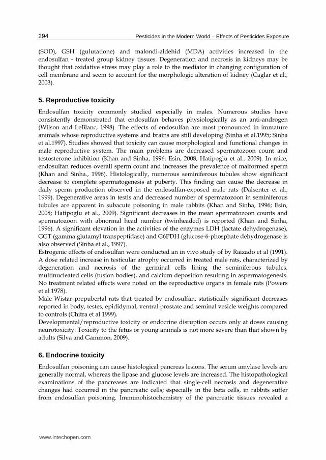
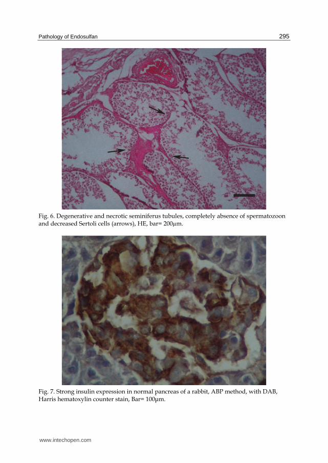
Fig. 6. Degenerative and necrotic seminiferus tubules, completely absence of spermatozoon and decreased Sertoli cells (arrows), HE, bar= 200µm.
Fig. 7. Strong insulin expression in normal pancreas of a rabbit, ABP method, with DAB, Harris hematoxylin counter stain, Bar= 100μm.
www.intechopen.com
Pesticides in the Modern World – Effects of Pesticides Exposure 296
marked reduction in concentration and distribution of insulin, proinsulin, and amylin. The
number of the endocrine cells in pancreas in endosulfan treated rabbits is significantly
decreased (Ozmen et al., 2010). In electron microscopy studies, swelling of mitochondria,
vacuoles in cytoplasm, dissolution of mitochondrial matrix, picnotic nucleus in ┚ cells in
Langerhans islet reported after endosulfan treatment (Kalender et al., 2004a)
Endosulfan poisoning may affect reproductive endocrine hormones. Recent information
indicates that endosulfan mimics non-uterotrophic E(2) actions, strengthening the
hypothesis that endosulfan is a widespread xenoestrogen (Varayoud et al., 2008), acts via a
membrane version of the estrogen receptor-┙ on pituitary cells and can provoke Ca++ influx
via L-type channels, leading to prolactin (PRL) secretion (Watson et al., 2007), and alters
circulating levels of prolactin, luteinizing hormone, growth hormone, and thyroid
stimulating hormone (Caride et al 2010).
In addition to inducing cell proliferation, endosulfan induced proliferation of the
progesterone receptor, another oestrogen-mimicking effect (Soto et al 1995). Parathyroid
hyperplasia occurred in treated males, as did medial calcification of the aorta and medial
calcification of the mesenteric artery, and calcium deposits in the stomach (Powers et al
1978). The adrenals of rabbits given a single dermal dose of 100 mg/kg of endosulfan
exhibited microscopic changes, including swollen cells with foamy cytoplasm and eccentric
nuclei (Gupta and Chandra, 1975).
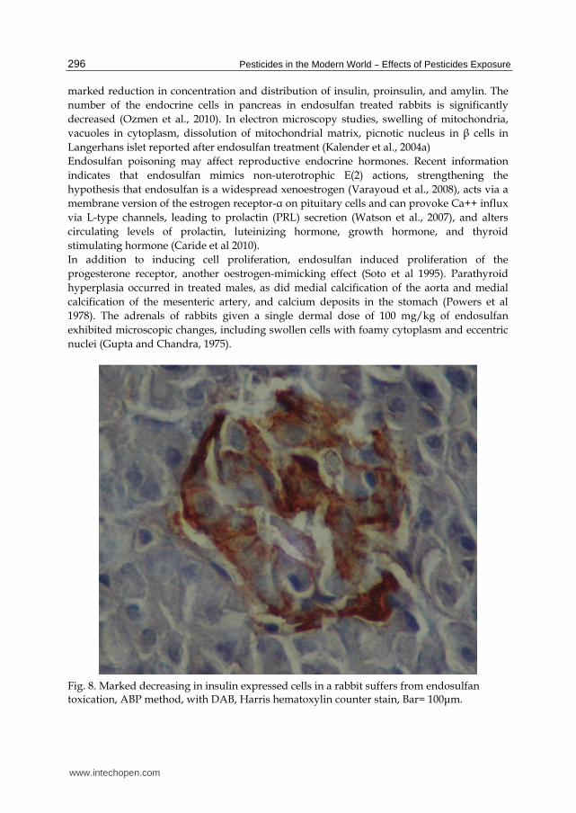
Fig. 8. Marked decreasing in insulin expressed cells in a rabbit suffers from endosulfan toxication, ABP method, with DAB, Harris hematoxylin counter stain, Bar= 100μm.
www.intechopen.com
Pathology of Endosulfan 297
7. Breast toxicity
Microscopic examinations of breast tissues of long term and short term exposure showed
lymphocytic infiltration in stroma of breast tissue. There is no neoplastic and dysplastic
changes in breast in endosulfan administration reported (Kurutas and Doran, 2001).
8. Muscle toxicity
Muscle necroses are reported in rats severely toxicated and under health stress (Benjamin et
al., 2006). Endosulfan can cause inhibitory effect on on skeletal muscle MDH of the
freshwater catfish Clarias batrachus (Misra and Shuckla, 2003).
9. Genotoxicity
Genotoxicity in tests for gene mutation, chromosomal aberration and DNA damage are
reported in endosulfan toxicity (Silva and Beauvais, 2010).
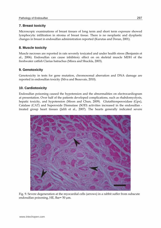
10. Cardiotoxicity
Endosulfan poisoning caused the hypotension and the abnormalities on electrocardiogram
at presentation. Over half of the patients developed complications, such as rhabdomyolysis,
hepatic toxicity, and hypotension (Moon and Chun, 2009). Glutathionperoxidase (Gpx),
Catalase (CAT) and Superoxide Dismutase (SOD) activities increased in the endosulfan -
treated group heart tissues (Jalili et al., 2007). The hearts generally indicated severe
Fig. 9. Severe degeneration at the myocardial cells (arrows) in a rabbit suffer from subacute endosulfan poisoning, HE, Bar= 50 µm.
www.intechopen.com
Pesticides in the Modern World – Effects of Pesticides Exposure 298
congestion, hemorrhages, with interstitial edema. In some places diapedesis of leukocytes
may be seen. Different degrees of degeneration can be seen in myocardium, granular
appearance with picnotic nuclei may observe in some myofibrils. Thickening of wall of
arteries were reported (Jalili et al.2007). In electron microscopic investigations cytoplasmic
edema and swelling and vacuolization of mitochondria of myocardial cells in endosulfan
toxicity may observed (Kalender et al., 2004a). Significantly decreased were reported in
serum calcium levels of endosulfan treated rats. No calcification was observed in heart
muscle tissues of the rats (Ozmen and Elcuman, 1998)
11. Other organ toxicity
Marked and extensive hemorrhages can be seen in the spleen in protein malnourished
and diabetic rats in endosulfan poisoning (Benjamin et al., 2006). Lungs are commonly
affected especially in acute poisoning (Mor and Ozmen, 2003). Lungs are generally
edematous and hemorrhagic (Mor and Ozmen, 2003; Hatipoglu et al., 2009; Fazekas, et al.,
2010). Subacute toxicity may affect almost all organs (Mor and Ozmen, 2010a). Gastro
intestinal system commonly affected by poisoning especially toxication occurs by oral
route. Hemorrhages in all part of the system can be seen (Mor and Ozmen, 2003;
Hatipoglu et al., 2009).
Although endosulfan is a poisonous component ameliorative effect of some anti-oxidants
like as vitamin C or E also reported (Muruguesan et al., 2005; Hatipoglu et al., 2009; Ozmen
et al., 2010; Mor and Ozmen, 2010a; Mor and Ozmen, 2010b).
12. Human toxicity
The major symptoms of acute endosulfan intoxication in human are nausea, vomiting,
gagging, diarrhea, agitation, writhing, loss of consciousness, cyanosis, dyspnea, foaming at
the mouth, noisy breathing, headache, and dizziness. Ingestion of endosulfan can also cause
restlessness, irritability, vertigo, muscle twitching, confusion, stupor, coma, abnormal blood
and urine chemistry. Patients may be asymptomatic but fatality can occur due to usually
pulmonary, renal or cardiovascular disorders. Severe metabolic acidosis with high anion
gap reported human suffer from endosulfan poisoning (Terziev et al., 1974; Blanco-
Coronado et al., 1992; Segasothy and Pang. 1992). Residues of endosulfan have been
detected in multiple human tissues including blood, fetal placenta, breast milk, and
mammary adipose tissue (Hernandez et al., 2002; Cerrillo et al., 2005).
Acute accidental or intentional ingestion of large amounts of endosulfan resulted in death in
humans. Autopsies revealed edema and congestion of the brain and lungs, hemorrhage of
the medullary layer of the kidneys, acute lung emphysema, and chromatolysis of the
neurons (Terziev et al., 1974). Dark-red/purple body and cyanotic face is also reported
Autopsy revealed edematous lungs (Demeter and Heyndrickx, 1978). Acute renal failure,
disseminated intravascular coagulation, thrombi in the pulmonary arteries and aorta, and
cardiogenic shock may be seen. Postmortem finding included bilateral pleural effusions,
hyaline membranes, microatelectasia, polymorphonuclear lymphocytes and red cells in the
alveoli, and interstitial fibrosis also reported (Blanco-Coronado et al., 1992). Cardio-
respiratory arrest was described in endosulfan poisoning in human (Lo et al., 1995; Yildiz et
al., 2008).
www.intechopen.com
Pathology of Endosulfan 299
In humans, endosulfan exposure has been associated with congenital defects,
developmental delays, and death (ATSDR, 2000).
13. Toxicity in chicken
Hyperexcitability, tremors, vocalization, violent beating of wings, ataxia and convulsions leading to death were reported in endosulfan treated chicken. Pathologically, liver, kidney and gall bladder enlargement, small spleen or splenomegaly was reported. Histopathologically lymphoid depletion in spleen and bursa and hyperamia can be seen in kidney. Congestion, focal neuronal degenerative changes, meningeal thickening and focal areas of gliosis were reported in the brain (Selvaraj et al., 2000).
14. Toxicity in birds
Endosulfan is immunosuppressive in bird species (Bhattacharya et al., 1993; Kurkure et al.,
1993; Khurana and Chauhan, 1998; Garg et al., 2004). Exposure of chicken eggs to extremely
low doses of endosulfan results in adverse effects on the liver and brain enzymes decreased
DNA and RNA in the brain, and immunosuppression (Pushpanjali et al., 2005). Exposure of
chickens to sublethal doses of endosulfan has adverse effects on metabolism (Garg et al.,
2004).
15. Aquatic toxicology
Numerous studies available about endosulfan toxicity in aquatic species especially fish. The sensitivity of aquatic animals to endosulfan has been well described (Naqvi and Vaishnavi, 1993; Pandey et al., 2001; Dorval and Hontela, 2003; Dorval et al., 2003; Matthiessen, 1981; Wan et al., 2005). Toxicity is primarily mediated by inhibition of important ion transport proteins in a variety of tissues (Naqvi and Vaishnavi, 1993), and endosulfan exposure may also induce oxidative stress (Pandey et al., 2001; Dorval and Hontela, 2003; Dorval et al., 2003). The toxicity of waterborne endosulfan is such that levels in agricultural run-off may exceed the median lethal concentration for many of the inhabitants of contaminated waterways (Matthiessen, 1981; Wan et al., 2005). Endosulfan is toxic to aquatic organisms and has been shown to damage the gills, liver and kidneys of fish (Altinok and Capkin, 2007). Even low environmental concentrations of endosulfan can have potentially harmful effects on exposed animals (Brunelli et al., 2009). Endosulfan can cause suppression growth and reproductive activity in zebrafish (Balasubramani and Pandian, 2008). The hepatic lesions in fish suffer from endosulfan toxicity are characterized by generalized toxic necrosis, focal necrosis, and subcapsular oedema, reduction in melanomacrophage, entres and perivascular haemopoietic tissue, and toxic accumulations of lipid are also reported. Focal necrosis is often seen in the hepatic tissue surrounding bile ducts. In brain, endosulfan-related changes are included encephalitis, meningitis and oedema, with an associated inflammatory infiltrate of eosinophilic granule cells. Severe focal encephalitis and intracerebral haemorrhage can be seen. In later stages, substantial glial scarring which probably resulted from the earlier encephalitis are reported. The pathological changes in brain show that endosulfan has neurotoxic effects in fish. The brain lesions are probably sufficient to cause behavioral changes. Fish become temporarily hyperactive and uncoordinated (Matthiessen and Roberts, 1982).
www.intechopen.com
Pesticides in the Modern World – Effects of Pesticides Exposure 300
Generally the tubular structure did not alter in the livers containing low doses residues of endosulfan. Sinusoids are dilated in most of the treated fish. Dark, atrophied hepatocytes with pyknotic nuclei usually present. Vacuolization in cytoplasm of hepatocytes can be seen. Lysis of cell membranes in liver containing endosulfan resulted in the loss of celularity in some livers. Numerous hepatocytes become shrunken and dark; their nuclei characterized by bizarre shape, condensation of chromatin, and smaller size. In many hepatocytes, the previous compartmentation in the areas of high metabolic activity and storage is lost. This is at least partly a consequence of proliferation of RER. At the microscopical observations, vacuolation can be observed, due to the presence of dilated RER, which often filled whole cytoplasm. Concentric membranous bodies of RER found in some hepatocytes. Myelinated bodies can be presented in the cytoplasm of hepatocytes in the livers of treated fish. They may be presented in the mitochondria. Fibrous material and myelinated bodies observed in the secondary lysosomes. Regression of hepatocyte microvilli in the space of Disse and bile canaliculi have been commonly seen in the livers of treated fish. Bile canaliculi may be dilated. The percentage of hepatocytes with proliferated and dilated RER is significantly greater in fish containing residues of endosulfan (Nowak, 1996). Histological lesions in gills are seen in liver, spleen, and trunk kidney of rainbow trout
exposed to endosulfan. The endosulfan poisoning can cause primarily of epithelial lifting of
the outer layer of the lamellar epithelium with the space under the epithelium filled with
eosinophilic material of gill filaments of rainbow trout (Altinok and Capkin, 2007).
Endosulfan exposure can cause enteropathology with vacuoles in the villi tips, and led to
loss of integrity of the epithelium. In severe cases, vacuolated epithelium, fusion, and
complete loss of integrity of areas of villi may be seen. In the very severe cases some necrosis
and loss of epithelium integrity on the tips of intestinal villi are reported. In the liver the
primary effects are glycogen depletion and lipidosis (Glower et al., 2007). One of the
important toxic causes of the fish is endosulfan (Ton et al., 2000).
Hyperplasia usually present as an increased number of epithelial cells at the distal or
basal portions. Endosulfan exposed fish gills are also caused hypertrophy of epithelial
cells on the lamellae, fusion of two or more lamellae, and epithelial necrosis (Altinok and
Capkin, 2007).
Morphological and ultra structural analysis of the hepatic cells from the fish exposed to
endosulfan revealed depletion on the concentration of liver glycogen and an apparent
proliferation of the endoplasmic reticulum. In studies by electron microscopy, with rainbow
trout are observed an increase in the size of the cell nucleus and depletion in the
concentration of hepatic glycogen. They also observed an increase in the hepatocytes
volume and diameter and a proliferation of the endoplasmic reticulum, a possible indication
of mixed-function oxygenase (MFO) induction in different species of fishes (Salvo et al.,
2007).
The trunk kidney of fish exposed to endosulfan had enlarged sinusoids within an
apparently decreased amount of hematopoietic tissue. Some nephrons can occlude
glomerular capillaries and separation of the renal tubular epithelium from the surrounding
connective tissue. Necrosis usually present in hematopoietic tissue, glomerular cells and
tubular cells. Glomeruli may contain eosinophilic exudate. The liver has a low number of
necrotic hepatocytes and enlarged hepatic perisinusoidal areas containing eosinophilic
material. Vacuolar dystrophy of hepatocytes and hypertrophy of hepatocytes also observed.
www.intechopen.com
Pathology of Endosulfan 301
Melanomacrophage centers (MMC) are scattered throughout spleen. Exudate and necrosis
in the splenic white pulp can observe (Altinok and Capkin, 2007).
16. Conclusion
Endosulfan can cause toxic effects in all tissue but some protective agents like as vitamin C
and E may have ameliorative affect in this toxicity both human and animals.
17. References
Altinok, İ., Capkin, E. Histopathology of Rainbow Trout Exposed to Sublethal
Concentrations of Methiocarb or Endosulfan. Toxicologic Pathology, 35: 405-410,
2007.
ATSDR (Agency for Toxic Substances and Disease Register). Toxicological Profile for
Endosulfan, September 2000. Available at:
http://www.atsdr.cdc.gov/toxprofiles/tp41.pdf
Balasubramani, A., Pandian, T.J. Endosulfan suppresses growth and reproduction in
zebrafish. Current Science, 94(7): 883-890, 2008.
Ballesteros, M.L., Wunderlin, D.A., Bistoni, M.A. Oxidative stres responses in different
organs of Jenynsia multidentata exposed toendosulfan. Ecotoxicology and
Environmental Safety 72: 199-205, 2009.
Benjamin, N., Kushwah, A., Sharma, R.K., Katiyar, AK. Histopathological changes in liver,
kidney and muscle of pesticides -exposed malnourished and diabetic rats. Indian
Journal of Experimental Biology, 44: 228-232, 2006.
Bhattacharya, S., Gosh, R.K., Mandal, T.K., Chakraborty, A.K., Basak, D.K. Some histological
changes in chronic endosulfan (Thionol) toxicity in poultry. Indian Journal of Animal
Health 32: 9-11, 1993.
Blanco-Coronado, J.L., Repetto, M., Ginestal, R.J., Vicente, J.R., Yelamos F., Lardelli A. Acute
Intoxication by Endosulfan. Clinical Toxicology, 30(4): 575-583, 1992.
Brunelli, E., Bernabo, I., Berg, C., Lundstedt-Enkel, K.,Bonacci, a., Tripepsi, S.
Environmentally relevant concentrations of endosulfan impair development,
metamorphosis and behaviour in Bufo bufo tadpoles. Aquatic Toxicology, 91(2): 135-
142, 2009.
Caglar, Y., Kaya, M., Belge, E., Mete, U.O. Ultrastructural evaluation of the effect of
endosulfan on mice kidney. Histology and Histopathology, 18: 703-708, 2003.
Caride, A., Lafuente, A., Cabaleiro, T. Endosulfan effects on pituitary hormone and both
nitrosative and oxidative stress in pubertal male rats. Toxicology Letters, 197(2): 106-
112, 2010
Cerrillo, I., Granada, A., Lopez-Espinosa, M.J., Olmos, B., Jimenez, M., Cao, A., Olea, N.,
Olea-Seranno, M. Endosulfan and its metabolites in fertile women, placenta, cord
blood, and human milk. Environ Research, 98(2): 233-239, 2005.
Chan,M.P.L., Morisawa, S., Nakayama, A., Kawamoto, Y., Yoneda, M. Development of an
in vitro blood–brain barrier model to study the effects of endosulfan on the
permeability of tight junctions and a comparative study of the cytotoxic effects of
www.intechopen.com
Pesticides in the Modern World – Effects of Pesticides Exposure 302
endosulfan on rat and human glial and neuronal cell cultures. Environmental
Toxicology, 21: 223-235, 2006.
Chitra, K.C., Latchoumycandane, C., Mathur, P.P. Chronic effect of endosulfan on the
testicular functions of rat. Asian Journal of Andrology, 1(4): 203-206, 1999.
Dalsenter, P.R., Dallegrave, E., Mello, J.R.B., Langeloh, A., Oliveira, R.T., Faqi, A.S.
Reproductive effects of endosulfan on male offspring of rats exposed during
pregnancy and lactation. Human and Experimental Toxicology, 18: 583-589, 1999.
Demeter, J., Heyndrickx, A. Two lethal endosulfan poisonings in man. Analytical Toxicology,
2: 68-74,1978.
Dorval, J., Hontela, A. Role of glutathione redox cycle and catalase in defense against
oxidative stress induced by endosulfan in adrenocortical cells of rainbow trout
(Oncorhynchus mykiss). Toxicology and Applied Pharmacology, 192: 191–200, 2003.
Dorval, J., Leblond, V.S., Hontela, A. Oxidative stress and loss of cortisol secretion in
adrenocortical cells of rainbow trout (Oncorhynchus mykiss) exposed in vitro to
endosulfan, an organochlorine pesticide. Aquatic Toxicology, 63: 229–241, 2003.
Esin, K.F. Biochemical evidence of free radical-induced lipid peroxidation for chronic
toxicity of endosulfan and malathion in liver, kidney and gonadal tissues of wistar
albino rats. Fresenius Environmental Bulletin, 17 (9A): 1340– 1343, 2008.
Fazekas, B., Orosz, E., Salyi, G. Poisonings caused by pesticides in dogs and cats. Magyar
Allatorvosok Lapja,132 (6): 355-360, 2010.
Garg, U.K., Pal, A.K., Jha, G.J., Jadhao, S.B. Haemato-biochemical and immuno-
pathophysiological effects of chronic toxicity with synthetic pyrethroid,
organophosphate and chlorinated pesticides in broiler chicks. International
Immunopharmacology, 4(13):1709-1722, 2004.
Glover, C.N., Petri, D., Tollefsen, K.E., Jørum, N., Handy, R.D., Berntssen, H.G. Assessing
the sensitivity of Atlantic salmon (Salmo salar) to dietaryendosulfan exposure using
tissue biochemistry and histology. Aquatic Toxicology, 84: 346–355, 2007.
Gupta, P.K. Endosulfan-induced neurotoxicity in rats and mice, Bulletin of Environmental
Contamination and Toxicology, 15 (6-9): 708–713, 1976.
Hack, R., Ebert, E., Leist, K.H. Chronic toxicity and carcinogenicity studies with the
insecticide endosulfan in rats and mice. Food and Chemical Toxicology, 33: 941–950,
1995.
Hatipoglu, F.S., Ozmen, O., Ata, A., Ileri-Buyukoglu, T., Sahinduran, S., Mor, F., Yildiz-
Gulay, O., Gulay, M.S. Ameliorating effect of ascorbic acid on subacute endosulfan
toxicity in male New Zealand White rabbits. Journal of Animal Science, 87, E-Suppl.
2, 2009.
Hayes, W.J., Laws, E.R. Handbook of pesticide toxicology. San Diego, CA: Academic Press,
1991.
Hernandez, F., Pitarch, E., Serrano, R., Gaspar, J.V., Olea, N. Multiresidue determination of
endosulfan and metabolic derivatives in human adipose tissue using automated
liquid chromatographic cleanup and gas chromatographic analysis. Journal of
Analytical Toxicology, 26(2): 94-103, 2002.
Howard PH. Handbook of environmental fate and exposure data for organic chemicals.
Chelsea: Lewis Publishers; 1991.
www.intechopen.com
Pathology of Endosulfan 303
Jalili, Sh., Ilkhanipour, M., Heydari, R., Farshid, A.A., Salehi, S., The effects of vitamin E on
endosulfan - induced oxidative stress in rat heart. Pakistan Journal of Nutrition 6 (4):
375-380, 2007.
Jia, Z., Misra, H.P. Developmental exposure to pesticides zineb and/or endosulfan renders
the nigrostriatal dopamine system more susceptible to these environmental
chemicals later in life. NeuroToxicology, 28: 727–735, 2007.
Kalender, S., Kalender, Y., Ogutcu, A., Uzunhisarcikli, M., Durak, D., Açikgoz, F.,
Endosulfan-induced cardiotoxicity and free radical metabolism in rats: the
protective effect of vitamin E. Toxicology, 202: 227–235, 2004a.
Kalender, Y., Kalender, S., Uzunhisarcikli, M., Ogutcu, A., Acikgoz, F., Durak, D. Effects of
endosulfan on B cells of Langerhans islets in rat pancreas. Toxicology, 200 (2-3): 205-
211, 2004b.
Kang, K.S., Park, J.E., Ryu, D.Y., Lee, Y.S. Effects and neuro-toxic mechanisms of
2,2’,4,4’,5,5’-hexaclorobiphenyl and endosulfan in neuronal stem cells. Journal of
Veterinary Medical Science, 63(11):1183-1190, 2001.
Kayhan, F.E.B., Koc, N.D., Contuk, G., Muslu; M.N., Sesal, N.C. Sıçan böbrek dokusunda
endosulfan ve malathion’ un oluşturduğu yapısal değişiklikler. Journal of Arts and
Sciences, 12:43-52, 2009. Khan, D.A., Hashmi, I., Mahjabeen, W., Naqvi, T.A., Monitoring health implications of
pesticide exposure in factory workers in Pakistan. Environmental Monitoring and
Assessment, 168 (1-4): 231-240, 2010.
Khan, P.K., Sinha, S.P. Ameliorating effect of vitamin C on murine sperm toxicity induced
by three pesticides (endosulfan, phosphamidon and mancozeb). Mutagenesis, 11
(1):33–36, 1996.
Khurana, S.K., Chauhan, R.S. Immunotoxic effects of cypermethrin and endosulfan on
macrophage functions of broiler chicks. Indian Journal of Animal Science, 68:105–106,
1998.
Kurkure, N.V., Bhandarkar, A.G., Joshi, M.V., Sadekar, R.D., Bhagwat, S.S.
Immunosuppressive and histotoxic effects of endosulfan in chicks. Indian Journal of
Animal Science, 63: 1258–1260, 1993.
Kurutas, E. B., Doran, F. The Effects of Endosulfan on Activity and Kinetic Properties of
Lactic Dehydrogenase Enzyme: A biochemical and histopathological study.
Turkiye Klinikleri Tip Bilimleri Dergisi, 21:11-16, 2001.
Lo, R.S.K., Chan, J.C.N., Cockram, C.S., Lai, F.M.M. Acute tubular necrosis following
endosulphan insecticide poisoning. Clinical Toxicology, 33:67-69. 1995.
Matthiessen, P. Haematological changes in fish following aerial spraying with endosulfan
insecticide for tsetse fly control in Botswana. Journal of Fish Biology, 18, 461–469,
1981.
Matthiessen, P., Roberts, R.J. Histopathological changes in the liver and brain of fish
exposed to endosulfan insecticide during tsetse fly control operations in Botswana.
Journal of Fish Diseases, 5: 153-159, 1982.
Misra, R., Shukla, S.P. Endosulfan effects on muscle malate dehydrogenase of the freshwater
catfish Clarias batrachus. Ecology and Environmantal Safety, 56(3):425-433, 2003.
www.intechopen.com
Pesticides in the Modern World – Effects of Pesticides Exposure 304
Moon, J.M., Chun, B.J. Acute endosulfan poisoning: a retrospective study. Human and
Experimental Toxicology, 28 (5): 309-316, 2009.
Mor, F., Ozmen, O. Acute endosulfan poisoning in cattle. Veterinary and Human Toxicology,
45(6): 323-324, 2003.
Mor, F., Ozmen, O. Effect of vitamin C in reducing the toxicity of endosulfan in liver in
rabbits. Experimental and Toxicologic Pathology, 62: 75-80, 2010a.
Mor, F., Ozmen, O. Endosulfan-induced neurotoxicity and serum acetylcholinesterase
inhibition in rabbits: The protective effect of Vit C. Pesticide Biochemistry and
Physiology, 96:108-112, 2010b.
Muruguesan, P., Muthusamy, T., Balasubramanian, K., Arunakaran, J. Studies on the
protective role of vitamin C and E against polychlorinated biphenyl (Aroclor 1254)-
induced oxidative damage in Leydig cells. Free Radial Research, 39: 1259-1272, 2005.
Naqvi, S.M., Vaishnavi, C. Bioaccumulative potential and toxicity of endosulfan insecticide
to nontarget animals. Comparative Biochemistry and Physiology Part C: Pharmacology,
Toxicoloxicology and Endocrinology, 105:347-361, 1993.
Nowak, B. Relationship between endosulfan residue level and ultrastruetural changes in the
liver of catfish, Tandanus tandanus. Archives Environmental Contamination Toxicology,
30: 195-202, 1996.
Oktay, C., Goksu, E., Bozdemir, N., Soyuncu, S. Unintentional toxicity due to endosulfan: a
case report of two patients and characteristics of endosulfan toxicity. Veterinary and
Human Toxicology, 45: 318–320, 2003.
Ozmen, G., Elcuman, E.A. Combined effects of endosulfan, dimethoate and carbaryl on
serum calcium levels and heart muscle of rats. Turkish Journal of Biology, 22: 317-322,
1998.
Ozmen, O., Sahinduran, S., Mor, F. Pathological and immunohistochemical examination of
the pancreas in subacute endosulfan toxicity in rabbits. Pancreas, 39 (3): 367-370,
2010.
Pandey, S., Ahmad, I., Parvez, S., Bin-Hafeez, B., Haque, R., Raisuddin, S. Effect of
endosulfan on antioxidants of freshwater fish Channa punctatus Bloch: 1.
Protection against lipid peroxidation in liver by copper preexposure. Archives
Environmental Contamination Toxicology, 41, 345-352, 2001.
Powers, M.B., Voelker, R.W., Olsen, W.A., Weatherholtz, W.M. National Cancer Institute.
Bioassay of endosulfan for possible carcinogenicity: 78-week dietary study in
Osborne-Mendel rats and B6C3F1 mice. NCI Study No.NCI-CG-TR62, Technical
Report Series No. 62. Carcinogenesis testing program, Division of Cancer Cause
and prevention, National Cancer Institute, National Institutes of Health, Bethesda,
Maryland, 1978.
Pushpanjali, Pal A.K, Prasad, R.L, Prasad, A., Singh, S.K., Kumar, A., Jadhao, S.B. In ovo
embryotoxicity of a-endosulfan adversely influences liver and brain metabolism
and the immune system in chickens. Pesticide Biochemistry and Physiology, 82: 103–
814, 2005.
Raizada, R.B., Srivastava, M.K., Dikshith, T.S.S. Lack of estrogenic effects of endosulfan: An
organochlorine insecticide in rat. National Academy Science Letter, 14, 103-107,
1991.
www.intechopen.com
Pathology of Endosulfan 305
Salvo, L.M., Sinhorini, I.L., Malucelli, B.E., Klemz, C., Sanchez, D.C.O., Nicareta, L.,
Malucelli, M.I.C., Bacila, M, de Assis, H.C.S. Effects of endosulfan sublethal
concentrations on carp (Cyprinus carpio, Linnaeus, 1758): Morphometric,
hystologic, ultrastructural analyses and cholinesterase activity evaluation.
Brazilian Journal of Veterinary Research and Animal Science, São Paulo, 45(2): 87-94,
2008.
Segasothy, M., Pang, K.S. Acute interstitial nephritis due to endosulfan. Nephron, 62: 118,
1992.
Selvaraj, J.; Balasubramaniam, G. A.; George, V. T.; Balachandran, C. Pathology of
endosulfan toxicity in chicken. Indian Veterinary Journal, 77(8): 665-668, 2000 Silva, M.H., Beauvais, S.L. Human health risk assessment of endosulfan. I: Toxicology and
hazard identification. Regulatory Toxicology and Pharmacology, 56: 4–17, 2010.
Silva, M.H., Gammon, D. An assessment of the developmental, reproductive, and
neurotoxicity of endosulfan. Birth Defects Research Part B-Developmental and
Reproductive Toxicology, 86(1):1-28, 2009.
Sinha, N., Narayan, R., Saxena, D.K. Effect of endosulfan on the testis of growing rats.
Bulletin of Environmental Contamination and Toxicology, 58: 79–86, 1997.
Sinha, N., Narayan, R., Shanker, R., Saxena, D.K. Endosulfan-induced biochemical changes
in the testis of rats. Veterinary and Human Toxicology, 37: 547-549, 1995.
Soto, A.M., Sonnenschein, C., Chung, K.L., Fernandez, M.F., Olea, N., Serrano, F.O. The E-
SCREEN assay as a tool to identify estrogens: an update on estrogenic
environmental pollutants. Environ Health Perspect, 103(Suppl 7): 113-122, 1995.
Terziev, G., Dimşitrova, N., Rusev, P. Forensic medical and forensic chemical study of acute
lethal poisonings with thiodan. Folia Medica, 16: 325-329, 1974.
Ton, P., Tovignan, S., Vodouhe, S.D. Endosulfan deaths and poisonings in Benin. Pesticides
News, 47:12-14, 2000.
Varayoud, J., Ramos, J.G., Bosquiazzo, V.L., Munoz-de,Toto, M., Lugue, E.H.
Developmental exposure to Bisphenol a impairs the uterine response to ovarian
steroids in the adult. Endocrinology, 149(11): 5848-5860, 2008.
Wan, M.T., Kuo, J.N., Buday, C., Schroeder, G., Van Aggelen, G., Pasternak, J. Toxicity of ┙-,
┚- (┙ + ┚)-endosulfan and their formulated and degradation products to Daphnia
magna, Hyalella azteca, Oncorhynchus mykiss, Oncorhynchus kisutch, and biological
implications in streams. Environmental Toxicology and Chemistry, 24, 1146–1154,
2005.
Watson CS, Bulayeva NN, Wozniak AL, Alyea, R.A. Xenoestrogens are potent activators of
nongenomic estrogenic responses. Steroids, 72(2): 124-134, 2007.
WHO. International programme on chemical safety (IPCS), 2002. Environmental Health
Criteria, vol. 79. Geneva: World Health Organization; p. 58, 2002.
Wilson, V., LeBlanc, G.A. Endosulfan elevates testosterone biotransformation and clearance
in CD-1 mice. Toxicology and Applied Pharmacology, 148:158-168, 1998.
Yavuz, Y., Yurumez, Y., Kucuker, H., Ela, Y., Yuksel, S. Two cases of acute endosulfan
toxicity. Clinical Toxicology, 45(5):.530–532, 2007.
www.intechopen.com
Pesticides in the Modern World – Effects of Pesticides Exposure 306
Yildiz, M., Gürger, M., Bozdemir M.N., Basturk, M., Atescelik, M., Kilicarslan, I., Eken, C.
Endosulfan Poisoning: A case report of Three Patients. Akademik Acil Tip Dergisi,
7(1): 44-46, 2008.
www.intechopen.com
Pesticides in the Modern World - Effects of Pesticides ExposureEdited by Dr. Margarita Stoytcheva
ISBN 978-953-307-454-2Hard cover, 376 pagesPublisher InTechPublished online 12, September, 2011Published in print edition September, 2011
InTech EuropeUniversity Campus STeP Ri Slavka Krautzeka 83/A 51000 Rijeka, Croatia Phone: +385 (51) 770 447 Fax: +385 (51) 686 166www.intechopen.com
InTech ChinaUnit 405, Office Block, Hotel Equatorial Shanghai No.65, Yan An Road (West), Shanghai, 200040, China
Phone: +86-21-62489820 Fax: +86-21-62489821
The introduction of the synthetic organochlorine, organophosphate, carbamate and pyrethroid pesticides by1950’s marked the beginning of the modern pesticides era and a new stage in the agriculturedevelopment. Evolved from the chemicals designed originally as warfare agents, the synthetic pesticidesdemonstrated a high effectiveness in preventing, destroying or controlling any pest. Therefore, theirapplication in the agriculture practices made it possible enhancing crops and livestock’s yields andobtaining higher-quality products, to satisfy the food demand of the continuously rising world’s population.Nevertheless, the increase of the pesticide use estimated to 2.5 million tons annually worldwide since 1950.,created a number of public and environment concerns. This book, organized in two sections, addresses thevarious aspects of the pesticides exposure and the related health effects. It offers a large amount of practicalinformation to the professionals interested in pesticides issues.
How to referenceIn order to correctly reference this scholarly work, feel free to copy and paste the following:
Ozlem Ozmen (2011). Pathology of Endosulfan, Pesticides in the Modern World - Effects of PesticidesExposure, Dr. Margarita Stoytcheva (Ed.), ISBN: 978-953-307-454-2, InTech, Available from:http://www.intechopen.com/books/pesticides-in-the-modern-world-effects-of-pesticides-exposure/pathology-of-endosulfan

© 2011 The Author(s). Licensee IntechOpen. This chapter is distributedunder the terms of the Creative Commons Attribution-NonCommercial-ShareAlike-3.0 License, which permits use, distribution and reproduction fornon-commercial purposes, provided the original is properly cited andderivative works building on this content are distributed under the samelicense.