Parada cardiaca Extra-hospitalaria e Hipotermia ...
82
Parada cardiaca Extra-hospitalaria e Hipotermia terapéutica en el Síndrome Coronario Agudo: riesgo de trombosis y hemorragias Gustavo David Jiménez Brítez Aquesta tesi doctoral està subjecta a la llicència Reconeixement- NoComercial – CompartirIgual 4.0. Espanya de Creative Commons. Esta tesis doctoral está sujeta a la licencia Reconocimiento - NoComercial – CompartirIgual 4.0. España de Creative Commons. This doctoral thesis is licensed under the Creative Commons Attribution-NonCommercial- ShareAlike 4.0. Spain License.
Transcript of Parada cardiaca Extra-hospitalaria e Hipotermia ...

Parada cardiaca Extra-hospitalaria e Hipotermia terapéutica en el Síndrome Coronario Agudo: riesgo de trombosis
y hemorragias
Gustavo David Jiménez Brítez
Aquesta tesi doctoral està subjecta a la llicència Reconeixement- NoComercial – CompartirIgual 4.0. Espanya de Creative Commons. Esta tesis doctoral está sujeta a la licencia Reconocimiento - NoComercial – CompartirIgual 4.0. España de Creative Commons. This doctoral thesis is licensed under the Creative Commons Attribution-NonCommercial-ShareAlike 4.0. Spain License.

ParadacardiacaExtra-hospitalariaeHipotermiaterapéuticaenelSíndromeCoronarioAgudo:
riesgodetrombosisyhemorragias
TesiselaboradaypresentadaporGustavoDavidJiménezBrítez
Directoresdetesis:Dr.ManelSabatéTenasyelDr.XavierFreixaRofastes
Líneadeinvestigación:Biopatologíaybioingeniería
respiratoria,cardiovascularyrenal.
GrupodeInvestigación:Aterosclerosisyenfermedadcoronaria
Barcelona,2018
AGRADECIMIENTOS:
Enprimerlugaramisdirectoresdetesis,ElDr.XavierFreixayelDr.Manel
Sabaté, profesionales a los que admiro y a los que agradezco sus consejos y
supervisióneneldesarrollodeestatesisyengeneralporsuayudaenmidesarrollo
comopersona,cardiólogo,investigadorycardiólogointervencionista.
AmipaísParaguaya travésdesuprogramanacionaldebecas“DonCarlos
AntonioLópez”quegraciasasuinmensaayudamepermitiócontinuarmiformación
profesionalycomoinvestigadorenBarcelona
AmiscompañerosderesidenciadelHospitalClinicdeBarcelona,hoyamigos
queme han ayudado en todo para la presente tesis y durantemi estancia en el
Hospital
A loscompañerosdetrabajodel ServiciodeCardiologíadelHospitalClinic,
incluidos los adjuntos, residentes, persona de enfermería, técnicos, auxiliares,
secretarías,camillerosydemásprofesionalesporsutratoafectuosoconmigocomo
residenteprimeroyfellowdespúesson8añosjuntos.
Amispadresyhermanos por todosuapoyoymotivacióndesiemprea la
distancia.
Alosamigosdelavidadeaquíyalláporestarsiempre.
Atodosmisfamiliaresporelcariñoyelsoportedesiempre
AAdrianacompañeradevidaporsupacienciayapoyoincondicional

PROLOGO
LapresentetesissedesarrolladentrodelprogramadeDoctoradoenmedicinadela
UniversidaddeBarcelona.
Sepresentaconelformatodecompendiodeartículospublicados.
Se incluyen3subproyectos:dossubproyectoscomoartículosoriginalespublicados
en la revista “Resuscitation Journal” y un subproyecto como carta científica
publicadoenlaRevistaEspañoladeCardiología.
Asítambiénseañadeenanexosycomopartedeladiscusiónunaeditorialsobreel
subproyecto1publicadoenlarevista“Resuscitation”ylacontinuidaddelproyecto
deinvestigaciónpresentadocomocomunicaciónenelcongreso“EUROPCR2018”en
París,Francia

ÍNDICE
1.INTRODUCCIÓN 13
1.1ParadaCardiacaExtra-Hospitalaria 15
1.1.1SíndromePostParadaCardíaca 15
1.2Hipotermiaterapéutica 16
1.2.1ProtocolodeHipotermia 18
1.2.2Efectossecundariosdelahipotermiaterapéutica 19
1.2.3ParadaCardíacaExtra-Hospitalaria,HipotermiaTerapéutica21
yTrombosisdeStent
1.2.4Hipotermiayhemorragia 22
1.3EvidenciaactualdetratamientoantiagreganteenParadaCardiaca 24
ExtraHospitalariaeHipotermiaterapéutica
1.3.1InhibidoresdelreceptorP2Y12 26
1.3.2InhibidoresdelaglicoproteínaIIb-IIIa 30
2.HIPÓTESIS 33
3.OBJETIVOS 37
4.MATERIALYMÉTODOS 41
5.ARTÍCULOSPUBLICADOS 45
6.DISCUSIÓN 63
7.CONTINUIDADDELTRABAJODEINVESTIGACIÓN 77
8.CONCLUSIONES 81
9.BIBLIOGRAFÍA 85
10.ANEXOS 95

1.INTRODUCCIÓN
15
1.1 PARADA CARDIACA EXTRA-HOSPITALARIA
La Parada Cardiaca Extra-hospitalaria (PCEH) es la principal causa de muerte en
Europa y Estados Unidos de América (EUA). El número de pacientes que anualmente
sufre PCHE en estas dos partes del mundo han sido reportadas en 275.000 y 420.000
respectivamente (1).
De éstos, algunos recuperan la circulación espontánea y son transferidos al hospital.
De los pacientes que sobreviven al evento inicial y son admitidos al hospital,
aproximadamente el 50% muere antes del alta hospitalaria y sólo dos tercios de los
restantes son dados de alta con buen pronóstico neurológico (2,3). En 2014, en EUA
la supervivencia al alta hospitalaria para las PCEH no traumáticas independiente del
primer ritmo inicial fue solo del 12% (3). Aproximadamente del 20 al 30% de las
PCHE tienen una arteria coronaria ocluida o una lesión coronaria inestable, incluso
en ausencia de cambios en el segmento ST del electrocardiograma (ECG) (4).
El síndrome coronario agudo (SCA) es una de las principales causas de PCHE. La
aparición de una arritmia cardiaca letal en el contexto de un infarto agudo de
miocardio es una de las principales causas de mortalidad en el medio extra
hospitalario. Diversos trabajos han demostrado el beneficio de un cateterismo
coronario con una intervención coronaria percutánea (ICP) primaria en la fase aguda
de todo paciente superviviente a una PCEH de probable origen cardíaco (4-6).
1.1.1 SINDROME POST PARADA CARDIACA
Los pacientes que sobreviven a una PCEH sufren una lesión por isquemia-reperfusión
general denominada síndrome post parada cardiaca (7), que puede conducir a mala
evolución neurológica y muerte. Este síndrome inicia una cascada de reacciones
16
inflamatorias nocivas en el organismo que puede continuar durante varios días. El
tratamiento dirigido a reducir al mínimo la respuesta inflamatoria y la muerte celular
en el periodo de reperfusión puede mejorar los resultados clínicos tras la parada
cardiaca. Una de las pocas estrategias de tratamiento intrahospitalario de eficacia
probada es la inducción de una hipotermia terapéutica (HT) (8). El International
Liaison Committee on Resuscitation (ILCOR) recomienda desde 2003 la HT. El
principal efecto protector de la HT consiste en reducir la lesión cerebral general a
través de efectos multifactoriales, disminuyendo el metabolismo corporal y cerebral
en general, la apoptosis, la entrada de Ca2+ en las células, la acidosis intracelular y
extracelular, la acumulación del neurotransmisor excitotóxico glutamato, la
liberación de glicina, la inflamación y la producción de óxido nítrico y radicales libres.
1.2 HIPOTERMIA TERAPEUTICA
La HT se define como una disminución inducida de la temperatura corporal hasta
conseguir rangos entre 32 y 34ºC mantenidos y controlados durante un período de
tiempo variable en función del tipo de agresión neurológica (8). En 1959, se describió
por primera vez el uso eficaz de la HT después de la parada cardiaca, tras aplicarla a
12 pacientes. Sin embargo, no fue hasta el año 2002, en que dos estudios
demostraron la validez clínica y su aplicabilidad en pacientes comatosos
supervivientes a una PCEH secundaria a ritmos desfibrilables (taquicardia ventricular
o fibrilación ventricular) (9,10). Como resultado, las guías de reanimación
cardiopulmonar de la European Resuscitation Council y la American Heart
Association recomendaron el uso la HT en este tipo de pacientes (11).
17
Aunque sólo se había demostrado su efecto beneficioso en pacientes con fibrilación
ventricular inicial, la ILCOR afirmó que este tratamiento podría ser beneficioso
también en otros ritmos iniciales de la parada cardiaca (12). Es importante señalar
que se produce una lesión por reperfusión cerebral en todos los pacientes con
parada cardiaca y en coma, sea cual sea el ritmo inicial; por lo que parece razonable
utilizar la HT con independencia de cuál sea el ritmo inicial cuando esté indicado un
tratamiento activo. Aunque los pacientes con ritmos no tratables mediante descarga
eléctrica tienen peor pronóstico y menor supervivencia, en los últimos años la HT ha
contribuido también a mejorar el pronóstico neurológico en estos pacientes (13).
El estudio Target Temperature Management After Cardiac Arrest (TTM), fue un
ensayo multicéntrico en el que se incluyeron a 950 pacientes con PCEH en el que se
comparó la asistencia estándar tras la reanimación con la temperatura de 33°C
frente a la de 36°C durante 24 h en pacientes supervivientes de una parada cardiaca
y en coma, con independencia de su ritmo cardiaco inicial. No se observaron
diferencias significativas en cuanto mortalidad y el pronóstico neurológico al alta
hospitalaria entre los grupos (14), considerando como principal factor protector el
evitar que los pacientes desarrollen fiebre, por lo que actualmente la guía Europea
de reanimación recomienda una estrategia de control de la temperatura entre 33º a
36º C, evitando sobre todo la fiebre en pacientes comatosos tras de una PCEH (5).
Este plan de tratamiento estandarizado incluye un enfoque inicial centrado en
optimizar la hemodinámica, la ventilación y la oxigenación, un tratamiento temprano
de la causa de la parada, es decir, la realización de una angiografía coronaria y la
consiguiente ICP si está indicada, evitando la hiperglucemia, y detectando y tratando
precozmente las crisis epilépticas.

18
La combinación de la aplicación de HT y la revascularización percutánea en contexto
de un SCA se ha mostrado tanto efectiva como segura (4-6). Sin embargo, la HT no
está exenta de complicaciones, siendo las alteraciones de la coagulación y las
complicaciones infecciosas las más destacadas (15). De la misma forma, en algunos
estudios se ha objetivado una mayor tendencia a producirse trombosis del stent
entre los pacientes sometidos a HT (16), si bien en otros no se ha observado esta
relación (17) y por tanto no se ha podido establecer aún una relación causal.
1.2.1 PROTOCOLO DE HIPOTERMIA
El tratamiento con HT puede dividirse en tres partes: inducción, mantenimiento y
recalentamiento. Aunque no se dispone de evidencia clara respecto al objetivo
óptimo de temperatura, cuando se decide aplicar un tratamiento activo, se
recomienda iniciar el enfriamiento con la mayor rapidez posible (18).
Se describe el protocolo utilizado actualmente por nuestra Unidad de Cuidados
Intensivos.
La HT se inicia en el área de emergencia por la administración de solución salina 4 °
C, 30 ml / kg (máximo: 2 l) infundido en 30 min. La infusión se detiene al llegar a la
temperatura de 33,5 ° C. En la Unidad de Cuidados Intensivos (UCI), los pacientes
reciben el tratamiento estándar que incluía la ventilación mecánica y la corrección
de inestabilidad cardiovascular. Todos los pacientes son sedados con una inyección
de midazolam y morfina a dosis que se ajustan para la ventilación mecánica. La
relajación neuromuscular se realiza con la infusión cisatracurio para evitar temblores
musculares. Se implanta un catéter urinario con sensor de temperatura (sonda de
Foley, Rüsch serie de sensores 400 [de silicio], Curity, Tyco, Athione, Irlanda). Se
19
utiliza el sistema de HT extracorpórea (Medivance Sistema dom Ártico, Louisville,
Colorado) para controlar la temperatura. Todos los pacientes deben alcanzar los 33 °
C en menos de 8 h de una PCEH, y se debe mantener esta temperatura durante 24
h. El calentamiento se lleva a cabo en forma gradual en 24 a 30 h, con una velocidad
de 0,10 a la 0,15º C / h (16).
1.2.2 EFECTOS SECUNDARIOS DE LA HIPOTERMIA TERAPEUTICA
La HT tiene efectos diversos en varios sistemas del organismo, los estudios clínicos
indican que la HT no aumenta el riesgo ni el número de complicaciones en
comparación con lo observado en otros pacientes similares no tratados con HT (9).
La neumonía por aspiración y/o la ventilación mecánica pueden ser la complicación
más importante durante el periodo tras la reanimación, con una incidencia de hasta
el 50% en varios estudios, aunque no es más frecuente en los pacientes tratados con
HT (9,10,13). La HT puede causar una disfunción tubular renal y un aumento de la
diuresis, lo que se debe tener en cuenta durante al menos las primeras 24 h, cuando
el paciente necesita un balance de líquidos positivo debido a la disfunción
miocárdica y el síndrome de tipo sepsis que se produce en la fase inicial en los
pacientes en parada cardiaca. La HT puede causar hipofosfatemia, hipomagnesemia,
hipocalcemia o hipopotasemia (7,13). Además, la HT puede inducir una
hiperglucemia al reducir la sensibilidad a la insulina y a su secreción, pero esto suele
ser fácil de controlar con la administración de insulina (13). Como consecuencia de
estos mecanismos, se recomienda claramente un control estricto de los electrolitos y
la glucosa en sangre, sobre todo en las fases de enfriamiento y recalentamiento.
20
La hemorragia con necesidad de transfusión es muy poco frecuente, y en un estudio
afectó a un 4% del total de pacientes; su riesgo era significativamente mayor si se
realizaban una angiografía e ICP (el 2,8 y el 6,2% respectivamente) (13). Sin embargo,
la angiografía y la ICP tempranas fueron también factores predictivos de buena
evolución, por lo que el efecto neto de la HT y la ICP es obviamente favorable (4-6)
Nielsen et al describieron los acontecimientos adversos que se produjeron en el
periodo tras la reanimación en pacientes tratados con HT, y exploraron mediante un
modelo multivariable su relación con la mortalidad. Es importante señalar que el
aumento de la frecuencia de hemorragia y sepsis después de procedimientos
invasivos (angiografía coronaria, ICP, dispositivos intravasculares de enfriamiento,
balón de contrapulsación intraaórtico) no se asoció a un aumento de la mortalidad.
En cambio, la hiperglucemia persistente y las crisis epilépticas tratadas con
anticonvulsivos sí se asociaron a mayor mortalidad en estos pacientes (19).
A nivel cardiaco los efectos secundarios más frecuentes en el periodo tras la
reanimación son las arritmias graves. La taquicardia y la bradicardia se observan en
un 33 y un 41% de los pacientes respectivamente (13). Además, tras la reanimación
aparece una disfunción miocárdica, reversible precozmente tras la parada cardiaca,
que afecta a la hemodinámica en el periodo inmediato tras la reanimación. Sin
embargo, a pesar de que la bradicardia se considera generalmente un efecto
secundario, en los pacientes tratados con HT después de una parada cardiaca puede
haber un efecto de bloqueo beta positivo en el corazón isquémico. En un estudio se
describió que en los pacientes en shock cardiogénico la HT aportó apoyo circulatorio,
aumentó la resistencia vascular sistémica con una reducción del uso de vasopresores
y redujo el consumo de oxígeno (20). Esto indica que la HT podría ser una opción
21
terapéutica para pacientes hemodinámicamente inestables, con independencia de la
parada cardiaca.
1.2.3 PARADA CARDIACA EXTRA-HOSPITALARIA, HIPOTERMIA TERAPEUTICA y
TROMBOSIS DE STENT
La trombosis del stent (TS) es una entidad que en la era actual afecta a menos del 1%
de los pacientes sometidos a angioplastia con stent (21) pero que se asocia a una
elevada morbilidad y mortalidad. Una TS implica en muchos casos una oclusión
completa de la arteria y por tanto la aparición de un infarto agudo de miocardio. En
pacientes conscientes, la reproducción de los síntomas es el signo de alarma que
permite realizar un ECG de manera precoz, diagnosticar el infarto y enviar el
paciente al laboratorio de hemodinámica para desobstruir el stent trombosado lo
antes posible. Por el contrario, en pacientes intubados y por tanto inconscientes, la
detección de un infarto agudo de miocardio es generalmente difícil y en muchos
casos tardía. Una falta de detección o una detección tardía de un infarto implica la
pérdida de los beneficios de una angioplastia precoz y por tanto un mayor tamaño
de infarto. El aumento del tamaño del infarto se asocia inevitablemente a un
aumento de la morbilidad y mortalidad tanto a corto como largo plazo (22). De esta
manera, los pacientes sometidos a HT, al estar intubados, son pacientes
especialmente susceptibles a la detección tardía de una potencial TS
La incidencia de TS en pacientes con PCEH en contexto de SCA ha sido pobremente
estudiada, con variables resultados entre 1,4 y 45,5% en pequeñas series (16,17,23-
28). Inicialmente Penela et al, reportaron de manera retrospectiva de un total de 28
pacientes que fueron sometidos a HT tras parada cardiaca. En 15 de ellos el

22
responsable del evento fue un SCA. En estos pacientes se implantaron un total de 11
stents. Durante el seguimiento se observaron 5 trombosis de stent, una aguda
(durante las primeras 24 horas) y 4 subagudas (durante la primera semana) (16).
Otros estudios confirmaron este hallazgo (23-25) sin embargo otros no (26,28), por
lo que la relación de HT y TS es controvertido. El estado post parada cardiaca podría
aumentar el riesgo de TS por los siguientes mecanismos: a) dificultad y retraso en la
administración de inhibidores de la P2Y12 en estos pacientes; b) dificultad en la
absorción de los fármacos antiplaquetarios; c) shock y el fallo de múltiples órganos
pueden reducir el metabolismo de los fármacos: y d) el uso de fármacos que
requieren metabolización hepática (clopidogrel, prasugrel) (23,29,30) Además de
estos mecanismos la HT crea un estado protrombótico con una activación
plaquetaria por el frío, una probable disfunción endotelial, y una disminución en el
flujo de las arterias coronarias (31-32)
Dadas las importantes implicaciones pronosticas de la TS y teniendo en cuenta las
dificultades de un diagnóstico precoz en pacientes con PCEH sometidos a HT, un
porcentaje tan elevado de TS resulta intolerable en la época actual y requiere un
análisis exhaustivo que permita detectar lo antes posible las causas que lo provocan
y aplicar estrategias que permitan minimizar este riesgo.
1.2.4 HIPOTERMIA Y HEMORRAGIA
La hemorragias mayor en pacientes con SCA es un factor predictor de mortalidad
post ICP (24).
La hipotermia por debajo de 33º afecta a la síntesis y la cinética de enzimas de
coagulación, la generación de trombina e inhibidores del activador del plasminógeno
23
y se relaciona con la disfunción de las plaquetas, y esto puede estar asociado con un
mayor riesgo de ambos eventos trombóticos y hemorrágicos (33,34). En este
sentido, Orban et al. reportaron un aumento de la hemorragia mayor en pacientes
con shock cardiogénico tratados con ICP primaria y HT en comparación con aquellos
sin HT (24). Del mismo modo, Gouffran et al observaron una tasa de hemorragia
mayor del 25,7%, según el Bleeding Academic Research Consortium (BARC) (35) en
una cohorte de 101 sobrevivientes de PCEH tratados con ICP primaria e HT (25).
Jacob et al (36) investigaron la influencia del control de la temperatura a 33º o 36º
en parametros estándar de coagulación y con la trombelastografía (TEG) en 171
PCEH. Ellos observaron que el recuento de plaquetas fue significativamente menor a
33ª, pero ningún parámetro de coagulación ni TEG mostraron diferencias entre los
grupos. No hubieron diferencias en la incidencia de sangrado entre los grupos. El
estado post parada cardiaca podría por si mismo aumentar el riesgo de sangrado de
estos pacientes (acidosis, daño de tejidos, altos niveles de catecolaminas intrínseca o
terapéutica, reanimación cardiopulmonar previa), por lo que el papel de HT en el
aumento de incidencia de sangrados es controvertido.
24
1.3 EVIDENCIA ACTUAL DE TRATAMIENTO ANTIAGREGANTE EN PARADA CARDIACA
EXTRA-HOSPITALARIA E HIPOTERMIA TERAPEUTICA
Actualmente no existen recomendaciones específicas para el tratamiento
antitrombótico en pacientes con PCEH causadas por un SCA (5).
Trabajos previos muestran cómo la hipotermia produce una activación sistémica de
las plaquetas, siendo el receptor plaquetario P2Y12 el principal mediador (37). El
receptor P2Y12 es la principal diana terapéutica de los fármacos antiagregantes que
se utilizan en asociación con la aspirina para reducir los eventos coronarios y en
especial la TS tras realizar una angioplastia.
Las guía europea sobre el tratamiento del infarto agudo de miocardio en pacientes
con elevación del segmento ST en su última actualización en 2017 (38), recomienda
el tratamiento con los inhibidores más potentes de la P2Y12 (ticagrelor, prasugrel)
como primera línea de tratamiento, el clopidogrel se reserva cuando estos no están
disponibles o contraindicados. Así también incluye como nueva recomendación en la
posibilidad de administrar el inhibidor de P2Y12 endovenoso, cangrelor si no se han
administrado previamente inhibidores vía oral. Los IGP IIb-IIIa solo deben
considerarse de rescate en casos de fenómeno de ausencia de reperfusión (no
reflow) o complicaciones trombóticas, desaparece la indicación de guías anteriores
de considerar el pre tratamiento con estos fármacos para pacientes de riesgo
transferidos para ICP primaria.
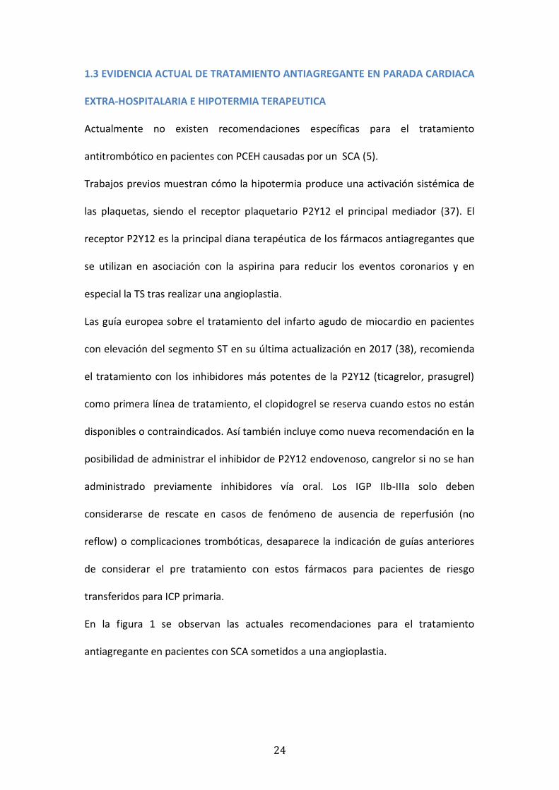
En la figura 1 se observan las actuales recomendaciones para el tratamiento
antiagregante en pacientes con SCA sometidos a una angioplastia.
25
Figura 1
Terapia antiplaquetaria peri y post procedimiento en pacientes sometidos a una angioplastia
primaria
Recomendaciones Clase Nivel
Terapia antiplaquetaria
Se recomienda un potente inhibidor del receptor P2Y12 (prasugrel,
ticagrelor) o clopidogrel si los otros no estan disponibles o
contraindicados.
I A
AAS (oral o endovenoso en pacientes que no pueden tragar) es
recomendado si no hay contraindicaciones.
I B
Inhibidores de la GP Iib-IIIa deberían ser considerado como rescate en
caso de complicaciones trombóticas o no reflow.
IIa C
Cangrelor podría ser considerado en pacientes que no han recibido un
inhibidor de los receptores de la P2Y12.
IIb A
Extraída de ESC 2017 Guidelines for Management of AMI-STEMI

26
1.3.1 INHIBIDORES DEL RECEPTOR P2Y12
En la actualidad, los fármacos más utilizados en la práctica clínica son clopidogrel,
prasugrel y ticagrelor. Los tres fármacos se administran por vía oral pero tienen un
metabolismo y una potencia antiagregante diferente (figura 2,3). Clopidogrel y
prasugrel requieren ser metabolizados por las enzimas hepáticas CYP2C19 a su
metabolito activo para conseguir bloquear el receptor P2Y12. En pacientes
sometidos a HT se ha observado un enlentecimiento del metabolismo hepático que
podría asociarse a un retraso de la activación de estos dos fármacos. Por el contrario,
ticagrelor es activo en su forma inicial ya que actúa directamente sobre el receptor
P2Y12 sin requerir una metabolización previa. Estudios experimentales y clínicos han
demostrado que ticagrelor tiene una potencia antiagregante entre el 30 y 40%
superior al clopidogrel (39,40).
Otro de los aspectos a tener en cuenta en pacientes bajo HT es la absorción de los
fármacos administrados por vía oral. Numerosos estudios han demostrado que la HT
reduce el metabolismo gastrointestinal y por tanto podría modificar la
farmacocinética y farmacodinámica de los fármacos administrados por vía oral (41).
En pacientes con SCA sometidos a implante de stent coronario no sólo es necesario
alcanzar unos niveles de antiagregación adecuados también es importante
conseguirlo lo más rápido posible. La consecución de una correcta antiagregación
durante la angioplastia se ha asociado a unos mejores resultados de la misma con
mejoría del flujo coronario y reducción del tamaño del infarto. Por este motivo, en
muchos centros se administra doble tratamiento antiagregante antes de cateterizar
al paciente e implantar un stent. No obstante, en los casos urgentes, esta estrategia
no es posible o el tiempo entre la administración del fármaco y el inicio de acción es

27
insuficiente (39). Este es el caso de los cateterismos urgentes o angioplastias
primarias en pacientes con paradas cardiacas sometidos a HT. El pico de acción tras
administrar un tratamiento antiagregante en bolus varía entre fármacos siendo más
rápido con ticagrelor (1-2 horas) y prasugrel (1-3 horas) que con clopidogrel (4-6
horas) (16). En este contexto, la administración de fármacos endovenosos de esta
(cangrelor) u otras familias como los inhibidores de la glicoproteína IIb-IIIa (IGP IIb-
IIIa) (abcximab, tirofiban y eptifibatide) podría suponer una ventaja respecto a los
fármacos orales al ayudar a conseguir un efecto antiagregante adecuado de forma
inmediata.
La doble terapia anti plaquetaria (DTAP) con aspirina y el inhibidor P2Y12 son el
tratamiento estándar para los pacientes después de la ICP. De acuerdo con ello,
DTAP se ha integrado en el tratamiento de pacientes después de la OHCA que se
someten a ICP y posteriormente se tratan con HT. En el ensayo PLATO (40) ticagrelor
se asoció con una reducción significativa de eventos cardiovasculares, mortalidad
cardiovascular, mortalidad por todas las causas y TS en pacientes con SCA en
comparación con clopidogrel. En este sentido, Tilemann et al. informaron que la
administración de ticagrelor triturado a través de sonda nasogástrica inhibió
confiablemente la función plaquetaria, independientemente de la presencia de
hipotermia en pacientes con PCEH en contexto de un SCA (41), y otros estudios
confirmaron estos hallazgos(30,43-44).
El cangrelor es un potente antagonista reversible del receptor P2Y12 análogo del
ATP , con una semivida < 10min. No es activo cuando se administra por vía oral y por
eso, a diferencia de todos los anteriores antagonistas del receptor del ADP, se
administra por vía endovenosa y puede desempeñar un papel importante en
28
pacientes en quienes los tratamientos enterales sean difíciles de administrar
(pacientes intubados o con hemesis intratables) o requieran una rápida inhibición
plaquetaria. De hecho, alcanza un alto grado de inhibición plaquetaria (> 90%) a los
pocos minutos tras su administración. Gracias a su mecanismo de acción reversible y
de desaparición rápida (una semivida extremadamente breve, 2–5min, a causa de
una rápida desactivación por ectonucleotidasas plasmáticas), se observa una
recuperación de la función plaquetaria 1–2h tras suspender la infusión del fármaco.
El cangrelor ha demostrado en pacientes con SCA una capacidad de inhibición
plaquetaria casi completa y en un tiempo comparable al alcanzado por el abciximab).
Es más, comparado con el abciximab, se ha demostrado mayor rapidez en el retorno
de la función plaquetaria tras la interrupción del tratamiento, lo que le confiere un
mejor perfil de seguridad (45).
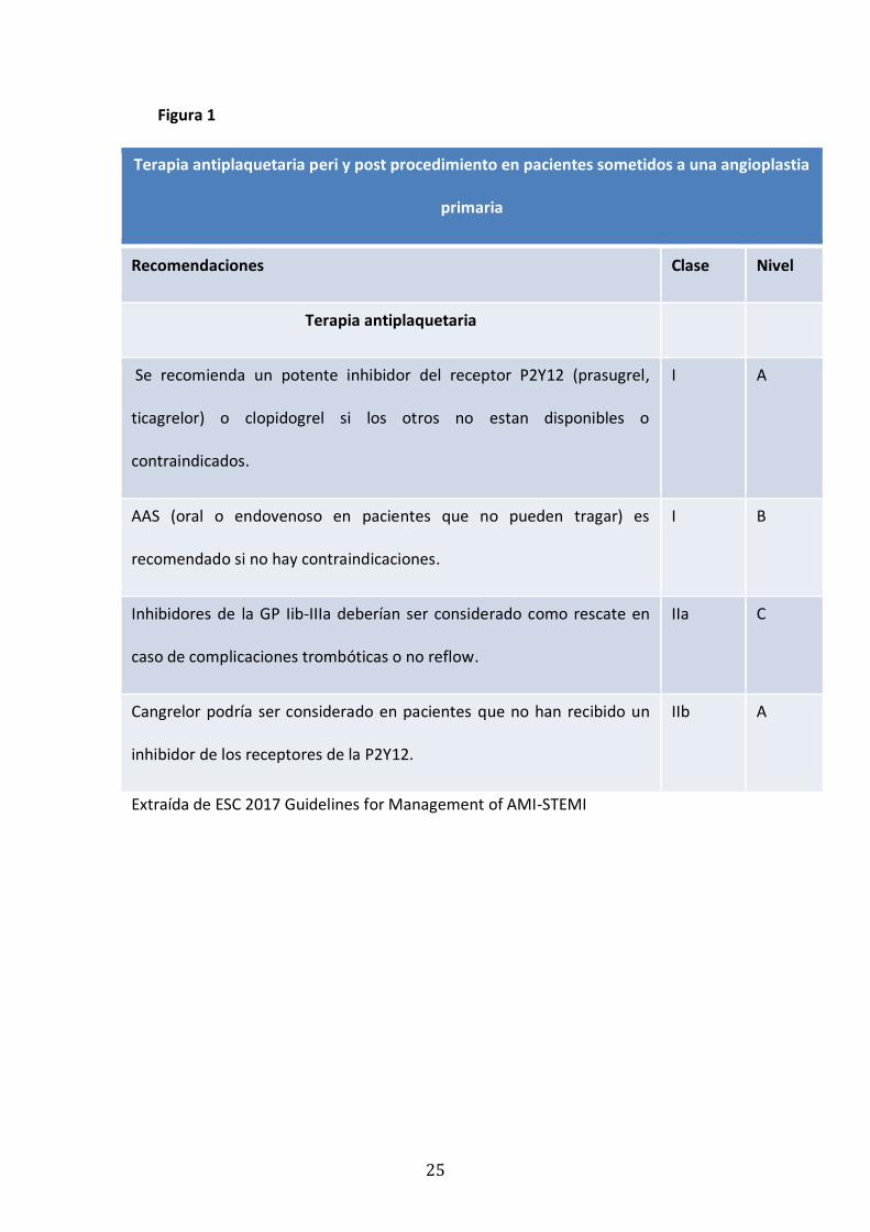
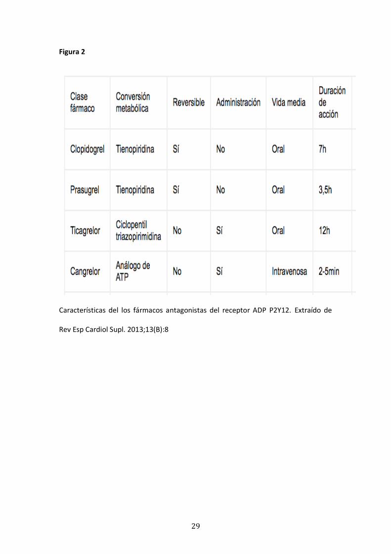
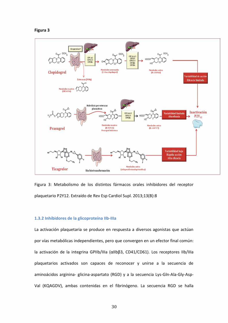
En la Figura 2 y 3 se observan las características y el metabolismo de los distintos
fármacos antagonistas del receptor plaquetario P2Y12.
29
Figura 2
Características del los fármacos antagonistas del receptor ADP P2Y12. Extraído de
Rev Esp Cardiol Supl. 2013;13(B):8
30
Figura 3
Figura 3: Metabolismo de los distintos fármacos orales inhibidores del receptor
plaquetario P2Y12. Extraído de Rev Esp Cardiol Supl. 2013;13(B):8
1.3.2 Inhibidores de la glicoproteína IIb-IIIa
La activación plaquetaria se produce en respuesta a diversos agonistas que actúan
por vías metabólicas independientes, pero que convergen en un efector final común:
la activación de la integrina GPIIb/IIIa (αIIbβ3, CD41/CD61). Los receptores IIb/IIIa
plaquetarios activados son capaces de reconocer y unirse a la secuencia de
aminoácidos arginina- glicina-aspartato (RGD) y a la secuencia Lys-Gln-Ala-Gly-Asp-
Val (KQAGDV), ambas contenidas en el fibrinógeno. La secuencia RGD se halla

31
también en otras sustancias como la vitronectina, el factor de von Willebrand y la
fibronectina, pero el fibrinógeno es el principal ligando debido a que contiene una
mayor concentración de esta secuencia de aminoácidos. El diseño de agentes
capaces de inhibir los receptores de GPIIb/IIIa permite el bloqueo de la etapa final
del proceso trombótico, cualquiera que sea el mecanismo o la sustancia que
inicialmente lo activara. Esto convierte a estos receptores en la diana ideal para el
tratamiento de los síndromes coronarios agudos. Por su mecanismo de acción, se
puede considerar dos tipos o familias de fármacos antagonistas de la GPIIb/IIIa
plaquetaria: los que bloquean de manera permanente los receptores plaquetarios
(abciximab) y los que los inhiben de manera competitiva y reversible, el lugar de
unión para la secuencia RGD, cuyo efecto depende de la concentración plasmática
(eptifibatida, tirofibán y lamifibán). Los cuatro fármacos se administran por vía
endovenosa (45). La activación del receptor de la glicoproteína IIb-IIIa es el eslabón
final común de la agregación plaquetaria, que produce una inhibición muy intensa de
la agregación plaquetaria y la unión de las plaquetas al tejido conectivo, por lo que
están asociados con un mayor tasa de hemorragias graves. Actualmente solo esta
indicado su uso en pacientes con complicaciones trombóticas en contexto de una ICP
(38). El uso de IGP IIb-IIIa parecía representar una opción muy atractiva para reducir
los episodios trombóticos en pacientes con HT por tres razones principales: 1) la
acción inmediata después de la administración; 2) la administración endovenosa en
pacientes que no pueden tragar; 3) los altos niveles de inhibición de plaquetas que
producen.

2.HIPÓTESIS

35
SUBPROYECTO 1 : Ticagrelor vs. Clopidogrel en pacientes comatosos luego de una
PCEH sometidos a ICP e hipotermia terapéutica
La HT en pacientes con SCA tiene un efecto protrombótico que está mediado
por una activación sistémica de la función de agregación plaquetaria y una
disminución de la acción de los fármacos antiagregantes por diferentes razones
(reducción de la absorción y metabolismo). Dado que el ticagrelor tiene perfil
de acción antiagregante más rápido y potente que el clopidogrel y no requiere
metabolización hepática, pensamos que en pacientes con PCEH e HT en
contexto de un SCA , se objetivara una disminución de la incidencia de TS con el
uso de ticagrelor comparando con clopidogrel.
SUBPROYECTO 2: Uso de inhibidores del la glicoproteína IIb-IIIa en pacientes
comatosos luego de una PCEH sometidos a ICP e hipotermia terapéutica
Como la HT y la PCEH causan alteraciones plaquetarias y de la coagulación se
objetivará una mayor tasa de sangrados con el uso de inhibidores del
glicoproteína IIb-IIIa (IGP IIb-IIIa) en pacientes comatosos supervivientes de una
PCEH en contexto de un SCA sometidos a HT comparando con pacientes con
PCEH en los que no se usaron IGP IIb-IIIa

36
SUBPROYECTO 3: Eventos trombóticos y hemorrágicos en pacientes comatosos
luego de una PCEH sometidos a ICP según el uso o no de HT
La PCEH por sí misma pueden causar alteraciones plaquetarias y en la
coagulación, así como una disminución en la absorción y metabolismo de los
fármacos antiagregantes. La HT podría relacionarse con una activación de la
agregación plaquetaria inducida por el frío, una menor para la absorción de los
fármacos antiagregantes por vía digestiva, un enlentecimiento de la
metabolización de los fármacos y una probable disfunción endotelial. Así
también la hipotermia por debajo de 33º afecta la síntesis y la cinética de
enzimas de la coagulación, la generación de trombina e inhibidores del
activador del plasminógeno y se relaciona con disfunción de plaquetas, y eso
podría favorecer la aparición de sangrados. Por lo que pensamos que en
pacientes con PCEH e SCA sometidos a HT se observarán mayor tasa de eventos
trombóticos y hemorrágicos que en controles históricos de PCEH sin HT.

3.OBJETIVOS
39
SUBPROYECTO 1 : Ticagrelor vs. Clopidogrel en pacientes comatosos luego de una
PCEH sometidos a ICP e hipotermia terapéutica
1.1 Comparar la incidencia de TS y eventos hemorrágicos entre el uso de ticagrelor o
clopidogrel en pacientes con SCA sometidos a una ICP después de una PCEH en
HT.
SUBPROYECTO 2: Uso de inhibidores del la glicoproteína IIb-IIIa en pacientes
comatosos luego de una PCEH sometidos a ICP e hipotermia terapéutica
2.1 Definir el beneficio clínico del uso de IGP IIb-IIIa en pacientes con PCEH en
contexto de SCA sometidos a ICP bajo tratamiento con HT, evaluando la
incidencia de eventos hemorrágicos y tromboticos según el uso o no IGP IIb-IIIa.
SUBPROYECTO 3: Eventos trombóticos y hemorrágicos en pacientes comatosos
luego de una PCEH sometidos a ICP según el uso o no de HT
3.1 Determinar la incidencia de eventos hemorrágicos y trombóticos en pacientes
con SCA después de una PCEH según recibieron o no HT.

4.MATERIALYMÉTODOS
43
SUBPROYECTO 1 : Ticagrelor vs. Clopidogrel en pacientes comatosos luego de una
PCEH sometidos a ICP e hipotermia terapéutica
La población de estudio del primer subproyecto estuvo constituida por todos los
pacientes con PCEH ingresados en nuestro hospital entre enero de 2010 y agosto de
2016 con SCA, sometidos a ICP primaria bajo HT. Los criterios de exclusión
incluyeron pacientes con SCA sin implantación de stent y pacientes que murieron
antes del procedimiento índice. Aunque los pacientes que recibieron prasugrel se
incluyeron en la cohorte principal, el pequeño número de sujetos en este grupo
impidió cualquier comparación específica con ticagrelor o clopidogrel. Se comparó la
incidencia de eventos trombóticos incluyendo la TS y la incidencia de eventos
hemorrágicos según el uso de ticagrelor o clopidogrel.
SUBPROYECTO 2: Uso de inhibidores del la glicoproteína IIb-IIIa en pacientes
comatosos luego de una PCEH sometidos a ICP e hipotermia terapéutica
En el segundo subproyecto la población de estudio estuvo constituida por todos los
pacientes con PCEH ingresados en nuestro hospital entre enero de 2010 y
septiembre de 2015, con PCEH y SCA sometidos a ICP bajo HT. Los criterios de
exclusión incluyeron el uso de derivados coumarinicos, el uso previo de agentes
fibrinolíticos y los pacientes que murieron antes de la intervención inicial. Se valoró
el beneficio clínico del uso de IGP IIb-IIIa en pacientes con PCEH en contexto de SCA
sometidos a ICP bajo tratamiento con HT, evaluando la incidencia de eventos
hemorrágicos y tromboticos según el uso o no IGP IIb-IIIa.
44
SUBPROYECTO 3: Eventos trombóticos y hemorrágicos en pacientes comatosos
luego de una PCEH sometidos a ICP según el uso o no de HT.
La población de estudio del tercer subproyecto estuvo constituida por todos los
pacientes con PCEH ingresados en nuestro hospital entre enero de 2005 y diciembre
de 2016 con SCA y PCEH sometidos a ICP. Desde enero de 2010, el protocolo HT se
inició en nuestro centro para pacientes comatosos luego de una PCEH de presunta
causa cardíaca, independientemente del ritmo inicial y se utiliza actualmente.
Comparamos la incidencia de eventos trombóticos que incluyeron: trombosis venosa
profunda, tromboembolismo pulmonar y trombosis de stent durante la
hospitalización, así como la incidencia de eventos hemorrágicos de estos pacientes
con un control histórico en los que no se realizó HT entre los años 2005 y 2009.
Los criterios de exclusión incluyeron el uso de antagonistas de la vitamina K, el uso
previo de un fibrinolítico, hemorragia intracraneal aguda sospechada o conocida o
accidente cerebrovascular, y pacientes que murieron antes del procedimiento índice.
La metodología especifica de cada uno de los subproyectos esta detallada en los
artículos publicados e incorporados a la presente tesis.

5.ARTÍCULOSPUBLICADOS
Resuscitation 114 (2017) 141–145
Contents lists available at ScienceDirect
Resuscitationjou rn al hom ep age : w ww.elsev ier .com/ locate / resusc i ta t ion
Clinical paper
Out-of-hospital cardiac arrest and stent thrombosis: Ticagrelor versusclopidogrel in patients with primary percutaneous coronaryintervention under mild therapeutic hypothermia�
Gustavo Jiménez-Brítez, Xavier Freixa ∗, Eduardo Flores-Umanzor, Rodolfo San Antonio,Gala Caixal, John Garcia, Marco Hernandez-Enriquez, Rut Andrea, Ander Regueiro,Mónica Masotti, Salvatore Brugaletta, Victoria Martin, Manel SabatéCardiology Department, Hospital Clinic de Barcelona, Institut d’Investigacions Biomèdiques August Pi i Sunyer (IDIBAPS), University of Barcelona,Barcelona, Spain
a r t i c l e i n f o
Article history:Received 9 December 2016Received in revised form 19 January 2017Accepted 15 February 2017
Keywords:Mild therapeutic hypothermiaOut-of-hospital cardiac arrestStent thrombosisTicagrelor
a b s t r a c t
Background: Out-of-Hospital Cardiac Arrest (OHCA) and mild therapeutic hypothermia (MTH) have beenlinked to increased risk of Stent Thrombosis (ST) in comatose survivors who undergo percutaneouscoronary intervention (PCI). In this sense, there is no formal recommendation about which antiplateletregimen should be used in patients with acute coronary syndromes (ACS) after OHCA.Aims: To compare the incidence of probable/definite ST and bleeding events between ticagrelor andclopidogrel, in patients with ACS under MTH after an OHCA.Methods and results: From January 2010 to August 2016, 144 patients underwent MTH after an OHCA.Overall, 114 had an ACS (79%) and 98 (67,3%) were treated with primary PCI and stent implantation.Among them, 61 (62,2%) were treated with clopidogrel, and 32 (32,6%) with ticagrelor. During hospital-ization, the incidence of probable or definite ST was significantly higher in patients receiving clopidogrelcompared to ticagrelor (11,4% vs. 0%; p: 0.04), and no significant differences in any (28,6% vs. 25%; p:0.645) or major bleeding (BARC 3 or 5) (11,4% vs. 12,5%; p: 0.685) were found. Hospital mortality did notdiffer between groups (26,2% vs. 25%; p: 0.862).Conclusions: In this study, as compared to clopidogrel, ticagrelor was associated with a lower rate of ST,without differences in haemorrhagic events in patients with OHCA for an ACS under MTH. Similarly toother settings, ticagrelor might be a valid alternative to clopidogrel in these patients.
© 2017 Elsevier B.V. All rights reserved.
Introduction
Acute coronary syndromes (ACS) are the commonest cause ofmalignant arrhythmias leading to sudden cardiac death.1 Mild ther-apeutic hypothermia (MTH) and emergent coronary angiographywith primary percutaneous coronary intervention (PCI) improves
Abbreviations: ACS, acute coronary syndrome; BARC, bleeding academicresearch consortium; BMS, bare metal stent; DAPT, dual antiplatelet therapy; DES,drug eluting stent; GP IIb-IIIa, glycoprotein IIb-IIIa receptors; ICU, intensive car-diac unit; LVEF, left ventricle ejection fraction; MTH, mild therapeutic hypothermia;OHCA, out-of-hospital cardiac arrest; PCI, percutaneous coronary intervention; ST,stent thrombosis.� A Spanish translated version of the abstract of this article appears as Appendix
in the final online version at http://dx.doi.org/10.1016/j.resuscitation.2017.02.015.∗ Corresponding author at: Cardiology Department, Hospital Clinic de Barcelona,
IDIBAPS, C/Villarroel 170, 08036, Barcelona, Spain.E-mail address: [email protected] (X. Freixa).
outcomes in the setting of Out-of-Hospital Cardiac Arrest (OHCA)after a coronary event.2,3 MTH has been proposed to preserve neu-rological status in these patients.4
MTH has been associated with haemostasis and coagulopathydisorders.5,6 The relationship between a higher risk of stent throm-bosis (ST) and OHCA is however very controversial. Whereas severalstudies have reported a higher risk of stent thrombosis (ST) afterprimary PCI in OHCA patients,7–12 some other studies did not findthis association.13,14 A recent study suggested that just the fact ofhaving suffered an OHCA by itself increases the risk of ST regardlessthe use of MTH.12 Alterations in platelet reactivity and pharma-cokinetics of antiplatelet agents with MTH may pre-dispose to STin these patients.9–11
Dual antiplatelet therapy (DAPT) with aspirin and P2Y12inhibitor are the standard of care for patients after PCI.15 Accord-ingly, DAPT has been integrated into management of patients afterOHCA who undergo PCI and subsequently are treated with MTH.
http://dx.doi.org/10.1016/j.resuscitation.2017.02.0150300-9572/© 2017 Elsevier B.V. All rights reserved.
142 G. Jiménez-Brítez et al. / Resuscitation 114 (2017) 141–145
Nonetheless there is no formal recommendations about whichantiplatelet regimen should be used in patients with ACS andOHCA.1 In the PLATelet inhibition and patient outcomes (PLATO)trial, ticagrelor was associated with a significant reduction of car-diovascular events, cardiovascular mortality, all cause mortalityand ST in patients with ACS compared to clopidogrel,16 In thisregard, Tilemann et al. reported that the administration of crushedticagrelor via nasogastric tube reliably inhibited platelet functionregardless of the presence of hypothermia in ACS patients,17 andother studies confirmed these findings.18–20
Previous reports showing increased ST in OHCA patients withMTH did not focus in antiplatelet treatment until, although surpris-ingly, Gouffran et al. observed an increase of ST in patients treatedwith new P2Y12 inhibitors receptors (ticagrelor or prasugrel) com-pared to clopidogrel in a cohort of 101 OHCAs treated with PCIand MTH.10 Thus, the aim of the study was to compare the inci-dence of ST and bleedings events between ticagrelor or clopidogrelin patients with ACS undergoing PCI after OHCA under MTH.
Methods
Patients
This was a single centre observational study. We retrospectivelyscreened consecutive patients admitted to our Hospital betweenJanuary 2010 and August 2016 with ACS and OHCA undergoingprimary PCI under MTH. Exclusion criteria included patients withACS without stent implantation, and patients who died before theindex procedure. Although patients who received prasugrel wereincluded in the main cohort, the small number of subjects in thisgroup precluded any specific comparison with ticagrelor or clopi-dogrel. The study was approved by Ethics Committee of our centre(approval reference number 2013/8596) and complies with princi-ples laid down in the Declaration of Helsinki.
Procedural characteristics
All surviving OHCA patients admitted to our centre withoutan evident extra cardiac cause were admitted immediately tothe cardiac catheterization laboratory regardless of the clinicaland ECG findings. If there was a high suspicion of ACS definedby ECG changes, initial shockable rhythm or previous chest pain,antithrombotic treatment with aspirin and heparin was initiatedby emergency team prior to admission. Primary PCI was attemptedif there was an acute coronary atherothrombotic lesion. The useof glycoprotein IIb-IIIa receptors inhibitors (GPI IIb-IIIa) and man-ual thrombus aspiration were left to the operator preference. Thelength, diameter and type of stent Drug Eluting Stent (DES) or BareMetal Stent (BMS), were also decided by the operator. After PCI allpatients were transferred to the Intensive Cardiac Unit (ICU).
In order to reduce delays, most of the patients arrived withoutnasogastric tubing at the cardiac catheterization laboratory. Naso-gastric tubing was therefore placed in the cath lab just after PCI,so the loading dose of P2Y12 inhibitors was crushed, dissolved andadministered right after the PCI. Unfortunately, there was no accu-rate estimation of the time delay between PCI and P2Y12 inhibitorsadministration. P2Y12 inhibitors were however always adminis-tered within the 30 min after PCI and prior to the ICU transfer.Although small, this variable delay in drug administration may havecontribute to the final results, as ticagrelor has a faster mechanismof action compared to clopidogrel.
The loading dose was followed by maintenance dose (clopido-grel 75 per day, prasugrel 10 mg per day, ticagrelor 90 mg bid.)
All patients received MTH according to the local ICU protocol. Allpatients reached 33 ◦C fewer than 8 h from cardiac arrest, and this
temperature was maintained for 24 h. Warming took place gradu-ally in 24–30 h, with a rate of 0.10–0.15 ◦C/h. This protocol has beencomprehensively described somewhere else.7 In the ICU, patientsreceived standard treatment that included mechanical ventilationand correction of cardiovascular instability.
Data analysis
The baseline and procedural data of patients were systemati-cally collected in a dedicated database. The primary endpoint wasthe occurrence of definite and probable stent thrombosis (ST),21
during hospitalization according to the Academic Research Con-sortium definitions, as well as the incidence of bleeding eventsaccording to the BARC criteria.22
A routine angiography was not compulsory after baseline PCIand was only performed in case of a clinical event, ECG or echocar-diography changes or severe hemodynamic instability.
Statistical analysis
Continuous variables were expressed as mean ± standard devi-ation and non-normally distributed variables were expressedas median [inter-quartile range]. Categorical variables wereexpressed as count and percentage. Baseline characteristicsbetween groups were compared using t test for continuous vari-ables and chi-square test for categorical variables. Results wereconsidered statistically significant at a p-value <0.05. Statisticalanalyses were carried out using SPSS package v20.0 (Chicago, IL,USA).
Results
From January 2010 to August 2016, 144 patients were treatedwith MTH after an OHCA. Overall, 114 had an ACS (79%) and 98(67,3%) underwent primary PCI with stent implantation. Amongthem, 61 (62,2%) were treated with clopidogrel, (clopidogrelgroup), 32 (32,6%) with ticagrelor (ticagrelor group) and 5 (5,1%)with prasugrel.
As shown in Table 1, baseline characteristics were similar amonggroups. Of note, most of the patients presented cardiac arrest sec-ondary to STEMI. Post-resuscitation shock was similar in bothgroups (65.5% in the clopidogrel group vs. 71.8% in the ticagrelorgroup; p = 0.67). As shown in Table 2, procedural data revealed nosignificant differences in the use of GP IIb-IIIa inhibitors or throm-boaspiration, but a significant higher use of DES in the ticagrelorgroup.
Clinical outcomes
During hospitalization, 7 (7.1%) patients presented definite orprobable ST, 7 (11,4%) in the clopidogrel group and none (0%) inthe ticagrelor group (p:0,04). None of five patients with prasugrelpresented ST (Table 3). Stent thrombosis was classified as acute in2 patients and sub acute in the other 5 patients. Two patients withDES and 5 with BMS presented definite ST.
DES were implanted in 33 patients (16 in the clopidogrel and 17in the ticagrelor group). Only 2 patients in the clopidogrel group(12.5%) presented ST and there were no significant differencesamong groups (12.5% vs. 0%, p = 0.13). An individual and more com-prehensive description of ST is provided in Table 4.
There were no significant differences in any bleeding (28,6%with clopidogrel vs. 25% with ticagrelor; p:0,645) and major bleed-ing (BARC 3 or 5) (11,4% vs. 12,5; p = 0,685) among groups (Table 3).
Sixteen (26,2%) patients in the clopidogrel group and 8 (25.0%)in the ticagrelor group died during hospitalization without signif-icant differences between groups. Among them, 2 patients who

G. Jiménez-Brítez et al. / Resuscitation 114 (2017) 141–145 143
Table 1Baseline clinical characteristics.
Clopidogrel (n:61) Ticagrelor (n:32) p:value
Age, years, mean ± SD 57, 4 ± 12 56, 5 ± 9,8 0.759Male, n (%) 52 (85,2) 28 (87,5) 0.766Smoking, n (%) 32 (52,4) 13 (40,6) 0.630Hypertension, n (%) 26 (42,6) 11 (34,3) 0.308Diabetes Mellitus, n (%) 10 (16,3) 6 (18,7) 0.359Hypercholesterolemia, n (%) 30 (49,1) 12 (37,5) 0.243Previous myocardial infarction, n (%) 11 (18) 6 (18,7) 0.139Renal failure, n (%) 5 (8,1) 1 (3,1) 0.252Initial shockable rhythm, n (%) 49 (80,3) 26 (81,2) 0.121Total ischemic time, min. (mean ± SD)a 125, 4 ± 72,0 141, 3 ± 52,1 0.338Time from cardiac arrest to return of spontaneous circulation, min. (mean ± SD) 30, 1 ± 16,9 26, 1 ± 10,6 0.292ST- segment elevation myocardial infarction, n (%) 54 (88,5) 45 27 (87) 0.675Post-resuscitation shock, n (%) 40 (65,5) 32 23 (71,8) 0.400LVEF mean ± SD 40, 3 ± 16,3 41, 9 ± 12,8 0.685
LVEF: left ventricular ejection fraction.a Time from symptom onset to coronary flow restoration.
Table 2Procedural characteristics.
Clopidogrel (n:61) Ticagrelor (n:32) p:value
Culprit coronary artery, n (%) 0.197LAD 27 (44,2) 17 (53,2)LCX 13 (21,3) 6 (18,7)RCA 19 (31,2) 8 (25)TIMI flow 0 or 1 before PCI, n (%) 34 (55,7) 16 (50) 0.537Thromboaspiration, n (%) 33 (54) 14 (43,7) 0.343Glycoprotein IIb-IIIa receptors inhibitors used, n (%) 13 (21,3) 6 (18,7) 0.771TIMI flow 3 after PCI, n (%) 52 (85,2) 30 (93,7) 0.644Number of implanted stents (mean ± SD) 1,28 ± 0,68 1,26 ± 0,44 0.888Patients treated with DES, n (%) 16 (26,2) 17 (53,1) 0.01Mean diameter stent, mm, (mean ± SD) 3,10 ± 0,65 3,33 ± 0,72 0.181Total stent length, mm, (mean ± SD) 23,4 ± 13,2 21,9 ± 8,8 0.608Bifurcation lesions, n (%) 8 (13,1) 8 (25) 0.149No reflow, n (%) 7 (11,4) 3 (9,3) 0.555IABP, n (%) 12 (19,6) 3 (9,3) 0.200
LAD: Left Anterior Descending; LCX: Left Circumflex; RCA: Right Coronary Artery; PCI: Percutaneous Coronary Intervention; DES: Drug Eluting Stent; IABP: Intra-AorticBalloon Pump.The values in bold mean that there are statistically significant differences in these variables.
Table 3Patient Outcomes.
Clopidogrel (n: 61) Ticagrelor (n: 32) p:
Definite stent thrombosis n (%) 7 (11.4) 0 (0) 0.046Probable stent thrombosis 0 (0) 0 (0)Any bleeding, n (%) 18 (28,6) 8 (25) 0.645BARC type 3 or 5, n(%) 7 (11,4) 4 (12,5) 0.685Mortality, (%) 16 (26,2) 8 (25.0) 0.862
BARC (bleeding academic research consortium).The values in bold mean that there are statistically significant differences in these variables.
Table 4Patients with stent thrombosis.
Artery No. of stents Pre-treatment PCI Post-procedure treatment Day of ST Death
1 RCA 1 ASA, Heparin BMS 3 × 18 mm Clopidogrel, abciximab 3 No2 LAD/LCX 2 ASA, Heparin DES
2.5 × 33, 3 × 33 mmClopidogrel, abciximab 4 No
3 LAD 1 ASA, Heparin, BMS2,5 × 14 mm
Clopidogrel, abciximab 2 No
4 LAD 1 ASA, Heparin, Tenecteplase BMS2,5 × 19 mm
Clopidogrel 3 Yes
5 LAD 4 ASA, Heparin BMS3 × 18, 3.5 × 8, 2,5 × 13,2.25 × 13
Clopidogrel, abciximab 1 Yes
6 LCX 1 ASA, Heparin DES2.5 × 18
Clopidogrel 2 No
7 RCA 1 ASA, Heparin BMS3 × 23
Clopidogrel 1 No
ASA: aspirin; BMS: bare metal stent; DES: drug eluting stent, LAD: left anterior descending; LCX: left circumflex, PCI: percutaneous coronary intervention; RCA: right coronaryartery; ST: stent thrombosis.

144 G. Jiménez-Brítez et al. / Resuscitation 114 (2017) 141–145
presented ST died (28.5% of them), both in the clopidogrel groupThe cause of mortality in these two patients was severe neurologicdamage and cardiogenic shock with end-organ failure respectively.
Discussion
The main finding of the present study was that the use of tica-grelor in patients undergoing PCI under MTH after OHCA wasassociated with a lower incidence of ST without differences inhaemorrhagic events compared to clopidogrel.
The present study shows a high incidence of ST (7,1% overall).Our group already reported a higher rate of ST in OHCA survivorstreated with primary PCI under MTH.7 Accordingly, other stud-ies confirmed the higher incidence of ST in OHCA survivors8–12,although other studies did not confirm these findings.13,14
The impact of MTH on ST is however not clear. In fact, Shan et al.reported high rates of ST in OHCA patients after PCI (4,7%) withoutdifferences in patients undergoing MTH or not (3,9% vs. 4,7%).12
Gouffran et al. reported a high incidence of ST (10.9%) in a cohort101 OHCA survivors treated with PCI and MTH. Surprisingly, morepatients presented ST with the use of new P2Y12 inhibitors thanthose receiving clopidogrel. The authors suggest that this findingmight be explained by the fact that ADP pathway may not be theonly target for antiplatelet strategies after OHCA.10 In contrast, Tile-mann et al. reported no ST in 27 patients undergoing MTH with PCItreated with crushed ticagrelor.17
The mechanisms explaining why there is an increased riskof ST in patients with OHCA and MTH are not well establishedyet. All these mechanisms would create a prothrombotic environ-ment leading to a higher incidence of thrombotic events.18–20 (a)Activation of platelet aggregation by cold, (b) antiplatelet absorp-tion disorders through the digestive tract; (c) drug metabolismslowing-down; and (d) probable endothelial dysfunction have beenproposed as these potential mechanisms among others. Sinceplatelet adenosine diphosphate receptor P2Y12 is a pivotal tar-get for antiplatelet treatment in ACS, particularly in patients withimplanted stent, and MTH might interfere with the action of theseagents, the use of conventional DAPT with aspirin and clopidogrelmay be inefficient after OHCA and hypothermic conditions.17–20 Inthis sense, ticagrelor may represent a valid alternative to reducethrombotic events in patients under MTH as it provides a morerapid and intense inhibition of platelet reactivity. Ticagrelor isa direct inhibitor of the P2Y12 receptor and does not requiremetabolic transformation. Additionally, it has been associated witha superior pharmacodynamic profile compared to clopidogrel inpatients undergoing PCI.18–20,23 In fact, Steblovik et al. reported afaster and stronger platelet inhibition with ticagrelor as comparedto clopidogrel in 37 comatose survivors of OHCA undergoing PCIand MTH before and after PCI23
Bednar et al. measured platelet inhibition by VASP (vasodilator-stimulated phosphoprotein) in 40 patients with ACS and OHCAtreated with MTH who received one P2Y12 inhibitor (clopidogrel,prasugrel or ticagrelor) and observed that the proportion of patientswith ineffective platelet inhibition after clopidogrel, prasugrel andticagrelor was (77% vs. 19% vs. 1%) on day 1, (77 vs. 17 vs. 0%) 2(85 vs. 6 vs. 0%) and 3 (p < 0,001).18 Moudgil et al. reported a morerapid (within 4 h) and sustained reduction (6 days) in platelet reac-tivity with ticagrelor compared to clopidogrel in 15 patients withACS after an OHCA treated with MTH.19 Rosencher et al. reported,in a cohort of 20 OHCA patients, a higher residual platelet activitywith clopidogrel than ticagrelor 4 h after the loading dose, not onlyduring MTH, but also after MTH up to day 7.20
There are currently no specific recommendations for antithrom-botic therapy in patients with OHCA caused by ACS.1 In this regard,conversely to Gouffran et al., our series showed a reduction of STin patients treated with ticagrelor without differences in haem-
orrhagic events compared to clopidogrel. These findings wouldsupport the clinical benefits of ticagrelor over clopidogrel thatwere already observed in pharmacodynamics and pharmacokinet-ics studies in OHCA patients.
In the present study, there were no significant differences inclassic risk factors for ST namely number, length and diameter ofthe implanted stents, diabetes, renal failure, bifurcated lesions, noreflow and cardiogenic shock. In the ticagrelor group, patients weretreated more frequently with DES compared to clopidogrel. Thismay have helped to reduce the rate of ST in the ticagrelor group.24
Although the incidence of any or major haemorrhagic eventswas high, no significant differences among groups were found. Theabsence of differences between groups might be related to thefact that post-resuscitation syndrome and MTH may produce alter-ations in haemostasis and coagulopathy. Hypothermia below 33◦
affects the synthesis and kinetics of clotting enzymes, thrombingeneration, and plasminogen activator inhibitors and is related toplatelet dysfunction, and this may be associated with an increasedrisk of haemorrhagic events25,26 regardless of the treatment withP2Y12 inhibitors.
Our group has already reported a very high incidence of majorbleedings (64,7% with BARC 3 o 5) without reduction of thromboticevents in OHCA patients undergoing primary PCI and MTH treatedwith GPI IIb-IIIa. The use of GP IIb-IIIa inhibitors might be seen as avalid alternative to reduce thrombotic events for the immediateaction and the intravenous administration in patients who can-not swallow. Nonetheless, the absence of differences in thromboticevents in patients with and without GP IIb-IIIa inhibitors and thehigher incidence of haemorrhagic events do not seem to supportthis option.27
Stent thrombosis is a multifactorial phenomenon, which isclearly not only driven by the lack of clopidogrel efficacy dur-ing MTH. However, in such a prothrombotic environment, itseems logical that these patients should be treated like high-riskpatients with the more effective and safe antiplatelet strategy. Newintra-venous P2Y12 inhibitors, like cangrelor could also play andimportant a role in this setting. Further clinical studies are requiredin this context.28
The present study has several limitations. The results must beinterpreted with caution, as this is a single-centre non-randomizedand retrospective study with a relatively small sample size. Thestudy includes patients that were treated in a long period (6 years)with most of them receiving clopidogrel. Ticagrelor was howeverused more often during the last years of the study and the observedresult may have been affected, at least in part, by improvementsin treatment over time. The limited number of patients precludedany further solid analysis of subgroups analysing classical ST riskfactors other than the use of DES. The number of patients receivingprasugrel in this setting was very small and therefore not includedin the analysis.
Conclusion
In this study, as compared to clopidogrel, ticagrelor was associ-ated with a lower rate of ST, without differences in haemorrhagicevents in patients with ACS under MTH. Similarly to other set-tings, ticagrelor might be a very valid alternative to clopidogrel inMTH. The results of the study are hypothesis generating and furtherrandomized data will be needed.
Conflict of interest statement
There is no conflict of interest and relationship with the industryby any authors.
G. Jiménez-Brítez et al. / Resuscitation 114 (2017) 141–145 145
References
1. Nikolaos I Nikolau, Hans-Richard Arntz, Abdelouahab Bellou, et al. Europeanresuscitation council guidelines for resuscitation 2015 section 8. Managementof acute coronary syndrome. Resuscitation 2015;95:264–77.
2. Dumas F, Cariou A, Manzo-Silberman S, et al. Immediate percutaneous coro-nary intervention is associated with better survival after out-of-hospital cardiacarrest: insights from the PROCAT (Parisian Region Out of hospital Cardiac Arrest)registry. Circ Cardiovasc Interv 2010;3:200–7.
3. Dumas F, White L, Stubbs BA, Cariou A, et al. Long-term prognosis followingresuscitation from out of hospital cardiac arrest: role of percutaneous coronaryintervention and therapeutic hypothermia. J Am Coll Cardiol 2012;60:21–7.
4. Holzer M, Cerchiari E, Martens P, et al. Mild therapeutic hypothermia to improvethe neurologic outcome after cardiac arrest. N Engl J Med 2002;346:549–56.
5. Spiel AO, Frossard M, Mayr FB, et al. Pronounced platelet hyperfunction inpatients with cardiac arrest achieving restoration of spontaneous circulation.Crit Care Med 2009;37:975–9.
6. Brinkman AC, Ten Tusscher BL, de Waard MC, et al. A minimal effects on ex vivocoagulation during mild therapeutic hypothermia post cardiac arrest patients.Resuscitation 2014;85:1359–63.
7. Penela D, Magaldi M, Sabate M, et al. Hypothermia in acute coronary syndrome:brain salvage versus stent thrombosis? J Am Coll Cardiol 2013;61:686–7.
8. Joffre J, Varenne O, Bougouin W, Rosencher J, Mira J-P, Cariou A. Stent thrombo-sis: an increased adverse event after angioplasty following resuscitated cardiacarrest. Resuscitation 2014;85:769–73.
9. Orban M, Mayer K, Morath T, et al. The impact of therapeutic hypothermia on on-treatment platelet reactivity and clinical outcome in cardiogenic shock patientsundergoing primary PCI for acute myocardial infarction: results from the ISAR-SHOCK registry. Thromb Res 2015;136:87–93.
10. Gouffran G, Rosencher J, Bougouin W, et al. Stent thrombosis after primarypercutaneous coronary intervention in comatose survivors of out-of-hospitalcardiac arrest: are the new P2Y12 inhibitors really more effective than clopido-grel? Resuscitation 2016;98:73–8.
11. Ibrahim K, Christoph M, Schmeinck S, et al. High rates of prasugrel and ticagrelornon-responder in patients treated with therapeutic hypothermia after cardiacarrest. Resuscitation 2014;85:649–56.
12. Shan N, Chaudhary R, Mehta K, et al. Therapeutic hypothermia and Stent Throm-bosis. JACC Cardiovasc Interv 2016;9:1801–11.
13. Chisholm GE, Greis A, Thim T, et al. Safety of therapeutic hypothermia combinedwith primary percutaneous coronary intervention after out-of-hospital cardiacarrest. Eur Heart J Acute Cardiovasc Care 2015;4:60–3.
14. Casella G, Carinci V, Cavallo P, et al. Combining therapeutic hypothermia andemergent coronary angiography in out-of-hospital cardiac arrest survivors:optimal post-arrest care for the best patient. Eur Heart J Acute Cardiovasc Care2015;4:579–88.
15. Windecker S, Kolh P, Alfonso F, et al. ESC/EACTS Guidelines on myocardial revas-cularization: the task force on myocardial revascularization of the EuropeanSociety of Cardiology (ESC) and the European Association for CardioThoracicSurgery (EACTS) Developed with the special contribution of the EuropeanAssociation of Percutaneous Cardiovascular Interventions (EAPCI). Eur Heart J2014;35:2541–619.
16. Wallentin L, Becjer RC, Budaj A, et al. Ticagrelor versus clopidogrel in patientswith acute coronary syndrome. N Engl J Med 2009;361:1045–57.
17. Tilemann LM, Stiepak J, Zelniker T, et al. Efficacy of enteral ticagrelor inhypothermic patients after out-of-hospital cardiac arrest. Clin Res Cardiol2016;105:332–40.
18. Bednar F, Kroupa J, Ondrakova M, et al. Antiplatelet efficacy of P2Y12 inhibitors(prasugrel, ticagrelor, clopidogrel) in patients treated with mild therapeutichypothermia after cardiac arrest due to myocardial infarction. J Thromb Throm-bolysis 2016;41:549–55.
19. Moudgil R, Al-Turbak H, Osborne C, et al. Superiority of ticagrelor over clopido-grel in patients after cardiac arrest undergoing therapeutic hypothermia. Can JCardiol 2014;30:1396–9.
20. Rosencher J, Gouffran G, Bougouin W, et al. Optimal antiplatelet therapy inout-hospital cardiac arrest patients treated by primary percutaneous coronaryintervention. Resuscitation 2015;90:7–8.
21. Cutlip DE, Windecker S, Mehran R, et al. Academic research consortium, clinicalend points in coronary stent trials: a case for standardized definitions. Circula-tion 2007;115:2344–51.
22. Mehran R, Rao SV, Bhatt DL, et al. Standardized bleeding definitions for cardio-vascular clinical trials: a consensus report from the Bleeding Academic ResearchConsortium. Circulation 2011;123:2736–47.
23. Steblovnik K, Blinc A, Mijovski MD, et al. Ticagrelor versus clopidogrel incomatose survivors of out-of-hospital cardiac arrest undergoing percutaneouscoronary intervention and hypothermia. Circulation 2016;134:2128–30.
24. Sabate M, Cequier A, Iniguez A, et al. Everolimus-eluting stent versus bare-metalstent in ST-segment elevation myocardial infarction (EXAMINATION): 1 yearresults of a randomised controlled trial. Lancet 2012;380:1482–90.
25. Brinkman AC, Ten Tusscher BL, de Waard MC, et al. A minimal effects on ex vivocoagulation during mild therapeutic hypothermia post cardiac arrest patients.Resuscitation 2014;85:1359–63.
26. Adrie C, Adib-Conquy M, Laurent I, et al. Successful cardiopulmonary resuscita-tion after cardiac arrest as a sepsis-like syndrome. Circulation 2002;106:562–8.
27. Jiménez-Britez G, Freixa X, Flores E, et al. Safety of glycoprotein IIb-IIIa inhibitorsin patients under therapeutic hypothermia admitted for an acute coronary syn-drome. Resuscitation 2016;106:108–12.
28. Steblovnik K, Blinc A, Bozic-Mijovski M, et al. Platelet reactivity in comatosesurvivors of cardiac arrest undergoing percutaneous coronary intervention andhypothermia. EuroIntervention 2015;10:1418–24.
Resuscitation 106 (2016) 108–112
Contents lists available at ScienceDirect
Resuscitationjou rn al hom epage : w ww.elsev ie r .com/ locate / resusc i ta t ion
Clinical paper
Safety of glycoprotein IIb/IIIa inhibitors in patients under therapeutichypothermia admitted for an acute coronary syndrome�
Gustavo Jiménez-Brítez, Xavier Freixa ∗, Eduardo Flores, Diego Penela,Marco Hernandez-Enríquez, Rodolfo San Antonio, Gala Caixal, John Garcia, Mercé Roqué,Victoria Martín, Salvatore Brugaletta, Mónica Masotti, Manel SabatéCardiology Department, Hospital Clinic de Barcelona, Institut d’Investigacions Biomèdiques August Pi i Sunyer (IDIBAPS), Barcelona, Spain
a r t i c l e i n f o
Article history:Received 6 May 2016Received in revised form 15 June 2016Accepted 28 June 2016
Keywords:Mild therapeutic hypothermiaGlycoprotein IIb-IIIa inhibitorsOut-of-hospital cardiac arrestBleeding/thrombotic events
a b s t r a c t
Background: Mild therapeutic hypothermia (MTH) is associated with an increased risk of both thromboticand bleeding events. Although little is known about the use of Glycoprotein IIb-IIIa inhibitors (GPi) inthis setting, the early action and the intravenous administration of these agents in patients who cannotswallow might potentially translate into clinical benefits in patients with acute coronary syndromes(ACS).Aims: To assess the incidence of bleeding/thrombotic events in patients with ACS under MTH after anOut-of-hospital cardiac arrest (OHCA) who received GPi or not.Methods and Results: From January 2010 to September 2015, 110 patients were treated with MTH afteran OHCA. Among them, 88 (80%) had an ACS and 71 patients (80.6%) underwent percutaneous coronaryintervention (PCI). In 17 (24%) GPi were administered in the cath-lab. During hospitalization, 11.7% in theGPi and 9.25%in the non GPi group presented thrombotic events (stent thrombosis, deep vein thrombosis,pulmonary embolism) without significant differences between groups (p = 0.762). The incidence of anybleeding (64.7% vs. 14.8%; p < 0.0001), and major bleeding (41.1% vs. 3.7; p < 0.0001) was significantlyhigher in patients receiving GPi. Finally, in-hospital mortality did not differ between groups (24% vs. 35,2%; p = 0.385).Conclusions: In this study, the use of GPi in patients with ACS undergoing PCI under MTH was associatedwith an increased bleeding risk without reduction of thrombotic events. According to these results, theuse of GPi should be carefully considered in this setting.
© 2016 Elsevier Ireland Ltd. All rights reserved.
Introduction
Acute coronary syndromes (ACS) are the leading cause of cardiacarrest.1,2 European guidelines recommend urgent coronary angiog-raphy with a view to primary percutaneous coronary intervention(PCI) in survivors of Out-of-hospital cardiac arrest (OHCA).2,3 Mild
Abbreviations: ACS, Acute coronary syndrome; BARC, Bleeding AcademicResearch Consortium; DVT, Deep vein thrombosis; GPi, Glycoprotein IIb-IIIainhibitors; ICU, Intensive Cardiac Unit; LVEF, Left ventricle ejection fraction; MTH,Mild therapeutic hypothermia; OHCA, Out-of-hospital cardiac arrest; PCI, Percuta-neous coronary intervention; PE, Pulmonary embolism; ST, Stent thrombosis.� A Spanish translated version of the abstract of this article appears as Appendix
in the final online version at http://dx.doi.org/10.1016/j.resuscitation.2016.06.031.∗ Corresponding author. Cardiology Department, Hospital Clinic de Barcelona,
IDIBAPS, C/Villarroel 170, 08036, Barcelona, Spain.E-mail address: [email protected] (X. Freixa).
therapeutic hypothermia (MTH) has been proposed as a validoption to preserve neurological status in these patients.2 However,post resuscitation syndrome and MTH may produce alterations inhaemostasis and coagulopathy.4 Hypothermia below 33◦ affectsthe synthesis and kinetics of clotting enzymes, thrombin genera-tion, and plasminogen activator inhibitors and is related to plateletdysfunction, and this may be associated with an increased risk ofboth thrombotic and bleeding events.5,6 In this regard, Orban et al.reported an increase in major bleeding in patients in cardiogenicshock treated with primary PCI and MTH compared to those with-out MTH.7 Similarly, Gouffran et al. observed a major bleeding rateof 25.7% according to the Bleeding Academic Research Consortium(BARC)8 in a cohort of 101 OHCA survivors treated with primary PCIand MTH.9 In contrast, other published series of patients under-going PCI and MHT have reported an increased risk of not onlybleeding but also thrombotic events including stent thrombo-sis. Among other factors, the increased platelet activity and the
http://dx.doi.org/10.1016/j.resuscitation.2016.06.0310300-9572/© 2016 Elsevier Ireland Ltd. All rights reserved.
G. Jiménez-Brítez et al. / Resuscitation 106 (2016) 108–112 109
insufficient platelet inhibition by P2Y12 inhibitors seem to be themajor reasons for these events.5,8,12,13
The use of Glycoprotein IIb-IIIa inhibitors (GPi) is consideredfor bailout situations or thrombotic complications in patients withACS.10 Although the use of GPi has been related to increased riskof bleeding, GPi have shown clinical benefits in patients with highthrombotic burden.11 Since MHT seems to be a pro-thrombotic set-ting, the early action of GPi and the intravenous administration inpatients who cannot swallow might potentially translate into clin-ical benefits in patients with acute coronary syndromes (ACS) afterOHCA.14 Thus, the aim of the study was to assess the clinical benefitof GPi in patients with ACS undergoing PCI under MTH by evaluat-ing the incidence of bleeding/thrombotic events based on the useof GPi or not.
Methods
Patients
This was a single centre and observational study. We screenedconsecutive patients admitted to our Hospital between January2010 and September 2015 with ACS and OHCA undergoing PCIunder MTH. Exclusion criteria included the use of coumadinderivates, previous use of fibrinolytic agents and patients who diedbefore the index procedure.
Procedural characteristics
All surviving OHCA patients with high suspicion of ACS wereadmitted to the cardiac catheterization laboratory regardless ofthe clinical and ECG findings. Patients were treated with aspirinand heparin prior to hospital admission or during the proce-dure. Primary PCI was attempted if there was an acute coronaryatherothrombotic lesion. The use of GPi and manual thrombus aspi-ration were left to the operator preference but generally used incases of large thrombotic burden. The arterial access (radial orfemoral) was also decided by the operator based on the quality ofthe artery and the clinical status of the patient. After PCI all patientswere transferred to the Intensive Cardiac Unit (ICU).
A loading dose of P2Y12 inhibitors (clopidogrel 600 mg, pra-sugrel 10 mg or ticagrelor 180 mg) was crushed, dissolved andadministered by nasogastric tubing right after PCI. The loading dosewas followed by maintenance dose (clopidogrel 75 per day, prasug-rel 10 mg per day, ticagrelor 90 mg bid).
Hypothermia therapy
All patients received MTH according to the local ICU protocol.MHT was started in the emergency area by the administration of4 ◦C saline, 30 ml/kg (maximum: 2 l) infused in 30 min. Infusion wasstopped if the temperature was 33.5 ◦C. In the intensive care unit,patients received standard treatment that included mechanicalventilation and correction of cardiovascular instability. All patientswere sedated with an infusion of midazolam and morphine at dosesthat were adjusted for the management of mechanical ventila-tion. Neuromuscular relaxation was achieved with cisatracuriuminfusion to avoid muscular tremor. A urinary catheter with tem-perature sensor (Foley catheter, Rusch sensor series 400 [silicon],Curity, Tyco, Athione, Ireland) was implanted. The extracorporealMHT system (Medivance Arctic Sun System, Louisville, Colorado)was used to control temperature. All patients reached 33 ◦C fewerthan 8 h from cardiac arrest, and this temperature was maintainedfor 24 h. Warming took place gradually in 24–30 h, with a rate of0.10–0.15 ◦C/h.
Data analysis
The baseline and procedural data of patients were systemati-cally collected in a dedicated database. The primary endpoint wasthe occurrence of thrombotic events including definite and prob-able stent thrombosis (ST),15 deep vein thrombosis (DVT), andpulmonary embolism (PE) during hospitalization, as well as theincidence of bleeding events according to the BARC criteria.8
Thrombus grade was classified according to TIMI criteria16 withlater reclassification, if possible, of total occlusions after initialflow restoration in accordance with Sianos et al.17: Grade 0, noangiographic characteristics of thrombus present; Grade 1, pos-sible thrombus present, with such angiography characteristics asreduced contrast density, haziness, irregular lesion contour, or asmooth convex meniscus at the site of total occlusion suggestive,but not diagnostic, of thrombus; Grade 2, definite thrombus withlargest dimension ≤½ the vessel diameter; Grade 3, definite throm-bus, with largest linear dimension >½ but <twice vessel diameter;Grade 4, definite thrombus, with the largest dimension ≥2 vesseldiameters; Grade 5, total occlusion, unable to assess thrombus bur-den due to total vessel occlusion. In this study the highest thrombusgrades (4 and 5) were grouped into a variable for study.
Statistical analysis
Continuous variables were expressed as mean ± standard devi-ation and non-normally distributed variables were expressedas median [inter-quartile range]. Categorical variables wereexpressed as count and percentage. Baseline characteristicsbetween groups were compared using t test for continuous vari-ables and chi-square test for categorical variables. Results wereconsidered statistically significant at a p-value <0.05. Statisticalanalyses were carried out using SPSS package v16.0 (Chicago, IL,USA).
Results
From January 2010 to September 2015, 110 patients weretreated with MTH after an OHCA. Among them, 88 (80%) had anACS of which 71 patients (80.6%) were treated with primary PCI,17 (24%) with GPi (GPi group) and 54 (76%) without GPi (non-GPigroup).
Baseline characteristics are presented in Table 1. No significantdifferences were observed but a lower mean age in the GPi group.Similarly, features related to clinical presentation did not differbetween groups (Table 2). Of note, all patients in the GPi and 83.3%in the non-GPi group presented cardiac arrest caused by STEMI. Pro-cedural data revealed no significant differences in the use of newP2Y12 inhibitors, but a higher thrombotic burden and higher useof manual thromboaspiration in the GPi group (Table 3). However,
Table 1Characteristics of the patients at baseline.
GPi (n: 17) Non GPi (n: 54) p: Value
Age, years, mean ± SD 52.76 ± 12.3 60.1 ± 11.5 0.027Male, no. (%) 15 (88.2) 45 (83.3) 0.626Smoking, no. (%) 11 (64.7) 25 (46.2) 0.260Hypertension, no. (%) 4 (23.5) 24 (44.4) 0.237Diabetes mellitus, no. (%) 2 (11.7) 8 (14.8) 0.804Hypercholesterolemia,
no. (%)7 (41.1) 21 (38.9) 0.722
Family history ofcoronary arterydisease, no. (%)
3 (17.6) 5 (9.2) 0.444
Previous myocardialinfarction, no. (%)
1 (5.9) 12 (22.2) 0.205
Renal failure, no. (%) 0 (0) 4 (7.4) 0.429
110 G. Jiménez-Brítez et al. / Resuscitation 106 (2016) 108–112
Table 2Clinical presentation.
GPi (n: 17) Non GPi (n: 54) p: Value
Initial shockablerhythm, no. (%)
16 (94.1) 39 (72.2) 0.294
Total ischaemic time,min Median (IR)a
90 (29–184) 118 (16–360) 0.68
Time from cardiacarrest to return ofspontaneouscirculation, minMedian (IR)
25 (5–69) 26 (5–90) 0.664
ST-segment elevationmyocardialinfarction, no. (%)
17 (100) 45 (83.3) 0.114
Inotropic drugs, no. (%) 13 (76.4) 32 (59.2) 0.107LVEF mean ± SD 42.0 ± 13.2 40.7 ± 19.9 0.753
LVEF (left ventricular ejection fraction).a Time from symptom onset to coronary flow restoration.
Table 3Procedural characteristics.
GPi (n: 17) Non GPi (n: 54) p: Value
Antiplatelets administered before or during catheterization procedure, no. (%)Aspirin + Clopidogrel 13 (76.4) 45 (83.3) 0.523Aspirin + Ticagrelor 3 (17.6) 5 (9.2) 0.340Aspirin + Prasugrel 1 (5.8) 4 (7.4) 0.830
Heparin doses (U),mean ± SD
5764 ± 1393 7735 ± 2104 0.001
Femoral vascularaccess, no. (%)
7 (41.1) 33 (61.1) 0.126
IABP, no. (%) 3 (17.6) 9 (16.6) 0.925Thromboaspiration, no.
(%)15 (88.2) 22 (40.7) 0.01
TIMI flow 0 or 1 beforePCI, no. (%)
11 (64.7) 32 (59.2) 0.944
TIMI Thrombus score, no. (%)Grade 4 or 5 14 (82.3) 29 (53.7) 0.035TIMI flow 3 after PCI,
no. (%)15 (88.2) 49 (90.7) 0.622
Culprit coronary artery, no. (%)LAD 8 (47.0) 24 (44.4) 0.850LCX 3 (17.6) 13 (24.0) 0.580RCA 5 (29.4) 10 (18.5) 0.337Other 1 (5.8) 7 (12.9) 0.421
IABP (intra-aortic balloon pump), LAD (left descending artery), LCX (left circumflexartery), RCA (Right coronary artery).
this is not surprising since these are generally the indications forusing them.
Thrombotic and Bleeding events
During hospitalization, 8 (11.2%) patients presented thromboticevents, 2 (11.7%) in the GPi group vs. 6 (11.1%) in the non-GPi group;p = 0.941, without any significant differences among groups (Fig. 1).A more comprehensive description of the events is provided inTable 4. In addition, 2 (11.7%) patients in the GPi group and 3 (5.5%)patients in the control group had their subsequent clinical coursecomplicated by ST.
As shown in Table 4, the incidence of any bleeding (64.7% vs.14.8%; p < 0.0001) and major bleeding (BARC 3 or 5) (41.1% vs. 3.7;p < 0.0001) was significantly higher in patients receiving GPi (Fig. 1).We observed 7 major bleedings in the GPi group: 4 gastrointestinal,2 cerebral and 1 alveolar, 2 major bleeding were observed in the nonGPi group both gastrointestinal bleeding.
One patient who initially did not receive GPi, presented ST 1 dayafter the index procedure requiring additional PCI and GPi treat-ment. The same patient had a major bleeding during hospitalization1 day after the second PCI.
Fig. 1. Definite thrombotic and bleeding events.
Table 4Outcomes.
Bleeding GPi (n: 17) Non GPi (n: 54) p: Value
Any, no. (%) 11 (64.7) 8 (14.8) <0.0001BARC type 3 or 5, no. (%) 7 (41.1) 2 (3.7) <0.0001Thrombotic events
Deep vein thrombo-sis/Pulmonaryembolism, no. (%)
0 (0) 3 (5.5) 0.321
Definite stentthrombosis, no. (%)
2 (11.7) 3 (5.5) 0.315
Total thrombotic events,no. (%)
2 (11.7) 6 (11.1) 0.941
Mortality, no. (%) 6 (35.2) 13 (24.0) 0.385
BARC (Bleeding Academic Research Consortium).
Mortality
Six (35.2%) patients in the GPi group and 13 (24.0%) in thenon-GPi group died during hospitalization without significant dif-ferences between groups. Among them, 1 patient (5.8%) in the GPigroup presented a thrombotic event, and 2 patients (11,7%) majorbleedings that after careful assessments were not considered to bethe final cause of the death. In the non-GPi group no thrombotic ormajor bleeding events were observed in patients who died.
Discussion
The main finding of the present study was that the use of GPi inpatients undergoing PCI under MTH was associated with a higherincidence of bleeding events without a reduction of thromboticevents when compared to those without GPi.
The present study shows a very high rate of bleeding eventswith the combination of GPi and MTH. In our series, 41% of patientspresented major bleedings as compared to the 3.7% in the non-GPigroup. Nonetheless, the observed bleeding rate in patients underMHT is in agreement with previous published series. Rosillo et al.reported a higher rate of major bleeding (8% vs.1.3%) in patientstreated with primary PCI and MTH compared with patients withoutMTH.18 The incidence of major bleeding was even higher (25.7%) inthe series published by Gouffran et al. in a cohort of 101 OHCA sur-vivors treated with primary PCI and MTH in whom 30,7% receivedGPi.8 Similarly, Nielsen et al. reported an increased rate of minorbleeding events with the combination of PCI and MTH.19 Tem-peratures below 35 ◦C can cause platelet dysfunction and a mild
G. Jiménez-Brítez et al. / Resuscitation 106 (2016) 108–112 111
decrease in platelet count. At temperatures of 33 ◦C, other stepsin the coagulation cascade, such as the synthesis and kinetics ofclotting enzymes and plasminogen activator inhibitors, may alsobe affected, leading to increased bleeding risk.4,5 Additionally, clin-ical factors as previous cardiopulmonary resuscitation and a highprevalence of cardiogenic shock (76% in the GPi group in our series)might increase vulnerability to haemorrhagic complications andmay explain such higher bleeding rates.20
Glycoprotein IIb-IIIa receptor activation is the common finallink of platelet aggregation. Several studies suggest that the useof GPi may reduce thrombotic complications in patients undergo-ing primary PCI.11 In addition, the use of GPi seemed to representa very attractive option to reduce thrombotic events in patientsunder MTH for three main reasons: (1) the immediate action afteradministration; (2) the intravenous administration in patients whocannot swallow; (3) the high levels of platelet inhibition. However,since MTH has been shown to be not only a high-thrombotic butalso a high-haemorrhagic environment,4,5,23 some specific analysiswas deemed necessary. In fact, there are currently no specific rec-ommendations for antithrombotic therapy in patients with OHCAcaused by ACS.10 In this regard, our series showed no benefit withthe use of GPi as the absence of thrombotic events reduction did notcounterbalance the higher incidence of bleeding complications. Ourstudy confirms the high incidence of ST (7.1% overall groups) with-out differences regarding the use of GPi. Our group already reporteda high rate of ST in OHCA survivors treated with primary PCI underMHT,12 and other studies confirmed this finding.8,13
The effect of MTH on coagulation in cardiac arrest patients isa matter of controversy.21 The specific effects of hypothermia onthe coagulation system have been comprehensively explored intrauma and sepsis patients.22 Hypothermia is associated with awide range of haemostatic changes other than platelet inhibitionthat increase the bleeding and thrombotic risks.22,23 Endothelialdamage, circulating catecholamines, and haemorrhage can com-plicate hypothermia-associated coagulopathy in trauma and sepsispatients, inducing hypercoagulability through both enhancedthrombin generation and clotting factor and thrombocyte con-sumption. Moreover, in cases of severe trauma the release oftissue-type and urokinase-type plasminogen activator is suspectedto induce hyperfibrinolysis.22,24 OHCA patients often present withacidosis, tissue damage, and high levels of intrinsic or thera-peutically administered catecholamines, paralleling circumstancesin trauma and sepsis, and potentially aggravating hypothermia-associated coagulopathy.24,25 In this regard, the lack of differencesin the rate of thrombotic events in our study might be a result of ahypercoagulability environment that is not counterbalanced by theeffects of GPi on platelet inhibition. In contrast, the adverse effectsof both MTH and GPi on both coagulation and platelet inhibitionmight be the most plausible explanation for such a high rate ofhaemorrhagic events.
In our study, all patients in the GPi group received abciximab,which produces an irreversible inhibition of glycoprotein IIb-IIIareceptors, and most of the bleeding events were observed withinthe first 48 h. Recent studies support the use of Gpi with a reversibleinhibition of glycoprotein IIb-IIIa receptor,14 or new intra-venousP2Y12 inhibitors, could have a role in this setting. Further clinicalstudies are required in this context.
The present study has several limitations that need to beacknowledged. The results must be interpreted with caution, asthis is a single-centre non-randomized study with a relativelysmall sample size. Thus, the differences in baseline or proceduralcharacteristics might have impacted on the final results of thestudy. Among them, the higher thrombotic burden in the patientsreceiving GPi might reflect a more pro-thrombotic environment.Nonetheless, these limitation may potentially impact on throm-botic but not haemorrhagic events.
Conclusion
In this study, the use of GPi in patients with ACS under MTH wasassociated with an increased bleeding risk without any reductionof thrombotic events. According to these results, GPi usage shouldbe carefully considered in this setting.
Conflict of Interest Statement
There is no conflict of interest and relationship with the industryfor any authors.
References
1. Dumas F, Cariou A, Manzo-Silberman S, et al. Immediate percutaneous coro-nary intervention is associated with better survival after out-of-hospital cardiacarrest: insights from the PROCAT (Parisian Region Out of hospital Cardiac Arrest)registry. Circ Cardiovasc Interv 2010;3:200–7.
2. Nikolau NI, Arntz HR, Bellou A, et al. European resuscitation council guide-lines for resuscitation 2015 section 5. Management of acute coronary syndrome.Resuscitation 2015;95:264–77.
3. Dumas F, White L, Stubbs BA, Cariou A, Rea TD. Long-term prognosis followingresuscitation from out of hospital cardiac arrest: role of percutaneous coronaryintervention and therapeutic hypothermia. J Am Coll Cardiol 2012;60:21–7.
4. Wolberg AS, Meng ZH, Monroe 3rd DM, Hoffman M. A systematic evaluation ofthe effect of temperature on coagulation enzyme activity and platelet function.J Trauma 2004;56:1221–8.
5. Spiel AO, Frossard M, Mayr FB, et al. Pronounced platelet hyperfunction inpatients with cardiac arrest achieving restoration of spontaneous circulation.Crit Care Med 2009;37:975–9.
6. Brinkman AC, Ten Tusscher BL, de Waard MC, et al. A minimal effects on ex vivocoagulation during mild therapeutic hypothermia post cardiac arrest patients.Resuscitation 2014;85:1359–63.
7. Orban M, Mayer K, Morath T, et al. The impact of therapeutic hypothermia on on-treatment platelet reactivity and clinical outcome in cardiogenic shock patientsundergoing primary PCI for acute myocardial infarction: results from the ISAR-SHOCK registry. Thromb Res 2015;136:87–93.
8. Mehran R, Rao SV, Bhatt DL, et al. Standardized bleeding definitions for cardio-vascular clinical trials: a consensus report from the Bleeding Academic ResearchConsortium. Circulation 2011;123:2736–47.
9. Gouffran G, Rosencher J, Bougouin W, et al. Stent thrombosis after primarypercutaneous coronary intervention in comatose survivors of out-of-hospitalcardiac arrest: are the new P2Y12 inhibitors really more effective than clopido-grel? Resuscitation 2016;98:73–8.
10. Windecker S, Kolh P, Alfonso F, et al. 2014 ESC/EACTS Guidelines on myocar-dial revascularization: the task force on myocardial revascularization of theEuropean Society of Cardiology (ESC) and the European Association for Car-dioThoracic Surgery (EACTS) Developed with the special contribution of theEuropean Association of Percutaneous Cardiovascular Interventions (EAPCI). EurHeart J 2014;35:2541–619.
11. Boersma E, Harrington RA, Moliterno DJ, et al. Platelet glycoprotein IIb/IIIainhibitors in acute coronary syndromes: a meta-analysis of all major randomisedclinical trials. Lancet 2002;359:189–98.
12. Penela D, Magaldi M, Fontanals J, et al. Hypothermia in acute coronary syn-drome: brain salvage versus stent thrombosis? J Am Coll Cardiol 2013;61:686–7.
13. Joffre J, Varenne O, Bougouin W, Rosencher J, Mira J-P, Cariou A. Stent thrombo-sis: an increased adverse event after angioplasty following resuscitated cardiacarrest. Resuscitation 2014;85:769–73.
14. Steblovnik K, Blinc A, Bozic-Mijovski M, Kranjec I, Melkic E, Noc M. Plateletreactivity in comatose survivors of cardiac arrest undergoing percutaneous coro-nary intervention and hypothermia. EuroIntervention: J Eur Collab Work GroupInterv Cardiol Eur Soc Cardiol 2015;10:1418–24.
15. Cutlip DE, Windecker S, Mehran R, et al. Academic research consortium, clinicalend points in coronary stent trials: a case for standardized definitions. Circula-tion 2007;115:2344–51.
16. Gibson CM, de Lemos JA, Murphy SA, et al. Combination therapy with abciximabreduces angiographically evident thrombus in acute myocardial infarction: aTIMI 14 substudy. Circulation 2001;103:2550–4.
17. Sianos G, Papafaklis MI, Serruys PW. Angiographic thrombus burden classifica-tion in patients with ST-segment elevation myocardial infarction treated withpercutaneous coronary intervention. J Invasive Cardiol 2010;22:6B–14B.
18. Rosillo SO, Lopez-de-Sa E, Iniesta AM, et al. Is therapeutic hypothermia a riskfactor for stent thrombosis? J Am Coll Cardiol 2014;63:939–40.
19. Nielsen N, Hovdenes J, Nilsson F, et al. Outcome, timing and adverse events intherapeutic hypothermia after out-of-hospital cardiac arrest. Acta AnaesthesiolScand 2007;51:137–42.
20. Soucková L, Opatrilová R, Suk P, et al. Impaired bioavailability and antiplateleteffect of high-dose clopidogrel in patients after cardiopulmonary resuscitation(CPR). Eur J Clin Pharmacol 2013;69:309–17.
21. Nielsen AK, Jeppesen AN, Kirkegaard H, Hvas AM. Changes in coagula-tion during therapeutic hypothermia in cardiac arrest patients. Resuscitation2016;98:85–90.

112 G. Jiménez-Brítez et al. / Resuscitation 106 (2016) 108–112
22. Jacob M, Hassager C, Bro-Jeppesen J, et al. The effect of targeted temperaturemanagement on coagulation parameters and bleeding events after out-of-hospital cardiac arrest of presumed cardiac cause. Resuscitation 2015;96:260–7.
23. Ostrowski SR, Sørensen AM, Larsen CF, et al. Thrombelastography and biomarkerprofiles in acute coagulopathy of trauma: a prospective study. Scand J TraumaResusc Emerg Med 2011;19:64.
24. Johansson PI, Ostrowski SR. Acute coagulopathy of trauma: balancing progres-sive catecholamine induced endothelial activation and damage by fluid phaseanticoagulation. Med Hypotheses 2010;75:564–7.
25. Adrie C, Adib-Conquy M, Laurent I, et al. Successful cardiopulmonary resus-citation after cardiac arrest as a sepsis-like syndrome. Circulation 2002;106:562–8.

Scientific letter
Thrombotic and Bleeding Events After PercutaneousCoronary Intervention in Out-of-hospital Cardiac ArrestWith and Without Therapeutic Hypothermia
Eventos tromboticos y hemorragicos despues de una intervencioncoronaria percutanea tras parada cardiaca extrahospitalariacon y sin hipotermia terapeutica
To the Editor,
Mild therapeutic hypothermia (MTH) has been linked to anincreased risk of both thrombotic and bleeding events in comatosesurvivors after out-of-hospital cardiac arrest (OHCA) and inpatients with acute coronary syndrome (ACS) after OHCA whoundergo percutaneous coronary intervention (PCI).1,2 Controver-sially, MTH is associated with an increased risk of stent thrombosis(ST).3 The postcardiac arrest (CA) state by itself might increase thethrombotic/bleeding risk regardless of MTH, so that the clinicaleffects of MTH by itself have been debated.3,4 Thus, the aim of thisstudy was to assess the incidence of thrombotic/bleeding events inpatients with ACS after an OHCA, depending on whether theyreceived MTH.
This was a single-center observational study. We screenedconsecutive patients admitted to our hospital between 2005 andJanuary 2016 with ACS and OHCA undergoing PCI. Since 2010, theMTH protocol has been used in our center for comatose patientsafter OHCA of presumed cardiac cause regardless of the initialrhythm. We compared outcomes in these patients with those inother patients not undergoing MTH from 2005 to 2009.
Exclusion criteria included OHCA patients with contraindica-tions for MTH (pregnancy, temperature on admission < 30 8C, theuse of coumadin products, previous use of a fibrinolytic, suspectedor known acute intracranial hemorrhage, or stroke), and patientswho died before the index procedure. The study was approved bythe Ethics Committee of our center (retrospective data collection).
All surviving OHCA patients with high suspicion of ACS(electrocardiogram changes, initial shockable rhythm, or previouschest pain) were admitted to the cardiac catheterization laborato-ry. Patients were treated with aspirin and heparin. PCI wasattempted if there was an acute coronary atherothrombotic lesion.A loading dose of P2Y12 inhibitors was crushed and administeredby nasogastric tubing immediately after PCI. The loading dose wasfollowed by a maintenance dose. Since 2010, patients receivedMTH to 33 8C according to the local intensive cardiac unit protocol.
The primary endpoint was the occurrence of thrombotic eventsincluding definite and probable ST, as well as the incidence ofbleeding events according to the Bleeding Academic ResearchConsortium criteria during hospitalization.
From 2005 to 2016, 204 patients were treated after OHCA in theintensive cardiac unit. Of these, 145 had an ACS. From 2005 to2009, 40 patients (38%) did not received MTH, whereas from
2010 to 2016, 105 patients (62%) were treated with MTH. Baselineclinical and procedural characteristics are presented in Table 1.During hospitalization, no differences among groups were seen forthrombotic events, any or major bleeding, and mortality (Table 2).All ST occurred in patients receiving clopidogrel. In those patientswho received clopidogrel there were no significant differences inthe incidence of ST with MTH: 14% vs no MTH 8.8%; P = .52. In amultivariable logistic regression model incorporating covariatessuch as MTH, thromboaspiration, stent type, and clopidogrel use,MTH was not a significant predictor of ST with an adjusted oddsratio, 1.1; 95% confidence interval, 0.24-4,95; P = .89.
The incidence of ST in OHCA patients has been poorly studiedwith variable results ranging from 1.4 to 45.5% in small series.3 Thisstudy shows a high incidence in ST in OHCA patients after PCI (6.8%overall). In most studies, the relationship between MTH and ST isstudied without an OHCA patient control group not undergoingMTH.
Our study did not show a higher incidence of thrombotic eventsin patients under MTH compared with the control group, similar toa previous report3 suggesting that the post-CA state by itself couldbe the trigger for ST. In the present study, the new P2Y12 inhibitorswere used more often in the last years. Previously, our group hasreported a reduction in the incidence of ST in patients with ACSafter OHCA and MTH with the use of ticagrelor compared withclopidogrel.5 The present study shows high rates of any and majorbleeding events (11%) in ACS patients after OHCA undergoing PCI.There were no differences depending on the use of MTH. Thebleeding rate in patients is in keeping with that of previouslypublished studies in OHCA patients.2 Thus, the clinical effects ofMTH in addition to the post-CA state remain controversial. Thecomatose state of patients hampers the administration of oralP2Y12 inhibitors, and multiorgan failure can result in delayedabsorption and metabolism of antiplatelet drugs and a subsequentincreased risk of ST. OHCA patients often present with acidosis,tissue damage, and high levels of intrinsic or therapeuticallyadministered catecholamines that might facilitate hemorrhagiccomplications.
The present study has several limitations. It is a single-centernonrandomized study that includes patients treated in a long periodand heterogeneous groups with changes in treatment protocols(MTH, thromboaspiration, radial access, new P2Y12 inhibitors, drug-eluting stents). The MTH group showed a reduction in mortality.These patients were those included in the last years of the study andprobably benefited from advances in the treatment. Although thisreduction was not statistically significant, this was probably due tothe small sample size of this study.
In conclusion, the incidence of thrombotic/bleedings events inthis study was high in patients with ACS undergoing PCI after anOHCA. The use of MTH was not associated with an increased risk ofthrombotic/bleeding events. The results are hypothesis-generatingand there is a need for further randomized data.
Rev Esp Cardiol. 2018;xx(x):xxx–xxx
G Model
REC-3740; No. of Pages 3
Please cite this article in press as: Garcıa J, et al. Thrombotic and Bleeding Events After Percutaneous Coronary Intervention in Out-of-hospital
Cardiac Arrest With and Without Therapeutic Hypothermia. Rev Esp Cardiol. 2018. https://doi.org/10.1016/j.rec.2018.04.022
https://doi.org/10.1016/j.rec.2018.04.022
1885-5857/�C 2018 Sociedad Espanola de Cardiologıa. Published by Elsevier Espana, S.L.U. All rights reserved.

Table 1Baseline Clinical and Procedural Characteristics
Hypothermia (n = 105) No hypothermia (n = 40) P
Age, y 57.2 � 10 .8 62.5 � 12.6 .015
Male sex 90 (85.7) 34 (85) .91
Smoking 50 (47.6) 19 (47.5) .99
Hypertension 44 (41.9) 19 (47.5) .54
Diabetes mellitus 19 (18.1) 6 (15) .65
Hypercholesterolemia 44 (41.9) 16 (40) .26
Renal failure 6 (5.7) 3 (7.5) .24
Initial shockable rhythm 81 (77.1) 26 (65) .13
ST-segment elevation myocardial infarction 84 (80) 29 (72.5) .33
Postresuscitation shock 71 (67.6) 29 (72.5) .57
LVEF 42.9 � 1633 41.3 � 13 .66
Culprit coronary artery .90
LAD 46 (43.8) 19 (47.5)
LCX 27 (25.7) 8 (20)
RCA 29 (27.6) 12 (30)
Other 3 (2.9) 1 (2.5)
Grace risk score 158.2 � 35 165.0 � 42.9 .55
Crusade risk score 20.6 � 9.6 23.8 � 17 .48
TIMI flow 0-1 pre-PCI 73 (69.5) 34 (85) .26
TIMI flow 3 post-PCI 92 (87.6) 35 (87.5) .53
Femoral access 54 (51.4) 33 (82.5) .001
Thromboaspiration 50 (47.6) 12 (30) .05
Heparin doses (U) 7542 � 2068 7100 � 1816 .64
Glycoprotein IIb-IIIa receptor inhibitors used 13 (21.3) 6 (18.7) .77
Anti P2Y12 inhibitor regimen .001
Clopidogrel 57 (54.3) 37 (92.5)
Ticagrelor 42 (40) 2 (5)
Prasugrel 6 (5.7) 1 (2.5)
Number of implanted stents 1.28 � 0.52 1.25 � 0.43 .78
Patients treated with DES 43 (41) 6 (15) .003
Maximum stent diameter, mm 3.72 � 1.83 3.33 � 1.73 .30
Total stent length, mm 23.4 � 10.2 20.7 �4.9 .12
Bifurcation lesions 14 (18.7) 6 (15.4) .66
No reflow 9 (9.2) 5 (12.5) .55
IABP 19 (18.1) 7 (17.5) .20
DES, drug-eluting stent; IABP, intra-aortic balloon pump; LAD, left descending artery; LCX, left circumflex artery; LVEF, left ventricular ejection fraction; PCI, percutaneous
coronary intervention; RCA, right coronary artery; TIMI, Thrombolysis In Myocardial Infarction.
Data are expressed as No. (%) or mean � standard deviation.
Table 2Outcomes
Hypothermia (n = 105) No hypothermia (n = 40) P
Thrombotic events
Stent thrombosis 7 (6.7) 3 (7.5) .86
Deep vein thrombosis/pulmonary embolism 4 (3.8) 1 (2.5) .69
Total thrombotic events 11 (10.4) 4 (10) .93
Bleeding
Any 24 (22.9) 8 (20) .71
BARC type 3 or 5 12 (11.4) 4 (10) .80
Gastrointestinal 9 2
Cerebral 2 1
Alveolar 1 0
Vascular access 0 1
Mortality 29 (27.6) 16 (40.0) .15
BARC, Bleeding Academic Research Consortium.
Data are expressed as No. (%).
Scientific letter / Rev Esp Cardiol. 2018;xx(x):xxx–xxx2
G Model
REC-3740; No. of Pages 3
Please cite this article in press as: Garcıa J, et al. Thrombotic and Bleeding Events After Percutaneous Coronary Intervention in Out-of-hospital
Cardiac Arrest With and Without Therapeutic Hypothermia. Rev Esp Cardiol. 2018. https://doi.org/10.1016/j.rec.2018.04.022
John Garcıa, Gustavo Jimenez-Brıtez,* Eduardo Flores-Umanzor,Guiomar Mendieta, Xavier Freixa, and Manel Sabate
Departamento de Cardiologıa, Hospital Clınic de Barcelona, Institut
d’Investigacions Biomediques August Pi i Sunyer (IDIBAPS),
Universidad de Barcelona, Barcelona, Spain
* Corresponding author:E-mail address: [email protected](G. Jimenez-Brıtez).
REFERENCES
1. Penela D, Magaldi M, Fontanals J, et al. Hypothermia in acute coronary syndrome:brain salvage versus stent thrombosis? J Am Coll Cardiol. 2013;61:686–687.
2. Rosillo SO, Lopez-de-Sa E, Iniesta AM, et al. Is therapeutic hypothermia a risk factorfor stent thrombosis? J Am Coll Cardiol. 2014;63:939–940.
3. Shah N, Chaudhary R, Mehta K, et al. Therapeutic hypothermia and Stent Thrombo-sis. JACC Cardiovasc Interv. 2016;9:1801–1811.
4. Aboal J, Lopez de Sa E, Martın-Asenjo R, Masip J, Sionis A. Selection of the Best of2017 on Acute Cardiac Care. Rev Esp Cardiol. 2018;71:123–124.
5. Jimenez-Brıtez G, Freixa X, Flores-Umanzor E, San Antonio R, Caixal G, Garcia J. Out-of-hospital cardiac arrest and stent thrombosis: Ticagrelor versus clopidogrel inpatients with primary percutaneous coronary intervention under mild therapeutichypothermia. Resuscitation. 2017;114:141–145.
Scientific letter / Rev Esp Cardiol. 2018;xx(x):xxx–xxx 3
G Model
REC-3740; No. of Pages 3
Please cite this article in press as: Garcıa J, et al. Thrombotic and Bleeding Events After Percutaneous Coronary Intervention in Out-of-hospital
Cardiac Arrest With and Without Therapeutic Hypothermia. Rev Esp Cardiol. 2018. https://doi.org/10.1016/j.rec.2018.04.022

6.DISCUSIÓN
65
SUBPROYECTO 1 : Ticagrelor vs. Clopidogrel en pacientes comatosos luego de una
PCEH sometidos a ICP e hipotermia terapéutica
El principal hallazgos del subproyecto 1 (46) fue que el uso de ticagrelor en pacientes
comatosos sometidos a ICP bajo HT se asoció con una menor incidencia de TS sin
diferencias en cuanto a la incidencia de eventos hemorrágicos en comparación con
el uso de clopidogrel.
El subproyecto 1 muestra una alta incidencia de TS (7, 1% en general). Previamente
nuestro grupo reportó una alta tasa de TS en PCEH tratados con ICP primaria bajo HT
(16). Siguiendo esta línea, otros estudios confirmaron una mayor incidencia de TS en
estos pacientes (23-26). Sin embargo otros estudios no confirmaron estos hallazgos
(27,28).
Gouffran et al. informaron una alta incidencia de ST (10.9%) en una cohorte 101
PCHE tratados con PCI y HT. Sorprendentemente, más pacientes presentaron TS con
el uso de nuevos inhibidores de P2Y12 comparando con clopidogrel. Los autores
sugirieron que este hallazgo podría explicarse por el hecho de que la vía del ADP
puede no ser el único objetivo de las estrategias antiplaquetarias después de una
PCEH (25). En contraste, Tilemann et al. no reportaron ninguna TS en 27 pacientes
sometidos a HT e ICP tratados con ticagrelor triturado (42). Los mecanismos que
explican por qué hay un mayor riesgo de TS en pacientes con PCHE e HT no están
bien establecidos. Probablemente los elementos involucrados que crearían un
ambiente protrombótico podrían ser los siguientes: a) administración difícil y
diferida de inhibidores de P2Y12 orales en pacientes comatosos; b) trastornos de
absorción de antiplaquetarios a través del tracto digestivo; c) el choque y la falla
multiorgánica pueden ralentizar el metabolismo del fármaco; y d) el uso de

66
medicamentos que requieren metabolismo hepático (clopidogrel, prasugrel)
(28). Además de estos mecanismos, se postula que la HT podría crear un ambiente
protrombótico a través de la activación de la agregación plaquetaria por frío, incluida
la probable disfunción endotelial y una disminución del flujo coronario (31,32). El
receptor de adenosina difosfato P2Y12 es un objetivo fundamental para el
tratamiento antiplaquetario en el SCA, particularmente en pacientes con stent
implantado. La PCEH e HT pueden interferir con la acción de los agentes inhibidores
del receptor P2Y12, por lo que el uso de doble antigregación convencional con
aspirina y clopidogrel podría ser ineficaz en estas condiciones (23-25,28). En este
sentido, el ticagrelor podría representar una alternativa válida para reducir los
eventos trombóticos en pacientes con PCEH e HT, ya que proporciona una inhibición
más rápida e intensa de la actividad plaquetaria. Ticagrelor es un inhibidor directo
del receptor P2Y12 y no requiere transformación metabólica. Además, se ha
asociado con un perfil farmacodinámico superior en comparación con el clopidogrel
en pacientes hospitalizados sometidos a ICP (30,39-43). De hecho, Steblovik et al. en
un reciente estudio reportaron una inhibición plaquetaria más fuerte y prolongada
con ticagrelor en comparación con clopidogrel en 37 PCEH sometidos a ICP e HT (47).
Así también hay otros estudios que evidencian una reducción más rápida y
sostenida de la reactividad plaquetaria, una menor actividad plaquetaria residual y
menos pacientes con inhibición plaquetaria ineficaz con ticagrelor que con
clopidogrel en PCEH sometidos a ICP (30,43-44). En un proyecto posterior al
presente trabajo de investigación, se analizaron prospectivamente 25 pacientes con
PCEH en contexto de SCA. Comparamos la eficacia antiplaquetaria de clopidogrel y
ticagrelor con el analizador (VerifyNow®) en diferentes momentos después de la ICP
67
primaria e HT. En este estudio, observamos una menor eficacia antiplaquetaria de
clopidogrel en comparación con ticagrelor en pacientes con PCEH, HT e ICP primaria
desde las 2 horas de la dosis carga. Esta disminución de la eficacia antiplaquetaria
persistió después del recalentamiento (48)
Actualmente no hay recomendaciones específicas para terapia antitrombotica en
pacientes con PCEH en contexto de un SCA (5). En este sentido, y en contra de loa
resultados de Gouffran et al, nuestra serie mostró una reducción de TS en pacientes
tratados con ticagrelor sin diferencias en los eventos hemorrágicos en comparación
con clopidogrel. Estos hallazgos clínicos apoyarían los beneficios del ticagrelor sobre
clopidogrel a los ya observados en los estudios de farmacodinamia y
farmacocinéticos. En el presente estudio, no hubo diferencias significativas en los
clásicos factores de riesgo para TS (número, longitud y diámetro de los stents
implantados, diabetes, insuficiencia renal, lesiones bifurcadas, no-reflow y shock
cardiogénico). En el grupo de ticagrelor, los pacientes fueron tratados con más
frecuencia con stent farmacoactivos (FA) en comparación con clopidogrel. Esto pudo
haber ayudado a reducir la tasa de TS en el grupo de ticagrelor. En el subproyecto 1
fueron implantados 33 stents FA (16 en el grupo de clopidogrel y 17 en el grupo de
ticagrelor). Sólo 2 pacientes del grupo de clopidogrel presentaron TS sin diferencias
significativas entre los grupos (12,5% vs.0%, p: 0.13), si bien la muestra es pequeña.
Aunque la incidencia de eventos hemorrágicos mayores fue alta, no se encontraron
diferencias significativas entre los grupos. La ausencia de diferencias entre los grupos
podría estar relacionada con el hecho de que el estado post parada-cardiaca y la HT
pueden producir alteraciones en la hemostasia y la coagulación que favorecería

68
eventos hemorrágicos (33,34) independientemente del tratamiento con inhibidores
de P2Y12.
Considerando la alta tasa de TS en estos pacientes, el uso de IGP IIb-IIIa podría
representar una opción muy atractiva para reducir los episodios trombóticos por tres
razones principales: 1) la acción inmediata después de su administración; 2) la
administración endovenosa en pacientes que no pueden tragar; 3) los altos niveles
de inhibición de plaquetaria que produce.
SUBPROYECTO 2: Uso de inhibidores del la glicoproteína IIb-IIIa en pacientes
comatosos luego de una PCEH sometidos a ICP e hipotermia terapéutica
Por este motivo se realizo el subproyecto 2 (49) en el que el principal hallazgo fue el
uso de IGP IIb-IIIa en PCEH sometidos a ICP bajo HT se asoció con una mayor
incidencia de hemorragias sin una reducción de eventos trombóticos en
comparación con los que no se usó IGP IIb-IIIa.
En el subproyecto 2 se observó una alta tasa de sangrados con la combinación de
IGP IIb-IIIa e HT. En nuestra serie, el 41% de los pacientes en los que se utilizó IGP IIb-
IIIa presentaron hemorragias mayores en comparación con el 3,7% en el grupo de no
IGP IIb-IIIa. La tasa de hemorragias observadas es similar al de con series publicadas
anteriormente (17,25). Gouffran et al reportaron una incidencia de hemorragia
mayor del 25,7% en una cohorte de 101 sobrevivientes de PCEH tratados con ICP
primaria y HT en los cuales 30,7% recibieron IGP IIb-IIIa (25). Las temperaturas por
debajo de 35 ° C pueden causar disfunción plaquetaria y una disminución leve del
recuento de plaquetas. A temperaturas de 33 ° C, otros pasos en la cascada de
coagulación, tales como la síntesis y la cinética de las enzimas de la coagulación e

69
inhibidores del activador del plasminógeno, pueden también verse afectados, lo que
aumenta el riesgo de hemorragia (33,34). Además, los factores clínicos como la
reanimación cardiopulmonar previa y una alta prevalencia de shock cardiogénico
(76% en el grupo IGP IIb-IIIa en nuestra serie) podrían facilitar complicaciones
hemorrágicas y pueden explicar estas tasas de hemorragias mayores (50).
La activación del receptor de la glicoproteína IIb-IIIa es el eslabón final común de la
agregación plaquetaria. Varios estudios sugieren que el uso de IGP IIb-IIIa puede
reducir las complicaciones trombóticas en pacientes sometidos a ICP primaria (51).
Sin embargo, la PCEH por si misma y la HT han demostrado crear no sólo un
ambiente de alta carga trombótica sino también de alto riesgo de hemorragias
(33,34). En el subproyecto 2 se confirma la alta incidencia de TS (7,1% en ambos
grupos), sin diferencias con respecto al uso de IGP IIb-IIIa.
La hipotermia se asocia con una amplia gama de cambios hemostáticos que
aumentan el sangrado y los riesgos trombóticos (52). Los efectos específicos de la HT
en la coagulación fueron estudiados en pacientes con trauma y sepsis. El daño
endotelial y las catecolaminas circulantes, pueden complicar la coagulopatía
asociada a la hipotermia en pacientes. Así también hay una inducción de
hipercoagulabilidad a través de la generación de trombina, factores de coagulación y
al consumo de trombocitos. A menudo estos pacientes se presentan con acidosis
debido al daño de tejidos, y altos niveles de catecolaminas intrínsecos o
administrados terapéuticamente, similarmente con las circunstancias en el trauma y
la sepsis, la hipotermia puede potencialmente agravar la coagulopatía asociada (53).
A este respecto, la falta de diferencias en la tasa de eventos trombóticos en nuestro
estudio podría ser el resultado de un entorno de hipercoagulabilidad que no está

70
compensado por los efectos de IGP IIb-IIIa sobre la inhibición de las plaquetas. En
contraste, los efectos adversos de ambos HT e IGP IIb-IIIa en la inhibición de la
coagulación y plaquetas podrían ser la explicación más plausible para una alta tasa
de eventos hemorrágicos.
En el subproyecto 2, todos los pacientes en el grupo IGP IIb-IIIa recibieron abciximab,
lo que produce una inhibición irreversible de los receptores de la glicoproteína IIb-
IIIa, y la mayoría de los eventos hemorrágicos se observaron en las primeras 48
horas. Estudios recientes apoyan el uso de IGP IIb-IIIa con una inhibición reversible
de receptor de la glicoproteína IIb-IIIa, o nuevos inhibidores P2Y12 endovenosos
como el cangrelor (54).
La trombosis aguda de stent es un fenómeno multifactorial, que no solo está
motivado por la falta de eficacia de clopidogrel durante la PCEH e HT. Sin embargo,
en un ambiente protrombótico, parece lógico que estos pacientes se deben tratar
como pacientes de alto riesgo con la estrategia antiplaquetaria más efectiva y segura
como el ticagrelor que en el subproyecto 1 se ha asociado con una reducción de TS
sin un aumento de eventos hemorrágicos. Los nuevos inhibidores intravenosos de
P2Y12, como el cangrelor, también podrían desempeñar un papel importante en
este contexto y actualmente están indicados como alternativa si no se ha
administrado previamente un inhibidor del receptor P2Y12 vía oral (45). Se
requieren más estudios clínicos del uso de cangrelor en este contexto.
El verdadero impacto de HT en la TS no está claro y es controvertido. La incidencia
de TS en pacientes con PCEH ha sido poco estudiada con resultados variables que
varían entre 1,4 y 45,5% (16,17,23-28) en series pequeñas. El principal estudio
realizado en este contexto fue un registro de la US Nationwide de 49109 PCEH en la
71
que describieron altas tasas de TS en pacientes con ICP después de una PCEH (4,7%).
En este estudio no se observó un aumento de la TS con el uso de HT o no (3,9%
frente a 4,7%) (28).
SUBPROYECTO 3: Eventos trombóticos y hemorrágicos en pacientes comatosos
luego de una PCEH sometidos a ICP según el uso o no de HT
Los principales hallazgos del subproyecto 3 (55) fueron que: 1) Existe una alta
incidencia de eventos trombóticos y hemorrágicos en pacientes sometidos a ICP
después de una PCEH; 2) el uso de HT no se asoció con una mayor incidencia de
eventos trombóticos / hemorrágicos en comparación con aquellos que no recibieron
HT.
Este estudio muestra una alta incidencia en TS en pacientes con PCEH después de la
ICP primaria (6,8% en total). Previamente se describieron los mecanismos que por lo
que el estado post parada cardiaca por si mismo podría aumentar el riesgo de TS y
los mecanismos adicionales por lo que la HT podría crear un ambiente
protrombótico (28,31.32). Una de las limitaciones de los trabajos previos fue la
ausencia de un grupo control de PCEH sin HT. Como se ha comentado previamente,
el estado post parada cardiaca por si mismo puede aumentar el riesgo de TS. Por lo
tanto para determinar la verdadera contribución de la HT en la ocurrencia de TS es
importante comparar pacientes con SCA y PCEH que se han sometidos a ICP con y sin
HT.
En la misma línea que Shan et al, nuestro estudio observó una alta tasa de TS
después de una PCEH e ICP, y no se objetivó una mayor incidencia de eventos
trombóticos en pacientes con HT en comparación con el grupo control (28)
72
En el subproyecto 3, la mayoría de los pacientes fueron tratados con clopidogrel, y
todas las TS fueron en pacientes que recibieron clopidogrel. Entre los pacientes que
recibieron clopidogrel no hubieron diferencias en la incidencia de TS con HT 14% vs.
8,8% no HT, p: 0,52. Así tampoco se identifico a la HT como predictor de TS en un
análisis multivariado tomando en cuenta variables como (tipo de stent, HT, uso de
clopidogrel, tromboaspiración).
Este estudio muestra altas tasas de eventos hemorrágicos mayores (11%) en
pacientes con SCA después de una PCEH sometidos a ICP. No hubo diferencias con el
uso o no de HT. Previamente, se reportó un aumento de hemorragias mayores en
pacientes con PCEH, ICP e HT comparando con pacientes con ICP sin PCEH (17). Por
lo que resulta evidente que el estado post parada cardiaca aumenta el riesgo de
eventos hemorrágicos en estos pacientes. Orban et al. en una cohorte de 145
pacientes con infarto de miocardio, shock cardiogénico e ICP, compararon pacientes
con HT (44%) versus pacientes sin HT (66%). Observaron una alta incidencia de
hemorragias en ambos grupos con una fuerte tendencia hacia un mayor riesgo de
hemorragia mayor en pacientes con HT (25% vs. 12%; p: 0.07) (24). A diferencia de
nuestro estudio, este grupo no investigó específicamente pacientes con parada
cardíaca como grupo de control. Otro estudio comparó la incidencia de hemorragias
en 31 PCEH en contexto de SCA a los que se realizó HT e ICP con un grupo control
histórico de PCEH sin HT. No se observó diferencia en cuanto a la incidencia de
hemorragias, y la necesidad de transfusiones entre los grupos (56). De forma similar
a nuestro estudio, observamos una alta incidencia de hemorragias mayores
independientemente del uso de HT
73
En el subproyecto 3, informamos una alta incidencia de eventos trombóticos incluido
la TS y de hemorragias en ambos grupos con y sin HT. Por lo tanto, los efectos
clínicos de la HT además del estado post parada-cardiaca siguen siendo
controvertidos.
Los resultados de la presente tesis podrían tener aplicaciones clínicas relevantes. En
primer lugar, con el subproyecto 1 se confirma los beneficios clínicos con la
reducción de la incidencia de TS a los ya reportados en la farmacodinamica y
farmacocinética con el uso ticagrelor comparando con clopidogrel en pacientes con
PCEH, SCA sometidos a ICP e HT (57).
En segundo lugar, en el subproyecto 2 se pone de manifiesto de que a pesar de ser
una opción atractiva por su potencia y administración endovenosa en pacientes con
PCEH se objetiva una incidencia muy importante de hemorragias mayores con el uso
de IGP IIb-IIIa en PCEH e HT sin una disminución de eventos trombóticos.
Por último, en el subproyecto 3 se confirma que el estado post parada cardiaca por
si mismo podría aumentar los eventos trombóticos y hemorrágicos en pacientes con
PCEH y efecto adicional de la HT en este contexto es controvertido.
Es preciso reconocer ciertas limitaciones de los subproyectos incluidos en la
presente tesis. En relación al primer subproyecto se trata de un estudio de un solo
centro, no aleatorizado y retrospectivo, con un tamaño de muestra relativamente
pequeño. El ticagrelor se usó con mayor frecuencia durante los últimos años del
estudio y el resultado observado se pudo haber visto afectado, al menos en parte,
por mejoras en el tratamiento a lo largo del tiempo. El número limitado de pacientes
excluye cualquier análisis sólido adicional de subgrupos que analicen los factores de
riesgo clásicos de TS distintos del uso de stent farmacoactivo. El número de

74
pacientes que recibieron prasugrel en este contexto fue muy pequeño y, por lo
tanto, no se incluyó en el análisis.
En cuanto al subproyecto 2, la principal limitación es que también se trata de un
estudio de un solo centro, no aleatorizado y retrospectivo. Los pacientes que
recibieron inhibidores de IGP IIb-IIIa tienen un componente trombótico mayor en las
arterias coronarias, sin embargo esta limitación debería afectar a los eventos
trombóticos no a los hemorrágicos.
El subproyecto 3 tiene las siguientes limitaciones. El estudio incluye pacientes que
fueron tratados en un período prolongado (12 años) con cambios en los protocolos
de tratamiento (HT, tromboaspiración, acceso radial, nuevos inhibidores de P2Y12,
DES). Por lo tanto, las diferencias en las características basales o de procedimiento
podrían haber afectado los resultados finales del estudio. En el grupo de HT, se
observa una reducción en la mortalidad. Estos pacientes son los incluidos en los
últimos años del estudio desde el comienzo del protocolo de hipotermia y
probablemente se hayan beneficiado de los avances en el tratamiento. Aunque no es
una reducción estadísticamente significativa, esto probablemente se deba al
pequeño tamaño de muestra de este estudio.
Por último, la medicación antiagregante es solo uno de los muchos factores que se
ha asociado a la TS. Muchos otros parámetros relacionados a la condición clínica de
los pacientes, la angioplastia realizada, la anatomía coronaria y las características de
los stents implantados podrían tener un importante rol en el complejo proceso de la
TS. Por lo que estudios mas amplios y aleatorizados son necesarios para ajustar
todas las variables de confusión. Así también es necesario el estudio del efecto de los

75
fármacos antiagregantes en PCEH a temperaturas más altas hasta 36º, que
actualmente esta siendo utilizada para protección cerebral en estos pacientes.

7.CONTINUIDADDELTRABAJODEINVESTIGACIÓN
79
El presente proyecto constituye el inicio de una línea de investigación sobre la
Hipotermia terapéutica y riesgo de trombosis: estudio de la actividad plaquetaria y la
función endotelial. Todavía tenemos muchas preguntas sin resolver . Al tratarse de
estudios transversales no podemos evaluar de forma fehaciente la asociación entre
la HT y los fenómenos trombóticos y tampoco definir la mejor estrategia
antiagregante en estos pacientes. Con intención de dar respuesta a estas preguntas
hemos iniciado un proyecto de investigación longitudinal. Que consta de dos partes
una clínica (estudio de agregación plaquetaria) y otra experimental (estudio de la
disfunción endotelial)
El estudio de agregación plaquetaria y el efecto de los diferentes fármacos
antiagregantes se va a realizar en un marco clínico en pacientes sometidos a HT tras
presentar una parada cardiaca secundaria a un SCA y con un grupo control en
pacientes con SCA sin HT con el sistema gold-standard (agregometría) y un método
de análisis rápido (VerifyNow®) Los resultados de este estudio nos permitirá :
1- Estudiar los efectos de la HT sobre la función plaquetaria en pacientes con muerte
súbita recuperada secundaria a ritmos desfibrilables (FV/TV) tras un infarto agudo de
miocardio con elevación del segmento ST tratados con angioplastia primaria.
2- Estudiar los efectos de la HT sobre la farmacodinámica y farmacocinética de los
antiagregantes plaquetarios clopidogrel y ticagrelor, comparándolos con un grupo
control (sin HT).
3- Comparar los efectos de clopidogrel y ticagrelor en pacientes bajo HT para saber
qué fármaco ofrece un mejor perfil antiagregante en este contexto.

80
El proyecto se va a complementar con una parte experimental en cerdos que va a
permitir estudiar desde un punto de vista fisiopatológico, el efecto de la HT sobre la
función endotelial coronaria así como la reversibilidad de estos cambios. Nos
permitirá:
1- Estudiar los efectos de la HT, hipotermia profunda y recalentamiento sobre la
disfunción endotelial mediante la medición de los niveles plasmáticos de endotelina-
1, vWF y VCAM.
2- Estudiar los efectos de la HT, hipotermia profunda y recalentamiento sobre la
disfunción endotelial mediante el análisis de la velocidad del flujo coronario con
cateterización invasiva y medición directa del flujo coronario con el TIMI Frame
Count.
Este estudio se ha iniciado en el Hospital Clinic de Barcelona, financiado con una
beca del Fondo de investigaciones Sanitarias (FIS). Los primeros resultados del
presente proyecto se encuentra en anexos del presente trabajo.

8.CONCLUSIONES
83
SUBPROYECTO 1 : Ticagrelor vs. Clopidogrel en pacientes comatosos luego de una
PCEH sometidos a ICP e hipotermia terapéutica
Comparado con clopidogrel, el ticagrelor se asoció con una menor tasa de TS, sin
diferencias en los eventos hemorrágicos en pacientes con PCEH en HT sometidos a
ICP en contexto de un SCA. De forma similar a otros contextos, el ticagrelor podría
ser una alternativa muy válida al clopidogrel en pacientes con PCEH, HT e ICP.
SUBPROYECTO 2: Uso de inhibidores del la glicoproteína IIb-IIIa en pacientes
comatosos luego de una PCEH sometidos a ICP e hipotermia terapéutica
El uso de IGP IIb-IIIa en pacientes con SCA bajo HT se asoció con un mayor riesgo de
hemorragia sin ninguna reducción de eventos trombóticos comparado con los que
no se utilizó IGP IIb-IIIa. De acuerdo con estos resultados, el uso de IGP IIb-IIIa se
debe ser considerar cuidadosamente en este contexto.
SUBPROYECTO 3: Eventos trombóticos y hemorrágicos en pacientes comatosos
luego de una PCEH sometidos a ICP según el uso o no de HT
La incidencia de eventos trombóticos y hemorrágicos es alta en pacientes con un
SCA sometidos a una ICP después de una PCEH. El uso de HT no se asoció con un
aumento del riesgo de eventos trombóticos y hemorrágicos. Los resultados son
generadores de hipótesis y futuros estudios aleatorizados serán necesarios, para
determinar el papel real de la HT respecto a los eventos trombóticos y hemorrágicos.

9.BIBLIOGRAFÍA

87
1. Gräsner JT, Lefering R, Koster RW, Masterson S, Böttiger BW, Herlitz J. EuReCa ONE-
27 Nations, ONE Europe, ONE Registry: A prospective one month analysis of out-of-
hospital cardiac arrest outcomes in 27 countries in Europe. Resuscitation. 2016
Aug;105:188-95
2. Mozaffarian D, Benjamin EJ, Go AS, et al. Heart disease and stroke statistics-2016
update: a report from the American Heart Association. Circulation 2016;133:e38–
360.
3. Fugate JE, Brinjikji W, Mandrekar JN, et al. Postcardiac arrest mortality is declining: a
study of the US National In patient Sample 2001 to 2009. Circulation 2012;126:546–
50
4. Dumas F, Cariou A, Manzo-Silberman S, et al. Immediate percutaneous coronary
intervention is associated with better survival after out-of-hospital cardiacarrest:
insights from the PROCAT (Parisian Region Out of hospital Cardiac Arrest registry).
Circ Cardiovasc Interv 2010;3:200–7.
5. Nikolaos I Nikolau, Hans-Richard Arntz, Abdelouahab Bellou, et al. European
resuscitation council guidelines for resuscitation 2015 section 8. Managementof
acute coronary syndrome. Resuscitation 2015;95:264–77.2.
6. Dumas F, White L, Stubbs BA, Cariou A, et al. Long-term prognosis followingr
esuscitation from out of hospital cardiac arrest: role of percutaneous coronary
intervention and therapeutic hypothermia. J Am Coll Cardiol 2012;60:21–7.4
7. Sunde K. Therapeutic hypothermia in cardiac arrest. Rev Esp Cardiol (Engl Ed). 2013
May;66(5):346-9.
8. Nolan JP, Morley PT, Vanden Hoek TL, Hickey RW, Kloeck WG, Billi J, et al.
Therapeutic hypothermia after cardiac arrest: an advisory statement by the
88
advanced life support task force of the International Liaison Committee on
Resuscitation. Circulation. 2003;108:118-21.
9. Bernard SA, Gray TW, Buist MD, Jones BM, Silvester W, Gutteridge G, et al.
Treatment of comatose survivors of out-of-hospital cardiac arrest with induced
hypothermia. N Engl J Med. 2002;346:557-63.
10. Hypothermia after Cardiac Arrest Study Group. Mild therapeutic hypothermia to
improve the neurologic outcome after cardiac arrest. N Engl J Med. 2002;346:549-
56.
11. Neumar RW, Shuster M, Callaway CW, et al. Part1: executive summary: 2015
American Heart Association guidelines update for cardiopulmonary resuscitation and
emergency cardiovascular care. Circulation 2015;132:S315–67.
12. Kim YM, Yim HW, Jeong SH, Klem ML, Callaway CW. Does therapeutic hypothermia
benefit adult cardiac arrest patients presenting with non-shockable initial rhythms?.:
A systematic review and meta-analysis of randomized and non-randomized studies.
Resuscitation. 2012;83:188-96.
13. Nielsen N, Hovdenes J, Nilsson F, Rubertsson S, Stammet P, Sunde K, et al.
Hypothermia Network. Outcome, timing and adverse events in therapeutic
hypothermia after out-of-hospital cardiac arrest. Acta Anaesthesiol Scand.
2009;53:926-34.
14. Nielsen N, Wetterslev J, Cronberg T, Erlinge D, Gasche Y, Hassager C, et al. Targeted
temperature management at 33°C versus 36°C after cardiac arrest N Engl J Med.
2013 Dec 5;369(23):2197-206
15. Varon J, Acosta P. Coagulopathy during therapeutic hypothermia: Where are the
data? Resuscitation. 2009 ;80(7):726-7.

89
16. Penela D, Magaldi M, Sabate M, et al. Hypothermia in acute coronary
syndrome:brain salvage versus stent thrombosis? J Am Coll Cardiol 2013;61:686–7.8
17. Rosillo SO, Lopez-de-Sa E, Iniesta AM, et al. Is therapeutic hypothermia a risk factor
for stent thrombosis? J Am Coll Cardiol. 2014;63:939–940.
18. Deakin CD, Nolan JP, Soar J, Sunde K, Koster RW, Smith GB, et al. European
Resuscitation Council Guidelines for Resuscitation 2010 Section 4. Adult advanced
life support. Resuscitation. 2010;81:1305-52.
19. Nielsen N, Sunde K, Hovdenes J, Riker RR, Rubertsson S, Stammet P, et al.
Hypothermia Network. Adverse events and their relation to mortality in out-of-
hospital cardiac arrest patients treated with therapeutic hypothermia. Crit Care
Med. 2011;39:57-64.
20. Zobel C, Adler C, Kranz A, Seck C, Pfister R, Hellmich M, et al. Mild therapeutic
hypothermia in cardiogenic shock syndrome. Crit Care Med. 2012;40:1715-23.
21. Sabate M, Cequier A, Iñiguez A, et al. Everolimus-eluting stent versus bare-
metalstent in ST-segment elevation myocardial infarction (EXAMINATION): 1 year
results of a randomised controlled trial. Lancet 2012;380:1482–90.25.
22. Liem AL, van 't Hof AW, Hoorntje JC, de Boer MJ, Suryapranata H, Zijlstra F. Influence
of treatment delay on infarct size and clinical outcome in patients with acute
myocardial infarction treated with primary angioplasty. J Am Coll Cardiol. 1998;
32(3):629-33.
23. Joffre J, Varenne O, Bougouin W, Rosencher J, Mira J-P, Cariou A. Stent thrombosis:
an increased adverse event after angioplasty following resuscitated cardiac arrest.
Resuscitation 2014;85:769–73
90
24. Orban M, Mayer K, Morath T, et al. The impact of therapeutic hypothermia on on-
treatment platelet reactivity and clinical outcome in cardiogenic shock patients
undergoing primary PCI for acute myocardial infarction: Results from the ISAR-
SHOCK registry. Thromb Res. 2015;136:87-93.
25. Gouffran G, Rosencher J, Bougouin W et al. Stent thrombosis after primary
percutaneous coronary intervention in comatose survivors of out-of-hospital cardiac
arrest: Are the new P2Y12 inhibitors really more effective than clopidogrel?.
Resuscitation. 2016 Jan;98:73-8.
26. Chisholm GE, Greis A, Thim T, et al. Safety of therapeutic hypothermia combined
with primary percutaneous coronary intervention after out-of-hospital cardiac
arrest. Eur Heart J Acute Cardiovasc Care 2015;4:60–3.
27. Casella G, Carinci V, Cavallo P, et al. Combining therapeutic hypothermia and
emergent coronary angiography in out-of-hospital cardiac arrest survivors: optimal
post-arrest care for the best patient. Eur Heart J Acute Cardiovasc Care 2015;4:579–
88.
28. Shan N, Chaudhary R, Mehta K, et al. Therapeutic hypothermia and Stent
Thrombosis. JACC Cardiovasc Interv 2016;9:1801–11.13.
29. Kern KB, Boyella RR, Patel RM, Slepian MJ. Stent thrombosis after aggressive post
resuscitation care: the beginning or the end? Resuscitation 2014;85:711–3.
30. Rosencher J, Gouffran G, Bougouin W,Varenne O. Optimal antiplatelet therapy in
out-hospital cardiac arrest patients treated by primary percutaneous coronary
intervention. Resuscitation 2015;90:e7–8.

91
31. Brugaletta S, Scalone G, Dantas AP, et al. Endothelial function impairment in STEMI
patients with out of hospital cardiac arrest under therapeutic hypothermia
treatment. Int J Cardiol. 2017 Apr 1;232:70-75.
32. Regueiro A, Freixa X, Heras M, et al. Impact oh therapeutic hypothermia on coronary
flow. Int J Cardiol. 2014 Mar 1;172(1):228-9
33. Spiel AO, Frossard M, Mayr FB, et al. Pronounced platelet hyperfunction inpatients
with cardiac arrest achieving restoration of spontaneous circulation. Crit Care Med
2009;37:975–9.
34. Brinkman AC, Ten Tusscher BL, de Waard MC, et al. A minimal effects on ex vivo
coagulation during mild therapeutic hypothermia post cardiac arrest
patients.Resuscitation 2014;85:1359–63.
35. Mehran R, Rao SV, Bhatt DL, et al. Standardized bleeding definitions for cardio-
vascular clinical trials: a consensus report from the Bleeding Academic Research
Consortium. Circulation 2011;123:2736–47.
36. Jacob M, Hassager C, Bro-Jeppesen J, et al. The effect of targeted temperature
management on coagulation parameters and bleedings events after out-of-hospital-
cardiac arrest of presumed cardiac cause. Resuscitation. 2015;96:260-7
37. Straub A, Krajewski S, Hohmann JD, Westein E, Jia F, Bassler N, et al. Evidence of
platelet activation at medically used hypothermia and mechanistic data indicating
ADP as a key mediator and therapeutic target. Arterioscler Thromb Vasc Biol. 2011;
31(7):1607-16
38. Ibanez B, James S, Agewall S, Antunes MJ, Bucciarelli-Ducci C, Bueno H, et al. 2017
ESC Guidelines for the management of acute myocardial infarction in patients
presenting with ST-segment elevation: The Task Force for the management of acute

92
myocardial infarction in patients presenting with ST-segment elevation of the
European Society of Cardiology (ESC). Eur Heart J. 2018 Jan 7;39(2):119-177
39. Bliden KP, Tantry US, Storey RF, Jeong YH, Gesheff M, Wei C, Gurbel PA. The effect of
ticagrelor versus clopidogrel on high on-treatment platelet reactivity: combined
analysis of the ONSET/OFFSET and RESPOND studies. Am Heart J. 201;162(1):160-5
40. Wallentin L, Becjer RC, Budaj A, et al. Ticagrelor versus clopidogrel in patientswith
acute coronary syndrome. N Engl J Med 2009;361:1045–57.17.
41. Van den Broek MP, Groenendaal F, Egberts AC, Rademaker CM. Effects of
hypothermia on pharmacokinetics and pharmacodynamics: a systematic review of
preclinical and clinical studies. Clin Pharmacokinet. 2010 May;49(5):277-94
42. Tilemann LM, Stiepak J, Zelniker T, et al. Efficacy of enteral ticagrelor inhypothermic
patients after out-of-hospital cardiac arrest. Clin Res Cardiol2016;105:332–40.18.
43. Bednar F, Kroupa J, Ondrakova M, et al. Antiplatelet efficacy of P2Y12
inhibitors(prasugrel, ticagrelor, clopidogrel) in patients treated with mild
therapeutichypothermia after cardiac arrest due to myocardial infarction. J Thromb
Throm-bolysis 2016;41:549–55.19.
44. Moudgil R, Al-Turbak H, Osborne C, et al. Superiority of ticagrelor over clopido-grel in
patients after cardiac arrest undergoing therapeutic hypothermia. Can JCardiol
2014;30:1396–9.
45. Badimón L, Vilahur G. Mecanismos de acción de los diferentes agentes
antiplaquetarios. Rev Esp Cardiol Supl. 2013;13(B):8-15
46. Jiménez-Brıítez G, Freixa X, Flores-Umanzor E, San Antonio R, Caixal G, Garcia J.
Outof- hospital cardiac arrest and stent thrombosis: Ticagrelor versus clopidogrel

93
inpatients with primary percutaneous coronary intervention under mild
therapeutichypothermia. Resuscitation. 2017;114:141–145.
47. Steblovnik K, Blinc A, Mijovski MD, et al. Ticagrelor versus clopidogrel incomatose
survivors of out-of-hospital cardiac arrest undergoing percutaneouscoronary
intervention and hypothermia. Circulation 2016;134:2128–30.24.
48. Jiménez G, Sabate M, Freixa X et al. Optimal antiplatelet therapy in out-hospital
cardiac arrest patients treated by primary PCI and therapeutic hypothermia. En:
EuroPCR. 2018 May 22-25. Paris, France.
49. Jiménez-Britez G, Freixa X, Flores E, et al. Safety of glycoprotein IIb-IIIa inhibitorsin
patients under therapeutic hypothermia admitted for an acute coronary syn-drome.
Resuscitation 2016;106:108–12.
50. Soucková L, Opatrilová R, Suk P, et al. Impaired bioavailability and antiplateleteffect
of high-dose clopidogrel in patients after cardiopulmonary resuscitation(CPR). Eur J
Clin Pharmacol 2013;69:309–17.21.
51. Boersma E, Harrington RA, Moliterno DJ, et al. Platelet glycoprotein IIb/IIIainhibitors
in acute coronary syndromes: a meta-analysis of all major randomisedclinical trials.
Lancet 2002;359:189–98.
52. Jacob M, Hassager C, Bro-Jeppesen J, et al. The effect of targeted
temperaturemanagement on coagulation parameters and bleeding events after out-
of-hospital cardiac arrest of presumed cardiac cause. Resuscitation 2015;96:260–7.
53. Ostrowski SR, Sørensen AM, Larsen CF, et al. Thrombelastography and
biomarkerprofiles in acute coagulopathy of trauma: a prospective study. Scand J
TraumaResusc Emerg Med 2011;19:64.24

94
54. Steblovnik K, Blinc A, Bozic-Mijovski M, Kranjec I, Melkic E, Noc M. Plateletreactivity
in comatose survivors of cardiac arrest undergoing percutaneous coro-nary
intervention and hypothermia. EuroIntervention: J Eur Collab Work GroupInterv
Cardiol Eur Soc Cardiol 2015;10:1418–24.15.
55. García J, Jiménez-Brítez G, Flores-Umanzor E, Mendieta G, Freixa X, Sabaté M.
Thrombotic and Bleeding Events After Percutaneous Coronary Intervention in Out-
of-hospital Cardiac Arrest With and Without Therapeutic Hypothermia. Rev Esp
Cardiol (Engl Ed). 2018 May 25.
56. Schefold JC, Storm C, Joerres A, Hasper D. Mild therapeutic hypothermia after
cardiac arrest and the risk of bleeding in patients with acute myocardial infarction.
Int J Cardiol. 2009 Mar 6;132(3):387-91.
57. Nikolaou NI. Stent thrombosis in patients undergoing coronary stenting after return
of spontaneous circulation. Does the choice of antiplatelet drug matter?.
Resuscitation. 2017 May;114:A8-A9

10.ANEXOS
Accepted Manuscript
Title: Stent thrombosis in patients undergoing coronarystenting after return of spontaneous circulation. Does thechoice of antiplatelet drug matter?
Author: Nikolaos I. Nikolaou
PII: S0300-9572(17)30119-3DOI: http://dx.doi.org/doi:10.1016/j.resuscitation.2017.03.019Reference: RESUS 7111
To appear in: Resuscitation
Received date: 10-3-2017Accepted date: 13-3-2017
Please cite this article as: Nikolaou Nikolaos I.Stent thrombosis in patients undergoingcoronary stenting after return of spontaneous circulation.Does the choice of antiplateletdrug matter?.Resuscitation http://dx.doi.org/10.1016/j.resuscitation.2017.03.019
This is a PDF file of an unedited manuscript that has been accepted for publication.As a service to our customers we are providing this early version of the manuscript.The manuscript will undergo copyediting, typesetting, and review of the resulting proofbefore it is published in its final form. Please note that during the production processerrors may be discovered which could affect the content, and all legal disclaimers thatapply to the journal pertain.

1
Stent thrombosis in patients undergoing coronary stenting after return of
spontaneous circulation. Does the choice of antiplatelet drug matter?
Nikolaos I Nikolaou, Konstantopouleio General Hospital
Athens
Return of spontaneous circulation (ROSC) is the first step towards complete
recovery from cardiac arrest (CA). Of those patients that remain comatose after
out of hospital cardiac arrest (OHCA) and are admitted to ICUs only 40–50% will
survive to be discharged from hospital. [1] Significant myocardial dysfunction is
common during the first 2–3 days after cardiac arrest. At that time most deaths
occur due to cardiovascular failure while brain injury accounts for most of the
later deaths. [2]
Maintaining a constant, target temperature between 32 º C and 36 º C for patients
who remain unresponsive after CA is key to prevention of brain damage. [3]
Acute coronary obstructions are common in patients with ST segment elevation
(STE) after ROSC and may also exist in one third of patients without STE. To
alleviate the detrimental effects of ongoing myocardial ischemia, cardiac
catheterization, followed by percutaneous coronary intervention (PCI) should be
performed immediately in the presence of STE. It should also be considered in
less than 2 h in patients without STE, in the absence of an obvious non-coronary
cause, particularly if they are haemodynamically unstable. [4-5]
Reports form the German Resuscitation Registry and other smaller clinical
studies indicate that the combination of therapeutic hypothermia (TH) and acute
PCI post ROSC is feasible and may be associated with benefits for the individual
patient [6,7]
Newer studies, however, raise concerns about the possibility of increased stent
thrombosis in patients undergoing coronary stenting in combination with TH
after ROSC. The reported incidence of stent thrombosis in these patients varied
widely among the various reports ranging from 2.6% to 45.5%. [8-11] The
number of patients in these studies was small, rendering the validity of these
results questionable. A much better insight was provided by a report from a

2
nationwide analysis including 49.109 patients from the US Nationwide Inpatient
Sample from 2006 to 2011. This study showed that the incidence of stent
thrombosis after primary PCI in patients with acute myocardial infarction
presenting with CA is high (4.7%) in comparison to patients with MI without CA.
It did not confirm, however, the presence of increased rates of stent thrombosis
in patients undergoing TH in comparison to patients with CA but without TH.
[13]
Several mechanisms have been proposed to explain the higher incidence of stent
thrombosis in patients after CA.
Post resuscitation syndrome may be associated with increased coagulation,
profound endothelial dysfunction and microcirculatory reperfusion disorders
leading to no-flow phenomena. [14] Moreover, it is often difficult to achieve
effective loading of comatose patients with dual antiplatelet therapy before PCI
because of the need of insertion of nasogastric tubes and the unreliable
absorption and metabolism of antiplatelet drugs due to circulatory shock.
Endothelial dysfunction, increased vasoconstriction, slow coronary flow,
enhanced thrombogenicity, increased platelet activation, impairment of
metabolism and bioavailability of antiplatelet drugs and reduced or ineffective
platelet inhibition have all been reported as possible mechanisms of increased
stent thrombosis in the presence of TH after CA. (15)
The optimal therapeutic strategy towards diminishing stent thrombosis in
patients with ROSC after CA undergoing TH and PCI remains unknown.
Pharmacodynamic studies have shown substantial interindividual variability in
the antiplatelet response to clopidogrel and an increased risk of adverse events
in clopidogrel hypo- and hyper-responders. The newer purinergic receptor G-
protein coupled 12 (P2Y12) inhibitors have a more rapid and consistent onset of
action compared with clopidogrel. Moreover ticagrelor is not a pro-drug and
does not require first pass hepatic metabolism. This offers an advantage when
rapid onset of action is needed. In the PLATO trial ticagrelor compared favorably
to clopidogrel, reducing the incidence of stent thrombosis in patients with acute
coronary syndromes, with consistent benefit across a broad range of patient,
stent, and treatment characteristics. This was achieved at the cost of a higher

3
rate of major bleeding not related to coronary artery bypass grafting but rates of
fatal or life-threatening bleeding were similar. [16]
However, it remains controversial whether this clinical benefit still holds for
patients undergoing stent implantation after CA. Recent clinical data show higher
rates of stent thrombosis compared with clopidogrel [12].
In their study [17], which is published in this issue of Resuscitation, Jiménez-
Brítez et al. compared the effects of ticagrelor vs. those of clopidogrel on stent
thrombosis and patient bleeding in a retrospective cohort of 98 patients that
were treated in their institution from January 2010 to August 2016 with ROSC
after OHCA. All patients underwent PCI with stent implantation and mild TH.
They observed a significantly higher (11.4%) incidence of probable or definite
stent thrombosis in 61 patients that had received clopidogrel as adjunctive
antiplatelet treatment in comparison to zero percent in those that had received
ticagrelor. There was no difference regarding total or major bleeding. There was
also no significant difference in hospital mortality between the 2 study groups.
These findings are in accordance with previous reports showing a superior
pharmacodynamic profile of ticagrelor on inhibiting platelet adenosine
diphosphate receptor P2Y12 in patients after CA. A recent randomized study
[18] showed faster and stronger platelet inhibition with ticagrelor in comatose
survivors of out-of-hospital cardiac arrest with ROSC undergoing immediate PCI
and mild therapeutic hypothermia at 32-34˚ C. There is also evidence for more
rapid and sustained reduction of platelet reactivity, lower residual platelet
activity and less patients with ineffective platelet inhibition with ticagrelor than
with clopidogrel. [19-20]
Thus, the use of a newer P2Y12 inhibitor seems promising as an alternative
pharmaceutical strategy for prevention of stent thrombosis in patients
undergoing stent implantation for treatment of ongoing myocardial ischemia
after ROSC.
However, the issue remains far from being settled. Antiplatelet medication is
only one of the many factors that are associated with stent thrombosis. Many
other parameters related to patients’ clinical condition, stenting procedure,
coronary anatomy and stent characteristics might play an important role in the
complex process leading to stent thrombosis. Hence, much larger studies would
4
be required in order to adjust for all possible confounders. The study of
Jiménez-Brítez et al. is one of the first on a very interesting topic and can stand as
hypothesis generating for future research. However, prospective studies and
randomized controlled trials are needed in order to draw firm conclusions about
the role of newer medications in patients undergoing stent implantation after
ROSC. Moreover, we need more evidence regarding the effect of antiplatelet
drugs on stent thrombosis at higher temperatures, up to 36 ˚ C, which are now in
use for protection of the brain. And of course it remains to be seen whether the
reliable platelet inhibition that is achieved with the use of intravenous P2Y12
inhibitors such as cangrelor, translates into a reduction in stent thrombosis
events and to overall clinical benefit.
I have no conflicts of interest to declare regarding this publication.

5
References
1. Nolan JP, Laver SR, Welch CA, Harrison DA, Gupta V, Rowan K. Outcome following admission to UK intensive care units after cardiac arrest: a sec- ondary analysis of the ICNARC Case Mix Programme Database. Anaesthesia 2007;62:1207–16. 2. Lemiale V, Dumas F, Mongardon N, et al. Intensive care unit mortality after cardiac arrest: the relative contribution of shock and brain injury in a large cohort. Intensive Care Med 2013;39:1972–80. 3. Nolan JP, Soar J, Cariou A, Cronberg T, Moulaert VR, Deakin CD, Bottiger BW,Friberg H, Sunde K, Sandroni C. European Resuscitation Council and European Society of Intensive Care Medicine Guidelines for Post-resuscitation Care 2015: Section 5 of the European Resuscitation Council Guidelines for Resuscitation 2015. Resuscitation. 2015;95:202-22. 4. Noc M, Fajadet J, Lassen JF, et al. Invasive coronary treatment strategies for out-of-hospital cardiac arrest: a consensus statement from the European association for percutaneous cardiovascular interventions (EAPCI)/stent for life (SFL) groups. EuroIntervention 2014;10:31–7. 5. Nikolaou NI, Welsford M, Beygui F et al. Acute Coronary Syndrome Chapter Collaborators.. Part 5: Acute coronary syndromes: 2015 International Consensus on Cardiopulmonary Resuscitation and Emergency Cardiovascular Care Science with Treatment Recommendations. Resuscitation. 2015;95:e121-46 6. Gräsner JT, Meybohm P, Caliebe A, et al. German Resuscitation Registry Study Group.. Postresuscitation care with mild therapeutic hypothermia and coronary intervention after out-of-hospital cardiopulmonary resuscitation: a prospective registry analysis. Crit Care. 2011;15(1):R61 7. Sunde K, Pytte M, Jacobsen D, Mangschau A, Jensen LP, Smedsrud C, Draegni T, Steen PA: Implementation of a standardised treatment protocol for post resuscitation care after out-of-hospital cardiac arrest. Resuscitation 2007, 73:29-39. 8. Ibrahim K. Increased rate of stent thrombosis due to clopidogrel resistance in patients in therapeutic hypothermia after sudden cardiac death. Eur Heart J 2011;32 Suppl:252. 9. Penela D, Magaldi M, Fontanals J, et al. Hypothermia in acute coronary syndrome: brain salvage versus stent thrombosis? J Am Coll Cardiol 2013;61:686–7. 10. Rosillo SO, Lopez-de-Sa E, Iniesta AM, et al. Is therapeutic hypothermia a risk factor for stent thrombosis? J Am Coll Cardiol 2014;63:939–40. 11. Joffre J, Varenne O, Bougouin W, Rosencher J, Mira JP, Cariou A. Stent thrombosis: an increased adverse event after angioplasty following resuscitated cardiac arrest. Resuscitation 2014;85: 769–73.
6
12. Gouffran G, Rosencher J, Bougouin W, et al. Stent thrombosis after primary percutaneous coronary intervention in comatose survivors of out-of-hospital cardiac arrest: are the new P2Y inhibitors really more effective than clopidogrel? Resuscitation 2015;98:73–8. 13. Shah N, Chaudhary R, Mehta K, et al. Therapeutic Hypothermia and Stent Thrombosis: ANationwide Analysis. JACC Cardiovasc Interv. 2016 Sep 12;9(17):1801-11. 14. Neumar RW, Nolan JP, Adrie C et al. Post-cardiac arrest syndrome: epidemiology, pathophysiology, treatment, and prognostication. A consensus statement from the International Liaison Committee on Resuscitation (American Heart Association, Australian and New Zealand Council on Resuscitation, European Resuscitation Council, Heart and Stroke Foundation of Canada, InterAmerican Heart Foundation, Resuscitation Council of Asia, and the Resuscitation Council of Southern Africa); the American Heart Association Emergency Cardiovascular Care Committee; the Council on Cardiovascular Surgery and Anesthesia; the Council on Cardiopulmonary, Perioperative, and Critical Care; the Council on Clinical Cardiology; and the Stroke Council. Circulation. 2008 2;118(23):2452-83. 15. Ibrahim K, Christoph M, Schmeinck S, et al. High rates of prasugrel and ticagrelor non- responder in patients treated with therapeutic hypothermia after cardiac arrest. Resuscitation 2014;85:649–56. 16. Wallentin L, Becker RC, Budaj A, et al; PLATO Investigators. Ticagrelor versus clopidogrel in patients with acute coronary syndromes. N Engl J Med. 2009 Sep 10;361(11):1045-57 17. Gustavo Jiménez-Brítez et al 18. Steblovnik K, Blinc A, Mijovski MB, Fister M, Mikuz U, Noc M. Ticagrelor Versus Clopidogrel in Comatose Survivors of Out-of-Hospital Cardiac Arrest Undergoing Percutaneous Coronary Intervention and Hypothermia: A Randomized Study. Circulation. 2016 20;134(25):2128-2130 19. Moudgil R, Al-Turbak H, Osborne C, et al. Superiority of ticagrelor over clopidogrel in patients after cardiac arrest undergoing therapeutic hypothermia. Can J Cardiol 2014;30:1396-9. 20. Bednar F, Kroupa J, Ondrakova M, et al. Antiplatelet efficacy of P2Y12 inhibitors (prasugrel, ticagrelor, clopidogrel) in patients treated with mild therapeutic hypothermia after cardiac arrest due to myocardial infarction. J Thromb Thrombolysis 2016;41:549- 55.

Abstract number: Euro18A-POS513
Abstract type: Poster
Reference: This abstract is a part of the EuroPCR 2018 programme, 22-25 May 2018, Paris
Link: https://abstractbook.pcronline.com/export/pdf/id/100676
Published on: 15 May 2018
Optimal antiplatelet therapy in out-hospital cardiac arrest patients treated by primaryPCI and therapeutic hypothermia
JIMENEZ G. (1), FLORES-UMANZOR E. (1), HERNANDEZ-ENRIQUEZ M. (1), REGUEIRO A. (1), MASOTTI M. (1),
SABATE M. (1), FREIXA X. (1)
(1) Hospital Clinic, Barcelona SPAIN
THEME: Coronary Interventions
TOPIC(S): STEMI
AIMS
Out-of-Hospital Cardiac Arrest (OHCA) and Mild therapeutic hypothermia (MTH) has been linked to an increased risk of
Stent Thrombosis (ST) in comatose survivors who underwent percutaneous coronary intervention (PCI). There is no formal
recommendation about which antiplatelet regimen should be used in patients with Acute Coronary Syndrome (ACS) after
OHCA. We compared antiplatelet efficacy of clopidogrel and ticagrelor with a point of care system (VerifyNow®) at different
time points after primary PCI in OHCA patients underwent MTH.
METHODS AND RESULTS
In a cohort of 25 consecutive OHCA patients included in our hospital, we compared antiplatelet efficacy of clopidogrel and
ticagrelor with a point of care system (VerifyNow®) at different time points after primary PCI and MTH. We confirmed that
residual platelet activity was higher with clopidogrel than with ticagrelor, expressed as platelet reaction units, 2h after
loading dose (229± 56 vs. 180 ± 30, p: 0,014), 6h after loading dose during MTH (203 ± 45 vs. 135 ± 51, p: 0,004), 24h
after loading dice during MTH (188 ± 52 vs. 58 ± 64, p:0,0001), after warming (200 ± 55 vs. 27 ± 24, p: 0.0001), and 24 hs
after warming (201 ± 57 vs. 31 ± 24, p: 0.0001)
CONCLUSIONS
In this study, we observed a lower antiplatelet efficacy of clopidogrel compared with ticagrelor in patients with OHCA after
PCI with MHT. This decrease in antiplatelet efficacy persists after the warming. Similarly to other settings, ticagrelor might
be a valid alternative to clopidogrel in those patients.
Copyright © The Author 2018. Published by PCRonline.com. All rights reserved.