OXYGEN ISOTOPE ANALYSIS OF HUMAN BONE … · OXYGEN ISOTOPE ANALYSIS OF HUMAN BONE AND TOOTH...
100
OXYGEN ISOTOPE ANALYSIS OF HUMAN BONE AND TOOTH ENAMEL: IMPLICATIONS FOR FORENSIC INVESTIGATIONS THESIS Presented to the Graduate Council of Texas State University-San Marcos in Partial Fulfillment of the Requirements for the Degree Master of ARTS by Connie L. Parks, B.A. San Marcos, Texas May 2009
Transcript of OXYGEN ISOTOPE ANALYSIS OF HUMAN BONE … · OXYGEN ISOTOPE ANALYSIS OF HUMAN BONE AND TOOTH...
OXYGEN ISOTOPE ANALYSIS OF HUMAN BONE AND TOOTH
ENAMEL: IMPLICATIONS FOR FORENSIC INVESTIGATIONS
THESIS
Presented to the Graduate Council of
Texas State University-San Marcos
in Partial Fulfillment
of the Requirements
for the Degree
Master of ARTS
by
Connie L. Parks, B.A.
San Marcos, Texas
May 2009
OXYGEN ISOTOPE ANALYSIS OF HUMAN BONE AND TOOTH
ENAMEL: IMPLICATIONS FOR FORENSIC INVESTIGATIONS
Committee Members Approved:
______________________________
Michelle D. Hamilton, Chair
______________________________
F. Jerome Melbye
______________________________
M. Katherine Spradley
Approved:
______________________________
J. Michael Willoughby
Dean of the Graduate College

COPYRIGHT
by
Connie L. Parks
2009
iv
ACKNOWLEDGEMENTS
Many times a day I realize how much my own life is built on the labors of my fellowmen,
and how earnestly I must exert myself in order to give in return as much as I have
received.
~ Albert Einstein
This manuscript was submitted March 23, 2009.
v
TABLE OF CONTENTS
Page
ACKNOWLEDGEMENTS............................................................................................iv
LIST OF TABLES ...................................................................................................... viii
LIST OF FIGURES........................................................................................................ix
CHAPTER
I. INTRODUCTION ............................................................................................1
Research Purpose ...........................................................................................................................3 Research Assumptions...................................................................................................................4 Research Hypothesis......................................................................................................................4 Research Contents..........................................................................................................................5
II. ELEMENTS AND ISOTOPES.........................................................................6
Introduction ....................................................................................................................................6 Isotopic Variation...........................................................................................................................7 Measurement and Instrumentation............................................................................................. 11 Standards and Best Practices ...................................................................................................... 13 Oxygen......................................................................................................................................... 14 Oxygen in Skeletal Tissue .......................................................................................................... 14
III. HUMAN BONE AND TOOTH ENAMEL .....................................................18
Introduction ................................................................................................................................. 18 Bone Structure and Composition ............................................................................................... 18 Tooth Structure and Composition .............................................................................................. 22 Bone and Tooth Enamel Remodeling ........................................................................................ 24 Diagenesis and Forensic Material .............................................................................................. 25
IV. PREVIOUS RESEARCH AND MODERN FORENSIC APPLICATION.......28
Introduction ................................................................................................................................. 28 Origin and Migration .................................................................................................................. 28 Modern Forensic Application..................................................................................................... 35
Mammoth Lakes Homicide Case ....................................................................................... 36
Previous Research Conclusion ................................................................................................... 37
V. MATERIALS AND METHODS ....................................................................38
Introduction ................................................................................................................................. 38

vi
Justification for Tissues Used in This Study............................................................................. 39 Collection Procedures ................................................................................................................. 41
Bone Sample Collection ............................................................................................................. 44 Tooth Enamel Sample Collection .............................................................................................. 44 Reference Water Sample Collection.......................................................................................... 44 Bone and Enamel Sample Preparation ...................................................................................... 45 Water Sample Preparation .......................................................................................................... 46 Mass Spectrometry: Bone and Enamel...................................................................................... 46 Mass Spectrometry: Water ......................................................................................................... 47 Data Adjustments and Comparisons.......................................................................................... 48
VI. RESULTS.......................................................................................................53
Introduction ................................................................................................................................. 53 Case 06-3784, Webb County ..................................................................................................... 54 Case 0A0760, Cameron County................................................................................................. 54 Case DMG-9319, Starr County.................................................................................................. 54 Case 06-5121, Webb County ..................................................................................................... 55 Case DMG-9821, Zapata County............................................................................................... 55 Case F2008-100, Webb County ................................................................................................. 55
VII. DISCUSSION.................................................................................................58
Introduction ................................................................................................................................. 58 Case 06-3784, Webb County ..................................................................................................... 58 Case 0A0760, Cameron County................................................................................................. 59 Case DMG-9319, Starr County.................................................................................................. 60 Case 06-5121, Webb County ..................................................................................................... 60 Case DMG-9821, Zapata County............................................................................................... 61 Case F2008-100, Webb County ................................................................................................. 62 Acetic Acid Treatment Analysis ................................................................................................ 62 Nursing Effect ............................................................................................................................. 64 Research Assumptions, Issues, and Limitations ....................................................................... 65
Assumption: Consumption of Local Water ....................................................................... 65 Assumption: Nursing........................................................................................................... 65 Issue: Fresh Water Versus Tap Water................................................................................ 66 Issue: Ethics ......................................................................................................................... 66 Limitation: Unknown Deposition Context........................................................................ 67 Limitation: Data Availability............................................................................................. 68
VIII. CONCLUSION...............................................................................................69
Summary...................................................................................................................................... 69 Conclusion and Suggestions....................................................................................................... 70 Implications ................................................................................................................................. 71
APPENDIX A...............................................................................................................73
APPENDIX B ...............................................................................................................75
APPENDIX C ...............................................................................................................77
APPENDIX D...............................................................................................................79

vii
LIST OF REFERENCES...............................................................................................81
viii
LIST OF TABLES
Table Page
1. Permanent Dentition, Mineralization and Eruption ....................................................24
2. Case Summary and Sample Data ...............................................................................42
3. Sub-sample A (white, acetic acid) and Sub-sample B (gray, no acetic acid). ..............43
4. Oxygen Isotope Analysis Results and Adjustments....................................................57
ix
LIST OF FIGURES
Figure Page
1. Sample Extraction Instruments ................................................................................50
2: Samples Post-extraction...........................................................................................50
3. Sample Extraction Process.......................................................................................51
4. Water Sample Collection Materials..........................................................................51
5. Thermo Finnigan DeltaPlus XP™ Stable Isotope Ratio Mass Spectrometer. ............52
6. Thermo Finnigan GasBench II™ Automated Gas Preparation System .....................52
1
CHAPTER I
INTRODUCTION
Forensic anthropological investigations of human remains primarily focus on
establishing the biological profile. The biological profile typically includes an
assessment of age, ancestry, sex, stature, trauma, pathological conditions, and any
potential individualizing characteristics present on the remains. Creating a biological
profile can be challenging or even impossible in the case of highly fragmented or
degraded remains, such as in cases of terrorism (e.g., September 11, 2001) or where
remains have been exposed to environmental and taphonomic processes for an extended
period of time (e.g., U.S. POW/MIAs from foreign conflicts). Even when remains are
relatively complete and in good condition, the basic profile offered by the forensic
anthropologist is sometimes not enough to ensure the successful identification of an
unknown individual. Confounding the issue further is the fact that human beings have
successfully developed transportation technologies that allow them a high degree of
mobility, a situation complicating the identification of unknown remains.
No longer blocked by geographic barriers, modern populations have the choice of
traveling to and relocating in nearly every conceivable corner of the earth with relative
ease. Gone are the days when individuals are born and live out their days in one general
location. Assuming that a set of discovered remains is of local origin is no longer an
2
assumption afforded those charged with the task of identification. It is precisely this
situation that has led to increased difficulties in the identification of human remains.
Given the fact that mass transportation has liberated people of their geographic
tethers, a demand exists for research and development of forensic techniques beyond the
traditionally employed visual and morphological methods––methods that inherently hold
assumptions and are limited in the information they provide. More sophisticated
techniques providing additional evidence, particularly techniques that extract evidence of
origin and recent movement, are toolkit essentials for the forensic anthropologist.
Such sophisticated techniques are found in the field of biomolecular research.
Biology, geology, archaeology, chemistry, medicine, and numerous other fields of study
and research are no strangers to molecular level research, nor is such research particularly
novel. What is novel is the application of these sophisticated techniques in regards to
modern cases of human identification. The past decade has witnessed an increase in the
use of non-traditional methods such as DNA typing and trace element analysis. One
trend has been the reliance on DNA typing, which is often thought of as the Holy Grail of
criminal and forensic investigations. Unfortunately this is a myth perpetuated by
Hollywood more than real life. DNA typing is often expensive, inapplicable, or useless
(as is the case with highly degraded remains) and possessing a DNA sample is only as
good as the database against which it is compared. Even the more than 6 million entries
(June 2008) in the Combined DNA Index System (CODIS) maintained by the FBI are
often not enough to provide identification in missing person cases. This problem results
from the fact that the majority of the CODIS entries are those of very specific groups,

3
such as convicted criminals, military personnel, government employees, and certain
health service professionals, not missing persons (Federal Bureau of Investigations 2008).
One potential source of assistance for forensic specialists is the largely untapped
technique of elemental isotope analysis. Although isotopic analysis has become a
cornerstone research tool for disciplines and sciences attempting to reconstruct the life-
ways of ancient populations (Evans et al. 2000, Prowse et al. 2007, White et al. 1998,
2000), it has only recently been recognized as a potential and powerful part of research in
the forensic sciences (Benson et al. 2006, Carter et al. 2005, Pye 2004). Practitioners of
forensic anthropology have yet to completely embrace its growing research significance.
The potential specificity of a multi-elemental, multi-isotopic analysis could theoretically
yield a 1 in 1.47 billion specificity, a figure rivaling fingerprint analyses and low loci
DNA matches (Meier-Augenstein and Liu 2004). Given such impressive figures,
elemental isotope analysis should prove itself an integral tool in forensic anthropological
research endeavors.
Research Purpose
This research will explore the utility of oxygen isotope chemistry as a means of
providing information in regards to the geographic origin and residence of unidentified
individuals. To facilitate this application, the bone and tooth enamel of six open forensic
cases curated by the Forensic Anthropology Center, Texas State University-San Marcos
will be analyzed for oxygen isotope composition and compared against the oxygen
composition of corresponding tap water samples from their respective discovery
locations. Given that research has demonstrated the oxygen in skeletal tissues is
primarily incorporated from ingested water, the oxygen signatures of the skeletal tissues

4
are expected to match the oxygen signature of their respective discovery location’s tap
water (Longinelli 1984).
Research Assumptions
In this research, two assumptions are made: (1) the primary source of drinking
water (including water used in food preparation) was secured from locally available
municipal (city) or fresh water (e.g., well water) sources, and (2) the oxygen signature of
the municipally available water is not significantly different than that of the local
precipitation. Therefore, individuals consuming a fresh, non-municipal water source
(e.g., well water) would reflect an oxygen signature similar to a municipal water source
located at the discovery location. The second assumption is supported by Bowen et al.’s
(2007) research demonstrating that municipally supplied tap water oxygen signatures
across the continental United States are strongly correlated (R=0.74) with the oxygen
signature of the corresponding local precipitation. In other words, municipal treatment
processes do not significantly alter the isotopic signatures present in tap waters originally
sourced by local precipitation. Therefore, fresh water sources such as well water will
present oxygen signatures similar to those of local municipal water sources. This
correlation accounts for any individuals within the sample set that may have consumed a
fresh water source (well water) instead of municipally supplied water.
Research Hypothesis
Because the deposition locales of the forensic cases used in this research are
documented, oxygen profiles resulting from the bone and tooth enamel of each case are
expected to match the oxygen profiles of the drinking water from their respective
5
deposition locations (H0). A match will suggest that the victim’s birth and/or recent
habitation is in the same general area as the deposition location. In the event the profiles
are not in agreement (H1), an inference will be made that the individual is not from the
region of deposition, thus either supporting a local origin and/or residence or disproving it
by way of exclusion. By demonstrating these matches, or mismatches, this research will
provide support for the potential value and use of isotopic analysis in modern forensic
investigations.
Research Contents
This research explores the potential forensic value of utilizing oxygen isotope
analysis of human bone and tooth enamel as a means of placing constraints on the
geographic origins and residential locales of unidentified individuals. However, to fully
understand the concept of isotopic analysis, a basic understanding of the oxygen element
and its manifestation within human skeletal tissue is necessary. An overview of the
oxygen element and its relationship with skeletal tissue is provided in Chapters II and III.
Chapter IV explores previous research that utilized oxygen isotope analysis of human
skeletal tissue as a means for extracting information for inference of habitation, natal
origin, and migration. Chapter V outlines the collection, preparation, and analysis of the
bone and enamel samples utilized in this study. Chapter VI reports the results of the
analyses, while Chapter VII discusses the inferences made from those results. Chapter
VII also provides a discussion of the research assumptions, issues, and limitations.
Finally, Chapter VIII concludes this research and offers suggestions for future research
on this topic.

6
CHAPTER II
ELEMENTS AND ISOTOPES
Introduction
The earth and its atmosphere are composed of more than 90 known naturally
occurring elements. An atom, the smallest unit of an element, is comprised of protons,
neutrons, and electrons, together referred to as subatomic particles. The atom has
essentially two parts, a nucleus and orbitals (pathways for the orbiting electrons). The
protons and neutrons are housed within the atom’s nucleus, while the electrons ‘float’ in
orbit around the nucleus. The number of protons and electrons are always equal in an
atom of any element, but the number of neutrons may vary. The number of protons in an
atom is called its atomic number, e.g., the element carbon has an atomic number of 6
because it contains six protons in the nucleus. The sum of all three constituent
components of an atom (protons, electrons, and neutrons) is its atomic mass, also often
referred to as the atomic weight. For example, 12
C has 6 protons, 6 electrons and 6
neutrons, giving it an atomic mass of 12 (Emsley 2001, Hoefs 2004). Generally,
electrons are not counted in the atomic mass since they contribute no significant amount
of mass to the atom.
Of the more than 90 naturally occurring elements that make up the Earth and its
atmosphere, approximately two-thirds are present in more than one form, called isotopes.

7
The number of isotopes each element presents varies. For example, oxygen has 17
known isotopes and more than 40 exist for the naturally occurring gas, xenon. Isotopes
of an element contain the same number of protons but differ in their number of neutrons,
i.e., isotopes differ in their atomic mass (weight). Isotopes are divided into two
fundamental groups: stable and unstable. Stable isotopes retain their nuclear
configuration (protons = neutrons) and do not decay (change their nuclear configuration)
over time. In contrast, and because of their imbalanced nuclear configuration (protons !
neutrons), unstable isotopes will spontaneously decay into one or more decay products
called daughter elements. The rate of decay of some unstable isotopes, such as 14
C, is
known. Utilizing these known decay rates of unstable isotopes as a means of dating
carbonaceous material has a long-standing history in many fields of research, particularly
in the field of archaeology (Buikstra et al. 2004, Longinelli 1984, Prowse et al. 2007,
Schwarcz et al. 1991, White et al. 1998). Similarly, stable isotopes, given that they do
not change over time, have also been heavily utilized in research, most notably in
paleoclimate research (Hoefs 2004).
Isotopic Variation
The earth’s abundances and ratios of elemental isotopes were fixed when the
Earth was formed and, globally, have not changed. This is untrue, however, of regional
abundances of isotopes. Some isotopes, for example oxygen, are in a state of constant
partitioning resulting in one isotope of oxygen existing more prevalently than another, a
process termed fractionation (Hoefs 2004). Isotopic fractionation within living
organisms results from both extraneous and intrinsic influences (Bryant and Froelich
1995, Hedges et al. 2005, Luz et al. 1984, Schoeller 1999). Extraneous influences that
8
can alter isotopic ratios include climatic and geographic conditions such as temperature,
altitude, humidity, and continentality (distance from the sea) (Dansgaard 1964). In turn,
and because organisms isotopically equilibrate with their environment, the tissues of the
plants and animals living in a particular region will reflect the environmental oxygen
ratio of that region.
Other non-climatic, human created, extraneous influences are also capable of
obscuring isotopic signals. For example, when analyzing materials for carbon content it
is imperative to know if the materials are pre- or post-1950s nuclear testing. The effects
of which drastically altered the atmospheric carbon concentrations, and therefore, the
concentrations incorporated into living organisms (Bowen and Wilkinson 2002,
Dansgaard 1964, Gat 1996, Hoefs 2004, Yurtsever and Gat 1981).
Intrinsic physiological influences may also alter isotopic ratios in living tissues. It
has been demonstrated that such factors as diet, metabolism, body size, and heat loss
mechanisms may have significant fractionation effects in certain species (Bryant and
Froelich 1995, Kohn 1996, Kohn et al. 1996). Thus, researchers must be aware of and
take into account the dietary habits and physiological adaptations of the analyzed species.
Bone remodeling rates (discussed in Chapter III) also affect isotopic compositions
due to the varying rates of bone turnover among the different skeletal elements and tissue
types. Therefore, sampling the same elements and/or tissue types during an isotopic
study is a sound research strategy and is implemented in this project (Martin et al. 1998,
Pate 1994).
In humans researchers have shown that although metabolic fractionation of
oxygen occurs as it travels from ingested water through the body water (e.g., blood), and
9
into skeletal tissues, a strong correlation between oxygen signatures of ingested water and
skeletal tissue remains. Longinelli (1984) demonstrated that a linear relationship exists
(R=0.98) between ingested water and body water. He further demonstrated a
corresponding relationship (R=0.98) between body water and skeletal tissue, suggesting
little change in the oxygen composition ingested via water and the oxygen composition
eventually incorporated into bony tissues. Analyzing previously published data from
Levinson et al. (1987), Luz and Kolodny (1985, 1989) indicate similarly strong
relationships between ingested water, body water, and skeletal tissues (R=0.99 for bone
and R=0.93 for teeth). Luz and Kolodny (1985, 1989) attribute the lower correlation
(R=0.93 vs. R=0.98) between Levinson et al. (1987) and Longinelli (1984) to sample age
difference. Longinelli’s samples were from individuals living in the first half of this
century, while Levinson’s samples were derived from more recent individuals (1970s-
1980s) whose drinking and dietary habits may have included water sources from
imported beverages and foods. Luz and Kolodny (1985) and Levinson et al. (1987) have
developed regression equations (Equation 1 and 2 respectively) for inferring the oxygen
composition of the water ingested based on the oxygen composition of bone and enamel.
The authors further suggested that an oxygen value from an isolated bone and/or tooth
enamel may be used to infer the source of that individual’s ingested water by working
backwards through the metabolic fractionations of the human body.
In two recent studies Daux and colleagues (Daux et al. 2005, Daux et al. 2008) re-
evaluated the regression equations of Luz and Kolodny (1985) and Levinson et al. (1987)
utilizing more modern teeth (post 1980), while taking into account the use of tap water
and modern consumption patterns. It must be noted here that the authors mentioned
10
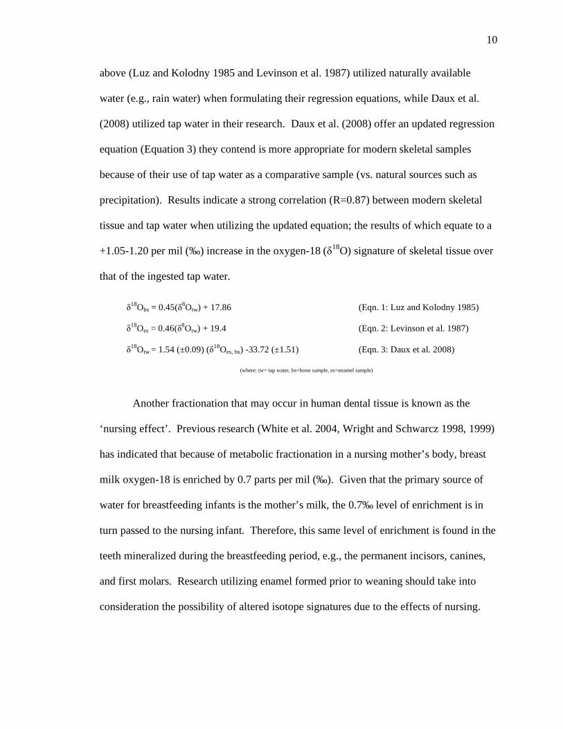
"18
Obs = 0.45("8Otw) + 17.86 (Eqn. 1: Luz and Kolodny 1985)
"18
Oes = 0.46("8Otw) + 19.4 (Eqn. 2: Levinson et al. 1987)
"18
Otw = 1.54 (±0.09) ("18
Oes, bs) -33.72 (±1.51) (Eqn. 3: Daux et al. 2008)
(where: tw= tap water, bs=bone sample, es=enamel sample)
above (Luz and Kolodny 1985 and Levinson et al. 1987) utilized naturally available
water (e.g., rain water) when formulating their regression equations, while Daux et al.
(2008) utilized tap water in their research. Daux et al. (2008) offer an updated regression
equation (Equation 3) they contend is more appropriate for modern skeletal samples
because of their use of tap water as a comparative sample (vs. natural sources such as
precipitation). Results indicate a strong correlation (R=0.87) between modern skeletal
tissue and tap water when utilizing the updated equation; the results of which equate to a
+1.05-1.20 per mil (‰) increase in the oxygen-18 ("18
O) signature of skeletal tissue over
that of the ingested tap water.
Another fractionation that may occur in human dental tissue is known as the
‘nursing effect’. Previous research (White et al. 2004, Wright and Schwarcz 1998, 1999)
has indicated that because of metabolic fractionation in a nursing mother’s body, breast
milk oxygen-18 is enriched by 0.7 parts per mil (‰). Given that the primary source of
water for breastfeeding infants is the mother’s milk, the 0.7‰ level of enrichment is in
turn passed to the nursing infant. Therefore, this same level of enrichment is found in the
teeth mineralized during the breastfeeding period, e.g., the permanent incisors, canines,
and first molars. Research utilizing enamel formed prior to weaning should take into
consideration the possibility of altered isotope signatures due to the effects of nursing.
11
Measurement and Instrumentation
Isotopic analysis is performed utilizing an isotope ratio mass spectrometer
(IRMS), a highly sensitive and specialized form of a mass spectrometer capable of
delivering instrumental precision of <0.02‰ and a standard deviation of < ±0.01‰
(Hoefs 2004, Paul and Skrzypek 2006). As the name implies, a mass spectrometer is
designed to measure the mass of the elements constituting a sample. Simplistically, mass
spectrometry equipment performs three basic functions: (1) breakdown of the sample into
its constituent elements, (2) separation of the elements by mass, and (3) collection and
analysis of the proportions of each element. In the past, a sample intended for mass
spectrometry analysis required conversion into a gaseous compound isotopically
representative of the solid parent material prior to its manual introduction into the mass
spectrometer. Mass spectrometry equipment at the time was equipped with only manual
inlets; no automated introduction mechanisms yet existed (Brazier 1995). This meant
that any sample not already in a gaseous state had to be manually converted and injected,
thus adding time, expense, and the possibility of contamination. As an alternative to
manual injection modern mass spectrometry equipment offers the option of interfacing
with a variety of on-line gas preparation and/or combustion chambers to fully automate
the solid to gas conversion and injection processes. Utilizing this type of technology
eliminates external manipulation; thereby minimizing both expense and the potential for
contamination (Meier-Augenstein and Liu 2004).
Although several methods of isotopic analysis exist, most fit into two general
categories: (1) bulk analysis, and (2) compound specific analysis. Bulk analysis provides
an averaged isotope signature for the sample as a whole, regardless of the isotopic
12
proportions housed within the constituent elemental compounds of the sample (see
Chapter III for a detailed description of the constituent compounds in bones and teeth).
In contrast, compound specific analysis separates the compounds within a complex
sample and delivers an isotopic signature for an individual compound (Carter et al. 2005,
Meier-Augenstein 2007). The bulk analysis method is utilized in this research.
Isotopes are generally measured as a ratio of the two most dominant forms of any
given element. For example, an oxygen isotope analysis would be reported as a ratio in
terms of the two most abundant oxygen isotopes, the heaver 18
O isotope over the lighter
16O isotope (denoted as
18O/
16O). The difference in abundances of two isotopes is
typically quite small, often with the observable difference beginning in the third or fourth
decimal digit (Schoeller 1999). Mass spectrometry data are not generally reported in
their decimal form, but instead are converted to a simpler more manageable form through
the use of standardized equations. The result of an isotopic analysis is reported as a delta
value (") and expressed as a per mil (denoted as ‰) deviation relative to an
internationally recognized standard (Coplen 1996). In the case of oxygen, the delta value
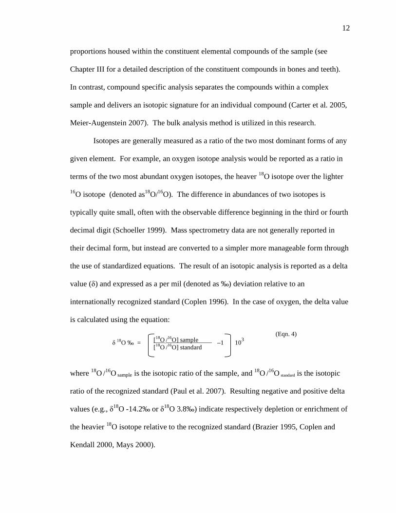
is calculated using the equation:
where 18
O /16
O sample is the isotopic ratio of the sample, and 18
O /16
O standard is the isotopic
ratio of the recognized standard (Paul et al. 2007). Resulting negative and positive delta
values (e.g., "18
O -14.2‰ or "18
O 3.8‰) indicate respectively depletion or enrichment of
the heavier 18
O isotope relative to the recognized standard (Brazier 1995, Coplen and
Kendall 2000, Mays 2000).
(Eqn. 4)
" 18
O ‰ = –1 103 [
18O /
16O] sample
[18
O /16
O] standard
13
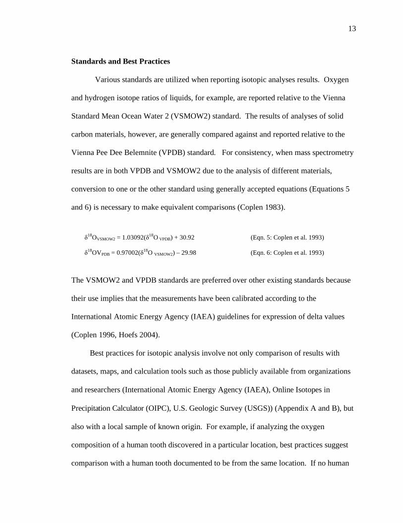
"18
OVSMOW2 = 1.03092("18
O VPDB) + 30.92 (Eqn. 5: Coplen et al. 1993)
"18
OVPDB = 0.97002("18
O VSMOW2) – 29.98 (Eqn. 6: Coplen et al. 1993)
Standards and Best Practices
Various standards are utilized when reporting isotopic analyses results. Oxygen
and hydrogen isotope ratios of liquids, for example, are reported relative to the Vienna
Standard Mean Ocean Water 2 (VSMOW2) standard. The results of analyses of solid
carbon materials, however, are generally compared against and reported relative to the
Vienna Pee Dee Belemnite (VPDB) standard. For consistency, when mass spectrometry
results are in both VPDB and VSMOW2 due to the analysis of different materials,
conversion to one or the other standard using generally accepted equations (Equations 5
and 6) is necessary to make equivalent comparisons (Coplen 1983).
The VSMOW2 and VPDB standards are preferred over other existing standards because
their use implies that the measurements have been calibrated according to the
International Atomic Energy Agency (IAEA) guidelines for expression of delta values
(Coplen 1996, Hoefs 2004).
Best practices for isotopic analysis involve not only comparison of results with
datasets, maps, and calculation tools such as those publicly available from organizations
and researchers (International Atomic Energy Agency (IAEA), Online Isotopes in
Precipitation Calculator (OIPC), U.S. Geologic Survey (USGS)) (Appendix A and B), but
also with a local sample of known origin. For example, if analyzing the oxygen
composition of a human tooth discovered in a particular location, best practices suggest
comparison with a human tooth documented to be from the same location. If no human
14
tooth of known origin is available, a tooth from local fauna may be substituted if species-
specific fractionation effects are considered (Kohn 1996, Kohn et al. 1996, Pye 2004).
Additionally, comparison of the oxygen composition of the human tooth with a water
source from that discovery location is also an acceptable research design (given that the
oxygen concentration in human tooth enamel is a function of ingested water, discussed
below). However, the research design and sample selection depends on the research
question under investigation and the element of focus, therefore the choice of a
comparative sample will vary accordingly (Pye 2004).
Oxygen
Oxygen is a non-metallic element that exists in abundance on Earth as a colorless,
odorless, two-atom gas (O2). There are three naturally occurring, non-radioactive (stable)
principal isotopes of oxygen existing in varying abundances: oxygen-16 (99.763%, 16
O),
oxygen-17 (0.0375%, 17
O), and oxygen-18 (0.1995%, 18
O) (Hoefs 2004). Oxygen is the
third most abundant element in the known Universe, and makes up 47% of the Earth’s
crust, 60% of the human body, 21% of the atmosphere (by volume), and 90% of the
water we drink (Emsley 2001). Because the oxygen concentration in the in meteoric
precipitation is geographically and climatically dependent, concentrations of this gas in
water sources vary across geographic locations (Bowen and Wilkinson 2002, Dansgaard
1964, Gat 1996, Yurtsever and Gat 1981).
Oxygen in Skeletal Tissue
The oxygen isotope analysis of human bone and tooth enamel is based on the
following underlying principles: (1) the strong linear relationship (R= 0.93–0.99) shown
15
to exist between the oxygen composition of skeletal tissues and the oxygen composition
of drinking water, (2) the oxygen composition of drinking water, in turn, reflects its
original source––local precipitation, and (3) the oxygen composition of precipitation is
controlled by geographic and climatic factors such as temperature, altitude, and distance
from the sea (Levinson et al. 1987, Longinelli 1984, Luz et al. 1984, Luz and Kolodny
1985, 1989).
In other words, as water evaporates from a body of water (e.g., the ocean),
condenses to form clouds, and moves inland to varying locations, precipitation occurs
and local water sources are imbued with the isotopic composition of the precipitation as
determined by the geographic location. Generalizing, the closer to the body of water the
more enriched the local precipitation is with the heavier 18
O isotope, given that it
precipitates from condensed water preferentially over the lighter 16
O due to its heavier
mass. As the evaporated moisture/condensation in the form of clouds moves farther
inland, less and less 18
O is available, and therefore less is precipitated. In other words,
the rainwater further inland has less 18
O compared to the lighter 16
O and is considered
depleted of 18
O. Different regions, therefore, have distinct oxygen isotope signatures.
Once deposited in the local water sources, the precipitated water––and its isotopic
signature–is then ingested and incorporated into the body tissues, thereby creating a
unique geo-location tracer within the organism (Hoefs 2004).
The oxygen composition of human bone and tooth enamel is a function of the
oxygen sources consumed. For humans, incorporation of oxygen into body tissues is
primarily (~60-70%) through drinking water (Bryant and Froelich 1995, Hedges et al.
2005, Podlesak et al. 2007, Sponheimer and Lee-Thorp 1999). As a result of this
16
incorporation process, the oxygen composition in the bones and teeth of an organism, as
previously stated, correlate linearly to the oxygen composition of the organism’s
environmental water, and by extension, ingested water.
Several factors can affect the oxygen composition of human bone and teeth.
Metabolically induced fractionation, individual consumption patterns, and diagenesis are
three such factors. Researchers have found that although physiological adaptations,
drinking habits, diet, and body size can significantly alter the linearity between body
tissue and drinking water in the bone and teeth of some species, for humans, intra-
population variation is low (1‰) and the linear relationship between body tissue and
drinking water remains (Bryant and Froelich 1995, Kohn 1996, Kohn et al. 1996,
Longinelli 1984). Additionally, it has been found that the inorganic mineral component
of bone and teeth, the focus of this research, is resistant to post-depositional processes
and preserves its original isotopic signature, particularly in remains not in prolonged
burial contexts (Hedges et al. 2005, Koch et al. 1997, Kohn et al. 1999, Pate 1994,
Sponheimer and Lee-Thorp 1999).
Given that the oxygen composition in the mineral component of bone and tooth
enamel has been demonstrated to be linearly correlated with the oxygen composition of
ingested water and is diagenetically resistant (discussed in Chapter III), bone and teeth
make for highly suitable proxy materials for isotopic research in regards to natal origin
and residential locale.
To fully understand the concept of oxygen isotope analysis of human bone and
tooth enamel, a basic understanding of the element’s presence within the composition of
human skeletal tissue is necessary. Therefore, an overview of the relationship between

17
oxygen and human bone and tooth enamel and a detailed exploration of the composition,
structure, and function of bone is provided in the following chapter. Also provided is a
justification for the selection of tissues used in this study.
18
CHAPTER III
HUMAN BONE AND TOOTH ENAMEL
Introduction
The exceptional durability of bone and teeth makes these tissues the most often,
and sometimes only, surviving substances of the human body subsequent to death. Their
consistent presence in the paleoanthropological, archaeological, and forensic records
makes them one of the most analyzed materials in anthropological research (Hedges
2005). Although the constituent components of bone and tooth enamel are similar, they
vary dramatically in proportion, formation, structure, and reaction to post-deposition
alteration (diagenesis). Given that isotopic analysis typically targets individual molecular
compounds, a basic comprehension of the composition differences between bone and
teeth is essential in the molecular level type research presented in this project. For this
reason, a brief review of the structure, biochemistry, and potential diagenetic changes of
human bone and tooth enamel is given.
Bone Structure and Composition
The average adult human skeleton is comprised of 206 bones, or elements, of four
general forms: (1) long bones––characterized by a hollow, tubular shaft (diaphysis) with
flaring, closed ends (epiphysis), found in the upper and lower extremities, and whose

19
main functions are weight bearing, locomotion, and manipulation, (2) flat bones–
characterized by thin, tabular shaped bones, found in the cranial vault, shoulder, rib
cage, and pelvis, and whose primary functions are protection and provision of large areas
for muscle attachment, (3) irregular bones––such as vertebrae, which exhibit a variety of
complex shapes and sizes depending on their location and function, and (4) short bones––
such as carpals and tarsals, are roughly cuboidal and like irregular bones exhibit a variety
of sizes, shapes, and functions (Bass 1995, White and Folkens 2000).
Based on porosity, two types of bone (osseous) tissue exist within adult human
skeletal elements: (1) compact tissue––also referred to as cortical tissue depending on if
the tissue is in the form of a thick or a thin layer––is a dense, tightly packed tissue of 5%-
10% porosity, and (2) cancellous tissue––also called spongy tissue, is a porous,
lightweight, honeycomb structured tissue, with porosity of 75%-95% (Martin et al. 1998).
Compact or cortical tissue forms the outer walls of the long and short bone diaphyses and
the external surfaces of the flat and irregular elements, varies in thickness depending on
the skeletal element, and comprises the bulk of the osseous tissue in the human skeleton.
Trabecular tissue makes up the portion of bone between the cortical layers of the flat and
irregular bones, the inner shafts of long bones, and is the major tissue constituting long
bone epiphyses (White and Folkens 2000). Some skeletal elements, such as the femur
and the tibia, have a high proportion of compact tissue, while others, such as vertebrae
and ribs, have higher proportions of cancellous tissue (Martin et al. 1998). In this
research the terms compact and cancellous will be used when referring to these two
osseous tissue types.
20
Although a wide array of shapes and sizes exist in the human skeleton, the basic
structure of osseous tissue is that of a highly organized structural matrix of protein with
mineral crystals interspersed within the gaps of the matrix, a composition producing a
tough, structural integrity in human bone. Within this basic structure human bone is
composed of two principal components: organic and inorganic (Martin et al. 1998).
The organic component makes up ~30% of dry bone by weight and is primarily
comprised of collagen (Martin et al. 1998). Collagen––a structural protein found
ubiquitously throughout vertebrates––is the most abundant protein found in the human
body and exists in several forms. Type I collagen, the primary form of collagen found in
human bone, is organized into strong, flexible fibers with alternating parallel and
perpendicular orientation to the bone surface (Martin et al. 1998). This alternating
organization provides the matrix for deposition of the mineral component of the inorganic
portion of bone (Martin et al. 1998).
The inorganic component makes up ~70% of dry bone by weight and is
comprised principally of a composite of calcium phosphate minerals closely resembling
the naturally occurring mineral, hydroxyapatite (Ca10(PO4)6(OH)2) (Martin et al. 1998).
Biological hydroxyapatite, or bioapatite, differs from the geological hydroxyapatite in its
lack of stoichiometry (i.e., ratio of atoms remain the same before and after a chemical
reaction), small crystal size, high rate of substitution, and structural disorganization
(Budd et al. 2001, Martin et al. 1998). The hydroxyapatite in bone is considered impure
due to its poor crystalline structure that allows for elemental substitutions, substitutions
supplied by ingested food and water or resulting from diagenetic processes. In humans,
the primary substitutions occur in the phosphate (PO4) and hydroxyl (OH) sites of the
21
hydroxyapatite molecule, mainly substituted with carbonate CO3 (although other
substitutions such as fluoride and chloride can also occur) (Hedges et al. 2005, Kohn et
al. 1999). The carbonate substitution renders a hydroxyapatite formula with a CO3
molecule in place of either or both of the PO4 or OH molecules (Shemesh 1990).
Therefore, human bone contains three sites where oxygen, in varying content, may be
found: 1) the phosphate site (PO4), containing 35% oxygen, 2) the hydroxyl site (OH),
containing 1.6% oxygen, and 3) the carbonate site (CO3), containing 3.3% oxygen
(Martin et al. 1998).
Although both the phosphate and carbonate sites within the mineral component of
bone house oxygen available for analysis, a large proportion of studies have primarily
utilized the phosphate site for several reasons. First is the high proportion of analytical
oxygen available in the phosphate site (35%) as compared to the amount available in the
carbonate site (3.3%) (Martin et al. 1998). Second, the P-O bond of the PO4 molecule is
stronger than the C-O bond of the CO3 molecule, suggesting the phosphate site is more
diagenetically resistant (Sponheimer and Lee-Thorp 1999). Third is the strong
correlation (R=0.93–0.99) demonstrated to exist between the phosphate oxygen and the
oxygen composition of ingested water (Longinelli 1984, Luz and Kolodny 1985, 1989).
Taken together, these characteristics render the phosphate site the most likely to yield
useful oxygen composition data, and therefore is a common analyte choice for oxygen
isotope research (Budd et al. 2001, Hedges et al. 2005, Koch et al. 1997).
The carbonate site, although containing less oxygen, is sometimes the chosen
analyte for two reasons. First, the strong correlation (R=0.98) demonstrated to exist
between the carbonate oxygen and phosphate oxygen indicates that, after conversion
22
(Equation 7), the carbonate site reflects the same isotopic signature as the phosphate site,
and therefore, is equally as informative in isotopic studies (Bryant et al. 1996, Iacumin et
al. 1996). Second is the ease of oxygen extraction from the carbonate molecule
compared to that of the more complex, lengthy process required for oxygen extraction
from the phosphate molecule (Iacumin et al. 1996, Sponheimer and Lee-Thorp 1999).
"18
O (phosphate) = 0.998* "18
O (carbonate) -8.5 (Eqn. 7: Iacumin et al. 1996)
The structural integrity and high mineral content of bone render it one of the
longest surviving and, therefore, most abundant tissues available for analysis following
the death of a vertebrate organism.
Tooth Structure and Composition
Although typically not as abundant, human tooth enamel—the hardest substance
is the human body–is also a tissue often chosen for isotopic analysis. Humans develop
two sets of dentition throughout their lifetime. One set, the deciduous, or baby teeth,
begin mineralizing (forming) in utero and erupt from early infancy though early
childhood. Deciduous teeth start erupting at approximately 6 months of age and typically
by the age of 2 years eruption is complete, resulting in a full set of 20 teeth. Beginning
around the age of 6 years the deciduous teeth begin to be displaced by the emerging
permanent teeth (Hillson 1996). The 20 deciduous teeth are progressively lost
throughout the early childhood and pre-teenage years when they are replaced by a full set
of permanent teeth (Anderson et al. 1976, Scheuer and Black 2004).
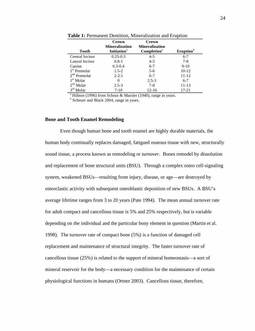
The second set of human dentition, the permanent teeth, mineralize from birth
through the early teenage years, and erupt from early childhood through the late teenage
years (see Table 1). Beginning around the age of 6 years the first permanent teeth
23
emerge and continue to emerge in a particular order until all 32 permanent teeth are fully
erupted. The first 28 permanent teeth erupt at predictable intervals, with completion
around the age of 12. The remaining four teeth, the third molars (wisdom teeth), exhibit
highly variable eruption and, in some cases, never erupt. The typical eruption period for
the third molars is approximately 18-21 years of age (Anderson et al. 1976, Hillson 1996,
Scheuer and Black 2004).
Both deciduous and permanent teeth have three types of hard tissue: (1) enamel–
the accreted layers of material covering the crown, (2) dentine––the bulk of the inner
tooth, and (3) cementum––the outer layer of the roots (Scheuer and Black 2004). As with
bone, these components contain varying organic and inorganic proportions. Tooth
enamel, the focus of this study, is composed of ~1% organic protein (amelogenin), ~2%
water, and ~97% inorganic minerals (hydroxyapatite) (Hillson 1996). The organic
portion of tooth enamel is principally (90%) composed of the protein amelogenin
functioning similarly to the collagen in bone, providing a structural matrix for the
deposition of inorganic minerals (Hillson 1996). The inorganic mineral portion of tooth
enamel functions similarly to bone, but is composed of larger crystals and a greater
structural organization, a structure allowing fewer ionic substitutions than is found in
bone. These properties, along with the high mineral content, afford enamel the
characteristic of being the most durable and elementally stable substance in the human
body.
24
Table 1: Permanent Dentition, Mineralization and Eruption
Tooth
Crown
Mineralization
Initiationa
Crown
Mineralization
Completiona
Eruptionb
Central Incisor 0.25-0.3 4-5 6-7
Lateral Incisor 0.8-1 4-5 7-8
Canine 0.3-0.4 6-7 9-10
1st Premolar 1.5-2 5-6 10-12
2nd
Premolar 2-2.5 6-7 11-12
1st Molar 0 2.5-3 6-7
2nd
Molar 2.5-3 7-8 11-13
3rd
Molar 7-10 12-16 17-21 a Hillson (1996) from Schour & Massler (1940), range in years.
b Scheuer and Black 2004, range in years.
Bone and Tooth Enamel Remodeling
Even though human bone and tooth enamel are highly durable materials, the
human body continually replaces damaged, fatigued osseous tissue with new, structurally
sound tissue, a process known as remodeling or turnover. Bones remodel by dissolution
and replacement of bone structural units (BSU). Through a complex osteo cell-signaling
system, weakened BSUs––resulting from injury, disease, or age––are destroyed by
osteoclastic activity with subsequent osteoblastic deposition of new BSUs. A BSU’s
average lifetime ranges from 3 to 20 years (Pate 1994). The mean annual turnover rate
for adult compact and cancellous tissue is 5% and 25% respectively, but is variable
depending on the individual and the particular bony element in question (Martin et al.
1998). The turnover rate of compact bone (5%) is a function of damaged cell
replacement and maintenance of structural integrity. The faster turnover rate of
cancellous tissue (25%) is related to the support of mineral homeostasis––a sort of
mineral reservoir for the body––a necessary condition for the maintenance of certain
physiological functions in humans (Ortner 2003). Cancellous tissue, therefore,
25
continually and quickly turns over constituent elemental components while compact
tissue does so more slowly.
In contrast to bone, tooth enamel is a dense, static tissue, and once formed
undergoes no post-mineralization remodeling or element exchange with its environment
(Hillson 1996). Therefore, enamel maintains its original elemental signature encoded
during mineralization. The lack of remodeling and the miniature archival quality of teeth
make them one of the most often chosen bio-materials for elemental isotopic analysis
(Lee-Thorp 2002).
Diagenesis and Forensic Material
Although an elementally durable material that tends to resist external influences,
the elemental analysis of bone and teeth has historically been riddled with difficulties,
difficulties brought about by the effects of diagenesis. Anthropologically, diagenesis is
defined as the chemical alteration of biological material, from both infiltration and
leaching within the depositional context, that occurs from the time of death to discovery
in the archaeological or forensic record (Darwent and Lyman 2002, Lyman 1994).
Although no skeletal tissue is immune to digenetic processes, the dense, highly
mineralized structure of tooth enamel affords it a measure of protection. Bone, on the
other hand, is a more porous tissue, allowing for greater potential of diagenetic exchange.
According to Sorg and Haglund (2002), the initial biochemical response of bone to its
depositional environment is similar to that of bone in a fresh, living state; a response
afforded it by the protection of retained moisture and organic components. As the length
of deposition increases and the organic components within and surrounding the skeletal
tissue decompose, the chemical composition of the osseous tissue may change. The type
26
and extent of this change is largely dependent on the surrounding environmental
conditions and whether the deposition is in a buried or surface context (Rodriguez 1997).
Within the deposition environment such extrinsic factors as moisture, temperature, soil
pH, and the presence of microorganisms are primary factors affecting the presence and
rate of diagenetic alteration of bone and teeth (Gill-King 1997).
Potential diagenetic effects on bony tissue in regards to modern forensic material
are often disregarded due to the depositional context and/or youth of the material being
analyzed (Hedges et al. 2005). Although it is true that much of the material forensically
analyzed is generally less than 60 to 70 years of age, these materials, as with
archaeological materials, are subject to diagenetic processes. Skeletal remains deposited
as the result of a crime are discovered in a wide variety of depositional contexts, many
containing the same varying microenvironments found within archaeological sites
(Hochrein 2002). It is important to keep in mind that diagenetic processes are not
uniform and, therefore, no concrete timeline can be developed that delineates when
osseous tissue becomes diagenetically susceptible (Haglund and Sorg 1997).
Disregarding the potential of postmortem alteration based on temporal criteria, even for
modern samples, is highly cautioned against (Hedges et al. 2005).
Hedges (2002) indicates a number of intrinsic factors of bone such as size, shape,
condition, and age that may also contribute to its chemical integrity. For example, the
highly porous, aged bones often found in the elderly may succumb to diagenetic
processes more quickly than would healthy, dense tissue found in younger adults.
Similarly, the incompletely mineralized bone of infants and children are also vulnerable
27
to diagenetic alteration and are highly susceptible to not only rapid chemical exchange,
but to external mechanical damage as well.
A variety of techniques exist to assess diagenetic alteration including (1)
consideration of the deposition context, (2) visual assessment for signs of color, weight,
porosity, and structure changes, and (3) spectral techniques such as X-ray diffraction,
infrared spectroscopy, and Fourier transform infrared spectroscopy (FTIR) (Hedges 2002,
Sandford and Weaver 2000, Shemesh 1990). When postmortem alteration is suspected,
various sample preparation treatments are utilized to remove diagenetically absorbed
contaminants. For bone and tooth enamel, contaminant removal is typically performed
via an acetic acid wash. (Garvie-Lok et al. 2004, Pate 1994). Once the acid wash is
complete, the element signature remaining more accurately reflects the in vivo isotope
condition.
Human bone and tooth enamel have been utilized extensively for research in a
variety of academic disciplines. Reviewed in the following chapter are studies utilizing
isotope analyses of human bone and tooth enamel to explore research questions, clarify
historical events, and challenge long standing misconceptions regarding past peoples. In
addition to formal research, a modern forensic homicide case in which isotopic analysis
was utilized as an investigative tool is discussed.
28
CHAPTER IV
PREVIOUS RESEARCH AND MODERN FORENSIC APPLICATION
Introduction
The use of oxygen isotope analysis for the acquisition of information to assist in
the reconstruction of ancient climates, migration patterns, diet, habitation, and origins of
both humans and animals is by no means a novel idea. Since their discovery by Thomson
and Aston in 1913, and the subsequent identification of most existing stable isotopes in
the 1930s, the use of isotopes in research has proven invaluable throughout a range of
disciplines (Katzenberg 2000). Previously a research cornerstone in medicine, chemistry,
and geology, the increased number of studies employing isotopic analysis from an array
of disciplines indicates its growing significance as a routine research tool, particularly in
the field of anthropology (Katzenberg and Harrison 1997, Sandford and Weaver 2000).
Although the most frequently utilized elements in isotopic investigations have
traditionally been carbon and nitrogen, oxygen is steadily becoming an analyte choice
(Mays 2000, Pye 2004). Explored below are studies utilizing oxygen isotope analysis of
human bone and tooth enamel for inference of habitation, natal origin, and migration.
Origin and Migration
Conventional methods for tracing the origins and migration of past peoples
primarily rely on identifying the artifactual similarities of material culture. Sites

29
(including burials) with similar artifacts, but located some distance apart, are often linked
via the causal explanation of migration or trade networks in existence between the sites
(Evans et al. 2006). Unfortunately the impermanent nature of moveable objects renders
concrete associations nearly impossible without corroborating evidence (e.g.,
hieroglyphic texts and iconography). The employment of isotopic studies in regards to
the study of past peoples is beginning to delineate the hazy boundary between actual
migration and what was simply trade or casual transport of material objects (Evans et al.
2006, Lee-Thorp 2002). Although isotopic studies investigating migration and origins
have typically utilized strontium (e.g. Ezzo et al. 1997, Ezzo and Price 2002, Price et al.
1994, 2002, Sealy et al. 1991), researchers are beginning to use oxygen analysis as an
additional means of elucidating the movement of past peoples (Lee-Thorp 2002).
The underlying principle of oxygen isotope analysis of human bone and teeth, as
stated previously (Chapter II), is the strong linear relationship between the oxygen
composition of body tissue formed at various stages of life, and that of the water ingested
during the formation of that tissue. Therefore, it is reasonable to assume that
geographically distinct isotopic signatures identifiable in human tissues are suggestive of
the geolocations of an organism at different developmental stages.
Several early studies utilizing oxygen isotopes for geolocation determination were
conducted by Keenleyside et al. (1997), Schwarcz et al. (1991), and White et al. (1998).
Keenleyside and colleagues (1997) used oxygen and lead isotope analyses to investigate
and determine the geographic origin of nearly 400 human skeletal elements and bone
fragments. These represented a minimum of 11 individuals thought to be from, but
inconclusively linked to, the ill-fated European crewed, America bound Franklin
30
Expedition of 1845-1848 (Melbye and Fairgrieve 1994). Results of the isotope analyses
reflected the birthplace and most recent habitation locale of two of the individuals
represented by the bones and fragments as Western Europe and not the Northern Arctic.
This, therefore, suggested the remains were potentially those of the Franklin Expedition
crew. Although not conclusive evidence of exact origin, the multi-elemental isotopic
evidence found in this study sufficiently allayed suspicions that the remains were of local
Inuit origin. To date, 3 of the represented 11 individuals have been positively identified
utilizing a multi-disciplinary approach combining both traditional and isotopic
techniques.
Schwarcz and associates (1991) conducted oxygen isotope analyses on human
skeletal remains from the War of 1812 interred in the Snake Hill military cemetery,
Ontario, Canada. This was done in an attempt to gain insight into the place of origin of 6
of the buried soldiers. Results of the study demonstrated a fairly uniform oxygen
signature, suggesting the interred individuals were from the same general region. Based
on isotopic values from local archaeological and modern reference samples, it was found
that the region, however, was not Ontario. Results instead indicated a lower latitude
region, most likely the Northeastern United States. This supported the authors’ original
intent to demonstrate that at least some of the military interments at the Canadian
cemetery were in fact US soldiers.
Similarly, White and colleagues (1998) conducted oxygen isotope analyses on
archaeological samples from Tlailotlacan, Mexico, a culturally homogenous enclave on
the western edge of Teotihucan in the Valley of Mexico. The authors theorized that
immigrants from the southern Valley of Oaxaca originally populated Tlailotlacan. Using
31
Monte Alban (Valley of Oaxaca) and the Teotihucan peripheral site of Tlajinga (Valley
of Mexico) as comparative baseline isotope ratios, the authors showed the isotopic
signatures of the Tlailotlacan enclave more closely resembled the Oaxacan Valley ratios
than those from the Valley of Mexico. These finding supported the authors’ contention
of an immigrant enclave in the Valley of Mexico.
In a 2000 study, White and colleagues once more focused their attention on the
Valley of Mexico, this time to determine the extent of Teotihuacan (Valley of Mexico)
influence over a neighboring region, Kaminaljuyu, Guatemala. Again utilizing oxygen
isotopes, White and associates (2000) analyzed enamel from the remains of 31 suspected
“foreigners” (based on archaeological evidence) in positions of power––be it a ruler or
an elite citizen––in the ancient Mayan city of Kaminaljuyu in an attempt to infer natal
origins of the 31 individuals. Results demonstrated only a minor biochemical link (3 of
31 samples) to the Valley of Mexico, challenging previously held beliefs of a larger
Teotihuacan presence in Kaminalijuyu. Instead, results indicated support for a more
recent view (Braswell 2004) that local Kaminalijuyu elite appropriated Teotihuacan
materials and symbols to elevate their personal status within the community. Authors
White et al. caution, however, against broad interpretation of results based solely on tooth
enamel since enamel is indicative of early childhood location only and provides no
evidence of later life residence or mobility. The concluding results of this study simply
suggest that the remains of what were believed to be foreign individuals were in fact born
locally in the Kaminalijuyu area.
In perhaps one of the better-known studies employing isotopic techniques,
Hoogewerff et al. (2001) and Muller et al. (2003) utilized data from strontium, carbon,
32
and oxygen isotope analyses to suggest the natal origins and adult residential locale in
relation to the discovery site of the 5300 year old mummified remains of the famed
Alpine iceman, “Otzi.” Results based on the analyses performed on bone and enamel
samples from Otzi’s rib, femur, canine and first premolar indicate Otzi most likely spent
the majority of his adult life at higher altitudes of the Alpine region, while his early years
were spent at lower altitudes. This evidence, the authors suggest, may assist in resolving
the still unanswered question of who really was this Late Neolithic individual that died an
obviously unexpected death in the inhospitable environment of the high Alps.
Long held beliefs supported by tangible archaeological evidence have not escaped
challenge when confronted with the results of isotopic analysis. Based on several lines of
archaeological evidence, anthropologists have generally accepted that the Mayan king,
K’inich Yax K’uk Mo (ruled A.D. 426-437), founder and first ruler of the Early Classic
dynasty of Copan in the central Peten region (c. A.D 250-600), was originally from a
region northwest of Copan, most likely the powerful political center at Teotihuacan in the
Valley of Mexico (Sharer and Traxler 2005). Additionally, hieroglyphic texts deciphered
at the Copan Acropolis also indicate that the Mayan lord originated from outside the
Copan region. Utilizing both strontium and oxygen isotopes, Buikstra and colleagues
(2004) investigated the natal origins of the Mayan king in relationship to his burial
location at Copan. Results from the analyses indicated that the remains archaeologically
identified as those of K’inich Yax K’uk Mo were more characteristic of Copan in the
Central Peten than of the Valley of Mexico. These results effectively challenged the
prevailing theory that the Mayan king was originally from the prestigious political site of
the northern located Teotihuacan.
33
In another study originating from the Valley of Mexico, Spence and associates
(2004) attempted to isotopically discern the origins of human maxillae trophy pendants
worn by sacrificed soldiers who were excavated from a mass grave at the Feathered
Serpent Pyramid (c. A.D. 200), Teotihuacan. Results of the oxygen analysis revealed two
interesting outcomes. First, the victims represented by the trophy maxillae were from
both nearby, perhaps even Teotihuacan, and distant regions, suggesting the soldiers may
have killed local residents as well as foreigners. Second, the archaeological evidence
reporting Teotihuacan’s military interactions with other Mesoamerican regions does not
appear until a century after the construction of the Feathered Serpent Pyramid. Evidence
from the isotopically analyzed trophy maxillae excavated from the Pyramid suggests
otherwise by demonstrating that interaction with more distant regions must have occurred
prior to the time indicated by archaeological evidence. As in the Copan study discussed
above, the utilization and results of isotopic analysis on the maxillae trophy pendants of
Teotihuacan again challenged the archaeologically based acceptance of long standing
theories concerning the timing of its military interactions with foreign regions.
Bentley and colleagues (2005) uniquely utilized isotopic analysis to support the
genetic and linguistic evidence indicating the presence of matrilocality during the
agricultural transition in the village of Ban Chiang, Thailand. Additionally, they utilized
isotope data to lend clarity to the contradictory genetic results suggesting that Thai male
ancestors originated elsewhere, while females shared a local maternal ancestor. The
results of carbon, strontium, and oxygen isotope analyses of the remains of 44 individuals
demonstrated support for the authors’ contention that males during this period emigrated
to Ban Chiang, while the females were of local origin. These data supported their theory
34
that the females of Ban Chiang shared a long, local maternal ancestry and were
practitioners of matrilocality.
In two recent studies, Evans and associates (2006) demonstrated the utility of
isotopic analysis as evidence for the inference of migratory behavior in Southern
England. In the first of these studies, Evans and fellow researchers performed oxygen
and strontium analysis on a set of 16 burials from a Late Roman cemetery in Winchester
to test their hypothesis that the exotically interred individuals were immigrants from the
distant Danube region in Central Europe. Although the isotopic results showed a varied
set of values for the burials, none were consistent with Southern England, indicating
support for the authors’ contention that the burials were indeed of non-indigenous origin.
The authors further suggested that such diverse results for the exotic interments provided
support for an additional hypothesis that the individuals did not relocate as one large
community group, but instead included small groups, families, and single individuals.
In a second study, Evans and Chenery (2006) utilized isotopic analysis to discern
the origins of an individual interred in a particularly rich fashion for the early Bronze
Age, the “Amesbury Archer,” discovered three miles Southeast of Stonehenge. Resulting
isotopic values from the Archer’s teeth revealed a childhood location other than
Amesbury or even England, but instead most likely a cooler locale in central Europe.
The authors further contended that the lavishly buried Archer, dating to approximately
2300 BC, was of particular anthropological interest in regards to the chronological
relationship with the large stone building phase of Stonehenge, raising question as to the
Archer’s involvement and potential ‘foreign’ influence on the construction of
Stonehenge. In the two studies discussed above Evans and colleagues successfully
35
demonstrated the ability of isotopic analysis to cast doubt on the belief that migrations
were most often a community or group affair.
Similarly, Prowse and colleagues (2007) attempted to demonstrate that migration
to the Italian island of Isla Sacra was not the predominantly single, adult male activity as
indicated by historical records. The authors theorize instead that movement of people to
the island was potentially a family affair as well. The authors conducted oxygen isotope
analysis of first and third molars of 61 individuals (both male and female) interred in the
necropolis of Isla Sacra (1st-3rd century AD). Isotopic results indicated that
approximately 23% of the individuals analyzed relocated during the time period between
crown completion of the first and third molars (ca. 2-3 and ca. 10-16 years of age
respectively). These results supported not only the authors' contention that migratory
behavior was not limited to adult males but also lent credibility to the migration
hypotheses tested by Evans and associates in the above mentioned studies conducted
in Southern England.
Modern Forensic Application
Application of isotopic analysis to modern human material is a relatively recent
development in regards to forensic investigations. Few studies conducted have been
specifically concerned with forensic applications and utilization of isotopic signatures in
modern remains, and fewer still have been conducted using the oxygen element for
inference of origin and habitation of an unidentified individual (Pye 2004).
One researcher utilizing oxygen isotope analysis for the specific purpose of
investigating natal origins is Henry P. Schwarcz of the Department of Anthropology,
McMaster University, Ontario, Canada. Following is the detailed account of a recent
36
homicide case for which Schwarcz conducted oxygen analyses on bone and tooth enamel
in an attempt to gain data regarding the origin and residence of the murdered individual
(Schwarcz 2007).
Mammoth Lakes Homicide Case
In late May 2003 a hiker discovered skeletonized human remains along a trail in
the Mammoth Lakes region of Central California. The remains were estimated to be
those of a 30-40 year old female who stood approximately 4’6” at the time of death, with
an estimated postmortem interval of 9 months. After the failure of several conventional
identification techniques (e.g., DNA typing) investigators pursued alternative methods,
inviting Schwarcz and his expertise in isotopic analysis into the case.
Schwarcz’s analysis resulted in a determination that the victim had imbibed water
as a child from a location other than where the remains were discovered. In fact, the
oxygen signature in the tooth enamel could have come only from an area much farther
south. Because other evidence connected the Mammoth Lakes victim with the southern
Mexican village of San Mateo in the Oaxacan region, Schwarcz also conducted an
oxygen analysis of a drinking water sample from that area. The drinking water analysis
resulted in a match to the oxygen value found in the tooth enamel, supporting a San
Mateo origin. Continuing, Schwarcz performed an oxygen analysis of a rib fragment––
information that would indicate residential locale for approximately the last eight to ten
years. The result of the rib analysis matched neither the tooth oxygen value (San Mateo)
nor the oxygen signature of the Mammoth Lakes region (established through the analysis
of a baby tooth from a lifelong resident). Knowing that the bone sample would reflect a
fairly recent geolocation, and taking into account the enamel and San Mateo water
37
oxygen values, as well as other case evidence, Schwarcz concluded that the victim was
most likely born and lived the majority of her life in the Oaxacan region. Additionally,
because no oxygen signature of the Mammoth Lakes region registered in the victim’s
teeth or bones, Schwarcz was confident the victim’s arrival to Central California was
recent, most likely within the last two years (Dotsie 2007, Page 2007, Schwarcz 2007).
Armed with this evidence investigators focused their efforts in the Southern Mexico
region of Oaxaca and were eventually able to tentatively identify the remains as Barbara
Pacheco Santiago; an identification currently awaiting DNA confirmation.
Previous Research Conclusion
Although this review illustrates a variety of archaeological applications for which
isotope analysis has been utilized, it demonstrates only a fraction of the research
conducted over the past several decades. Consequently, a wealth of comparative
information exists for those attempting to reconstruct the lives of past peoples. To date,
however, the use of isotope analysis in a modern forensic context has yet to be fully
realized or extensively applied.
38
CHAPTER V
MATERIALS AND METHODS
Introduction
In an effort to test the efficiency of oxygen isotope analysis of modern forensic
cases, fourteen unsolved forensic cases curated at the Forensic Anthropology Center at
Texas State University-San Marcos were examined for potential inclusion in this study.
Only adult remains (estimated age #18) with the required elements (see Justification for
Tissues Used in This Study below) were selected for inclusion in this research. To
minimize the possibility of erroneous isotope readings, cases exhibiting obvious
pathology were not considered for this study. Three analyses (hereafter referred to as a
‘set’) were performed on each selected case. A set contained one sample each of
cancellous tissue, compact tissue, and dental enamel. The cancellous and compact tissue
samples were taken from the shaft of the femur and the dental sample from an intact
maxillary molar (see Justification for Tissues Used in This Study below). To establish a
local oxygen signature for the known discovery locations (counties) of the cases, a fourth
analysis was performed utilizing a sample of municipally available drinking water
collected from a city within each county (Chapter II) (Bowen 2007).
In an effort to minimize potential postmortem contaminants, all skeletal elements
were visually assessed and any remains exhibiting signs of postmortem diagenetic
39
alteration were precluded from this study. Diagenetic alteration was assessed based on a
visual assessment for differential coloring, weight, porosity, structural abnormalities, and
weathering. The six cases chosen (see Table 2) for inclusion presented a uniform
appearance and exhibited no signs of structural damage or advanced weathering, e.g.,
cracking and/or flaking of the outer surfaces. These cases represent unidentified
individuals from four South Texas counties.
As previously discussed in Chapter III, a variety of methods exist for assessing
and accounting for postmortem alteration. The standard protocol most commonly
utilized is the acetic acid wash (Hedges 2002). Although no diagenetic alteration was in
evidence, it is standard procedure to perform a contamination removal treatment as a
precaution even when no contamination is suspected. Therefore, an acetic acid
contamination removal treatment was utilized on a portion of the samples in this study.
Two sub-samples were created from each primary bone and enamel sample, heretofore
referred to as sub-sample A and B. Sub-sample A was treated with the acetic acid soak
described below, while sub-sample B was left untreated (see Table 3). As a side analysis,
isotope values from both sub-sample sets were compared to suggest the necessity of
diagenetic contaminant removal treatments for forensic material (see Chapter III).
Justification for Tissues Used in This Study
Although several tissues (bone, teeth, dry tissue, and hair) are often recovered
with a set of remains, the selection of tissue for elemental isotopic analysis is a function
of the research objectives. In this study, the primary objectives are the exclusionary
inference of: (1) most recent habitation locale, (2) long term habitation locale, and (3)
early childhood location based on a match/mismatch with the isotopic signature of the

40
local drinking water for each of the individuals analyzed. These objectives demand the
analysis of tissues that provide supporting evidence. Therefore, the tissues selected for
analysis in this study are: (1) compact and cancellous tissue from the proximal one-third
of a femoral diaphysis and, (2) enamel from a first permanent maxillary molar.
The decision to utilize a tissue sample from the femur is based on the thickness of
its proximal shaft. This portion of the element houses some of the thickest compact
tissue (along with the tibia) in the human body, and therefore, provides the longest
elemental life history for the individual. The femur, with its thick outer compact tissue
layer and slow turnover rate discussed previously, can provide a life history extending to
~20 years prior to death (Hill and Orth 1998). The cancellous tissue of the femur is also
highly useful as a residential indicator. The quick turnover rate of this tissue makes it an
ideal choice for rendering evidence of recent habitation, on the order of $ 8-10 years
(Simmons et al.1991). Taken in concert, the outer compact and inner cancellous tissue of
a large, thick element such as the femur holds the potential to yield evidence for
objectives #1 and #2, recent and long-term geo-habitation.
The choice of the first permanent maxillary molar (#3 or #14) for this study is
derived from the fact that the chronological mineralization of individual teeth, although
somewhat variable, is predictable and well known (Hillson 1996). Therefore, the
selection of a tooth for analysis in this study is based on the objective of obtaining
information to infer early childhood residence. This objective demands the analysis of
one of the earliest forming adult teeth. The permanent first molars, incisors, and canines
begin mineralization in utero and during early infancy, with crown (enamel) completion
at 2-3, 4-5, and 6-7 years of age respectively (see Table 1), thus providing a static
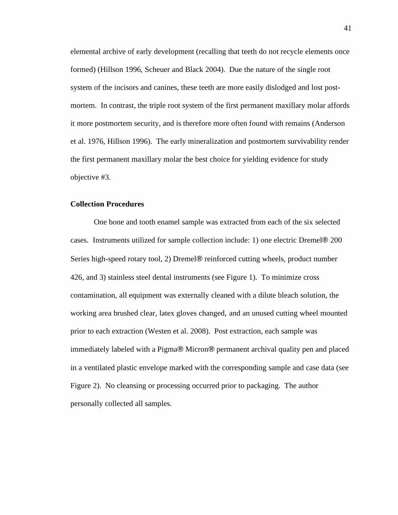
41
elemental archive of early development (recalling that teeth do not recycle elements once
formed) (Hillson 1996, Scheuer and Black 2004). Due the nature of the single root
system of the incisors and canines, these teeth are more easily dislodged and lost post-
mortem. In contrast, the triple root system of the first permanent maxillary molar affords
it more postmortem security, and is therefore more often found with remains (Anderson
et al. 1976, Hillson 1996). The early mineralization and postmortem survivability render
the first permanent maxillary molar the best choice for yielding evidence for study
objective #3.
Collection Procedures
One bone and tooth enamel sample was extracted from each of the six selected
cases. Instruments utilized for sample collection include: 1) one electric Dremel! 200
Series high-speed rotary tool, 2) Dremel! reinforced cutting wheels, product number
426, and 3) stainless steel dental instruments (see Figure 1). To minimize cross
contamination, all equipment was externally cleaned with a dilute bleach solution, the
working area brushed clear, latex gloves changed, and an unused cutting wheel mounted
prior to each extraction (Westen et al. 2008). Post extraction, each sample was
immediately labeled with a Pigma! Micron! permanent archival quality pen and placed
in a ventilated plastic envelope marked with the corresponding sample and case data (see
Figure 2). No cleansing or processing occurred prior to packaging. The author
personally collected all samples.
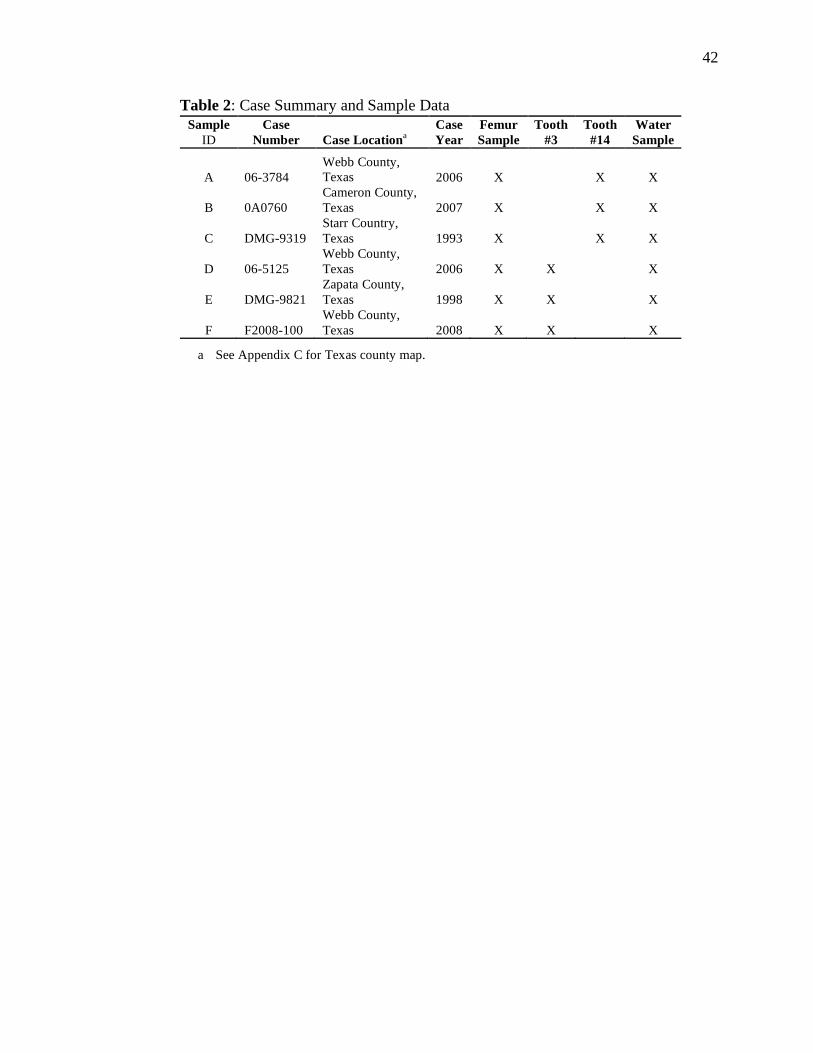
42
Table 2: Case Summary and Sample Data
Sample
ID
Case
Number
Case Locationa
Case
Year
Femur
Sample
Tooth
#3
Tooth
#14
Water
Sample
A 06-3784
Webb County,
Texas 2006 X X X
B 0A0760
Cameron County,
Texas 2007 X X X
C DMG-9319
Starr Country,
Texas 1993 X X X
D 06-5125
Webb County,
Texas 2006 X X X
E DMG-9821
Zapata County,
Texas 1998 X X X
F F2008-100
Webb County,
Texas 2008 X X X
a See Appendix C for Texas county map.
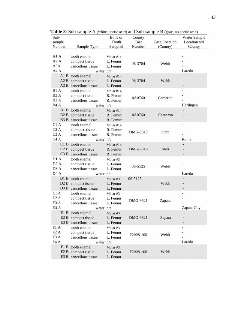
43
Table 3: Sub-sample A (white, acetic acid) and Sub-sample B (gray, no acetic acid)
Sub-
sample
Number Sample Type
Bone or
Tooth
Sampled
County
Case
Number
Case Location
(County)
Water Sample
Location w/i
County
A1 A tooth enamel Molar #14 -
A2 A compact tissue L. Femur -
A3A cancellous tissue L. Femur -
A4 A water n/a
06-3784 Webb
Laredo
A1 B tooth enamel Molar #14 -
A2 B compact tissue L. Femur -
A3 B cancellous tissue L. Femur
06-3784 Webb
-
B1 A tooth enamel Molar #14 -
B2 A compact tissue R. Femur -
B3 A cancellous tissue R. Femur -
B4 A water n/a
0A0760 Cameron
Harlingen
B1 B tooth enamel Molar #14 -
B2 B compact tissue R. Femur -
B3 B cancellous tissue R. Femur
0A0760 Cameron
-
C1 A tooth enamel Molar #14 -
C2 A compact tissue R. Femur -
C3 A cancellous tissue R. Femur -
C4 A water n/a
DMG-9319 Starr
Roma
C1 B tooth enamel Molar #14 -
C2 B compact tissue R. Femur -
C3 B cancellous tissue R. Femur
DMG-9319 Starr
-
D1 A tooth enamel Molar #3 -
D2 A compact tissue L. Femur -
D3 A cancellous tissue L. Femur -
D4 A water n/a
06-5125 Webb
Laredo
D1 B tooth enamel Molar #3 06-5125 -
D2 B compact tissue L. Femur -
D3 B cancellous tissue L. Femur
Webb
-
E1 A tooth enamel Molar #3 -
E2 A compact tissue L. Femur -
E3 A cancellous tissue L. Femur -
E4 A water n/a
DMG-9821 Zapata
Zapata City
E1 B tooth enamel Molar #3 -
E2 B compact tissue L. Femur -
E3 B cancellous tissue L. Femur
DMG-9821 Zapata
-
F1 A tooth enamel Molar #3 -
F2 A compact tissue L. Femur -
F3 A cancellous tissue L. Femur -
F4 A water n/a
F2008-100 Webb
Laredo
F1 B tooth enamel Molar #3 -
F2 B compact tissue L. Femur -
F3 B cancellous tissue L. Femur
F2008-100 Webb
-
44
Bone Sample Collection
One 2 centimeter (cm), semi-lunar shaped bone sample was removed from the
proximal one-third of the femoral diaphysis from each of the aforementioned cases. Due
to the incomplete nature of the remains of the six cases, both right and left femora are
represented in the samples. Each femur was measured to locate the proximal one-third,
and distally from that point a 2 cm semi-lunar section was measured, marked, and
extracted. The semi-lunar section was further divided in two, one portion for compact
tissue analysis and one portion for cancellous tissue analysis (see Figures 3a-3d).
Tooth Enamel Sample Collection
One first permanent molar (#3 or #14) was extracted from the maxilla of each
case. Only teeth exhibiting no evidence of modification, fracture, restorative procedures,
or obvious pathology were selected for inclusion in this research. To minimize damage
to both the molar and the alveolar process, the surrounding alveolar tissue was worked
away from the lingual surface of the selected tooth with dental instruments prior to
extraction (see Figure 3e). Once the roots were partially exposed, a hooked dental pick
was inserted between the root projections and the tooth was carefully worked from the
maxilla.
Reference Water Sample Collection
As stated previously, the sample against which the bone and tooth enamel
samples were compared was the local drinking water collected from the known discovery
location of each case. The author visited each case locale and a water sample was
obtained from an unfiltered municipally supplied water source (e.g., restaurants and
45
hotels). Unused 250ml Fisher! clear glass bottles with polyvinyl-lined caps (#02-911-
732) were utilized for water collection (see Figure 4). To minimize contamination, the
cold water tap was run for ~30 seconds after which each bottle rinsed three times with the
water source prior to sample collection. Bottles were filled completely, leaving no air
space between the water sample and the cap. After sampling, the bottles were
immediately capped and labeled with relative location and case data. Samples were
placed in dark, room temperature (~24ºC) storage until the analyses were performed (~4
weeks) (following Bowen et al. 2007).
Bone and Enamel Sample Preparation
Bone and enamel sample preparation was performed at the Center for
Archaeological Studies, The University of Texas-San Antonio following a protocol
adapted from Garvie-Lok et al. (2004). Sub-sample A bone specimens were exteriorly
cleaned of adhering matrix by mechanical abrasion using a Dremel! rotary tool fitted
with a brush attachment. After exterior cleaning, specimens were ultrasonically cleaned
with double distilled water (ddH20) for 60 minutes as many times as necessary to remove
any remaining surface contaminants and dried at 50 Cº for 2 hours. Utilizing
ultrasonically cleaned drill bits and a Dremel! rotary tool, 100-250 milligram (mg) of
bone powder was removed from each bone specimen. Following removal, the bone
powder samples were placed in 20 ml test tubes with ddH20, centrifuged, supernatant
removed, and dried at 50 Cº until the ddH20 was evaporated. Once water was
evaporated, 10 milliliter (ml) of 2% sodium hypochlorite (NaOCL) was added to the test
tubes, agitated, and allowed to stand for 12 hours. After 12 hours, the supernatant was
decanted and the process repeated every 12 hours for 48 hours. The bone powder
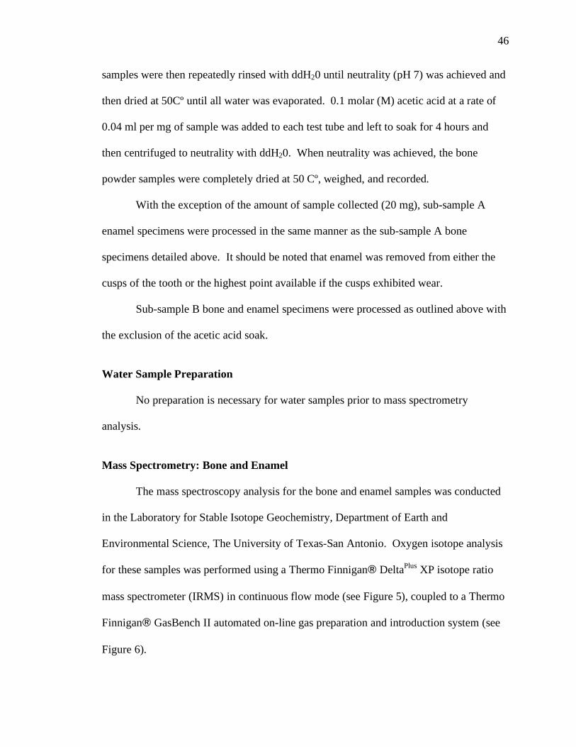
46
samples were then repeatedly rinsed with ddH20 until neutrality (pH 7) was achieved and
then dried at 50Cº until all water was evaporated. 0.1 molar (M) acetic acid at a rate of
0.04 ml per mg of sample was added to each test tube and left to soak for 4 hours and
then centrifuged to neutrality with ddH20. When neutrality was achieved, the bone
powder samples were completely dried at 50 Cº, weighed, and recorded.
With the exception of the amount of sample collected (20 mg), sub-sample A
enamel specimens were processed in the same manner as the sub-sample A bone
specimens detailed above. It should be noted that enamel was removed from either the
cusps of the tooth or the highest point available if the cusps exhibited wear.
Sub-sample B bone and enamel specimens were processed as outlined above with
the exclusion of the acetic acid soak.
Water Sample Preparation
No preparation is necessary for water samples prior to mass spectrometry
analysis.
Mass Spectrometry: Bone and Enamel
The mass spectroscopy analysis for the bone and enamel samples was conducted
in the Laboratory for Stable Isotope Geochemistry, Department of Earth and
Environmental Science, The University of Texas-San Antonio. Oxygen isotope analysis
for these samples was performed using a Thermo Finnigan! DeltaPlus
XP isotope ratio
mass spectrometer (IRMS) in continuous flow mode (see Figure 5), coupled to a Thermo
Finnigan! GasBench II automated on-line gas preparation and introduction system (see
Figure 6).

47
Bone and enamel powder was prepared for IRMS analysis following the protocol
reported by Paul and Skrzypek (2007) as adapted from Spotl and Vennemann (2003).
The bone and enamel powder samples (~200-400 %g each, %g=microgram=1/1,000,000
of a gram) were weighted into helium flushed (to remove air from the vials) 10 ml butyl
rubber sealed glass vials where they were reacted with 100% ortho-phosphoric acid at
70ºC for 30 minutes to produce carbon dioxide gas (CO2). The resulting CO2 gas was
automatically collected (via the GasBench II) and injected into the IRMS. Once
introduced into the IRMS, samples were analyzed and referenced against CO2 gasses
derived from one internal (CO2 tank at 99.995%) and three external (NBS 19, NBS 18,
and LSVEC) standards. For quality assurance during each analysis, two replicates each
of the external standards and two replicates of the internal standard were simultaneously
run with each batch of unknown samples. Precision for the bone and enamel sample suite
was <± 0.05 ‰.
Mass Spectrometry: Water
The mass spectroscopy analysis for the water samples was conducted at Coastal
Science Laboratories, Austin, Texas. Oxygen isotope analysis for these samples was
performed using one of two laboratory isotope ratio mass spectrometers (IRMS), a VG
Micromass 602D or a VG Micromass SIRA Series II.
Oxygen isotope analysis of the water samples was prepared for IRMS analysis
using the carbon dioxide (CO2) equilibration method of Epstein and Mayeda (1953) as
described by Yoshida and Yoshihiko (1986). Six ml of sample water was drawn into a
common 20 ml syringe (the equilibration vessel) and then fitted with a 25 mm 22 gauge
needle. Air bubbles were removed as the water volume was reduced to 5 ml. Seven ml of

48
99.9% CO2 was then introduced into the syringe through a rubber septum attached to a
mercury manometer. The CO2 pressure in the syringe was adjusted to 1 atmosphere,
removed from the manometer, and the needle sealed with a 000 rubber stopper. The syringe
was then attached to a rack in a 25Cº water bath and agitated for 2 hours. After 2 hours the
syringe was removed from the water bath and the equilibrated CO2 gas manually
introduced into an evacuated injection port on the IRMS. Once introduced into the
IRMS, samples were analyzed and referenced against gasses derived from two carbonate
(CO2 tank at 99.9% and NBS 19) and one external (Austin Meteoric Water) standard.
For quality assurance during each analysis, one standard was measured after every ten
samples. Additionally, as a precision check (± 0.2 ‰), replicate analyses were performed
on 10% of the sample suite.
The sample " values were calculated using Equation 1 (Chapter II) and reported in
‰ relative to the VSMOW2 standard for oxygen and VPDB standard for carbonates as
established by the IAEA (Coplen 1996).
Data Adjustments and Comparisons
As discussed previously, researchers have estimated adjustments necessary to
account for metabolically induced oxygen fractionation that occurs from the time the
oxygen enters the body to the time it is incorporated into the bone and enamel tissues.
One such fractionation correction, the nursing effect, as reported by White et al. (2004)
and Wright and Schwarcz (1998, 1999) was applied to the enamel samples in the current
study. In concert with the nursing effect adjustment, the regression equation developed
by Daux et al. (2008) was also applied. Summarizing, the following data comparisons
will be analyzed and discussed:
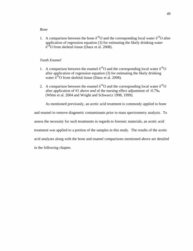
49
Bone
1. A comparison between the bone "18
O and the corresponding local water "18
O after
application of regression equation (3) for estimating the likely drinking water
"18
O from skeletal tissue (Daux et al. 2008).
Tooth Enamel
1. A comparison between the enamel "18
O and the corresponding local water "18
O
after application of regression equation (3) for estimating the likely drinking
water "18
O from skeletal tissue (Daux et al. 2008).
2. A comparison between the enamel "18
O and the corresponding local water "18
O
after application of #1 above and of the nursing effect adjustment of -0.7‰
(White et al. 2004 and Wright and Schwarcz 1998, 1999).
As mentioned previously, an acetic acid treatment is commonly applied to bone
and enamel to remove diagenetic contaminants prior to mass spectrometry analysis. To
assess the necessity for such treatments in regards to forensic materials, an acetic acid
treatment was applied to a portion of the samples in this study. The results of the acetic
acid analyses along with the bone and enamel comparisons mentioned above are detailed
in the following chapter.
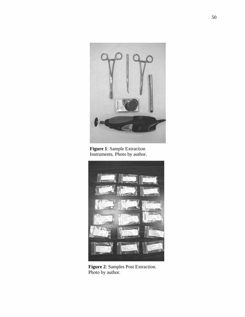
50
Figure 1: Sample Extraction
Instruments. Photo by author.
Figure 2: Samples Post Extraction.
Photo by author.
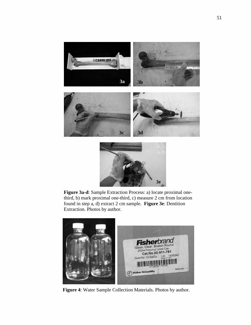
51
Figure 4: Water Sample Collection Materials. Photos by author.
3a
Figure 3a-d: Sample Extraction Process: a) locate proximal one-
third, b) mark proximal one-third, c) measure 2 cm from location
found in step a, d) extract 2 cm sample. Figure 3e: Dentition
Extraction. Photos by author.
3b
3c 3d
3e
3a
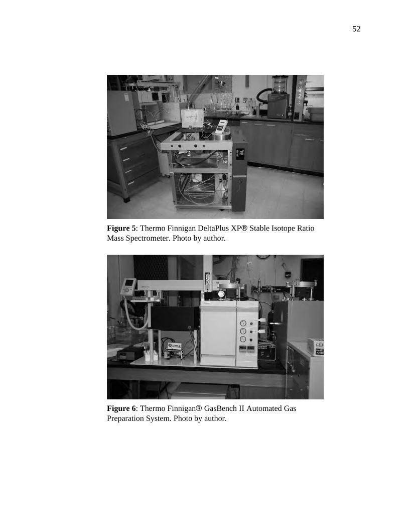
52
Figure 5: Thermo Finnigan DeltaPlus XP! Stable Isotope Ratio
Mass Spectrometer. Photo by author.
Figure 6: Thermo Finnigan! GasBench II Automated Gas
Preparation System. Photo by author.
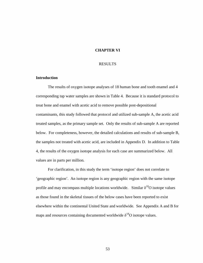
53
CHAPTER VI
RESULTS
Introduction
The results of oxygen isotope analyses of 18 human bone and tooth enamel and 4
corresponding tap water samples are shown in Table 4. Because it is standard protocol to
treat bone and enamel with acetic acid to remove possible post-depositional
contaminants, this study followed that protocol and utilized sub-sample A, the acetic acid
treated samples, as the primary sample set. Only the results of sub-sample A are reported
below. For completeness, however, the detailed calculations and results of sub-sample B,
the samples not treated with acetic acid, are included in Appendix D. In addition to Table
4, the results of the oxygen isotope analysis for each case are summarized below. All
values are in parts per million.
For clarification, in this study the term ‘isotope region’ does not correlate to
‘geographic region’. An isotope region is any geographic region with the same isotope
profile and may encompass multiple locations worldwide. Similar "18
O isotope values
as those found in the skeletal tissues of the below cases have been reported to exist
elsewhere within the continental United State and worldwide. See Appendix A and B for
maps and resources containing documented worldwide "18
O isotope values.
54
Case 06-3784, Webb County
Three distinct "18
O values are present for case 06-3784. The "18
O values for the
compact (-11.04) and cancellous (-9.31) tissues are clearly out of agreement with the
corresponding tap water "18
O value (-4.7). While the expected "18
O value for the tooth
enamel (-5.06) is also in disagreement with the corresponding tap water (-4.7), it is
comparable with the low end of the enamel range at two standard deviations (-5.39 to -
4.73).
Case 0A0760, Cameron County
The compact tissue sample for case 0A0760 was contaminated during processing
and no data was available for analysis. The "18
O values for the tooth enamel and
cancellous tissues are isotopically similar to one another (-7.79 and -7.17 respectively),
but clearly disagree with the corresponding tap water "18
O value (-1.9). At two standard
deviations, the enamel (-7.25 to -7.09) and cancellous (-7.80 to -7.78) tissue results
remain in disagreement with the corresponding tap water.
Case DMG-9319, Starr County
The "18
O values for the enamel (-6.75), compact (-7.39), and the cancellous
(-9.19) tissues disagree with the corresponding tap water "18
O value (-3.2). At two
standard deviations, the enamel (-6.89 to -6.62), compact (-7.44 to -7.33), and cancellous
(-9.34 to -9.04) tissue results remain in disagreement with the corresponding tap water.
Although in disagreement with the local tap water, the enamel and compact tissue "18
O
values are isotopically similar to one another, differing by 0.64.
55
Case 06-5121, Webb County
The "18
O values for the enamel (-11.50), compact (-12.27), and the cancellous
(-14.04) tissues are clearly out of agreement with the corresponding tap water "18
O value
(-4.7). At two standard deviations, the enamel (-11.92 to -11.08), compact (-12.78 to -
11.76), and cancellous (-14.76 to -13.32) tissue results remain in disagreement with the
corresponding tap water. Although in disagreement with the local tap water, the enamel
and compact tissue "18
O values are isotopically similar to one another (0.77 difference)
with overlapping ranges at two standard deviations.
Case DMG-9821, Zapata County
The "18
O values for the tooth enamel and compact tissues are isotopically similar
to one another (-6.17 and -6.37 respectively), but are in disagreement with the
corresponding tap water "18
O value (-4.7). The cancellous tissue value range (-5.72 to -
5.14), although not in absolute agreement, is within 0.44 of the corresponding tap water
"18
O value (-4.7). At two standard deviations, the enamel (-6.37 to -5.97), compact (-
6.55 to -6.19), and cancellous (-5.72 to -5.14) tissue results remain in disagreement with
the corresponding tap water.
Case F2008-100, Webb County
Three distinct "18
O values are present for case F2008-100. The "18
O values for
the enamel (-10.73), compact (-12.70), cancellous (-14.07) tissues are clearly out of
agreement with the corresponding tap water "18
O value (-4.7). At two standard
deviations, the enamel (-11.06 to -10.40), compact (-13.26 to -12.14), and cancellous (-
14.79 to -13.35) tissue results remain in disagreement with the corresponding tap water.

56
Minimal overlap (0.09) exists only between the compact and cancellous ranges at two
standard deviations
A detailed discussion of the results for each case outlined above is given in the
following chapter. Also discussed are the possible consequences of the nursing effect,
acetic acid side analysis, and research assumptions, issues and limitations.

Table 4: Oxygen Isotope Analysis Results and Adjustmentsa
57
Actual
Tap Water
Sample
Case
Number County Type
Carbonate
18O‰
PDB
Carbonate
18O‰
b
Phosphate
18O‰
c
Calculated
Ingested
Waterd 18
O‰
A1-A 06-3784 Webb tooth enamel -3.64 27.17 18.61 -5.06 -5.39 -4.73 -4.69 -4.03
A2-A compact -7.42 23.27 14.73 -11.04 -10.67 -11.40
A3-A cancellous -6.32 24.40 15.85 -9.31 -9.14 -9.48
A4-A tap water (-4.7)
B1-A 0A0760 Cameron tooth enamel -5.36 25.39 16.84 -7.79 -7.80 -7.78 -7.10 -7.08
B2-A compact No data. Sample contaminated during processing.
B3-A cancellous -4.97 25.80 17.24 -7.17 -7.25 -7.09
B4-A tap water (-1.9)
C1-A DMG-9319 Starr tooth enamel -4.71 26.07 17.51 -6.75 -6.89 -6.62 -6.19 -5.92
C2-A compact -5.11 25.65 17.10 -7.39 -7.44 -7.33
C3-A cancellous -6.25 24.48 15.93 -9.19 -9.04 -9.34
C4-A tap water (-3.2)
D1-A 06-5125 Webb tooth enamel -7.71 22.98 14.43 -11.50 -11.08 -11.92 -10.38 -11.22
D2-A compact -8.19 22.47 13.93 -12.27 -11.76 -12.78
D3-A cancellous -9.31 21.32 12.78 -14.04 -13.32 -14.76
D4-A tap water (-4.7)
E1-A DMG-9821 Zapata tooth enamel -4.34 26.45 17.89 -6.17 -6.37 -5.97 -5.67 -5.27
E2-A compact -4.47 26.31 17.76 -6.37 -6.55 -6.19
E3-A cancellous -3.88 26.92 18.37 -5.43 -5.72 -5.14
E4-A tap water (-4.7)
F1-A F2008-100 Webb tooth enamel -7.22 23.47 14.93 -10.73 -10.40 -11.06 -9.70 -10.36
F2-A compact -8.47 22.19 13.65 -12.70 -12.14 -13.26
F3-A cancellous -9.32 21.31 12.76 -14.07 -13.35 -14.79
F4-A tap water (-4.7)
a Unless otherwise stated all values are relative to VSMOW2.
b Coplen et al. 1983.
c Iacumin et al. 1996.
d Daux et al. 2008.
e White et al. 2004.
Calculated
Ingested Water Ranged
18O‰ 2sd
With Nursing
Effecte
18O‰
58
CHAPTER VII
DISCUSSION
Introduction
As discussed previously (Materials and Methods, page 45), a number of data
comparisons, adjustments, and applications of regression formulae were made against the
bone and tooth enamel sample data. The results of these comparisons are detailed for
each case below. The nursing effect and the acetic acid side analysis mentioned earlier
(Materials and Methods, page 38) are discussed separately from the individual cases. A
discussion of the research assumptions, issues, and limitations is also provided.
Case 06-3784, Webb County
The enamel !18
O value for case 06-3784 indicates that the individual likely
ingested water during early infancy and childhood (0-3 years) with a similar isotopic
signature as that found in the discovery location, Webb County. In contrast, the compact
and cancellous values suggest the individual lived in at least two isotopically distinct
locations as an adult. Both values are indicative of either a cooler or higher altitude
climate where isotope values are more negative than in warmer or lower altitude areas.
Based on the three !18
O values found within the tissues of case 06-3784, a
suggestion is made that the individual represented by the remains resided in an
59
isotopically similar area as that found in Webb County during early childhood. The
individual did not, however, live in said area recently (~8-10 years) or up to
approximately 20 years prior to death. Because the discovery location !18
O value was
not found in any of the individual’s bone tissues, the individual was most likely not from
or had only recently (<2 years) relocated to the area. Because of the close
correspondence between the enamel !18
O value and the Webb County tap water !18
O
range at two standard deviation, an alternative explanation may be that the individual
spent a portion of his or her early childhood in or near Webb County, but later moved
from the area.
Case 0A0760, Cameron County
The compact tissue sample for case 0A0760 was contaminated during processing
and no data for that tissue was available for analysis. Enamel and cancellous !18
O values
represent ingested water during early childhood (0-3 years) and recent residence (~8-10
years). Similar !18
O values in these tissues, therefore, indicate the individual resided in
an area during these stages with a similar !18
O value to one another or in the same
location for both stages of development. The enamel and cancellous !18
O values for this
case are similar to one another, but not similar to the discovery location, Cameron
County. This suggests that the individual likely lived in the same area during early
childhood and for approximately the last 8-10 years of his or her life. Due to the absence
of the compact tissue, a suggestion cannot be given in regards to long-term residence
(~20 yeas prior).
In summary, the individual represented by the remains of case 0A0760 resided in
an isotopically similar area during the first three years of life and for approximately 8-10
60
years prior to death. That area was, however, most likely not the discovery location,
Cameron County. Although no discovery location !18
O value was found in either of the
individual’s tissues, this does not preclude the possibility that the individual only recently
(<2 years) relocated to the Cameron County region.
Case DMG-9319, Starr County
The enamel and compact tissue !18
O values for case DMG-9319 are distinct from
the tap water !18
O, but isotopically similar to one another (0.64 difference) and most
likely represent the same or adjacent isotope regions of residence during early childhood
(0-3 years) and ~20 years prior to death. The cancellous tissue !18
O value is distinct from
the other two tissues and the corresponding tap water !18
O values. The highly negative
cancellous tissue !18
O value is indicative of either a cooler or higher altitude climate
where isotope values are more negative than in warmer or lower altitude areas.
In summary, the individual represented by the remains of case DMG-9319 resided
in an isotopically similar area during the first three years of life and for approximately 20
years prior to death. Isotope values further indicate that recent residence was in a cooler
or higher altitude region. These areas were most likely not the discovery location, Starr
County. Although no discovery location !18
O value was found in any of the individual’s
tissues, this does not preclude the possibility that the individual only recently (<2 years)
relocated to the Starr County region.
Case 06-5121, Webb County
The enamel and compact tissue !18
O values for case 06-5121 are similar and most
likely represent the same or adjacent isotope regions of residence during early childhood
61
(0-3 years) and ~20 years prior to death. In contrast, the cancellous !18
O value is clearly
distinct from the other two tissues and the corresponding tap water !18
O value. The
cancellous tissue value is indicative of residence that is either a cooler or higher altitude
climate where isotope values are more negative than in warmer or lower altitude areas.
In summary, the individual represented by the remains of case DMG-5121 resided
in an isotopically similar area during the first three years of life and for approximately 20
years prior to death. Isotope values further indicate that recent residence was in a cooler
or higher altitude region. These areas were, however, most likely not the discovery
location, Webb County. Although no discovery location !18
O value was found in any of
the individual’s tissues, this does not preclude the possibility that the individual only
recently (<2 years) relocated to the Webb County region.
Case DMG-9821, Zapata County
The enamel and compact tissue !18
O values for case DMG-9821 are distinct from
the tap water !18
O, but isotopically similar to one another (0.20 difference) and most
likely represent the same or adjacent isotope regions of residence during early childhood
(0-3 years) and ~20 years prior to death. The cancellous !18
O value, although not in
absolute agreement, is within 0.44 of the corresponding tap water !18
O value at two
standard deviations. This value suggests a possible recent relocation to a region with an
isotope value similar to the Zapata County tap water. If the relocation was in fact to the
Zapata area, the difference between the cancellous tissue and the tap water (0.44) further
indicates incomplete isotopic acclimation; therefore suggesting the relocation was likely
" 8 years.

62
In summary, the individual represented by the remains of case DMG-9821 resided
in an isotopically similar area during early childhood and approximately 20 years prior to
death. That area was, however, most likely not the discovery location, Zapata County. In
contrast, recent residence was in an isotope region similar to that of the Zapata region,
indicating the individual likely moved to the Zapata area in the last 8 years.
Case F2008-100, Webb County
Three distinct !18
O values are present for case F2008-100. Although minimal
overlap (0.09) exists between the compact and cancellous !18
O ranges produced when
applying two standard deviations, it is unlikely the values are indicative of residence in
similar isotope regions. This is particularly true given that a standard deviation of .165 is
significant when differences in !18
O values begin registering at the 4th or 5
th decimal
position. Most likely the !18
O values found in this case represent three distinct isotope
regions. The highly negative values found (enamel:-10.73, compact:-12.70, and
cancellous: -14.07) are more typical of !18
O values found in cooler areas or regions of
higher altitudes.
In summary, the individual represented by the remains of case F2008-100 resided
in at least three isotopically distinct locations during his or her lifetime. That location
was, however, not the discovery location, Webb County, but instead, a cooler and/or high
altitude region.
Acetic Acid Treatment Analysis
Acetic acid treatments are typically used to remove contaminants in
archaeological samples resulting from diagenetic activity within the deposition context.

63
Unlike most archaeological materials, modern human skeletal tissues are not
environmentally exposed for great lengths of time and should, theoretically, hold little to
no contamination. To test for the necessity of a contaminate removal treatment (acetic
acid) in regards to modern human skeletal material, a separate analysis was performed on
a sub-set of the bone and enamel samples in this study.
Half of each bone and enamel sample was processed utilizing the acetic acid
treatment (sub-sample A) described in Chapter V, while the other half of the sample was
processed with no acetic acid treatment (sub-sample B). The difference in each set, with
versus without treatment, showed no overall trend toward more positive or more negative
isotope values (see Table 4 and Appendix D). After the acid treatment on sub-sample A,
seven of the 18 sample pairs (~39%) resulted in more negative isotope values, while 11
of the 18 were more positive (~61%).
A two-tailed, paired-samples t-test revealed no significant difference in the
oxygen values before and after the acetic acid treatment, t (17) = 0.93, p = 0.37. This
indicates that the mean oxygen value after the acetic acid treatment (M = -6.53) was not
significantly different than the mean before the acetic acid treatment (M = -6.71), i.e., a
mean difference of 0.18 between treated and untreated samples.
Based on these few observations and small sample size, it is difficult to
conclusively state the necessity of a contaminate removal treatment for modern, forensic
bone and tooth enamel. Although statistically no difference is demonstrated, the fact that
nearly ~39% of the samples resulted in more negative isotope values and ~61% in more
positive values after the treatment prevents a definite conclusion based on this sample set.
The mean difference between the acid and non-acid treated sets was 0.18. Interestingly,

64
both the percentage of samples affected (~39% and ~61%) and the mean difference
(0.18) are similar to the results of a previous study utilizing a similar acetic acid test
(30%, more negative, 60% more positive, 10% no change, and 0.19 mean difference,
Regan 2006). Further research in regards to sample preparation is critical to the
reliability and acceptance of isotope analysis as a valid technique in the pursuit of
investigating the identity of modern human remains.
Nursing Effect
Researchers have demonstrated the existence of a metabolically induced
fractionation of the oxygen composition of tooth enamel formed during nursing (Evans et
al. 2006, White et al. 2000, Wright and Schwarcz 1998, 1999). Due to this fractionation,
breast milk is oxygen enriched by 0.7‰ compared to the intake water of the mother. The
breast milk enrichment is passed to the infant where it is subsequently incorporated into
the bone and enamel tissues. Enamel is a static tissue and maintains its isotopic record
through life and after death. In contrast, and because of the remodeling nature of bone,
no nursing effect oxygen imprint will be found in the bony elements unless the infant
perished during or shortly after the nursing period.
In this study the nursing effect adjustment was applied to the calculated ingested
water ranges. Comparison of the results (Table 4) do not significantly alter the
conclusions drawn in the above discussion. The results do, however, support the
conclusion suggested in case 06-3784 that the individual represented by the remains most
likely formed his or her first permanent molar enamel in an isotopic region similar to the
Webb County discovery location.
65
Research Assumptions, Issues, and Limitations
Assumption: Consumption of Local Water
In this study the oxygen compositions within the skeletal tissues of unidentified
human remains were compared against the oxygen composition of their respective
discovery location tap waters. Utilizing only tap water as the sample against which the
skeletal tissue is compared does not take into account imported food supplies and cultural
dietary practices that may alter oxygen signature within the body. In today’s global
economy and transportation of foodstuffs, it is likely that some individuals, such a those
living within large population centers located some distance from agricultural activity,
consume little, if any, of the local food oxygen signature. Confounding the issue further
is the possibility that individuals ingest a large portion of non-local water, either by
choice or because the municipally supplied water is piped from a distant source. For
these reasons, utilizing the oxygen composition of the locally supplied tap water as a
means of comparison to the oxygen composition found in the skeletal tissues may not be
the best choice of comparative samples. A more suitable candidate for comparison is a
bone and/or tooth sample from an individual known to have originated and continually
resided in the discovery location.
Assumption: Nursing
Discussed and applied in this study is the concept that nursing activity affects the
oxygen composition statically imprinted within the enamel of dentition formed during the
nursing period. The importance placed on the nursing effect is primarily determined by
the research intent and goals. If attempting to determine ages at which weaning may
have occurred in past populations, the application of the nursing effect is of the utmost
66
importance. In contrast, the amount of alteration demonstrated to exist (0.7, Wright and
Schwarcz 1998, 1999) is unlikely to alter results to the extent that an origin from an
entirely distinct isotope region is indicated. Examination of the oxygen values resulting
from application of the nursing effect adjustment did not lead to alterations in the
conclusions discussed above.
Issue: Fresh Water Versus Tap Water
As discussed in Chapter I, a strong correlation (R=0.70) exists between the
oxygen composition of corresponding local fresh (e.g., precipitation or well water) and
tap waters. Although Bowen et al. (2007) demonstrate this relationship, the authors
recognize the fact that 30% of the variation between the two water sources remains
unidentified. Responsible for the remaining variation, contend Bowen and colleagues, is
principally the nature of the source (e.g., an open, shallow well vs. deep aquifers), local
geology, and municipal water treatment processes. Without detailed knowledge and
incorporation of these factors into a research design, the acceptance of fresh and tap
water oxygen compositions as equal factors in the determination of likely ingested water
may result in erroneous data. Therefore, the assumption that the oxygen in the skeletal
tissues from an individual consuming only fresh water (e.g., well water) is still indicative
of the geolocation based on the oxygen in a corresponding tap water sample may lead to
inaccurate results.
Issue: Ethics
An ethical conflict arises as a result of this methodology. That concern centers on
the destructive nature of the process. Even though advances in technology (e.g., direct
67
laser fluorination (Lindars et al. 2001) and thermal laser ablation (Passey and Cerling
2006) now allow for isotopic analysis of minute amounts of skeletal tissue, the fact
remains that the process destroys the original sample. When skeletal tissue is limited or
falls under protective legislation (e.g., NAGPRA), destructive procedures are
unacceptable. Even when destructive analyses are allowed, the long-term integrity of the
samples and/or collection must be considered and made a priority (Katzenberg 2000).
Although the integrity of human skeletal material should be a concern for all
professionals dealing with human remains, the bigger picture must also be considered.
One of the primary goals of modern forensic casework is the identification of the human
remains and the eventual return of those remains to his or her family. Despite the current
destructive nature of this method, the opportunity to provide potentially helpful
information leading to the identification of an unknown individual may quite possibly
outweigh the sample loss resulting from isotopic techniques.
Limitation: Unknown Deposition Context
The deposition contexts of the six cases in this study are unknown. This absence
presents implications for the interpretation of the isotope data. As discussed in Chapter
III, the potential for diagenetic alteration caused by the burial context (e.g., buried,
frozen, submerged, or burned) can be substantial depending on the burial type and
microenvironment (Sorg and Haglund 2002). Because of this, the interpretations based
solely on the isotopic data of this study are limited. Therefore, future studies attempting
interpretation of isotope data from bone and tooth enamel should include documented
knowledge of the deposition context.
68
Limitation: Data Availability
A number of the researchers discussed in this study (Katzenberg and Harrison
1997, Prowse et al. 2007, Spence et al. 2004) have expressed concerns in regards to the
dearth of isotope databases against which data may be compared. This study
demonstrates well this limitation. Although multiple pieces of information resulted from
the isotope analyses conducted as part of this research, the usefulness and interpretation
of the data is limited to the chosen comparative sample or to the few studies thus far
conducted. Further research and database compilation is imperative if the utilization of
isotope signatures is to be used, and even more importantly, accepted as viable evidence
towards the identification of an unknown individual.
69
CHAPTER VIII
CONCLUSION
At death the human skeleton holds an in vivo molecular history of the stressors,
diet, mobility, and health of the individual, a history that holds the potential of immense
information and wide scientific exploration. The use of molecular approaches for
extracting that recorded history, however, remains limited in forensic research
applications. As reviewed in the previous research section of this study, the ability of
biochemical techniques to extract the subtle clues left behind in skeletal tissue hold the
potential as an informative and valuable avenue in future research, particularly in the
field of forensic anthropology (Meier-Augenstein 2007, Ortner 2003).
Summary
The primary intent of this research was an exploration into the value of oxygen
isotope chemistry as a means of providing information with regards to the identification
of unknown individuals. To facilitate this application, the bone and tooth enamel of six
open, unidentified forensic cases curated by the Forensic Anthropology Center, Texas
State University-San Marcos were analyzed for oxygen isotope composition and
compared together with the oxygen composition of corresponding tap water samples
from their respective discovery locations. Given that the oxygen composition of skeletal
tissues is strongly correlated with the oxygen composition of ingested water, oxygen

70
signatures of the skeletal tissues were expected to match the oxygen signature of their
respective discovery location’s tap water. Although the analyses resulted in additional
information in regards to the individuals’ residential histories, the data were too general
in nature to formulate conclusive statements without further analysis of the remains and a
broader research design.
Conclusion and Suggestions
As demonstrated in this study, oxygen isotope analysis has been used in attempts
to answer a variety of research questions, clarify historical events, and even challenge
prevailing theories. Strikingly few studies, however, have utilized isotopic analysis as a
data gathering technique in modern forensic investigations of unidentified human
remains. Those studies that do exist have primarily applied isotopic analyses in order to
narrow down the geographic origin of the remains of missing American soldiers found on
foreign soil (Beard and Johnson 2000, Regan 2006,). Such studies facilitate
identification by providing scientists and investigators with a means to create a shortlist
from the more than 88,000 missing American soldiers (Joint POW-MIA Accounting
Command 2008). It stands to reason that applying a similar methodology to modern
forensic investigations would result in the same: a shortlist of missing persons to which a
set of unidentified remains may belong. Multiply this application by potential uses in
mass fatality incidents, acts of terrorism, or human rights violations and the value of
isotopic analysis is readily apparent.
Although this research demonstrated the predicted exclusionary value of oxygen
isotope analysis in regards to unknown human remains, it is believed that future research
utilizing a multiple elemental approach and documented modern bone and tooth samples–
71
–to account for contemporary food, water, and cultural dietary practices–– will yield less
broad results that allow for more conclusive interpretation.
Additionally, a concerted effort is needed among researchers to compile and make
publicly available isotope databases against which data may be compared. Such a
collaborative effort is imperative to the future success and acceptability of isotope
research as viable means of exposing the valuable but hidden elemental information that
resides within human skeletal material.
Finally, further research should be conducted to clarify the diagenetic effects on
modern human remains. In addition to the primary intent, this research also conducted an
analysis in an attempt to discern the necessity of treating modern skeletal material for
diagenetic contamination. The small sample size and results preclude a conclusive
statement concerning the need for such treatments. Future researchers would benefit
from a larger study whose primary focus is to determine both the type and necessity of
contamination removal treatments on modern skeletal material.
Implications
So why choose oxygen isotope analysis as a means of extracting information from
a set of human remains? Although a number of answers are valid and were outlined in
Chapter II, two additional reasons warrant attention. First, the cost of a single oxygen
analysis is fairly inexpensive, ranging from $20-$40. This cost is likely affordable by
most law enforcement agencies. In contrast, some laboratory techniques (DNA typing)
can easily cost hundreds per sample. Such costs are often prohibitive to small law
enforcement communities. Second, the time required for a lab to complete an elemental
analysis is often only a few weeks depending on the lab chosen. During this research a

72
number of laboratories around the country were found to not only offer oxygen analysis
but also offer an expedited service of less than two weeks if requested and the additional
fees paid. Laboratory techniques such as DNA typing require a year or more for
processing and the return of results. This turn around time causes delay in both the
investigation and closure to the families. Given these reasons, perhaps the more
appropriate question would be why not choose isotope analysis if even a small chance
exists of obtaining information that may bring a lost individual home.
While it is true that the information gleaned from an oxygen analysis of skeletal
material tends to be somewhat broad in nature, as the results in this research indicated,
the data do in fact provide information about the origins and residence patterns of the
represented individuals. Although the information appears to be primarily exclusionary,
this does not mean that the information resulting from isotopic analysis, however small or
broad, is of little value to those charged with the responsibility of identifying a set of
human remains or to the family of the missing individual. Armed with the information
provided through isotope profiling, investigators possess the ability to geographically
constrain (or expand) investigations, thereby potentially narrowing what may be a vast
list of possible victims. Elemental isotope analysis of human skeletal material, when
used as one of the many lines of evidence in the pursuit of identification of an unknown
individual, holds the potential to be a key factor in the identification of those who can no
longer identify themselves.

73
APPENDIX A
OBSERVED !18
O ISOTOPE RATIOS FOR TAP WATERS
IN THE CONTIGUOUS UNITED STATES
Observed !18
O Isotope Ratios for Tap Waters in the Contiguous United States (Bowen 2007)
74

75
APPENDIX B
DOCUMENTED OXYGEN ISOTOPE VALUES
RESOURCES
76
Isotope Data Resources
• Stable Hydrogen and Oxygen Isotope Ratios for Selected Sites of the U.S.
Geological Survey's NASQAN and Benchmark Surface-water Networks.
Available at: http://pubs.usgs.gov/of/2000/ofr00-160.
• Water Isotope System for Data Analysis, Visualization and Electronic Retrieval
(WISER). Available at: http://nds121.iaea.org/wiser.
• Stable Isotope Ratios of Tap Water in the Contiguous United States. Information
available from the author at: [email protected].
• Waterisotopes.org. Provides information, data, and resources for scientific
applications involving spatial variation in the isotopes of hydrogen and oxygen.
• The Online Isotopes in Precipitation Calculator (OIPC). An interactive tool
allowing on-demand estimation of the stable water isotope ratios of precipitation
at any site. Available at: http://wateriso.eas.purdue.edu/waterisotopes/
pages/data_access/oipc.html
77
APPENDIX C
TEXAS COUNTY MAP
INDICATING
CASE COUNTIES, OXYGEN ISOTOPE SIGNATURE, AND
WATER COLLECTION LOCATIONS
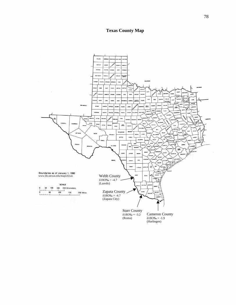
78
Texas County Map
www.lib.utexas.edu/maps/texas
.html
Cameron County !18O‰ = -1.9 (Harlingen)
Starr County !18O‰ = -3.2 (Roma)
Zapata County !18O‰ = -4.7 (Zapata City)
Webb County !18O‰ = -4.7 (Laredo)
www.lib.utexas.edu/maps/texas
79
APPENDIX D
SUB-SAMPLE B OXYGEN ISOTOPE ANALYSIS
RESULTS AND ADJUSTMENTS
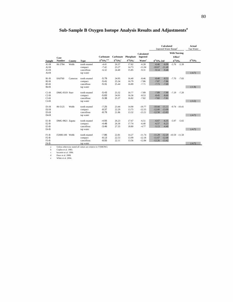
80
Sub-Sample B Oxygen Isotope Analysis Results and Adjustmentsa
Actual
Tap Water
Sample
Case
Number County Type
Carbonate
18O‰
PDB
Carbonate
18O‰
b
Phosphate
18O‰
c
Calculated
Ingested
Waterd 18
O‰
A1-B 06-3784 Webb tooth enamel -4.41 26.37 17.82 -6.28 -6.46 -6.09 -5.76 -5.39
A2-B compact -7.42 23.27 14.73 -11.04 -10.67 -11.40
A3-B cancellous -6.32 24.40 15.85 -9.31 -9.14 -9.48
A4-B tap water (-4.7)
B1-B 0A0760 Cameron tooth enamel -5.79 24.95 16.40 -8.46 -8.40 -8.53 -7.70 -7.83
B2-B compact -5.41 25.34 16.79 -7.86 -7.87 -7.86
B3-B cancellous -5.31 25.44 16.89 -7.71 -7.73 -7.69
B4-B tap water (-1.9)
C1-B DMG-9319 Starr tooth enamel -5.43 25.32 16.77 -7.89 -7.89 -7.90 -7.19 -7.20
C2-B compact -5.83 24.91 16.36 -8.53 -8.45 -8.60
C3-B cancellous -5.38 25.37 16.82 -7.82 -7.82 -7.81
C4-B tap water (-3.2)
D1-B 06-5125 Webb tooth enamel -7.25 23.44 14.90 -10.77 -10.44 -11.11 -9.74 -10.41
D2-B compact -8.37 22.29 13.75 -12.55 -12.00 -13.09
D3-B cancellous -8.79 21.86 13.32 -13.21 -12.58 -13.83
D4-B tap water (-4.7)
E1-B DMG-9821 Zapata tooth enamel -4.55 26.23 17.67 -6.51 -6.67 -6.35 -5.97 -5.65
E2-B compact -4.48 26.30 17.74 -6.40 -6.57 -6.23
E3-B cancellous -3.46 27.35 18.80 -4.77 -5.13 -4.40
E4-B tap water (-4.7)
F1-B F2008-100 Webb tooth enamel -7.86 22.81 14.27 -11.74 -11.29 -12.20 -10.59 -11.50
F2-B compact -8.13 22.53 13.99 -12.18 -11.67 -12.68
F3-B cancellous -8.55 22.11 13.56 -12.84 -12.26 -13.42
F4-B tap water (-4.7)
a Unless otherwise stated all values are relative to VSMOW2.
b Coplen et al. 1983.
c Iacumin et al. 1996.
d Daux et al. 2008.
e White et al. 2004.
Calculated
Ingested Water Ranged
18O‰ 2sd
With Nursing
Effecte
18O‰

81
LIST OF REFERENCES
Anderson, D. L., G. W. Thompson, and F. Popovich
1976 Age of Attainment of Mineralization Stages of the Permanent Dentition.
Journal of Forensic Sciences 21(1):191-200.
Bass, William M.
1995 Human Osteology: A Laboratory and Field Manual. Columbia: Missouri
Archaeological Society.
Beard, Brian L., and Clark M. Johnson
2000 Strontium isotope composition of skeletal material can determine the birthplace
and geographic mobility of humans and animals. Journal of Forensic Sciences
45(5):1049-1061.
Benson, Sara, Chris Lennard, Philip Maynard, and Claude Roux
2006 Forensic Applications of Isotope Ratio Mass Spectrometry-A Review. Forensic
Science International 157(1):1-22.
Bentley, R. Alexander, Michael Pietrusewsky, Michael T. Douglas, and Tim C. Atkinson
2005 Matrilocality During the Prehistoric transition to Agriculture in Thailand?
Antiquity 79(306):865-881.
Bowen, Gabriel J., James R. Ehleringer, Lesley A. Chesson, Erik Stange, and Thure E.
Cerling
2007 Stable Isotope Ratios of Tap Water in the Contiguous United States. Water
Resource Research W03419 43:1-12.
Bowen, Gabriel J., and Bruce Wilkinson
2002 Spatial Distribution of !18
O in Meteoric Precipitation. Geology 30(4):315-318.
Braswell, Geoffrey E.
2004 The Maya and Teotihuacan. Reinterpreting Early Classic Interaction. Austin:
The University to Texas Press.
Brazier, Jean Louis
1995 Use of Isotope Ratios in Forensic Analysis. In Forensic Applications of Mass
Spectrometry. Jehuda Yinon, ed. Boca Raton: CRC Press. Pp. 259-288.
82
Bryant, J. Daniel, and Phillip N. Froelich
1995 A Model of Oxygen Isotope Fractionation in Body Water of Large Mammals.
Geochimica et Cosmochimica Acta 59(21):4523-4537.
Bryant, J. Daniel, Paul L. Koch, Phillip N. Froelich, William J. Showers, and Bernard J.
Genna
1996 Oxygen Isotope Partitioning Between Phosphate and Carbonate in Mammalian
Apatite. Geochimica et Cosmochimica Acta 60(24):5145-5148.
Budd, P., J. Montgomery, J. Evans, and C. Chenery
2001 Combined Pb-, Sr- and O-Isotope Analysis of Human Dental Tissue for the
Reconstruction of Archaeological Residential Mobility. In Plasma Source Mass
Spectrometry: The New Millennium. Grenville Holland and Scott D. Tanner, eds.
Pp. 311-323. Cambridge: The Royal Society of Chemistry.
Buikstra, Jane, T. Douglas Price, Lois Wright, and James A. Burton
2004 Tombs from the Copan Acropolis: A Life History Approach. In Understanding
Early Classic Copan. Ellen E. Bell, Marcello A. Canuto, and Robert J. Sharer,
eds. Pp. 191-214. Los Angeles: Cotsen Press.
Carter, James F., Richard Sleeman, Jenny G. Hill, Fay Idoine, and Emma L. Titterton
2005 Isotope Ratio Mass Spectrometry as a Tool for Forensic Investigation
(Examples from Recent Studies). Science and Justice 45(3):141-149.
Coplen, Tyler B.
1996 New Guidelines for Reporting Stable Hydrogen, Carbon, and Oxygen Isotope-
ratio Data. Geochimica et Cosmochimica Acta 60(17):3359-3360.
Coplen, Tyler B., and Carol Kendall
2000 Stable Hydrogen and Oxygen Isotopes Ratios for Selected Sites of the U.S.
Geological Survey’s NASQAN and Benchmark Surface-Water Networks. United
States Geologic Survey, Reston, VA. Open File Report 00-160. Accessible at:
http://pubs.usgs.gov/of/2000/of00-160.
Dansgaard, Willi
1964 Stable Isotopes in Precipitation. Tellus 16(4):436-468.
Darwent, Christyann M., and R. Lee Lyman
2002 Detecting the Postburial Fragmentation of Carpals, Tarsals, and Phalanges. In
Advances in Forensic Taphonomy: Method, Theory, and Archaeological
Perspectives. William D. Haglund and Marcella H. Sorg, eds. Pp. 355-377. Boca
Raton: CRC Press.
83
Daux, Valerie, Christophe Lecuyer, Frederic Adam, Francois Martineau, and Francoise
Vimeux
2005 Oxygen Isotope Composition of Human Teeth and the Record of Climate
Changes in France (Lorraine) During the Last 1700 Years. Climatic Change
70(3):445-464.
Daux, Valerie, Christophe Lecuyer, Marie-Anne Heran, Romain Amiot, Laurent Simon,
et al.
In press; N.d. Oxygen Isotope Fractionation Between Human Phosphate and Water
Revisited. Journal of Human Evolution.
Dotsie, Paul
2007 Murder in Mammoth Lakes. Forensic Magazine. June-July: 4(3):12-18.
Emsley, John
2001 Nature’s Building Blocks: An A-Z Guide to the Elements, 1st edition. Pp. 297-
304. Oxford University Press: New York.
Epstein, S., and TK Mayeda
1953 Variation of 18
O Content of Waters from Natural Sources. Geochimica et
Cosmochimica Acta, 4(5):213-224.
Evans J.A., and Carolyn A. Chenery
2006 Bronze Age Childhood Migration of Individuals Near Stonehenge, Reveals by
Strontium and Oxygen Isotope Tooth Enamel Analysis. Archaeometry 48(2):30-
321.
Evans, Jane, Nick Stoodley, and Carolyn Chenery
2006 A Strontium and Oxygen Isotope Assessment of a Possible Fourth Century
Immigrant Population in a Hampshire Cemetery, Southern England. Journal of
Archaeological Science 33(2):265-272.
Ezzo, Joseph A., and T. Douglas Price
2002 Migration, Regional Reorganization, and Spatial Group Composition at
Grasshopper Pueblo, Arizona. Journal of Archaeological Science 29(5):499-520.
Ezzo, Joseph A., Clark M. Johnson, and T. Douglas Price
1997 Analytical Perspectives on Prehistoric Migration: A Case Study From East-
Central Arizona. Journal of Archaeological Science 24(5):447-466.
Federal Bureau of Investigations
2008 CODIS-NDIS Statistics. Electronic document,
http://www.fbi.gov/hq/lab/codis1, accessed August 2008.
84
Garvie-Lok, Sandra J., Tamara L. Varney, and M. Anne Katzenberg
2004 Preparation of Bone Carbonate for Stable Isotope Analysis: the Effects of
Treatment Time and Acid Concentration. Journal of Archaeological Science
31(6):763-776.
Gat, J. R.
1996 Oxygen and Hydrogen Isotopes in the Hydrologic Cycle. Annual Review of
Earth and Planetary Sciences 24:225-262.
Gill-King H.
1997 Chemical and Ultrastructural Aspects of Decomposition. In Forensic
Taphonomy: The Postmortem Fate of Human Remains. William D. Haglund and
Marcella H. Sorg, eds. Pp. 93-108. Boca Raton: CRC Press.
Haglund, William D., and Marcella H. Sorg
1997 Method and Theory of Forensic Taphonomy Research. In Forensic Taphonomy:
The Postmortem Fate of Human Remains. William D. Haglund and Marcella H.
Sorg, eds. Pp. 13-26. Boca Raton: CRC Press.
Hedges, Robert E.M.
2002 Bone Diagenesis: An Overview of Processes. Archaeometry 44(3):319-328.
Hedges, Robert E.M., Rhiannon E. Stevens, and Paul L. Koch
2005 Isotopes in Bones and Teeth. In Isotopes in Paleoenvironmental Research.
Melanie J. Leng, editor. Pp. 117-138. New York: Springer.
Hill, P.A., and M. Orth
1998 Bone Remodeling. British Journal of Orthodontics 25(2):101-107.
Hillson, Simon
1996 Dental Anthropology. Cambridge: Cambridge University Press.
Hochrein, Michael J.
2002 An Autopsy of the Grave: Recognizing, Collecting, and Preserving Forensic
Geotaphonomic Evidence. In Advances in Forensic Taphonomy: Method, Theory,
and Archaeological Perspectives. William D. Haglund and Marcella H. Sorg, eds.
Pp. 45-70. Boca Raton: CRC Press.
Hoefs, Jochen
2004 Stable Isotope Geochemistry, 5th edition. Berlin: Springer Verlag.
Hoogewerff, Jurian, Wolfgang Papesch, Martin Kralik, Margit Berner, Pieter Vroon,
Hermann Miesbauer, Othmar Gaber, Karl-Heinz Künzel, and Jos Kleinjans
2001 The Last Domicile of the Iceman from Hauslabjoch: A geochemical Approach
Using Sr, C and O Isotopes and Trace Element Signatures. Journal of
Archaeological Science 28(9):983-989.
85
Iacumin, P., H. Bocherens, H., A. Mariotti, and A. Longinelli
1996 An Isotopic Paleoenvironmental Study of Human Skeletal Remains from the
Nile Valley. Paleogeography, Paleoclimatology, Paleoecology 126(1-2):15-30.
International Atomic Energy Agency
2006 Isotope Hydrology Information System. The ISOHI-WISER Database.
Accessible at: http://www-naweb.iaea.org/napc/ih/IHS_resources_ISOHIS2.html.
Joint Pow-MIA Accounting Command (JPAC)
2008 Wars and Conflicts. Electronic document, http://www.jpac.pacom.mil/
index.php?page=wars_and_conflicts&size =100&ind=1, accessed August 2008.
Katzenberg, M. Anne
2000 Stable Isotope Analysis: A Tool for Studying Past Diet, Demography, and life
history. In Biological Anthropology of the Human Skeleton. M.A. Katzenberg
and Shelley R. Saunders, eds. Pp.305-327. New York: Wiley-Liss.
Katzenberg, M. Anne, and Roman G. Harrison
1997 What's in a bone? Recent Advances in Archaeological Bone Chemistry. Journal
of Archaeological Research 5(3):1573-7756.
Keenleyside, Anne, Margaret Bertulli, and Henry C. Fricke
1997 Final days of the Franklin Expedition: new skeletal evidence. Arctic 50(1):36-
46.
Koch, Paul L., Noreen Tuross, and Mailyn L. Fogel
1997 The Effects of Sample Treatment and Diagenesis on the Isotopic Integrity of
Carbonate in Biogenic Hydroxylapatite. Journal of Archaeological Science
24(5):417-429.
Kohn, Matthew J.
1996 Predicting !18
O: Accounting for Diet and Physiological Adaptation.
Geochimica et Cosmochimica Acta 60(23):4811-4829.
Kohn, Matthew J., Margaret J. Schoeninger, and John W. Valley
1996 Herbivore Tooth Oxygen Isotope Compositions: Effects of Diet and Physiology.
Geochimica et Cosmochimica Acta 60(20):3889-3896.
Kohn, Matthew L., Margaret J. Schoeninger, and William W. Barker
1999 Altered States: Effects of Diagenesis on Fossil Tooth Chemistry. Geochimica et
Cosmochimica Acta 63(18):2737-2747.
Lee-Thorp, Julia A.
2002 Two Decades of Progress towards Understanding Fossilization Processes and
Isotopic Signals in Calcified Tissue Minerals. Archaeometry 44(3):435-446

86
Levinson, A.A., and Boaz Luz, and Yehoshua Kolodny
1987 Variations in Oxygen Isotopic Compositions of Human Teeth and Urinary
Stones. Applied Geochemistry 2(4):367-371.
Lindars, Elaine S., Stephen T. Grimes, David P. Mattey, Margaret E. Collinson, Jerry J.
Hooker, and Tim P. Jones
2001 Phosphate 18O Determination of Modern Rodent Teeth by Direct Laser
Fluorination: An Appraisal of Methodology and Potential Application to
Palaeoclimate Reconstruction. Geochimica et Cosmochimica Acta 65(15):2535-
2548.
Longinelli, Antonio
1984 Oxygen Isotopes in Mammal Bone phosphate: a New Tool for
Paleohydrological and Paleoclimatological Research? Geochimica et
Cosmochimica Acta 48(2):385-390.
Luz, Boaz, and Yehoshua Kolodny
1985 Oxygen Isotope Variations in Phosphate of Biogenic Apatites, 4. Mammal
Teeth and Bones. Earth and Planetary Science Letters 75(1):29-36.
Luz, Boaz, and Yehoshua Kolodny
1989 Oxygen Isotopic Variation in Bone Phosphate. IV. Mammal Teeth and Bones.
Applied Geochemistry 4(3):317-323.
Luz, Boaz, Yehoshua Kolodny, and Michal Horowitz
1984 Fractionation of Oxygen Isotopes Between Mammalian Bone-Phosphate and
Environmental Drinking Water. Geochimica et Cosmochimica Acta 48(8):1689-
1693.
Lymann R. Lee
1994 Vertebrate Taphonomy. Cambridge: Cambridge University Press.
Martin R. Bruce, David B. Burr, and Neil A. Sharkey
1998 Skeletal Tissue Mechanics. New York: Springer.
Mays, Simon
2000 New Directions in the Analysis of Stable Isotopes in Excavated Bone and Teeth.
In Human Osteology in Archaeology and Forensic Science. Margaret Cox and
Simon Mays eds. Pp. 425-438 London: Greenwich Medical Media Ltd.
Meier-Augenstein, Wolfram
2007 Stable Isotope Fingerprinting-Chemical Element “DNA”? In Forensic Human
Identification: An Introduction. Tim Thompson and Sue Black eds. Pp. 29-53.
Boca Raton: CRC Press.
87
Meier-Augenstein, Wolfram, and R.H. Liu
2004 Forensic Applications of Isotope Ratio Mass Spectrometry. In Advances in
Forensic Applications of Mass Spectrometry. J. Yinon, ed. Pp. 149-180. Boca
Raton: CRC Press.
Melbye, Jerry and Scott I. Fairgrieve
1994 A Massacre and Possible Cannibalism in the Canadian Arctic: New Evidence
from the Saunaktuk Site (NgTn-1). Arctic Anthropology 31(2):57-77.
Muller, Wolfgang , Henry Fricke, Alex N. Halliday, Malcolm T. McCulloch, and Jo-
Anne Wartho
2003 Origin and Migration of the Alpine Iceman. Science 302(5646):862-866.
Online Isotopes in Precipitation Calculator (OIPC)
2008 Accessible at: http://wateriso.eas.purdue.edu/waterisotopes/pages/ data_access/oipc.html.
Ortner, Donald J.
2003 Identification of Pathological Conditions in Human Skeletal Remains, 2nd
edition. San Diego: Academic Press.
Page, Douglas
2007 Murder in Mammoth Lakes. Forensic Magazine. June-July: 4(3):12-18.
Passey, Benjamin H., and Thure E. Cerling
2006 In Situ Stable Isotope Analysis (d13C, d18O) of Very Small Teeth Ssing Laser
Ablation GC/IRMS. Chemical Geology 235(3-4):238-249.
Pate, F. Donald
1994 Bone Chemistry and Paleodiet. Journal of Archaeological Method and Theory
1(2):161-209.
Paul, Debajyoti, Grzegorz Skrzypek, and Istvan Forizs
2007 Normalization of Measured Stable Isotopic Compositions to Isotope Reference
Scales - A Review. Rapid Communications in Mass Spectrometry 21(18):3006-
3014.
Paul, Debajyoti, and Grzegorz Skrzypek
2007 Assessment of carbonate-phosphoric acid analytical technique performed using
GasBench II in continuous flow isotope ratio mass spectrometry. International
Journal of Mass Spectrometry 262(3):180-186.
Podlesak David W., Ann-Marie Torregrossa, James R. Ehleringer, M. Denise Dearing,
Benjamin H. Passey, and Thure E. Cerling
2007 Turnover of Oxygen and Hydrogen Isotopes in the Body Water, CO2, Hair, and
Enamel of a Small Mammal. Geochimica et Cosmochimica Acta 72(1):19-35.
88
Price, T. Douglas, James H. Burton, and Bentley, R. Alexander
2002 The Characterization of Biologically Available Strontium Isotope Ratios for the
study of Prehistoric Migration. Archaeometry 44(1):117-135.
Price, T. Douglas, Clark M. Johnson, Joseph A. Ezzo, Jonathan Ericson, and James H.
Burton
1994 Residential Mobility in the Prehistoric Southwest United States: A Preliminary
Study using Strontium Isotope Analysis. Journal of Archaeological Science
21(3):315-330.
Paul, Debajyoti, and Grzegorz Skrzypek
2006 Flushing Time and Storage Effects on the Accuracy and Precision of Carbon
and Oxygen Isotope Ratios of Sample Using the Gasbench II Technique. Rapid
Communications in Mass Spectrometry 20(19):2033-2040.
Prowse, Tracy L., Henry P. Schwarcz, Peter Garnsey, Martin Knyf, Roberto
Macchiarelli, and Luca Bondioli
2007 Isotopic Evidence for Age-Related Immigration to Imperial Rome. American
Journal of Physical Anthropology 132(4):510-519.
Pye, Kenneth
2004 Isotope and Trace Element Analysis of Human Teeth and Bones for Forensic
Purposes. Geological Society, London, Special Publications 232(1):215-236.
Rodriguez, William C. III
1997 Decomposition of Buried and Submerged Bodies. In Forensic Taphonomy: The
Postmortem Fate of Human Remains. William D. Haglund and Marcella H. Sorg,
eds. Pp. 13-26. Boca Raton: CRC Press.
Sandford, Mary K. and David S. Weaver
2000 Trace Element Research in Anthropology: New Perspectives and Challenges. In
Biological Anthropology of the Human Skeleton. M.A. Katzenberg and Shelley
R. Saunders, eds. Pp.305-327. New York: Wiley-Liss.
Sealy, Judith C., Nikolaas J. van der Merwe, Andrew Sillen, F. Johan Kruger, and Harold
W. Krueger
1991 87
Sr/86
Sr as a Dietary Indicator in Modern and Archaeological Bone. Journal of
Archaeological Science 18(3):399-416.
Scheuer, Louise, and Sue Black
2004 The Juvenile Skeleton. San Diego: Academic Press.
Schoeller, Dale A.
1999 Isotopic Fractionation: Why Aren’t We What We Eat? Journal of
Archaeological Science 26(6):667-673.

89
Schwarcz, Henry P.
2007 Tracing Unidentified Skeletons Using Stable Isotopes. Forensic Magazine.
June-July: 4(3):28-31.
Schwarcz, Henry P., Linda Gibbs, and Martin Knyf
1991 Oxygen Isotopic Analysis as an Indicator of Place of Origin. In Snake Hill: An
Investigation of a Military Cemetery from the War of 1812 Susan Pfeiffer and
Ronald F. Williamson eds. Pp. 263-266. Toronto: Dundurn Press.
Sharer, Robert, and Loa Traxler
2005 The Ancient Maya, 6th edition. Palo Alto: Stanford University Press.
Shemesh, Aldo
1990 Crystallinity and Diagenesis of Sedimentary Apatites. Geochimica et
Cosmochimica Acta 54(9):2433-2438.
Simmons, E. D, K. P. H. Pritzker, and M. D. Grynpas
1991 Age-related Changes in the Human Femoral Cortex. Journal Orthopaedic
Research 9(2):155-167.
Skrzypek, Grzegorz, and Debajyoti Paul
2006 !13
C Analysis of Calcium Carbonate: Comparison Between the GasBench and
Elemental Analyzer Techniques. Rapid Communications in Mass Spectrometry
20(19):2915-2920.
Socki, Richard A, Haraldur R. Karlsson, and Everett K. Gibosn, Jr.
1992 Extraction Technique for the Determination of Oxygen-18 in Water Using
Preevacuated Glass Vials. Analytical Chemistry 64(7):829-831.
Sorg, Marcella H., and William D. Haglund
2002 Advancing Forensic Taphonomy: Purpose, Theory, and Process. In Advances in
Forensic Taphonomy: Method, Theory, and Archaeological Perspectives.
William D. Haglund and Marcella H. Sorg, eds. Pp. 3-29. Boca Raton: CRC
Press.
Spence, Michael W., Christine D. White, Fred J. Longstaffe, and Kimberley R. Law
2004 Victims of the Victims: Human Trophies Worn by Sacrificed Soldiers from the
Feathered Serpent Pyramid Teotihuacan. Ancient Mesoamerica 15(1):1-15.
Sponheimer, Matt, and Julia A. Lee-Thorp
1999 Oxygen Isotopes in Enamel Carbonate and their Ecological Significance.
Journal of Archaeological Science 26(6):723-728.
Spotl, Christoph, and T. Vennemann
2003 Continuous-flow Isotope Ratio Mass Spectrometric Analysis of Carbonate
Minerals. Rapid Communications in Mass Spectrometry 17(9):1004-1006.

90
Westen, Antoinette A., Reza R. R. Gerretsen, and George J. R. Maat
2008 Femur, Rib, and Tooth Sample Collection for DNA Analysis in Disaster Victim
Identification (DVI). Forensic Science, Medicine and Pathology 4(1):15-21.
White, Christine D., Michael W. Spence, Fred J. Longstaffe, and Kimberley R. Law
2004 Demography and Ethnic Continuity in the Tlailotlacan Enclave of Teotihuacan:
The Evidence from Stable Oxygen Isotopes. Journal of Anthropological
Archaeology 23(4):385-403.
White, Christine D., Michael W. Spence, Fred J. Longstaffe, and Kimberley R. La
2000 Testing the Nature of Teotihuacán Imperialism at Kaminaljuyú Using Phosphate
Oxygen-Isotope Ratios. Journal of Anthropological Research 56(4):535-558.
White, Christine D., Michael W. Spence, Hilary Le Q. Stuart-Williams, and Henry P.
Schwarcz
1998 Oxygen Isotopes and the Identification of Geographical Origins: The Valley of
Oaxaca versus the Valley of Mexico. Journal of Archaeological Science
25(7):643-655.
White Tim D., and Pieter Arend Folkens
2000 Human Osteology. San Diego: Academic Press.
Wright, Lori E., and Henry P. Schwarcz
1999 Correspondence Between Stable Carbon, Oxygen and Nitrogen Isotopes in
Human Tooth Enamel and Dentine: Infant Diets at Kaminalijuyu. Journal of
Archaeological Science 26(9):1159-1170.
Wright, Lori E., and Henry P. Schwarcz
1998 Stable Carbon and Oxygen Isotopes in Human Tooth Enamel: Identifying
Breastfeeding and Weaning in Prehistory. American Journal of Physical
Anthropology 106(1):1-18.
Yoshida, Naohiro, and Mizutani Yoshihiko
1986 Preparation of Carbon Dioxide for 18
O Determination of Water by use of a
Plastic Syringe. Analytical Chemistry 58(6):1273-1275.
Yurtsever, Y., and Gat J. R.
1981 Atmospheric Waters. In Stable Isotope Hydrology. Deuterium and Oxygen-18
in the Water Cycle. J. R. Gat and R. Gonfiantini, eds. Pp. 103-142. Technical
Report Series, No. 210. International Atomic Energy Agency: Vienna.

VITA
Connie L Parks was born in Canton, Illinois to her parents Carrie L. Parks and
Clarence E. Parks. After receiving her diploma from Spoon River Valley High School,
she relocated to Texas and began life. Although dabbling in university courses over the
years, it was some seventeen years later that she was finally able to return to university
life to seek her undergraduate degree in earnest. Connie received her undergraduate
degree in December 2006. In August of 2007 she entered the graduate program at Texas
State University-San Marcos. In May 2009 Connie received her degree of Master of Arts
in Anthropology with a focus in physical anthropology and an emphasis in forensic
anthropology.
Permanent Address: [email protected]
This thesis was typed by Connie L. Parks.