Original Article Interventional effects of da-cheng-qi ... · Radix et Rhizoma Rhei, Natrii Sulfas,...
7
Int J Clin Exp Med 2015;8(11):20302-20308 www.ijcem.com /ISSN:1940-5901/IJCEM0010307 Original Article Interventional effects of da-cheng-qi decoction on enteric nerve system in a rat model of multiple organ dysfunction syndrome Ming-Zheng Xie, Peng Luo, Bin Ma, Lu Li, De-Hua Wang, Qing-Hui Qi Department of General Surgery, First Affiliated Hospital of Dalian Medical University, Dalian 116011, Liaoning Province, China Received May 17, 2015; Accepted September 10, 2015; Epub November 15, 2015; Published November 30, 2015 Abstract: In this study, we investigate the morphologic changes of enteric nerve system (ENS) and the expression of neurotransmitters, acetylcholine (ACh), substance P (SP), vasoactive intestinal peptide (VIP) and nitric oxide syn- thase (NOS), in small bowel of rats undergoing multiple organ dysfunction syndrome (MODS). Undergoing MODS, fluorescence integral optical density (IOD) value of enteric nerve fibers were significantly decreased (P<0.05), and the network structure of ENS was destroyed. The expression of ACh, SP, VIP and NOS was inhibited, IOD value of the four neurotransmitters was significantly decreased (P<0.05). After intervention of DCQD, the fluorescence IOD value of enteric nerves were significantly increased (P<0.05), and the network structure of ENS was repaired. The expression of ACh, SP, VIP and NOS was recovered, fluorescence IOD value of the four neurotransmitters was signifi- cantly increased (P<0.05). In conclusion, the gastrointestinal motility disorders undergoing MODS may be closely related to the morphology destroy of ENS and down regulation of neurotransmitters (ACh, SP, VIP and NOS) expres- sion. DCQD could promote gastrointestinal motility through protecting the morphology of ENS and up regulation of neurotransmitters (ACh, SP, VIP and NOS) expression. Keywords: Multiple organ dysfunction syndrome, enteric nerve system, gastrointestinal motility, da-cheng-qi de- coction Introduction Gastrointestinal motility is reduced during mul- tiple organ dysfunction syndrome (MODS), and also participates in the development of MODS [1]. Studies found that ileus in both the stom- ach and small bowel caused the proximal gut becoming a reservoir for pathogens and toxins that contributed to sepsis-associated multiple_ organ_failure (MOF) [2]. The gut can be both an instigator and a victim of MODS [2]. Improving the recovery of gastrointestinal motility func- tion could effectively prevent the advance and onset of MODS [3]. Enteric nervous system (ENS) is a peripheral nerve loop with high degree of autonomy, not dependent on the central nervous system (CNS) [4]. ENS is comprised of a large number of glial cells, neurons, nerve fibers and neu- rotransmitters, which are organized into a net- work throughout the overall length of the gut wall [5]. ENS provides the intrinsic neural con- trol of the gastrointestinal tract and regulates virtually all gastrointestinal tract functions [6]. Therefore, altered neuronal activity within the ENS is a key contributing factor of various gas- trointestinal tract disorders underlies stress [6]. The Chinese medicine recipe Da-Cheng-Qi decoction (DCQD) is a famous herbal formula, having been used in clinical practice for thou- sand years, composed of four Chinese herbs: Radix et Rhizoma Rhei, Natrii Sulfas, cortex magnoliae officinalis, and Fructus Aurantii Immaturus. A recent study showed that DCQD could recover gastrointestinal motility by im- proving gastric dysrythmia, enhancing gastroin- testinal motility, adjusting the synchronized recovery of the alimentary tract, and increasing plasma ghrelin after abdominal surgery [7]. But
-
Upload
phungthien -
Category
Documents
-
view
215 -
download
0
Transcript of Original Article Interventional effects of da-cheng-qi ... · Radix et Rhizoma Rhei, Natrii Sulfas,...
Int J Clin Exp Med 2015;8(11):20302-20308www.ijcem.com /ISSN:1940-5901/IJCEM0010307
Original ArticleInterventional effects of da-cheng-qi decoction on enteric nerve system in a rat model of multiple organ dysfunction syndrome
Ming-Zheng Xie, Peng Luo, Bin Ma, Lu Li, De-Hua Wang, Qing-Hui Qi
Department of General Surgery, First Affiliated Hospital of Dalian Medical University, Dalian 116011, Liaoning Province, China
Received May 17, 2015; Accepted September 10, 2015; Epub November 15, 2015; Published November 30, 2015
Abstract: In this study, we investigate the morphologic changes of enteric nerve system (ENS) and the expression of neurotransmitters, acetylcholine (ACh), substance P (SP), vasoactive intestinal peptide (VIP) and nitric oxide syn-thase (NOS), in small bowel of rats undergoing multiple organ dysfunction syndrome (MODS). Undergoing MODS, fluorescence integral optical density (IOD) value of enteric nerve fibers were significantly decreased (P<0.05), and the network structure of ENS was destroyed. The expression of ACh, SP, VIP and NOS was inhibited, IOD value of the four neurotransmitters was significantly decreased (P<0.05). After intervention of DCQD, the fluorescence IOD value of enteric nerves were significantly increased (P<0.05), and the network structure of ENS was repaired. The expression of ACh, SP, VIP and NOS was recovered, fluorescence IOD value of the four neurotransmitters was signifi-cantly increased (P<0.05). In conclusion, the gastrointestinal motility disorders undergoing MODS may be closely related to the morphology destroy of ENS and down regulation of neurotransmitters (ACh, SP, VIP and NOS) expres-sion. DCQD could promote gastrointestinal motility through protecting the morphology of ENS and up regulation of neurotransmitters (ACh, SP, VIP and NOS) expression.
Keywords: Multiple organ dysfunction syndrome, enteric nerve system, gastrointestinal motility, da-cheng-qi de-coction
Introduction
Gastrointestinal motility is reduced during mul-tiple organ dysfunction syndrome (MODS), and also participates in the development of MODS [1]. Studies found that ileus in both the stom-ach and small bowel caused the proximal gut becoming a reservoir for pathogens and toxins that contributed to sepsis-associated multiple_organ_failure (MOF) [2]. The gut can be both an instigator and a victim of MODS [2]. Improving the recovery of gastrointestinal motility func-tion could effectively prevent the advance and onset of MODS [3].
Enteric nervous system (ENS) is a peripheral nerve loop with high degree of autonomy, not dependent on the central nervous system (CNS) [4]. ENS is comprised of a large number of glial cells, neurons, nerve fibers and neu-rotransmitters, which are organized into a net-
work throughout the overall length of the gut wall [5]. ENS provides the intrinsic neural con-trol of the gastrointestinal tract and regulates virtually all gastrointestinal tract functions [6]. Therefore, altered neuronal activity within the ENS is a key contributing factor of various gas-trointestinal tract disorders underlies stress [6].
The Chinese medicine recipe Da-Cheng-Qi decoction (DCQD) is a famous herbal formula, having been used in clinical practice for thou-sand years, composed of four Chinese herbs: Radix et Rhizoma Rhei, Natrii Sulfas, cortex magnoliae officinalis, and Fructus Aurantii Immaturus. A recent study showed that DCQD could recover gastrointestinal motility by im- proving gastric dysrythmia, enhancing gastroin-testinal motility, adjusting the synchronized recovery of the alimentary tract, and increasing plasma ghrelin after abdominal surgery [7]. But
The effects of da-cheng-qi decoction on enteric nerve system
20303 Int J Clin Exp Med 2015;8(11):20302-20308
little is known about the pathogenesis of gas-trointestinal motility disorder during MODS and the intervention mechanism of DCQD. In this study, we elucidate the morphological changes of ENS and the expression of the neurotrans-mitters, which determine neuronal excitability within the ENS, such as Ach, SP, NO and VIP, to reveal novel mechanism of DCQD promoting gastrointestinal motility undergoing MODS.
Materials and methods
Animals
Fifty healthy adult Wistar rats, half male and half female with weighting 200-250 g, were supplied by Animal Experimental Center of Dalian Medical University. After conventional breeding for 3 days, rats with no abnormalities were taken into the experiment. Fifty rats were divided into control group (n=10), MODS model group (n=20) and DCQD treated group (n=20) by means of random number table.
Drugs
DCQD was supplied by the pharmaceutical preparation section of Nankai Hospital (Tianjin, China). The formula was composed of Chinese
rhubarb, Glauber’s salt, Magnolia, citrus auran-tium, in the ratio of 4:2:3:3 (Radix et Rhizoma Rhei 12 g, Natrii Sulfas 6 g, cortex magnoliae officinalis 9 g, Fructus Aurantii Immaturus 9 g). Granules were dissolved into sterile distilled water, to prepare medicament containing crude drug 100%, equivalent to crude drug 1 g/mL.
Reagents and instrument
Mouse IgG antibody (β-tubulin III), rabbit IgG antibody (Ach, SP, VIP, NOS) , donkey anti mouse IgG secondary antibody (PE) and donkey anti rabbit IgG secondary antibody (FITC) were supplied by Santa Cruz, USA and mounting medium were supplied by Vector, USA. Tris-Hcl and Triton X-100 were supplied by Amresco, USA. Albumin bovine was supplied by Roche, Germany. Microscopic anatomical tweezers was provided by REGINE EPOXY, Switzerland. Dissection microscope (SMZ800) was provided by Nikon, Japan. Nikon AZ-C1 laser scanning confocal microscope was provided by Nikon, Japan.
MODS rats modeling and grouping
Sixty Wistar rats were randomly divided into control group (n=20), MODS group (n=20) and DCQT treatment group (DCQT group) (n=20). Based on method of Qi QH, et al [8], the rats of control group were injected 1 ml normal saline into abdominal cavity under sterile condition. To establish MODS model caused by bacteria, rats in MODS and DCQD group were injected 1 ml E.coloi ×108 cfu/ml suspension with 10% BaSO4 adjuvant. Rats in DCQD group were gaveged with DCQD 2 days before establishing MODS model, twice a day, 2 ml for each rat.
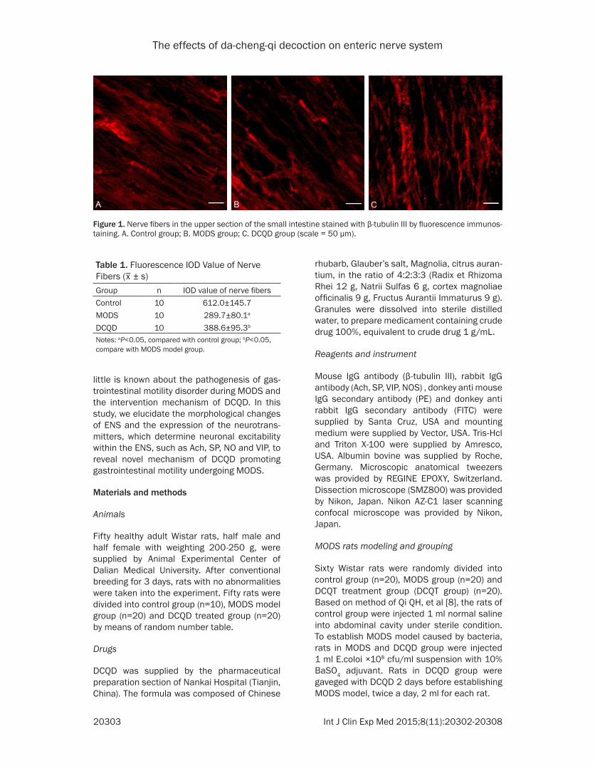
Figure 1. Nerve fibers in the upper section of the small intestine stained with β-tubulin III by fluorescence immunos-taining. A. Control group; B. MODS group; C. DCQD group (scale = 50 μm).
Table 1. Fluorescence IOD Value of Nerve Fibers (
_x ± s)
Group n IOD value of nerve fibersControl 10 612.0±145.7MODS 10 289.7±80.1a
DCQD 10 388.6±95.3b
Notes: aP<0.05, compared with control group; bP<0.05, compare with MODS model group.

The effects of da-cheng-qi decoction on enteric nerve system
20304 Int J Clin Exp Med 2015;8(11):20302-20308
All protocols were conducted in accordance with the Guidance Suggestions for Care and Use of Laboratory Animals, formulation by Ministry of Science and Technology of People’s Republic of China [9].
Whole-mount preparation
Twenty four hours after modeled, upper section of the small intestine with 2 cm length from pyloric was taken from survived rats in control group, MODS group, and DCQT group, and rinsed by 4°C PBS solution. After small intes-tine was cut along the mesentery, contents were removed by PBS solution. The tissue was cut into 1×1 cm blocks, placed into 4% parafor-maldehyde solution, storing in 4°C refrigerator overnight. The blocks were rinsed by PBS, and the mucosa and submucosa were dissected by anatomy microscope (Nikon SMZ800, Japan).
Immunofluorescence
Reference to Axel Brehmer’s method [10], brief-ly described as follows. The preparations were incubated in 0.05 mol/l Tris-HCl containing 0.5% TritonX-100 at 37 for 4 h, 1% BSA was then added at room temperature and kept for 1 h. Mouse IgG antibody (β-tubulin III) was added
(1:200), and the preparations were kept at 4°C for 48 h. After rinsed by 0.1 mol/l PBS (3×5 min), donkey anti mouse IgG secondary anti-body (PE) (1:100) was added and kept for 2 h. Rabbit IgG antibody (VAChT, VIP, SP, NOS1) was added (1:200), and the preparations were kept at 4°C for 48 h. After rinsed by 0.1 mol/l PBS (3×5 min), donkey anti rabbit IgG secondary antibody (FITC) (1:100) was added and kept for 2 h. The slides were observed by laser scanning confocal microscope.
Data and statistical analysis
Data were analyzed using ANOVA test by SPSS13.0 software. All data were expressed as mean ± standard deviation. P<0.05 was con-sidered as statistically significant.
Results
Morphology of enteric nerve system
In control group, the morphology of small intes-tine neurons was clear, distributing uniformly and continually. Neurons connected with adja-cent neurons through weeny synapses. Some neurons assembled in some region. In MODS group, the morphological structure of intestinal nerve was indistinct, distributing sparsity. Compared with the control group, the number of nerve fibers was significantly reduced (P<0.05). The nerve synapses was short, or even completely disappeared, and the gap between adjacent nerves was increased. The morphological structure and continuity of intes-tinal nerve was seriously destructed. In DCQD group, compared with MODS group, the num-ber of nerves increased significantly (P<0.05),
Figure 2. The expression of ACh and the structure of nerve fibers by fluorescence immunostaining. Notes: nerve fibers were marked by red fluorescence; ACh was marked by green fluorescence. A. Normal Group; B. MODS Group; C. DCQD Group (scale = 50 μm).
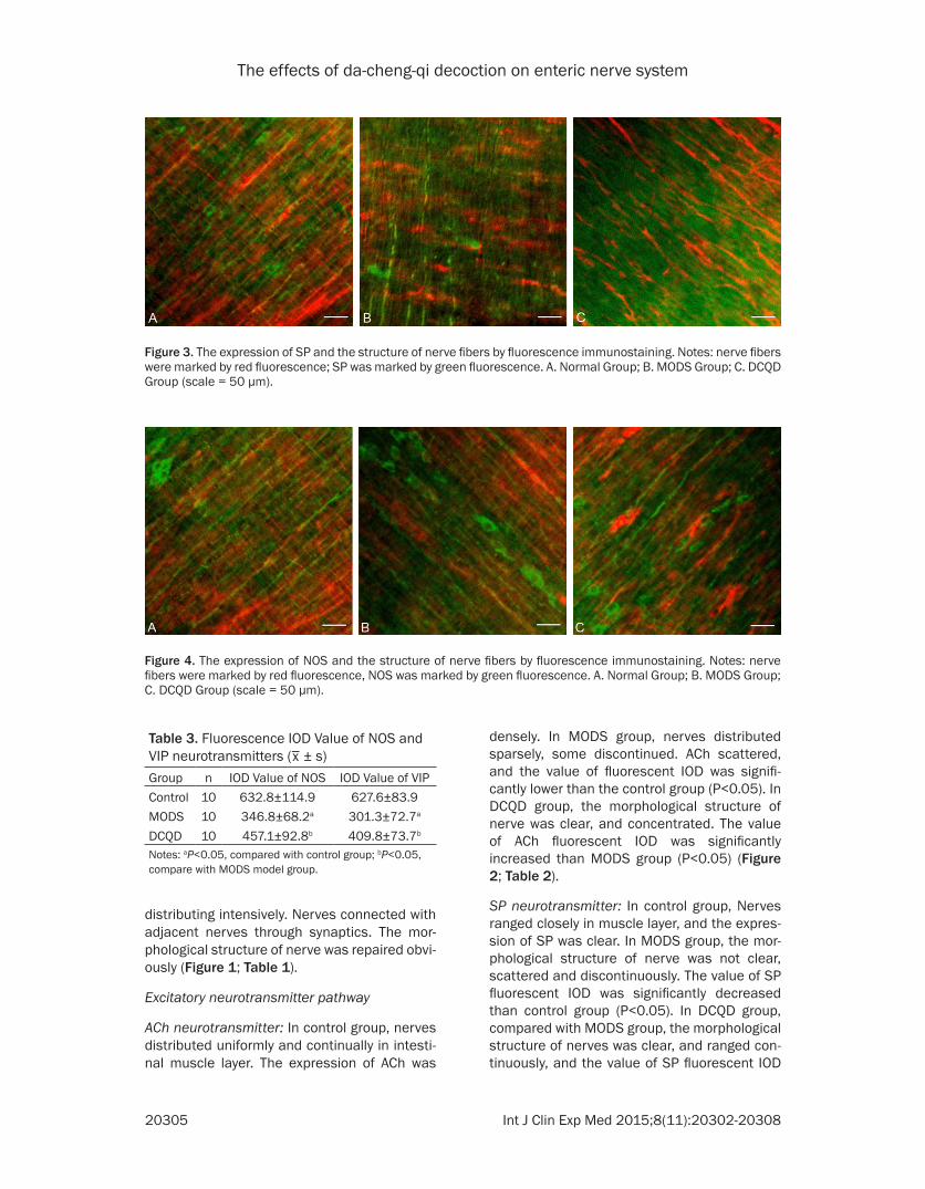
Table 2. Fluorescence IOD Value of ACh and SP neurotransmitters (
_x ± s)
Group n IOD Value of ACh IOD Value of SPControl 10 540.1±82.4 517.0±98.0MODS 10 263.9±85.4a 244.7±65.4a
DCQD 10 342.6±75.9b 400.8±70.0b
Notes: aP<0.05, compared with control group; bP<0.05, compare with MODS model group.
The effects of da-cheng-qi decoction on enteric nerve system
20305 Int J Clin Exp Med 2015;8(11):20302-20308
distributing intensively. Nerves connected with adjacent nerves through synaptics. The mor-phological structure of nerve was repaired obvi-ously (Figure 1; Table 1).
Excitatory neurotransmitter pathway
ACh neurotransmitter: In control group, nerves distributed uniformly and continually in intesti-nal muscle layer. The expression of ACh was
densely. In MODS group, nerves distributed sparsely, some discontinued. ACh scattered, and the value of fluorescent IOD was signifi-cantly lower than the control group (P<0.05). In DCQD group, the morphological structure of nerve was clear, and concentrated. The value of ACh fluorescent IOD was significantly increased than MODS group (P<0.05) (Figure 2; Table 2).
SP neurotransmitter: In control group, Nerves ranged closely in muscle layer, and the expres-sion of SP was clear. In MODS group, the mor-phological structure of nerve was not clear, scattered and discontinuously. The value of SP fluorescent IOD was significantly decreased than control group (P<0.05). In DCQD group, compared with MODS group, the morphological structure of nerves was clear, and ranged con-tinuously, and the value of SP fluorescent IOD
Figure 3. The expression of SP and the structure of nerve fibers by fluorescence immunostaining. Notes: nerve fibers were marked by red fluorescence; SP was marked by green fluorescence. A. Normal Group; B. MODS Group; C. DCQD Group (scale = 50 μm).
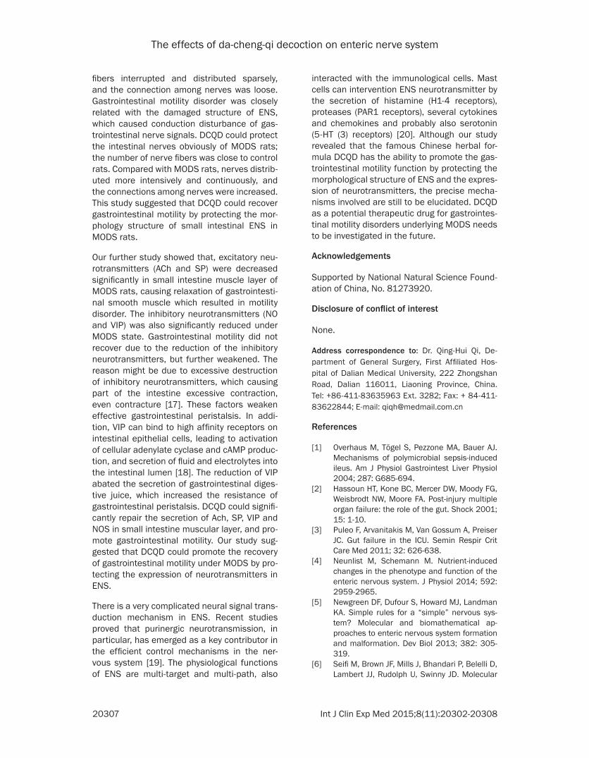
Table 3. Fluorescence IOD Value of NOS and VIP neurotransmitters (
_x ± s)
Group n IOD Value of NOS IOD Value of VIPControl 10 632.8±114.9 627.6±83.9MODS 10 346.8±68.2a 301.3±72.7a
DCQD 10 457.1±92.8b 409.8±73.7b
Notes: aP<0.05, compared with control group; bP<0.05, compare with MODS model group.
Figure 4. The expression of NOS and the structure of nerve fibers by fluorescence immunostaining. Notes: nerve fibers were marked by red fluorescence, NOS was marked by green fluorescence. A. Normal Group; B. MODS Group; C. DCQD Group (scale = 50 μm).

The effects of da-cheng-qi decoction on enteric nerve system
20306 Int J Clin Exp Med 2015;8(11):20302-20308
was significantly increased (P<0.05) (Figure 3; Table 2).
Inhibition neurotransmitter pathway
NOS neurotransmitter: In control group, the morphological structure of nerves was clear, and NOS neurotransmitter expressed near by the nerves in intestinal muscle layer neural. In MODS group, the nerves scatted and discontin-ued. The value of NOS fluorescent IOD was significantly decreased than control group (P<0.05). In DCQD group, compared with MODS group, the morphological structure of nerves was clearly, ranged continuously. The value of NOS fluorescent IOD was significantly increased (P<0.05) (Figure 4; Table 3).
VIP neurotransmitter: In Control group, nerves distributed density in intestinal muscle layer, and the expression of VIP is clear. In MODS group, compared with the control group, nerves distributed sparsely and discontinuously. The value of VIP fluorescent IOD decreased signifi-cantly (P<0.05). In DCQD group, compared with the MODS group, the morphological structure of nerves was clearly and distributed densely. The value of VIP fluorescent IOD was signifi-cantly increased (P<0.05) (Figure 5; Table 3).
Discussion
Enteric nervous system (ENS) is formed by a large number of neurons and ganglion cells, as well as the nerve fibers and neurotransmitters in the gut wall [11]. ENS is a complete periph-eral nerve loop, with high degree of autonomy. Anatomic study found that ENS is mainly com-
posed by the myenteric plexus and submucous plexus [11, 12]. The myenteric plexus distribute between the circular and longitudinal muscle larger, containing a large number of neurons, and can regulate secretion of glands and move-ment of smooth muscle within gastrointestine [5]. Submucosal plexus contain less neurons with smaller volume, and connections among ganglions are also very subtle. Submucosal plexus contains sensory neurons, which can receive afferent signals from myenteric plexus, and also can stimulate secretion of the gastric intestinal crypt cells [11]. ENS is different from other peripheral nervous systems, not depend-ing on the brain to regulate the glandular secre-tion, blood supply and smooth muscle contrac-tion and other physiological functions of ali-mentary system. The current view is that, ENS has a relative independence, a large number of different neuronal subtypes cells compose a local loop. That is similar to the information pro-cessing in the brain and spinal cord, thereby ENS can autonomously regulating gastrointes-tinal physiological activity [13].
ENS can regulate movement of gastrointestinal smooth muscle mainly through the release of excitatory and inhibitory neurotransmitter. The major excitatory neurotransmitters including acetylcholine (ACh), substance P (SP), and the inhibitory neurotransmitter is mainly the nonad-renergic noncholinergic (NANC) neurotransmit-ters, including nitric oxide (NO), vasoactive intestinal peptide (VIP) [14-16].
This study showed that the IOD value of nerve fibers in the small intestine of MODS rats decreased significantly, the continuity of nerve
Figure 5. The expression of VIP and the structure of nerve fibers by fluorescence immunostaining. Notes: nerve fibers were marked by red fluorescence; VIP was marked by green fluorescence. A. Normal Group; B. MODS Group; C. DCQD Group (scale = 50 μm).
The effects of da-cheng-qi decoction on enteric nerve system
20307 Int J Clin Exp Med 2015;8(11):20302-20308
fibers interrupted and distributed sparsely, and the connection among nerves was loose. Gastrointestinal motility disorder was closely related with the damaged structure of ENS, which caused conduction disturbance of gas-trointestinal nerve signals. DCQD could protect the intestinal nerves obviously of MODS rats; the number of nerve fibers was close to control rats. Compared with MODS rats, nerves distrib-uted more intensively and continuously, and the connections among nerves were increased. This study suggested that DCQD could recover gastrointestinal motility by protecting the mor-phology structure of small intestinal ENS in MODS rats.
Our further study showed that, excitatory neu-rotransmitters (ACh and SP) were decreased significantly in small intestine muscle layer of MODS rats, causing relaxation of gastrointesti-nal smooth muscle which resulted in motility disorder. The inhibitory neurotransmitters (NO and VIP) was also significantly reduced under MODS state. Gastrointestinal motility did not recover due to the reduction of the inhibitory neurotransmitters, but further weakened. The reason might be due to excessive destruction of inhibitory neurotransmitters, which causing part of the intestine excessive contraction, even contracture [17]. These factors weaken effective gastrointestinal peristalsis. In addi-tion, VIP can bind to high affinity receptors on intestinal epithelial cells, leading to activation of cellular adenylate cyclase and cAMP produc-tion, and secretion of fluid and electrolytes into the intestinal lumen [18]. The reduction of VIP abated the secretion of gastrointestinal diges-tive juice, which increased the resistance of gastrointestinal peristalsis. DCQD could signifi-cantly repair the secretion of Ach, SP, VIP and NOS in small intestine muscular layer, and pro-mote gastrointestinal motility. Our study sug-gested that DCQD could promote the recovery of gastrointestinal motility under MODS by pro-tecting the expression of neurotransmitters in ENS.
There is a very complicated neural signal trans-duction mechanism in ENS. Recent studies proved that purinergic neurotransmission, in particular, has emerged as a key contributor in the efficient control mechanisms in the ner-vous system [19]. The physiological functions of ENS are multi-target and multi-path, also
interacted with the immunological cells. Mast cells can intervention ENS neurotransmitter by the secretion of histamine (H1-4 receptors), proteases (PAR1 receptors), several cytokines and chemokines and probably also serotonin (5-HT (3) receptors) [20]. Although our study revealed that the famous Chinese herbal for-mula DCQD has the ability to promote the gas-trointestinal motility function by protecting the morphological structure of ENS and the expres-sion of neurotransmitters, the precise mecha-nisms involved are still to be elucidated. DCQD as a potential therapeutic drug for gastrointes-tinal motility disorders underlying MODS needs to be investigated in the future.
Acknowledgements
Supported by National Natural Science Found- ation of China, No. 81273920.
Disclosure of conflict of interest
None.
Address correspondence to: Dr. Qing-Hui Qi, De- partment of General Surgery, First Affiliated Hos- pital of Dalian Medical University, 222 Zhongshan Road, Dalian 116011, Liaoning Province, China. Tel: +86-411-83635963 Ext. 3282; Fax: + 84-411-83622844; E-mail: [email protected]
References
[1] Overhaus M, Tögel S, Pezzone MA, Bauer AJ. Mechanisms of polymicrobial sepsis-induced ileus. Am J Physiol Gastrointest Liver Physiol 2004; 287: G685-694.
[2] Hassoun HT, Kone BC, Mercer DW, Moody FG, Weisbrodt NW, Moore FA. Post-injury multiple organ failure: the role of the gut. Shock 2001; 15: 1-10.
[3] Puleo F, Arvanitakis M, Van Gossum A, Preiser JC. Gut failure in the ICU. Semin Respir Crit Care Med 2011; 32: 626-638.
[4] Neunlist M, Schemann M. Nutrient-induced changes in the phenotype and function of the enteric nervous system. J Physiol 2014; 592: 2959-2965.
[5] Newgreen DF, Dufour S, Howard MJ, Landman KA. Simple rules for a “simple” nervous sys-tem? Molecular and biomathematical ap-proaches to enteric nervous system formation and malformation. Dev Biol 2013; 382: 305-319.
[6] Seifi M, Brown JF, Mills J, Bhandari P, Belelli D, Lambert JJ, Rudolph U, Swinny JD. Molecular
The effects of da-cheng-qi decoction on enteric nerve system
20308 Int J Clin Exp Med 2015;8(11):20302-20308
and functional diversity of GABA-A receptors in the enteric nervous system of the mouse co-lon. J Neurosci 2014; 34: 10361-10378.
[7] Jian W, Heng L, Hui QQ. Effect of Da-Cheng-Qi-Tang on gastrointestinal motility in patients undergoing laparotomy. Hepatogastroentero- logy 2011; 58: 1887-1892.
[8] Qi QH, Li Y, Yao CH, Liang GG, Guo HS. Morphological changes in network of enteric nerve-interstitial cells of Cajal-smooth muscle cells in rats with multiple organ dysfunct- ion syndrome and therapeutic effects of Dachengqi decoction. Chin J Integr Med 2010; 16: 422-429.
[9] The Ministry of Science and Technology of the People’s Republic of China. Guidance Suggestions for the Care and Use of Laboratory Animals 2006-09-30.
[10] Brehmer A, Rupprecht H, Neuhuber W. Two submucosal nerve plexus in human intestines. Histochem Cell Biol 2010; 133: 149-161.
[11] Furness JB, Callaghan BP, Rivera LR, Cho HJ. The enteric nervous system and gastrointesti-nal innervation: integrated local and central control. Adv Exp Med Biol 2014; 817: 39-71.
[12] Gfroerer S, Metzger R, Fiegel H, Ramachandran P, Rolle U. Differential changes in intrinsic in-nervation and interstitial cells of Cajal in small bowel atresia in newborns. World J Gastroenterol 2010; 16: 5716-5721.
[13] Burns AJ, Pachnis V. Development of the en-teric nervous system: bringing together cells, signals and genes. Neurogastroenterol Motil 2009; 21: 100-102.
[14] Hens J, Gajda M, Scheuermann DW, Adria- ensen D, Timmermans JP. The longitudinal smooth muscle layer of the pig small intestine is innervated by both myenteric and submu-cous neurons. Histochem Cell Biol 2002; 117: 481-492.
[15] Burnstock G. Cotransmission in the autonomic nervous system. Handb Clin Neurol 2013; 117: 23-35.
[16] Sadeghinezhad J, Sorteni C, Di Guardo G, D’Agostino C, Agrimi U, Nonno R, Chiocchetti R. Neurochemistry of myenteric plexus neu-rons of bank vole (Myodes glareolus) ileum. Res Vet Sci 2013; 95: 846-853.
[17] Krajewska WM, Masłowska I. Caveolins: struc-ture and function in signal transduction. Cell Mol Biol Lett 2004; 9: 195-220.
[18] Lebowitz-Amit R, Mete O, Asa SL, Ezzat S, Joshua AM. Malignant pheochromocytoma se-creting vasoactive intestinal peptide and res-ponse to sunitinib: a case report and literature review. Endocr Pract 2014; 20: e145-50.
[19] Mutafova-Yambolieva VN, Durnin L. The puri-nergic neurotransmitter revisited: A single sub-stance or multiple players? Pharmacol Ther 2014; 144: 162-91.
[20] Buhner S, Schemann M. Mast cell-nerve axis with a focus on the human gut. Biochim Biophys Acta 2012; 1822: 85-9.