Optimally designed narrowband guided-mode resonance ...
16
Optimally designed narrowband guided-mode resonance reflectance filters for mid-infrared spectroscopy Jui-Nung Liu, 1,2,3 Matthew V. Schulmerich, 3 Rohit Bhargava, 2,3,4,5 and Brian T. Cunningham 1,2,4,* 1 Department of Electrical and Computer Engineering, University of Illinois at Urbana-Champaign, Urbana, Illinois 61801, USA 2 Micro and Nanotechnology Laboratory, University of Illinois at Urbana-Champaign, Urbana, Illinois 61801, USA 3 Beckman Institute for Advanced Science and Technology, University of Illinois at Urbana-Champaign, Urbana, Illinois 61801, USA 4 Department of Bioengineering, University of Illinois at Urbana-Champaign, Urbana, Illinois 61801, USA 5 Department of Mechanical Science and Engineering, University of Illinois at Urbana-Champaign, Urbana, Illinois 61801, USA *[email protected] Abstract: An alternative to the well-established Fourier transform infrared (FT-IR) spectrometry, termed discrete frequency infrared (DFIR) spectrometry, has recently been proposed. This approach uses narrowband mid-infrared reflectance filters based on guided-mode resonance (GMR) in waveguide gratings, but filters designed and fabricated have not attained the spectral selectivity (≤ 32 cm −1 ) commonly employed for measurements of condensed matter using FT-IR spectroscopy. With the incorporation of dispersion and optical absorption of materials, we present here optimal design of double-layer surface-relief silicon nitride-based GMR filters in the mid-IR for various narrow bandwidths below 32 cm −1 . Both shift of the filter resonance wavelengths arising from the dispersion effect and reduction of peak reflection efficiency and electric field enhancement due to the absorption effect show that the optical characteristics of materials must be taken into consideration rigorously for accurate design of narrowband GMR filters. By incorporating considerations for background reflections, the optimally designed GMR filters can have bandwidth narrower than the designed filter by the antireflection equivalence method based on the same index modulation magnitude, without sacrificing low sideband reflections near resonance. The reported work will enable use of GMR filters-based instrumentation for common measurements of condensed matter, including tissues and polymer samples. ©2011 Optical Society of America OCIS codes: (050.0050) Diffraction and gratings; (050.1970) Diffraction optics; (310.0310) Thin films; (310.2790) Guided waves; (300.6340) Spectroscopy, infrared; (300.6300) Spectroscopy, Fourier transforms. References and links 1. L. Mashev and E. Popov, “Zero order anomaly of dielectric coated gratings,” Opt. Commun. 55(6), 377–380 (1985). 2. H. Bertoni, L. Cheo, and T. Tamir, “Frequency-selective reflection and transmission by a periodic dielectric layer,” IEEE Trans. Antenn. Propag. 37(1), 78–83 (1989). 3. M. T. Gale, K. Knop, and R. H. Morf, Proc. Soc. Photo Opt. Instrum. Eng. 1210, 83 (1990). 4. B. T. Cunningham, P. Li, B. Lin, and J. Pepper, “Colorimetric resonant reflection as a direct biochemical assay technique,” Sens. Actuators B Chem. 81(2-3), 316–328 (2002). 5. B. T. Cunningham, B. Lin, J. Qiu, P. Li, J. Pepper, and B. Hugh, “A plastic colorimetric resonant optical biosensor for multiparallel detection of label-free biochemical interactions,” Sens. Actuators B Chem. 85(3), 219–226 (2002). #153807 - $15.00 USD Received 1 Sep 2011; revised 27 Oct 2011; accepted 31 Oct 2011; published 11 Nov 2011 (C) 2011 OSA 21 November 2011 / Vol. 19, No. 24 / OPTICS EXPRESS 24182
Transcript of Optimally designed narrowband guided-mode resonance ...
Optimally designed narrowband guided-mode
resonance reflectance filters for mid-infrared
spectroscopy
Jui-Nung Liu,1,2,3
Matthew V. Schulmerich,3 Rohit Bhargava,
2,3,4,5
and Brian T. Cunningham1,2,4,*
1Department of Electrical and Computer Engineering, University of Illinois at Urbana-Champaign, Urbana, Illinois
61801, USA 2Micro and Nanotechnology Laboratory, University of Illinois at Urbana-Champaign, Urbana, Illinois 61801, USA
3Beckman Institute for Advanced Science and Technology, University of Illinois at Urbana-Champaign, Urbana,
Illinois 61801, USA 4Department of Bioengineering, University of Illinois at Urbana-Champaign, Urbana, Illinois 61801, USA
5Department of Mechanical Science and Engineering, University of Illinois at Urbana-Champaign, Urbana, Illinois
61801, USA
Abstract: An alternative to the well-established Fourier transform infrared
(FT-IR) spectrometry, termed discrete frequency infrared (DFIR)
spectrometry, has recently been proposed. This approach uses narrowband
mid-infrared reflectance filters based on guided-mode resonance (GMR) in
waveguide gratings, but filters designed and fabricated have not attained the
spectral selectivity (≤ 32 cm−1
) commonly employed for measurements of
condensed matter using FT-IR spectroscopy. With the incorporation of
dispersion and optical absorption of materials, we present here optimal
design of double-layer surface-relief silicon nitride-based GMR filters in the
mid-IR for various narrow bandwidths below 32 cm−1
. Both shift of the
filter resonance wavelengths arising from the dispersion effect and
reduction of peak reflection efficiency and electric field enhancement due to
the absorption effect show that the optical characteristics of materials must
be taken into consideration rigorously for accurate design of narrowband
GMR filters. By incorporating considerations for background reflections,
the optimally designed GMR filters can have bandwidth narrower than the
designed filter by the antireflection equivalence method based on the same
index modulation magnitude, without sacrificing low sideband reflections
near resonance. The reported work will enable use of GMR filters-based
instrumentation for common measurements of condensed matter, including
tissues and polymer samples.
©2011 Optical Society of America
OCIS codes: (050.0050) Diffraction and gratings; (050.1970) Diffraction optics; (310.0310)
Thin films; (310.2790) Guided waves; (300.6340) Spectroscopy, infrared; (300.6300)
Spectroscopy, Fourier transforms.
References and links
1. L. Mashev and E. Popov, “Zero order anomaly of dielectric coated gratings,” Opt. Commun. 55(6), 377–380
(1985).
2. H. Bertoni, L. Cheo, and T. Tamir, “Frequency-selective reflection and transmission by a periodic dielectric
layer,” IEEE Trans. Antenn. Propag. 37(1), 78–83 (1989).
3. M. T. Gale, K. Knop, and R. H. Morf, Proc. Soc. Photo Opt. Instrum. Eng. 1210, 83 (1990).
4. B. T. Cunningham, P. Li, B. Lin, and J. Pepper, “Colorimetric resonant reflection as a direct biochemical assay
technique,” Sens. Actuators B Chem. 81(2-3), 316–328 (2002).
5. B. T. Cunningham, B. Lin, J. Qiu, P. Li, J. Pepper, and B. Hugh, “A plastic colorimetric resonant optical
biosensor for multiparallel detection of label-free biochemical interactions,” Sens. Actuators B Chem. 85(3),
219–226 (2002).
#153807 - $15.00 USD Received 1 Sep 2011; revised 27 Oct 2011; accepted 31 Oct 2011; published 11 Nov 2011(C) 2011 OSA 21 November 2011 / Vol. 19, No. 24 / OPTICS EXPRESS 24182
6. L. L. Chan, M. F. Pineda, J. T. Heeres, P. J. Hergenrother, and B. T. Cunningham, “A General Method for
Discovering Inhibitors of Protein-DNA Interactions using Photonic Crystal Biosensors,” ACS Chem. Biol. 3(7),
437–448 (2008).
7. P. C. Mathias, N. Ganesh, W. Zhang, and B. T. Cunningham, “Graded Wavelength One-Dimensional Photonic
Crystal Reveals Spectral Characteristics of Enhanced Fluorescence,” J. Appl. Phys. 103(9), 094320 (2008).
8. P. C. Mathias, H.-Y. Wu, and B. T. Cunningham, “Employing two distinct photonic crystal resonances for
improved fluorescence enhancement,” Appl. Phys. Lett. 95(2), 021111 (2009).
9. A. Pokhriyal, M. Lu, V. Chaudhery, C.-S. Huang, S. Schulz, and B. T. Cunningham, “Photonic crystal enhanced
fluorescence using a quartz substrate to reduce limits of detection,” Opt. Express 18(24), 24793–24808 (2010).
10. F. Yang, G. Yen, and B. T. Cunningham, “Integrated 2D photonic crystal stack filter fabricated using
nanoreplica molding,” Opt. Express 18(11), 11846–11858 (2010).
11. G. Niederer, H. P. Herzig, J. Shamir, H. Thiele, M. Schnieper, and C. Zschokke, “Tunable, Oblique Incidence
Resonant Grating Filter for Telecommunications,” Appl. Opt. 43(8), 1683–1694 (2004).
12. S. T. Thurman and G. M. Morris, “Controlling the Spectral Response in Guided-Mode Resonance Filter Design,”
Appl. Opt. 42(16), 3225–3233 (2003).
13. M. G. Moharam, E. B. Grann, D. A. Pommet, and T. K. Gaylord, “Formulation for stable and efficient
implementation of the rigorous coupled-wave analysis of binary gratings,” J. Opt. Soc. Am. A 12(5), 1068–1076
(1995).
14. M. G. Moharam, D. A. Pommet, E. B. Grann, and T. K. Gaylord, “Stable implementation of the rigorous
coupled-wave analysis for surface-relief gratings: enhanced transmittance matrix approach,” J. Opt. Soc. Am. A
12(5), 1077–1086 (1995).
15. D. Shin, S. Tibuleac, T. A. Maldonado, and R. Magnusson, “Thin-film optical filters with diffractive elements
and waveguides,” Opt. Eng. 37(9), 2634–2646 (1998).
16. Z. Hegedus and R. Netterfield, “Low Sideband Guided-Mode Resonant Filter,” Appl. Opt. 39(10), 1469–1473
(2000).
17. S. Tibuleac and R. Magnusson, “Narrow-linewidth bandpass filters with diffractive thin-film layers,” Opt. Lett.
26(9), 584–586 (2001).
18. G. Bao and K. Huang, “Optimal design of guided-mode grating resonance filters,” IEEE Photon. Technol. Lett.
16(1), 141–143 (2004).
19. G. Bao and K. Huang, “Computational design for guided-mode grating resonances,” J. Opt. Soc. Am. A 22(7),
1408–1413 (2005).
20. W. Liu, Z. Lai, H. Guo, and Y. Liu, “Guided-mode resonance filters with shallow grating,” Opt. Lett. 35(6),
865–867 (2010).
21. D. C. Fernandez, R. Bhargava, S. M. Hewitt, and I. W. Levin, “Infrared spectroscopic imaging for
histopathologic recognition,” Nat. Biotechnol. 23(4), 469–474 (2005).
22. R. Bhargava, “Towards a practical Fourier transform infrared chemical imaging protocol for cancer
histopathology,” Anal. Bioanal. Chem. 389(4), 1155–1169 (2007).
23. A. K. Kodali, M. Schulmerich, J. Ip, G. Yen, B. T. Cunningham, and R. Bhargava, “Narrowband midinfrared
reflectance filters using guided mode resonance,” Anal. Chem. 82(13), 5697–5706 (2010).
24. R. Bhargava, D. C. Fernandez, S. M. Hewitt, and I. W. Levin, “High throughput assessment of cells and tissues:
Bayesian classification of spectral metrics from infrared vibrational spectroscopic imaging data,” Biochim.
Biophys. Acta 1758(7), 830–845 (2006).
25. S. S. Wang and R. Magnusson, “Theory and applications of guided-mode resonance filters,” Appl. Opt. 32(14),
2606–2613 (1993).
26. S. S. Wang and R. Magnusson, “Design of waveguide-grating filters with symmetrical line shapes and low
sidebands,” Opt. Lett. 19(12), 919–921 (1994).
27. A. Sharon, D. Rosenblatt, and A. A. Friesem, “Resonant grating–waveguide structures for visible and near-
infrared radiation,” J. Opt. Soc. Am. A 14(11), 2985–2993 (1997).
28. D. Rosenblatt, A. Sharon, and A. A. Friesem, “Resonant grating waveguide structures,” IEEE J. Quantum
Electron. 33(11), 2038–2059 (1997).
29. S. L. Chuang, Physics of Optoelectronic Devices (Wiley, New York, 1995).
30. A. Yariv and P. Yeh, Photonics: Optical Electronics in Modern Communications, 6th ed. (Oxford U. Press, New
York, 2007).
31. M. Klanjšek Gunde and M. Maček, “Infrared Optical Constants and Dielectric Response Functions of Silicon
Nitride and Oxynitride Films,” Phys. Status Solidi 183, 439–449 (2001).
32. M. Rubin, “Optical properties of soda lime silica glasses,” Sol. Energy Mater. 12(4), 275–288 (1985).
33. S. M. Norton, G. M. Morris, and T. Erdogan, “Experimental investigation of resonant-grating filter lineshapes in
comparison with theoretical models,” J. Opt. Soc. Am. A 15(2), 464–472 (1998).
34. T. Sun, J. Wang, J. Ma, Y. Jin, H. He, J. Shao, and Z. Fan, “Ultra-narrow bandwidth resonant reflection grating
filters using the second diffracted orders,” Opt. Commun. 282(4), 451–454 (2009).
35. T. Sun, J. Ma, J. Wang, Y. Jin, H. He, J. Shao, and Z. Fan, “Electric field distribution in resonant reflection
filters under normal incidence,” J. Opt. A, Pure Appl. Opt. 10(12), 125003 (2008).
36. J. N. Liu, M. Schulmerich, R. Bhargava, and B. T. Cunningham, University of Illinois at Urbana-Champaign,
Urbana, IL are preparing a manuscript to be called “Effects of collimation on narrowband guided-mode
resonance filters in mid-infrared,”
#153807 - $15.00 USD Received 1 Sep 2011; revised 27 Oct 2011; accepted 31 Oct 2011; published 11 Nov 2011(C) 2011 OSA 21 November 2011 / Vol. 19, No. 24 / OPTICS EXPRESS 24183
37. S. S. Wang and R. Magnusson, “Multilayer waveguide-grating filters,” Appl. Opt. 34(14), 2414–2420 (1995).
38. A. L. Fehrembach, A. Talneau, O. Boyko, F. Lemarchand, and A. Sentenac, “Experimental demonstration of a
narrowband, angular tolerant, polarization independent, doubly periodic resonant grating filter,” Opt. Lett.
32(15), 2269–2271 (2007).
1. Introduction
Since the introduction of narrowband reflectance filters using subwavelength periodic grating
nanostructures [1–3], guided-mode resonance (GMR) filters (as they have come to be called,
but also known as photonic crystal slabs or photonic crystal surfaces) have found application
in label-free biodetection [4–6], fluorescence enhancement [7–9], optical limiting [10], and
telecommunications [11, 12]. Despite the large number of papers that utilize electromagnetic
simulation methods such as rigorous coupled wave analysis (RCWA) [13, 14] to study and
design GMR filters, none fully consider the effects of optical losses from the materials that
the GMR filter is comprised of and the resulting effects of optical loss upon the resonant filter
characteristics. Indeed, for filter design that must meet stringent performance criteria that
include filter bandwidth, out-of-band rejection, and filter efficiency, these considerations
become critical for determining whether or not a design is feasible with a set of materials.
Without the effects of optical loss, GMR filters may be designed to provide ~100% reflection
efficiency and vanishingly small bandwidth [15–20], but real materials often fail to be
completely lossless. Optical properties of GMR filters such as the resonance wavelength, peak
reflection efficiency, and resonant linewidth are heavily dependent upon the complex
refractive index of materials which is a function of wavelength, so it is actually necessary to
consider the dispersion (dn/dν ≠ 0 and dκ/dν ≠ 0, where n and κ are the real and imaginary
part of the complex refractive index, respectively) of optical materials to obtain designs that
accurately reflect experimentally measured behavior. With accurate incorporation of material
parameters into design models, it is possible to adjust the dimensional parameters of a GMR
filter to achieve a particular resonant wavelength. For practical fabrication of devices, it is
desirable to adjust dimensional parameters that are easily controlled (such as grating height
and thin film layer thickness) in contrast to parameters that are adjustable only with increased
cost or effort (such as grating fill factor).
An application for which accurate GMR filters design is especially critical is infrared (IR)
absorption spectroscopic imaging. The efficiency and expense of IR imaging spectroscopy
can be greatly improved using narrowband filters for applications such as automated cancer
pathology [21, 22] by acquisition of data only from a few specific regions in the spectrum that
correspond to known spectral features that differentiate cell and disease types [23, 24].
Compared to Fourier Transform Infrared (FTIR) spectroscopic imaging, discrete-frequency
IR (DFIR) spectroscopy can be performed rapidly with simple and inexpensive
instrumentation due to elimination of the need for an interferometer for spectral selectivity.
DFIR spectroscopy requires a set of optical filters with narrow bandwidth (typically ≤ 32 cm−1
for solids and liquids) for spectroscopy. One method for obtaining illumination from these
wavelengths is to reflect a broadband light source against a resonant reflectance filter, such as
a GMR filter and to direct the narrow band of reflected wavelengths through a biological
sample. In DFIR spectroscopy, a set of GMR filters with predetermined resonant wavelengths
are illuminated by the broadband light source in sequence, and separate absorption intensity
images are gathered for each wavelength using an appropriate infrared imaging camera, such
as a microbolometer array. For DFIR spectroscopy to be effective, it is necessary to fabricate
a series of GMR filters that simultaneously provide high reflectance efficiency for the
resonant wavelength, low out-of-band reflectance, and narrow bandwidth. In our recent work,
GMR filters in the mid-IR were demonstrated [23]. RCWA simulations incorporating the
absorption and dispersion of materials were used to design GMR filter structures, where
bandwidth of the designed filters is larger than 32 cm−1
and background reflection reaches up
to 17% near resonance wavenumbers. Spectroscopic measurements of a set of fabricated
#153807 - $15.00 USD Received 1 Sep 2011; revised 27 Oct 2011; accepted 31 Oct 2011; published 11 Nov 2011(C) 2011 OSA 21 November 2011 / Vol. 19, No. 24 / OPTICS EXPRESS 24184
filters were compared with theory with good agreement. In order to realize GMR filters with
bandwidth ≤ 32 cm−1
and lower sideband reflections for DFIR spectroscopy, design
optimization of filter structures is essential to guide for fabrication.
In this paper, we describe a design optimization approach for obtaining narrow bandwidth
GMR filters in the mid-IR, with absorption and dispersion characteristics of materials taken
into consideration. We first study the impact of absorption and dispersion of materials on
GMR filters when bandwidth becomes narrow. Second, we describe how to achieve an
optimal GMR filter design at one specific resonance wavenumber for various bandwidths.
Finally, we compare the optimal filter design developed in this paper and the filter design
with conventional antireflection (AR) equivalence method. Although we focus upon design of
GMR filters in the mid-IR part of the spectrum, the approach described in this work can be
extended to any wavelength.
2. GMR structure and physical principle
The double-layer GMR filter in this study has a Si3N4 surface-relief periodic grating structure
built on top of a soda lime glass substrate. The grating period (Λ), grating depth (dgr
), and
waveguide layer thickness (dwg
) of the GMR filter structure are depicted in Fig. 1. Thin film
layer of Si3N4 can be easily deposited onto the substrate by plasma enhanced chemical vapor
deposition (PECVD), and can be etched using reactive ion etching (RIE). In addition to
simple fabrication, Si3N4 has a transparent window in mid-IR (λ<~6 µm), making it an
appropriate material candidate for GMR filters in a DFIR instrument that can be used to
measure absorption caused by C-H, O-H and S-H stretching vibrational modes (> 2600 cm−1
).
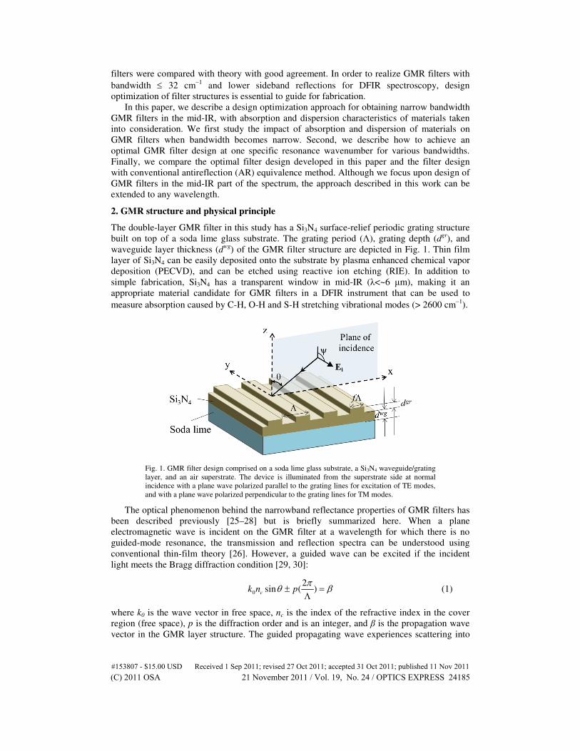
Fig. 1. GMR filter design comprised on a soda lime glass substrate, a Si3N4 waveguide/grating
layer, and an air superstrate. The device is illuminated from the superstrate side at normal
incidence with a plane wave polarized parallel to the grating lines for excitation of TE modes,
and with a plane wave polarized perpendicular to the grating lines for TM modes.
The optical phenomenon behind the narrowband reflectance properties of GMR filters has
been described previously [25–28] but is briefly summarized here. When a plane
electromagnetic wave is incident on the GMR filter at a wavelength for which there is no
guided-mode resonance, the transmission and reflection spectra can be understood using
conventional thin-film theory [26]. However, a guided wave can be excited if the incident
light meets the Bragg diffraction condition [29, 30]:
0
2sin ( )ck n p
πθ β± =
Λ (1)
where k0 is the wave vector in free space, nc is the index of the refractive index in the cover
region (free space), p is the diffraction order and is an integer, and β is the propagation wave
vector in the GMR layer structure. The guided propagating wave experiences scattering into
#153807 - $15.00 USD Received 1 Sep 2011; revised 27 Oct 2011; accepted 31 Oct 2011; published 11 Nov 2011(C) 2011 OSA 21 November 2011 / Vol. 19, No. 24 / OPTICS EXPRESS 24185
specular and transmitted directions assisted by the grating periodic structure as well as optical
absorption in the waveguide layer structure. For this reason, such guided modes are also
called “leaky modes.” The constructive interference of waves in the specular direction creates
peak reflection efficiency; at the same time the destructive interference of waves in the
transmitted direction generates a dip in the transmission spectra [27, 28]. The filter property
of GMR filters can be used to admit light in a small region of the spectrum, with light outside
the reflection band being rejected. When illuminated at the resonant condition, the guided
mode in GMR filters is not only excited and extracted simultaneously via periodic grating
structures, but is also coupled to the guided wave propagating along the other direction via
Bragg diffraction. The constructive interference of the two counter-propagating guided waves
establishes an electromagnetic standing wave, leading to near-field enhancement and storage
of photon energy.
3. Results and discussion
3.1 Effects of dispersion and absorption of optical materials on narrowband GMR filters
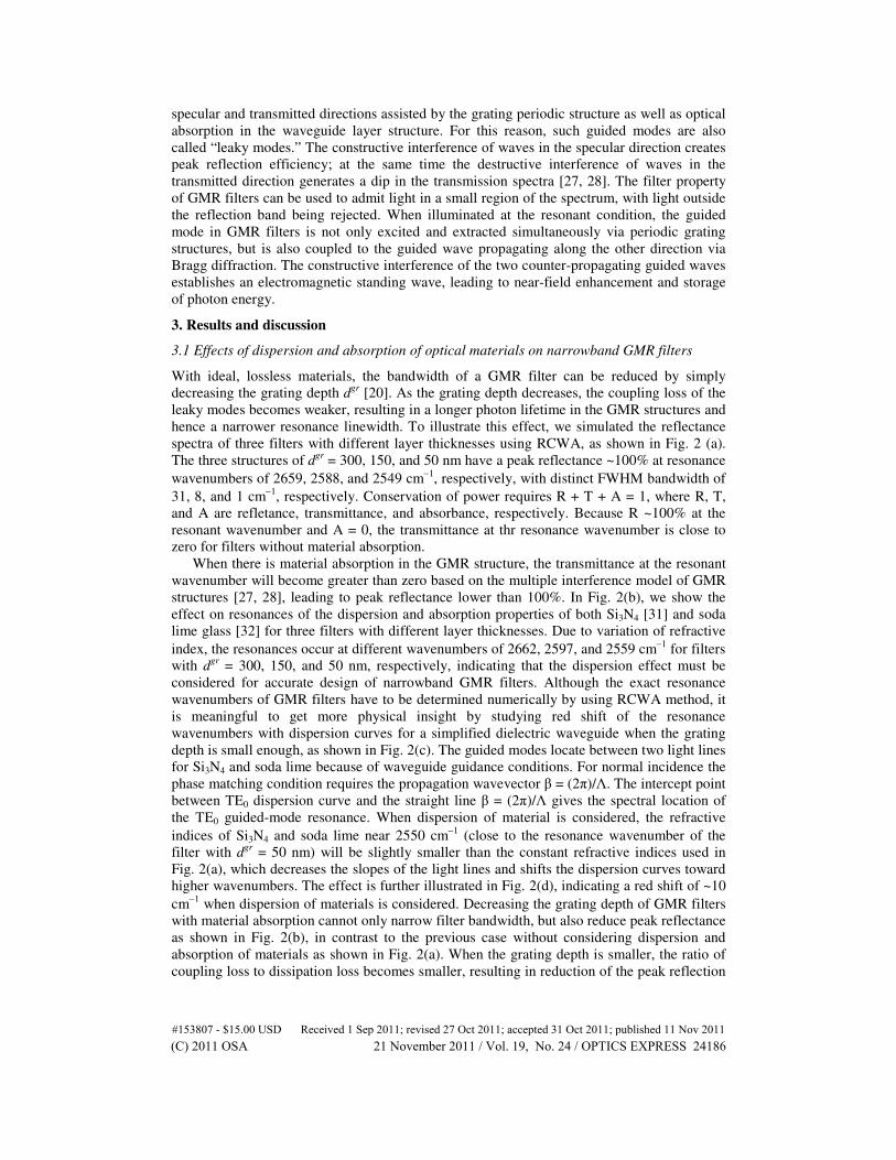
With ideal, lossless materials, the bandwidth of a GMR filter can be reduced by simply
decreasing the grating depth dgr
[20]. As the grating depth decreases, the coupling loss of the
leaky modes becomes weaker, resulting in a longer photon lifetime in the GMR structures and
hence a narrower resonance linewidth. To illustrate this effect, we simulated the reflectance
spectra of three filters with different layer thicknesses using RCWA, as shown in Fig. 2 (a).
The three structures of dgr
= 300, 150, and 50 nm have a peak reflectance ~100% at resonance
wavenumbers of 2659, 2588, and 2549 cm−1
, respectively, with distinct FWHM bandwidth of
31, 8, and 1 cm−1
, respectively. Conservation of power requires R + T + A = 1, where R, T,
and A are refletance, transmittance, and absorbance, respectively. Because R ~100% at the
resonant wavenumber and A = 0, the transmittance at thr resonance wavenumber is close to
zero for filters without material absorption.
When there is material absorption in the GMR structure, the transmittance at the resonant
wavenumber will become greater than zero based on the multiple interference model of GMR
structures [27, 28], leading to peak reflectance lower than 100%. In Fig. 2(b), we show the
effect on resonances of the dispersion and absorption properties of both Si3N4 [31] and soda
lime glass [32] for three filters with different layer thicknesses. Due to variation of refractive
index, the resonances occur at different wavenumbers of 2662, 2597, and 2559 cm−1
for filters
with dgr
= 300, 150, and 50 nm, respectively, indicating that the dispersion effect must be
considered for accurate design of narrowband GMR filters. Although the exact resonance
wavenumbers of GMR filters have to be determined numerically by using RCWA method, it
is meaningful to get more physical insight by studying red shift of the resonance
wavenumbers with dispersion curves for a simplified dielectric waveguide when the grating
depth is small enough, as shown in Fig. 2(c). The guided modes locate between two light lines
for Si3N4 and soda lime because of waveguide guidance conditions. For normal incidence the
phase matching condition requires the propagation wavevector β = (2π)/Λ. The intercept point
between TE0 dispersion curve and the straight line β = (2π)/Λ gives the spectral location of
the TE0 guided-mode resonance. When dispersion of material is considered, the refractive
indices of Si3N4 and soda lime near 2550 cm−1
(close to the resonance wavenumber of the
filter with dgr
= 50 nm) will be slightly smaller than the constant refractive indices used in
Fig. 2(a), which decreases the slopes of the light lines and shifts the dispersion curves toward
higher wavenumbers. The effect is further illustrated in Fig. 2(d), indicating a red shift of ~10
cm−1
when dispersion of materials is considered. Decreasing the grating depth of GMR filters
with material absorption cannot only narrow filter bandwidth, but also reduce peak reflectance
as shown in Fig. 2(b), in contrast to the previous case without considering dispersion and
absorption of materials as shown in Fig. 2(a). When the grating depth is smaller, the ratio of
coupling loss to dissipation loss becomes smaller, resulting in reduction of the peak reflection
#153807 - $15.00 USD Received 1 Sep 2011; revised 27 Oct 2011; accepted 31 Oct 2011; published 11 Nov 2011(C) 2011 OSA 21 November 2011 / Vol. 19, No. 24 / OPTICS EXPRESS 24186
efficiency [12, 27, 28, 33]. Therefore, for GMR filters with material absorption, the inherent
tradeoff between bandwidth and reflection efficiency puts a limitation on performance of
narrowband GMR filters, and the design method of narrowband filters needs to be improvised
as simply decreasing the grating layer thickness to obtain GMR filters with narrow linewidth
[20] is not effective.
Fig. 2. Effect on guided-mode resonances of dispersion and absorption properties of Si3N4 and
soda lime, when a TE-polarized wave is normally incident on GMR filters with grating period
Λ = 2.5 µm, duty cycle f = 0.5, and Si3N4 total thickness dgr + dwg = 0.6 µm. (a) Reflectance
spectra of three filters with different grating depths without considering the dispersion
characteristics of both Si3N4 and soda lime. The refractive indices chosen are nSiN = 2.02 and
nSL = 1.47. (b) Reflectance spectra considering the dispersion characteristics of both Si3N4 [31]
and soda lime glass [32]. (c) The dispersion relation for a simplified dielectric waveguide,
where the refractive indices used are the same as those in (a). The phase matching condition
requires β = (2π)/Λ, where the grating period Λ = 2.5 µm. (d) Explanation of the observed red
shift of guided mode resonances by comparing dispersion curves for the TE0 mode using nSiN =
2.01 and nSL = 1.46 near 2550 cm−1 with that in (c). (e-f) Distribution of the electric field
amplitude (|E|) in a unit cell of the filter with grating depth dgr = 50 nm for illumination at (e) υ
= 2549 cm−1 without considering the dispersion characteristics of both Si3N4 and soda lime and
at (f) υ = 2559 cm−1 considering the dispersion properties of both Si3N4 and soda lime. (g-h)
Comparison of electric field amplitude profiles shown in (e) and (f) at x = 1.25 µm (left) and
near the center of the waveguide layer at z = 200 nm (right).
The material absorption influences not only the spectral response of GMR filters but also
their electric field enhancement. In Fig. 2(c) we show the electric field enhancement
#153807 - $15.00 USD Received 1 Sep 2011; revised 27 Oct 2011; accepted 31 Oct 2011; published 11 Nov 2011(C) 2011 OSA 21 November 2011 / Vol. 19, No. 24 / OPTICS EXPRESS 24187
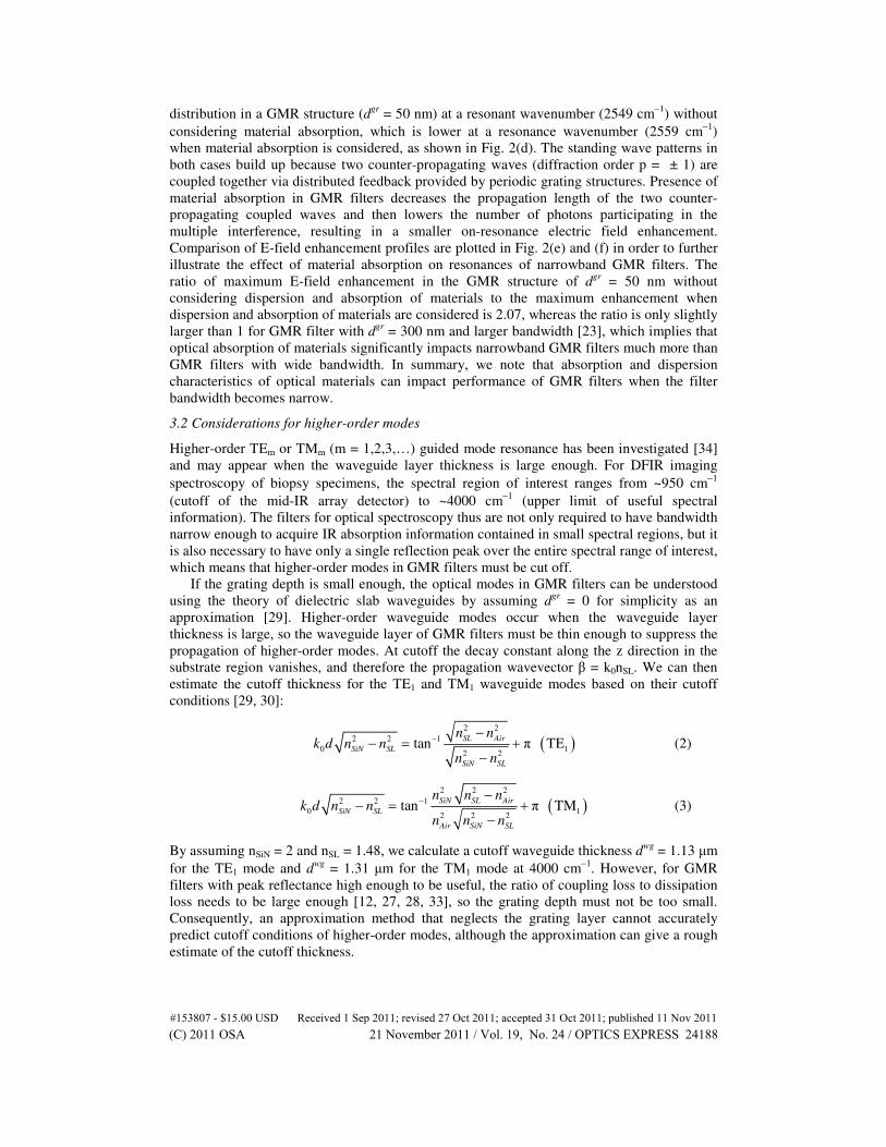
distribution in a GMR structure (dgr
= 50 nm) at a resonant wavenumber (2549 cm−1
) without
considering material absorption, which is lower at a resonance wavenumber (2559 cm−1
)
when material absorption is considered, as shown in Fig. 2(d). The standing wave patterns in
both cases build up because two counter-propagating waves (diffraction order p = ± 1) are
coupled together via distributed feedback provided by periodic grating structures. Presence of
material absorption in GMR filters decreases the propagation length of the two counter-
propagating coupled waves and then lowers the number of photons participating in the
multiple interference, resulting in a smaller on-resonance electric field enhancement.
Comparison of E-field enhancement profiles are plotted in Fig. 2(e) and (f) in order to further
illustrate the effect of material absorption on resonances of narrowband GMR filters. The
ratio of maximum E-field enhancement in the GMR structure of dgr
= 50 nm without
considering dispersion and absorption of materials to the maximum enhancement when
dispersion and absorption of materials are considered is 2.07, whereas the ratio is only slightly
larger than 1 for GMR filter with dgr
= 300 nm and larger bandwidth [23], which implies that
optical absorption of materials significantly impacts narrowband GMR filters much more than
GMR filters with wide bandwidth. In summary, we note that absorption and dispersion
characteristics of optical materials can impact performance of GMR filters when the filter
bandwidth becomes narrow.
3.2 Considerations for higher-order modes
Higher-order TEm or TMm (m = 1,2,3,…) guided mode resonance has been investigated [34]
and may appear when the waveguide layer thickness is large enough. For DFIR imaging
spectroscopy of biopsy specimens, the spectral region of interest ranges from ~950 cm−1
(cutoff of the mid-IR array detector) to ~4000 cm−1
(upper limit of useful spectral
information). The filters for optical spectroscopy thus are not only required to have bandwidth
narrow enough to acquire IR absorption information contained in small spectral regions, but it
is also necessary to have only a single reflection peak over the entire spectral range of interest,
which means that higher-order modes in GMR filters must be cut off.
If the grating depth is small enough, the optical modes in GMR filters can be understood
using the theory of dielectric slab waveguides by assuming dgr
= 0 for simplicity as an
approximation [29]. Higher-order waveguide modes occur when the waveguide layer
thickness is large, so the waveguide layer of GMR filters must be thin enough to suppress the
propagation of higher-order modes. At cutoff the decay constant along the z direction in the
substrate region vanishes, and therefore the propagation wavevector β = k0nSL. We can then
estimate the cutoff thickness for the TE1 and TM1 waveguide modes based on their cutoff
conditions [29, 30]:
( )2 2
2 2 1
0 12 2
tan π TESL Air
SiN SL
SiN SL
n nk d n n
n n
−−
− = +−
(2)
( )2 2 2
2 2 1
0 12 2 2
tan π TMSiN SL Air
SiN SL
Air SiN SL
n n nk d n n
n n n
−−
− = +−
(3)
By assuming nSiN = 2 and nSL = 1.48, we calculate a cutoff waveguide thickness dwg
= 1.13 µm
for the TE1 mode and dwg
= 1.31 µm for the TM1 mode at 4000 cm−1
. However, for GMR
filters with peak reflectance high enough to be useful, the ratio of coupling loss to dissipation
loss needs to be large enough [12, 27, 28, 33], so the grating depth must not be too small.
Consequently, an approximation method that neglects the grating layer cannot accurately
predict cutoff conditions of higher-order modes, although the approximation can give a rough
estimate of the cutoff thickness.
#153807 - $15.00 USD Received 1 Sep 2011; revised 27 Oct 2011; accepted 31 Oct 2011; published 11 Nov 2011(C) 2011 OSA 21 November 2011 / Vol. 19, No. 24 / OPTICS EXPRESS 24188
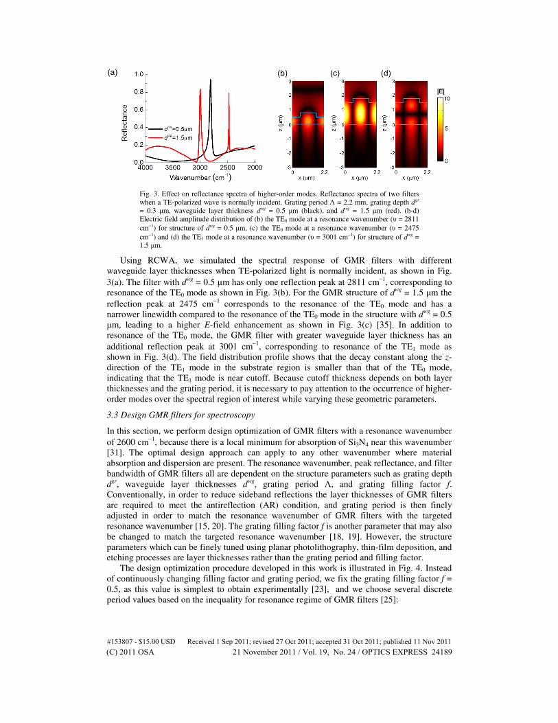
Fig. 3. Effect on reflectance spectra of higher-order modes. Reflectance spectra of two filters
when a TE-polarized wave is normally incident. Grating period Λ = 2.2 mm, grating depth dgr
= 0.3 µm, waveguide layer thickness dwg = 0.5 µm (black), and dwg = 1.5 µm (red). (b-d)
Electric field amplitude distribution of (b) the TE0 mode at a resonance wavenumber (υ = 2811
cm−1) for structure of dwg = 0.5 µm, (c) the TE0 mode at a resonance wavenumber (υ = 2475
cm−1) and (d) the TE1 mode at a resonance wavenumber (υ = 3001 cm−1) for structure of dwg =
1.5 µm.
Using RCWA, we simulated the spectral response of GMR filters with different
waveguide layer thicknesses when TE-polarized light is normally incident, as shown in Fig.
3(a). The filter with dwg
= 0.5 µm has only one reflection peak at 2811 cm−1
, corresponding to
resonance of the TE0 mode as shown in Fig. 3(b). For the GMR structure of dwg
= 1.5 µm the
reflection peak at 2475 cm−1
corresponds to the resonance of the TE0 mode and has a
narrower linewidth compared to the resonance of the TE0 mode in the structure with dwg
= 0.5
µm, leading to a higher E-field enhancement as shown in Fig. 3(c) [35]. In addition to
resonance of the TE0 mode, the GMR filter with greater waveguide layer thickness has an
additional reflection peak at 3001 cm−1
, corresponding to resonance of the TE1 mode as
shown in Fig. 3(d). The field distribution profile shows that the decay constant along the z-
direction of the TE1 mode in the substrate region is smaller than that of the TE0 mode,
indicating that the TE1 mode is near cutoff. Because cutoff thickness depends on both layer
thicknesses and the grating period, it is necessary to pay attention to the occurrence of higher-
order modes over the spectral region of interest while varying these geometric parameters.
3.3 Design GMR filters for spectroscopy
In this section, we perform design optimization of GMR filters with a resonance wavenumber
of 2600 cm−1
, because there is a local minimum for absorption of Si3N4 near this wavenumber
[31]. The optimal design approach can apply to any other wavenumber where material
absorption and dispersion are present. The resonance wavenumber, peak reflectance, and filter
bandwidth of GMR filters all are dependent on the structure parameters such as grating depth
dgr
, waveguide layer thicknesses dwg
, grating period Λ, and grating filling factor f.
Conventionally, in order to reduce sideband reflections the layer thicknesses of GMR filters
are required to meet the antireflection (AR) condition, and grating period is then finely
adjusted in order to match the resonance wavenumber of GMR filters with the targeted
resonance wavenumber [15, 20]. The grating filling factor f is another parameter that may also
be changed to match the targeted resonance wavenumber [18, 19]. However, the structure
parameters which can be finely tuned using planar photolithography, thin-film deposition, and
etching processes are layer thicknesses rather than the grating period and filling factor.
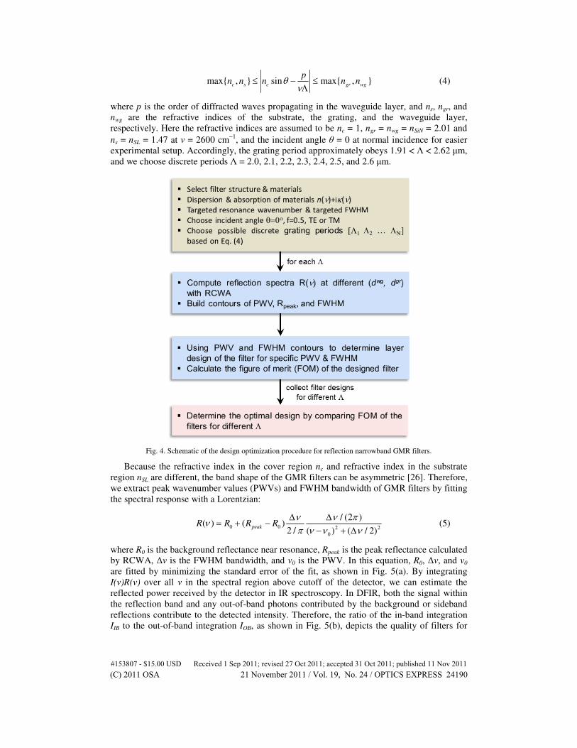
The design optimization procedure developed in this work is illustrated in Fig. 4. Instead
of continuously changing filling factor and grating period, we fix the grating filling factor f =
0.5, as this value is simplest to obtain experimentally [23], and we choose several discrete
period values based on the inequality for resonance regime of GMR filters [25]:
#153807 - $15.00 USD Received 1 Sep 2011; revised 27 Oct 2011; accepted 31 Oct 2011; published 11 Nov 2011(C) 2011 OSA 21 November 2011 / Vol. 19, No. 24 / OPTICS EXPRESS 24189
max{ , } sin max{ , }c s c gr wg
pn n n n nθ
ν≤ − ≤
Λ (4)
where p is the order of diffracted waves propagating in the waveguide layer, and ns, ngr, and
nwg are the refractive indices of the substrate, the grating, and the waveguide layer,
respectively. Here the refractive indices are assumed to be nc = 1, ngr = nwg = nSiN = 2.01 and
ns = nSL = 1.47 at ν = 2600 cm−1
, and the incident angle θ = 0 at normal incidence for easier
experimental setup. Accordingly, the grating period approximately obeys 1.91 < Λ < 2.62 µm,
and we choose discrete periods Λ = 2.0, 2.1, 2.2, 2.3, 2.4, 2.5, and 2.6 µm.
Fig. 4. Schematic of the design optimization procedure for reflection narrowband GMR filters.
Because the refractive index in the cover region nc and refractive index in the substrate
region nSL are different, the band shape of the GMR filters can be asymmetric [26]. Therefore,
we extract peak wavenumber values (PWVs) and FWHM bandwidth of GMR filters by fitting
the spectral response with a Lorentzian:
0 0 2 2
0
/ (2 )( ) ( )
2 / ( ) ( / 2)peakR R R R
ν ν πν
π ν ν ν
∆ ∆= + −
− + ∆ (5)
where R0 is the background reflectance near resonance, Rpeak is the peak reflectance calculated
by RCWA, ∆ν is the FWHM bandwidth, and ν0 is the PWV. In this equation, R0, ∆ν, and ν0
are fitted by minimizing the standard error of the fit, as shown in Fig. 5(a). By integrating
I(ν)R(ν) over all ν in the spectral region above cutoff of the detector, we can estimate the
reflected power received by the detector in IR spectroscopy. In DFIR, both the signal within
the reflection band and any out-of-band photons contributed by the background or sideband
reflections contribute to the detected intensity. Therefore, the ratio of the in-band integration
IIB to the out-of-band integration IOB, as shown in Fig. 5(b), depicts the quality of filters for
#153807 - $15.00 USD Received 1 Sep 2011; revised 27 Oct 2011; accepted 31 Oct 2011; published 11 Nov 2011(C) 2011 OSA 21 November 2011 / Vol. 19, No. 24 / OPTICS EXPRESS 24190
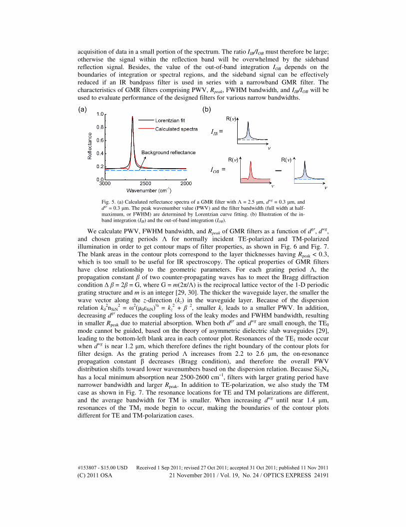
acquisition of data in a small portion of the spectrum. The ratio IIB/IOB must therefore be large;
otherwise the signal within the reflection band will be overwhelmed by the sideband
reflection signal. Besides, the value of the out-of-band integration IOB depends on the
boundaries of integration or spectral regions, and the sideband signal can be effectively
reduced if an IR bandpass filter is used in series with a narrowband GMR filter. The
characteristics of GMR filters comprising PWV, Rpeak, FWHM bandwidth, and IIB/IOB will be
used to evaluate performance of the designed filters for various narrow bandwidths.
Fig. 5. (a) Calculated reflectance spectra of a GMR filter with Λ = 2.5 µm, dwg = 0.3 µm, and
dgr = 0.3 µm. The peak wavenumber value (PWV) and the filter bandwidth (full width at half-
maximum, or FWHM) are determined by Lorentzian curve fitting. (b) Illustration of the in-
band integration (IIB) and the out-of-band integration (IOB).
We calculate PWV, FWHM bandwidth, and Rpeak of GMR filters as a function of dgr
, dwg
,
and chosen grating periods Λ for normally incident TE-polarized and TM-polarized
illumination in order to get contour maps of filter properties, as shown in Fig. 6 and Fig. 7.
The blank areas in the contour plots correspond to the layer thicknesses having Rpeak < 0.3,
which is too small to be useful for IR spectroscopy. The optical properties of GMR filters
have close relationship to the geometric parameters. For each grating period Λ, the
propagation constant β of two counter-propagating waves has to meet the Bragg diffraction
condition ∆ β = 2β = G, where G = m(2π/Λ) is the reciprocal lattice vector of the 1-D periodic
grating structure and m is an integer [29, 30]. The thicker the waveguide layer, the smaller the
wave vector along the z-direction (kz) in the waveguide layer. Because of the dispersion
relation k02nSiN
2 = ω
2(µ0εSiN)
½ = kz
2 + β
2, smaller kz leads to a smaller PWV. In addition,
decreasing dgr
reduces the coupling loss of the leaky modes and FWHM bandwidth, resulting
in smaller Rpeak due to material absorption. When both dgr
and dwg
are small enough, the TE0
mode cannot be guided, based on the theory of asymmetric dielectric slab waveguides [29],
leading to the bottom-left blank area in each contour plot. Resonances of the TE1 mode occur
when dwg
is near 1.2 µm, which therefore defines the right boundary of the contour plots for
filter design. As the grating period Λ increases from 2.2 to 2.6 µm, the on-resonance
propagation constant β decreases (Bragg condition), and therefore the overall PWV
distribution shifts toward lower wavenumbers based on the dispersion relation. Because Si3N4
has a local minimum absorption near 2500-2600 cm−1
, filters with larger grating period have
narrower bandwidth and larger Rpeak. In addition to TE-polarization, we also study the TM
case as shown in Fig. 7. The resonance locations for TE and TM polarizations are different,
and the average bandwidth for TM is smaller. When increasing dwg
until near 1.4 µm,
resonances of the TM1 mode begin to occur, making the boundaries of the contour plots
different for TE and TM-polarization cases.
#153807 - $15.00 USD Received 1 Sep 2011; revised 27 Oct 2011; accepted 31 Oct 2011; published 11 Nov 2011(C) 2011 OSA 21 November 2011 / Vol. 19, No. 24 / OPTICS EXPRESS 24191
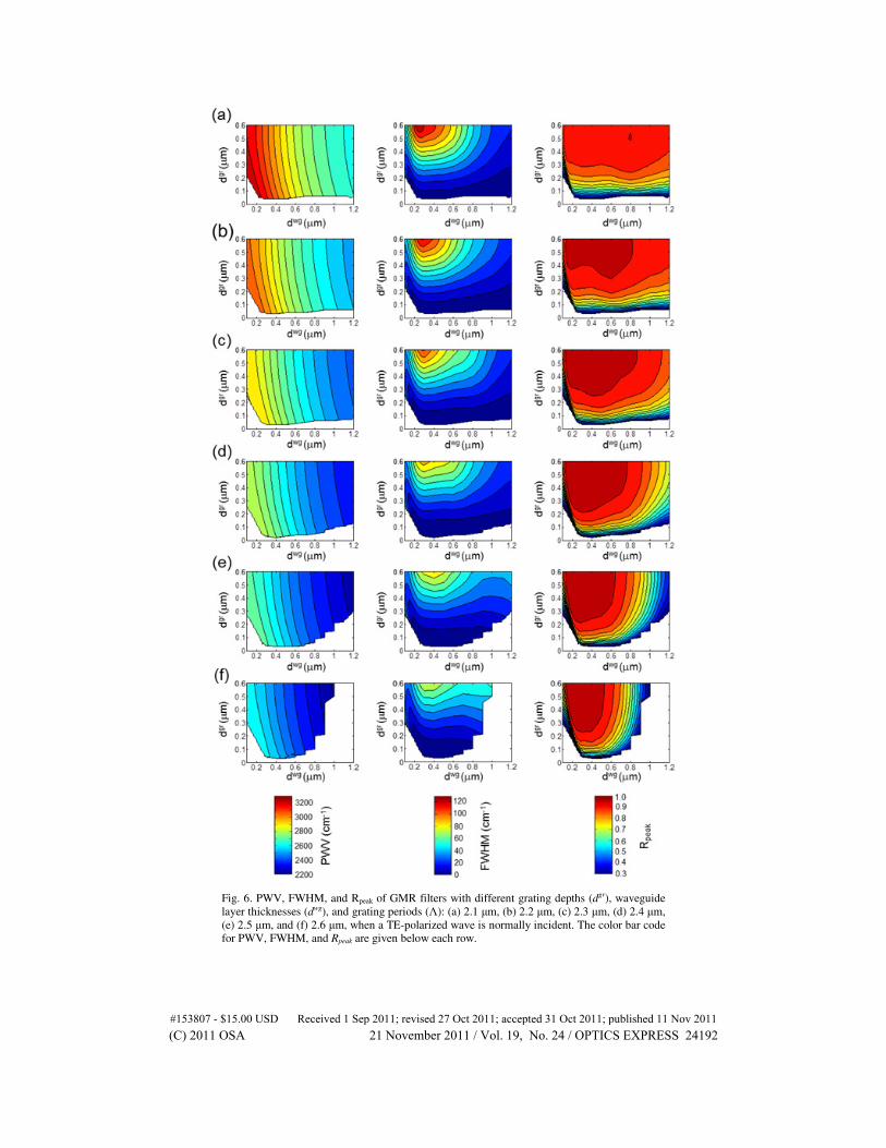
Fig. 6. PWV, FWHM, and Rpeak of GMR filters with different grating depths (dgr), waveguide
layer thicknesses (dwg), and grating periods (Λ): (a) 2.1 µm, (b) 2.2 µm, (c) 2.3 µm, (d) 2.4 µm,
(e) 2.5 µm, and (f) 2.6 µm, when a TE-polarized wave is normally incident. The color bar code
for PWV, FWHM, and Rpeak are given below each row.
#153807 - $15.00 USD Received 1 Sep 2011; revised 27 Oct 2011; accepted 31 Oct 2011; published 11 Nov 2011(C) 2011 OSA 21 November 2011 / Vol. 19, No. 24 / OPTICS EXPRESS 24192
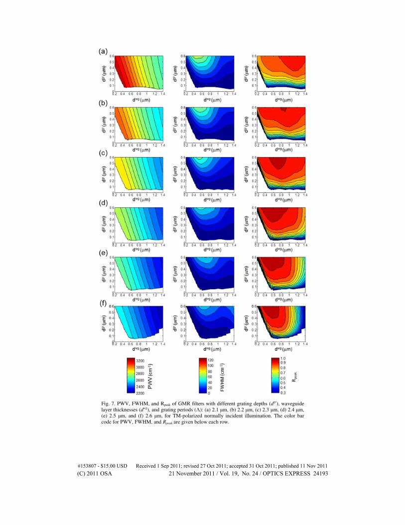
Fig. 7. PWV, FWHM, and Rpeak of GMR filters with different grating depths (dgr), waveguide
layer thicknesses (dwg), and grating periods (Λ): (a) 2.1 µm, (b) 2.2 µm, (c) 2.3 µm, (d) 2.4 µm,
(e) 2.5 µm, and (f) 2.6 µm, for TM-polarized normally incident illumination. The color bar
code for PWV, FWHM, and Rpeak are given below each row.
#153807 - $15.00 USD Received 1 Sep 2011; revised 27 Oct 2011; accepted 31 Oct 2011; published 11 Nov 2011(C) 2011 OSA 21 November 2011 / Vol. 19, No. 24 / OPTICS EXPRESS 24193
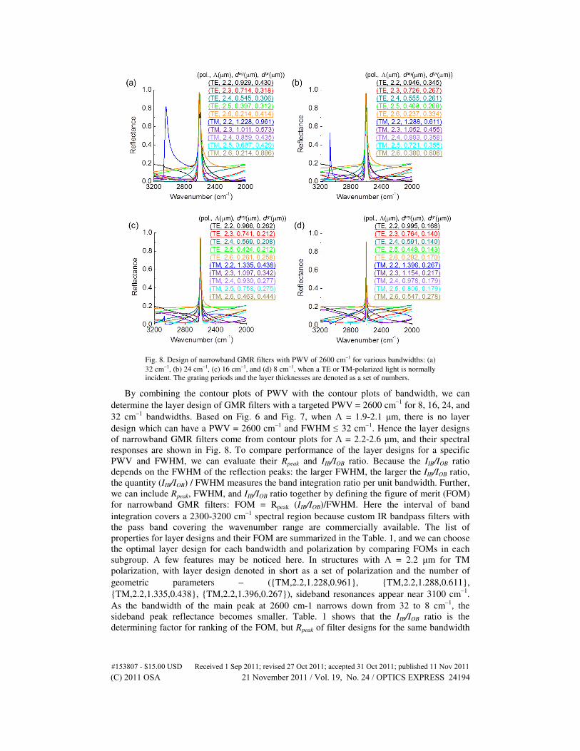
Fig. 8. Design of narrowband GMR filters with PWV of 2600 cm−1 for various bandwidths: (a)
32 cm−1, (b) 24 cm−1, (c) 16 cm−1, and (d) 8 cm−1, when a TE or TM-polarized light is normally
incident. The grating periods and the layer thicknesses are denoted as a set of numbers.
By combining the contour plots of PWV with the contour plots of bandwidth, we can
determine the layer design of GMR filters with a targeted PWV = 2600 cm−1
for 8, 16, 24, and
32 cm−1
bandwidths. Based on Fig. 6 and Fig. 7, when Λ = 1.9-2.1 µm, there is no layer
design which can have a PWV = 2600 cm−1
and FWHM ≤ 32 cm−1
. Hence the layer designs
of narrowband GMR filters come from contour plots for Λ = 2.2-2.6 µm, and their spectral
responses are shown in Fig. 8. To compare performance of the layer designs for a specific
PWV and FWHM, we can evaluate their Rpeak and IIB/IOB ratio. Because the IIB/IOB ratio
depends on the FWHM of the reflection peaks: the larger FWHM, the larger the IIB/IOB ratio,
the quantity (IIB/IOB) / FWHM measures the band integration ratio per unit bandwidth. Further,
we can include Rpeak, FWHM, and IIB/IOB ratio together by defining the figure of merit (FOM)
for narrowband GMR filters: FOM = Rpeak (IIB/IOB)/FWHM. Here the interval of band
integration covers a 2300-3200 cm−1
spectral region because custom IR bandpass filters with
the pass band covering the wavenumber range are commercially available. The list of
properties for layer designs and their FOM are summarized in the Table. 1, and we can choose
the optimal layer design for each bandwidth and polarization by comparing FOMs in each
subgroup. A few features may be noticed here. In structures with Λ = 2.2 µm for TM
polarization, with layer design denoted in short as a set of polarization and the number of
geometric parameters − ({TM,2.2,1.228,0.961}, {TM,2.2,1.288,0.611},
{TM,2.2,1.335,0.438}, {TM,2.2,1.396,0.267}), sideband resonances appear near 3100 cm−1
.
As the bandwidth of the main peak at 2600 cm-1 narrows down from 32 to 8 cm−1
, the
sideband peak reflectance becomes smaller. Table. 1 shows that the IIB/IOB ratio is the
determining factor for ranking of the FOM, but Rpeak of filter designs for the same bandwidth
#153807 - $15.00 USD Received 1 Sep 2011; revised 27 Oct 2011; accepted 31 Oct 2011; published 11 Nov 2011(C) 2011 OSA 21 November 2011 / Vol. 19, No. 24 / OPTICS EXPRESS 24194
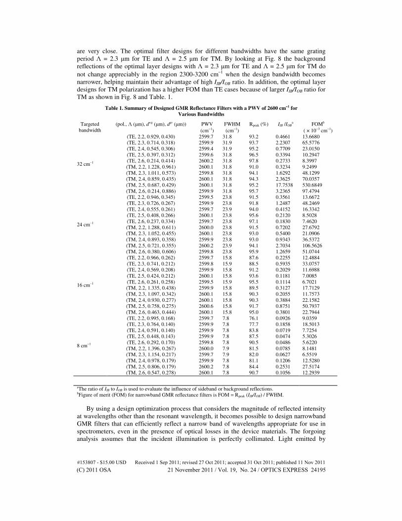
are very close. The optimal filter designs for different bandwidths have the same grating
period Λ = 2.3 µm for TE and Λ = 2.5 µm for TM. By looking at Fig. 8 the background
reflections of the optimal layer designs with Λ = 2.3 µm for TE and Λ = 2.5 µm for TM do
not change appreciably in the region 2300-3200 cm−1
when the design bandwidth becomes
narrower, helping maintain their advantage of high IIB/IOB ratio. In addition, the optimal layer
designs for TM polarization has a higher FOM than TE cases because of larger IIB/IOB ratio for
TM as shown in Fig. 8 and Table. 1.
Table 1. Summary of Designed GMR Reflectance Filters with a PWV of 2600 cm−−−−1 for
Various Bandwidths
Targeted
bandwidth (pol., Λ (µm), dwg (µm), dgr (µm)) PWV
(cm−1) FWHM
(cm−1) Rpeak (%) IIB /IOB
a FOMb
( × 10−3 cm−1)
32 cm−1
(TE, 2.2, 0.929, 0.430) 2599.7 31.8 93.2 0.4661 13.6680 (TE, 2.3, 0.714, 0.318) 2599.9 31.9 93.7 2.2307 65.5776 (TE, 2.4, 0.545, 0.306) 2599.4 31.9 95.2 0.7709 23.0150 (TE, 2.5, 0.397, 0.312) 2599.6 31.8 96.5 0.3394 10.2947 (TE, 2.6, 0.214, 0.414) 2600.2 31.8 97.8 0.2733 8.3997 (TM, 2.2, 1.228, 0.961) 2600.1 31.8 91.0 0.3234 9.2499 (TM, 2.3, 1.011, 0.573) 2599.8 31.8 94.1 1.6292 48.1299 (TM, 2.4, 0.859, 0.435) 2600.1 31.8 94.3 2.3625 70.0357 (TM, 2.5, 0.687, 0.429) 2600.1 31.8 95.2 17.7538 530.6849 (TM, 2.6, 0.214, 0.886) 2599.9 31.8 95.7 3.2365 97.4794
24 cm−1
(TE, 2.2, 0.946, 0.345) 2599.5 23.8 91.5 0.3561 13.6672 (TE, 2.3, 0.726, 0.267) 2599.9 23.8 91.8 1.2487 48.2469 (TE, 2.4, 0.555, 0.261) 2599.7 23.9 94.0 0.4152 16.3342 (TE, 2.5, 0.408, 0.266) 2600.1 23.8 95.6 0.2120 8.5028 (TE, 2.6, 0.237, 0.334) 2599.7 23.8 97.1 0.1830 7.4620 (TM, 2.2, 1.288, 0.611) 2600.0 23.8 91.5 0.7202 27.6792 (TM, 2.3, 1.052, 0.455) 2600.1 23.8 93.0 0.5400 21.0906 (TM, 2.4, 0.893, 0.358) 2599.9 23.8 93.0 0.9343 36.5372 (TM, 2.5, 0.721, 0.355) 2600.2 23.9 94.1 2.7034 106.5626 (TM, 2.6, 0.380, 0.606) 2599.8 23.8 95.9 1.2659 51.0744
16 cm−1
(TE, 2.2, 0.966, 0.262) 2599.7 15.8 87.6 0.2255 12.4884 (TE, 2.3, 0.741, 0.212) 2599.8 15.9 88.5 0.5935 33.0757 (TE, 2.4, 0.569, 0.208) 2599.9 15.8 91.2 0.2029 11.6988 (TE, 2.5, 0.424, 0.212) 2600.1 15.8 93.6 0.1181 7.0085 (TE, 2.6, 0.261, 0.258) 2599.5 15.9 95.5 0.1114 6.7021 (TM, 2.2, 1.335, 0.438) 2599.9 15.8 89.5 0.3127 17.7129 (TM, 2.3, 1.097, 0.342) 2600.1 15.8 90.3 0.2055 11.7573 (TM, 2.4, 0.930, 0.277) 2600.1 15.8 90.3 0.3884 22.1582 (TM, 2.5, 0.758, 0.275) 2600.6 15.8 91.7 0.8751 50.7937 (TM, 2.6, 0.463, 0.444) 2600.1 15.8 95.0 0.3801 22.7944
8 cm−1
(TE, 2.2, 0.995, 0.168) 2599.7 7.8 76.1 0.0926 9.0359 (TE, 2.3, 0.764, 0.140) 2599.9 7.8 77.7 0.1858 18.5013 (TE, 2.4, 0.591, 0.140) 2599.9 7.8 83.8 0.0719 7.7254 (TE, 2.5, 0.448, 0.143) 2599.9 7.8 87.5 0.0474 5.3026 (TE, 2.6, 0.292, 0.170) 2599.8 7.8 90.5 0.0486 5.6220 (TM, 2.2, 1.396, 0.267) 2600.0 7.9 81.5 0.0785 8.1481 (TM, 2.3, 1.154, 0.217) 2599.7 7.9 82.0 0.0627 6.5519 (TM, 2.4, 0.978, 0.179) 2599.9 7.8 81.1 0.1206 12.5280 (TM, 2.5, 0.806, 0.179) 2600.2 7.8 84.4 0.2531 27.5174 (TM, 2.6, 0.547, 0.278) 2600.1 7.8 90.7 0.1056 12.2939
aThe ratio of IIB to IOB is used to evaluate the influence of sideband or background reflections. bFigure of merit (FOM) for narrowband GMR reflectance filters is FOM = Rpeak (IIB/IOB) / FWHM.
By using a design optimization process that considers the magnitude of reflected intensity
at wavelengths other than the resonant wavelength, it becomes possible to design narrowband
GMR filters that can efficiently reflect a narrow band of wavelengths appropriate for use in
spectrometers, even in the presence of optical losses in the device materials. The forgoing
analysis assumes that the incident illumination is perfectly collimated. Light emitted by
#153807 - $15.00 USD Received 1 Sep 2011; revised 27 Oct 2011; accepted 31 Oct 2011; published 11 Nov 2011(C) 2011 OSA 21 November 2011 / Vol. 19, No. 24 / OPTICS EXPRESS 24195
extended thermal sources used in spectrometers, however, is not perfectly collimated.
Because the spectral response of narrowband GMR filters is very sensitive to the incident
angle [23], such angle of divergence arising from imperfect collimation can degrade the
performance of narrowband GMR filters by reducing peak reflectance and broadening filter
bandwidth [36]. The trade-off between narrow bandwidth and performance limitation by
imperfect collimation must also be taken into consideration when selecting bandwidth of
GMR filters for DFIR spectroscopy, and this will be a topic of a future publication.
The IIB/IOB ratio depends on the spectral interval of integration. If the spectral interval of
integration becomes wider than the region of 2300-3200 cm−1
, the sideband reflections will
increase for optimal layer designs but decrease for others, making the advantage of optimal
layer designs for having low background reflections near the resonance wavenumber of 2600
cm−1
indistinguishable. By co-locating optimized narrowband GMR filters with one IR
bandpass transmission filter with wide bandwidth such as 2300-3200 cm−1
, the optical power
outside the pass band can be blocked, and the signal from the small spectral region of interest
can then be further improved with high IIB/IOB ratio powered by the optimal layer design.
3.4 Comparison to the antireflection thin-film equivalence method
It is meaningful to compare the optimal layer design of GMR filters developed in this paper
with the conventional design method with antireflection (AR) thin-film equivalence concept
[26, 37]. For double-layer GMR filter structures, AR conditions for TE require that layer
thicknesses of grating layer and waveguide layer are mλres/4(εgr,eff)1/2
and mλres/4(εwg)1/2
,
respectively, where m = 1, 3, 5, …, εgr,eff = (εc + εSiN)/2, λres = 1/PWV, and εc = 1 (air). Here
we choose m = 1 because smaller grating depth can result in narrower bandwidth, which is
favorable for applications to IR spectroscopy. Because the imaginary part of the permittivity
of Si3N4 at 2600 cm−1
is ~2.2 × 10⁻³
, which is much smaller than the real part of its
permittivity (~4.04), we use the real part of permittivity in Si3N4 approximately in the AR
design. In Fig. 9(a) the blue curve shows the spectral response of the filter with AR design,
where dgr
= 0.606 µm and dwg
= 0.478 µm. The grating period Λ = 2.42 µm is finely adjusted
to match the filter resonance wavenumber with the targeted 2600 cm−1
, as shown in the inset.
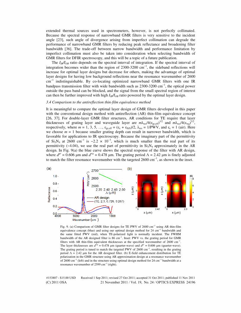
Fig. 9. (a) Comparison of GMR filter designs for TE PWV of 2600 cm−1 using AR thin-film
equivalence concept (blue) and using our optimal design method for 24 cm−1 bandwidth and
the same fitted PWV (red), when TE-polarized light is normally incident. The FWHM
bandwidth of the AR designed filter is 86 cm−1. Inset: PWV vs. the grating period for GMR
filters with AR thin-film equivalent thicknesses at the specified wavenumber of 2600 cm−1.
The layer thicknesses are dwg = 0.478 µm (quarter-wave) and dgr = 0.606 µm (quarter-wave).
The grating period is tuned to match the targeted PWV of 2600 cm−1, resulting in the grating
period Λ = 2.42 µm for the AR designed filter. (b) E-field enhancement distribution for TE
polarization in the GMR structure using AR approximation design at a resonance wavenumber
of 2600 cm−1 (left) and in the structure using optimal design method for 24 cm−1 bandwidth at a
resonance wavenumber of 2599 cm−1 (right).
#153807 - $15.00 USD Received 1 Sep 2011; revised 27 Oct 2011; accepted 31 Oct 2011; published 11 Nov 2011(C) 2011 OSA 21 November 2011 / Vol. 19, No. 24 / OPTICS EXPRESS 24196
In Fig. 9(a) the red curve shows the optimal layer design {TE, 2.3, 0.726, 0.267} for
bandwidth of 24 cm−1
. However, the FWHM bandwidth of the filter with AR equivalence
design is 86 cm−1
, which is not narrow enough to detect the signal in the narrow spectral
region of interest in routine IR spectroscopy. The large bandwidth of the filter with AR design
arises because the modulation index, (εSiN - εAir)/ (εSiN + εAir) = 0.60, is too large to correctly
model the heavily modulated grating layer with effective permittivity [37]. Incorporating one
more layer in the surface-relief filter structures (triple-layer GMR filters) may further reduce
sidebands of the filters with AR design, but the resonance linewidth cannot be effectively
improved because the bandwidth of GMR filters is mainly determined by the magnitude of
the modulation index. The minimum out-of-bandpass reflections of the filter with AR design
and the filter created with the optimal design method are 2.5% and 1.2% near resonance,
respectively, indicating that the optimally designed filter can have narrower linewidth without
sacrificing low out-of-bandpass reflections. In Fig. 9(b) the on-resonance E-field
enhancement distributions of the two filters are compared, which shows that the filter with
optimal design {TE, 2.3, 0.726, 0.267} has a higher maximum field enhancement due to
narrower linewidth and longer photon lifetime. Besides the optimized filters for 24 cm−1
bandwidth, filters with optimal layer designs for narrower bandwidths such as 8 and 16 cm−1
can have narrower linewidth and greater E-field enhancement factor at a specific resonance
wavenumber, which are promising for applications to optical sensing in mid-IR as well as
DFIR spectroscopy.
Although GMR filters are polarization dependent, DFIR spectroscopy requires only one
polarization. Therefore, optimally designed narrowband GMR filters presented in this work
can serve this specific purpose. However it is also interesting to consider polarization
independent optical filters such as two-dimensional periodic grating filters or photonic
crystals [38].
4. Conclusions
In summary, we demonstrate design optimization of narrowband GMR filters in the mid-IR,
with absorption and dispersion properties of materials taken into consideration. We illustrate
the effect of absorption on GMR filters with narrow bandwidth, which limits the performance
of GMR filters by creating a trade-off between filter bandwidth and peak reflectance. The
optimal design method developed in this work provides high-performance GMR filter designs
with accurate control of filter resonance wavenumber, reduced background reflection, and
various narrow bandwidths. Based on the same index modulation value, the designed filter by
the AR equivalence method has a bandwidth of 86 cm−1
, but the designed filters with the
optimal design method developed here can have bandwidth smaller than 32 cm−1
without
sacrificing low sideband reflections near resonance. We anticipate that the optimal design
method developed here will guide the design and fabrication of narrowband GMR filters for
DFIR spectroscopy and many other applications that can take advantage of the unique
properties of GMR filters.
Acknowledgments
This work was funded by the National Institutes of Health (grant number R01 EB009745A).
J. N. Liu acknowledges the support of Taiwan’s Government Fellowship for Studying
Abroad.
#153807 - $15.00 USD Received 1 Sep 2011; revised 27 Oct 2011; accepted 31 Oct 2011; published 11 Nov 2011(C) 2011 OSA 21 November 2011 / Vol. 19, No. 24 / OPTICS EXPRESS 24197